
Submitted:
27 April 2023
Posted:
28 April 2023
You are already at the latest version
Abstract
Introduction: Austere environments include the wilderness and many lower-and-middle-income countries, many of these facing unrest and war. The access to advanced diagnostic equipment is often unaffordable even if available and is often liable to breakdown. This short review paper examines the options available to medical professionals to undertake clinical and point of care diagnostic testing in resource constrained environments and also illustrates the development of mobile advanced diagnostic equipment. The aim is to provide an overview of the spectrum and functionality of these devices beyond clinical acumen.
Keywords:
Diagnosis
; Point-of-care
; Mobile laboratory
; imaging
; LMIC
- 1)
- Introduction
Lower- and Middle-income countries (LMIC’s) are almost synonymous with being resource deprived. It is probably no coincidence that many of these very areas experience armed conflicts. This combination spells disaster for healthcare in these areas, further impacting the huge disparity in healthcare from higher income countries (HIC’s). People in poor countries have less access to healthcare than those in better off countries, and within countries the poor have less access to health services. [1]
Access to point-of-care (POC) diagnostics services is essential for ensuring accurate yet rapid disease diagnosis, emergency resuscitation and further management. POC testing services can improve healthcare by improving access to services especially where healthcare infrastructure is weak, such as in LMIC’s and access to medical care is a challenge as may be the case in an austere environment. [1]
Bearing in mind several factors which will influence the accessibility of point of care diagnostics in LMIC’s, Desmond Kuupiel and co-workers suggest a lean and agile supply chain management framework. [2] The factors to be considered being; test selection, test quantification, procurement and storage, quality assurance, distribution of tests and inventory management.
Tivani Mashamba-Thompson highlighted the need to assess affordability and ensure quality assurance of current services before adopting new POC diagnostics and scaling up current POC diagnostics. [3] This review will briefly examine the role of point of care diagnostics in the following categories: history taking and clinical examination, Vital signs monitoring, imaging and laboratory type analysis focussing on the austere environment and what is relevant to LMIC’s.
A number of factors will be discussed that affect the uptake and utility of POC devices from the basics to more advanced aspects. This includes staff education on the basics of clinical acumen and the use of assistive POC devices to enhance the diagnostic capacity, rather than replace good clinical judgement. Then the various devices and the capacity thereof will be covered.
- 2)
- Applying the basics: History taking and clinical examination
It must be emphasized that staff education is paramount. All medical students, at the beginning of the clinical years are informed that they are now embarking on being clinicians. One is reminded that about 80% of the diagnoses are made once a comprehensive history from the patient is recorded and a thorough examination is completed.
Clinical examination and history-taking is a learned skill with many available books to guide the learner. [4,5] Combining history, signs and symptoms in most cases one would arrive at least at the correct differential diagnosis. Unfortunately, often today, comprehensive history-taking and comprehensive clinical examination is no longer the norm. The inclination towards and reliance on special investigations, partially as a result in an increase in litigation, has reduced the value of clinical acumen to the detriment of patient care. [6,7] Therefore, emphasis should be placed on educating clinicians working in resource-limited environments to optimise history taking and clinical examination skills. These LMIC’s usually have less litigation risk levels. Then they will develop the skill to appropriately and rationally use side-room and electronic point of care devices wisely and cost-effectively.
Good clinical examination will allow for focussed imaging within the limitations of the accessible equipment. Sometimes clinical aspects will allow for the avoidance of unnecessary imaging. Examples of this include the various cervical spine imaging rules (e.g. Canadian C-spine rule or NEXUS) that will ensure that appropriately selected patients receive imaging when needed. [8]
The chest is easy to evaluate clinically and imaging can be guided by clinical findings suggesting a diagnosis (See Table 1).
Clinical assessment of the conscious patient allows for identification of peritonitis, leading to operative intervention. Complete neurological examination allows for assessment of higher function, establishing a sensory level, or motor deficit using dermatome or myotome assessment. This will assist in establishing where the pathology is. Thereby special investigations can be streamlined.
Becoming familiar with signs and symptoms pathognomic for certain conditions will also expedite diagnoses and allow prompt management without unnecessary special investigations, an example being a lucid interval, with skull fracture on palpation, associated pupil dilatation on the same side of the is most likely an extradural haemorrhage on the side of the fracture. This can allow for focussed imaging to guide surgery.
- 3)
- Vital signs Monitors
State of the art multi-function vital signs monitors are used in most first world emergency departments or intensive care units. However, these are costly and also require constant electricity supply and regular maintenance. They are also not always portable. The advance in technology has made available battery operated, portable and affordable vital signs monitors.
The target market is usually what determines the quality of these instruments; home-based care devices are meant for low volume usage and the complexity of usage is minimal. Typical examples of this would be glucometers, blood pressure measuring devices and temperature measuring devices. A common mistake is procuring these for usage in other areas such as peripheral clinics. Although they may be cheaper, their life span is very short if over used, and their reliability and reproducibility of accurate results is questionable. [9,10]
Another dimension that has recently emerged is consumer wearable digital devices for this purpose. These devices are already being used by many consumers despite the lack of confirmatory testing in clinical settings. Christina Hahnen and co-workers tested two devices and found that both devices met accuracy guidelines for heart rate measurements, but they failed to meet the predefined accuracy guidelines for other vital sign measurements. [11] Caution should be exercised if these devices are being used especially in life threatening situations
Although a single monitor is probably the easiest manner in which to monitor patient vital signs, Covid home-based care taught the world how to adapt. Battery operated isolated pulse-oximeters have proven to be a useful tool. In addition to providing a saturation level it is usually accompanied by the measurement of the pulse rate. Add to this a battery-operated blood pressure monitor complements vital sign monitoring. Unfortunately, this combination excludes ECG monitoring that is often required. The advantage of such a combination is in the simplicity of its use, with the relatively small price tag compared to high tech equipment.
It can be concluded that in LMIC’s vital sign monitoring can be adequately performed across a spectrum, the limiting factor being the availability of resources.
- 4)
- Plain radiography
Plain radiographs are cheap, easily accessible and usually available in LMIC’s and even austere environments occasionally. Limiting their use based on clinical findings will reduce unnecessary usage and also prolong the life-span of equipment used to acquire them. Being able to interpret plain radiographs supplements history and clinical findings in attaining a final diagnosis. To derive maximal benefit from plain X-rays it is recommended to have a “reporting” system to work through each film, thereby increasing ability to detect pathology and minimising missing pathology.
Portable X-Ray imaging (radiography) technology has advanced considerably to where it is now small enough, cheap enough, and accurate enough to give diagnostic quality images sent wirelessly to a computer. An example of this in Figure 1 is the Leonardo nano mobile X-ray system [https://www.or-technology.com/en/products/maritime/leonardo-dr-nano.html]
This entire x-ray system fits into a back pack. It is a portable wireless X-ray system. It comprises of only two components: a wireless detector and a laptop with the professional acquisition and analysis software. Its image quality is superb. It can be used for both limbs and torso images.
A system such as this would be ideal for taking services to communities in LMIC’s however its price tag is the limiting factor. Many military units are using this system or an equivalent. Other devices, similar in concept, yet less complex (and much cheaper) are also alternatives. See Figure 2 for an image of one such device.
An example of such a more cost effective system is the Mobile 1-piece x-ray system [Novalion instrument company, Jiangsu, China] The advantages of a machine as illustrated in Figure 2 are portability, the requirement for low amounts of electricity and that the device can even be powered by a battery-inverter combination. The image is displayed on the screen and negates the need for further processing. Disadvantages are that image quality is not at the same level of detail as standard devices. Another limitation is acquiring chest or abdominal x-rays with such a device is far from ideal. A machine such as this is definitely an option in LMIC’s and other austere environments, where access to any imaging device is minimal.
With these devices comes the need to ensure reliable interpretation of the images obtained. Adopting a clinician-based reporting system to assess radiographs will allow interpretation of these x-rays to optimise identifying pathology and minimise missed diagnosis. For C-spine x-rays: in interpreting a lateral c-spine x-ray the following system may be adopted: adequacy (C1 to top of T1), check alignment along four lines namely A Anterior vertebral line, B Anterior spinal line, C Posterior spinal line, D Spinous processes. For the Chest X-ray a recommended system for interpreting a plain AP Chest x-ray would be following an ABCDEF approach (illustrated in Figure 3): Airway: position of trachea and of endotracheal tube if present; Breathing: Lung field assessment looking specifically for: Haemothorax, Pneumothorax, Contusion Circulation, Mediastinal width, Heart shape and size; Diaphragms and any Extras : any extra visible eg: ECG electrodes, Nasogastric tube etc. Finally look for Fractures: follow a system from medially to laterally: Ribs (anterior and posterior), Clavicle, Scapulae Humerus, Vertebrae if visible. A similar system can be used for the pelvis, limbs and the vertebrae plain X-rays as well.
- 5)
- Ultrasound
Ultrasound usage has evolved from being a purely diagnostic instrument to use in assisting with interventions and reassessment post intervention.
Ultrasound machines have reduced in size with better quality resolution. Surprisingly, a mid-range hand held portable ultrasound probe, which can be linked to a mobile device, (telephone or tablet) is relatively inexpensive – one is illustrated in Figure 4 as an example. These portable probes are exceptionally useful in LMIC’s and especially so in the austere environment. They can be transported and charged with ease.
Point-of-care ultrasound is a low-cost imaging modality that can be used for the diagnosis and management of diseases that affect low- and middle-income countries. [12] LMIC’s are faced with healthcare challenges including lack of specialized healthcare workforce and limited diagnostic infrastructure. Task shifting for point-of-care ultrasound (POCUS) can overcome both shortcomings. [13]
Advances in technology have led to more user-friendly ultrasound devices being developed. With appropriate knowledge and training, bedside ultrasound examinations can be used to better diagnose pathology and guide perioperative strategies. Cardiac ultrasound examination was the initial emphasis in anaesthesiology, with now expansion into lung and gastric ultrasound imaging. [14]
Mass casualty
Ultrasound is a valuable tool for the management of mass casualties by improving triage, especially when surgical resources are limited. In some situations, Ultrasound can also correct a diagnosis or improve prehospital therapeutic choices. Field medical teams should be trained to integrate Ultrasound into their prehospital protocols. [2]
Emergency department
Point-of-care ultrasound (POCUS) is a useful tool for clinicians in the management of patients in an emergency department. The role of POCUS is ever increasing due to the need for a rapid assessment of critically ill patients and to speed up the diagnostic process. Hand-carried ultrasound devices are particularly useful in emergency setting and allow rapid assessment of patient even in prehospital setting. [15]
Trauma
The use of point-of-care ultrasound in trauma is widespread. Focused Assessment with Sonography for Trauma examination is a bedside examination used to rapidly determine need for intervention and appropriate patient disposition. This is a largely binary examination, aiming to establish the presence or not of free fluid in the belly or the pericardial sac. When performed by properly trained individuals, FAST is an accepted, rapid, and reliable study for identifying intraperitoneal fluid. It has the advantage of being repeatable and can also detect pericardial tamponade [16]
FAST includes examination of four regions: Positive examination on unstable patients directs operative approach, while in stable patients demands further detailed assessment. The role of POCUS in trauma, however, has expanded beyond the Focused Assessment with Sonography for Trauma examination. Advancements in diagnostics include contrast-enhanced ultrasound, thoracic, and musculoskeletal applications. Ultrasound is also an important tool for trauma providers for procedural guidance including vascular access and regional anaesthesia. Its portability, affordability, and versatility have made ultrasound an invaluable tool in trauma management in resource-limited settings. [17]
Obstetrics
Point of care ultrasound in the field of obstetric anaesthesiology, including cardiac, pulmonary, neuraxial, gastric, and airway ultrasound, can facilitate rapid diagnosis, management, and clinical decision-making surrounding common maternal peripartum comorbidities, and obstetric complications. Routine and proficient utilization of POCUS can ultimately aid in anaesthesiology as well as the role of midwives and obstetricians in the multidisciplinary practice on labour and delivery. [18] The role in diagnosis of pregnancy, determination of gestational age and fetal cardiac monitoring is well established. Ultrasound can provide important and potentially life-saving information. Two of the most basic yet important uses of ultrasound in the pregnant patient are to provide information concerning the gestational age of the pregnancy and the foetal heart rate. [19]
Intensive care
Spreading beyond the realm of tertiary academic medical centres, point-of-care ultrasound in the intensive care unit is an important diagnostic tool. Real-time feedback leads to critical and clinically relevant changes in management and decreases potential complications. Bedside ultrasound evaluation in the intensive care setting with small, portable equipment is well-suited for placement of central lines, lumbar puncture, thoracocentesis or other bedside ICU procedures and in the evaluation of cardiac activity, pleural and abdominal cavity and the overall fluid volume. [20] One such example is measurement of inferior caval collapsibility in the assessment of shock. Ultrasonographic assessment of IVC could be useful in the evaluation of volume status. [20]
Vascular
The use of point-of care ultrasound (POCUS) in the evaluation of vascular emergencies including abdominal aortic aneurysm and deep vein thrombosis is well established. Direct visualization of the vasculature via B-mode, colour Doppler, and pulsed-wave Doppler has been reported to have assisted in the diagnosis of the following: 1) an acute, post-catheterization thrombus of the proximal radial artery; 2) a complete, traumatic radial artery transection; 3) a forearm hematoma with active arterial extravasation; 4) a traumatic arteriovenous fistula; 5) an acute thrombosis of an artery bypass graft; and 6) an infected pseudoaneurysm. POCUS usage in vascular patients allowed rapid identification of pathology within both arteries and veins and improved patient management and prevented potentially harmful complications [21]
Orthopaedics
Recently, musculoskeletal ultrasonography has become more common for diagnosis and treatment in the field of orthopaedics. Musculoskeletal ultrasound technology has rapidly advanced in recent years, and has many advantages, including no exposure to radiation, non-invasive, wide availability, cost-effectiveness, and the ability to be used in real-time in the general outpatient clinic. Traditional radiography was not able to detect soft tissue injury, but musculoskeletal ultrasonography enables the diagnosis of not only musculoskeletal disorders including soft tissue injury, but also fractures by dynamic examination. [22]
Diagnosis in orthopaedics often requires further imaging beyond history taking, clinical examination and plain radiographs. In these cases POCUS can be useful for ruling out occult fractures, diagnosing joint effusions and tendon ruptures. By aiding a speedy diagnosis, unnecessary immobilisation is reduced, as are inpatient stays, while allowing introduction of early mobilisation, and thus reduced harm to patients. [23]
POCUS has a high sensitivity in diagnosing Long Bone fractures. POCUS has a high sensitivity in identifying fracture characteristics. POCUS can be used as an alternative imaging method to XR in the diagnosis of Long Bone fractures and in the determination of fracture characteristics. [24,25] The addition of POCUS to a physical examination significantly improves diagnostic accuracy for dislocations, proximal humeral fractures and reduction confirmation. [26]
Military
Military clinicians demonstrated the ability to perform focused exams, including FAST exams and fracture detection with acceptable sensitivity and specificity. POCUS in the hands of trained military clinicians has the potential to improve diagnostic accuracy and ultimately improve care for the combatant and civilian casualty. [27,28]
Ultrasound training
It is imperative that training in ultrasound usage needs to be incorporated into medical degree curriculums. The Toronto Addis Ababa Academic Collaboration in Emergency Medicine (TAAAC-EM) recently established an introductory POCUS rotation within the Emergency Medicine residency program at Addis Ababa University. [29] Results were very promising.
A POCUS training course that comprised 7 sessions of 2 hours each with didactics and proctored skills stations covering ultrasound applications for trauma (Focused Assessment with Sonography for Trauma (FAST) examination), obstetrics, vascular, soft tissue, regional anaesthesia, focused echocardiography, and ultrasound guidance for procedures was introduced into General Surgery Training Program in Seattle, Washington. Results yielded surgical residents had improved self-efficacy and confidence levels across a broad range of skills. [30] In South Africa a national emergency POCUS curriculum is established with two levels of certification. [31]
A Point-of-care ultrasound (POCUS) training program was introduced at a Family Medicine residency in Zambia. Zambian resident physicians perceived POCUS to be very helpful in their clinical decision-making. These data support the need to advance POCUS education at the residency level throughout LMICs, which may be an ideal strategy to promote widespread utilization of POCUS in low-resource settings globally. [32] Adequate proctoring is, however essential. [33]
- 6)
- Laboratory devices
Initial establishment of laboratory services requires a number of aspects to be addressed, including: skilled staff (e.g. haemotology, biochemistry, pathology, etc.), a suitable physical structure; an appropriate location within the structure and appropriate equipment, along with a system of specimen collection and processing including the publication of results. Post initiation these laboratories and devices also need regular maintenance and a constant reliable power supply. [34]
LMIC’s and austere environments are sometimes lacking in all three of these aspects and end users are sometimes left with making do with whatever is available. This is where reliable POC devices can largely assist the clinician through clinician-performed testing.
Bed-side machines such bed-side haemoglobinometers are very useful. Although not as accurate as formal laboratory testing, using these for trend measurements in critically ill patients remains a useful option. These devices usually use single-use consumables, which need to be procured and used with specific devices, so the health technology services must be intimately involved with the clinician in obtaining these consumables. Strip and stick tests for usually endocrine or microbiological testing are also useful in these environments. Here again accuracy and reliability may be the limiting factor, depending on whether they have been tested against a formal laboratory standard. [35,36,37]
The use of point-of-care testing (POCT) in different clinical applications is justified by the fact that the time to release the result is shortened, allowing the physician to define the diagnosis and most appropriate therapy in a shorter time. However, the negative aspects must also be highlighted and studied so that advancement occurs with the use of these devices. These negative aspects include greater analytical imprecision compared to laboratory automation, the variability between different equipment from different manufacturers, the risk of inappropriate use, a low level of global regulation, higher costs compared with laboratory testing and cost ineffectiveness in terms of health care. [38]
The military, and some civilian emergency medical services in higher income countries, use state of the art portable devices that can do almost any investigation, such as the iStat device often used by military medical teams operating in forward field projection points. This is illustrated in Figure 5. (https://www.globalpointofcare.abbott/en/index.html) The machine itself is expensive, costing approximately $ 4500 for the system. The cartridges are what the limiting factor would be for LMIC’s as these are expensive for repeated usage as an alternative to a fully-fledged laboratory. However, where the fully-fledged laboratory is unavailable these POC devices may be life-saving.
The iStat machine is able to do the following groups of investigations: Blood gases, Haematology, Coagulation, Chemistry and electrolytes, Lactate, Cardiac markers and Endocrinology. This analyser is easy to use, reliable, and portable, and therefore suitable for the operating room, for analyses during emergencies, on peripheral wards, for preclinical screening, or at times when availability of lab tests is time-consuming or limited. The test accuracy for electrolytes, blood gases, and haemoglobin is high enough to justify routine use of the i-STAT analyser in clinical practice. While the nationally required quality standards for Calcium, pH, and Haemoglobin were not met during test is not of significance, because the measured deviation was too small to have clinical relevance. [39]
An alternative would be the Piccolo Xpress chemistry analyser (https://www.globalpointofcare.abbott/en/index.html). This system offers the following: 31 blood chemistry tests that range from liver, kidney and metabolic functions to lipids, electrolytes, and other specialty analytes for routine test, general health screening, and chronic conditions. These devices and others from other suppliers are robust and accurate in the results provided. [40]
Microbiology
Point-of-care (POC) laboratories that deliver rapid diagnoses of infectious diseases were invented to balance the centralization of core laboratories. POC laboratories operate 24 h a day and 7 days a week to provide diagnoses within 2 h, largely based on immunochromatography and real-time PCR tests. These tests are conveniently combined into syndrome-based kits that facilitate sampling and test operations. POC laboratories provide clinical microbiology testing for populations distant from laboratories in LMIC’s. The cost-effectiveness of POC laboratories has been established for the rapid diagnosis of tuberculosis and sexually transmitted infections in both HIC and LMIC’s. [41]
Parasitology
Infectious diseases are more common in resource limited environments and have the potential to have serious complications because they are transmissible. Early detection is therefore paramount. Point of care testing has the potential to provide an alternative to centralised laboratory testing. It has the advantage of being less time consuming and also less resource requiring. The use of point of care tests (POCT) has increased worldwide because they provide real time rapid diagnosis. This is very useful for starting initial treatment. These POCT provide excellent solutions for rural and less accessible areas where parasitic diseases are most prevalent. The diagnostic tests for parasitic diseases barring malaria are not easily available especially in low resources settings where they are needed most. Despite the high demand, a relatively limited number of validated rapid diagnostics are commercially available for parasitic infections. [42]
Intra-operative
Perioperative POCT includes arterial blood gas monitoring, chemistry, CO2-oximetry panels, parathyroid hormone assays, and coagulation testing. Parathyroid hormone assays continue to guide surgical resection of the parathyroid glands. Point of care coagulation testing (such as viscoelastic assays) aids in diagnosis of coagulopathy as well as therapeutic optimization of anticoagulants such as clopidogrel and aspirin. [43] In modern resuscitation practice cartridge-based clinician-performed thrombelastogram (TEG™, [Haemoscope Corporation, USA]) or rotational thrombelastometry (ROTEM® [TEM Innovations GmbH, Germany]) can be helpful in guiding resuscitation and blood product transfusion. Several studies have demonstrated a reduction in transfusion of blood components with TEG/ROTEM in both the civilian and military environments. [44,44]
- 7)
- Conclusion:
There are multiple POC devices on the market that can increase access to diagnostics and enhance time to diagnosis. Many are designed for use in LMIC’s and the austere environment and cover the entire spectrum of care from basic observations to complex diagnosis. Cost effectiveness and availability, along with training, quality-assurance, and maintenance are the determining factors for uptake and implementation of these resources.
Author Contributions
These authors contributed equally to this work. (I) Conception and design: F.G. and T.C.H.; (II) administrative support: none; (III) provision of study materials or patients: F.G. and T.C.H.; (IV) collection and assembly of data: F.G. and T.C.H.; (V) data analysis and interpretation: F.G. and T.C.H.; (VI) manuscript writing: F.G. and T.C.H.; (VII) final approval of manuscript: F.G. and T.C.H. All authors have read and agreed to the published version of the manuscript.
Funding
This research received no external funding.
Institutional Review Board Statement
Not applicable. Literature all in the public domain.
Informed Consent Statement
Not applicable.
Data Availability Statement
The study did not report on any new data.
Conflicts of Interest
The authors declare no conflict of interest. The funding sponsors had no role in the design of the study; in the collection, analyses, or interpretation of data; in the writing of the manuscript; or in the decision to publish the results.
References
- Peters, D.H.; Garg, A.; Bloom, G. Poverty and access to health care in developing countries. Ann. N. Y Acad. Sci. 2008, 1136, 161–171. [Google Scholar] [CrossRef]
- Kuupiel, D.; Bawontuo, V.; Mashamba-Thompson, T.P. Improving the Accessibility and Efficiency of Point-of-Care Diagnostics Services in Low- and Middle-Income Countries: Lean and Agile Supply Chain Management. Diagn. (Basel). 2017, 7, 58. [Google Scholar] [CrossRef] [PubMed]
- Mashamba-Thompson, T.P.; Jama, N.A.; Sartorius, B.; Drain, P.K.; Thompson, R.M. Implementation of Point-of-Care Diagnostics in Rural Primary Healthcare Clinics in South Africa: Perspectives of Key Stakeholders. Diagn. (Basel). 2017, 7, 3. [Google Scholar] [CrossRef] [PubMed]
- Talley, N.; O’Connor, S. Clinical examination (International Edition), 2018, Elsevier, Australia, ISBN 9780729542890.
- Innes, J.A.; Dover, A.R.; Fairhurst, K. Macleod's Clinical Examination, 14th Edition. Elsevier, Amsterdam NL, 2018, ISBN 9780702069932.
- Reschovsky, J.D.; Saiontz-Martinez, C.B. Malpractice Claim Fears and the Costs of Treating Medicare Patients: A New Approach to Estimating the Costs of Defensive Medicine. Health Serv. Res. 2018, 53, 1498–1516. [Google Scholar] [CrossRef] [PubMed]
- Cunningham, W.; Wilson, H. Complaints, shame and defensive medicine. BMJ Qual. Saf. 2011, 20, 449–452. [Google Scholar] [CrossRef] [PubMed]
- Vazirizadeh-Mahabadi, M.; Yarahmadi, M. Canadian C-spine Rule versus NEXUS in Screening of Clinically Important Traumatic Cervical Spine Injuries; a systematic review and meta-analysis. Arch. Acad. Emerg. Med. 2023, 11, e5. [Google Scholar] [CrossRef] [PubMed]
- Peprah, Y.A.; Lee, J.Y.; Persell, S.D. Validation testing of five home blood pressure monitoring devices for the upper arm according to the ISO 81060-2:2018/AMD 1:2020 protocol. J Hum Hypertens. 2023, 37, 134–140. [Google Scholar] [CrossRef] [PubMed]
- Salacinski, A.J.; Alford, M.; Drevets, K.; Hart, S.; Hunt, B.E. Validity and Reliability of a Glucometer Against Industry Reference Standards. J. Diabetes Sci. Technol. 2014, 8, 95–99. [Google Scholar] [CrossRef]
- Hahnen, C.; Freeman, C.G.; Haldar, N.; Hamati, J.N.; Bard, D.M.; Murali, V.; Merli, G.J.; Jeffrey, I.; van Helmond, N. Accuracy of Vital Signs Measurements by a Smartwatch and a Portable Health Device: Validation Study. JMIR Mhealth Uhealth. 2020, 8, e16811. [Google Scholar] [CrossRef]
- Tran, T.T.; Hlaing, M.; Krause, M. Point-of-Care Ultrasound: Applications in Low- and Middle-Income Countries. Curr. Anesth. Rep. 2021, 11, 69–75. [Google Scholar] [CrossRef]
- Abrokwa, S.K.; Ruby, L.C.; Heuvelings, C.C.; Bélard, S. Task shifting for point of care ultrasound in primary healthcare in low- and middle-income countries-a systematic review. EClinical Med. 2022, 45, 101333. [Google Scholar] [CrossRef]
- Bledsoe, A.; Zimmerman, J. Ultrasound: The New Stethoscope (Point-of-Care Ultrasound). Anesth. Clin. 2021, 39, 537–553. [Google Scholar] [CrossRef]
- Mancusi, C.; Carlino, M.V.; Sforza, A. Point-of-care ultrasound with pocket-size devices in emergency department. Echocardiography 2019, 36, 1755–1764. [Google Scholar] [CrossRef]
- American College of Surgeons Committee on Trauma. Chapter 5 Abdominal and pelvic trauma. IN: Advanced Trauma Life Support® Student Course, Manual 10th Ed. Chicago Il. ISBN 78-0-9968262-3-5.
- Gleeson, T.; Blehar, D. Point-of-Care Ultrasound in Trauma. Semin. Ultrasound CT MR. 2018, 39, 374–383. [Google Scholar] [CrossRef] [PubMed]
- Neumann, K.E.; Banayan, J.M. Point of Care Ultrasound on Labor and Delivery. Anesth. Clin. 2021, 39, 811–837. [Google Scholar] [CrossRef] [PubMed]
- Hsu, S.; Euerle, B.D. Ultrasound in pregnancy. Emerg. Med. Clin. N. Am. 2012, 30, 849–867. [Google Scholar] [CrossRef] [PubMed]
- Campbell, S.J.; Bechara, R.; Islam, S. Point-of-Care Ultrasound in the Intensive Care Unit. Clin. Chest Med. 2018, 39, 79–97. [Google Scholar] [CrossRef]
- Ragaisyte, E.; Bardauskiene, L.; Zelbiene, E.; Darginavicius, L.; Zemaityte, E.; Jasinskas, N.; Stasaitis, K. Evaluation of volume status in a prehospital setting by ultrasonographic measurement of inferior vena cava and aorta diameters. Turk. J. Emerg. Med. 2018, 18, 152–157. [Google Scholar] [CrossRef] [PubMed]
- Drake, A.; Dreyer, N.; Hoffer, M.; Boniface, K. Point-of-care Ultrasound for the Evaluation of Acute Arterial Pathology in the Emergency Department: A Case Series. Clin. Pr. Cases Emerg. Med. 2022, 6, 1–7. [Google Scholar] [CrossRef]
- Kanaya, Y.; Taniguchi, N. [Usefulness of POCUS in Orthopedic and Trauma Fields]. Rinsho Byori. 2015, 63, 725–732. [Google Scholar]
- Oluku, J.; Stagl, A.; Cheema, K.S.; El-Raheb, K.; Beese, R. The Role of Point of Care Ultrasound (PoCUS) in Orthopaedic Emergency Diagnostics. Cureus. 2021, 13, e13046. [Google Scholar] [CrossRef]
- Avci, M.; Kozaci, N.; Tulubas, G.; Caliskan, G.; Yuksel, A.; Karaca, A.; Doganay, F.; Etli, I. Comparison of Point-of-Care Ultrasonography and Radiography in the Diagnosis of Long-Bone Fractures. Med. (Kaunas). 2019, 55, 355. [Google Scholar] [CrossRef] [PubMed]
- Biancardi, M.A.A.; Jarman, R.D.; Cardona, T. Diagnostic accuracy of point-of-care ultrasound (PoCUS) for shoulder dislocations and reductions in the emergency department: A diagnostic randomised control trial (RCT). Emerg. Med. J. 2022, 39, 655–661. [Google Scholar] [CrossRef] [PubMed]
- Savell, S.C.; Baldwin, D.S.; Blessing, A.; Medelllin, K.L.; Savell, C.B.; Maddry, J.K. Military Use of Point of Care Ultrasound (POCUS). J. Spec. Oper. Med. 2021, 21, 35–42. [Google Scholar] [CrossRef] [PubMed]
- Dubecq, C.; Dubourg, O.; Morand, G.; Montagnon, R.; Travers, S.; Mahe, P. Point-of-care ultrasound for treatment and triage in austere military environments. J. Trauma. Acute Care Surg. 2021, 91 (Suppl 2), S124–S129. [Google Scholar] [CrossRef] [PubMed]
- Aspler, A.; Kegel, F.; Beyene, T.; Zewdu, T.; Tesfaye, B.; McKnight, A.; Cheung, E.; Bryan, J.; Acton, C. Establishing a Self-sustaining Emergency Medicine Point-of-Care Ultrasound Curriculum in an Academic Teaching Hospital in Ethiopia. Ethiop. J. Health Sci. 2022, 32, 533–538. [Google Scholar] [CrossRef]
- Kotagal, M.; Quiroga, E.; Ruffatto, B.J.; Adedipe, A.A.; Backlund, B.H.; Nathan, R.; Roche, A.; Sajed, D.; Shah, S. Impact of point-of-care ultrasound training on surgical residents' confidence. J. Surg. Educ. 2015, 72, e82–e87. [Google Scholar] [CrossRef]
- Emergency Medicine Society of South Africa. Guidelines for the training and credentialing in emergency point-of-care ultrasound. Available online: https://emssa.org.za/wp-content/uploads/2021/04/POSITION-STATEMENT-Emergency-Medicine-Society-of-South-Africa-21.pdf (accessed on 20 April 2023).
- Haldeman, M.S.; Kunka, E.; Makasa, M.; Birkland, B. Resident perception on the impact of point-of-care ultrasound in clinical care at a family medicine training program in Zambia. Ultrasound J. 2022, 14, 18. [Google Scholar] [CrossRef] [PubMed]
- Waweru-Siika, W.; Barasa, A.; Wachira, B.; Nekyon, D.; Karau, B.; Juma, F.; Wanjiku, G.; Otieno, H.; Bloomfield, G.S.; Sloth, E. Building focused cardiac ultrasound capacity in a lower middle-income country: A single centre study to assess training impact. Afr. J. Emerg. Med. 2020, 10, 136–143. [Google Scholar] [CrossRef] [PubMed]
- Iskandar, K.; Molinier, L.; Hallit SSartelli, M.; Hardcastle, T.C.; Haque, M.; Lugova, H.; Dhingra, S.; Sharma, P.; Islam, S.; Mohammed, L.; et al. Surveillance of antimicrobial resistance in low- and middle-income countries: A scattered picture. Antimicrob. Resist. Infect. Control 2021, 10, 63. [Google Scholar] [CrossRef]
- Zamanzad, B. Accuracy of dipstick urinalysis as a screening method for detection of glucose, protein, nitrites and blood. East. Mediterr. Health J. 2009, 15, 1323–1328. [Google Scholar] [PubMed]
- Freckmann, G.; Pleus, S.; Link, M.; Baumstark, A.; Schmid, C.; Högel, J.; Haug, C. Accuracy Evaluation of Four Blood Glucose Monitoring Systems in Unaltered Blood Samples in the Low Glycemic Range and Blood Samples in the Concentration Range Defined by ISO 15197. Diabetes Technol Ther. 2015, 17, 625–634. [Google Scholar] [CrossRef] [PubMed]
- Hornedo-González, K.D.; Jacob, A.K.; Burt, J.M.; Higgins, A.A.; Engel, E.M.; Hanson, A.C.; Belch, L.; Kor, D.J.; Warner, M.A. Non-invasive hemoglobin estimation for preoperative anemia screening. Transfusion. 2023, 63, 315–322. [Google Scholar] [CrossRef] [PubMed]
- Sumita, N.M.; Ferreira, C.E.S.; Martino, M.D.V.; Franca, C.N.; Faulhaber, A.C.L.; Scartezini, M.; Pinho, J.R.R.; Dias, C.M.; Cesar, K.R.; Pariz, V.M.; et al. Clinical Applications of Point-of-Care Testing in Different Conditions. Clin. Lab. 2018, 64, 1105–1112. [Google Scholar] [CrossRef] [PubMed]
- Schneider, J.; Dudziak, R.; Westphal, K.; Vettermann, J. [The i-STAT analyzer. A new, hand-held device for the bedside determination of hematocrit, blood gases, and electrolytes]. Anaesthesist 1997, 46, 704–714. [Google Scholar] [CrossRef]
- Yonel, Z.; Kuningas, K.; Sharma, P.; Dutton, M.; Jalal, Z.; Cockwell, P.; Webber, J.; Narendran, P.; Dietrich, T.; Chapple, I.L.C. Concordance of three point of care testing devices with clinical chemistry laboratory standard assays and patient-reported outcomes of blood sampling methods. BMC Med. Inf. Decis. Mak. 2022, 22, 248. [Google Scholar] [CrossRef]
- Drancourt, M.; Michel-Lepage, A.; Boyer, S.; Raoult, D. The Point-of-Care Laboratory in Clinical Microbiology. Clin. Microbiol. Rev. 2016, 29, 429–447. [Google Scholar] [CrossRef] [PubMed]
- Ghoshal, U.; Jain, M. A review on point of care tests in parasitology. Indian. J. Med. Microbiol. 2022, 40, 337–341. [Google Scholar] [CrossRef]
- Rhee, A.J.; Kahn, R.A. Laboratory point-of-care monitoring in the operating room. Curr Opin Anaesthesiol, 2010, 23, 741–748. [Google Scholar] [CrossRef]
- Schmidt, A.E.; Israel, A.K.; Refaai, M.A. The Utility of Thromboelastography to Guide Blood Product Transfusion. Rev. Am. J. Clin. Pathol. 2019, 152, 407–422. [Google Scholar] [CrossRef]
- Rizoli, S.; Min, A.; Adic Peez Sanchez, A.P.; Shek, P.; Grodecki, R.; Veigas, P.; Peng, H.T. In Trauma, Conventional ROTEM and TEG Results Are Not Interchangeable But Are Similar in Clinical Applicability. Mil Med. 2016, 181 (Suppl. 5), 117–126. [Google Scholar] [CrossRef] [PubMed]
Figure 1.
Image of a Leonardo nano mobile X-ray system.
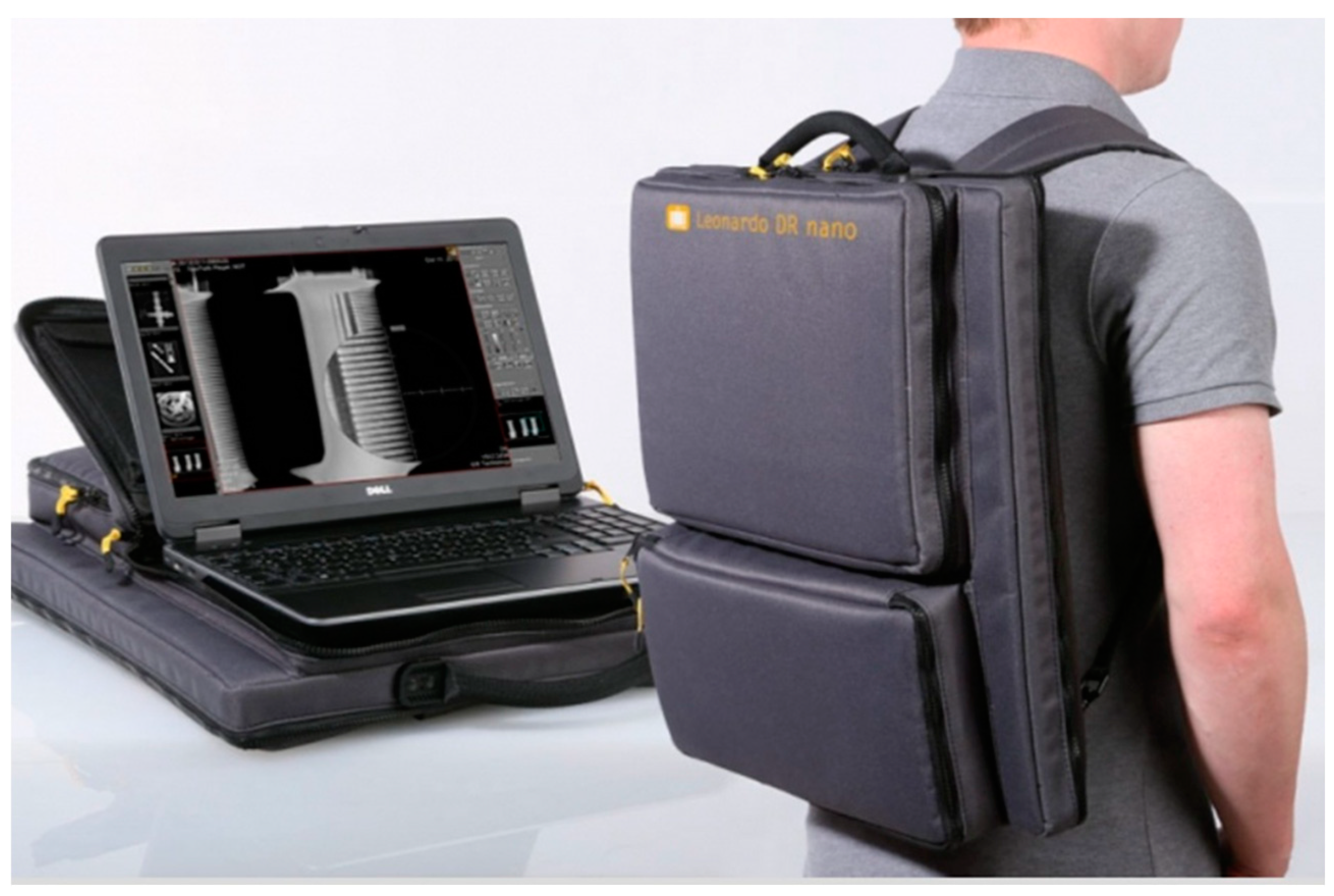
Figure 2.
Mobile 1 piece X-ray system.
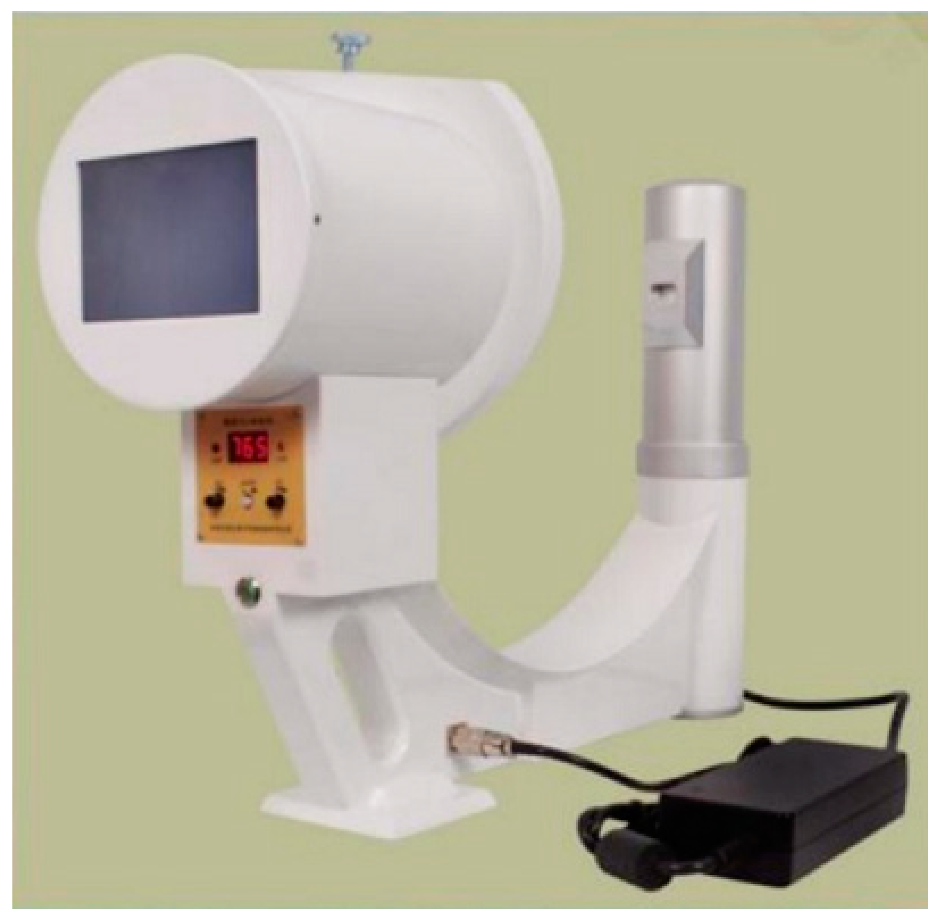
Figure 3.
The ABDCEF approach to viewing a chest x-ray.
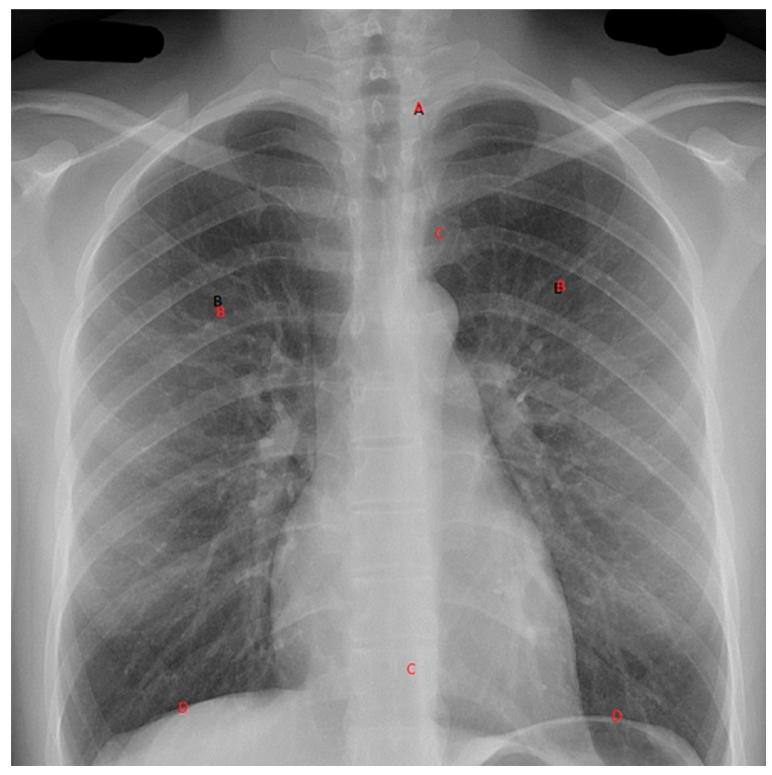
Figure 4.
Image of ultrasound probe with linked mobile device onto which image is transmitted. There are a number of such devices, some with wire-to-the-phone and others with Bluetooth® or WiFi connectivity.
Figure 4.
Image of ultrasound probe with linked mobile device onto which image is transmitted. There are a number of such devices, some with wire-to-the-phone and others with Bluetooth® or WiFi connectivity.
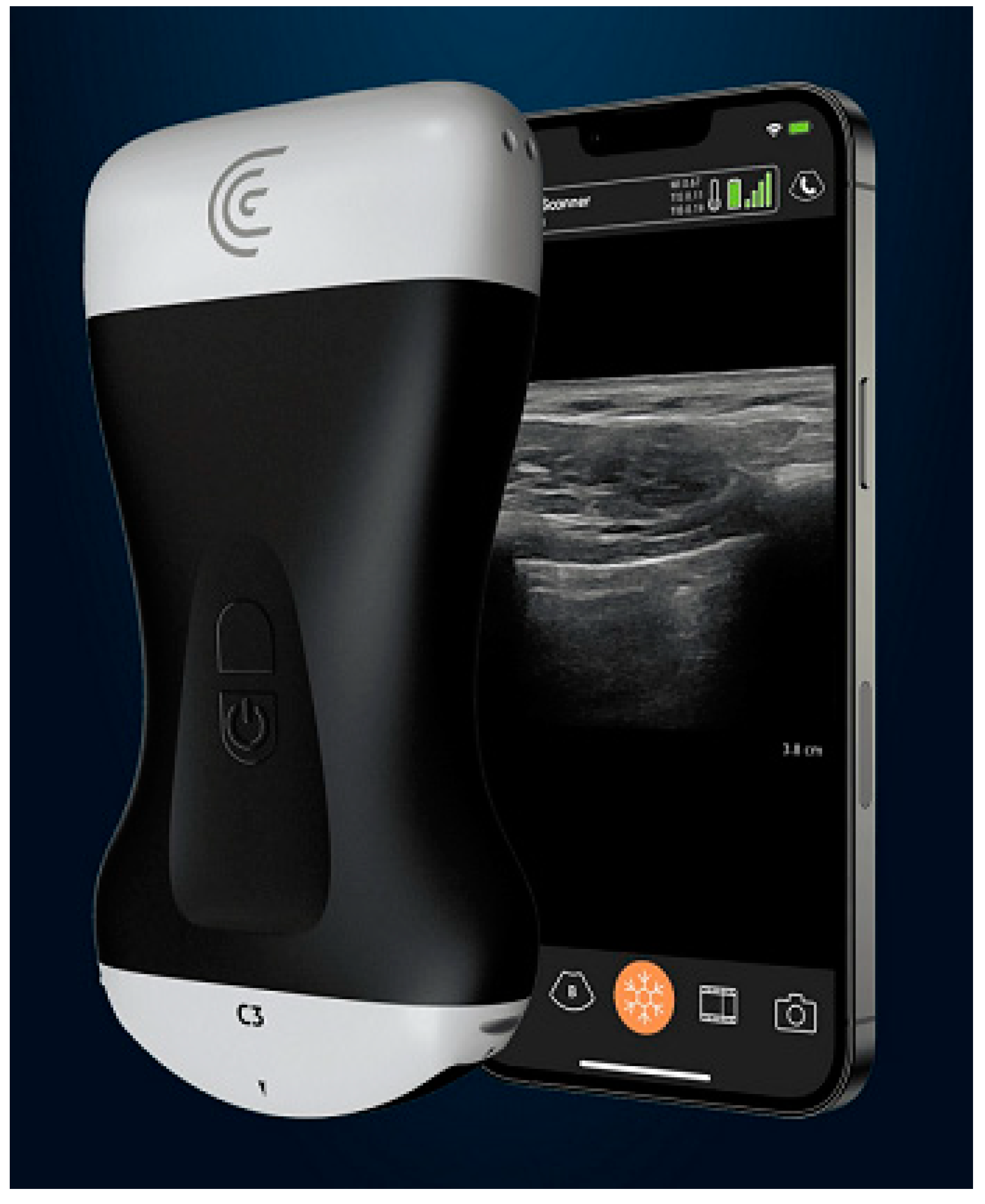
Figure 5.
Image of complete iStat system (Abbott Laboratories, Chicago Il.).
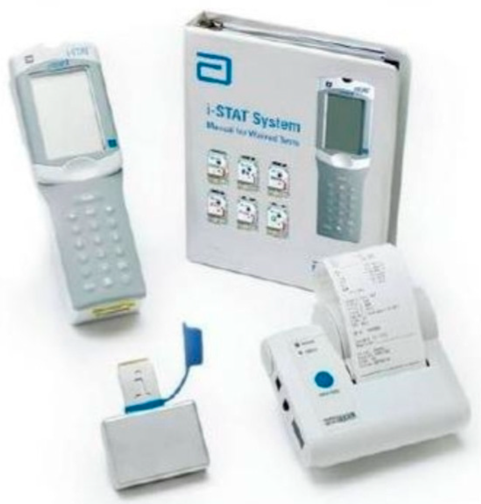

Table 1.
Chest clinical features associated with trauma.
| Inspect | Palpate | Percuss | Auscultate | |
|---|---|---|---|---|
| Pneumothorax | ↓chest movement | Trachea central | ↑ resonance | ↓ air entry |
| Tension pneumothorax | ↓ chest movement | Trachea deviated away from side of pathology | ↑ resonance | ↓ air entry |
| Haemothorax | ↓ chest movement | Trachea central | ↓ resonance | ↓ air entry |
| Massive haemothorax | ↓ chest movement | Trachea deviated away from side of pathology | ↓ resonance | ↓ air entry |
Disclaimer/Publisher’s Note: The statements, opinions and data contained in all publications are solely those of the individual author(s) and contributor(s) and not of MDPI and/or the editor(s). MDPI and/or the editor(s) disclaim responsibility for any injury to people or property resulting from any ideas, methods, instructions or products referred to in the content. |
© 2023 by the authors. Licensee MDPI, Basel, Switzerland. This article is an open access article distributed under the terms and conditions of the Creative Commons Attribution (CC BY) license (http://creativecommons.org/licenses/by/4.0/).
Copyright: This open access article is published under a Creative Commons CC BY 4.0 license, which permit the free download, distribution, and reuse, provided that the author and preprint are cited in any reuse.


