
Submitted:
18 April 2023
Posted:
19 April 2023
You are already at the latest version
Abstract
The objective of this study was to analyze the in vitro stability and toxicity of liposomes containing guarana in skin cell lines. The liposomes were produced by the reverse phase evaporation method containing 1 mg/mL guarana. The stability of the liposomes was evaluated by physical-chemical parameters for up to 90 d using three different storage conditions. The cytotoxicity of guarana (GL), liposomes (B-Lip), and guarana-loaded liposomes (G-Lip) was evaluated on spontaneously immortalized human keratinocyte cell lines (HaCaT), murine swiss albino fibroblasts (3T3), and human fibroblast (1BR.3.G). The evaluation was performed using cellular viability analysis. The techniques used were 3-(4,5-dimethylthiazol-2-yl)-2,5-diphenyltetrazolium bromide (MTT) and neutral red capturing (NRU) and the analyses were conducted after 24, 48, and 72 h exposure of these cells to the different treatments. The G-Lip exhibited physical-chemical stability for 60 d when the samples were stored in a refrigerator. The GL, B-Lip, and G-Lip demonstrated low cytotoxicity in the three cell cultures tested since a low reduction in cell viability was only observed at the highest concentrations. In addition, greater cell damage was observed for B-Lip; however, guarana protects cells from this damage. Thus, G-Lip structures can be considered as promising systems for topical applications.

Keywords:
Paullinia cupana
; natural products
; nanoparticles
; stability
; cytotoxicity
; cell culture
1. Introduction
Plants are important sources of bioactive compounds in modern medicine; and, approximately one-third of the best-selling pharmaceuticals are from natural products or their derivatives [1,2,3]. In contrast, many natural active ingredients are unstable compounds that may undergo degradation or oxidation reactions, or both. In addition, these reactions may lead to a decrease or loss of efficacy of the active compounds. For example, incorrect storage may promote the loss of active compounds, whether for physical or biological reasons [4,5].
One alternative that attempts to resolve these limitations is the incorporation of nanoparticles in natural product-based delivery systems, which increases the stability of the compounds and consequently preserves the therapeutic effects using techniques that involve nanotechnology [6,7,8]. These nanoparticles can significantly increase both the in vitro and in vivo bioavailability of natural products [1]. From this perspective, the pharmaceutical industry has increasingly used nanotechnology-based products to create cosmetic formulations [9,10,11,12].
Liposomes are among the nanoparticles used for the development of nanocosmetic products [4,13,14,15]. Liposomes are structures formed by a lipid bilayer that can fuse with the layers when applied to the skin, thereby promoting the release of active compounds, making them useful as carriers for cosmetic application [16].
In addition to having a simple means of preparation, the use of these systems can promote a greater absorption of the active compounds in the skin, being an interesting system when talking about cosmeceuticals. The enhanced absorption is associated with a prolonged release, thereby promoting a greater effect [9,16,17].
The lipid structure of the liposomes facilitates the fusion of active compounds with different layers of the skin. This is more advantageous than other nanostructured systems in the transport of nanocosmetics, such as vitamins, minerals, antioxidants and anti-aging materials, making it a useful tool in the field of pharmaceuticals and cosmetics [19,20].
Studies of the interaction of these nanoparticles with biological systems, such as their bioavailability, biodegradability, and toxicity are of utmost importance. Hence, it is essential to know the physicochemical properties of these particles, such as: size, shape, surface area, morphology, and stability [21]. In addition, precise and predictive risk assessment approaches are required for understanding the potential health and environmental hazards associated with exposure to nanomaterials [22].
The use of animals in scientific research is the most commonly used method to ensure safety and low-level toxicity. However, the increase in ethical discussions and regulatory standards on the protection of animals used for scientific purposes (Directive 2010/63/EU) [23,24], as well as the growing interest in the search for predictive toxicology, have been changing the perspective in this line of research [25,26,27,28].
Russell and Burch (1959) [29] postulated the 3Rs principle, which is primarily aimed at the reduction, refinement, and replacement of laboratory animals. Researchers state that good science and animal welfare must go hand in hand. Hence, several alternative methods are proposed in an attempt to reduce the number of animals used in experimentation and the cost of experiments [24,30,31].
Guarana, Paullinia cupana var. sorbilis (Mart.) Ducke (Sapindaceae), is a native Brazilian species of considerable economic and social importance [32]. Among the Amazonian species, guarana is one of the most promising species in the Brazilian flora [33]. Guarana has a long history of use as a stimulant, mainly by indigenous tribes in Brazil, and is a versatile plant due to its potential utility in the food industry, such as in the preparation of energy drinks, soft drinks, and food supplements [34,35,36,37].
Furthermore, guarana is widely used in the pharmaceutical industry in the production of drugs and is listed in the Brazilian Pharmacopoeia [38]. It is present in several cosmetic products, due to its antimicrobial [39,40] and antioxidant activity [34,35,36,39,41,42,43,44,45,46,47,48,49,50]. Due to the high content of alkaloids in guarana, extracts are added to products for the treatment of gynoid lipodystrophy and to anti-aging creams [51,52]. Given the importance of guarana and the increasing use of its seeds, there has been increased interest in the quality of the products containing this compound because its chemical structure is predominantly unsaturated and susceptible to oxidation [53].
In the present study, we evaluated the physicochemical stability of liposomes containing 1 mg/mL guarana powder by reverse phase evaporation. Moreover, based on the potential topical application of these new nanocarriers, the in vitro cytotoxicity of guarana (GL), the blank liposome (without guarana, B-Lip), and the liposome containing 1 mg/mL guarana powder (G-Lip) were tested. The evaluations were conducted in different cultures of skin cells, fibroblasts (3T3 and 1BR.3.G) and keratinocytes (HaCaT).
2. Materials and methods
2.1. Materials
Acetonitrile analytical standard, dimethyl sulfoxide (DMSO), 3-(4,5-Dimethylthiazol-2-yl)-2,5-diphenyltetrazolium bromide (MTT), neutral red dye (NR), cholesterol, and polysorbate 80 were purchased from Sigma-Aldrich® (St. Louis, MO, USA). Methanol and trifluoroacetic acid (TFA) were acquired from J.T.Baker® (Mexico City, Mexico). Ethanol was acquired from Synth® (São Paulo, Brazil). Monobasic potassium phosphate was acquired from F. Maia® (São Paulo, Brazil). Sodium chloride and dibasic sodium phosphate were obtained from Nuclear® (São Paulo, Brazil). Potassium chloride was obtained from Qhemis® (São Paulo, Brazil). Lipoid S100® was obtained from Lipoid® (Ludwigshafen, Alemanha), and vitamin E from Alpha química® (Porto Alegre, Brazil). Dulbecco's modified Eagle's medium (DMEM), fetal bovine serum (FBS), phosphate buffered saline (PBS), L-glutamine solution (200 mM), trypsin-EDTA solution (170,000 U/L trypsin and 0.2 g/L EDTA) and penicillin-streptomycin solution (10,000 U/mL penicillin and 10 mg/mL streptomycin) were obtained from Lonza (Verviers, Belgium). The 75 cm2 flasks and 96-well plates were obtained from TPP® (Trasadingen, Switzerland). Guarana powder was kindly provided by Agropecuary Research Brazilian Enterprise (EMBRAPA Western Amazon in Manaus, Amazon, Brazil).
2.2. Preparation and characterization of guarana-loaded liposomes
Liposomes containing 1 mg/mL guarana powder (G-Lip) were prepared by reverse phase evaporation [54,55], after being previously developed and standardized by our research group [56]. The soy phosphatidylcholine (0.8 g), cholesterol (0.15 g), and vitamin E (0.02 g) were solubilized in ethanol (40 mL) with the aid of an ultrasound for 5 min. Then, an aliquot of an aqueous solution (4 mL) of guarana powder (0.1 g) and polysorbate 80 (0.15 g) in PBS pH 7.4 was sonicated for 5 min, thereby yielding a dispersion of reverse micelles. The organic solvent was removed by evaporation to form an organogel. The organogel reverted to vesicles after the addition of the remainder of the aqueous phase by stirring (300 rpm) for 30 min using a rotary evaporator in a water bath at 40 °C. The vesicles were homogenized at room temperature using filtering sequences through the use of 0.45 and 0.22 µm filter membranes (Millex Syringe Filter®). Blank liposomes (B-Lip) were also produced by the same method under identical experimental conditions previously described; except, at this stage, the guarana was suppressed from the formulation.
For the characterization, the average diameter parameters were evaluated by two different techniques: laser diffraction (Microtrac S3500®, EUA) using the undiluted dispersions and dynamic light scattering (Zetaziser Nano-ZS®, Malverm, Reino Unido) using samples diluted in ultra-pure water (1:500 v/v). The latter method also determined the polydispersity index (PDI). In order to determine the homogeneity of the suspended vesicles, the span index was calculated from the data obtained by laser diffraction analysis, using the following formula:
where: dv is the size(µm) em 10, 50 e 90%.
The zeta potential values of the liposomes were evaluated by the determination of electrophoretic mobility (Zetaziser Nano-ZS®, Malverm, Reino Unido). The measurements were performed after diluting the formulations in 10 mM NaCl aqueous solution. The pH values of the formulations were directly determined using a calibrated potentiometer (Digimed DM22®, Brazil). The organoleptic characteristics (appearance, color, and odor) were visually evaluated and the changes in the initial sample (time zero) observed.
The five main active compounds present in guarana powder (theobromine, theophylline, caffeine, catechin, and epicatechin) were quantified. The quantification end encapsulation/incorporation efficiency was determined by reversed-phase high-performance liquid chromatography (RP-HPLC) using a Prominence® chromatograph (Shimadzu®, Japan), according to methodologies described and validated by our group [46,56].
The chromatographic instruments and conditions were: a Shimadzu HPLC system (Kyoto, Japan) equipped with an LC-20AT pump, an SPD-M20A photodiode array (PDA) detector, a CBM-20A system controller, a C18 Phenomenex (4 × 3.0 mm, 5 µm) precolumn, and an RP-18 Phenomenex column (250 mm × 4.5 mm, 5 µm). Water was used as the mobile phase with 0.1% TFA (pH 4.2, A) and a methanol:acetonitrile solution (25:75 v/v, B) in a 90:10 v/v ratio (A:B) at an isocrative flow rate (1 mL/min). The injection volume was 20 μL. The detection was performed at 280 nm. The tests were based on the methodology described by Klein et al. (2012) [57] and some modifications were validated by our research group [56].
2.3. Physicochemical stability study of guarana-loaded liposomes
The liposome samples containing 1 mg/mL guarana powder were prepared in triplicates and stored at room temperature (RT 25 ± 2 °C), in a climatic chamber (CC 40 ± 2 °C and 75% relative humidity), and under refrigeration (RE 5 ± 2 °C) and analyzed at 0, 7, 15, 30, 60, and 90 d. The parameters analyzed were the organoleptic characteristics (appearance, color, and odor), precipitate formation or phase separation, mean vesicle diameter, polydispersity index, zeta potential, pH, concentration of active compounds, and encapsulation/incorporation efficiency, using the methodology described previously (Section 2.2).
2.4. Culture of 3T3, HaCaT and 1BR.3.G Cell Lines
The HaCaT, 3T3, and 1BR.3.G were grown in DMEM (4.5 g/L glucose), supplemented with 10% FBS, L-glutamine (2 mM), penicillin (100 U/mL), and streptomycin (100 µg/mL) at 37 °C, 5% CO2. The cells were routinely cultured in 75 cm2 culture flasks and trypsinized using trypsin-EDTA when the cells reached approximately 80% confluence. The HaCaT cell lines were obtained from the Eucellbank (University of Barcelona, Spain), whereas 3T3 was obtained from ECACC (Sigma-Aldrich®) and 1BR3.G was donated by Prof. Ramon Mangues (Biomedical Research Institute Sant Pau of the Hospital de Sant Pau, Barcelona, Spain).
2.5. Analysis of liposome interference with cell viability assays
To eliminate the potential interference of the liposomes with the cell viability assays, an interference test was performed, prior to the experiments, using the methodology described by Nogueira et al. (2013) [58]. In this analysis, the G-Lip (500 μL) was suspended in DMEM (500 μL, without FBS and phenol red) containing the MTT dye (0.5 mg/mL) or NR (0.05 mg/mL). These solutions were prepared in triplicate and incubated at 37 °C in 5% CO2.
After 3 h of incubation, the liposomes were centrifuged (10 min at 19,000 rpm). The supernatant was removed and the DMSO (1 mL) or a solution (1 mL) containing 50% absolute ethanol and 1% acetic acid in distilled water were added for the MTT and NR experiments, respectively. These solutions were shaken and transferred to a quartz cuvette where a scanning spectrum was plotted in the range of 300 to 700 nm. The absorbance was recorded at 550 nm using a Shimadzu double beam UV-160A-Vis spectrophotometer (Shimadzu®, Kyoto, Japan).
2.6. Cytotoxicity Assays
The cytotoxic effect of GL, G-Lip, and B-lip were measured by MTT tetrazolium salt assay, described by Mosmann (1983) [59], and neutral red uptake (NRU) assay, described by Borenfreund and Puerner (1985) [60]. The 3T3, HaCaT, and human fibroblast cells were seeded in the 60 central wells of a 96-well plate at a density of 1 × 10⁵, 6.5 × 10⁴, and 5.5 × 10⁴ cells/mL for 24, 48, and 72 h, respectively. After incubation (24 h, 5% CO2, 37 °C), the medium was removed and 100 μL of the DMEM supplemented with 5% FBS containing the different treatments at the required concentration (3.91–500 µg/mL) was added. After incubation under identical conditions as before, the medium was removed and 100 μL of MTT in PBS (5 mg/mL) diluted (in a 1:10 ratio) in DMEM without FBS. The phenol red was then added to the cells, with a final concentration of 0.5 mg/mL. Similarly, 100 μL of 0.05 mg/mL NR solution in DMEM without FBS and phenol red was added to each well for the NRU assay. The control used in the experiments consisted of cells and medium, without any treatment. The plates were again incubated for 3 h after the medium was removed. Then, for the MTT assay, 100 μL of DMSO was added to each well to dissolve the purple formazan product. For the NRU assay, 100 μL of a solution containing 50% absolute ethanol and 1% acetic acid in distilled water was added. After 10 min on a microtiter plate shaker at room temperature, the absorbance of the resulting solutions was measured at 550 nm using a microplate reader. The cell viability was calculated by considering the mean absorbance of each concentration with respect to that of the controls.
2.7. Statistical analysis
All the experiments were evaluated in triplicates. The results are expressed as mean ± standard deviation (SD) and the statistical analyses were performed using one-way analysis of variance (ANOVA) to determine the differences between the data sets, followed by the Dunnett or Tukey post hoc test for multiple comparisons using the GraphPad Prism 5.0 software®. The differences were considered significant at p < 0.05.
3. Results and discussion
3.1. Stability study of guarana-loaded liposomes
Considering the hydrophilic and lipophilic characteristics of the active compounds present in guarana, liposomes were the nanostructures selected in our study as vehicles for the incorporation of guarana powder. This is because the structures allow the incorporation of compounds with different characteristics [20,61,62,63,64].
In a previous study conducted by our research group [46], we evaluated two different methods of producing liposomes (ethanol injection and reverse phase evaporation) and different concentrations of guarana powder associated with these structures (1, 5, and 10 mg/mL). From this initial study, the reverse phase evaporation method and the concentration of 1 mg/mL were selected as the optimum conditions. Hence, the results herein refer to liposomes produced under the aforementioned conditions (Figure 1).
To verify the formation of a homogeneous and nanometric system, the vesicle diameter distribution analysis was performed by the laser diffraction technique (Microtrac®). The initial mean diameter was 147 ± 0.01 nm; this parameter was measured for 90 d under different storage conditions. The diameter remained stable, with no significant differences, for 7, 15, and 60 d when stored at CC, RT, and RE, respectively (Figure 1A).
From these results, the span index was calculated, through which it is possible to determine the homogeneity of the suspended vesicles. The initial span values of 0.31 ± 0.10 indicate a close distribution of the vesicles. These values remained low (0.72 ± 0.11, 0.79 ± 0.41, and 0.69 ± 0.31) for up to 90 d when the samples were stored at RT, RE, and CC, respectively. The presence of low dispersion nanometric vesicles was also observed by the dynamic light scattering technique (Figure 1B).
We observed initial vesicles 165 ± 8.27 nm in size and a PDI of 0.250 ± 0.03, which remained largely similar for 7 d when stored in RT (172 ± 3.98 nm, PDI 0.273 ± 0.03) and CC (174 ± 3.03 nm, PDI 0.237 ± 0.02), under this same condition, the destabilization of the system was observed in 30 d of storage in the presence of micrometric vesicles (1.355 μm) (Figure 1B). This increase was associated with alterations in the organoleptic characteristics of appearance, color, and odor in 15 d and intensified in 30 d (Figure 2).
In contrast, the vesicles were not significantly different for 30 d when stored under RE (181 ± 4.70 nm, PDI 0.285 ± 0.04) (Figure 1B), whereas changes in the organoleptic characteristics were confirmed in 90 d of analysis (Figure 2).
The formulations were also analyzed for the zeta potential by the electrophoretic mobility technique, using the data presented in Figure 1D. The liposomes had an initial zeta potential of -9.78 ± 0.98 mV and remained without significant difference for 15 d when stored in RT (-11.56 ± 3.47 mV) and in CC (-6.40 ± 1.40 mV). For the samples stored under RE, the initial characteristics were maintained up to 60 d of storage (-9.81 ± 2.28 mV).
The low zeta potential values agree with the characteristics of the phospholipid (soy phosphatidylcholine) used for the production of these liposomes [65]. The findings in this paper corroborated the results obtained by Karn, Parkl and Hwangl, (2013) [66], where they produced liposomes using Lipoid S100® and cholesterol obtained vesicles with potentials of -6.8 to -7.7 mV.
The initial pH values of 7.24 ± 0.04 were maintained without significant difference for up to 7 d of analysis when the samples were stored at RT (6.92 ± 0.04) and in CC (7.03 ± 0.01). For the samples stored under RE, no significant difference was observed during the 90 d (Figure 1E).
The pH values were expected, based on the preparation of these nanostructures, where PBS (pH 7.4) was used as the aqueous phase. The pH change associated with higher temperatures (RT and CC) is directly related to the stability of the liposomes. It is believed that the reduction in pH when the samples were stored under these conditions could be related to the hydrolysis of the lipids present in the liposomal structures, which at high temperatures can undergo chemical degradation, leading to a loss of stability [67,68].
It is known that the evaluation of the organoleptic characteristics is of great importance because the physical processes such as aggregation, flocculation, fusion or coalescence can alter the utility of the liposomes, which can result in the loss of the associated liposomes and changes in the size of the structures [69,70]. The result regarding the organoleptic characteristics is shown in Figure 2.
In summary, it was observed that the samples stored in CC revealed alterations in colour, from 7 d of storage (Figure 2). At 15 d, the samples stored at RT and CC exhibited an intense rancid odor, with significant changes in the vesicle diameter and polydispersity index (Figure 1B and 1C) and pH (Figure 1E). These changes intensified at 30 d of storage, mainly under the CC condition, with changes in the zeta potential as well (Figure 1D). Under these two conditions, complete phase separation was observed after 60 d. For the samples only stored in RE at 90 d, changes in organoleptic characteristics were observed
It should also be noted that blank liposomes (in the absence of guarana) were produced and characterized at the same times under identical conditions (data not shown). The results were similar to those of the liposomes containing 1 mg/mL of guarana powder; that is, guarana did not alter the characteristics of the liposomal structures.
The total content and encapsulation/incorporation efficiency of the five main active compounds (theobromine, theophylline, caffeine, catechin, and epicatechin) were also evaluated. The results obtained for the total content of the assets are depicted in Figure 3.
The quantification results (Figure 3) indicated the presence of 20.61 μg/mL methylxanthines (0.14 μg/mL (0.028%) TEOB, 0.47 μg/mL (0.094%) TEOF, and 20.00 μg/mL (4.0%) CAF) and 26.00 μg/mL polyphenols (13.00 μg/mL (2.6%) CAT and 13.00 μg/mL (2.6%) EPICAT) in the guarana powder sample.
For TEOB, the initial content of 104.73 ± 1.11% decreased to 64.80 ± 18.11% (15 d) and 63.99 ± 2.06% (90 d) when the liposomes were stored in RT and CC, respectively. When the samples were stored in RE, there were no significant differences in the concentration until 90 d, with a final concentration of 95.34 ± 1.02% (Figure 3A).
For TEOF, the initial content (91.99 ± 1.07%) significantly changed in 15 d at RT (84.08 ± 58.71%) and in CC (84.24 ± 58.98%). When the liposomes were stored under RE, the decrease in the content of this active compound was only observed in 90 d, with a final content of 40.07 ± 4.93% (Figure 3B).
The CAF demonstrated a reduction in the initial content (100.96 ± 0.59%) after 15 d when the samples were stored at RT (35.55 ± 31.77%) and in CC (62.90 ± 25.71%). The initial content of the CAF exhibited no significant difference for 90 d, when the sample was stored under RE (98.89 ± 4.37%) (Figure 3C).
The polyphenols (CAT and EPICAT) (Figure 3D and 3E) were the active compounds that demonstrated the greatest reduction in the content, which were already observable in 7 d of stability, when the samples were stored in CC. The CAT initially showed a content of 92.90 ± 1.07% and was significantly reduced to 10.65 ± 6.90% under this storage condition. The same was observed for EPICAT that had an initial content of 85.35% ± 2.99 and in 7 d, a stability content of 21.51% ± 18.66. When these compounds (CAT and EPICAT) were at RT, the content was significantly altered in 15 d, with concentrations of 80.70 ± 0.45 and 77.90 ± 3.18% for CAT and EPICAT, respectively. Under refrigeration, the concentration was reduced in 60 d for both active compounds, with content of 63.87 ± 30.03 and 53.78 ± 22.06% for CAT and EPICAT, respectively.
In general, the condition that yielded the highest stability of the active compounds was under RE. For TEOB and CAF, the content remained unchanged throughout the stability study. When stored in RE, the CAT and EPICAT exhibited a significant reduction in 60 d of stability, whereas TEOF demonstrated a reduction in 90 d.
The reduction in the content, particularly for the polyphenols (CAT and EPICAT) when the samples were stored in CC, could be caused by the oxidation of these compounds at higher temperatures. In previous studies, the polyphenols present in cocoa exhibited enzymatic oxidation when temperature and high humidity were used to dry this product [71,72].
The literature also describes the polyphenols as unstable structures that may undergo possible oxidative processes when under neutral and alkaline conditions. This instability was visualized through three degradation processes: decomposition into smaller molecules, polymerization in other molecules, and oxidation to oxidative molecules under natural conditions [73].
In our study, we demonstrated a protection against this alkaline degradation when the polyphenols were incorporated into liposomal structures. When the formulations were stored in RE, the content of polyphenols (CAT and EPICAT) was maintained without significant differences for up to 60 d of stability. In previous studies, when the degradation of these CAT and EPICAT compounds present in guarana were evaluated at baseline alkaline conditions (0.1 M NaOH), the final CAT and EPICAT contents were approximately 62.13 and 23.25%, respectively, when the guarana powder was exposed to this condition for 15 min.
Data from the literature indicated that green tea liposomes containing polyphenols, such as catechin, prepared with lecithin, cholesterol, and phosphate buffer at pH 6.62, exhibited a greater stability of this active against oxidative processes when compared to the non-nanostructured green tea [74].
After the total quantification of the active compounds present in guarana, the encapsulation/incorporation of these active substances was determined in the liposome sample. The results of this evaluation are presented in Table 1.
Table 24. hours of preparation. ** The final condition was considered for the samples stored in RE, 90 days of experiments.
It was not possible to determine the encapsulation/incorporation for TEOB and TEOF because the concentration of these active components in the sample of guarana analyzed was very low, 0.14 μg/mL (TEOB) and 0.47 μg/mL (TEOF). Although the values satisfied the detection limits of the method, they were below the limits of quantification.
The compounds analyzed in our study exhibited different interactions with the liposomal structures, resulting in different encapsulation/incorporation. There are two kinds of substance that may be stably associated with liposomes, highly water soluble substances and highly lipid soluble substances. In this context, the hydro or lipophilicity of each active compound will determine whether it will be encapsulated or incorporated into the lipid bilayer. The higher hydrophilicity of CAF compared with those of CAT and EPICAT may justify its lower incorporation into the liposome structure. Similarly, the composition of soy phosphatidylcholine confers a higher permeability to the membrane, leading to a lower incorporation for the compounds with hydrophilic characteristics [67,75].
For CAF, the initial encapsulation/incorporation was 17.02 ± 0.60% but increased to 30.13 ± 0.23% after 90 d of storage under RE. From these results, it is believed that the CAF is free in the dispersion, evidencing a low encapsulation/incorporation because it is a highly hydrophilic compound. With the passage of time, a greater interaction or permeability may occur with the liposomal system, thereby resulting in a better internalization and a subsequent increase in the encapsulation/incorporation. This hypothesis was proven when new tests for the encapsulation/incorporation of CAF were performed after 7 d of liposome stability stored under refrigeration. In this period, the encapsulation/incorporation of CAF was 32.61 ± 0.36% and was maintained at 30.13 ± 0.23% until 90 d of stability.
On the other hand, for highly lipophilic materials, such as CAT and EPICAT, when produced by preparation methods using organic solvents, the incorporation into the lipid bilayer is usually close to 100%. This is due to the fact that these compounds interact with the lipid layers of the liposomes, thus increasing their encapsulation/incorporation [76].
The CAT and EPICAT revealed higher incorporation when compared to the CAF, with values of 74.34 ± 1.93 and 87.53 ± 0.94%, respectively. It should be noted that the incorporation for these compounds was elevated for up to 90 d of stability (CAT, 51.65 ± 0.77% and EPICAT, 70.88 ± 2.17%) when the samples were stored under RE conditions, even the total content of these compounds exhibited a significant reduction in 60 d and were ~60% in 90 d, under this same condition.
3.2. Cytotoxicity studies
Oxidative stress is one of the main mechanisms contributing to the aging of skin [77]. In this respect, the use of products with potential antioxidant effects can exert beneficial actions on the same, thereby protecting it against aging [78].
In previous studies [34,35,36,39,41,42,43,79,80,81], guarana demonstrated a potent antioxidant activity, thereby making it a product of great interest in the cosmetic industry. In studies previously conducted by our research group, it was observed that guarana's antioxidant activity was maintained when it was incorporated into liposomes [46].
The immortalized human keratinocytes are cell lines that retain the capacity for epidermal differentiation. They are the most abundant cells in the epidermis. Therefore, they are kept directly in contact with the active substances that are capable of permeating the stratum corneum. Likewise, the fibroblast cell lines are the most abundant cell type in the human dermis and allow the verification of possible damages when the developed product penetrates to this layer.
The MTT and NRU assays used for the evaluation of cytotoxicity are based on the color detection of the substances by spectrophotometry and the refraction or light absorption ability. Some nanoparticles may interfere with the spectrophotometric reading system [82,83,84].
Before the cellular viability experiments, the possible interferences of the liposomes with MTT and NR were evaluated. The scanning spectra for the liposome samples were found to be similar to the controls, both for MTT and NR. These results indicated that there is no interference of liposomes with the cell viability techniques used, thus showing reliability in the results obtained.
The results for the cell viability of 3T3 cells are presented in Figure 4.
For the 3T3 cells, the NRU assay (Figure 4.1A and 4.2A) demonstrated a decrease in the cell viability at the highest concentration tested (500 μg/mL) for the three different treatments (GL, B-Lip, and G-Lip) after 24 and 48 h exposure. Moreover, a decrease in cell viability at concentrations of 250 and 500 μg/mL was observed after 72 h. This reduction was visualized for the different treatments, showing no statistically significant differences among them (p > 0.05). For GL, after 72 h (Figure 4.3A), the viability reduction occurred from the concentration of 125 μg/mL (with a final viability of 77.74%). The liposomes (B-Lip and G-Lip) maintained a viability higher than 90% at this concentration.
The cell viability determined by the MTT assay (Figure 4.1B, 4.2B, and 4.3B) exhibited a decrease in the B-Lip cell viability at a concentration of 31.25 μg/mL after 24 h of cellular exposure and remained low after 48 and 72 h. On the other hand, the GL induced a decrease in the cell viability at concentrations of 500 μg/mL (after 24 and 48 h) and 250 μg/mL (after 72 h). The G-Lip viability reduction was only observed at a concentration of 500 μg/mL. The results of the cell viability for HaCaT evaluation are shown in Figure 5.
The cell viability of HaCaT cells determined by the NRU assay demonstrated a decrease in 500 μg/mL after 24 h (GL, 87.36 ± 7.09%) (Figure 5.1A), and 48 h (GL, 82.63 ± 8.74%; B-Lip, 80.54 ± 7.02%; and G-Lip, 71.02 ± 5.83%) (Figure 5.2A). When these cells were exposed to the different treatments for 72 h, the viability reduction occurred at a concentration in 250 μg/mL (GL, 88.49 ± 3.62% and B-Lip, 86.38 ± 8.24%) (Figure 5.3A). On the other hand, at the lowest concentrations assayed (3.91, 7.91, and 15.63 μg/mL), a slight cell proliferation occurred for the liposomes (B-Lip and G-Lip).
The cell viability of the HaCaT cells by the MTT assay showed a decrease at 500 µg/mL after 24 h (B-Lip, 66.41% ± 10.13) (Figure 5.1B), and 48 h (GL, 75.95 ± 14.91%; B-Lip, 55.41 ± 10.04%; and G-Lip, 65.42 ± 6.30%) for the different treatments (Figure 5.2B). Similarly, this occurred at a concentration of 125 μg/mL (B-Lip) and 500 μg/mL for the different treatments after 72 h (Figure 5.3B).
The results of the human fibroblast cells treated with guarana (GL) and liposomes (B-Lip and G-Lip) are shown in Figure 6.
Changes in the cell viability determined by the NRU assay were only observed for human fibroblasts at the highest concentration (500 μg/mL) for GL, with a slight reduction (13.16%) after 72 h (Figure 6.3A).
The MTT assay (Figure 6B) demonstrated significant changes (p < 0.05) after 72 h from a concentration of 62.5 μg/mL for B-Lip (Figure 6.3B). It was also observed that, after 24 h (Figure 6.1B), GL and B-Lip exhibited a significant increase (p < 0.05) at 500 μg/mL, indicating slight cell proliferation.
A comparison of the different cell viabilities assessed by MTT and NRU, revealed a higher cytotoxic response by the MTT than by NRU, independent of the cell line tested. This observation has already been described by Nogueira et al. (2011) [85] in a prior study. According to the authors, differences in cytotoxic responses may be related to the mechanisms of toxicity exerted by the compounds involving an initial effect on the metabolic activity of the cells primarily detected by the MTT technique. However, the plasma membrane and the lysosomal compartments may be affected in one phase, exhibiting less damage when analyzed by the NRU technique.
According to Oliveira (2009) [86], the reduction of the cell viability using nanostructures is acceptable up to 90%. From this, it establishes a standard classification for viability, considering the non-cytotoxic percent change for viability > 90%, slightly cytotoxic from 80–89%, moderately cytotoxic from 50 to 79%, and highly cytotoxic below 50%. Liposomes (B-Lip) have on average moderate cytotoxicity.
The results of the cellular viability observed in our study suggest a protective effect of guarana against the damage of cytotoxicity caused by liposomes for the different cell lines tested since a greater reduction in viability was observed in B-Lip when compared to G-Lip and GL.
The possible protective effect against cytotoxic damage evidenced by guarana may be related to the antioxidant activity exerted by their content on phenolic compounds (present in catechins), which are mainly found in guarana seeds. These compounds actively protect the body against the effects of free radicals, helping to prevent diseases [79]. It is worth noting that one of the components of the G-Lip formulation tested here is Vitamin E, which has antioxidant activity, however, based on experiments carried out previously [46], vitamin E did not express antioxidant activity in the formulation when evaluated by the DPPH method, since no antioxidant activity was observed for the control formulation (B-Lip), on the other hand, the antioxidant activity was observed for the G-Lip formulation, proving that guarana is responsible for the antioxidant activity observed in the G-Lip formulations.
In previous studies, Peirano et al. (2011) [51] demonstrated that guarana presented 23% higher cellular esterase activity than formulations without guarana, exerting a vitalizing effect on skin fibroblasts.
Basile et al. (2005) [39] evaluated the antioxidant activity of guarana in 3T3 cells by the malondialdehyde test (MDA) following cell damage by the ferric ammonium citrate (FAC) test. A reduction of 62.5% in the lipid peroxidation was observed when 2 μg/mL guarana concentrations were used, given that it is dose dependent. Likewise, the antioxidant potential correlated with the presence of phenolic compounds.
Also, in another study, Bittencourt et al. (2013) [35] determined the protective effect of guarana extract by the MTT technique after exposing fibroblast cells (NIH-3T3) to sodium nitroprusside (SNP, 10 μM) for 6 h. The assay was conducted at a concentration that was able to decrease > 90% of the cellular viability of 3T3 cells. With the addition of guarana extract at concentrations of 0.5, 1, 5, 10, and 20 mg/mL, the authors observed that guarana was able to revert the SNS toxicity, especially at lower concentrations (< 5 mg/mL), indicating a protective effect of this compound.
4. Conclusions
This study describes the stability of liposomes containing 1 mg/mL guarana powder and produced by the reverse phase evaporation. These liposomes revealed physicochemical characteristics suitable for the type of nanostructure under study and demonstrated stability for 60 d when the formulations were stored under refrigeration conditions (RE ± 5 °C). The in vitro cytotoxicity studies for skin cells, 3T3, HaCaT, and human fibroblasts demonstrated a low reduction in cell viability. However, the reduction in cell viability for B-Lip was greater when compared with those for GL and G-Lip, thus evidencing a possible protection by guarana from cytotoxic effects. In this sense, guarana-loaded liposomes present a potential application for topical administration.
Author Contributions
Methodology: Isabel Roggia, Ana Julia Figueiró Dalcin, Patrícia Gomes, Aline Ferreira Ourique, Montserrat Mitjans, M. Pilar Vinardell; writing-original draft preparation: Isabel Roggia, Ana Julia Figueiró Dalcin; writing-review and editing: Isabel Roggia, Patrícia Gomes, Aline Ferreira Ourique, Montserrat Mitjans, M. Pilar Vinardell; project administration: Isabel Roggia, Patrícia Gomes, M. Pilar Vinardell; supervision: Patrícia Gomes, Aline Ferreira Ourique, Montserrat Mitjans, M. Pilar Vinardell; funding acquisition: Patrícia Gomes, Ivana Beatrice Mânica da Cruz, Euler E. Ribeiro, M. Pilar Vinardell. All authors have read and agreed to the published version of the manuscript.
Acknowledgments
This study was financed in part by the Coordenação de Aperfeiçoamento de Pessoal de Nível Superior - Brazil (CAPES) - Finance Code 001 and CNPq/JSTproject (Process: 490760/2013-9) for financial support. This study was also financed by project 307629 of Fundació Bosch & Gimpera - Universitat de Barcelona.
Conflicts of Interest statement
The authors declare that they have no known competing financial interests or personal relationships that could have appeared to influence the work reported in this paper.
Disclosure statement
The authors report there are no competing interests to declare.
References
- Watkins, R., Wu, L., Zhang, C., Davis, R.M., Xu, B. 2015. Natural product-based nanomedicine: recent advances and issues, Int J Nanomedicine. 10, 6055-6074. [CrossRef]
- Calixto, J.B. 2019. The role of natural products in modern drug discovery, An Acad Bras Cienc. 91, e20190105. [CrossRef]
- Fidan, O., Ren, J., Zhan, J. 2022. Engineered production of bioactive natural products from medicinal plants, World J Tradit Chin Med. 8, 59-76. [CrossRef]
- Daudt, R.M., Emanuelli, J., Külkamp-Guerreiro, I.C., Pohlmann, A.R., Guterres, S.S. 2013. A nanotecnologia como estratégia para o desenvolvimento de cosméticos, Cienc. Cult. 65(3), 28-31. [CrossRef]
- Marques, L.L.M., Ferreira, E.D.F., De Paula, M.N., Klein, T., Palazzo de Mello, J.C. 2019. Paullinia cupana: a multipurpose plant – a review, Rev. Bras. Farmacogn. 29, 77–110. [CrossRef]
- Bonifácio, B.V., Silva, P.B., Ramos, M.A., Negri, K.M., Bauadb, T.M. Chorillii, M. 2014. Nanotechnology-based drug delivery systems and herbal medicines: a review, Int J Nanomedicine. 9, 1–15. [CrossRef]
- Zorzi, G., Carvalho, E., Poser, G.V., Teixeira, H.F. 2015. On the use of nanotechnology-based strategies for association of complexes matrices from plant extracts, Rev. Bras. Farmacogn. 25(4), 426-436. [CrossRef]
- Vanti, G. 2021. Recent strategies in nanodelivery systems for natural products: a review, Environ. Chem. Lett. 19, 4311-4326. [CrossRef]
- Kaul, S., Gulati., N., Verma., D., Mukherjee, S., Nagaich, U. 2018. Role of Nanotechnology in Cosmeceuticals: A Review of Recent Advances. Journal of Pharmaceutics. 19, Article ID 3420204. [CrossRef]
- Khezri, K., Saeedi, M., Dizaj, S.M. 2018. Application of nanoparticles in percutaneous delivery of active ingredients in cosmetic preparations, Biomed Pharmacother. 106, 1499-1505. [CrossRef]
- Aziz, Z.A.A., Mohd-Nasir, H., Ahmad, A., Setapar, S.H.M., Peng, W.L., Chuo, S.C., Khatoon, A., Umar, K., Yaqoob, A.A., Ibrahin, M.N.M. 2019. Role of Nanotechnology for Design and Development of Cosmeceutical: Application in Makeup and Skin Care, Front. Chem. 7. [CrossRef]
- Ferraris, C., Rimicci, C., Garelli, S., Ugazio, E., Battaglia, L. 2021. Nanosystems in Cosmetic Products: A Brief Overview of Functional, Market, Regulatory and Safety Concerns, Pharmaceutics 13, 1408. [CrossRef]
- Dimer, F.A., Friedrich, R.B., Beck, R.C.R., Guterres, S.S. 2013. Impactos na nanotecnologia na saúde: Produção de medicamentos, Quim. Nova. 36(10), 1520-1526. [CrossRef]
- Mali, A.D., Bathe, R.S. 2015. Updated review on nanoparticles as drug delivery systems, IJAPBS. 4, 18-34.
- Ahmadi, A.H.R., Bishe, P.L.N., Nilforoushzadeh, M.A., Zare, S. 2016. Liposomes in Cosmetics, J Skin Stem Cell. 3(3), e65815. [CrossRef]
- Nsairat, H., Khater, D., Sayed, U., Odeh, F., Bawab, A.A., Alshaer, W. 2022. Liposomes: structure, composition, types, and clinical applications, Heliyon, 8(5), e09394. [CrossRef]
- Raj, S., Jose, S., Sumod, U.S., Sabitha, M. 2012. Nanotechnology in cosmetics: Opportunities and challenges. J Pharm Bioallied Sci. 4, 186-193. [CrossRef]
- Soni, V., Chandel, S., Jain, P., Asati, S. 2016. Role of liposomal drug- delivery system in cosmetics, in Nanobiomaterials in Galenic Formulations and Cosmetics, ed A. M. Grumezescu (William Andrew Publishing), 93–120. [CrossRef]
- Shokri, J. 2017. Nanocosmetics: benefits and risks. Bioimpacts. 7(4), 207-208. [CrossRef]
- Castañeda-Reyes, E.D., Perea-Flores, M.J., Davila-Ortiz, G., Lee,Y., De Mejia, E.G.2020. Development, Characterization and Use of Liposomes as Amphipathic Transporters of Bioactive Compounds for Melanoma Treatment and Reduction of Skin Inflammation: A Review, Int J Nanomedicine.15, 7627-7660.
- SCCS (Scientific Committee on Consumer Safety) (2012). Guidance on the Safety Assessment of Nanomaterials in Cosmetics. Available on: SCCS/1484/12: https://ec.europa.eu/health/sites/health/files/scientific_committees/consumer_safety/ docs/sccs_s_005.pdf.
- SCENIHR (Scientific Committee on Emerging and Newly Identified Health Risks) Risk Assessment of Products of Nanotechnologies, 2009 [Last accessed on 2019]. Available on: http://www.ec.europa.eu/health/ph_risk/committees/04_scenihr/docs/scenihr_o_023.pdf.
- UE, 2010: EP and Council of the EU, 2010. Directive 2010/63/EU of the European Parliament and of the Council of 22 September 2010 on the protection of animals used for scientific purposes. O.J.L 276/33.
- Pietrzykowski, T. 2021. Ethical Review of Animal Research and the Standards of Procedural Justice: A European Perspective, Journal of Bioethical Inquiry, 18, 525-534. [CrossRef]
- Burden, N., Creton, S., Weltje, L., Maynard, S.K., Wheeler, J.R. 2014. Reducing the number of fish in bioconcentration studies with general chemicals by reducing the number of test concentrations, Regul. Toxicol. Pharmacol. 70(2), 442-445. [CrossRef]
- Piersma, A.H., Ezendam, J. Luijten, M., Muller, J.J., Rorije, E., Van Der Ven, L.T., Van Benthem, J. 2014. A critical appraisal of the process of regulatory implementation of novel in vivo and in vitro methods for chemical hazard and risk assessment, Crit Rev Toxicol. 44(10), 876-894. [CrossRef]
- Ramirez, R. Beken, S., Da Chlebus, M., Ellis, G., Griesinger, C., De Jonghe, S., Manou, I., Mehling, A., Reisinger, K., Rossi, L.H., Benthem, J.V., Laan, J.W.V.D., Weissenhorn, R., Sauer, U.G. 2015. Knowledge sharing to facilitate regulatory decision-making in regard to alternatives to animal testing: Report of an EPAA workshop, Regul. Toxicol. Pharmacol. 73(1), 210-26. [CrossRef]
- Rouquié, D., Heneweer, M., Botham, J., Ketelslegers, H., Markell, L., Pfister, T., Steiling, W., Strauss, V., Hennes, C. 2015. Contribution of new technologies to characterization and prediction of adverse effects, Crit Rev Toxicol. 45(2), 172-183. [CrossRef]
- Russell, W. M. S., & Burch, R. L. (1959). The principles of humane experimental technique. Methuen.
- Morales, M.M. 2008. Métodos alternativos à utilização de animais em pesquisa científica: mito ou realidade? Cienc. Cult. On-line version ISSN 2317-6660, 60(2), 33-36.
- Tannenbaum, J., Bennett, T.B. 2015. Russell and Burch’s 3Rs Then and Now: The Need for Clarity in Definition and Purpose, J Am Assoc Lab Anim Sci. 54 (2), 120–132. PMCID:PMC4382615.
- Atroch, A.L., Filho, F.J.N. 2018. Guarana—Paullinia cupana Kunth var. sorbilis (Mart.) Ducke. Exotic Fruts, 225-236. [CrossRef]
- Miranda, M.V., Metzner, B.S. 2010. Paullinia cupana: revisão da matéria médica, Rev. homeopatia. 73, 1-17.
- Antonelli-Ushirobira, T.M., Kameshima, E.N., Gabriel, M., Audi, E.A., Marques, L.C., Mello, J.C.P. 2010. Acute and subchronic toxicological evaluation of the semipurified extract of seeds of guaraná (Paullinia cupana) in rodents, Food Chem Toxicol. 48, 1817-1820. [CrossRef]
- Bittencourt, L.S., Machado, D.C., Machado, M.M., Dos Santos, G.F.F., Algarve, T.D., Marinowic, D.R., Ribeiro, E.E., Soares, F.A.A., Barbisan, F., Athayde, M.L., Cruz, I.B.M. 2013. The protective effects of guaraná extract (Paullinia cupana) on fibroblast NIH-3T3 cells exposed to sodium nitroprussid. Food Chem Toxicol. 53, 119-125. [CrossRef]
- Yonekura, L., Martins, C.A., Sampaio, G.R., Monteiro, P.M., César, L.A.M., Mioto, B.M., Mori, C.S., Mendes, T.M.N., Ribeiro, M.L., Arçari, D.P., Torres, E.A.F.D. 2016. Bioavailability of catechins from guaraná (Paullinia cupana) and its effect on antioxidant enzymes and other oxidative stress markers in healthy human subjects, Food Funct. 13(7), 2970-2978. [CrossRef]
- Torres, E.A.F.S., Pinaffi-Langley, A.C.C., Figueira, M.S., Cordeiro, K.S., Negrão, L.D., Soares, M.J., Da Silva, C.P., Alfino, M.C.Z., Sampaio, G.R., De Camargo, A.C. 2021. Effects of the consumption of guarana n human heath: A narrative review, Compr. Rev. Food Sci. Food Saf. 21, 272-295. [CrossRef]
- .
- Basile, A., Ferrara, L., Pezzo, M.D., Mele, G., Sorbo, S., Bassi, P., Montesano, D. 2005. Antibacterial and antioxidant activities of ethanol extract from Paullinia cupana. Mart, J. Ethnopharmacol. 102, 32–36. [CrossRef]
- Majhenic, L., Kerget, M.S., Knez, E.Z. 2007. Antioxidant and antimicrobial activity of guarana seed extracts, Food Chem. 104, 1258-1268. [CrossRef]
- Portella, R.L., Barcelos, R.P., Da Rosa, E.J.F., Ribeiro, E.E., Cruz, I.B.M., Suleiman, L., Soares, F.A.A. 2013. Guaraná (Paullinia cupana Kunth) effects on LDL oxidation in elderly people: an in vitro and in vivo study, Lipids Health Dis. 12, 1-9. [CrossRef]
- Matsuura, E., Godoy, J.S.R., Bonfim-Mendonça, P.S., De Mello, J.C.P., Svidzinski, T.I.E. Gasparetto, A., Maciel, S.M. 2015. In vitro effect of Paullinia cupana (guaraná) on hydrophobicity, biofilm formation, and adhesion of Candida albicans to polystyrene, composites, and buccal epithelial cells, Arch Oral Bio. 60, 471–478. [CrossRef]
- Klein, T., Longhini, R., Bruschi, M.L., De Mello, J.C.P. 2015. Microparticles containing guaraná extract obtained by spray-drying technique: development and characterization, Rev. Bras. Farmacogn. 25, 292–300. [CrossRef]
- Arantes, L.P, Machado M.L, Zamberlan D.C, Silveira T.L, Silva T.C, Cruz B.M, et al. Mechanisms involved in anti-aging effects of guarana (Paullinia cupana) in Caenorhabditis elegans. Braz. J. Med. Biol. Res. 2018; 51(9). PubMed PMID: 29972429.
- Boasquívis, P. F., Silva, G. M. M., Paiva, F. A., Cavalcanti, R. M., Nunez, C. V., & de Paula Oliveira, R. (2018). Guarana (Paullinia cupana) Extract Protects Caenorhabditis elegans Models for Alzheimer Disease and Huntington Disease through Activation of Antioxidant and Protein Degradation Pathways. Oxidative Medicine and Cellular Longevity, 2018, 1–16. [CrossRef]
- Roggia, I., Dalcin, A.J.F., Ourique, A.F., Da Cruz, I.B.M., Ribeiro, E.E., Mitjans, M., Vinardell, M.P., Gomes, P. 2020. Protective effect of guarana-loaded liposomes on hemolytic activity, Colloids Surf B Biointerfaces. 187:110636. [CrossRef]
- Aldhahrani A. Protective effects of guarana (Paullinia cupana) against methotrexate-induced intestinal damage in mice. Food Sci Nutr. 2021 May; 9(7). PubMed PMID: 34262701.
- Machado, K.N., Barbosa, A.De P., De Freitas, A.A., Alvarenga, L.F., De Pádua, R.M., Faraco, A.A.G., Braga, F.C., Viana-Soares, C.D., Castilho, R.O. TNF-α inhibition, antioxidant effects and chemical analysis of extracts and fraction from Brazilian guaraná seed poder. Food Chemistry. v.355, p. 129563, 2021. [CrossRef]
- Reigada, I.; Kapp, K.; Maynard, C; Weinkove, D.; Valero, M.S.; Langa, E.; Hanski, L.; Gómez-Rincón, C. Alterations in Bacterial Metabolism Contribute to the Lifespan Extension Exerted by Guarana in Caenorhabditis elegans. Nutrients 2022, 14, 1986. [CrossRef]
- Marques, L.L.M., Ribeiro, F.M., Nakamura, C.V., Simianato, A.S., Andrade, G., Ziwlinski, A.A.F., Carollo, C.A., Da Silva, D.B., De Oliveira, A.G., De Mello, J.C.P. Metabolomic profiling and correlations of supercritical extracts of guarana, Natural Product Research, 2022. [CrossRef]
- Peirano, R.I., Achterberg, V., Dusing, H.J., Akhiani, M., Koop, U., Jaspers, S., Kruger, A., Schwengler, H., Hamann, T., Wenck, H., Stab, F., Gallinat, S.B.T. 2011. Dermal penetration of creatine from a face-care formulation containing creatine, guarana and glycerol is linked to effective antiwrinkle and antisagging efficacy in male subjects, J Cosmet Dermatol. 10(4), 273-81. [CrossRef]
- Marchei, E., Orsi, D., Guarino, C., Donato, S., Pacifici, R., Pichini, S. 2013. Measurement of iodide and caffeine content in cellulite reduction cosmetic products sold in the European Market, Anal. Methods. 5, 376-383. [CrossRef]
- Silva, W.G. Rovellini, P., Fusari, P., Venturini, S. 2015. Guaraná - Paullinia cupana, (H.B.K): Estudo da oxidação das formas em pó e em bastões defumados Guaraná, Revista de Ciências Agroveterinárias. (edição on-line) Lages, 14(2), 235-241.
- Mertins, O., Sebben, M., Pohlmann, A., Da Silveira, N. 2005. Production of soybean phosphatidylcholine chitosan nanovesicles by reverse phase evaporation: a step by step study, Chem Phys Lipids. 138, 29-37. [CrossRef]
- Oliveira, C.B., Rigo, L.A., Rosa, L.D., Gressler, L.T., Zimmermann, C.E.P., Ourique, A.F., Silva, A.S., Miletti, L.C., Beck, R.C.R., Monteiro, S.G. 2014. Liposomes produced by reverse phase evaporation: in vitro and in vivo efficacy of diminazene aceturate against Trypanosoma evansi, Parasitology. 141, 761-769. [CrossRef]
- Roggia, I., Dalcin, A.J.F., De Souza, D., Machado, A.K., De Souza, D.V., Da Cruz, I.B.M., Ribeiro, E.E., Ourique, A.F., Gomes, P. Guarana: Stability-Indicating RP-HPLC method and safety profile using microglial cells. Journal of Food Composition and Analysis. V.94, p. 103629, 2020. [CrossRef]
- Klein, T., Longhini, R., Palazzo De Mello, J.C. 2012. Development of an analytical method using reversed-phase HPLC-PDA for a semipurified extract of Paullinia cupana var. sorbilis (guaraná), Talanta, 88, 502-506. [CrossRef]
- Nogueira, D.R., Morán, C.M., Mitjans, M., Martínez, V., Pérez, L., Vinardel, M.P. 2013. New cationic nanovesicular systems containing lysine-based surfactants for topical administration: Toxicity assessment using representative skin cell lines, Eur J Pharm Biopharm. 83, 33–43. [CrossRef]
- Mosmann, T. 1983. Rapid colorimetric assay to cellular growth and survival: application to proliferation and cytotoxicity assays, J. Immunol. Methods. 65, 55-63. [CrossRef]
- Borenfreund, E., Puerner, J. 1985. Toxicity determined in vitro by morphological alterations and neutral red absorption, Toxicol. Lett. 24, 119-124. [CrossRef]
- Laouini, A., Jaafar-Maalej, C., Limayem-Blouza, I., Sfar, S., Charcosset, C., Fessi, H. 2012. Preparation, Characterization and Applications of Liposomes: State of the Art, J. Colloid Sci. Biotechnol. 1, 147–168. [CrossRef]
- Bozzuto, G., Molinari, A. 2015. Liposomes as nanomedical devices, Int J Nanomedicine. 10, 975–999. [CrossRef]
- Alavi, M., Karimi, N., Safaei, M. 2017. Application of Various Types of Liposomes in Drug Delivery Systems, Adv Pharm Bull. 7(1), 3-9. [CrossRef]
- Neves, M.T., Dos Santos, F.R., Gonçalves, D.J.R., Fernandes, J.G., Justino, H.F.M., Júnior, B.R.C.L., Vieira, E.N.R. 2021. Use of liposome technology in the encapsulation of bioactive compounds – Review, J. Eng. Exact Sci. 07(04), 1-20. [CrossRef]
- Heurtault, B., Saulnier, P., Pech, B., Proust, J.E., Benoit, J.P. 2003 Physico-chemical stability of colloidal lipid particles, Biomaterials, 24, 4283–4300. [CrossRef]
- Karn, P.R., Parkl, H.J., Hwangl, S.J. 2013. Characterization and stability studies of a novel liposomal cyclosporin A prepared using the supercritical fluid method: comparison with the modified conventional, Int J Nanomedicine. 8, 365–377. [CrossRef]
- Batista, C.M., De Carvalho, C.M.B., Magalhães, N.S.S. 2007. Lipossomas e suas aplicações terapêuticas: Estado da arte, Rev. Bras. Cienc. Farm. 43(2), 167-179. [CrossRef]
- Roy, A., Saha, D., Mandal, P.S., Mukherjee, A., Talukdar, P. 2016. pH-Gated Chloride Transport by a Triazine-Based Tripodal Semicage, Chem. Eur. 23(6), 1241-1247. [CrossRef]
- Yadav, A.V., Murthy, M.S., Shete, A.S., Sfurti, S. 2011. Stability Aspects of Liposomes, Ind J Pharm Edu Res. 45(4), 402-413.
- Yo, J.Y., Chuesiang, p., Shin, G.H., Park, H.J. 2021. Post-Processing Techniques for the Improvement of Liposome Stability, Pharmaceutics. 13(7), 1023. [CrossRef]
- Kyi, T.M., Daud, W.R.W., Mohammad, A.B., Samsudin, M.W., Kadhum, A.A.H., Talib, M.Z.M. 2005. The kinetics of polyphenol degradation during the drying of Malaysian cocoa beans, Institute of Food Science & Technology 40, 323-331. [CrossRef]
- Alean, J., Chejne, F., Rojano, B. 2016. Degradation of polyphenols during the cocoa drying process, J. Food Eng. 189, 99-105. [CrossRef]
- Wan Yong, F. 2006. Metabolism of Green Tea Catechins: An Overview, Curr Drug Metab. 7(7), 755-809. doi. 10.2174/138920006778520552.
- Lu, Q., Li, D.C., Jiang, J.G. 2011. Preparation of a Tea Polyphenol Nanoliposome System and Its Physicochemical Properties, J. Agric. Food Chem. 59, 13004–13011. [CrossRef]
- Akbarzadeh, A., Rezaei-Sadabady, R., Davaran, S., Joo, S.W., Zarghami, N., Hanifehpour, Y., Samiei, M., Kouhi, M., Nejati-Koshki, K. 2013. Liposome: classification, preparation, and applications, Nanoscale Res Lett. 8(1), 102. [CrossRef]
- Thompson, A.K., Couchoud, A., Singh, H. 2009. Comparison of hydrophobic and hydrophilic encapsulation using liposomes prepared from milk fat globule-derived phospholipids and soya phospholipids, Dairy Sci Technol. 89(1), 99-113. [CrossRef]
- Papaccio, F., D’Arino, A., Caputo, S., Bellei, B. 2022. Focus on the Contribution of Oxidative Stress in Skin Aging, Antioxidants. 11(6), 1121. [CrossRef]
- Balboa, E.M., Soto, M.L., Nogueira, D.R., González-López, N., Enma Conde, E., Moure, A., Vinardell, M.P., Mitjans, M., Domínguez, H. 2014. Potential of antioxidant extracts produced by aqueous processing of renewable resources for the formulation of cosmetics, Ind Crop Prod. 58, 104–110. [CrossRef]
- Yamagguti-Sasaki, E., Ito, L.A., Canteli, V.C.D., Ushirobira, T.M.A., Ueda-Nakamura, T., Dias Filho, B.P., Nakamura, C.V., Mello, J.C.P. 2007. Antioxidant Capacity and In Vitro Prevention of Dental Plaque Formation by Extracts and Condensed Tannins of Paullinia cupana, Molecules. 12, 1950-1963. [CrossRef]
- Schimpl, F.C., Da Silva, J.F., Gonçalves, J.F.C., Mazzafera, P. 2013. Guarana: Revisiting a higtly caffeinated plant from the Amazon, J. Ethnopharmacol. 150, 14-31. [CrossRef]
- Bittencourt, L.S., Bortolin, R.C., Kolling, E.A., Schnorr, C.E., Zanotto-Filho, A., Gelain, D.P., Moreira, J.C.F. 2016. Antioxidant Profile Characterization of a Commercial Paullinia cupana (Guarana) Extracts, J. Nat. Prod. Resour. 2(1), 47–52.
- Stone, V., Johnston, H., Schins, R.P. 2009. Development of in vitro systems for nanotoxicology: methodological considerations, Crit Rev Toxicol. 39, 613-626. [CrossRef]
- Kroll, A., Pillukat, M.H., Hahn, D., Schnekenburger, J. 2012. Interference of engineered nanoparticles with in vitro toxicity assays, Arch Toxicol. 86, 1123-1136. [CrossRef]
- Guadagnini, R., Kenzaoui, B.H., Cartwright, L., Pojana, G., Magdolenova, Z., Bilanicova, D., Saunders, M., Juillerat, L., Marcomini, A., Huk, A., Dusinska, M., Fjellsbo, L.M., Marano, F., Boland, S. 2015. Toxicity screenings of nanomaterials: challenges due to interference with assay processes and components of classic in vitro tests, Nanotoxicology. 9, 13-24. [CrossRef]
- Nogueira, D.R., Mitjans, M., Infante, M.R., Vinardell, M.P. 2011. Comparative sensitivity of tumor and non-tumor cell lines as a reliable approach for in vitro cytotoxicity screening of lysine-based surfactants with potential pharmaceutical applications, Int J Pharm. 420, 51– 58. [CrossRef]
- Oliveira, M.P. Análise in vitro da citotoxicidade e proliferação celular em equivalentes de pele humana [In vitro analysis of cytotoxicity and cell proliferation in human skin equivalents] MS thesis. Porto Alegre (RS): Universidade Católica do Rio Grande do Sul, Brasil. 2009.
Figure 1.
– A: Distribution of the mean diameter by the laser diffraction technique; B: Distribution of the mean diameter by dynamic light scattering; C: Polydispersity index; D: Zeta potential; E: pH values. Results are expressed as mean ± standard deviation (n = 3). RT (room temperature, 25 ± 2 °C), CC (climatic chamber, 40 ± 2 °C and 75% relative humidity), and RE (under refrigeration, 5 ± 2 °C). ∗Significant difference (p <0.05) in relation to the initial values. ∗∗micrometric vesicles diameter (μm).
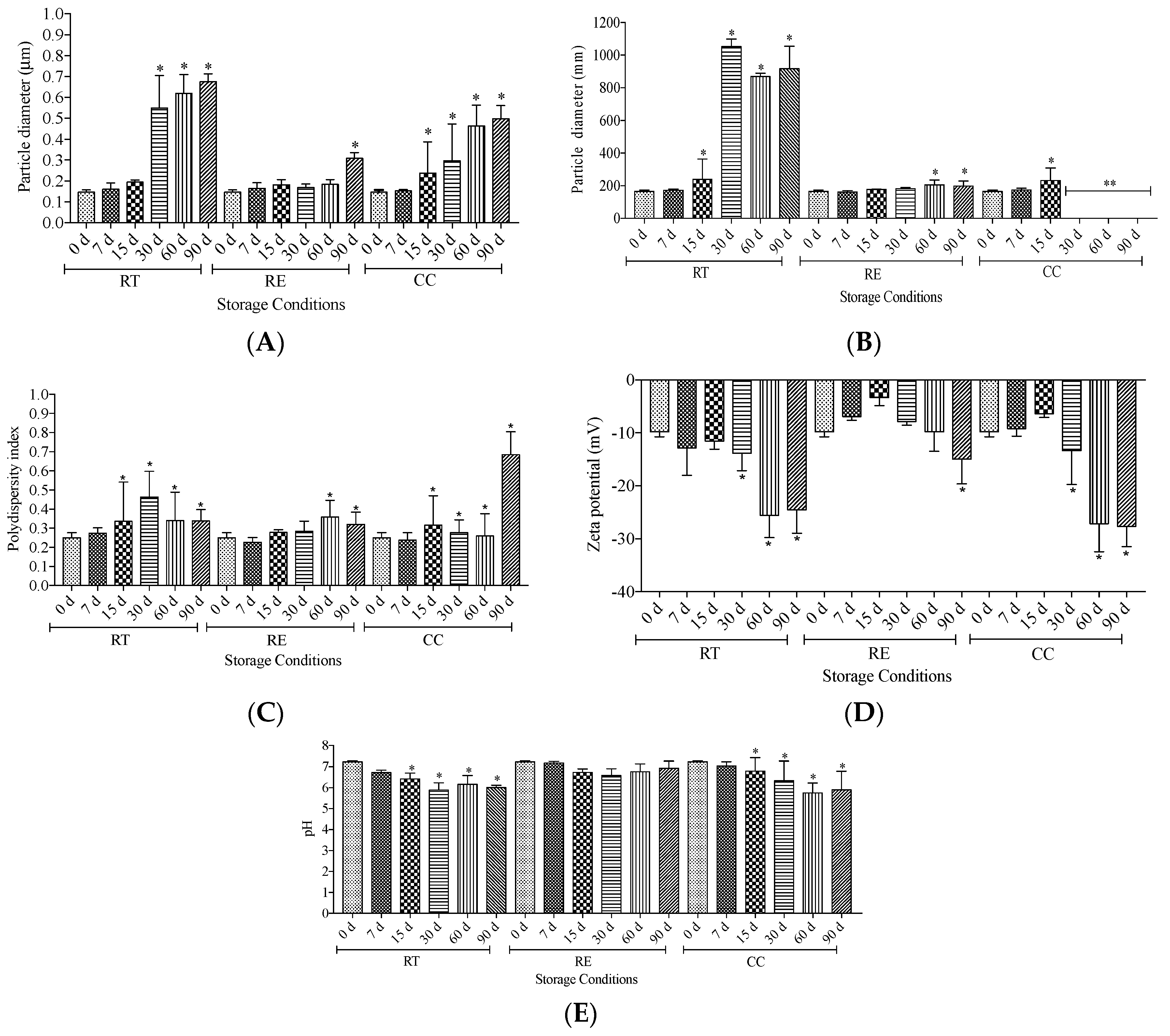
Figure 2.
- Macroscopic characteristics for the liposomes prepared by the reverse phase evaporation method, stored under different storage conditions. RT (room temperature, 25 ± 2 °C), CC (climatic chamber, 40 ± 2 °C and 75% relative humidity) and RE (under refrigeration, 5 ± 2 °C).
Figure 2.
- Macroscopic characteristics for the liposomes prepared by the reverse phase evaporation method, stored under different storage conditions. RT (room temperature, 25 ± 2 °C), CC (climatic chamber, 40 ± 2 °C and 75% relative humidity) and RE (under refrigeration, 5 ± 2 °C).
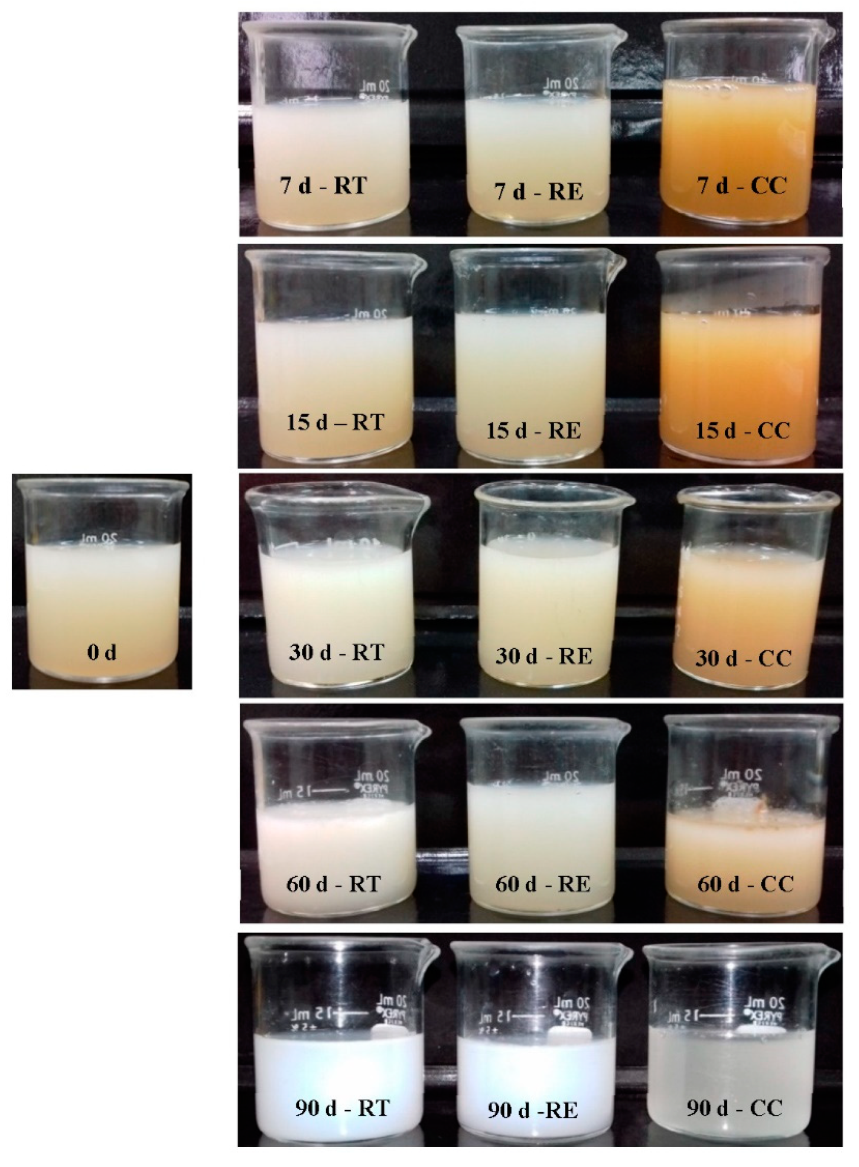
Figure 3.
- Total content of liposomal active ingredients containing 1 mg/mL of guarana powder. Results are expressed as mean ± standard deviation (n = 3). A (theobromine), B (theophylline), C (caffeine), D (catechin), and E (epicatechin). RT (room temperature, 25 ± 2 °C), CC (climatic chamber, 40 ± 2 °C and 75% relative humidity), and RE (under refrigeration, 5 ± 2 °C). ∗Significant difference (p <0.05) in relation to the initial values.


Figure 4.
– Cell viability for 3T3 cells, analyzed by the NRU and MTT after 24h, 48h, and 72h. Cell viability by the NRU (A) and MTT (B), after 24h (4.1), 48h (4.2), and 72h (4.3), respectively. GL (1 mg/mL guarana powder), B-Lip (blank liposomes), and G-Lip (liposomes containing 1 mg/mL guarana powder). Results are expressed as mean ± standard deviation (n = 3). *Significant difference (p<0.05) in relation to the control cells (100% viability).
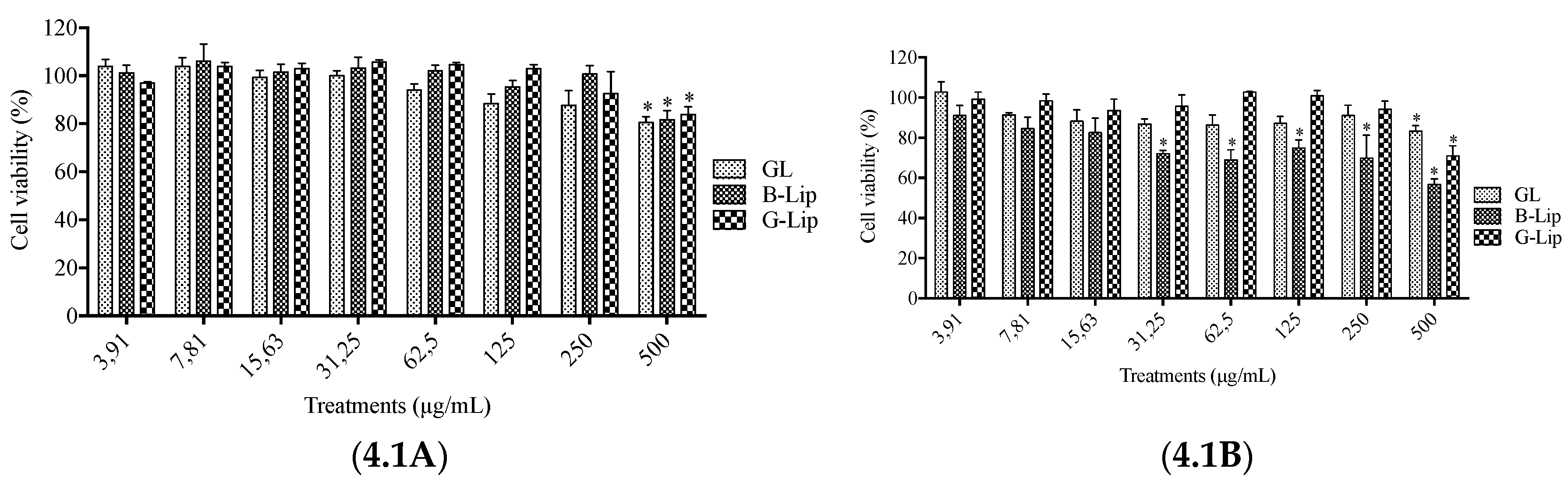
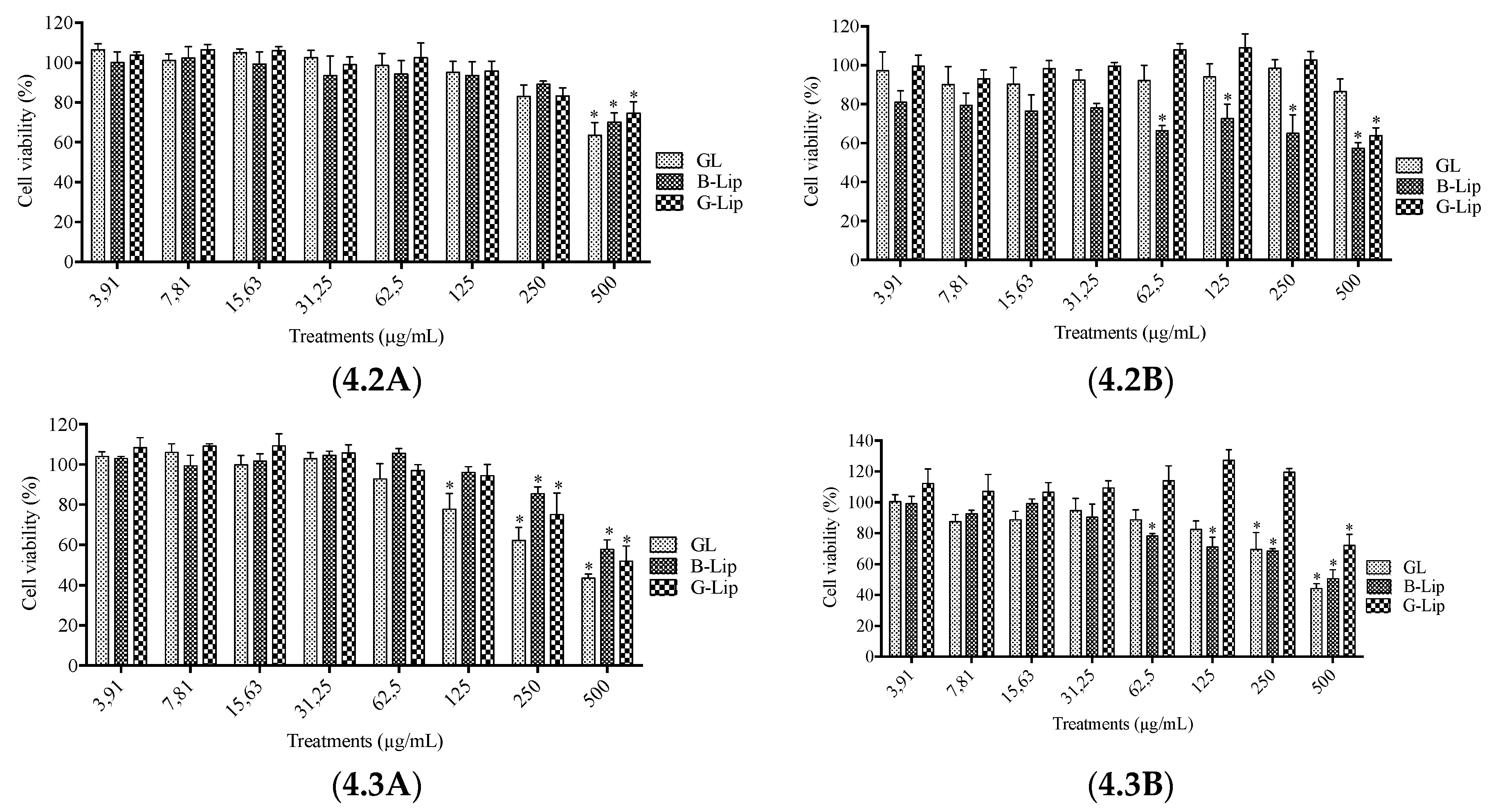
Figure 5.
– Cell viability of HaCaT cells, analyzed by the NRU and MTT techniques, at 24h, 48h, and 72h. Cell viability measured by the NRU (A) and MTT (B), analyzes in 24h (5.1), 48h (5.2) and 72h (5.3), respectively. GL (1 mg/mL guarana powder), B-Lip (blank liposomes), and G-Lip (liposomes containing 1 mg/mL guarana powder). Results are expressed as mean ± standard deviation (n = 3). *Significant difference (p<0.05) in relation to control cells (100% viability).
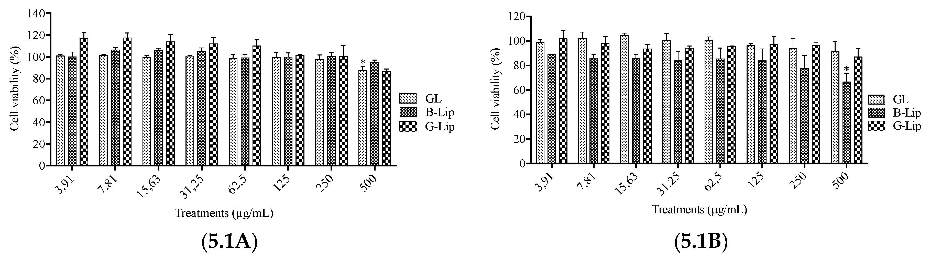
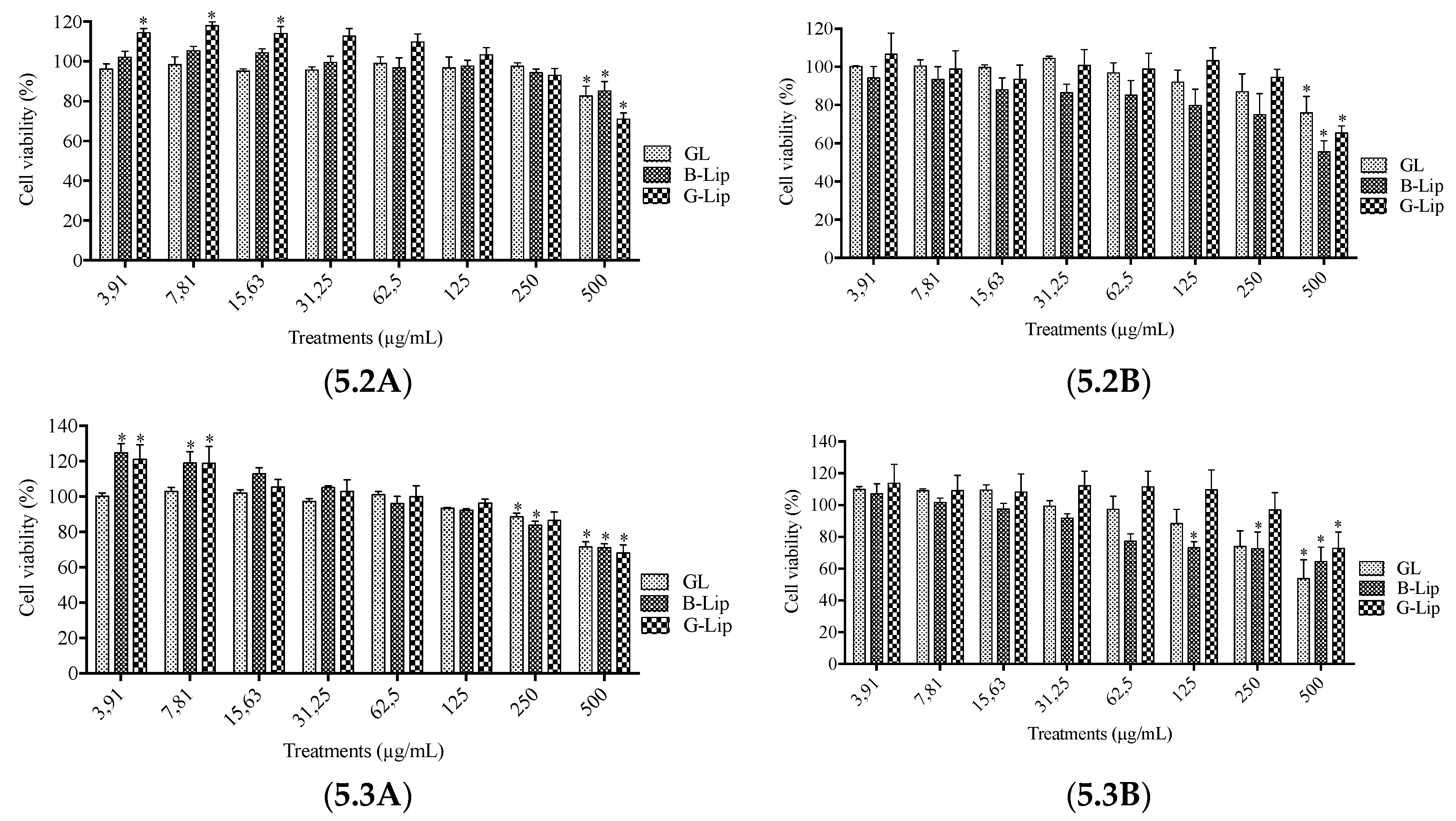
Figure 6.
– Cell viability of human fibroblasts cells, assayed by the NRU and MTT techniques, after 24h, 48h, and 72h. Cell viability measured by the NRU (A) and MTT (B), analyzes in 24h (6.1), 48h (6.2), and 72h (6.3), respectively. GL (1 mg/mL guarana powder), B-Lip (blank liposomes), and G-Lip (liposomes containing 1 mg/mL guarana powder). Results are expressed as mean ± standard deviation (n = 3). *Significant difference (p<0.05) in relation to control cells (100% viability).
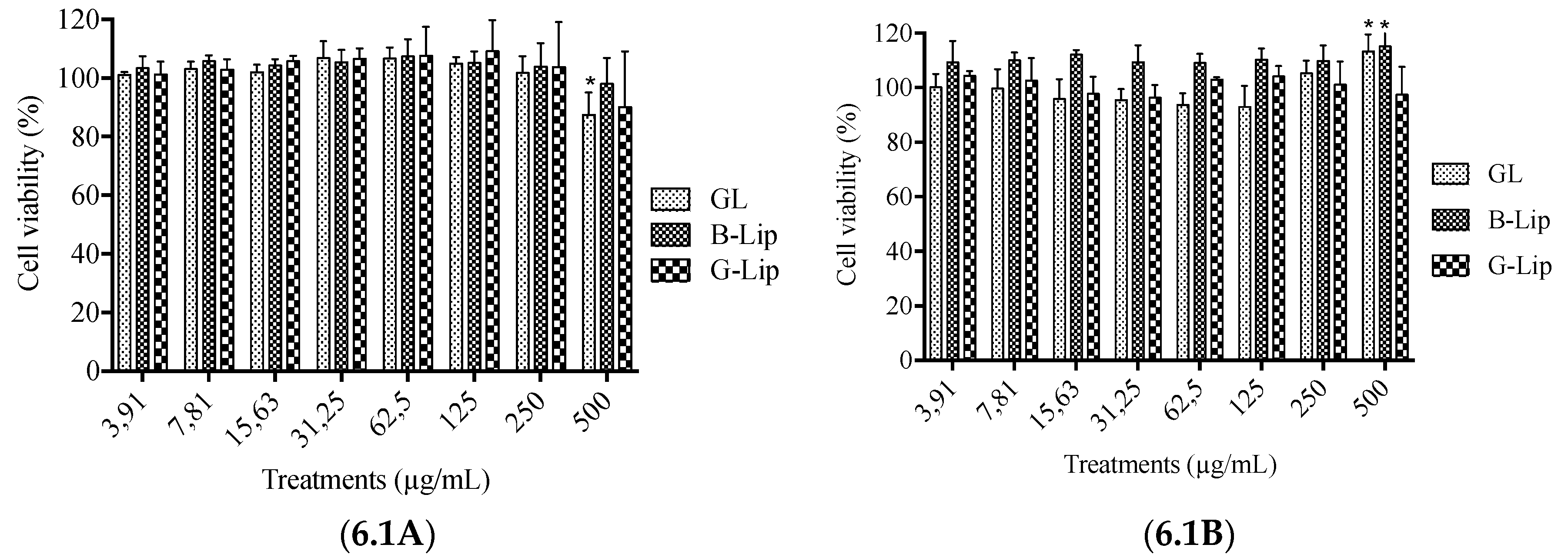

Table 1.
- Efficiency of encapsulation/incorporation of liposomes containing 1 mg/mL of guarana powder, prepared by the reverse phase evaporation method (n = 2).
Table 1.
- Efficiency of encapsulation/incorporation of liposomes containing 1 mg/mL of guarana powder, prepared by the reverse phase evaporation method (n = 2).
| Reverse phase evaporation | ||
| Active | Initial (%)* | 90 d (%)** |
| TEOB | Not determined | Not determined |
| TEOF | Not determined | Not determined |
| CAF | 17.02 ± 0.60 | 30.13 ± 0.23 |
| CAT | 74.34 ± 1.93 | 51.65 ± 0.77 |
| EPICAT | 87.53 ± 0.94 | 70.88 ± 2.17 |
Disclaimer/Publisher’s Note: The statements, opinions and data contained in all publications are solely those of the individual author(s) and contributor(s) and not of MDPI and/or the editor(s). MDPI and/or the editor(s) disclaim responsibility for any injury to people or property resulting from any ideas, methods, instructions or products referred to in the content. |
© 2023 by the authors. Licensee MDPI, Basel, Switzerland. This article is an open access article distributed under the terms and conditions of the Creative Commons Attribution (CC BY) license (http://creativecommons.org/licenses/by/4.0/).
Copyright: This open access article is published under a Creative Commons CC BY 4.0 license, which permit the free download, distribution, and reuse, provided that the author and preprint are cited in any reuse.


