
Submitted:
11 April 2023
Posted:
12 April 2023
You are already at the latest version
Abstract
Liposomes have been used for several decades for the encapsulation of drugs and bioactives in cosmetics and cosmeceuticals. On the other hand, the use of these phospholipid vesicles in food applications is more recent and has been increasing significantly in the last ten years. Although in different stages of technological maturity - in the case of cosmetics, many products are on the market - processes to obtain liposomes suitable for the encapsulation and delivery of bioactives are highly expensive, especially aiming at scaling-up. Among the bioactives proposed for cosmetics and food applications, vitamins are the most frequently used. Despite the differences between the administration routes (oral for food and mainly dermal for cosmetics), some challenges are very similar (e.g., stability, bioactive load, average size, increase of drug bioaccessibility and bioavailability). In the present work, a systematic review of the technological advancements in the nanoencapsulation of vitamins using liposomes and related processes was performed; challenges and future perspectives were also discussed in order to underline the advantages of these drug loaded biocompatible nanocarriers for cosmetics and food applications.
Keywords:
vitamins
; nanoencapsulation
; nanodispersions
; phospholipid vesicles
; liposomes
; cosmetics
; food application
1. Why Encapsulating Vitamins for Food and Cosmetics Application?
Vitamins are organic compounds required for metabolic processes, and not produced by the human body in sufficient amounts. Each vitamin has specific functions in the body and cannot be replaced by any other substance [1]. Having different chemical structures, vitamins can be classified in two main groups: fat-soluble and water-soluble compounds. Thirteen main vitamins have been identified; they are characterized by different ways of action and different beneficial roles in the body. In addition, vitamins play a key role in transforming energy and regulating the metabolism pathways of the human body [2].
Regarding food products, the incorporation of vitamins is generally performed: (i) to fortify; (ii) to replace vitamin losses during processing; and (iii) to use them as antioxidants and/or natural colorants. On the other hand, in cosmetics, vitamins are present in products for skin care, hair, and oral health [2].
Vitamins A, D, E, and K are fat-soluble compounds; therefore, they require a certain amount of fat in the diet to be efficiently absorbed. On the other hand, group B vitamins and vitamin C are water-soluble compounds and cannot be stored in the body, requiring daily consumption or supply. All these vitamins (A, D, E, K, B, and C) can be used as ingredients in cosmetics according to European Union legislation, with the exception of vitamin D3 (cholecalciferol); whereas, in USA and Japan, there are no restrictions on the use of vitamin D3 in cosmetics [2]. Chemical structure, ways of obtainment and food sources of some main vitamins are summarized in Table 1.
1.1. Hydrophobic Vitamins
Vitamin A can be found as retinoids (retinol, retinal, and retinoic acid) and provitamin A carotenoids (mainly β-carotene) [3]. It is important to emphasize that vitamin A is not produced endogenously. The retinol is the first precursor of two vital active metabolites: retinal, which plays an important role in the development of vision, and retinoic acid that acts as an intracellular signal that alters the transcription of a range of genes. Vitamin A is not found directly in plants; however, plants can contain carotenoids, like β-carotene, which is transformed in vitamin A in the intestine and other body tissues. Therefore, vitamin A supply is necessarily obtained through ingestion (dietary sources or vitamin supplements) [4]. Foods such as fish, meat (mainly liver), eggs, and whole milk are animal sources of retinol [2,3]. Fruits and vegetables such as carrots, spinach, and mango are sources of carotenoids. In terms of biological activity, vitamin A is important for the growth and development of children and for the maintenance of the immune system [5]. In adults, vitamin A deficiency can lead to adverse effects on the immune system, reproductive function, and eyes, such as night blindness [3,4,6]. On the other hand, excessive exposures to vitamin A are associated with adverse health effects, such as teratogenicity [7]. In the cosmetic industry, vitamin A is widely used due to its ability to delay photoaging effects. Being the main bioactive for skin treatment, it promotes the regeneration of the skin aged by UV radiation, reduces wrinkles and improves skin elasticity [2]. Vitamin A is not degraded by heat; but it is easily oxidized, and care is required during its processing. To reduce this undesired effect, antioxidants are added to vitamin A products as, for example, vitamin E [2].
Vitamin D group is composed of ergocalciferol (vitamin D2) and cholecalciferol (vitamin D3). Vitamin D3, or cholecalciferol, is a fat-soluble compound synthesized by the human epidermis by the irradiation of UV light on 7-dehydrocholesterol [8]. The precursor molecule of vitamin D is ergosterol (or 7-dehydrocholesterol), a rigid structure that is inserted by the body when absorbed by the lipid layer of the plasmatic membrane. The production of provitamin D occurs after solar incidence on the aromatic ring of ergosterol. The structure, then, becomes less rigid, promoting an increase in its permeability and, thus, allowing the incorporation of numerous ions into its interior, including calcium. It can be synthesized by the human epidermis or consumed in the form of supplements or fortified foods. A few foods naturally contain vitamin D in their composition: fish, such as salmon and sardines, butter, and eggs being the main sources. Some benefits conferred to its consumption are the improvement of calcium absorption by the intestine and the homeostasis of this mineral in the bloodstream, preventing diseases such as osteoporosis [9]. Currently, a large part of the urban population is deficient in vitamin D3 mainly due to the lack of sun exposure and loss of 7-hydrocholesterol reserves in the epidermis with aging [10]. Vitamin D deficiency is responsible for bone diseases; but also for other diseases such as cancer, asthma, arthritis, hypertension, osteoporosis and cardiovascular problems. Signs and symptoms of its deficiency may include bone pain and muscle weakness [11]. Vitamin D is used in the cosmetic industry as it prevents photo-damage, wrinkles and other morphological skin changes [2]. Some studies indicated its topical application for the treatment of psoriasis [12]. However, it has an adverse effect on calcium metabolism and limits its use for topical applications [13].
Vitamin E refers to different compounds, tocopherols and tocotrienols, and they are identified by the prefixes: α, β, γ, and δ. α-tocopherol is the most common occurring form of vitamin E and has much higher bioavailability than the other vitamin E forms [14]. Vegetable oils of peanut, soya, palm, corn, safflower, sunflower, and wheat germ are the most important sources of vitamin E. Vitamin E has relevant physiological functions, including antioxidant, immunity regulation, anti-inflammatory, and neuroprotective capacities [15]. This vitamin also plays the role of protecting body tissues from oxidation by metabolic processes and exogenous agents, in addition to assisting the body in the synthesis of vitamin A [2]. However, its incorporation in food can be challenging, as it is extremely sensitive to high temperatures, light, oxygen, and alkaline conditions, and has low solubility in water [15]. Encapsulation techniques can help to overcome these obstacles allowing the application of vitamin E in food, cosmetics and nutraceuticals. In the cosmetic industry, vitamin E is generally used as an antioxidant for the skin, aiding in softening and promoting hydration [2].
1.2. Hydrophilic Vitamins
Vitamin K occurs in three forms: vitamin K1 (phylloquinone, phytonadione, phytomenadione), vitamin K2 (menaquinone), and vitamin K3 (menadione). Vitamin K1 is found in plants; K2 is synthesized by bacteria in the human and animal intestines; and K3 is a synthetic compound that is converted to K2 in the intestinal tract [2,16,17]. Green leafy vegetables (such as spinach, kale, broccoli, and cauliflower) are good dietary sources of this vitamin that is also found in smaller amounts in liver, lean meat, cow's milk, egg yolks, and whole wheat products [2]. Although differing in structure, the vitamin K1 and K2 act as cofactor for the enzyme gamma-glutamylcarboxylase, encompassing both hepatic and extrahepatic activity. In addition, vitamin K2 has been shown to be necessary in regulating osteoporosis, atherosclerosis, cancer and inflammatory diseases without risk of negative side effects or overdosing [16,17]. Also, vitamin K is effective in treating dark circles and bruises on the face, and its application to reduce the effects of bruising after certain dermatological procedures has been also studied [18].
Group B vitamins comprises B1 (thiamine), B2 (riboflavin), B3 (niacin), B5 (pantothenic acid), B6 (pyridoxine), B8 (D-biotin), B9 (folic acid), and B12 (cobalamin) vitamin. Vitamin B1 has been found in brewer's dried yeast and pork, lamb and beef, poultry, whole grains, nuts, vegetables, and legumes, but in small amounts. It is essential for the breakdown of food, especially carbohydrates, to release energy, and for healthy nerve and muscle function. Deficiency causes growth retardation and disorders of the nervous and cardiac systems, such as 'beri-beri' disease. In the cosmetic industry, it acts as a coenzyme in the metabolism of aminoacids and keeps the skin healthy [2]. Vitamin B2 (riboflavin) is found in all plants and animal cells; but these are poor sources. The commonest dietary sources are milk and dairy products, meat, eggs and leafy green vegetables; however, animal sources of riboflavin are better absorbed than vegetable sources. Riboflavin is vital for the release of energy from foods and for healthy skin, eyes and growth. It plays a major role in oxidation and reduction processes in cells. Deficiency is rare, and generally occurs in combination with deficiencies of other water-soluble vitamins. Riboflavin is not used in skin care cosmetics; but, due to its bright deep yellow color, it can be used as a colorant in cosmetic products [2]. Vitamin B3 (nicotinic acid or niacin) is found in yeast, liver, poultry, lean meats, nuts, and legumes. Niacin is used for the treatment of lipid disorders and cardiovascular diseases, essential for growth and hormone synthesis [19]. Together with vitamins B1 and B2, it maintains a healthy nervous and digestive system. Pantothenic acid is necessary for the release of energy from food, for the production of antibodies and healthy growth. Vitamin B5 requires vitamin A, vitamin B3, vitamin B6, vitamin B9, and vitamin B12 in order to act properly. Vitamin B5 is used in many cosmetic skin and hair care products. Its main functions can improve wound healing, anti-inflammatory, and moisturizing activities [2]. Pantothenic acid is present in almost every type of food; but it is particularly abundant in yeast and organs of animals (liver, kidney, heart, brain). However, eggs, milk, vegetables, legumes, and whole-grain cereals are probably the most common sources of this vitamin. Vitamin B12 is essential for DNA synthesis and red blood cell production. Its deficiency results in megaloblastic macrocytic anemia (characterized by tachycardia, palpitations, generalized weakness, fatigue, and pallor), cognitive impairment, peripheral neuropathies, sensory and neurological abnormalities [20].
Vitamin C or ascorbic acid is largely used in cosmetic and pharmaceutical products due to its antioxidant activity. However, the biggest challenge in the incorporation of vitamin C is to maintain its stability and improve its delivery to the active site [21]. It is naturally found in citrus fruits, currants, peppers, parsley, cauliflower, potatoes, sweet potatoes, broccoli, brussels sprouts, strawberries, guava, and mangoes. Vitamin C is also frequently used as a natural antioxidant in foods and beverages spoilage. This molecule is also important for the production of collagen, connective tissue and protein fibers that give strength to teeth, gums, muscles, blood vessels, and skin. In the immune system, vitamin C helps the white blood cells to fight infection. It also helps the body to absorb iron [2,21].
2. Formation and Classification of Liposomes
In 1965, it was reported, for the first time, that phospholipid molecules were able to instantaneously form closed bilayer vesicles in aqueous media due to the amphiphilic nature of phospholipids [22]. Liposomes are vesicular structures formed by one or more phospholipid bilayers that encapsulate part of the aqueous medium in which they are dispersed [23]. Their average diameters range from 20 nm to several microns. Phospholipids are the main constituents of vesicles, being an amphiphilic molecule in which the hydrophilic polar head groups are oriented towards the aqueous phase and the hydrophobic non-polar hydrocarbon tails are oriented towards each other in an ordered bilayer structure. A wide variety of phospholipids can be used for the production of liposomes, such as eggs, soy, milk that are natural and safe sources. The most used phospholipid for the production of liposomes in the food and cosmetic industry is phosphatidylcholine (PC). In addition to PC, phospholipids such as lysophosphatidylcholine (LPC), phosphatidylinositol (PI), phosphatidylethanolamine (PE) and phosphatidylglycerol (PG) are also used. Phospholipids contain different amounts of unsaturated fatty acids that are prone to oxidative reactions, and the resulting products can cause changes in the permeability of the lipid bilayers. In addition, interactions between phospholipids and bioactives can alter the overall chemical stability of the system.
Phospholipids, when added to an aqueous medium, due to their low solubility in water, associate with each other resulting in lipid bilayers, and by adding a sufficient amount of energy to the system (which can be provided by external methods such as sonication, heating or homogenization), the unfavorable interaction between molecules of fatty acids and water is eliminated and the bilayers arrange themselves in their organized form. In this process, liposomes are capable of incorporating hydrophilic compounds in their aqueous core, in addition to hydrophobic compounds in the internal regions of the lipid bilayer [24].
Liposomes are among the current commercialized lipid nanocarriers most used for various nutraceutical purposes and are increasingly studied for incorporation into foods for functional applications [25,26].
During formation, liposomes acquire structural characteristics (number of layers) and various sizes [27,28,29]. Based on their size and number of bilayers, liposomes can be classified in two main categories: unilamellar vesicles and multilamellar vesicles (Table 2). Liposomes that contain only a single bilayer membrane are called small unilamellar vesicles (SUV) with a size of 20 to 200 nm; large unilamellar vesicles (LUV), with size above 200 nm, and giant unilamellar vesicles (GUV), with size higher than 1 µm. Liposomes that contain more than one bilayer membrane are called multilamellar vesicles (MLV) and are composed of several concentrically arranged vesicles with a size of 0.5 to 5 µm, or multivesicular vesicles (MVV), where there are smaller vesicles within a vesicle of larger size [12,24]. Such size characteristics directly depend on the chosen production method and the type(s) of phospholipid(s) used.
Despite their versatility, liposomes are physicochemically unstable. This is due to the fact that the lipids present in their structure can undergo to natural degradation through oxidation or hydrolysis, or even because the particles can form agglomerates. Liposomes originally show repulsive forces between their particles that provide a certain physical stability; but external factors, such as high temperatures or pH changes, can affect their structure and change the permeability of the bilayer, causing the release of the encapsulated compound or the formation of agglomerates. In order to reduce these undesired effects, a possible solution is the coating of liposomes with biopolymers that can increase the physical stability of these particles through steric and electrostatic factors, thus creating a hybrid system. Among the biopolymers used for the coating of liposomes, starches, gelatin, proteins, cellulose, pectin and chitosan can be mentioned.
3. Methods for the Production of Vitamin-Loaded Liposomes
Generally, the successful encapsulation of bioactive molecules in liposomes, with desired and specific size and structure, depends on several factors such as the correct choice of the main lipid, the affinity between the liposome and the bioactive of interest, and, most of all, the production method. The latter is considered as extremely important when encapsulating vitamins for several reasons that may include: (i) the efficiency of encapsulation, as some methods may result in higher encapsulation efficiencies than others; (ii) the stability of the vitamin, as certain methods may be less harmful to them, which is particularly important for sensitive vitamins that can be easily degraded by heat, light, or exposure to oxygen; (iii) the control of particle size and distribution, which are directly related to the release rate of the vitamin from the encapsulating material and also to its bioaccessibility and bioavailability; and (iv) the process scalability, to ensure both consistent quality and efficient production.
Conventional methods of liposome production for vitamin encapsulation are still on the spotlight of many researchers due to their simplicity, recognition and wide range of applications. On the other hand, novel methods have arisen thanks to their advantages over the conventional ones, such as higher encapsulation efficiencies, reduced toxicity, versatility, and scalability. Overall, the production method used for encapsulating vitamins plays a critical role in ensuring the stability, efficacy, and quality of the final product. The main methods currently found in the literature for liposome production are discussed below.
3.1. Conventional Methods
In the conventional methods, five main steps are basically followed: (i) lipids are dissolved in an organic solvent (ethanol, ether, chloroform, dichloromethane); (ii) the organic solvent is removed using evaporation, rotary evaporation, or distillation; (iii) the resultant lipid layer is hydrated using an aqueous medium (distilled water, buffer solutions, serum-containing media, or physiological saline solutions) and agitated; (iv) vesicles are analyzed and eventually treated by downsizing steps (see Supplementary Material for the detailed description of these methods), depending on the liposome final use; and (v) post-formation processing (purification or sterilization) is carried out to increase the stability of the liposomes [30]. Vitamins must to be dissolved in the liquid medium in which they are easily solubilized; i.e., in the organic solvent if lipophilic (A, D, E, and K) or in the hydration medium if hydrophilic (complex B vitamins and C) vitamins are selected.
3.1.1. Thin-Film Hydration (TFH) Method (Bangham Method)
The thin-film hydration technique, also referred as the Bangham method due to the British biochemist that first described it in 1965, is the simplest method used for the preparation of MLVs. It is mostly used for the encapsulation of lipophilic molecules and it is based on the production of a thin lipid film by the evaporation at 45 - 60 °C of an organic solvent from a lipid-solvent solution during flask rotation under vacuum and its further hydration using an aqueous media. When small volumes of liposomes are desired, the organic solvent can be dried by using argon steam or dry nitrogen in a fume hood [31]. Hydration processes should be performed above the phase-transition temperature of the lipids (e.g., 60 - 70 °C) for a duration of at least 1 - 2 h [30]. Besides the ability to encapsulate both hydrophilic and hydrophobic molecules, the TFH method presents some limitations such as low encapsulation efficiencies and batch-to-batch variability. The production of liposomes using TFH method is usually followed by a downsizing process to obtain SUVs.
Bi et al. [32] used egg-PC, cholesterol (Chol), and vitamin D3 (3:1:1 w/w) to produce vitamin D3-loaded MLVs by the TFH method followed by high-pressure homogenization. Obtained liposomes presented a particle size of 169 nm and an encapsulation efficiency (EE%) of 62%. Vitamin B12-loaded liposomes were produced by Andrade et al. [33] prepared liposomes by the TFH technique followed by ultrasonication, using 1,2-distearoyl-sn-glycero-3-phosphocholine (DSPC), Chol, polyethylene glycol 2000 (PEG200) and amine 1,2-distearoyl-sn-glycero-3-phosphoethanolamine-N-[amino(polyethylene glycol)-2000] (DSPE-PEG2000) in a 52:45:3:0.06 molar ratio. LUVs showed sizes of 116 nm and an EE% of 14%. The authors stated that the the low EE% was due to the vitamin B12 that is an extremely water-soluble molecule. Campani et al. [34] produced a liposome-based formulation loaded with vitamin K1 by the TFH technique followed by extrusion through polycarbonate membranes with decreasing porosity (400, 200, and 100 nm). Lipid films were prepared using soy phosphatidylcholine (SPC) and were rehydrated by PBS at pH 7.4 or aqueous solution at 0.01% w/v of benzalkonium chloride. Samples produced in these conditions were all in the nanometric range (131 - 147 nm) and presented vitamin K1 encapsulation ratios of 3.4 - 154.0 µg VK1/mg SPC. Vitamin E-loaded nanoliposomes were efficiently prepared by Qu et al. [35] by using egg lecithin, cholesterol, sodium deoxycholate, and vitamin E, in a 5.8:1:1.1:1.8 mass ratio, by the TFH method followed by sonication and extrusion. The obtained vesicles presented an average hydrodynamic diameter of 231 nm, EE% of 97%, a narrow size distribution (polydispersity index, PDI = 0.217) and high zeta potential (-52.39 mV).
3.1.2. Reverse-Phase Evaporation (RPE) Method
This technique was first described by Szoka & Papahadjopoulos in 1978 [36] and is generally used to encapsulate large amounts of hydrophilic bioactives with high encapsulation efficiencies [37]. Similarly to the TFH method, lipids are first dissolved in an organic solvent, such as diethyl ether, chloroform, isopropyl ether, or a mixture of two solvents, in order to form inverted micelles [38]. After the addition of an amount of aqueous phase, water-in-oil (W/O) microemulsions are formed due to the rearrangement of lipids at the interface between water and oil. During this step, a large amount of the aqueous phase is encapsulated within the microemulsion, as well as the hydrophilic molecules. The organic solvent can be slowly removed using a rotary evaporator under vacuum until the conversion of the micelles to a semi-solid viscous gel-like structure is obtained. The gradual removal of the solvent favors the disruption of the gel and promotes the formation of LUVs [30]. As discussed for the TFH method, a downstream step such as sonication or extrusion is required to reduce the average size of liposomes and to obtain a narrow size distribution [39]. The main drawbacks related to this technique include: (i) the use of a large amount of organic solvents and the presence of residual solvent at the end of the process; (ii) the high complexity and difficulty to industrial scaling-up; (iii) the unsuitability to be employed for the encapsulation of sensitive molecules due to the long-lasting contact with the organic solvent; (iv) the time-consuming process; and (v) the sterile boundary is quite hard to establish [40].
There are a few studies exploring this method for the encapsulation of vitamins, mainly due to their high degradability rates. Favarin et al. [41] employed the RPE method to encapsulate vitamin C into liposomes. In their study, the aqueous phase was composed of polysorbate 80, at pH 3.65, and vitamin C; whereas the organic phase was composed of phospholipid Lipoid® S100, cholesterol and ethanol. The inverted micelles were formed after the addition of the aqueous phase to the organic phase under ultrasonic conditions. The mixture was, then, submitted to a slow evaporation in a rotary evaporator operating at 80 rpm and 35 °C for the removal of the organic solvent, and the formation of the gel-like structure. Liposomes were produced after the addition of the aqueous phase under agitation. To homogenize the vesicles size, samples were extruded using membranes with pores of 0.45 μm and 0.22 μm. Vitamin C-loaded liposomes showed size of 160 nm and PDI of 0.23, besides a slight negative zeta potential of -7.3 mV and an EE% equal to 19%.
3.1.3. Injection Methods
The injection methods are based on the dissolution of lipids into an organic solvent (ethanol or diethyl ether) and the further injection of the resulting solution into an aqueous phase. Among all the liposome production techniques, the injection methods are suitable to operate continuously [42].
- Ethanol injection (EI) method: this method was first described by Batzri & Korn [43] and is based on the dissolution of phospholipids in ethanol and, then, the injection of the solution into a rapidly stirred aqueous phase [43]. Part of the ethanol evaporates upon contact with the aqueous phase, producing a lipid film that forms liposomes upon hydration. Vesicles are formed due to the immediate diffusion of the ethanol in the aqueous medium, leading the lipid molecules to precipitate and to form bilayered planar structures that tend to liposomes [44]. A change in the solubility of lipids leads to the spontaneous formation of vesicles that encapsulate a small volume of the aqueous phase. This method is relatively simple and produces liposomes characterized by a high entrapment efficiency of hydrophobic molecules. SUVs can be formed if proper process parameters, such as low lipid concentrations and fast rate of injection, are used. A disadvantage of ethanol injection is that the encapsulation efficiency of hydrophilic compounds is low, and the control of both size and size distribution of the resulting liposomes can be challenging. On the other hand, some advantages of this method include: (i) it is a straightforward method, which makes it a popular choice for liposome production; (ii) its high reproducibility; and (iii) its easy scale-up, making it a practical choice for commercial production [44]. However, this method is limited by the need for subsequent processing to solvent evaporation and the residual content [45]. The use of the EI method to produce liposomes for food or cosmetics is usually hindered by the low encapsulation efficiency of hydrophilic bioactive molecules such as vitamin C. This occurs because hydrophilic bioactives tend to be preferentially retained in the external aqueous phase of vesicles instead of in their small aqueous core [46]. Charcosset et al. [47] developed a continuous process for the ethanol injection method coupled with membrane extrusion, in which vitamin E-loaded MLVs were produced using Lipoid® S100 as the source of phospholipids. Liposomes were stored in a vacuumed double jacketed reactor in which the ethanol residue was constantly removed. Using this method, volumes between 60 mL to 3 L of liposomes with sizes ranging from 89 to 118 nm were produced.
- Ether injection method: this method requires the dissolution of lipids in diethyl ether and, then, a slow injection of the solution into an aqueous phase under high pressure. The ether rapidly evaporates upon contact with the aqueous phase under warming, resulting in a lipid film that forms liposomes upon hydration [48]. This method results in a concentrated liposomal (LUVs) product with a narrow size distribution and high entrapment efficiency [45]. Some differences can be pointed out between this method and EI: (i) ethanol is a polar solvent, unlike ether that is nonpolar; this can affect the lipid solubility and the self-assembly of lipids in liposomes, resulting in differences in size, size distribution, and other properties of liposomes; (ii) ether injection method may be more complex and time-consuming than EI method; and (iii) different effects on the stability of lipids and liposomes as the use of ether may cause a larger lipid degradation or oxidation than the use of ethanol due to the formation of peroxides [30]. However, an advantage of using the ether injection method consists of the more efficient removal of the organic solvent from the final product [30]. For food purposes, it is generally preferable to use ethanol instead of ether. Ether is highly flammable and can pose a fire hazard, which may be a concern in a food production setting. Ethanol, on the other hand, is a safer solvent that is commonly used in food processing and is generally recognized as safe (GRAS) by the US Food and Drug Administration (FDA). Ethanol is also a polar solvent, thus more compatible with the polar environment of aqueous foods. The ether injection method is not commonly used for the encapsulation of vitamins in liposomes, basically because of its high toxicity [49].
3.1.4. Detergent Removal (Depletion) Method
The detergent depletion method is a mild process capable of producing highly homogeneous liposomes. This method is based on the formation of mixed micelles of detergents and lipids and the further removal of the detergent to form LUVs. The size of vesicles is based on the rate at which the detergent is removed from the formulation and the initial detergent to phospholipid ratio [50]. Detergents, such as sodium cholate, sodium deoxycholate, and octyl glycoside, are often used in the initial stages of liposome preparation to solubilize the lipids and to form a homogeneous solution. However, detergents can also destabilize the liposome membrane and lead to aggregation and fusion of the liposomes. To remove the detergent and to obtain stable liposomes, various methods are used. One of the most commonly used method is dialysis [51]. Using this method, the detergent-containing the liposome dispersion is submitted to a dialysis step. This process is typically carried out over several hours to several days, depending on the amount of detergent and the desired degree of detergent removal [52]. Another method for detergent removal is the use of adsorbents such as Bio-Beads [53]. The obtained liposomes are, then, separated from the beads containing adsorbed detergent molecules by filtration or centrifugation, resulting in detergent-free liposomes [53].
Vitamin-loaded liposomes can also be produced by the detergent depletion method. In this case, vitamins should be solubilized with the lipids in a detergent-containing solution. Hydrophilic compounds are generally encapsulated more efficiently in liposomes produced by detergent removal than hydrophobic bioactives since these latter may be more prone to being removed along with the detergent during the depletion process. However, in the case of food applications, the choice of detergents used in the liposome production should be carefully considered; some safety concerns or regulatory restrictions may apply. Secondly, this process can be time-consuming and labor-intensive, and may not be easily scalable to commercial production levels. Some detergents such as Tween 20, Tween 80, and Brij-35 are considered safe for food applications, as they are approved by FDA and European Food Safety Authority (EFSA). Triton X-100 cannot longer be used for food applications; but its use in cosmetics is still feasible due to the regulation approved by several agencies. It is important to notice that the choice of the detergent can affect the properties of the resulting liposomes, including their size, stability, and encapsulation efficiency [45]. Therefore, it is necessary to carefully optimize the detergent concentration and preparation method to achieve the desired properties of the liposomes for the intended final application. Additionally, it is important to ensure that any residual detergent is removed from the liposomes during the detergent depletion process to avoid potential health risks [54]. When producing liposomes for cosmetic applications, it is important to choose a detergent that is safe for use on the skin and does not cause irritation or other adverse effects on humans.
3.1.5. Double-Emulsion Method
The double emulsion method, also known as the water-in-oil-in-water (W/O/W) emulsion method, is a technique for encapsulating both hydrophilic and hydrophobic molecules in liposomes [55]. Hydrophilic bioactives such as proteins are more suitable to be encapsulated using this technique due to the multiple aqueous phases involved in the emulsification process. In this context, the method involves two emulsions, one inside the other one [56]. The first step is to prepare a W/O emulsion. This is performed by mixing a small amount of an aqueous solution containing the hydrophilic compound of interest with an immiscible organic solvent, such as chloroform or toluene, and a lipid solution. The mixture is vigorously shaken to create small droplets of the aqueous solution surrounded by a lipid bilayer. The second step is to create a second emulsion by adding the W/O emulsion to a large amount of the aqueous phase containing a stabilizer, such as a surfactant or polymer. This is typically carried out by sonication or homogenization. The resulting W/O/W emulsion contains small droplets of the aqueous phase plus the hydrophilic compound, surrounded by a lipid bilayer, which in turn is surrounded by a second aqueous phase. Liposomes are formed after the removal of the organic solvent by stripping gas or vacuum pressure, which leads to the direct contact between the external and internal oil-water phases and the formation of the lipid bilayer [57]. The double emulsion method presents some advantages over the other liposome production methods such as high encapsulation efficiency of hydrophilic compounds, drug controlled release, and versatility. However, this process can be time-consuming since it requires several steps to form liposomes, besides the low yield, and the need for specialized equipments such as sonicators or homogenizers [39].
A modification of this method, named “freeze-drying of double emulsions”, has been efficiently used to produce liposomes characterized by improved stability and prolonged shelf-life. This technique involves three main steps: (i) the formation of a double emulsion by the emulsification of an aqueous phase with a lipid phase, followed by a new emulsification with an external aqueous phase; (ii) the freezing of the double emulsion to produce a frozen matrix of liposomes, which helps to stabilize the liposomes and to prevent aggregation or coalescing phenomena; and (iii) the freeze-drying process, in which the frozen matrix is submitted to freeze drying for the removal of water through sublimation under vacuum. This step transforms the frozen matrix of liposomes in a dry powder that can be easily reconstituted with water or other appropriate solvents. Some advantages related to this method include: (i) improved stability, making them suitable for long-term storage and transportation; (ii) preservation of structural integrity, thus maintaining their biological activity; and (iii) flexibility, as liposomes can be produced at different sizes and compositions depending on the final destination. However, this technique shows some drawbacks, such as the requirement for specialized equipments, the potential for loss of encapsulated compounds during the freeze-drying process, and some damage to the freeze-dryer apparatus due to residual solvents [58].
Li et al. [59] produced complex liposomes containing medium-chain fatty acids (MCFAs) and vitamin C using the double emulsion method followed by dynamic high pressure microfluidization. A mass ratio of 100:25:4 soybean phospholipids, cholesterol, and vitamin E, altogether with an ethanolic solution containing MCFAs, was used throughout the production. The procedure was followed by the injection of a small volume of twice-distilled water under vigorous stirring at 50 °C. The primary emulsion was formed after evaporation of part of the solvent under reduced pressure. The aqueous phase composed by twice-distilled water, Tween 80 and vitamin C was, then, incorporated into the organic phase. After agitation, the residual solvent was removed through rotary evaporation under reduced pressure. These authors obtained liposomes with a mean diameter of approximately 93 nm, encapsulation efficiencies of MCFAs and vitamin C of 49% and 64%, respectively, and a good stability over 90 days at 4 °C. In another study, Yang et al. [60] used the same protocol as Li et al. [59] to produce complex liposomes encapsulating MCFAs and vitamin C; but these authors added a freeze-drying step at the end of the process to increase vesicle stability. The reconstituted liposomes presented a mean diameter of 110 nm and an encapsulation efficiencies of MCFAs and vitamin C of 44% and 62%, respectively. Vesicles remained stable for 60 days at 4 °C. In a more recent study, Pattnaik & Mishra [61] produced a multivitamin (A, D, B9 and B12) liposome using the double emulsion technique and a mix of soy lecithin and vegetable oil blend, besides a polymer solution containing milk protein isolate and trehalose at different concentrations. Liposomes were stable, with size varying from 143 to 396 nm and zeta potential from -20 to -33.5 mV. Interestingly, these authors observed that hydrophilic vitamins showed lower entrapment efficiency than the hydrophobic vitamins (vitamin B9, EE% = 78-97.57% and vitamin B12, EE% = 96-99.9% vs. vitamin A, EE% > 99.9% and vitamin D, EE% > 98%) and stated that this behavior was probably due to the interaction between the lipophilic vitamins with the hydrophobic tails of phospholipids during freeze-drying, which might have caused a protective effect over the vitamins.
3.1.6. Hydration of Proliposomes
This method of production uses a powder mixture of dry phospholipids and bioactives, called proliposomes. Several techniques can be used to obtain these powders, including fluidized bed, spray drying, freeze-drying, coating of micronized sugars, milling, and supercritical techniques. Proliposomes are defined as dry, free-flowing powders that contain the bioactives to be encapsulated [62]. Upon hydration under appropriate conditions, MLVs are formed. Their solid form confers stability and offers advantages such as improved transport convenience, storage, distribution, and dosage. Proliposomes can be manufactured using industrialized procedures as tablets or capsules, which eliminates the stability problems of liquid liposomes and may increase the oral bioavailability of bioactives [63].
Spray drying is a commonly used technique in the pharmaceutical and food industries for the production of powdered materials. This method is widely used for the encapsulation of oils, flavors and fragrances. Its large application is mostly due to its ability to evaporate moisture rapidly from a sample, maintaining a low temperature in the particles [64]. Wall materials such as polysaccharides and proteins are commonly used throughout the process [65]. In the liposome field, spray drying acts as a post-processing step to convert the liquid form of dispersions to a high stable solid form [66]. First, the liposome dispersion must be produced selecting the desired bioactive molecule by any method of interest. This solution is, then, atomized in small droplets using a spray nozzle and dried using a hot gas stream. The resulting powder is collected and can be used for further processing or packaging. Spray drying has several advantages for the production of bioactive-loaded liposomes. It is a fast and efficient method for producing large quantities of powdered material, and it can be easily scaled-up for commercial production [67]. The resulting powder is also stable and can be stored for long periods of time without degradation. However, there are some challenges associated with spray drying regarding the encapsulation of vitamins. This process can cause damage to the liposome structure, leading to a loss of the encapsulated material. Additionally, the high temperatures used during spray drying can degrade sensitive vitamins [68]. However, some recent studies on thermosensitive vitamins such as vitamin B1, B9 and vitamin B12 showed that they may not experience significant degradation during the spray drying process depending on the wall material and process parameters [69,70]. The microencapsulation of lipophilic vitamins such as vitamin D using spray drying is rare due to the mandatory water-dispersed form that is needed for the processing. Also, the porosity of the resultant material can act as an advertisement about the risk of degradation of more sensitive materials due to oxygen exposure [68]. The use of lower temperatures or shorter drying times during the spray drying process may prevent early thermal degradation.
Spray drying was used to produce β-carotene-loaded proliposomes by Toniazzo et al. [71]. Phospholipids, sucrose and β-carotene were solubilized in anhydrous ethanol and treated by spray drying using an inlet temperature of 90 °C and outlet temperature of 85 °C, at a flow rate of 10 mL/min in a 1 mm-diameter spray nozzle with co-current airflow. Proliposomes were, then, rehydrated with deionized water and thickeners xanthan and guar gums were used as stabilizers. The MLVs produced by this method were useful to protect β-carotene over 95 days of storage with an EE% up to 96% when a 0.10% w/w mixture of xanthan and guar guns was incorporated during the hydration step.
3.2. Novel Methods
Novel methods for liposome production have been developed and improved over the years, with the aim of overcoming the drawbacks related to the traditional methods. Differently from the liposomes produced using the conventional methods, vesicles produced by these new methods are mainly unilamellar and show a more homogeneous size distribution (lower values of PDI). Therefore, post-processing techniques such as sonication or extrusion are rarely required. Moreover, conventional methods are based on the use of organic solvents and detergents, which can limit their use in food and cosmetics, and are often not environmentally friendly. Currently, research efforts around liposomes focused on the optimization of green technologies that can be scaled-up to industrial levels.
3.2.1. Heating Method
This method was first described by Mozafari et al. [73] and involves the hydration of phospholipids with a 3% v/v glycerol solution at increasing temperature up to 60 °C or 120 °C. The processing temperature depends on the absence or presence of Chol. The use of glycerol is due to its physicochemical characteristics such as water-solubility and non-toxicity, besides its ability to avoid sedimentation or coagulation of vesicles. Sterilization of the resulting liposomes is not required due to the high temperature already used during processing (120 °C), leading to cost reduction. The heating method has been improved over the years resulting in a new method called “the Mozafari method”, in which large-scale production of liposomes can be achieved without the need for the prehydration step of raw materials and without the use of toxic solvents or detergents [74]. Although vitamins are mostly thermosensitive, equally sensible molecules such as DNA are being incubated at room temperature with preformed liposomes produced by the heating method [73].
3.2.2. Membrane Contactor Method
In this technique, a porous membrane with a defined pore size is used to separate two fluid compartments [75,76]. The organic phase (generally ethanol + lipids) is flowed across one side of the membrane, while an aqueous solution is flowed across the other side. The two fluids come in contact at the pores of the membrane, and liposomes are formed thanks to the presence of water. Liposomes then diffuse tangentially through the pores of the membrane and are collected on the surface of the other size. The organic solvent is removed by evaporation under reduced pressure. This method offers some advantages over traditional liposome production methods. Firstly, it is a continuous process, able to be scaled-up, thus allowing the production of large quantities of liposomes. Secondly, it is a gentle method, with minimal shear stress, which reduces the potential damage to liposomes or to the encapsulated molecules. Thirdly, it offers a better control over the size and size distribution of the liposomes, as the pore size of the membrane can be precisely controlled [77]. Also, membranes can be regenerated by washing using a water/ethanol mixture.
This method was already used to produce vitamin E-loaded liposomes [78]. Vesicles were produced using 20-50 mg/mL of 1-palmitoyl-2-oleoyl-sn-glycero-3-phosphocholine (POPC) or Lipoid® E80, 5-12.5 mg/mL of a stabilizer (Chol, stearic acid or cocoa butter), and up to 5 mg/mL of vitamin E. Shear stress on the membrane surface ranged from 0.80 to 16 Pa. Mean particle size under optimal conditions was 84 and 59 nm for Lipoid® E80 and POPC liposomes, respectively. EE% up to 99% was obtained in MLVs produced using Lipoid® E80.
3.2.3. Electroformation
This method was developed by Angelova & Dimitrov [79] and consists of the formation of GUVs under electric fields by the hydration of a lipid film deposited on electrodes. This method relies on the ability of lipids to self-assemble in bilayer structures in the presence of an electric field. The electric field creates a potential difference across the lipid solution, causing the lipid molecules to move towards the electrode, where they self-assemble in bilayer structures. The electroformation process typically involves the following steps: (i) preparation of the lipid solution, typically by mixing lipids in a suitable organic solvent; (ii) deposition of the lipid solution on an electrode or a conductive substrate, typically made of glass, quartz, or indium tin oxide; (iii) application of an electric field to the lipid solution by using two electrodes, typically made of platinum or gold; and (iv) collection of the vesicles from the electrode surface and further purification using size exclusion chromatography, ultracentrifugation, or dialysis [80]. The main disadvantages of this method include its low production yield and limited scalability.
Vitamin E-loaded GUVs were prepared by Di Pasquale et al. [81] in which the influence of vitamin E on the membrane organization was corroborated by SANS and fluorescence microscopy. Samples were produced using 1,2-dipalmitoyl-sn-glycero-3-phosphocholine (DPPC), 1,2-dioleoyl-sn-glycero-3-phosphocholine (DOPC) and Chol at a 37.5:37.5:25 mole ratio and vitamin E at a mole fraction of 0.10.
3.2.4. Nanoprecipitation
This is a scalable method that allows the production of liposomes in a single step, without the necessity of homogenizing the vesicle size. The process involves the mixing of a lipid solution and an aqueous solution, where the lipid molecules join together spontaneously to form liposomes, as they become less soluble in the aqueous phase. The size of the liposomes can be controlled by adjusting the composition and processing parameters. Due to the self-assembly process, the resulting liposomes are characterized by a narrow size range and can be produced with high reproducibility [82].
Jash & Rizvi [83] prepared vitamin C-vitamin E-co-loaded coated liposomes by the nanoprecipitation method. In this case, polyethyleneglycol and acetic acid-sodium acetate buffer 6 mM at pH 4.5 were used as solvent and non-solvent, respectively. Polyanionic block copolymer Eudragit® S100 was used to coat the liposomal surface. The resulting solution was then added dropwise to the acetic acid-sodium acetate buffer. The coated liposomes were concentrated by centrifugation. The main results showed that Eudragit® S100 protected the encapsulated payload from deleterious gastric environment and facilitated site-specific pH-triggered release in the simulated intestinal condition.
3.2.5. Microfluidics Method
Microfluidics is a set of techniques that use channels with micrometric cross-sectional dimensions (from 5 to 500 µm), to intermediate fluid flows. Its advantages include axial mixing governed by diffusion and continuous operation at low volumes [67]. Microfluidic techniques have been demonstrated to generate uniformly dispersed liposomes and to provide a direct control over liposome size through precise adjustments to either the volumetric flow ratio or total flow rate [84]. This process requires low Reynolds numbers (laminar flow) and a diffusive mass transfer. The formation of liposomes by microfluidics is described in detail in Carugo et al. [85]. In general, a stream of lipids dissolved in alcohol is pushed through the central channel of the device. The lipid stream is sheathed and crossed by two lateral streams of water phase, generally distilled water or an aqueous buffer. By adjusting the volumetric flow rate ratio between the lipid and water phase streams and the total flow rate, the size of the focused stream can be adjusted. Liposome formation in microfluidic chips is driven by the diffusion of various molecular species (mostly alcohol and water, but also lipids) at the liquid interface between the solvent and non-solvent phases. The lipids initially solubilized in alcohol diffuse into the water (and concurrently the water diffuses into the alcohol) until the alcohol concentration decreases to a critical level below the lipid solubility limit. The decrease in alcohol concentration causes the formation of liposomes through self-assembly of lipids. Alcohol and water reciprocal diffusion across the focused alcohol/water interface is thought to cause the lipid to precipitate, resulting in intermediate structures in the form of micelles, which subsequently form liposomes [85]. Despite its high versatility and flexibility, microfluidic approach has some main drawbacks related to the use of organic solvents, the requirement for sensitive mechanical agitation, and the challenges associated with large-scale production.
Dalmoro et al. [86] prepared vitamin D3 and vitamin K2-loaded uncoated and chitosan-coated nanoliposomes by a novel simil-microfluidic device. PC, Chol and each vitamin were diluted in alcohol, whereas deionized water was used as the hydration solution. Higher encapsulation efficiencies were obtained for vitamin D3-loaded liposomes (EE% = 88 - 98%) and vitamin K2-loaded liposomes (EE% = 95 - 98%) at chitosan concentrations of 0.01% and 0.005% w/v, respectively.
3.2.6. Dual Asymmetric Centrifugation (DAC)
The DAC method involves a unique form of centrifugation in which a vial containing the liposome mixture is rotated around both the main axis and its own center. Whereas the main rotation moves the sample material outward in response to centrifugal forces, the rotation of the vial around its own center moves the sample material inward due to the adhesion between the sample material and the vial. This inward transport of the sample material is effective if the material has sufficient viscosity and adhesion to the vial material. The vesicular phospholipid gel that is formed after DAC processing can be then diluted to form liposomes. These gels can be particularly suitable for obtaining high liposome content to be applied in products such as creams, lotions or hydrogels [87]. The DAC method has also shown to produce a highly uniform population of liposomes with improved drug release properties compared to other methods [88]. Also, the equipment is of small size, easy to operate and offers good reproducibility. Liposomes with high EE% for water-soluble bioactives can be produced by this method, which does not require the use of organic solvents. Drawbacks of this method include the high shear force and the need for formulations that contain a high amount of phospholipids to increase viscosity and to form the vesicular gel [89]. To the best of our knowledge, there is no literature on the encapsulation of vitamins by the DAC method; however, it appears as a suitable method to encapsulate mainly water-soluble vitamins.
3.2.7. Crossflow Filtration Detergent Depletion
This method was developed by Peschka et al. [90] by combining the conventional detergent depletion technique with a cross-flow filtration system, providing a solution for a rapid detergent removal. The cross-flow filtration unit consists of a starting reservoir, a pump, a filtration device (membrane system), tubing with an integrated rotary slide valve, and a manometer to monitor the retentate pressure. By increasing the pressure on the membrane, the retentate pressure also increases, leading to the rapid removal of the detergent. The starting reservoir contains a mixed micelle solution that undergoes tangential filtration. The filtration unit includes a single membrane or membrane cassettes with a selected molecular weight cutoff. This cross-flow filtration process enables the production of liposomes with uniform size, homogeneity, and high stability [89]. Compared with other detergent removal methods, large quantities of liposomes can be produced in a significantly shorter time [91]. This method also allows for the production of sterile products by using sterile filtered mixed micelles and autoclaved devices. Additionally, the wasted filtrate can be recycled to minimize production costs [91].
3.2.8. Inkjet Method
The ethanol injection method has been modernized by the development of the inkjet method, which allows a precise control over liposome size and has the potential for scalable production [92]. Amphiphilic compounds are dissolved in ethanol and printed into an aqueous solution using an inkjet device, resulting in uniform liposome droplets in the range of 20 - 100 nm. Additionally, the inkjet method has been utilized to produce unilamellar lipid vesicles by transforming a bioactive solution into a jet of uniform droplets, which then collide with a solution containing a lipid bilayer membrane at the liquid/air interface. This sequence of events leads to the formation of each lipid vesicle, as the membrane undergoes deformation, collapse, and separation [93]. Although it is not currently used for the encapsulation of vitamins in liposomes, inkjets have been used to produce edible films for oral delivery of B-complex vitamins [94].
3.2.9. Supercritical Technologies
Supercritical fluid technologies have emerged as attractive methods for producing liposomes [56]. CO2 is the most commonly used gas in supercritical fluid technology for several reasons, including low cost, non-toxicity, safety, and large availability. It is therefore considered a safe gas for use in food, pharmaceutical, and cosmetic applications [56]. Most processes of liposome production that uses supercritical CO2 (sc-CO2) involves dissolving a lipid mixture in a solution of CO2 at high pressure and temperature. The CO2 solution then acts as a solvent for the lipids, leading them to self-assemble and to form vesicles. The pressure and temperature of the CO2 solution can be easily controlled to modify the size and shape of the samples. One of the key advantages of sc-CO2-based processes is the absence of organic solvents, which can be toxic and difficult to remove from the final product. Besides being non-toxic, CO2 readily evaporates, leaving no residue in the final product. Over the years, many supercritical technologies for liposomes production have been explored, and some of them are discussed below.
- Depressurization of expanded liquid organic solution into aqueous solution (DELOS-susp): this is a compressed fluid-based method that enables the reproducible and scalable production of nanovesicular systems with exceptional physicochemical properties, including uniformity, morphology, and particle size [95]. To prepare the samples, the reagents are initially dissolved in an organic solvent and subsequently treated using pressurized CO2 until saturation. Then, the sample is rapidly depressurized from the bottom, experiencing a significant pressure drop from 10 MPa to ambient pressure. CO2 molecules are released from phospholipid bilayers to temporarily disrupt them into highly dispersed phospholipids that undergo a rapid reorganization due to hydrophobic and van der Waals interactions, then packing themselves into liposomes [96,97]. This process produces small and uniform liposomes due to the high rate of depressurization and can be used to encapsulate thermo-sensitive materials since works under slight conditions [98]. Adaptations of this method have been proposed to remove the need for organic solvents or surfactants during production, some of them still resulting in SUVs at high storage stability [99].
- Depressurization of expanded solution into aqueous media (DESAM): this is an alternative dense gas technology, in which pressure requirements for liposome production are reduced to 4 - 5.5 MPa [100]. This method involves dissolving lipids in a solvent and pressurizing the solution with a dense gas to create an expanded lipid solution. The expanded solution is, then, released in a controlled manner into heated aqueous media, with pressure maintained through the addition of more dense gas. Care is taken to keep the pressurization and expansion below a certain threshold to avoid solute precipitation. The dense gas and solvent can be separated and reused. The resulting fine droplets disperse the lipid in the aqueous phase and improve component interaction, resulting in uniform liposome formation. The high temperature in the vesicle formation chamber can also aid in removing organic solvent from the product [100]. Liposomes produced by this method are mainly unilamellar, with sizes ranging from 50 to 200 nm, PDI not exceeding 0.29, and highly stable for periods of 8 months [142]. Recently, a continuous process named nano-carrier by a continuous dense gas (NADEG) technique appeared as an evolution of the DESAM method [101,102].
- Rapid expansion of supercritical solution (RESS): this is a technology currently used for micronization, co-precipitation and encapsulation. Lipids are dissolved in a mixture of sc-CO2 plus 5 - 10% v/v of ethanol within an extractor. This primary dissolution in a co-solvent (ethanol) is strictly necessary because the natural phospholipids are poorly soluble in sc-CO2 [103]. This solution is then released through a heated small nozzle in a low-pressure chamber and mixed with an aqueous solution. A rapid depressurization follows, and the pressure drop results in the lipids desolvation, which favors the formation of layers around the droplets due to solute supersaturation. Small particles are obtained from the gas stream [104,105]. This process produces small particles, with a uniform size. However, this method shows problems such as the difficult separation between vesicles and co-solvents during depressurization, which increases production costs [106]. Nevertheless, the RESS method is one of the most studied supercritical technologies for vitamin-loaded liposome production [107-112]. Han et al. [154] produced vitamin E acetate-loaded liposomes using an optimized RESS process without any organic solvent. Operating conditions were controlled using the single-factor analysis and the response surface methodology combined with Box-Behnken design. Samples were produced using polyvinyl acetate grafted phospholipids and vitamin E acetate in a 6.35:1 mass ratio and resulted in vesicles characterized by EE% = 93%, size of 247 nm, PDI of 0.295, and zeta potential of -42.55 mV. Jiao et al. [108] also used the Box-Behnken design to optimize the process parameters for the production of vitamin C-loaded liposomes using PC as wall material. Vesicles presented size of 270 nm, PDI of 0.254, zeta potential of -41.7 mV, and EE% of 75%. Sharifi et al. [111] produced ironized multivitamin-loaded liposomes containing lecithin, Chol, iron sulfate, and hydrophilic and hydrophobic vitamins (C and E, respectively) by a new venturi-based method called Vent-RESS, in which RESS was combined with Bernoulli principles. Liposomes with unimodal size distribution were obtained and EE% of bioactive molecules were improved when operating pressure increased from 12 to 18 MPa. The Vent-RESS process was also used in Jash, Ubeyitogullari, & Rizvi [110] to produce vitamin C-vitamin E-co-loaded liposomes in milk fat globule membrane phospholipids (MFGM) or sunflower phosphatidylcholine (SFPC). These authors verified that MFGM-based ULVs were smaller in size than SFPC-based ones (533 nm vs. 761 nm, respectively), with higher zeta potential (-57 mV vs. -37 mV, respectively).
- Supercritical reverse phase evaporation process (scRPE): this is a batch method developed by Otake et al. [113] that enables the efficient formation of liposomes using a one-step process. It acts similarly to the RPE method; but, in this case, sc-CO2 substitutes the organic solvent. It involves the mixing of sc-CO2, lipids, and ethanol and, then, the introduction of small amounts of water to generate a liposome dispersion through an emulsion formation. The procedure is carried out in a stirred volume cell at a temperature above the lipid phase transition temperature. As the aqueous solution is gradually added to the reactor, sc-CO2 is released, resulting in the formation of liposomes upon depressurization [97,114]. Some years later, Otake et al. [115] optimized the scRPE method in a way that organic solvents were no longer needed. Zhao & Temelli [116] developed a similar process in which liposomes are formed by simple pressurization and depressurization of a sc-CO2-lipid-based aqueous solution. Liposomes size can vary from 100 nm to 1.2 µm by using this method, being SUVs or MLVs [117].
- Supercritical antisolvent (SAS): it involves a continuous spraying of an organic solution of phospholipids into sc-CO2, which serves as an antisolvent for phospholipid precipitation. As soon as sc-CO2 contacts the liquid phospholipid phase, it quickly diffuses and divides the liquid phase in tiny droplets. At the same time, the organic solvent evaporates from the droplets as a consequence of the dissolution in sc-CO2. This mass transfer creates a supersaturation of phospholipids within the droplets, leading to the formation of small phospholipid particles through nucleation and aggregation. A pure CO2 washing step can be performed to remove any trace of the organic solvent. Finally, spherical micro- or nano-liposomes are formed upon hydration with an aqueous buffer. Xia et al. [118] produced vitamin D3-loaded proliposomes using a SAS based technology. Hydrogenated PC was used as a lipid source. Conditions as T = 45 °C, P = 8 MPa and 15% w/w lipid to vitamin D3 ratio resulted in hydrated liposomes with an EE% = 100% and an effective loading of 12.9%.
- Supercritical assisted liposome formation (SuperSomes): this is a continuous sc-CO2 based process proposed by Reverchon and co-workers [119] in which, differently from the other methods, water particles are first formed by atomization and, then, covered by lipids dissolved in an expanded liquid mixture. The expanded liquid mixture is composed of phospholipids, ethanol and sc-CO2. The main idea is that lipids reorganize themselves around the water droplets forming inverted micelles, which tend to form liposomes as soon as they come in contact with a water pool located at the bottom of the vessel [120,121]. Process parameters, as water flow rate, injector diameter, phospholipid concentration, pressure, and gas to liquid ratio, have been constantly optimized during SuperSomes studies [122]. This apparatus has been efficiently used to encapsulate both hydrophobic and hydrophilic bioactive molecules [123-126]. The process is reproducible, therefore, allowing a good control of vesicle size distribution, and nanometric vesicles at high EE%. Recent studies showed the feasibility of using SuperSomes apparatus to encapsulate vitamin D3 into nanoliposomes [127,128]. In both studies, liposomes were produced using different ratios of soy hydrogenated and egg-yolk nonhydrogenated phospholipids. In Chaves et al. [127], samples produced using only egg-yolk phosphatidylcholine presented sizes of 132 nm. Also, a 10 mL/min water flow rate also led to highly homogeneous vesicles produced using a maximum of 20% of soy hydrogenated phospholipids with size of 218 nm, PDI of 0.253, and an EE% of 89%. In Chaves et al. [128], the effect of the incorporation of vitamin D3 in curcumin-loaded liposomes was investigated. The addition of vitamin D3 reduced the overall size of liposomes from approximately 220 nm to 130 nm; but also promoted a decrease in EE% of curcumin. The authors stated that this behavior was probably due to the competition between the two hydrophobic bioactives for the inner region of lipid bilayers.
- Aerosol solvent extraction system (ASES): this method was originally developed to produce a sterile product containing a biologically degradable carrier and a molecule embedded within it [129]. Using this technique, organic liquids are sprayed through a nozzle into a bulk of sc-CO2, which quickly extracts them and facilitates the rapid precipitation of solutes from the solution. After a short period of drying with circulating sc-CO2, the residual solvent can be easily removed from the precipitates. This is because the pressure reduction during particle formation and drying only affects the circulating supercritical gas phase containing the organic liquids. The ASES process has been successfully applied to liposome preparation, enabling the production of dry and reconstitutable pharmaceutical liposomes suitable for large-scale manufacturing [130,131].
- Particles from gas saturated solution (PGSS): this is a cutting-edge method that uses supercritical fluids to produce particles of a precise size. This process operates at mild temperatures, generally between 40 - 60 °C, in an inert environment, and uses CO2 and water as solvents [132]. It offers an alternative to conventional techniques such as spray drying and freeze drying. One of the main advantages of the PGSS-drying method is its efficient atomization, due to the rapid release and expansion of gas from the solution during depressurization from supercritical to ambient conditions. This technique also allows a drying at lower temperatures in the spray tower, reducing the exposure of the bioactive material to harmful high temperatures [133]. Moreover, it enables the use of carrier materials that cannot be processed by conventional spray drying due to their low melting temperature.
4. Techniques for the Quantification of Vitamins into Liposomes
In 2018, the Center for Drug Evaluation and Research of Food and Drug Administration (CDER-FDA) developed a guidance summarizing the main characterization techniques that producers should consider when developing liposomes at industrial scale. In this context, the liposome formulation should be able to contain and retain as much as possible the molecule in the appropriate structure. Vesicles should be characterized in terms of morphology, surface characteristics, encapsulation efficiency, bioactive loading, particle size, phase transition temperature, in vitro release of the molecule, leakage rate from liposomes throughout shelf life, and integrity changes in response to changes in factors such as salt concentration, pH, temperature, or addition of other excipients.
Liposome composition, production method, and bilayer membrane rigidity can impact EE%. EE% is calculated as the percentage of bioactive inside the liposomes compared to the total amount of bioactive used. Separation of the free bioactive is necessary to quantify the amount within liposomes. Several techniques are used for separation, including chromatography, gravitation or centrifugation, dialysis membrane, and ultracentrifugation. Indirect and direct methods are used to determine EE%. Conventional techniques for measuring drug concentration include UV–Vis and fluorescence spectroscopy, enzyme or protein-based assays, HPLC, UPLC, LC-MS, and GC–MS. ESR and 1H NMR can also be used for bioactive quantification. Table 3 summarizes some studies in which EE% tests were performed on vitamin-loaded liposomes. Solvents and other chemicals commonly used to disrupt the liposome membranes, besides the techniques carried out to quantify the amount of hydrophobic and/or hydrophilic vitamins released from vesicles, are also presented.
5. Applications of Vitamin-Loaded Liposomes
5.1. In Food
Liposomes have become increasingly popular in the food industry for functional purposes due to their ability to increase bioactive dissolution rate and bioavailability, protect sensitive ingredients, improve stability during processing, storage and digestion, and to confine undesirable flavors. They have also been used to achieve controlled release to specific targets. The encapsulation of food bioactives using liposomes has been investigated, and there is a wide range of opportunities for research in real applications of liposomes in different food formulations. A proper application of bioactive-loaded liposomes in the food industry should involve excipient materials and bioactives that are generally recognized as safe (GRAS), fully incorporated within the liposomal structure, and not reactive with the core ingredients. Liposomes can be used to encapsulate an aqueous phase in order to decrease the vapor pressure of a matrix, allowing the lowering of the water activity without decreasing the moisture content, thus preventing the growth of microorganisms in foods that contain nutrients such as proteins or sugars [161]. Studies on the evaluation of shelf-life and sensorial acceptance, as well as oral processing, digestibility, and bioaccessibility of encapsulated compounds in liposomes designed for food applications, should receive more attention in the literature.
Vitamins are not easily incorporated into foods. Liposoluble vitamins are hardly introduced into aqueous-based food formulations and are easily oxidized in the air. Most vitamins are also thermolabile and can be readily degraded after thermal treatments as pasteurization. On the other hand, the addition of vitamins into liposomes can protect their activity, besides increasing their absorption and bioavailability [86]. Some studies have been carried out in order to test the effects of the incorporation of vitamin-loaded liposomes in food matrices. Marsanasco et al. [150] produced SPC-based liposomes encapsulating vitamins E and C for orange juice fortification. The hydration of a thin film lipid method was used to prepare these vesicles. To substitute cholesterol, which is related to health issues as atherosclerosis and high blood pressure, the authors tested other molecules to increase the rigidity of membranes as stearic acid (SA) and calcium stearate (CaS). The lipid films were constituted of mixtures of SPC and SA or CaS in a 1:0.25 molar ratio and vitamin E from a stock solution, and were further hydrated with acetic acid 3% v/v containing vitamin C to form liposome dispersions. These dispersions were, then, added to orange juice samples, which were pasteurized at 65 °C for 30 min and stored at 4 °C. In another study, Marsanasco et al. [162] incorporated SPC:SA and SPC:CaS based vitamin E-folic acid-co-loaded liposomes to enrich chocolate milk. Vesicles were prepared using the thin lipid film hydration at a 1:0.25 molar ratio of SPC and SA or CaS. Vitamin E was incorporated in the lipids before hydration step; whereas folic acid was incorporated in the hydration buffer. Samples were pasteurized and, then, added in a 1/100 ratio in chocolate milk kept at 4 °C. These authors observed a protective action of vitamin E over folic acid as oxidative stability of folic acid remained unchanged in the presence of vitamin E after pasteurization.
Banville, Vuillemard, & Lacroix [152] produced vitamin D-loaded liposomes aiming to fortify Cheddar cheese. Vesicles were prepared by hydrating a proliposome mixture (Prolipo-DuoTM) using a vitamin D solution. The resulting MLVs were then incorporated into raw milk, which was employed for the cheese manufacturing. The results showed that vitamin D-loaded liposomes promoted a higher final concentration of the vitamin in the processed cheeses than a commercial water-soluble vitamin D emulsion (Vitex-D). Wechtersbach, Ulrih, & Cigić [163] incorporated vitamin C-loaded liposomes based in DPPC and Chol in both apple juice and fermented milk that underwent through pasteurization at 72 °C; they observed higher retentions of vitamins after encapsulation than when added in free form. These authors also verified a higher retention of the molecule in vesicles containing DPPC and Chol than those containing DPPC alone. Mohammadi, Ghanbarzadeh, & Hamishehkar [164] produced vitamin D3-loaded liposomes aiming at beverage fortification. Samples were produced using SPC and Chol at various concentrations by the thin film hydration method. The resulting MLVs were subjected to homogenization at 60 °C for 15 min and to probe sonication at 20 kHz and 70% of strength in order to decrease their size and turn them undetectable by the human eye. Size of vesicles were up to approximately 87 nm after 30 days of refrigerated storage for samples produced at a 50:10 PC:Chol ratio with an EE% up to 95%. Vitamin D3-loaded liposomes were also incorporated into dark and white chocolate by Didar [165,166]. The ethanol inject method was used to prepare these liposomes based on phospholipids and Chol. A concentration of 5 µg/10 g of vitamin D3 in chocolate was fixed for free form-enriched and encapsulated enriched samples. The author verified a reduction of vitamin D3 after 60 days of storage in samples fortified with the free form of the vitamin instead of encapsulation in liposomes. Also, no impacts on rheological, colorimetric or sensory characteristics of chocolate were observed in the samples enriched with liposomes. Another study regarding the enrichment of foods using vitamin D3 was conducted by Chaves et al. [167], in which pineapple yogurt was fortified with nanoliposomes coencapsulating vitamin D3 and curcumin. The nanovesicles were produced using different ratios of purified and unpurified phospholipids by the hydration of proliposomes, which in turn were produced by coating of micronize sucrose. Dispersions were produced upon hydration of proliposomes, which were stabilized by xanthan and guar gums at a 0.02% w/v concentration. Liposomes were incorporated into formulated pineapple yogurts at 5% v/v and maintained under refrigeration at 4 °C. These authors verified that increased concentration of purified phospholipids were beneficial to increase the EE% of both bioactives in dispersions. Liu et al. [168] produced vitamin C-loaded nanoliposomes by self-assembly of alginate and chitosan to enrich mandarin juice. Samples were prepared by the thin lipid film hydration method combined with microfluidization at 120 MPa for 2 cycles. The main ingredients included SPC, Chol, Tween 80 and vitamin E at a 6:1:1.8:0.12 ratio. Samples were rehydrated using PBS 0.05 M at pH 7.4 containing 5 mg/mL of vitamin C. Vesicles were, then, coated with alginate and chitosan using electrostatic deposition. Finally, liposomes were incorporated into mandarin juice samples and pasteurized at 90 °C for 10 s and stored at 4 °C. Liposome coverage by alginate and chitosan was responsible for an increase in size and a reduction in zeta potential of the samples, besides promoting aggregation of vesicles at day 90. However, lipid peroxidation and vitamin C release were lower than non-coated samples, suggesting a protective effect. It is worth mentioning that several other bioactive molecules that are not vitamins have been incorporated into liposomes and tested in food matrices, such as nisin in Minas frescal cheese [170], rutin for chocolate coating [171], quercetin in cornstarch [172], betanin in gummy candy [173], baicalin in mushrooms [174], basil essential oil in pork [175], clove oil in tofu [169], ginger extract in wheat bread [176], and fish oil in yogurts [177].
5.2. In Cosmetics
Nanotechnology-based approaches in cosmetics are growing exponentially with the aim of developing novel formulations that can confer aesthetic and therapeutic benefits to people [178,179]. In particular, such cosmetic formulations are referred to as “cosmeceuticals” when they have both cosmetic and medicinal functions [180]. Liposome-based nanoformulations are gaining particular interest since can represent a promising strategy to prepare antiperspirants, creams, lipsticks, deodorants, moisturizers, hair care products, etc., and can be also successfully used to deliver vitamins, like vitamin A, B12, E, K, antioxidants (like coenzyme Q10, lycopene, carotenoids, etc.), and other bioactive molecules [46,143,181].
In the literature, in vitro and in vivo studies demonstrated the improved cosmeceutical effect of bioactives when incorporated into vesicles; e.g., in reducing pigmentation disorders, skin aging and solar exposure problems. These positive effects are due to the incorporation of the bioactives into vesicles that favors the penetration through the stratum corneum of the skin and promotes their activity at the damaged site [182,183]. For instance, the loading of vitamin C into liposomes, by facilitating its percutaneous transport, can increase its concentration in the dermis more than five times compared with the application of the pure vitamin [184]. The encapsulation of vitamin B12 in vesicles and its slow release enhances the bioactive absorption and bioavailability, in addition to the protection of the vitamin from the degradation induced by heat, light, air, and a no correct storage [143]. Jiao et al. [142] to improve vitamin C (VC) and folic acid (FA) stability, co-loaded these antioxidant molecules in liposomes (VCFA-Lip) and chitosan-coated liposomes (CS-VCFA-Lip). The mean vesicles size of VCFA-Lip and CS-VCFA-Lip was 138 nm and 249 nm, respectively; whereas the encapsulation efficiency of both drugs in CS-VCFA-Lip was much higher than that measured for VCFA-Lip. Moreover, the stability study revealed that the chitosan coating can efficiently improve the physical stability of VCFA-Lip. Dreier et al. [185] suggested that the main mechanism of liposomes action is their rupture and merging with the lipids in the stratum corneum. However, the effectiveness of liposomes in the skin delivery of bioactive is influenced by numerous parameters, such as their size, surface charge, number of layers (uni- or multi-lamellar vesicles), and the flexibility of the bilayer. Touti et al. [186] showed that systems at higher lipid bilayer flexibility can effectively pass through narrow constriction of the skin; whereas multilayer liposomes at high flexibility and larger dimensions can penetrate much deeper into the skin than smaller vesicles. This behaviour is due to the “abrasion” of liposomes and the loss of their outer layers during the diffusion in the skin. On the other side, the ultradeformable lipid carriers with sizes below 150 nm would be able to penetrate the skin and to release an active molecule in the deeper layers without allowing adsorption on keratinocytes. Transdermal transport through the skin can also be improved by the positive charge of phospholipid carriers: e.g., the stronger electrostatic interactions with the skin surface favor the accumulation of liposomes on it or within hair follicles, through which they can diffuse more easily. Choi et al. [187] showed that flexible cationic liposomes can promote a larger penetration than their less charged counterparts.
6. Discussion and Concluding Remarks
From the data and methods presented in this review, emerged that the encapsulation of vitamins in liposomes is a relevant field to be explored, both for food and cosmetics. Several methods of liposome production, from the most conventional to the highly innovative ones, have been described and the application of liposomes in vitamin incorporation was found and contextualized. However, the scale-up and the use of liposomes in food and cosmetic products are in distinct technological stages, as in cosmetics there are already products on the market.
It is quite important to highlight that the number of publications in this subject has been constantly growing over the last 20 years; but particularly in the last ten years, according to Figure 1.
In the case of food, the oral route is the main challenge to be faced, mainly due to the known sensitivity of liposomes to the gastric environment. Most vitamins should be released at the end of the gastric phase or only in the intestine to be absorbed, which often requires the liposomes to be gastroresistant. This is one of the main challenges in vitamin-loaded liposomes that need to be overcame, and some approaches have been tried, such as the coating using polysaccharides (chitosan and pectin, for example) [188,189]. However, studies about the digestion (both in vitro and in vivo) to determine the bioaccessibility and bioavailability of vitamin-loaded liposomes in food products are still scarce. Other challenges that can be cited for the liposomes to be used in foods are: (i) the high price of phospholipids when considering a large scale; (ii) restrictions about the use of organic solvents, which is a limitation for the use of some of the most common methods to produce liposomes already scaled-up.
On the other hand, as the main route for the application of vitamin-loaded liposomes in cosmetics is through the skin, the elasticity and flexibility of the vesicles are pointed out among the main challenges, together with their average size. Some important rewards come from the topical administration in comparison to the oral route, such as the possibility of lower fluctuations in plasma bioactive levels, site-specific delivery, avoidance of first-pass metabolism, and better patient compliance [190]. Also, as the skin is the first defensive barrier against external factors and prevents several bioactives from penetrating the underlying layers or going into systemic circulation, there are some limitations to deliver the active ingredient to the target site [191]. Ultradeformable liposomes, as transfersomes, ethosomes, and transethosomes are the new generation of elastic liposomes suitable for a more effective transdermal delivery of therapeutics, including vitamins [190]. Other challenges related to the use of liposomes in cosmetic products, including their use as cosmetics, are low drug loading, physical and chemical instability, and scarce reproducibility of the results [192].
Therefore, although both types of utilization (food and cosmetics) of vitamin-loaded liposomes are in different stages of technology maturity, the development of new technologies to face the challenges presented in this work can be mutually beneficial.
Author Contributions
M.A.C.: Conceptualization, Formal Analysis, Writing – Original Draft Preparation, Writing – Review & Editing; L.S.F.: Conceptualization, Formal Analysis, Writing – Original Draft Preparation; L.B.: Conceptualization, Formal Analysis, Writing – Original Draft Preparation, Writing – Review & Editing; S.C.P.: Conceptualization, Formal Analysis, Writing – Original Draft Preparation, Writing – Review & Editing, Supervision, Funding Acquisition; E.R.: Conceptualization, Formal Analysis, Writing – Review & Editing, Supervision.
Funding
The research is supported by the São Paulo Research Foundation (FAPESP, grant numbers 2017/10954-2; 2019/08345-3, and 2022/01236-7 awarded to M.A.C.), the Coordination of Superior Level Staff Improvement (CAPES, finance code 001 awarded to L.S.F.) and the National Council for Scientific and Technological Development (CNPq, grant number: 306816/2019-9 awarded to S.C.P.).
Data Availability Statement
The data presented in this study are available on request from the corresponding author.
Conflicts of Interest
The authors declare no conflict of interest.
References
- Tolonen, M. Vitamins. In: Vitamins and Minerals in Health and Nutrition; Woodhead Publishing, 1990, pp. 99–147. [CrossRef]
- Casas, C. Vitamins. In Analysis of Cosmetic Products, 1st ed.; Salvador, A., Chisvert, A., Eds.; Elsevier: Amsterdam, The Netherlands, 2007; pp. 364–379. [Google Scholar] [CrossRef]
- Cilla, A.; Zanirato, V.; Rodriguez-Estrada, M.T.; Garcia-Llatas, G. Nutriential Hazards: Micronutrients: Vitamins and Minerals. In Encyclopedia of Food Safety. Motarjemi, Y., Ed.; Waltham: Academic Press, 2014, pp. 86–94. [CrossRef]
- Hon, S.L. Vitamin A. In: Encyclopedia of Toxicology, 3rd ed.; Wexler, P., Ed.; Academic Press, 2014, pp. 960–961. [CrossRef]
- Al Tanoury, Z.; Piskunov, A.; Rochette-Egly, C. Vitamin A and retinoid signaling: genomic and nongenomic effects. J. Lipid Res. 2013, 54, 1761–1775. [Google Scholar] [CrossRef]
- Ross, A.C.; Stephensen, C.B. Vitamin A and retinoids in antiviral responses. FASEB J. 1996, 10, 979–985. [Google Scholar] [CrossRef]
- Rothman, K.J.; Moore, L.L.; Singer, M.R.; Nguyen, U.S.; Mannino, S.; Milunsky, A. Teratogenicity of high vitamin A intake. N. Engl. J. Med. 1995, 333, 1369–1373. [Google Scholar] [CrossRef]
- Holick, M.F. The vitamin D deficiency pandemic: Approaches for diagnosis, treatment and prevention. Rev. Endocr. Metab. Disord. 2017, 18, 153–165. [Google Scholar] [CrossRef] [PubMed]
- Caroli, A.; Poli, A.; Ricotta, D.; Banfi, G.; Cocchi, D. Invited review: Dairy intake and bone health: a viewpoint from the state of the art. J. Dairy Sci. 2011, 94, 5249–5262. [Google Scholar] [CrossRef] [PubMed]
- Chaves, M.A.; Oseliero-Filho, P.L.; Jange, C.G.; Sinigaglia-Coimbra, R.; Oliveira, C.L.P.; Pinho, S.C. Structural characterization of multilamellar liposomes coencapsulating curcumin and vitamin D3. Colloids Surf. A: Physicochem. Eng. Asp. 2018, 549, 112–121. [Google Scholar] [CrossRef]
- Barkoukis, H. Nutrition recommendations in elderly and aging. Med. Clin. North Am. 2016, 100, 1237–1250. [Google Scholar] [CrossRef]
- Kira, M.; Kobayashi, T.; Yoshikawa, K. Vitamin D and the skin. J. Dermatol. 2003, 30, 429–437. [Google Scholar] [CrossRef] [PubMed]
- Mitani, H.; Naru, E.; Yamashita, M.; Arakane, K.; Suzuki, T.; Imanari, T. Ergocalciferol promotes in vivo differentiation of keratinocytes and reduces photodamage caused by ultraviolet irradiation in hairless mice. Photodermatol. Photoimmunol. Photomed. 2004, 20, 215–223. [Google Scholar] [CrossRef]
- Jiang, Q. Metabolism of natural forms of vitamin E and biological actions of vitamin E metabolites. Free Radic. Biol. Med. 2022, 179, 375–387. [Google Scholar] [CrossRef]
- Ribeiro, A.M.; Estevinho, B.N.; Rocha, F. The progress and application of vitamin E encapsulation – A review. Food Hydrocoll. 2021, 121, 106998. [Google Scholar] [CrossRef]
- Halder, M.; Petsophonsakul, P.; Akbulut, A.C.; Pavlic, A.; Bohan, F.; Anderson, E.; Maresz, K.; Kramann, R.; Schurgers, L. Vitamin K: Double bonds beyond coagulation insights into differences between vitamin K1 and K2 in health and disease. Int. J. Mol. Sci. 2019, 20, 896. [Google Scholar] [CrossRef]
- Weish, J.; Bak, M.J.; Narvaez, C.J. New insights into vitamin K biology with relevance to cancer. Trends Mol. Med. 2022, 28, 864–881. [Google Scholar] [CrossRef]
- Shah, N.S.; Lazarus, M.C.; Bugdodel, R.; Hsia, S.L.; He, J.; Duncan, R.; Baumann, L. The effects of topical vitamin K on bruising after laser treatment. J. Am. Acad. Dermatol. 2002, 47, 241–244. [Google Scholar] [CrossRef]
- Kamanna, V.S.; Kashyap, M.L. Mechanism of action of niacin. Am. J. Cardiol. 2008, 101, S20–S26. [Google Scholar] [CrossRef]
- Rémond, D.; Shakar, D.R.; Gille, D.; Pinto, P.; Kachal, J.; Peyron, M-A.; dos Santos, C.N.; Walther, B.; Bordoni, A.; Dupont, D.; Tomás-Cobos, L.; Vergères, G. Understanding the gastrointestinal tract of the elderly to develop dietary solutions that prevent malnutrition. Oncotarget. 2015, 6, 13858–13898. [Google Scholar] [CrossRef]
- Caritá, A.C.; Fonseca-Santos, B.; Schultz, J.D.; Michniak-Kohn, B.; Chorilli, M.; Leonardi, G.R. Vitamin C: One compound, several uses. Advances for delivery, efficiency and stability. Nanomed.: Nanotechnol. Biol. Med. 2020, 24, 102117. [Google Scholar] [CrossRef]
- Bangham, A.D.; Standish, M.M.; Watkins, J.C. Diffusion of univalent ions across the lamellae of swollen phospholipids. J. Mol. Biol. 1965, 13, 238–252. [Google Scholar] [CrossRef]
- Lasic, D.D. Novel applications of liposomes. Trends Biotechnol. 1998, 16, 307–321. [Google Scholar] [CrossRef]
- Emami, S.; Azadmard-Damirchi, S.; Peighambardoust, S.H.; Valizadeh, H.; Hesari, J. Liposomes as carrier vehicles for functional compounds in food sector. J. Exp. Nanosci. 2016, 11, 737–759. [Google Scholar] [CrossRef]
- Shukla, S.; Haldorai, Y.; Hwang, S.K.; Bajpai, V.K.; Huh, Y.S.; Han, Y.-K. Current demands for food-approved liposome nanoparticles in food and safety sector. Front. Microbiol. 2017, 8, 2398. [Google Scholar] [CrossRef] [PubMed]
- Khorasani, S.; Danaei, M.; Mozafari, M.R. Nanoliposome technology for the food and nutraceutical industries. Trends Food Sci. 2018, 79, 106–115. [Google Scholar] [CrossRef]
- Thompson, A.K.; Mozafari, M.R.; Singh, H. The properties of liposomes produced from milk fat globule membrane material using different techniques. Lait 2007, 87, 349–360. [Google Scholar] [CrossRef]
- Van Hoogevest, P.; Wendel, A. The use of natural and synthetic phospholipids as pharmaceutical excipients. Eur. J. Lipid Sci. Technol. 2014, 116, 1088–1107. [Google Scholar] [CrossRef] [PubMed]
- Priya, S.; Desai, V.M.; Singhvi, G. Surface modification of lipid-based nanocarriers: A potential approach to enhance targeted drug delivery. ACS Omega 2023, 8, 74–86. [Google Scholar] [CrossRef] [PubMed]
- Lombardo, D.; Kiselev, M.A. Methods of liposomes preparation: Formation and control factors of versatile nanocarriers for biomedical and nanomedicine application. Pharmaceutics 2022, 14, 543. [Google Scholar] [CrossRef]
- Liu, P.; Chen, G.; Zhang, J. A review of liposomes as a drug delivery system: Current status of approved products, regulatory environments, and future perspectives. Molecules 2022, 27, 1372. [Google Scholar] [CrossRef]
- Bi, H.; Xue, J.; Jiang, H.; Gao, S.; Yang, D.; Fang, Y.; Shi, K. Current developments in drug delivery with thermosensitive liposomes. Asian J. Pharm. 2019, 14, 365–379. [Google Scholar] [CrossRef]
- Andrade, S.; Ramalho, M.J.; Loureiro, J.A.; Pereira, M.C. Transferrin-functionalized liposomes loaded with vitamin VB12 for Alzheimer’s disease therapy. Int. J. Pharm. 2022, 626, 122167. [Google Scholar] [CrossRef]
- Campani, V.; Marchese, D.; Pitaro, M.T.; Pitaro, M.; Grieco, P.; de Rosa, G. Development of a liposome-based formulation for vitamin K1 nebulization on the skin. Int. J. Nanomedicine 2014, 9, 1823–1832. [Google Scholar] [CrossRef]
- Qu, Y.; Tang, J.; Liu, L.; Song, L.; Chen, S.; Gao, Y. α-Tocopherol liposome loaded chitosan hydrogel to suppress oxidative stress injury in cardiomyocytes. Int. J. Biol. Macromol. 2019, 125, 1192–1202. [Google Scholar] [CrossRef] [PubMed]
- Szoka Jr., F.; Papahadjopoulos, D. Procedure for preparation of liposomes with large internal aqueous space and high capture by reverse-phase evaporation. Proc. Natl. Acad. Sci. 1978, 75, 4194–4198. [Google Scholar] [CrossRef] [PubMed]
- Shah, S.; Dhawan, V.; Holm, R.; Nagarsenker, M.S.; Perrie, Y. Liposomes: Advancements and innovation in the manufacturing process. Adv. Drug Deliv. Rev. 2020, 154-155, 102–122. [Google Scholar] [CrossRef] [PubMed]
- Guimarães, D.; Cavaco-Paulo, A.; Nogueira, E. Design of liposomes as drug delivery system for therapeutic applications. Int. J. Pharm. 2021, 601, 120571. [Google Scholar] [CrossRef] [PubMed]
- Bigazzi, W.; Penoy, N.; Evrard, B.; Piel, G. Supercritical fluid methods: An alternative to conventional methods to prepare liposomes. Chem. Eng. J. 2020, 383, 123106. [Google Scholar] [CrossRef]
- Andra, V.V.S.N.L.; Pammi, S.V.N.; Bhatraju, L.V.K.P.; Ruddaraju, L.K. A comprehensive review on novel liposomal methodologies, commercial formulations, clinical trials and patents. BioNanoScience 2022, 12, 274–291. [Google Scholar] [CrossRef]
- Favarin, F.R.; Gündel, S.S.; Ledur, C.M.; Roggia, I.; Fagan, S.B.; Gündel, A.; Fogaça, A.O.; Ourique, A.F. Vitamin C as a shelf-life extender in liposomes. Braz. J. Pharm. Sci. 2022, 58, e20492. [Google Scholar] [CrossRef]
- Laouini, A.; Jaafar-Maalej, C.; Limayem-Blouza, I.; Charcosset, C.; Fessi, H. Preparation, characterization and applications of liposomes: State of the art. J. Colloid Sci. Biotechnol. 2012, 1, 147–168. [Google Scholar] [CrossRef]
- Batzri, S.; Korn, E.D. Single bilayer liposomes prepared without sonication. Biochim. Biophys. Acta 1973, 16, 1015–1019. [Google Scholar] [CrossRef]
- Tsai, W.-C.; Rizvi, S.S.H. Liposomal microencapsulation using the conventional methods and novel supercritical fluid processes. Trends Food Sci. Technol. 2016, 55, 61–71. [Google Scholar] [CrossRef]
- Woodle, M.C.; Papahadjopoulos, D. Liposome preparation and size characterization. Meth. Enzymol. 1989, 171, 193–217. [Google Scholar] [CrossRef]
- Van Tran, V.; Moon, J.-Y.; Lee, Y.-C. Liposomes for delivery of antioxidants in cosmeceuticals: Challenges and development strategies. J Control Release 2019, 300, 114–140. [Google Scholar] [CrossRef] [PubMed]
- Charcosset, C.; Juban, A.; Valour, J.-P.; Urbaniak, S.; Fessi, H. Preparation of liposomes at large scale using the ethanol injection method: Effect of scale-up and injection devices. Chem. Eng. Res. Des. 2015, 94, 508–515. [Google Scholar] [CrossRef]
- Deamer, D.W.; Bangham, A.D. Large volume liposomes by an ether vaporization method. Biochim. Biophys. Acta 1973, 443, 629. [Google Scholar] [CrossRef]
- Farag, M.A.; Elimam, D.M.; Afifi, S.M. Outgoing and potential trends of the omega-3 rich linseed oil quality characteristics and rancidity management: A comprehensive review for maximizing its food and nutraceutical Applications. Trends Food Sci. Technol. 2021, 114, 292–309. [Google Scholar] [CrossRef]
- Maherani., B.; Arab-Tehrany, E.; Mozafari, M.R.; Gaiani, C.; Linder, M. Liposomes: A review of manufacturing techniques and targeting strategies. Curr. Nanosci 2011, 7, 136–452. [Google Scholar] [CrossRef]
- Cardoza, J.D.; Kleinfeld, A.M.; Stallcup, K.C.; Merscher, M.F. Hairpin configuration of H-2Kk in liposomes formed by detergent dialysis. Biochemistry 1984, 23, 4401–4409. [Google Scholar] [CrossRef]
- Zumbuehl, O.; Weder, H.G. Liposomes of controllable size in the range of 40 to 180 nm by defined dialysis of lipid/detergent mixed micelles. Biochim. Biophys. Acta Biomembr. 1981, 640, 252–262. [Google Scholar] [CrossRef]
- Lasch, J.; Weissig, V.; Brandl, M. Preparation of liposomes. In Liposomes – a practical approach, 2nd ed.; Torchilin, V.P., Weissig, V., Eds.; Oxford University Press: Oxford, UK, 2003. [Google Scholar]
- Mastrogiacomo, D.; Lenucci, M.S.; Bonfrate, V.; di Carolo, M.; Piro, G.; Valli, L.; Rescio, L.; Milano, F.; Comparelli, R.; de Leo, V.; Fiotta, L. Lipid/detergent mixed micelles as a tool for transferring antioxidant power from hydrophobic natural extracts into bio-deliverable liposome carriers: the case of lycopene rich oleoresins. RSC Adv. 2015, 5, 3081–3093. [Google Scholar] [CrossRef]
- Shum, H.C.; Lee, D.; Yoon, I.; Kodger, T.; Weitz, D.A. Double emulsion templated monodisperse phospholipid vesicles. Langmuir 2008, 24, 7651–7653. [Google Scholar] [CrossRef]
- Meure, L.A.; Foster, N.R.; Dehghani, F. Conventional and dense gas techniques for the production of liposomes: A review. AAPS PharmSciTech 2008, 9, 798–809. [Google Scholar] [CrossRef]
- Filipczak, N.; Pan, J.; Yalamarty, S.S.K.; Torchilin, V.P. Recent advancements in liposome technology. Adv. Drug Deliv. Rev. 2020, 156, 4–22. [Google Scholar] [CrossRef] [PubMed]
- Kuroiwa, T.; Horikoshi, K.; Suzuki, A.; Neves, M.A.; Kobayashi, I.; Uemura, K.; Nakajima, M.; Kanazawa, A.; Ichikawa, S. Efficient encapsulation of a water-soluble molecule into lipid vesicles using W/O/W multiple emulsions via solvent evaporation. J. Am. Oil Chem. Soc. 2016, 93, 421–430. [Google Scholar] [CrossRef]
- Li, T.; Yang, S.; Liu, W.; Liu, C.; Liu, W.; Zheng, H.; Zhou, W.; Tong, G. Preparation and characterization of nanoscale complex liposomes containing medium-chain fatty acids and vitamin C. Int. J. Food Prop. 2015, 18, 113–124. [Google Scholar] [CrossRef]
- Yang, S.; Liu, C.; Liu, W.; Yu, H.; Zheng, H.; Zhou, W.; Hu, Y. Preparation and characterization of nanoliposomes entrapping medium-chain fatty acids and vitamin C by lyophilization. Int. J. Mol. Sci. 2013, 14, 19763–19773. [Google Scholar] [CrossRef]
- Pattnaik, M.; Mishra, H.N. Effect of ultrasonication and wall materials on the stability, rheology, and encapsulation efficiency of vitamins in a lipid-based double emulsion template. J. Food Process Eng. 2022, e14201. [Google Scholar] [CrossRef]
- Payne, N.I.; Timmins, P.; Ambrose, C.V.; Ward, M.D.; Ridgway, F. Proliposomes: a novel solution to an old problem. J. Pharm. Sci. 1986, 75, 325–329. [Google Scholar] [CrossRef]
- Xu, H.; He, L.; Nie, S.; Guan, J.; Zhang, X.; Yang, X.; Pan, W. Optimized preparation of vinpocetine proliposomes by a novel method and in vivo evaluation of its pharmacokinetics in New Zealand rabbits. J. Control. Release. 2009, 140, 61–68. [Google Scholar] [CrossRef]
- Dhakal, S.P.; He, J. Microencapsulation of vitamins in food applications to prevent losses in processing and storage: A review. Food. Res. Int. 2020, 137, 109326. [Google Scholar] [CrossRef]
- Lee, S.J.; Wong, M. Nano- and microencapsulation of phytochemicals. In Nano- and Microencapsulation for Foods; Kwak, H-S., Ed.; John Wiley & Sons, Ltd, 2014, pp. 117–165. [CrossRef]
- Yu, J.Y.; Chuesiang, P.; Shin, G.H.; Park, H.J. Post-processing techniques for the improvement of liposome stability. Pharmaceutics 2021, 13, 1023. [Google Scholar] [CrossRef]
- Has, C.; Sunthar, P. A comprehensive review on recent preparation techniques of liposomes. J. Liposome Res. 2020, 30, 336–365. [Google Scholar] [CrossRef] [PubMed]
- Maurya, V.K.; Bashir, K.; Aggarwal, M. Vitamin D microencapsulation and fortification: Trends and technologies. J. Steroid Biochem. Mol. Biol. 2020, 196, 105489. [Google Scholar] [CrossRef] [PubMed]
- Carlan, I.C.; Estevinho, B.N.; Rocha, F. Production of vitamin B1 microparticles by a spray drying process using different biopolymers as wall materials. Can. J. Chem. Eng. 2020, 98, 1682–1695. [Google Scholar] [CrossRef]
- Estevinho, B.N.; Mota, R.; Leite, J.P.; Tamagnini, P.; Gales, L.; Rocha, F. Application of a cyanobacterial extracellular polymeric substance in the microencapsulation of vitamin B12. Powder Technol. 2019, 343, 644–651. [Google Scholar] [CrossRef]
- Toniazzo, T.; Berbel, I.F.; Cho, S.; Fávaro-Trindade, C.S.; Moraes, I.C.F.; Pinho, S.C. β-carotene-loaded liposome dispersions stabilized with xanthan and guar gums: Physico-chemical stability and feasibility of application in yogurt. LWT 2014, 59, 1265–1273. [Google Scholar] [CrossRef]
- Chaves, M.A.; Pinho, S.C. Unpurified soybean lecithins impact on the chemistry of proliposomes and liposome dispersions encapsulating vitamin D3. Food Biosci. 2020, 37, 100700. [Google Scholar] [CrossRef]
- Mozafari, M.R.; Reed, C.J.; Rostron, C.; Kocum, C.; Piskin, E. Constructing of stable anionic liposome-plasmid particles using the heating method: A preliminary investigation. Cell. Mol. Biol. Lett. 2002, 7, 923–928. [Google Scholar]
- Mozafari, M.R. Liposomes: Na overview of manufacturing techniques. Cell. Mol. Biol. Lett. 2005, 10, 711–719. [Google Scholar]
- Charcosset, C.; Limayem, I.; Fessi, H. The membrane emulsification process – a review. J. Chem. Technol. Biotechnol. 2004, 79, 209–218. [Google Scholar] [CrossRef]
- Jaafar-Maalej, C.; Charcosset, C.; Fessi, H. A new method for liposome preparation using a membrane contactor. J. Liposome Res. 2011, 21, 213–220. [Google Scholar] [CrossRef]
- Laouini, A.; Jaafar-Maalej, C.; Sfar, S.; Charcosset, C.; Fessi, H. Liposome preparation using a hollow fiber membrane contactor—Application to spironolactone encapsulation. Int. J. Pharm. 2011, 415, 53–61. [Google Scholar] [CrossRef] [PubMed]
- Laouini, A.; Charcosset, C.; Fessi, H.; Holdich, R.G.; Vladisavljević, G.T. Preparation of liposomes: a novel application of microengineered membranes - investigation of the process parameters and application to the encapsulation of vitamin E. RSC Adv. 2013, 3, 4985–4994. [Google Scholar] [CrossRef]
- Angelova, M.I.; Dimitrov, D.S. Liposome electroformation. Faraday Discuss. 1986, 81, 303–311. [Google Scholar] [CrossRef]
- Angelova, M.I.; Soléau, S.; Méléard, Ph.; Faucon, F.; Bothorel, P. Preparation of giant vesicles by external AC electric fields. Kinetics and applications. In Trends in Colloid and Interface Science VI; Progress in Colloid & Polymer Science, volume 89; Lösche, M., Möhwald, H., Helm, C., Eds.; Steinkopff: Darmstadt, Germany, 1992. [Google Scholar] [CrossRef]
- DiPasquale, M.; Nguyen, M.H.L.; Rickeard, B.W.; Cesca, N.; Tannous, C.; Castillo, S.R.; Katsaras, J.; Kelley, E.G.; Heberle, F.A.; Masquardt, D. The antioxidant vitamin E as a membrane raft modulator: Tocopherols do not abolish lipid domains. Biochim. Biophys. Acta Biomembr. 2020, 1862, 183189. [Google Scholar] [CrossRef] [PubMed]
- Cheung, C.C.L.; Monaco, I.; Kostevšek, N.; Franchini, M.C.; Al-Jamal, W.T. Nanoprecipitation preparation of low temperature-sensitive magnetoliposomes. Colloids Surf. B. 2021, 198, 111453. [Google Scholar] [CrossRef] [PubMed]
- Jash, A.; Rizvi, S.S.H. Heat-stable liposomes from milk fat globule membrane phospholipids for pH-triggered delivery of hydrophilic and lipophilic bioactives. Innov. Food Sci. Emerg. Technol. 2022, 79, 103030. [Google Scholar] [CrossRef]
- Ottino, J.M.; Wiggins, S. Introduction: Mixing in microfluidics. Phil. Trans. R. Soc. Lond. A 2004, 362, 923–935. [Google Scholar] [CrossRef]
- Carugo, D.; Bottaro, E.; Owen, J.; Stride, E.; Nastruzzi, C. Liposome production by microfluidics: potential and limiting factors. Sci. Rep. 2016, 6, 25876. [Google Scholar] [CrossRef]
- Dalmoro, A.; Bochicchio, S.; Lamberti, G.; Bertoncin, P.; Janssens, B.; Barba, A.A. Micronutrients encapsulation in enhanced nanoliposomal carriers by a novel preparative technology. RSC Adv. 2019, 9, 19800–19812. [Google Scholar] [CrossRef]
- Ingebrigtsen, S.G.; Škalko-Basnet, N.; Holsæter, A.M. Development and optimization of a new processing approach for manufacturing topical liposomes-in-hydrogel drug formulations by dual asymmetric centrifugation. Drug Dev. Ind. Pharm. 2016, 42, 1375–1383. [Google Scholar] [CrossRef]
- Massing, U.; Cicko, S.; Ziroli, V. Dual asymmetric centrifugation (DAC)—A new technique for liposome preparation. J. Control. Release 2008, 125, 16–24. [Google Scholar] [CrossRef] [PubMed]
- Huang, Z.; Li, X.; Zhang, T.; Song, Y.; She, Z.; Li, J.; Deng, Y. Progress involving new techniques for liposome preparation. Asian J. Pharm. 2014, 9, 176–182. [Google Scholar] [CrossRef]
- Peschka, R.; Dennehy, C.; Szoka, F.C., Jr. A simple in vitro model to study the release kinetics of liposome encapsulated material. J. Control. Release 1998, 56, 41–51. [Google Scholar] [CrossRef] [PubMed]
- Kapoor, B.; Gupta, R.; Gulati, M.; Singh, S.K.; Khursheed, R.; Gupta, M. The Why, Where, Who, How, and What of the vesicular delivery systems. Adv. Colloid Interface Sci. 2019, 271, 101985. [Google Scholar] [CrossRef] [PubMed]
- Hauschild, S.; Lipprandt, U.; Rumplecker, A.; Borchert, U.; Rank, A.; Schubert, R.; Förster, S. Direct preparation and loading of lipid and polymer vesicles using inkjets. Small 2005, 1, 1177–1180. [Google Scholar] [CrossRef] [PubMed]
- Bnyan, R.; Cesarini, L.; Khan, I.; Roberts, M.; Ehtezazi, T. The effect of ethanol evaporation on the properties of inkjet produced liposomes. DARU J. Pharm. 2020, 28, 271–280. [Google Scholar] [CrossRef] [PubMed]
- Eleftheriadis, G.; Monou, P.K.; Andriotis, E.; Mitsouli, E.; Moutafidou, N.; Markopoulou, C.; Bouropoulos, N.; Fatouros, D. Development and characterization of inkjet printed edible films for buccal delivery of b-complex vitamins. Pharmaceutics 2020, 13, 203. [Google Scholar] [CrossRef]
- Merlo-Mas, J.; Tomsen-Melero, J.; Corchero, J-L.; González-Mira, E.; Font, A.; Pedersen, J.N.; García-Aranda, N.; Cristóbal-Lecina, E.; Alcaina-Hernando, M.; Mendoza, R.; Garcia-Fruitós, E.; Lizarraga, T.; Resch, S.; Schimpel, C.; Falk, A.; Pulido, D.; Royo, M.; Schwartz Jr, S.; Abasolo, I.; Pedersen. J.S.; Danino, D.; Soldevila, A.; Veciana, J.; Sala, S.; Ventosa, N.; Córdoba, A. Application of Quality by Design to the robust preparation of a liposomal GLA formulation by DELOS-susp method. J. Supercrit. Fluids 2021, 173, 105204. [CrossRef]
- Cano-Sarabia, M.; Ventosa, N.; Sala, S.; Patiño, C.; Arranz, R.; Veciana, J. Preparation of uniform rich cholesterol unilamellar nanovesicles using CO2-expanded solvents. Langmuir 2008, 24, 2433–2437. [Google Scholar] [CrossRef]
- Leitgeb, M.; Knez, Ž.; Primožič, M. Sustainable technologies for liposome preparation. J, Supercrit. Fluids 2020, 165, 104984. [Google Scholar] [CrossRef]
- Zhao, L.; Temelli, F. Preparation of liposomes using a modified supercritical process via depressurization of liquid phase. J. Supercrit. Fluids 2015, 100, 110–120. [Google Scholar] [CrossRef]
- Elizondo, E.; Moreno, E.; Cabrera, I.; Córdoba, A.; Sala, S.; Veciana, J.; Ventosa, N. Liposomes and other vesicular systems: Structural characteristics, methods of preparation, and use in nanomedicine. Prog. Mol. Biol. Transl. Sci. 2011, 104, 1–52. [Google Scholar] [CrossRef] [PubMed]
- Meure, L.A.; Knott, R.; Foster, N.R.; Dehgani, F. The depressurization of an expanded solution into aqueous media for the bulk production of liposomes. Langmuir 2009, 25, 326–337. [Google Scholar] [CrossRef] [PubMed]
- Beh, C.C.; Mammucari, R.; Foster, N.R. Process intensification: Nano-carrier formation by a continuous dense gas process. Chem. Eng. J. 2015, 266, 320–328. [Google Scholar] [CrossRef]
- Beh, C.C.; Wong, M.G.; Olet, V.; Foster, N. Development of a novel continuous dense gas process for the production of residual solvent-free self-assembled nano-carriers. Chem. Eng. Process. 2019, 143, 107589. [Google Scholar] [CrossRef]
- Karn, P.R.; Cho, W.; Hwang, S.-J. Liposomal drug products and recent advances in the synthesis of supercritical fluid-mediated liposomes. Nanomedicine 2013, 8. [Google Scholar] [CrossRef] [PubMed]
- Frederiksen, L.; Anton, K.; van Hoogevest, P.; Keller, H.R.; Leuenberger, H. Preparation of liposomes encapsulating water-soluble compounds using supercritical carbon dioxide. J. Pharm. Sci. 2000, 86, 921–928. [Google Scholar] [CrossRef] [PubMed]
- Wen, Z.; Liu, B.; Zheng, Z.K.; You, X.K.; Pu, Y.T.; Li, Q. Preparation of liposome particle of atractylone by supercritical carbon dioxide process. Adv. Mater. Res. 2010, 92, 177–182. [Google Scholar] [CrossRef]
- Trucillo, P.; Campardelli, R.; Reverchon, E. Liposomes: From Bangham to supercritical fluids. Processes 2020, 8, 1022. [Google Scholar] [CrossRef]
- Tsai, W.-C.; Rizvi, S.S.H. Simultaneous microencapsulation of hydrophilic and lipophilic bioactives in liposomes produced by an ecofriendly supercritical fluid process. Food Res. Int. 2017, 99, 256–262. [Google Scholar] [CrossRef]
- Jiao, Z.; Wang, X.; Han, S.; Zha, X.; Xia, J. Preparation of vitamin C liposomes by rapid expansion of supercritical solution process: Experiments and optimization. J. Drug Deliv. Sci. Technol. 51, 1–6. [CrossRef]
- Jiao, Z.; Han, S.; Wang, W.; Song, J.; Cheng, J. Preparation and optimization of Vitamin E acetate liposomes using a modified RESS process combined with response surface methodology. Part. Sci. Technol. 38, 863–875. [CrossRef]
- Jash, A.; Ubeyitogullari, A.; Rizvi, S.S.H. Liposomes for oral delivery of protein and peptide-based therapeutics: challenges, formulation strategies, and advances. J. Mater. Chem. B, 2021, 9, 4773–4792. [CrossRef]
- Sharifi, F.; Jash, A.; Abbaspourrad, A.; Rizvi, S.S.H. Generation of ironized and multivitamin-loaded liposomes using venturi-based rapid expansion of a supercritical solution (Vent-RESS). Green Chem. 2020, 22, 1618–1629. [Google Scholar] [CrossRef]
- Han, Y.; Cheng, J.; Ruan, N.; Jiao, Z. Preparation of liposomes composed of supercritical carbon dioxide-philic phospholipids using the rapid expansion of supercritical solution process. J. Drug Deliv. Sci. Technol. 2021, 64, 102568. [Google Scholar] [CrossRef]
- Otake, K.; Imura, T.; Sakai, H.; Abe, M. Development of a new preparation method of liposomes using supercritical carbon dioxide. Langmuir 2001, 17, 3898–3901. [Google Scholar] [CrossRef]
- Imura, T.; Otake, K.; Hashimoto, S.; Gotoh, T.; Yuasa, M.; Yokoyama, S.; Sakai, H.; Rathman, J.F.; Abe, M. Preparation and physicochemical properties of various soybean lecithin liposomes using supercritical reverse phase evaporation method. Colloids Surf. B 2003, 27, 133–140. [Google Scholar] [CrossRef]
- Otake, K.; Shimomura, T.; Goto, T.; Imura, T.; Furuya, T.; Yoda, S.; Takebayashi, Y.; Sakai, H.; Abe, M. Preparation of liposomes using an improved supercritical reverse phase evaporation method. Langmuir 2006, 22, 2543–2550. [Google Scholar] [CrossRef]
- Zhao, L.; Temelli, F. Preparation of anthocyanin-loaded liposomes using an improved supercritical carbon dioxide method. Innov. Food Sci. Emerg. Technol. 2017, 39, 119–128. [Google Scholar] [CrossRef]
- Lesoin, L.; Boutin, O.; Crampon, C.; Badens, E. CO2/water/surfactant ternary systems and liposome formation using supercritical CO2: A review. Colloids Surf. A: Physicochem. Eng. Asp. 2011, 377, 1–14. [Google Scholar] [CrossRef]
- Xia, F.; Jin, H.; Zhao, Y.; Guo, X. Supercritical antisolvent-based technology for preparation of vitamin D3 proliposome and its characteristics. Chin. J. Chem. Eng. 2011, 19, 1039–1046. [Google Scholar] [CrossRef]
- Espírito-Santo, I.; Campardelli, R.; Albuquerque, E.C.; de Melo, S.V.; della Porta, G.; Reverchon, E. Liposomes preparation using a supercritical fluid assisted continuous process. Chem. Eng. J. 2014, 249, 153–159. [Google Scholar] [CrossRef]
- Campardelli, R.; Espírito-Santo, I.; Albuquerque, E.C.; de Melo, S.V.; della Porta, G.; Reverchon, E. Efficient encapsulation of proteins in submicro liposomes using a supercritical fluid assisted continuous process. J. Supercrit. Fluids 2016, 107, 163–169. [Google Scholar] [CrossRef]
- Espírito-Santo, I.; Campardelli, R.; Albuquerque, E.C.; de Melo, S.A.B.V.; Reverchon, E.; della Porta, G. Liposomes size engineering by combination of ethanol injection and supercritical processing. J. Pharm. Sci. 2015, 104, 3842–3850. [Google Scholar] [CrossRef] [PubMed]
- Trucillo, P.; Campardelli, R.; Scognamiglio, M.; Reverchon, E. Control of liposomes diameter at micrometric and nanometric level using a supercritical assisted technique. J. CO2 Util. 2019, 32, 119–127. [Google Scholar] [CrossRef]
- Campardelli, R.; Trucillo, P.; Reverchon, E. A supercritical fluid-based process for the production of fluorescein-loaded liposomes. Ind. Eng. Chem. Res. 2016, 55, 5359–5365. [Google Scholar] [CrossRef]
- Trucillo, P.; Campardelli, R.; Reverchon, E. Supercritical CO2 assisted liposomes formation: Optimization of the lipidic layer for an efficient hydrophilic drug loading. J. CO2 Util. 2017, 18, 181–188. [Google Scholar] [CrossRef]
- Trucillo, P.; Campardelli, R.; Reverchon, E. Production of liposomes loaded with antioxidants using a supercritical CO2 assisted process. Powder Technol. 2018, 323, 155–162. [Google Scholar] [CrossRef]
- Trucillo, P.; Campardelli, R.; Reverchon, E. A versatile supercritical assisted process for the one-shot production of liposomes. J. Supercrit. Fluids 2019, 146, 136–143. [Google Scholar] [CrossRef]
- Chaves, M.A.; Baldino, L.; Pinho, S.C.; Reverchon, E. Supercritical CO2 assisted process for the production of mixed phospholipid nanoliposomes: Unloaded and vitamin D3-loaded vesicles. J. Food. Eng. 316, 110851. [CrossRef]
- Chaves, M.A.; Baldino, L.; Pinho, S.C.; Reverchon, E. Co-encapsulation of curcumin and vitamin D3 in mixed phospholipid nanoliposomes using a continuous supercritical CO2 assisted process. J. Taiwan Inst. Chem. Eng. 132, 104120. [CrossRef]
- Bleich, J.; Müller, B.W. Production of drug loaded microparticles by the use of supercritical gases with the Aerosol Solvent Extraction System (ASES) process. J. Microencapsul. 1996, 13, 131–139. [Google Scholar] [CrossRef]
- Kunastitchai, S.; Pichert, L.; Sarisuta, N.; Müller, B.W. Application of aerosol solvent extraction system (ASES) process for preparation of liposomes in a dry and reconstitutable form. Int. J. Pharm. 2006, 316, 93–101. [Google Scholar] [CrossRef] [PubMed]
- Kunastitchai, S.; Sarisuta, N.; Panyarachun, B.; Müller, B.W. Physical and chemical stability of miconazole liposomes prepared by Supercritical Aerosol Solvent Extraction System (ASES) process. Pharm. Dev. 2007, 12, 361–370. [Google Scholar] [CrossRef] [PubMed]
- De Paz, E.; Martín, Á.; Cocero, M.J. Formulation of β-carotene with soybean lecithin by PGSS (Particles from Gas Saturated Solutions)-drying. J. Supercrit. Fluids 2012, 72, 125–133. [Google Scholar] [CrossRef]
- Varona, S.; Martín, Á.; Cocero, M.J. Liposomal incorporation of lavandin essential oil by a thin-film hydration method and by particles from gas-saturated solutions. Ind. Eng. Chem. Res. 2011, 50, 2088–2097. [Google Scholar] [CrossRef]
- Rovoli, M.; Pappas, I.; Lalas, S.; Gortzi, O.; Kontopidis, G. In vitro and in vivo assessment of vitamin A encapsulation in a liposome–protein delivery system. J. Liposome. Res. 2019, 29, 142–152. [Google Scholar] [CrossRef] [PubMed]
- Lee, D.-U.; Park, H.-W.; Lee, S.-C. Comparing the stability of retinol in liposomes with cholesterol, β-sitosterol, and stigmasterol. Food Sci. Biotechnol. 2021, 30, 389–394. [Google Scholar] [CrossRef] [PubMed]
- Pezeshky, A.; Ghanbarzadeh, B.; Hamishehkar, H.; Moghadam, M.; Babazadeh, A. Vitamin A palmitate-bearing nanoliposomes: Preparation and characterization. Food Biosci. 2016, 13, 49–55. [Google Scholar] [CrossRef]
- Fan, C.; Feng, T.; Wang, X.; Xia, S.; Swing, C.J. Liposomes for encapsulation of liposoluble vitamins (A, D, E and K): Comparation of loading ability, storage stability and bilayer dynamics. Food Res. Int. 2023, 163, 112264. [Google Scholar] [CrossRef]
- Cansell, M.; Moussaoui, N.; Lefrançois, C. Stability of marine lipid based-liposomes under acid conditions. Influence of xanthan gum. J. Liposome Res. 2001, 11, 229–242. [Google Scholar] [CrossRef]
- Fathima, S.J.; Fathima, I.; Abhishek, V.; Khanum, F. Phosphatidylcholine, an edible carrier for nanoencapsulation of unstable thiamine. Food Chem. 2016, 197, 562–570. [Google Scholar] [CrossRef]
- Ahmad, I.; Arsalan, A.; Ali, S.A.; Sheraz, M.A.; Ahmed, S.; Anwar, Z.; Munir, I.; Shah, M.R. Formulation and stabilization of riboflavin in liposomal preparations. J. Photochem. Photobiol. B Biol. 2015, 153, 358–366. [Google Scholar] [CrossRef] [PubMed]
- Ota, A.; Istenič, K.; Skrt, M.; Šegatin, N.; Žnidaršič, N.; Kogej, K.; Ulrih, N.P. Encapsulation of pantothenic acid into liposomes and into alginate or alginate–pectin microparticles loaded with liposomes. J. Food Eng. 2018, 229, 21–31. [Google Scholar] [CrossRef]
- Jiao, Z.; Wang, X.; Yin, Y.; Xia, J.; Mei, Y. Preparation and evaluation of a chitosan-coated antioxidant liposome containing vitamin C and folic acid. J. Microencapsul. 2018, 35, 272–280. [Google Scholar] [CrossRef] [PubMed]
- Marchianò, V.; Matos, M.; Serrano, E.; Álvarez, J.R.; Marcet, I.; Blanco-López, M.C.; Gutiérrez, G. Lyophilised nanovesicles loaded with vitamin B12. J. Mol. Liq. 2022, 365, 120129. [Google Scholar] [CrossRef]
- Bochicchio, S.; Barba, A.A.; Grassi, G.; Lamberti, G. Vitamin delivery: Carriers based on nanoliposomes produced via ultrasonic irradiation. LWT 2016, 69, 9–16. [Google Scholar] [CrossRef]
- Liu, X.; Wang, P.; Zou, Y.-X.; Luo, Z.-G.; Tamer, T.M. Co-encapsulation of vitamin C and β-Carotene in liposomes: Storage stability, antioxidant activity, and in vitro gastrointestinal digestion. Food Res. Int. 2020, 136, 109587. [Google Scholar] [CrossRef] [PubMed]
- Łukawski, M.; Dałek, P.; Borowik, T.; Foryś, A.; Langner, M.; Witkiewicz, W.; Przybyło, M. New oral liposomal vitamin C formulation: properties and bioavailability. J. Liposome Res. 2020, 30, 227–234. [Google Scholar] [CrossRef]
- Huang, Z.; Brennan, C.S.; Zhao, H.; Liu, J.; Guan, W.; Mohan, M.S.; Stipkovits, L.; Zheng, H.; Kulasiri, D. Fabrication and assessment of milk phospholipid-complexed antioxidant phytosomes with vitamin C and E: A comparison with liposomes. Food Chem. 2020, 324, 126837. [Google Scholar] [CrossRef]
- Kunthia, A.; Kumar, R.; Premjit, Y.; Mitra, J. Release behavior of vitamin C nanoliposomes from starch–vitamin C active packaging films. J. Food Process Eng. 2022, 45, e14075. [Google Scholar] [CrossRef]
- Farhang, B.; Kakuda, Y.; Corredig, M. Encapsulation of ascorbic acid in liposomes prepared with milk fat globule membrane-derived phospholipids. Dairy Sci. Technol. 2012, 92, 353–366. [Google Scholar] [CrossRef]
- Marsanasco, M.; Márquez, A.L.; Wagner, J.R.; Alonso, S.V.; Chiaramoni, N.S. Liposomes as vehicles for vitamins E and C: An alternative to fortify orange juice and offer vitamin C protection after heat treatment. Food Res. Int. 2011, 44, 3039–3046. [Google Scholar] [CrossRef]
- Liu, H.; Meng, X.; Li, L.; Hu, X.; Fang, Y.; Xia, Y. Synergistic effect on antioxidant activity of vitamin C provided with acidic vesiculation of hybrid fatty acids. J. Funct. Foods 2021, 85, 104647. [Google Scholar] [CrossRef]
- Banville, C.; Vuillemard, J.C.; Lacroix, C. Comparison of different methods for fortifying Cheddar cheese with vitamin D. Int. Dairy J. 2000, 10, 375–382. [Google Scholar] [CrossRef]
- Dałek, P.; Drabik, D.; Wołczańska, H.; Foryś, A.; Jagas, M.; Jędruchniewicz, N.; Przybyło, M.; Witkiewicz, W.; Langner, M. Bioavailability by design — Vitamin D3 liposomal delivery vehicles. Nanomed.: Nanotechnol. Biol. Med. 2022, 43, 102552. [Google Scholar] [CrossRef] [PubMed]
- Xu, T.; Zhang, J.; Jin, R.; Cheng, R.; Wang, X.; Yuan, C.; Gan, C. Physicochemical properties, antioxidant activities and in vitro sustained release behaviour of co-encapsulated liposomes as vehicle for vitamin E and β-carotene. J. Sci. Food Agric. 2022, 102, 5759–5767. [Google Scholar] [CrossRef]
- Jiao, Z.; Han, S.; Wang, W.; Song, J.; Cheng, J. Preparation and optimization of Vitamin E acetate liposomes using a modified RESS process combined with response surface methodology. Part. Sci. Technol. 2020, 38, 863–875. [Google Scholar] [CrossRef]
- Amiri, S.; Ghanbarzadeh, B.; Hamishehkar, H.; Hosein, M.; Babazadeh, A.; Adun, P. Vitamin E loaded nanoliposomes: effects of gammaoryzanol, polyethylene glycol and lauric acid on physicochemical properties. Colloids Interface Sci. Commun. 2018, 26, 1–6. [Google Scholar] [CrossRef]
- Ma, Q.H.; Kuang, Y.Z.; Hao, X.Z.; Gu, N. Preparation and characterization of tea polyphenols and vitamin E loaded nanoscale complex liposome. J. Nanosci. Nanotechnol. 2009, 9, 1379–1383. [Google Scholar] [CrossRef]
- Souri, J.; Almasi, H.; Hamishehkar, H.; Amjadi, S. Sodium caseinate-coated and β-cyclodextrin/vitamin E inclusion complex-loaded nanoliposomes: A novel stabilized nanocarrier. LWT 2021, 151, 112174. [Google Scholar] [CrossRef]
- Xia, S.; Tan, C.; Xue, J.; Lou, X.; Zhang, X.; Feng, B. Chitosan/tripolyphosphate-nanoliposomes core-shell nanocomplexes as vitamin E carriers: shelf-life and thermal properties. Int. J. Food Sci. 2013, 49, 1367–1374. [Google Scholar] [CrossRef]
- Samadi, N.; Azar, P.A.; Husain, S.W.; Maibach, H.I.; Nafisi, S. Experimental design in formulation optimization of vitamin K1 oxide-loaded nanoliposomes for skin delivery. Int. J. Pharm. 2020, 579, 119136. [Google Scholar] [CrossRef] [PubMed]
- Kim, H.-H. Y.; Baianu, I.C. Novel liposome microencapsulation techniques for food applications. Trends Food Sci. Technol. 1991, 2, 55–61. [Google Scholar] [CrossRef]
- Marsanasco, M.; Márquez, A.L.; Wagner, J.R.; Chiaramoni, N.S.; Alonso, S.V. Bioactive compounds as functional food ingredients: characterization in model system and sensory evaluation in chocolate milk. J. Food Eng. 2015, 166, 55–63. [Google Scholar] [CrossRef]
- Wechtersbach, L.; Ulrih, N.P.; Cigić, B. Liposomal stabilization of ascorbic acid in model systems and in food matrices. LWT 2012, 45, 43–49. [Google Scholar] [CrossRef]
- Mohammadi, M.; Ghanbarzadeh, B.; Hamishehkar, H. Formulation of nanoliposomal vitamin D3 for potential application in beverage fortification. Adv. Pharm. Bull. 2014, 4, 569–575. [Google Scholar] [CrossRef] [PubMed]
- Didar, Z. Enrichment of dark chocolate with vitamin D3 (free or liposome) and assessment quality parameters. J. Food Sci. Technol. 58, 3065–3072. [CrossRef] [PubMed]
- Didar, Z. Inclusion of vitamin D3 (free or liposome) into white chocolate and an investigation of its stability during storage. J. Food Process. Preserv. 45, e15231. [CrossRef]
- Chaves, M.A.; Franckin, V.; Sinigaglia-Coimbra, R.; Pinho, S.C. Nanoliposomes coencapsulating curcumin and vitamin D3 produced by hydration of proliposomes: Effects of the phospholipid composition in the physicochemical characteristics of vesicles and after incorporation in yoghurts. Int. J. Dairy Technol. 2021, 74, 107–117. [Google Scholar] [CrossRef]
- Liu, W.; Tian, M.; Kong, Y.; Lu, J.; Li, N.; Han, J. Multilayered vitamin C nanoliposomes by self-assembly of alginate and chitosan: Long-term stability and feasibility application in mandarin juice. LWT 2017, 75, 608–615. [Google Scholar] [CrossRef]
- Cui, H.; Zhao, C.; Lin, L. The specific antibacterial activity of liposome-encapsulated Clove oil and its application in tofu. Food Control 2015, 56, 128–134. [Google Scholar] [CrossRef]
- Malheiros, P.S.; Sant’Anna, V.; Barbosa, M.S.; Brandelli, A.; Franco, B.D.G.M. Effect of liposome-encapsulated nisin and bacteriocin-like substance P34 on Listeria monocytogenes growth in Minas frescal cheese. Int. J. Food Microbiol. 2012, 156, 272–277. [Google Scholar] [CrossRef] [PubMed]
- Lopez-Polo, J.; Silva-Weiss, A.; Zamorano, M.; Osorio, F.A. Humectability and physical properties of hydroxypropyl methylcellulose coatings with liposome-cellulose nanofibers: Food application. Carbohydr. Polym. 2020, 231, 115702. [Google Scholar] [CrossRef] [PubMed]
- Toniazzo, T.; Galeskas, H.; Dacanal, G.C.; Pinho, S.C. Production of cornstarch granules enriched with quercetin liposomes by aggregation of particulate binary mixtures using high shear process. J. Food Sci. 2017, 82, 2626–2633. [Google Scholar] [CrossRef] [PubMed]
- Amjadi, S.; Ghorbani, M.; Hamishehkar, H.; Roufegarinejad, L. Improvement in the stability of betanin by liposomal nanocarriers: Its application in gummy candy as a food model. Food Chem. 2018, 256, 156–162. [Google Scholar] [CrossRef] [PubMed]
- Lu, S.; Tao, J.; Liu, X.; Wen, Z. Baicalin-liposomes loaded polyvinyl alcohol-chitosan electrospinning nanofibrous films: Characterization, antibacterial properties and preservation effects on mushrooms. Food Chem. 2022, 371, 131372. [Google Scholar] [CrossRef] [PubMed]
- Li, C.; Bai, M.; Chen, X.; Hu, W.; Cui, H.; Lin, L. Controlled release and antibacterial activity of nanofibers loaded with basil essential oil-encapsulated cationic liposomes against Listeria monocytogenes. Food Biosci. 2022, 46, 101578. [Google Scholar] [CrossRef]
- Pinilla, C.M.B.; Thys, R.C.S.; Brandelli, A. Antifungal properties of phosphatidylcholine-oleic acid liposomes encapsulating garlic against environmental fungal in wheat bread. Int. J. Food. Microbiol. 2019, 293, 72–78. [Google Scholar] [CrossRef]
- Ghorbanzade, T.; Jafari, S.M.; Akhavan, S.; Hadavi, R. Nano-encapsulation of fish oil in nano-liposomes and its application in fortification of yogurt. Food Chem. 2017, 216, 146–152. [Google Scholar] [CrossRef]
- Hua, S. Lipid-based nano-delivery systems for skin delivery of drugs and bioactives. Front. Pharmacol. 2015, 6, 219. [Google Scholar] [CrossRef]
- Bozzuto, G.; Molinari, A. Liposomes as nanomedical devices. Int. J. Nanomed. 2015, 10, 975–999. [Google Scholar] [CrossRef]
- US Food and Drug Administration (FDA), Cosmeceutical, https://www.fda.gov/cosmetics/labeling/claims/ucm127064.htm, 2018.
- Dubey, S.K.; Dey, A.; Singhvi, G.; Pandey, M.M.; Singh, V.; Kesharwani, *!!! REPLACE !!!*. Emerging trends of nanotechnology in advanced cosmetics. Colloids Surf. B 2022, 214, 112440. [Google Scholar] [CrossRef] [PubMed]
- Figueroa-Robles, A.; Antunes-Ricardo, M.; Guajardo-Flores, D. Encapsulation of phenolic compounds with liposomal improvement in the cosmetic industry. Int. J. Pharm. 2021, 593, 120125. [Google Scholar] [CrossRef] [PubMed]
- Dymek, M.; Sikora, E. Liposomes as biocompatible and smart delivery systems – the current state. Adv. Colloid Interface Sci. 2022, 309, 102757. [Google Scholar] [CrossRef] [PubMed]
- Maione-Silva, L.; de Castro, E.G.; Nascimento, T.L.; Cintra, E.R.; Moreira, L.C.; Cintra, B.A.S.; Valadares, M.C.; Lima, E.M. Ascorbic acid encapsulated into negatively charged liposomes exhibits increased skin permeation, retention and enhances collagen synthesis by fibroblasts. Sci. Rep. 2019, 9, 1–14. [Google Scholar] [CrossRef] [PubMed]
- Dreier, J.; Sørensen, J.A.; Brewer, J.R. Superresolution and fluorescence dynamics evidence reveal that intact liposomes do not cross the human skin barrier. PLoS One 2016, 11, 1–15. [Google Scholar] [CrossRef] [PubMed]
- Touti, R.; Noun, M.; Guimberteau, F.; Lecomte, S.; Faure, C. What is the fate of multilamellar liposomes of controlled size, charge and elasticity in artificial and animal skin? Eur. J. Pharm. Biopharm. 2020, 151, 18–31. [Google Scholar] [CrossRef] [PubMed]
- Choi, J.U.; Lee, S.W.; Pangeni, R.; Byun, Y.; Yoon, I.S.; Park, J.W. Preparation and in vivo evaluation of cationic elastic liposomes comprising highly skin-permeable growth factors combined with hyaluronic acid for enhanced diabetic wound-healing therapy. Acta Biomater. 2017, 57, 197–215. [Google Scholar] [CrossRef] [PubMed]
- Caddeo, C.; Manconi, M.; Fadda, A.M.; Lai, F.; Lampis, S.; Diez0Sales, O.; Sinico, C. Nanocarriers for antioxidant resveratrol: Formulation approach, vesicle self-assembly and stability evaluation. Colloids Surf. B 2013, 111, 327–332. [Google Scholar] [CrossRef]
- Nguyen, S.; Alund, S.J.; Hiorth, M.; Kjøniksen, A.-L.; Smistad, G. Studies on pectin coating of liposomes for drug delivery. Colloids Surf. B 2011, 88, 664–673. [Google Scholar] [CrossRef]
- Nadaf, S.J.; Killedar, S.G. Ultradeformable liposomal nanostructures: Role in transdermal delivery of therapeutics. In Nanoscale Processing; Thomas, S., Balakrishnan, P., Eds.; Elsevier: Amsterdam, The Netherlands, 2021; pp. 492–516. [Google Scholar] [CrossRef]
- Ahmadi Ashtiani, H. R.; Bishe, P.; Lashgari, N.; Nilforoushzadeh, M. A.; Zare, S. Liposomes in Cosmetics. J. Skin Stem. Cell. 2016, 3, e65815. [Google Scholar] [CrossRef]
- Shaw, T.K.; Paul, P.; Chatterjee, B. Research-based findings on scope of liposome-based cosmeceuticals: an updated review. Futur. J. Pharm. Sci. 2022, 8, 46. [Google Scholar] [CrossRef]
Figure 1.
Evolution of the publications about the encapsulation of vitamins in liposomes over the last
20 years.
Figure 1.
Evolution of the publications about the encapsulation of vitamins in liposomes over the last
20 years.
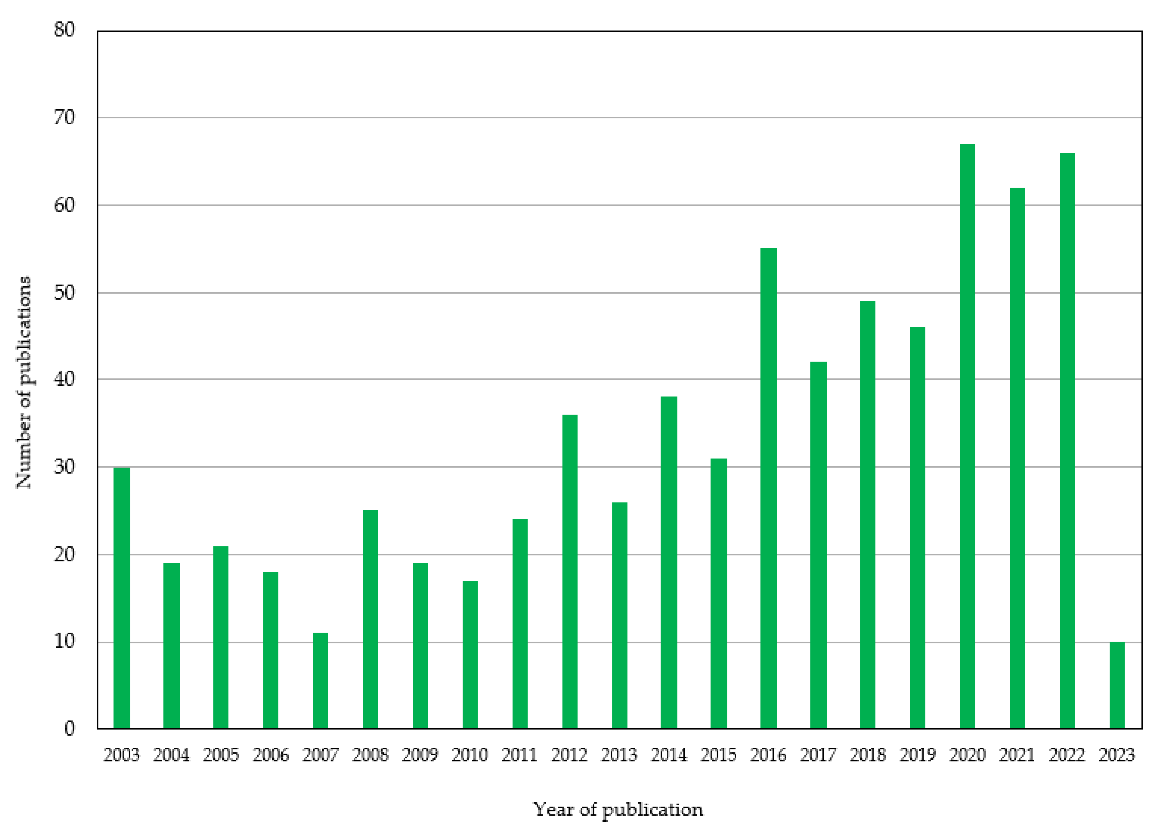

Table 1.
Chemical structure,ways of obtainment and main sources of vitamins in foods.
| Vitamin | Chemical structure | Way of obtainment | Sources |
|---|---|---|---|
| A (retinol) |  |
Intake of dietary sources or vitamin supplements | Fish, meat, eggs, whole milk, carrots, spinach, and mango |
| D3 (cholecalciferol) | 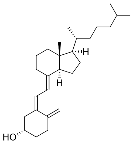 |
Synthesized by the human epidermis or consumed in the form of supplements or fortified foods | Fish, as salmon and sardines, butter and eggs |
| E (α-tocopherol) | 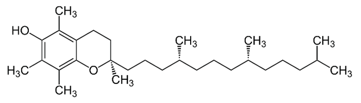 |
Intake of dietary sources or vitamin supplements | Vegetable oils of peanut, soya, palm, corn, safflower, sunflower, and wheat germ |
| K1 (phylloquinone) | 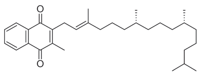 |
Plants | Green leafy vegetables, liver, lean meat, cow's milk, egg yolks, and whole wheat products |
| K2 (menaquinone) | 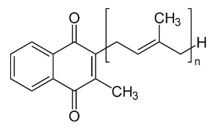 |
Synthesized by bacteria in the human and animal intestines | - |
| K3 (menadione) | 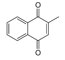 |
Converted to K2 in the intestinal tract | - |
| B1 (thiamine) | 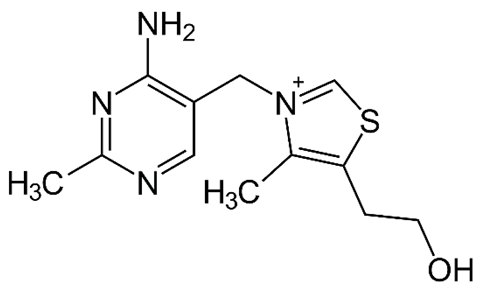 |
Intake of dietary sources or vitamin supplements | Brewer's dried yeast and pork, lamb and beef, poultry, whole grains, nuts, vegetables, and legumes |
| B2 (riboflavin) | 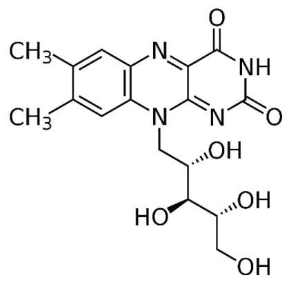 |
Plants and animal cells | Milk and milk products, meat, eggs, and leafy green vegetables |
| B3 (niacin) | 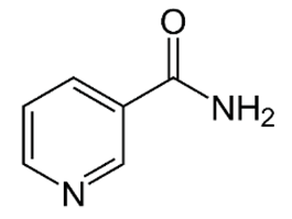 |
Intake of dietary sources or vitamin supplements | Yeast, liver, poultry, lean meats, nuts, and legumes |
| B5 (pantothenic acid) | 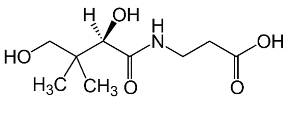 |
Intake of dietary sources or cosmetic products | Organ of animals, eggs, milk, vegetables, legumes, and whole-grain cereals |
| B12 (cobalamin) | 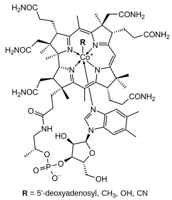 |
Intake of dietary sources or vitamin supplements | Milk, and dairy products, eggs, salmon |
| C (ascorbic acid) | 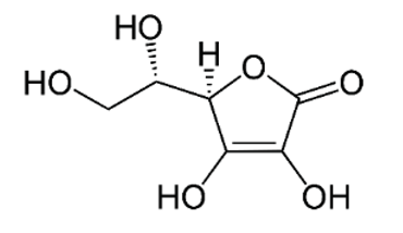 |
Intake of dietary sources or vitamin supplements | Citric fruits, currants, peppers, parsley, cauliflower, potatoes, sweet potatoes, broccoli, brussels sprouts, strawberries, guava, and mango |
Table 2.
Classification of liposomes based on size and lamellarity.
| Type of liposome | Abbreviation | Characteristic | Diameter | Schematic representation |
|---|---|---|---|---|
| Small unilamellar vesicles | SUV | Small unilamellar vesicles | 20 to 200 nm | 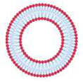 |
| Large unilamellar vesicles | LUV | Unilamellar liposomes with average diameters higher than SUV | Above 200 nm | 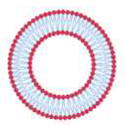 |
| Giant unilamellar vesicles | GUV | Unilamellar liposomes with average diameters higher than LUV | Higher than 1 µm | 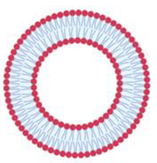 |
| Multilamellar vesicles | MLV | Several concentrically arranged vesicles | 0.5 to 5 µm | 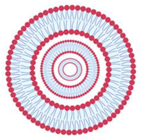 |
| Multivesicular vesicles | MVV | Smaller vesicles within a vesicle of large size | Higher than 1 µm | 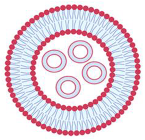 |
Table 3.
Chemicals and methods currently used for the extraction and quantification of vitamins in liposomes.
Table 3.
Chemicals and methods currently used for the extraction and quantification of vitamins in liposomes.
| Vitamin | Liposome type/size | Chemical used to demulsify/precipitate the liposomes | Method for vitamin quantification | EE% | Reference |
|---|---|---|---|---|---|
| Vitamin A | SUV | Methanol | HPLC | 50.6-56.2% | [134] |
| Vitamin A | MLV | Chloroform /Methanol 2:1 v/v | UV-Vis spectrophotometry | 99% | [135] |
| Vitamin A | MLV/SUV | Chloroform | HPLC | 15.8% | [136] |
| Vitamins A, D, E, and K | n.s. | Tween 80 | HPLC | 20-100% | [137] |
| Vitamin B1 | MLV | n-octyl β-D-glucopyranoside | Optical density | 31.2% | [138] |
| Vitamin B1 | n.s. | PBS | HPLC | 97% | [139] |
| Vitamin B2 | n.s. | Triton X-100 | Indirect method (photolysis) | 42.3% | [140] |
| Vitamin B5 | n.s. | Chloroform/Methanol | Direct method (mass weight) | 75% | [141] |
| Vitamin B9 | n.s. | Ethanol | Ultrafiltration in centrifuge tube followed by HPLC | 87.4% | [142] |
| Vitamin B12 | MLV | Methanol/Water 1:10 v/v | HPLC | 70% | [143] |
| Vitamin B12 | LUV | - | Centrifugation followed by UV-Vis spectroscopy | 27% | [33] |
| Vitamins B12, D2 and E | SUV | Ethanol | UV-Vis spectrophotometry | 56-76% | [144] |
| Vitamin C | n.s. | Ethanol | Dialysis followed by UV-vis spectrophotometry | 77.9% | [145] |
| Vitamin C | n.s. | Methanol, chloroform, and ammonium buffer | HPLC | 99.2% | [146] |
| Vitamin C | n.s. | Ethanol | HPLC | 75.4% | [108] |
| Vitamins C and E | n.s. | Triton X-100 / DMSO | UV-Vis spectrophotometry | 93-95% | [83] |
| Vitamins C and E | n.s. | Assay kit BC1235 | Indirect method | 98.5% | [147] |
| Vitamin C | SUV/LUV | Meta-phosphoric acid | Titration with indophenol solution | 94.2% | [148] |
| Vitamin C | SUV/MLV | - | Gel permeation chromatography | 5-16% | [149] |
| Vitamins C and E | MLV/MVV | Protamine sulfate solution followed by Triton X-100 | Protamine aggregation method followed by UV-Vis spectrophotometry | 12-88% | [111] |
| Vitamins C and E | MLV | Acetic acid and protamine solution | UV-Vis spectrophotometry | 38% | [150] |
| Vitamin C | n.s. | - | Dialysis followed by UV-Vis spectrophotometry | 100% | [151] |
| Vitamin D | MLV | Triton X-100 | Ultracentrifugation followed by HPLC | 61.5% | [152] |
| Vitamin D3 | n.s. | Methanol | Ultrafiltration in centrifuge tube followed by HPLC | 74% | [127] |
| Vitamin D3 | SUV/LUV | Methanol | HPLC | 90.2% | [72] |
| Vitamin D3 | SUV/MLV | Water | Ultrafiltration in centrifuge tube followed by RP-HPLC | 100% | [153] |
| Vitamins D3 and K2 | SUV/LUV | Ethanol | UV-Vis spectrophotometry | 98% | [86] |
| Vitamin E | n.s. | Methanol | Dialysis followed by HPLC | 92.5% | [154] |
| Vitamin E | n.s. | Ethanol | UV-Vis spectrophotometry | 98.6% | [155] |
| Vitamin E | n.s. | Chloroform/Methanol 2:1 v/v | UV-Vis spectrophotometry | 78% | [156] |
| Vitamin E | n.s. | Water | Centrifugation followed by HPLC | 94% | [157] |
| Vitamin E | n.s. | - | Centrifugation followed by UV-Vis spectrophotometry | 83.8% | [158] |
| Vitamin E | n.s. | Chloride acid, Tween 80:ethanol and n-pentane | UV-Vis spectrophotometry | 97% | [159] |
| Vitamin K1 | n.s. | - | Ultracentrifugation followed by HPLC | 79.2% | [160] |
| Vitamin K1 | n.s. | Methanol | HPLC | 3.4-154 µg/mg | [34] |
Disclaimer/Publisher’s Note: The statements, opinions and data contained in all publications are solely those of the individual author(s) and contributor(s) and not of MDPI and/or the editor(s). MDPI and/or the editor(s) disclaim responsibility for any injury to people or property resulting from any ideas, methods, instructions or products referred to in the content. |
© 2023 by the authors. Licensee MDPI, Basel, Switzerland. This article is an open access article distributed under the terms and conditions of the Creative Commons Attribution (CC BY) license (http://creativecommons.org/licenses/by/4.0/).
Copyright: This open access article is published under a Creative Commons CC BY 4.0 license, which permit the free download, distribution, and reuse, provided that the author and preprint are cited in any reuse.


