Submitted:
09 January 2023
Posted:
16 January 2023
You are already at the latest version
Abstract
Diffuse optical tomography (DOT) is a medical imaging procedure using light to measure the geometric and working properties of cells, for instance, oxygen consumption, water content, and fat percentage in the tissue by performing three-dimensional visualization of the tissue. This paper aims to explain the theory behind diffuse optical tomography imaging and how the technology works. The paper explains how photon migration techniques based on diffusion theory can be used to image the optical properties of tissue. There are several reasons why near-infrared (NIR) imaging is the most effective method in terms of recovering optical parameters quantitatively in the near-infrared region. The author discusses the methods in detail. This research also presents various advantages, practical uses, and potential problems that have been related to DOT in this work. There is also a brief discussion of current research developments in medical imaging using near-infrared wavelengths, and what the future holds for that area.
Keywords:
Inverse problem
; Biomedical
; Diffuse Optics
; Imaging
; Tomography
; NIRS
1. What is Diffuse Optical Tomography
Diffuse optical tomography (DOT) is a light-based imaging concept applied in medicine that is capable of imaging the geometrical properties of tissues and their working characteristics, such as the concentration of different components of the tissue [1,2]. DOT is based on the principle that measured near-infrared (NIR) signals are signature of the optical properties of the underlying biological tissues [1,3]. It is possible to reconstruct optical tomographic images using the spatial distribution of these measured optical signals. The problem of image reconstruction is an inverse problem that can be described as determining the internal spatial distribution of the objective function using the measurements taken at the boundaries of the tissues [4,5]. There are large attenuation and scattering factors in diffusive waves, which makes this problem ill-posed. In DOT, the tissue is illuminated by NIR light sources, one at a time. Arrays of detectors are used to measure the diffuse light that comes out of the boundary. Next, the region of interest optical tissue properties of the radiated tissue are derived and estimated using a model.
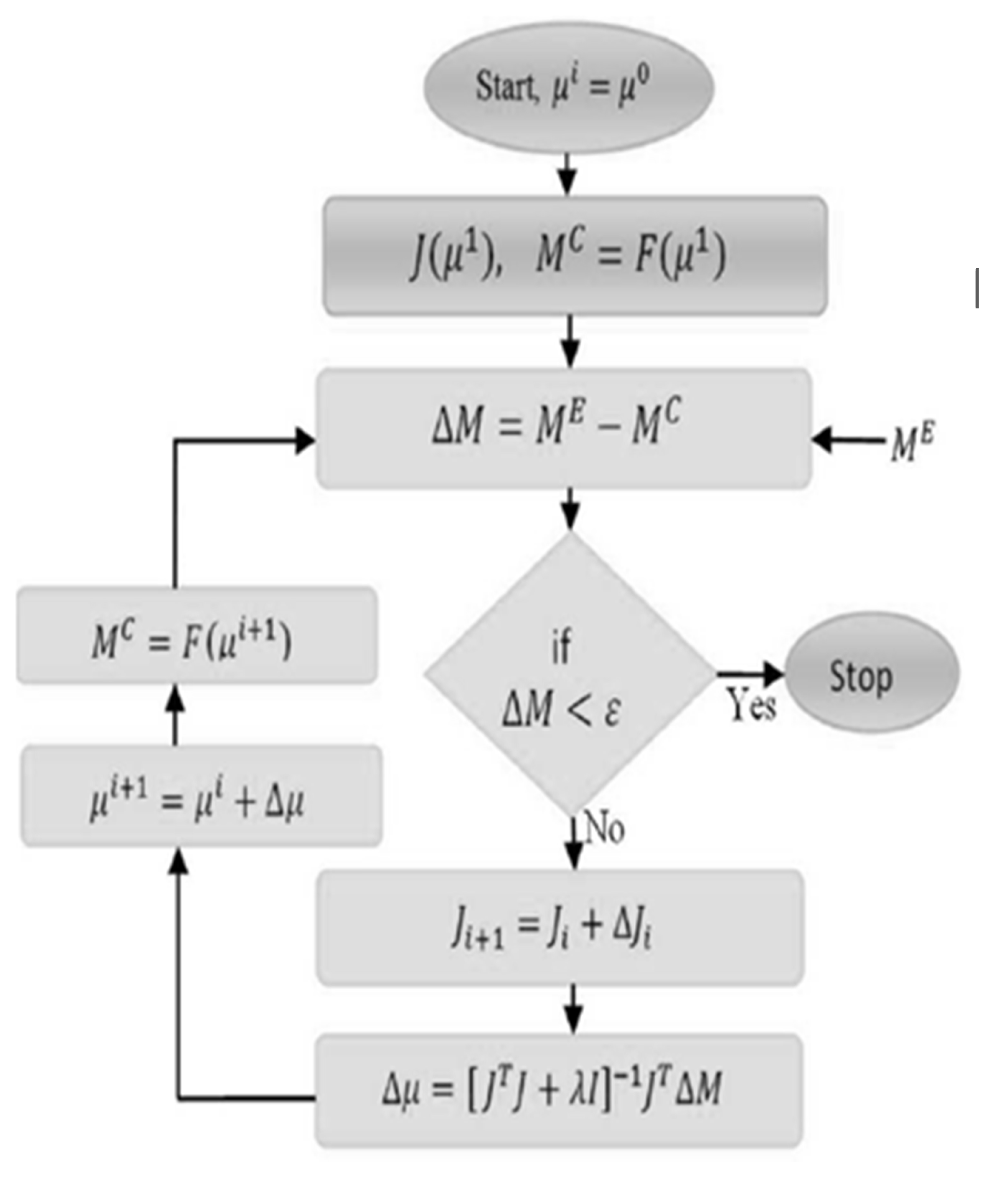
As DOT can provide functional images of tissue using the nonionizing NIR light, it has gained a lot of attention in the past decades. DOT is mostly used to image brains and breasts. Measurements at the tissue boundary are critical for estimating the internal distribution of optical properties [1,4]. NIR interaction with tissue is driven primarily by scattering, so the estimation problem, or the inverse problem, isn’t linear, ill-posed, and underdetermined [1]. A computationally intensive model is needed to solve an inverse problem. Calculated optical properties of tissue are matched iteratively with experimental data using models in a least-squares sense [1]. Figure 2 shown an algorithm used for DOT image reconstruction [6]. Because these computational models run repeatedly, obtaining real-time optical images is a big challenge [1,2].
Figure 1.
Absorption spectra of and .
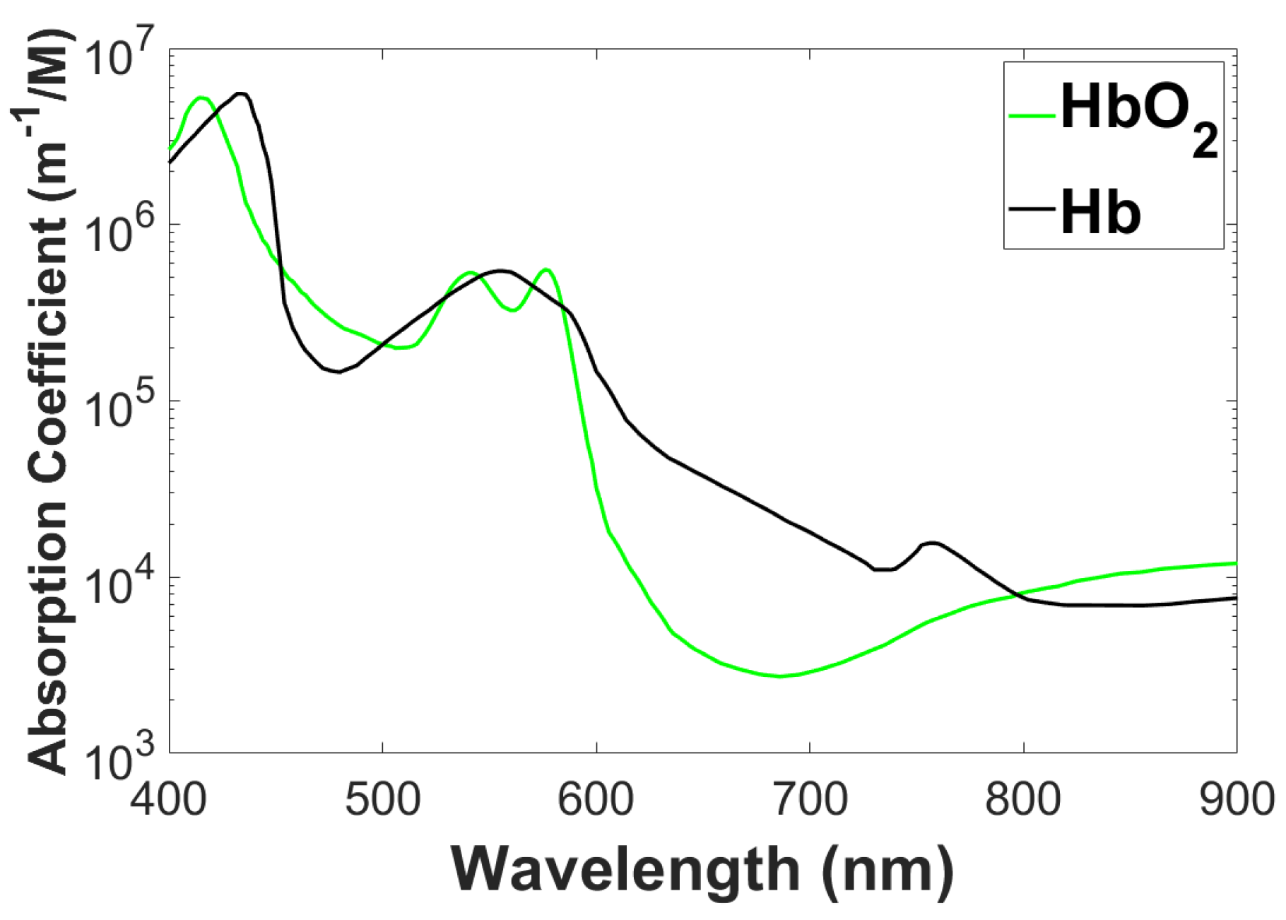
DOT comes with its most obvious advantage, which is the possibility of spectroscopic delineation of tissue chromophores and the concomitant ability for early diagnosis of malignancy on the basis of the functional information that spectroscopy provides [7]. The disadvantages are caused by the turbid nature of the tissue through which light is modeled to diffuse. The diffusion process with a finite length scale associated with it imposes a limitation on the spatial resolution achievable in DOT images. More importantly, the penetration of light into the human body or organ is rather limited, which limits the application of DOT only to soft-tissue organs such as the breast, prostate, muscles, and neonatal head. One of the reasons for choosing NIR light is to maximize penetration because, in the range of 600 - 900 nm, optical absorption in tissue is relatively small [6]. The absorption spectra of and is shown in Figure 1. It’s not as spatially detailed as other imaging methods, like magnetic resonance imaging or X-ray CT, but DOT lets us see a lot of physiological parameters that we wouldn’t be able to see otherwise, like hemodynamics and other functional processes that happen every few microseconds. In spite of the earlier mentioned disadvantages, spectroscopy-based prediction of such useful functional parameters as total hemoglobin concentration, oxygen partial pressure, etc., has made DOT imaging the subject of intense research leading to the development of either stand-alone NIR clinical imagers or MRI-assisted DOT imagers. Furthermore, at a relatively low cost, DOT can be implemented in small and portable instrumentation.
Figure 2.
Image reconstruction algorithm.
1.1. Application of DOT
DOT has various applications and here are some of them:
- Breast cancer imaging: X-ray mammography can detect breast cancer. To improve the assessment and characterization of breast tumors, a wide range of other techniques such as ultrasound, Electrical Impedance Tomography (EIT), and Magnetic Resonance Imaging (MRI) are being used. Positron Emission Tomography (PET) and MRI are becoming more popular because they provide fundamentally different information than traditional structural pictures. It gives us direct access to physiological data like amount of blood, metabolic state, flow of blood, and oxygen level. Tumor angiogenesis alters these tissue characteristics, which are also used to track a tumor’s response to therapy. These functional parameters can be imaged by DOT. Because tumors are more vascularized than surrounding tissue, they absorb light differently. Through spectroscopic variation in , the relative concentration of oxygenated hemoglobin can be determined, and thus the oxygen demand-supply ratio can be determined. Furthermore, it can be used to differentiate tumors from background tissue, malignant tumors from non-malignant tumors, and tumors with varying levels of activity (degrees of malignancy).
- Brain function imaging: A DOT assessment of brain function complements positron emission tomography (PET), functional MRI (fMRI), electroencephalogram (EEG), and magnetoencephalography (MEG). PET images changes in metabolic activity but has a low temporal and spatial resolution. fMRI provide high spatial and temporal resolution images of blood flow and deoxy-hemoglobin concentration, but cannot measure oxyhemoglobin level simultaneously. EEG as well as MEG can monitor electrical activity of the brain with much higher time resolution (50 to 1 kHz), but pin pointing sources of these electrical and magnetic fields is difficult and resolution in space is not upto as expectation compared with fMRI. While its spatial resolution is inferior to that of fMRI, the DOT, when used in combination with fMRI, can simultaneously measure oxy- and deoxyhemoglobin concentrations and blood volume. Combining light-based imaging with fMRI and MEG/EEG could provide result in a complete picture that is more useful than any of the parts alone.
- Stroke: It may be possible to detect ischemic strokes and hemorrhagic strokes more quickly and accurately using DOT, which is essential before applying neuroprotective drugs as it can effectively treat stroke patients in case of ischemic strokes, but can lead to fatality in case of hemorrhagic strokes. It is also possible to monitor the progression of a stroke and treatment response using DOT at the bedside.
- Monitoring Brain Trauma and Surgical Intervention: Detecting a brain hemorrhage early can greatly improve a patient’s recovery and long-term effects if the hemorrhage occurs because of brain injury. Current screening methods include cognitive testing and invasive monitoring (e.g., measurement of cranial pressure). A continuous DOT monitor at the bedside could provide continuous monitoring on bleeding site and spreading, which is an advantage over these techniques. Collateral damage can also be minimized through monitoring during brain surgery. The use of an EEG while performing a surgery could disrupt the critical surgical functions, but the placement of electrodes is a time-consuming and painful process. There is also the option of using an fMRI, but a special room with a costly magnet is required for this. An inexpensive alternative could be the DOT that can provide optical imaging on the surface of the body.
2. Theoretical Basis of Diffuse Optical Tomography
In the past two decades, diffusion theory has been increasingly used to study radiative transfer, particularly in laser diagnosis. The light interesction of tissue could be monitored by photon transport concept derived from the diffusion theory [8]. To detect objects embedded within tissue phantoms, photon-density waves have been used as solutions to the diffusion equation that exhibit strong damping [9,10]. The laser light must be transported through optical fibers placed on the tissue surface in order for these laser techniques to be noninvasive. As a result, tissue boundaries are inevitable. Diffusion theory must take this boundary into account if optical properties are to be measured without errors of more than 50%. As biomedical research with diffusion light moves away from high spatial resolution, it is now focused more on functional imaging. Despite the fact that diffuse optical imaging cannot compete with anatomical imaging methods (such as x-ray, ultrasound, and MRI imaging), it offers several distinct advantages when it comes to sensitivity to functional changes, safety, cost, and convenience. Through the use of diffuse optical spectroscopy in the near-infrared, it is possible to uncover physiological information noninvasively that cannot be obtained otherwise.
When studying spectroscopic optical parameters of highly scattering media such as tissue, NIR-based imaging is the most viable method with respect to the of recovering of spectroscopic tissue light characteristics. In the area of photon as a light migration, the biggest advance was the accepting the light diffusion as a mechanism for transporting light over long distances. By using this model, we can quantify the scattering of tissue and absorption of tissue and to precisely include boundarie conditions, for example tissue-air interfaces, with the theory of transport [11,12]. Diffusion approximation allows detailed, quantitative investigations of mean volume of species in majorly scattering space due to the separation of scattering and absorption [13,14,15,16,17,18].
Figure 3.
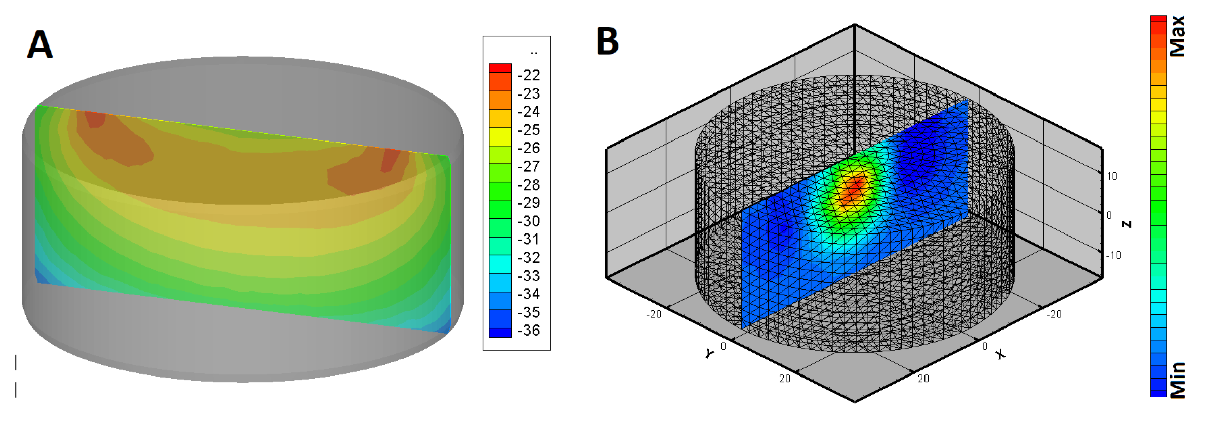
Diffuse optical tomography. (A) sensitivity matrix and (B) Reconstructed image from a phantom having inclusion at the center.
Figure 3.
Diffuse optical tomography. (A) sensitivity matrix and (B) Reconstructed image from a phantom having inclusion at the center.
2.1. Photon Diffusion Equation
A diffusion equation well describes the moving of light via strongly scattering tissue, according to Patterson’s experiment in 1988 [11]. Light from a source to a detector yield a banana-shaped sensitivity as shown in Figure 3A. A comparison could be drawn between the theory of light diffusion within tissues, the concept of heat diffusion and neutron diffusion. In order to fit their measurements to the diffusion solution, researchers had to incorporate the diffusion solution. Experimental measurements were conducted using a pulsed laser source and a time-resolved laser source. Researchers later realized that amplitude-modulated light sources could be used to measure the amplitude and phase of diffusing photon density waves in the frequency domain [19]. It is possible to solve the Helmholtz formula with simple solution of spherical wave when light energy density oscillates for amplitude-modulated sources in homogeneous media. In biological tissue, the waves are considered as randomly moving photons exhibiting a random moving step of approximately 1 mm. However, macroscopically, they form a damping wave of photon density with 10 cm wavelength. A diffusing photon density wave (DPDW) is referred to as this wave. As a result of wave analysis, we are able to draw analogies from electromagnetic radiation, which provides us with useful insights and ease of computation. In contrast to time-resolved imaging, frequency domain measurements are less expensive and more stable, and pulsed illumination data in the form of the temporal point spread function (TPSF) can be viewed as frequency domain data with multiple frequencies at the same time. Fishkin. et al. [19] and Patterson et al. [12] developed analytic solutions in the frequency domain and performed some of the first frequency domain measurements to non-invasively recover optical properties. The properties were verified in both bio-models [20] and human breast studies [21].
3. Difficulties in DOT Imaging
Image reconstruction is a non-linear as well as an ill-posed problem. DOT presents few challenges for the following reasons:
- The scattering nature of photons traveling through tissue makes DOT an attractive tool for the noninvasive imaging of diseases. The strong scattering of light by biological tissue leads to poor depth localization in DOT due to the attenuation of detection sensitivity exponentially with depth.
- The tissue is like a turbid property with heavily scattering property. Light travels through the tissue in a complicated zigzag path. As a result, strength attenuated. this renders the relation between the output photon density and the optical properties dependent on stochastically defined paths.
- In the imaging for frequency space, although the amplitude changes during modulation at Mega Hertz, the wavelength of DPWD (Diffuse Photon Density wave), which is owing to the intensity modulated illumination, is of the order of a few cm, much greater than the typical size of inhomogeneities. This is the fundamental reason for poor resolution in images from DOT.
- When one illuminates the turbid object with a short pulse, the ballistic photons are very few, or none. If ballistic photons are of sufficient strength reconstruction from such photons can give better-resolved images.
- The background properties being not known in advance leads to difficulties in measuring and interpreting measurements for inverse reconstructions.
- As a result of the quantum nature of noise, modeling it is a challenging task. This is because the sources of noise include both thermal noise in the amplification unit and the noise generated by the shots due to the quantum source nature.
- Absorption/scattering coefficients and field amplitude and phase are nonlinearly related. Due to these considerations, either a linearized approximation like Born or Rytov must be used or a nonlinear forward model must be used to reduce the numerical burden.
- Depending on the geometry and physical conditions, light may propagate in greatly different ways, for example through significantly scattered brain tissue that is covered by slightly scattering cerebrospinal fluid. In order for dealing with such inclusions the DE is inadequate as a model for light transport. One should rely on the RTE, which also takes into account the angles of scattering.
- The light interection of tissue are characterized by two parameters. Simultaneous reconstruction of both parameters complicates and may induce cross-talk between the images.
- Ill-posedness may also arise from the fact that very small changes in optical parameters can give rise to large changes in measurement or vice versa. Thus inverse solutions must take care to see that the effect of noise, which gives rise to large swings in reconstruction, is properly accounted for.
- In addition, one might estimate the absorption or scattering coefficient at many locations in space, several times of amplitude more than the taken datapoints. This is an ill-posed problem. This also becomes another source of non-uniqueness of solutions.
3.1. Progress and Future Directions
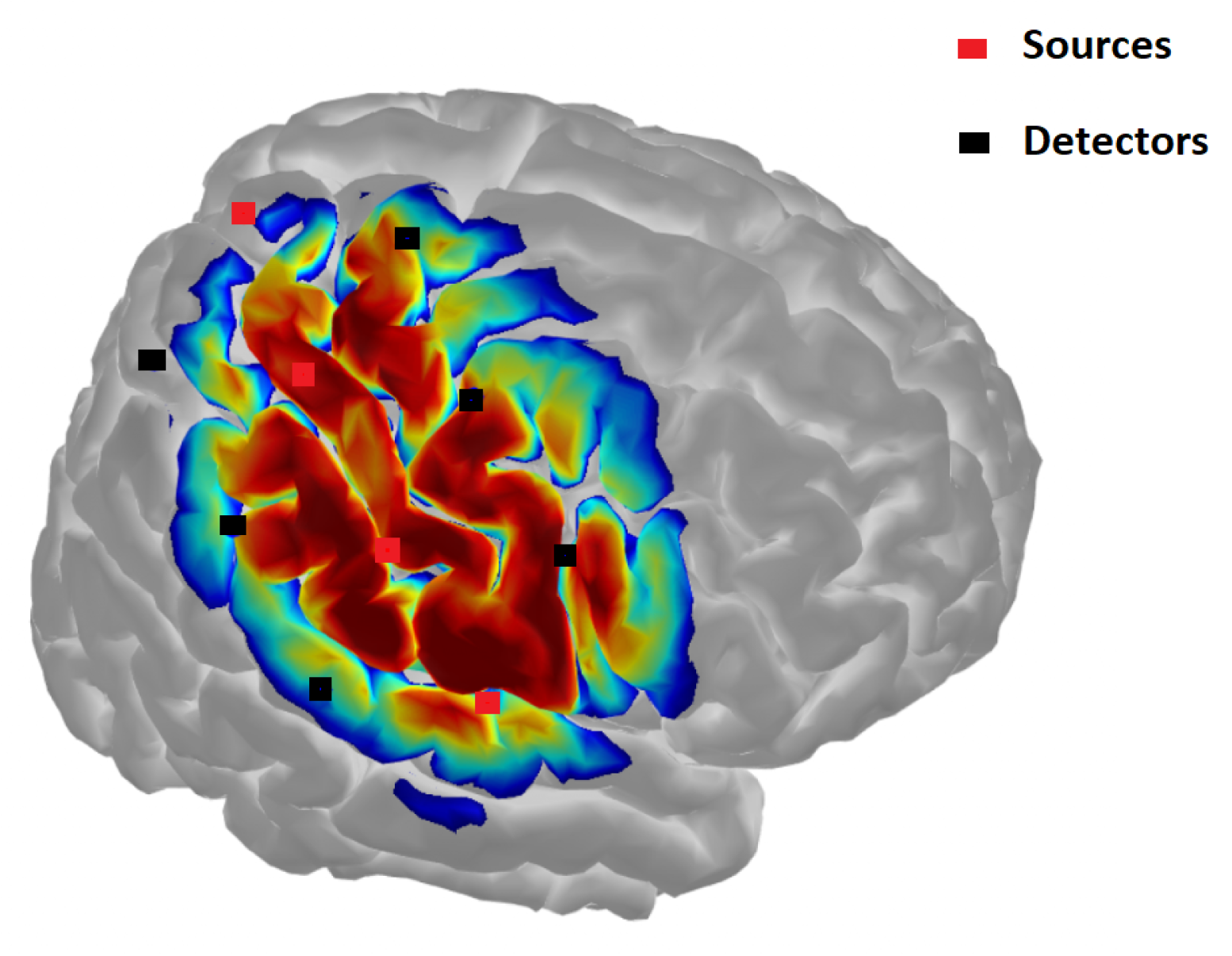
Diffuse optical tomography (DOT) is based on taking measurement of Near-infrared (NIR) light at multiple locations on the tissue surface, transmitted or reflected from the deep tissue to probe the optical properties of the tissues. It requires source-detector multiplexing techniques to acquire data and system calibration to remove uncertainty [22]. The computational challenge is in the estimation of internal optical properties of tissue using a few measurements on the tissue boundary [1,4]. However, there is an algorithm developed for high-speed 3D DOT image reconstruction [23]. This algorithm can also be implemented on multiple GPUs for further speed up the image reconstruction [24] and real-time imaging of DOT is an another possibility [25]. Method for scanning a small region of the tissue as an efficient imaging technique was proposed [26,27]. Another interesting tissue imaging method based on DOT, particularly for the brain, is functional near-infrared spectroscopy (fNIRS). Figure 4 shown an fNIRS image of the brain. It can be used to measure brain activity, such as mental workload [28]. fNIRS system can be built with few hardware components. The hardware can also be in the form of a patch for brain imaging [29,30]. In fNIRS technology, the light sources and detectors are generally referred to as optodes [31]. There is a trend for portable fNIRS [32]. New concepts, such as internet-of-things, were also implemented in fNIRS system development [33,34,33]. Further, machine learning-based classification of the cognitive status of individuals in real-time is possible [35,36]. Researchers are exploring new applications of fNIRS and fNIRS has great future potential. Usually the DOT hardware for breast imaging is bulky. There is a possibility of using LEDs and photodetectors [37] to develop compact DOT systems. Further, DOT can be used as a point-of-care imaging system [32,38]. Some of the systems were designed for teaching purposes [39]. While probing tissue the unwanted signal from the superficial layer of the tissue is often added to the signal of interest. These unwanted signals can be removed my measuring them separately and accounting them in the image reconstruction [40].
4. Discussion and Conclusion
It is essential to address both the theoretical, computational, and hardware challenges involved in nurturing the idea of imaging using NIR light into a fully developed NIR imaging system. It is an ill-posed (and nonlinear) problem to recover the optical property distribution inside a large object from noisy boundary measurements of light. When creating direct 3-D images, this is especially critical. From a theoretical perspective, it is difficult to determine the distribution of optical properties in the interior of a large object. This is due to the noisy boundary measurement data obtained on the boundary region. There are several reasons for this, but it is especially prevalent when it comes to the reconstruction of direct 3-D images. A computational-theoretical procedure is used to solve the nonlinear ill-posed problem of recovering a large-dimensional unknown parameter vector by an iterative procedure. Because of ill-posedness, DOT requires regularization to yield proper results. Therefore, there is a need for the development of a robust and accurate 3-D optical tomography reconstruction system. This paper suggests the direction toward implementing the computationally intensive part of the 3-D DOT image reconstruction algorithm on GPU. The GPUs have the potential to provide massively parallel computational power for continuous 3D DOT imaging.
Figure 4.
Functional near-infrared spectroscopy (fNIRS).
References
- Arridge, S.R. Optical tomography in medical imaging. Inverse Problems 1999, 15, R41. [Google Scholar] [CrossRef]
- Gibson, A.P.; Hebden, J.C.; Arridge, S.R. Recent advances in diffuse optical imaging. Physics in Medicine and Biology 2005, 50, R1. [Google Scholar] [CrossRef] [PubMed]
- Saikia, M.J. Design and development of a functional diffuse optical tomography probe for real-time 3D imaging of tissue. SPIE 2021, 11639, 213–218. [Google Scholar]
- Arridge, S.R.; Hebden, J.C. Optical imaging in medicine: II. Modelling and reconstruction. Physics in Medicine and Biology 1997, 42, 841. [Google Scholar] [CrossRef]
- Poorna, R.; Kanhirodan, R.; Saikia, M.J. Square-waves for frequency multiplexing for fully parallel 3D diffuse optical tomography measurement. SPIE 2021, 11639, 219–226. [Google Scholar] [CrossRef]
- Saikia, M.J. A spectroscopic diffuse optical tomography system for the continuous 3D functional imaging of tissue -a phantom study. IEEE Transactions on Instrumentation and Measurement 2021, 1. [Google Scholar] [CrossRef]
- Saikia, M.J.; Kanhirodan, R. Development of handheld near-infrared spectroscopic medical imaging system. Optical Society of America, 2019, p. DS1A.6.
- Chance, B.; Leigh, J.S.; Miyake, H.; Smith, D.S.; Nioka, S.; Greenfeld, R.; Finander, M.; Kaufmann, K.; Levy, W.; Young, M. Comparison of time-resolved and -unresolved measurements of deoxyhemoglobin in brain. Proceedings of the National Academy of Sciences of the United States of America 1988, 85, 4971–4975. [Google Scholar] [CrossRef]
- Knüttel, A.; Schmitt, J.M.; Knutson, J.R. Spatial localization of absorbing bodies by interfering diffusive photon-density waves. Applied optics 1993, 32, 381–389. [Google Scholar] [CrossRef] [PubMed]
- Boas, D.A.; O’Leary, M.A.; Chance, B.; Yodh, A.G. Scattering of diffuse photon density waves by spherical inhomogeneities within turbid media: analytic solution and applications. Proceedings of the National Academy of Sciences of the United States of America 1994, 91, 4887–4891. [Google Scholar] [CrossRef]
- Patterson, M.S.; Chance, B.; Wilson, B.C. Time resolved reflectance and transmittance for the noninvasive measurement of tissue optical properties. Appl. Opt. 1989, 28, 2331–2336. [Google Scholar] [CrossRef]
- Patterson, M.S.; Moulton, J.D.; Wilson, B.C.; Berndt, K.W.; Lakowicz, J.R. Frequency-domain reflectance for the determination of the scattering and absorption properties of tissue. Appl. Opt. 1991, 30, 4474–4476. [Google Scholar] [CrossRef] [PubMed]
- Chance, B.; Nioka, S.; Kent, J.; McCully, K.; Fountain, M.; Greenfeld, R.; Holtom, G. Time-resolved spectroscopy of hemoglobin and myoglobin in resting and ischemic muscle. Analytical biochemistry 1988, 174, 698–707. [Google Scholar] [CrossRef] [PubMed]
- Duncan, A.; Whitlock, T.L.; Cope, M.; Delpy, D.T. Multiwavelength, wideband, intensity-modulated optical spectrometer for near-infrared spectroscopy and imaging. SPIE 1993, 1888, 248–257. [Google Scholar] [CrossRef]
- Ferrari, M.; Wei, Q.; Carraresi, L.; Blasi, R.A.D.; Zaccanti, G. Time-resolved spectroscopy of the human forearm. Journal of photochemistry and photobiology. B, Biology 1992, 16, 141–153. [Google Scholar] [CrossRef] [PubMed]
- Ferrari, M.; Wilson, D.A.; Hanley, D.F.; Hartmann, J.F.; Rogers, M.C.; Traystman, R.J. Noninvasive determination of hemoglobin saturation in dogs by derivative near-infrared spectroscopy. The American journal of physiology 1989, 256, H1493–9. [Google Scholar] [CrossRef] [PubMed]
- Sevick, E.M.; Chance, B.; Leigh, J.; Nioka, S.; Maris, M. Quantitation of time- and frequency-resolved optical spectra for the determination of tissue oxygenation. Analytical biochemistry 1991, 195, 330–351. [Google Scholar] [CrossRef] [PubMed]
- Svaasand, L.O.; Tromberg, B.J.; Haskell, R.C.; Tsay, T.T.; Berns, M.W. Tissue characterization and imaging using photon density waves. Optical Engineering 1993, 32, 258–266. [Google Scholar] [CrossRef]
- Tromberg, B.J.; Svaasand, L.O.; Tsay, T.T.; Haskell, R.C. Properties of photon density waves in multiple-scattering media. Applied optics 1993, 32, 607–616. [Google Scholar] [CrossRef] [PubMed]
- Fishkin, J.B.; Gratton, E.; VandeVen, M.J.; Mantulin, W.W. Diffusion of intensity modulated near-infrared light in turbid media. SPIE 1991, 1431, 122–135. [Google Scholar] [CrossRef]
- O’Leary, M.A.; Boas, D.A.; Chance, B.; Yodh, A.G. Experimental images of heterogeneous turbid media by frequency-domain diffusing-photon tomography. Optics letters 1995, 20, 426–428. [Google Scholar] [CrossRef]
- Saikia, M.J. An embedded system based digital onboard hardware calibration for low-cost functional diffuse optical tomography system. SPIE 2021, 11632, 1–8. [Google Scholar] [CrossRef]
- Saikia, M.J.; Kanhirodan, R.; Vasu, R.M. High-speed GPU-based fully three-dimensional diffuse optical tomographic system. International Journal of Biomedical Imaging 2014, 2014. [Google Scholar] [CrossRef]
- Saikia, M.J.; Kanhirodan, R. High performance single and multi-GPU acceleration for Diffuse Optical Tomography. Institute of Electrical and Electronics Engineers Inc., 2014; pp. 1320–1323. [Google Scholar] [CrossRef]
- Saikia, M.J.; Rajan, K.; Vasu, R.M. 3-D GPU based real time Diffuse Optical Tomographic system. IEEE Computer Society, 2014; pp. 1099–1103. [Google Scholar] [CrossRef]
- Saikia, M.J.; Kanhirodan, R. Development of DOT system for ROI scanning. Optical Society of America (OSA), 2014, p. T3A.4. [CrossRef]
- Saikia, M.J.; Kanhirodan, R. Region-of-interest diffuse optical tomography system. Review of Scientific Instruments 2016, 87, 013701. [Google Scholar] [CrossRef]
- Saikia, M.J.; Besio, W.G.; Mankodiya, K. The Validation of a Portable Functional NIRS System for Assessing Mental Workload. Sensors 2021, 21, 3810. [Google Scholar] [CrossRef] [PubMed]
- Abtahi, M.; Cay, G.; Saikia, M.J.; Mankodiya, K. Designing and testing a wearable, wireless fNIRS patch. Institute of Electrical and Electronics Engineers Inc., 2016; Volume 2016-Octob, pp. 6298–6301. [Google Scholar] [CrossRef]
- Saikia, M.J.; Mankodiya, K. A Wireless fNIRS Patch with Short-Channel Regression to Improve Detection of Hemodynamic Response of Brain. Institute of Electrical and Electronics Engineers Inc., 2018; pp. 90–96. [Google Scholar] [CrossRef]
- Saikia, M.; Mankodiya, K. 3D-printed human-centered design of fNIRS optode for the portable neuroimaging. 2019, 10870. [Google Scholar] [CrossRef]
- Saikia, M.; Besio, W.; Mankodiya, K. WearLight: Toward a Wearable, Configurable Functional NIR Spectroscopy System for Noninvasive Neuroimaging. IEEE Transactions on Biomedical Circuits and Systems 2019, 13. [Google Scholar] [CrossRef]
- Saikia, M.J.; Cay, G.; Gyllinsky, J.V.; Mankodiya, K. A Configurable Wireless Optical Brain Monitor Based on Internet-of-Things Services. Institute of Electrical and Electronics Engineers Inc., 2018; pp. 42–48. [Google Scholar] [CrossRef]
- Saikia, M.J. Internet of things-based functional near-infrared spectroscopy headband for mental workload assessment. SPIE 2021, 11629, 143–150. [Google Scholar] [CrossRef]
- Saikia, M.J.; Kuanar, S.; Borthakur, D.; Vinti, M.; Tendhar, T. A machine learning approach to classify working memory load from optical neuroimaging data. 2021, p. 69. [CrossRef]
- Saikia, M.J.; Brunyé, T.T. K-means clustering for unsupervised participant grouping from fNIRS brain signal in working memory task. SPIE, 2021, Vol. 11629, pp. 159–164. [CrossRef]
- Saikia, M.; Manjappa, R.; Kanhirodan, R. A cost-effective LED and photodetector based fast direct 3D diffuse optical imaging system. 2017, Vol. 10412. [CrossRef]
- Saikia, M.J.; Mankodiya, K.; Kanhirodan, R. A point-of-care handheld region-of-interest (ROI) 3D functional diffuse optical tomography (fDOT) system. SPIE, 2019, Vol. 10874, p. 90. [CrossRef]
- Saikia, M.J.; Kanhirodan, R. A tabletop Diffuse Optical Tomographic (DOT) experimental demonstration system. SPIE, 2019, Vol. 10869, p. 11. [CrossRef]
- Saikia, M.J.; Manjappa, R.; Mankodiya, K.; Kanhirodan, R. Depth sensitivity improvement of region-of-interest diffuse optical tomography from superficial signal regression. OSA - The Optical Society, 2018, Vol. Part F99-C, p. CM3E.5. [CrossRef]
Disclaimer/Publisher’s Note: The statements, opinions and data contained in all publications are solely those of the individual author(s) and contributor(s) and not of MDPI and/or the editor(s). MDPI and/or the editor(s) disclaim responsibility for any injury to people or property resulting from any ideas, methods, instructions or products referred to in the content. |
© 2023 by the authors. Licensee MDPI, Basel, Switzerland. This article is an open access article distributed under the terms and conditions of the Creative Commons Attribution (CC BY) license (http://creativecommons.org/licenses/by/4.0/).
Copyright: This open access article is published under a Creative Commons CC BY 4.0 license, which permit the free download, distribution, and reuse, provided that the author and preprint are cited in any reuse.


