Submitted:
18 September 2022
Posted:
20 September 2022
You are already at the latest version
Abstract
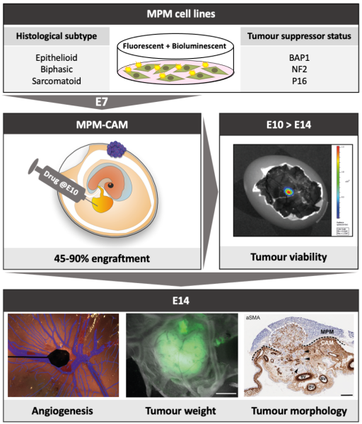
Malignant pleural mesothelioma (MPM) has limited treatment options and poor prognosis. Frequent inactivation of the tumour suppressors BAP1, NF2 and P16 may differentially sensitise tumours to treatments. We have established chick chorioallantoic membrane (CAM) xenograft models of low-passage MPM cell lines and protocols for evaluating drug responses. Ten cell lines, representing the spectrum of histological subtypes and tumour suppressor status, were dual labelled for fluorescence/bioluminescence imaging and implanted on the CAM at E7. Bioluminescence was used to assess viability of primary tumours, which were excised at E14 for immunohistological staining or real-time PCR. All MPM cell lines engrafted efficiently forming vascularised nodules, however their size, morphology and interaction with chick cells varied. MPM phenotypes including local invasion, fibroblast recruitment, tumour angiogenesis and vascular remodelling were evident. Bioluminescence imaging could be used to reliably estimate tumour burden pre- and post-treatment, correlating with tumour weight and Ki-67 staining. In conclusion, MPM-CAM models recapitulate important features of the disease and are suitable to assess therapies using a broad range of MPM cell lines that allow histological or genetic stratification. They are amenable to multi-modal imaging, offering a time and cost-efficient, 3Rs-compliant alternative to rodent xenograft models to prioritise candidate compounds from in vitro studies.
Keywords:
mesothelioma
; chick embryo
; CAM
; xenograft
; bioluminescence
; fluorescence
; histology
; MRI
; preclinical
; 3Rs
Copyright: This open access article is published under a Creative Commons CC BY 4.0 license, which permit the free download, distribution, and reuse, provided that the author and preprint are cited in any reuse.


