
Submitted:
05 September 2021
Posted:
06 September 2021
You are already at the latest version
Abstract
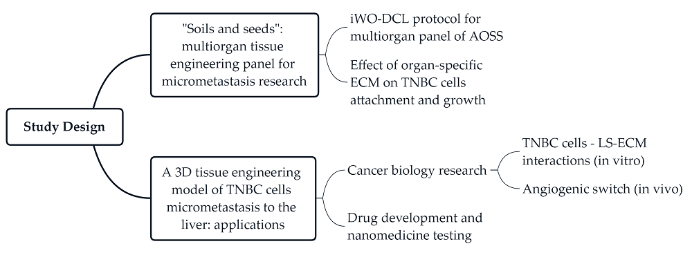
Colonization of distant organs by tumor cells is a critical step of cancer progression. The initial avascular stage of this process (micrometastasis) remains almost inaccessible to study due to the lack of relevant experimental approaches. Here, we introduce an in vitro/in vivo model of organ-specific micrometastases of triple-negative breast cancer (TNBC) that is fully implemented in a cost-efficient chick embryo (CE) experimental platform. The model is built as three-dimensional (3D) tissue engineering constructs (TECs) combining human MDA-MB-231 cells and decellular-ized CE organ-specific scaffolds. TNBC cells colonized CE organ-specific scaffolds in 2-3 weeks, forming tissue-like structures. The feasibility of this methodology for basic cancer research, drug development and nanomedicine was demonstrated on a model of hepatic micrometastasis of TNBC. We revealed that MDA-MB-231 differentially colonize parenchymal and stromal com-partments of the liver-specific extracellular matrix (LS-ECM) and become more resistant to the treatment with molecular Doxorubicin (Dox) and Dox-loaded mesoporous silica nanoparticles than in monolayer cultures. When grafted on CE chorioallantoic membrane, LS-ECM-based TECs induced angiogenic switch. These findings may have important implications for the diag-nosis and treatment of TNBC. The methodology established here is scalable and adaptable for pharmacological testing and cancer biology research of various metastatic and primary tumors.
Keywords:
chick embryo
; 3D culture
; tumor models in vitro
; tissue engineering
; metastasis
; triple-negative breast cancer
; liver
; mesoporous silica nanoparticles
; doxorubicin
; micrometastasis
Copyright: This open access article is published under a Creative Commons CC BY 4.0 license, which permit the free download, distribution, and reuse, provided that the author and preprint are cited in any reuse.


