Submitted:
30 November 2020
Posted:
02 December 2020
You are already at the latest version
Abstract
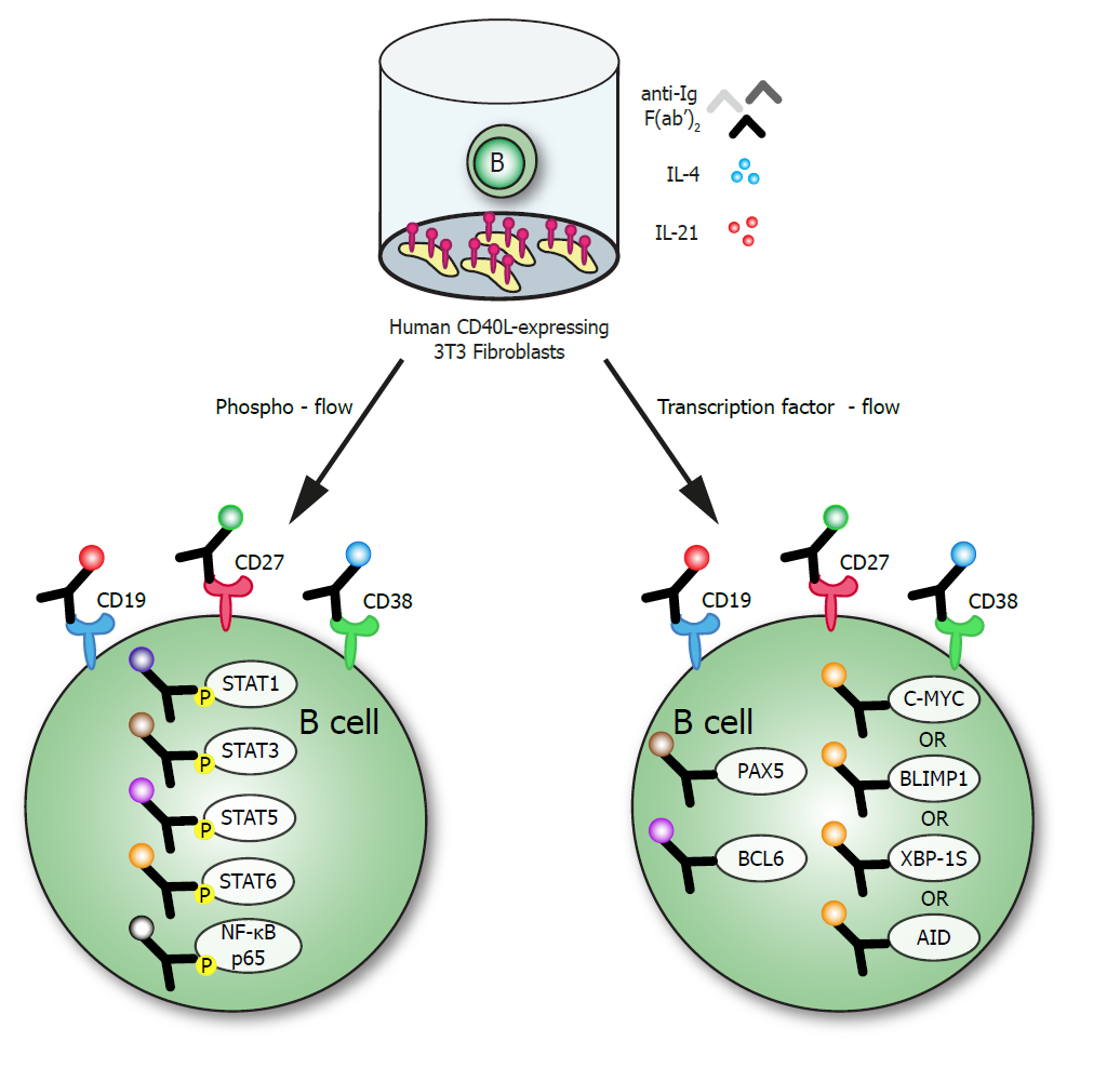
Flow cytometric detection of intracellular (IC) signaling proteins and transcription factors (TFs) will help elucidate the regulation of B cell survival, proliferation and differentiation. However, simultaneous detection of signaling proteins or TFs, with membrane markers (MM) can be challenging as required fixation and permeabilization procedures can affect functionality of conjugated antibodies. Here, a phosphoflow method is presented for detection of activated NF-κB p65 and phosphorylated STAT1, STAT3, STAT5 and STAT6 together with B cell differentiation MM CD19, CD27 and CD38. Additionally, a TF-flow method is presented that allows detection of B cell TFs; PAX5, c-MYC, BCL6, AID and antibody-secreting cell (ASC) TFs BLIMP1 and XBP-1s together with MM. Applying these methods on in vitro induced human B cell differentiation cultures showed significantly different steady-state levels, and responses to stimulation, of phosphorylated signaling proteins in CD27-expressing B cell and ASC populations. The TF-flow protocol and UMAP analysis revealed heterogeneity in TF-expression within stimulated CD27 or CD38-expressing B cell subsets. The methods presented here allow for sensitive analysis of STAT and NF-κB p65 signaling and TFs together with B cell differentiation MM at single-cell resolution. This will aid further investigation of B cell responses in both health and disease.
Keywords:
Differentiation
; germinal center
; antibody-secreting cells
; phosphorylated STATs
; NF-κB1
Copyright: This open access article is published under a Creative Commons CC BY 4.0 license, which permit the free download, distribution, and reuse, provided that the author and preprint are cited in any reuse.


