Preprint Review Version 1 Preserved in Portico This version is not peer-reviewed

What the Salamander Eye Has Been Telling the Vision Scientist’s Brain

Version 1 : Received: 3 March 2020 / Approved: 5 March 2020 / Online: 5 March 2020 (02:51:38 CET)
Version 2 : Received: 16 April 2020 / Approved: 19 April 2020 / Online: 19 April 2020 (08:06:38 CEST)

A peer-reviewed article of this Preprint also exists.

Rozenblit F, Gollisch T (2020) What the salamander eye has been telling the vision scientist’s brain. Semin Cell Dev Biol 106:61-71. Rozenblit F, Gollisch T (2020) What the salamander eye has been telling the vision scientist’s brain. Semin Cell Dev Biol 106:61-71.

Abstract

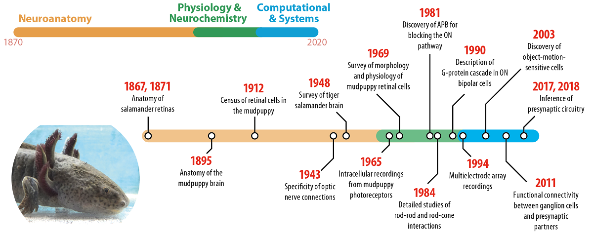
Salamanders have been habitual residents of research laboratories for more than a century, and their history in science is tightly interwoven with vision research. Nevertheless, many vision scientists – even those working with salamanders – may be unaware of how much our knowledge about vision, and particularly the retina, has been shaped by studying salamanders. In this review, we take a tour through the salamander history in vision science, highlighting the main contributions of salamanders to our understanding of the vertebrate retina. We further point out specificities of the salamander visual system and discuss the perspectives of this animal system for future vision research.

Keywords

retina; vision; ambystoma; salamander; mudpuppy; axolotl

Subject

Biology and Life Sciences, Animal Science, Veterinary Science and Zoology

Comments (2)

Comment 1
Received: 6 March 2020
Commenter:
The commenter has declared there is no conflict of interests.
Comment: Great article from Prof. Dr. Tim Gollisch, Ph.D.. I add an important event in CNS science that the salamander eye enabled. Working with Frank Werblin at Berkeley in the mid 1980s, we were the first to elicit a physiological response in single, identified neurons in a brain slice preparation using a natural stimulus, i.e. light to stimulate neurons in the retinal slice. This allowed single neurons to be unambiguously classified based on their voltage (current clamp) and current (volatage clamp) responses to their natural stimulus, along with the single neuron's morphological and histochemical features. Few thought I could elicit a light response in a slice prep, but using the salamander retina, working in dark-adapted conditions, and leaving the RPE attached would allow light responses in all the cell types from which I recorded: ACs, BCs, and IPCs. Thank you for paying tribute to this beautiful animal from which we've learned so much about vision.
+ Respond to this comment
Response 1 to Comment 1
Received: 14 March 2020
Commenter:
The commenter has declared there is no conflict of interests.
Comment: Thanks a lot for the note and for pointing this out. Indeed, getting light responses in the slice from different cell types was quite an achievement! Maybe we can follow this up.

We encourage comments and feedback from a broad range of readers. See criteria for comments and our Diversity statement.

Leave a public comment
Send a private comment to the author(s)
* All users must log in before leaving a comment
Views 0
Downloads 0
Comments 2
Metrics 0


×
Alerts
Notify me about updates to this article or when a peer-reviewed version is published.
We use cookies on our website to ensure you get the best experience.
Read more about our cookies here.