Submitted:
08 February 2020
Posted:
09 February 2020
You are already at the latest version
Abstract
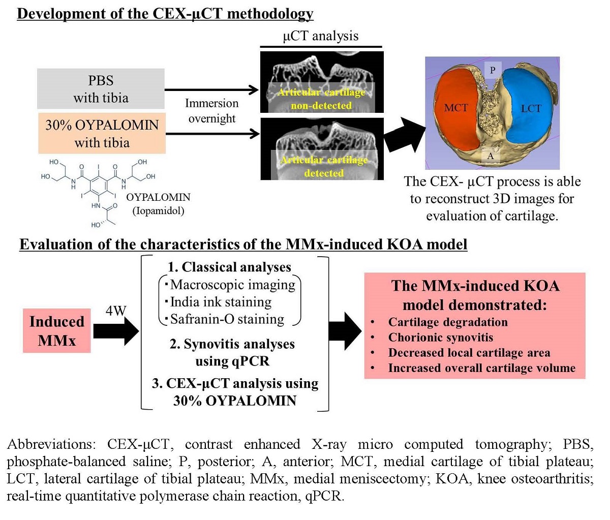
The aim of this study was to clarify degradation characteristics in each tissue of the knee complex of a medial meniscectomy (MMx)-induced knee osteoarthritis (KOA) animal model using classical methods and a new comprehensive evaluation method called contrast-enhanced X-ray micro-computed tomography (CEX-μCT), which was developed in the study. Surgical MMx was performed in the right knee joints of five male Wistar rats to induce KOA. At 4 wk post-surgery, the synovitis was evaluated using qPCR. Degradations of the articular cartilage of the tibial plateau were evaluated using classical methods and CEX-μCT. Evaluation of the synovitis demonstrated significantly increased expression levels of inflammation-associated marker genes in MMx-treated knees compared to that in sham-treated knees. Evaluation of the articular cartilage using classical methods showed that MMx fully induced degradation of the cartilage. Evaluation using CEX-μCT showed that local areas of the medial cartilage of the tibial plateau were significantly reduced in MMx-treated knees compared to that in sham-treated knees. On the other hand, total cartilage volumes were significantly increased in MMx-treated knees. Based on the findings of this study, the researchers in KOA research could be helped to select an optimal KOA model to discover new drugs.
Keywords:
osteoarthritis
; synovitis
; articular cartilage
; microfocus X-ray CT
; 3D analysis
Copyright: This open access article is published under a Creative Commons CC BY 4.0 license, which permit the free download, distribution, and reuse, provided that the author and preprint are cited in any reuse.


