
Submitted:
14 May 2018
Posted:
24 May 2018
You are already at the latest version
Abstract
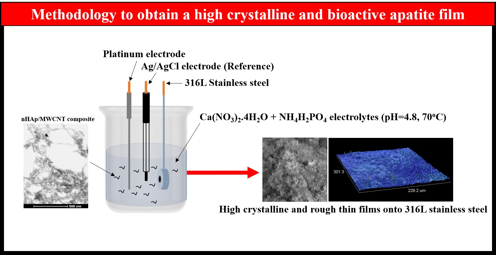
Herein, we evaluated the electrophoretic deposition of nanohydroxyapatite/superhydrophilic multiwalled carbon nanotube composites (nHAp/MWCNT) onto stainless steel biomedical alloys for applications in bone tissue engineering. First, nHAp/MWCNT composites were dispersed into 0.042 mol L−1 of Ca(NO3)2·4H2O + 0.025 mol L−1 NH4H2PO4 electrolytes (pH = 4.8) at two different concentrations. Next, a voltage of −2 V was applied using 316L stainless steel as a working electrode and (0.27 cm2), a high-purity platinum coil wire as the auxiliary electrode, and an Ag/AgCl(3 M) electrode was used as the reference electrode. The nHAp/MWCNT composites were characterized by transmission electron microscopy. The deposited nHAp and nHAp/MWCNT films were characterized by profilometry, scanning electron microscopy, X-Ray diffractometry and Raman spectroscopy. Human osteoblast cells were cultivated with the different materials, and in vitro cytotoxicity was evaluated using lactate dehydrogenase (LDH) assay. The osteogenesis process was evaluated by mRNA levels of the three genes that are directly related to bone repair: Alkaline Phosphatase, Osteopontin and Osteocalcin. We showed that rough, crystalline apatite thin films containing phases of nHAp were successfully deposited onto 316L stainless steel alloys. Also, we noticed that nHAp/MWCNT thin films deposited onto 316L stainless steel alloys upregulated the expression of important genes related to bone mineralization and maturation. Our results strongly support the possibility of this new alternative to modify the surface of metallic biomedical alloys to promote bone tissue regeneration.
Keywords:
316L
; electrodeposition
; nano-hydroxyapatite
; carbon nanotubes
; osteoblasts
; gene expression
Copyright: This open access article is published under a Creative Commons CC BY 4.0 license, which permit the free download, distribution, and reuse, provided that the author and preprint are cited in any reuse.


