

Submitted:
17 November 2026
Posted:
18 November 2026
You are already at the latest version
Abstract
Porcine circovirus type 2 (PCV2) is an important virus due to its association with a range of clinical syndromes. PCV2 is classified into eight genotypes, PCV2a-PCV2h, of which PCV2a, PCV2b, and PCV2d are the major genotypes. Porcine circovirus type 3 (PCV3) is a more recently identified virus classified into two genotypes, PCV3a and PCV3b. To investigate the genetic diversity of PCV2 and PCV3 in Danish pigs, samples collected between 2014-2025 from herds without specific clinical signs associated with PCV2 or PCV3 were analyzed and sequenced. Phylogenetic analysis of 139 PCV2 strains showed that PCV2b was the dominant genotype until 2017, when PCV2d was first detected in Denmark. Since 2019, PCV2d has been the dominant genotype, while sporadic cases of PCV2a and PCV2b continue to occur. The occurrence of PCV3 was investigated in samples from 113 herds. PCV3 was detected in a high proportion of herds across all herd types, but there was no evidence that it caused specific clinical disease. Phylogenetic analysis of 21 PCV3 strains showed that all belonged to genotype PCV3a. This study provides insight into the long-term genetic diversity of PCV2 and PCV3 and serves as a basis for prevention and control of infections with these viruses.
Keywords:
Denmark
; genetic diversity
; pigs
; porcine circovirus type 2
; porcine circovirus type 3
1. Introduction
Porcine circoviruses (PCVs) belong to the family Circoviridae within the genus Circovirus and are non-enveloped DNA viruses with a single-stranded (ssDNA) circular genome. Several open reading frames (ORFs) have been identified, but ORF1 and ORF2 appear to be essential for viral viability. ORF1 encodes the replicase (Rep) protein, which is required for viral genome replication, while ORF2 encodes the capsid (Cap) protein, which is involved in host-cells attachment [1,2,3]. PCVs are classified into four types; porcine circovirus type 1 (PCV1), 2 (PCV2), 3 (PCV3) and 4 (PCV4) [4]. Recently, a new type, tentatively named type 5 (PCV5), has been described [5].
PCV2 has been an important virus in modern pig production worldwide during the last 25 years [6]. It has been extensively studied due to its association with a range of clinical syndromes collectively referred to as porcine circovirus-associated diseases (PCVAD) including PCV2-systemic disease (PCV2-SD), PCV2-reproductive disease (PSV2-RD), PCV2-subclinical infection (PSV2-SI) and porcine dermatitis and nephropathy syndrome (PDNS) [7,8,9].
PCV2 is characterized by a remarkably high evolutionary rate for a DNA virus, of approximately 10-4-10-3 substitutions/site/year, which has facilitated the emergence of multiple genetically distinct variants [10,11]. Based on the ORF2 gene, these variants are classified into eight well-defined genotypes, designated as PCV2a-PCV2h, and a newly described genotype PCV2i [12,13]. Among these, PCV2a, PCV2b, and PCV2d are considered the major genotypes due to their widespread prevalence, global distribution, and sustained circulation in pig populations [1,12]. Of these major genotypes, PCV2a was the first genotype to be identified and dominated until the early 2000s, when a shift toward PCV2b occurred (around 2000–2003). Between 2010-2014, PCV2d began to emerge and has since become the predominant genotype in many countries [10,14,15]. Notably, PCV2d appears to possess enhanced replication efficiency, partly due to its improved uptake by host monocytes, which may contribute to its epidemiological success [16,17]. Despite the dominance of PCV2d, both PCV2a and PCV2b continue to be sporadically detected in pig herds [15].
Due to the significant impact of PCV2 on pig health and productivity, widespread vaccination programs were introduced in Europe in 2006 to mitigate clinical disease and economic losses [18,19]. The first commercial PCV2 vaccines were based on the PCV2a genotype, some in combination with PCV1, while a newer vaccine formulation has incorporated two genotypes (PCV2a and PCV2b) to broaden antigenic coverage. In 2025, the first PCV2d vaccine (Cirbloc® M Hyo, Ceva) was introduced to the European and Danish market. Some studies have shown that vaccines based on PCV2a generally provide cross-protection against other genotypes [20,21]. However, another study has demonstrated that PCV2d vaccines induce higher neutralizing antibody titers against PCV2d, whereas PCV2a-based vaccines may result in a delayed antibody response [22].
PCV3 is a more recently identified member of the genus Circovirus, first discovered in pigs in the United States in 2016 through a metagenomic approach [23,24]. Since its initial detection, PCV3 has been reported in pig populations across North and South America, Asia, and Europe, indicating a global distribution. Like PCV2, PCV3 has been associated with a range of clinical manifestations, including PCV3-reproductive disease (PCV3-RD), PCV3-systemic disease (PCV3-SD) and PDNS [7,23,24,25,26]. It has also been frequently detected in clinically healthy pigs, which complicates the assessment of their pathogenicity and virulence potential. Consequently, its role in disease remains uncertain and is still under investigation [27,28].
PCV3 is more genetically stable and has a lower mutation rate compared to PCV2 [29]. There is currently not full consensus on the genetic classification of PCV3, which has led to the proposal of different genetic groupings—typically referred to as PCV3a, PCV3b, and PCV3c, or Group A and Group B [25,30,31,32,33]. Later, two genotypes (PCV3a and PCV3b) were defined based on standardized criteria for analyzing sequencing data across different analytical approaches. However, since one of these genotypes (PCV3b) includes only two strains, only a single genotype (PCV3a) has been formally recognized so far [34].
The objective of this study was to investigate the genetic diversity of PCV2 and PCV3 in Danish pig populations over the periods 2014–2025 and 2017–2025, respectively. In addition, the study aimed to assess the presence of PCV3 in Danish pigs using samples obtained through research projects, diagnostic submissions, and screening activities.
2. Materials and Methods
2.1. Study Design and Samples
The samples analyzed in this study were archival, originating from previous research projects, national diagnostic submissions, and screening activities. All samples were collected from pigs in conventional herds in Denmark in the period 2014-2025.
To investigate the genetic diversity of PCV2 in Danish pigs, a total of 139 samples were analyzed, consisting of 64 screening and 75 diagnostic samples collected between 2014 and 2025. Screening samples were collected from herds without any specific clinical signs associated with PCV2. In contrast, diagnostic samples were submitted for laboratory investigation from herds experiencing health issues. The samples were blood (serum), tissue (lung, lymph node, fetuses), oral fluid, feces, nasal swab or lung swab (Table S1). Similarly, to assess the genetic diversity of PCV3 in Danish pigs, 21 samples were analyzed, including 13 diagnostic and eight randomly selected samples collected from 2017 to 2025. These samples included blood (serum), tissue (lung, lymph node, fetus, liver), and oral fluid (Table S2).
To investigate the presence and occurrence of PCV3, samples from seven different research projects (Study 1-7) conducted in 2017-2018 were included.
Study 1: A case-control study was conducted in 2017 involving 12 herds—six with a high farrowing rate (> 90%) and six with a low farrowing rate (< 85%). Reproductive organs were collected at slaughter and sent to the Veterinary Laboratory Kjellerup (Denmark). Parauterine lymph nodes were pooled (up to five per pool) from pigs within the same herd and collected on the same day. In total, 153 lymph nodes were pooled into 38 samples: 18 from high-farrowing herds and 20 from low-farrowing herds.
Study 2: A cross-sectional study was conducted in 2017 involving 60 herds with piglet health issues. A total of 501 deceased piglets were submitted to the Veterinary Laboratory in Kjellerup (Denmark), ranging from 3 to 91 piglets per herd (average 7.8). The piglets were 1–21 days old (average 6.4 days) and suffered from septicemia, intestinal, respiratory, skin and joint disorders. Inguinal and ileal lymph nodes were collected and pooled per herd, resulting in one sample per herd.
Study 3: A repeated cross-sectional study was conducted in 2018 in six sow herds [35]. Samples were collected monthly over three months (September-November) from nursery units. Each month, oral fluid and fecal sock samples were taken from two pens in each of three sections, representing pigs approximately two, five, and seven weeks after weaning. A total of 106 oral fluid and 108 fecal samples were collected.
Study 4: A repeated cross-sectional study was conducted in 2018 in three finishing units [35]. Sampling occurred monthly for three consecutive months (September-November). Oral fluid and fecal sock samples were collected from two pens in each of three age groups (early, mid, and late finishing stages). A total of 54 oral fluid samples and 54 fecal samples were included.
Study 5: A case study in 2017-2018 involving 13 herds selected by the herd practitioners due to suspected PCV2 infection or unexplained poor production performance. Blood samples were submitted from nine herds (42 pooled serum samples), oral fluid samples from two herds (nine samples), fetal tissue from two sows in one herd, and kidney and lymph node samples from a deceased pig in another herd.
Study 6: A case study was conducted in 2017 in nine herds selected by the Veterinary Laboratory Kjellerup (Denmark) based on suspected PCV2/PMWS during autopsy. A total of 24 lymph nodes were collected from these case herds.
Study 7: A cross-sectional study was conducted in 2018 in ten high-health specific pathogen free (SPF) breeding herds. Twenty blood samples were collected from sows in each herd and pooled into four samples, each consisting of five individual samples.
2.2. Nucleic Acid Extraction
Prior to extraction, tissue samples (180 mg) were homogenized in 1300 µl ATL buffer (QIAGEN, Hilden, Germany) in a TissueLyser II (QIAGEN) for 3 min at 30 Hz followed by centrifugation for 3 min at 5,500 g, and 200 μl of the supernatant was used for extraction. Blood samples were centrifuged for 10 min at 1,550 g and 200 µl of the serum was used for DNA extraction. Oral fluid samples were homogenized in a TissueLyser II (QIAGEN) for 15 sec at 30Hz, and the homogenate was centrifuged for 3 min at 5,500 g, and 200 μl of the supernatant was used for DNA extraction. For feces samples, a 10% dilution was made in phosphate-buffered saline (PBS), and the samples were homogenized in a TissueLyser II (QIAGEN) for 20 sec at 15Hz. For feces samples, the homogenate was centrifuged for 90 sec at 6,700 g, and either 350 µL (samples extracted before 2020) or 200 µL (samples extracted after 2020) of the supernatant was used for extraction.
Viral DNA was extracted from the serum, tissue and swab samples using the extraction robot QIAcube Connect (QIAGEN) and the QIAamp DNA mini kit (QIAGEN) according to the manufacturer’s instructions. Extraction of viral DNA from feces samples from before 2020 was automated on the QIAsymphony SP extraction robot (QIAGEN) using the QIAsymphony DSP virus/pathogen mini kit (QIAGEN) according to the manufacturer’s instructions. DNA extraction from feces samples collected from 2020 onwards was automated on the QIAcube HT (QIAGEN) using the IndiSpin QIAcube HT pathogen kit (Indical Bioscience, Leipzig, Germany) according to the manufacturer’s instructions. The viral DNA extraction from the oral fluid samples was performed using either of the above-mentioned robots and associated kit. Choice of method depended on the year in which the samples were purified due to availability of the instruments. Positive and negative (nuclease-free water) controls were included in each extraction set-up. The nucleic acid extracts were stored at −80°C until further analysis.
2.3. Detection of PCV2 and PCV3 by Quantitative Real-Time PCR
PCV2 quantitative real-time PCR (qPCR) analysis was performed in a final volume of 25 µl using AmpliTaq Gold™ master mix (Applied Biosystems, Massachusetts, USA) with 3 µl DNA. The PCV2 assay was run as described by Hjulsager at al., 2009 (laboratory 1) [36], and the used primer and probe sequences are listed in Table 1.
PCV3 qPCR analysis was performed in a final volume of 25 μl using AmpliTaq Gold™ master mix (Applied Biosystems) with 3 µl DNA. The qPCR mix consisted of 2.5 μl Gold buffer (10x), 2.5 μl MgCl2 (25 mM), 0.75 μl PCV3-Fw (10 μM), 0.75 µl PCV3-Rv (10 µM), 0.5 μl PCV3-P (10 μM), 0.5 μl dNTPs (10 mM), 0.25 μL AmpliTaq Gold DNA polymerase (5 U/μL), and 14.25 µl nuclease-free water. The used primer and probe sequences are listed in Table 1. The PCR reactions were run on the Rotorgene Q qPCR platform (QIAGEN) with the following thermal cycling conditions: 94°C for 10 min, followed by 45 cycles x (94°C for 15 sec, and 60°C for 60 sec). Data obtained from the PCV2 and PCV3 assays were analyzed (Rotor-Gene Q series software v.2.3.5; QIAGEN) with the following parameter adjustments: dynamic tube normalization, on; noise slope correction, on; ignore first cycle; outlier removal, 10%; Ct threshold, 0.01 (PCV3) or 0.02 (PCV2). All reactions were run in duplicates with positive (a dilution from a respective plasmid standard curve) and negative controls.
2.4. Conventional PCR and Sequencing of Complete Genomes
PCV2 samples sequenced in 2014-2017 (September included) were analyzed as described by Dupont et al., 2008 [14], while the sequencing procedure for samples analyzed in 2017 (from October)-2025 is described here.
The PCV2 and PCV3 conventional PCR analyses were performed in a final volume of 25 μl using the AccuPrime™ Taq DNA Polymerase, High Fidelity kit (Invitrogen, Thermo Fischer Scientific, Roskilde, Denmark) with 5 µl (PCV2) and 3 µl (PCV3) DNA. For PCV2, three (PCV2-1-3) or two (JK) primer pairs were used to amplify the complete PCV2 genome, while four primer pairs were used to amplify the complete PCV3 genome (Table 1). The PCR mix for both PCV2 and PCV3 consisted of 2.5 µl PCR buffer I (10x), 0.25 µl Tag high fidelity, 0.50 µl MgSO4, 1.25 µL of each primer (Table 1) and 14.25 µL (PCV2) or 16.25 µl (PCV3) nuclease-free water. The PCR reactions were run on either a T3 Thermocycler (Biometra, Fredensborg, Denmark) or a PCRmax Alpha thermocycler (Cole-Parmer, St Neots, United Kingdom) with the following thermal cycle conditions: 94°C for 2 min, followed by 35 cycles x (94°C for 30 sec, 59°C (PCV2; test 1)/69°C (PCV2; test 12)/61°C (PCV3) for 30 sec and 59°C (PCV2; test 1)/68°C (PCV2; test 12, PCV3) for 2.5 min) and 68°C for 10 min. The PCR products were visualized using a 0.8% (PCV3) or 2% (PCV2) E-Gel™ Agarose Gels with SYBR™ Safe DNA Gel Stain (Invitrogen).
Prior to sequencing, the PCR products were purified using High Pure PCR Product Purification kit (Roche, Basel, Switzerland) according to the manufacturer’s instructions. The concentration of the purified PCR products was measured with a NanoDrop™ One Spectrophotometer (Thermo Fisher Scientific). The purified PCR products were mixed with a PCR primer (5 μM, Table 1) and submitted for sequencing by the SANGER method (Sanger et al., 1977) to LGC Genomics (Berlin, Germany, samples from 2014-2024) or Eurofins Genomics (Ebersberg, Germany, samples from 2024-2025).
Sequence Data Analysis and Phylogenetic Analysis
Raw data obtained from the Sanger sequencing was imported into CLC Main Workbench (version 22.0, QIAGEN) wherein the sequences were assembled into consensus sequences using reference sequences (PCV2: EF565344 or EF565353 and PCV3: MF162299) and proofread. The consensus sequences were analyzed using BLAST search [38] to confirm the correctness of the PCV species.
The consensus nucleotide sequences (complete genomes and ORF2) were aligned with relevant published reference sequences obtained from the GenBank database (NCBI Resource Coordinators, 2017) (NCBI, http://ncbi.nlm.nih.gov) using the MUSCLE algorithm [39] in CLC Main Workbench (version 22.0, QIAGEN). For PCV2 and PCV3, reference sequences representing the current genotype classifications were selected from Franzo et al. (2018, 2024) [1,12] and Franzo et al. (2020) [34], respectively. Additionally, the top BLAST hit sequences were included as reference sequences in the alignments. Model testing was performed in CLC Main Workbench (Version 22.0, QIAGEN) to determine the best fitting substitution model, which was GTR + G + T for PCV2 and PCV3. The MUSCLE alignments were used for construction of maximum likelihood phylogenetic trees with 1,000 bootstrap replicates using the software IQ-TREE (version 3.1.4) [40]. The phylogenetic trees were visualized and annotated using Interactive Tree Of Life (iTOL, v6) [41], and final figure formatting, including color adjustments and text annotations, was performed in Inkscape (version 1.4.2, available at https://inkscape.org). Furthermore, the ORF2 nucleotide sequences were translated into amino acid sequences using CLC Main Workbench (Version 22.0, QIAGEN) with the standard genetic code. The nucleotide and amino acid sequences were subjected to a pairwise comparison, using CLC Main Workbench (version 22.0, QIAGEN), to reveal the overall sequence identity within the PCV species and genotypes.
The nucleotide sequences of this study are available from the GenBank database (NCBI, http://ncbi.nlm.nih.gov) with the accession numbers QB135240-QB135378 for PCV2 and QB135221-QB135239 for PCV3.
2.5. Analysis of the Amino Acid Variability in the Cap Protein of PCV2 and PCV3
Comparative analyses of the amino acid sequences of the Cap protein from the 139 obtained PCV2 strains and 21 PCV3 strains were performed to identify substitutions within each PCV2 genotype and PCV3, respectively. MUSCLE alignments of the amino acid sequences for each PCV2 genotype and PCV3 were generated using CLC Main Workbench (version 22.0, QIAGEN), and the amino acids at each position were visualized using sequence logos.
3. Results
3.1. Genetic Diversity of PCV2 in Danish Pigs
A total of 139 PCV2 positive samples collected between 2014 and 2025 were sequenced and genotyped (1-36 samples per year) (Figure 1). Two sequences, one from 2018 and one from 2025, were identified as genotype PCV2a. In comparison, 52 sequences belonged to genotype PCV2b, and 85 sequences to genotype PCV2d. From 2014 to 2016, only PCV2b was detected, whereas in 2017, both PCV2b (64%) and PCV2d (36%) were detected, with PCV2b being the most common type. In 2018, the distribution of PCV2b (47%) and PCV2d (47%) was equal. By 2019, PCV2d (76%) became the predominant genotype. Since 2019, PCV2b has only been detected sporadically, while PCV2d has remained the dominant genotype.
Complete genome sequences (1767 nucleotides) were obtained for 134 of the PCV2 samples, while for five samples, only ORF2 sequences were obtained. Pairwise comparison showed that the two PCV2a sequences were highly similar, sharing 99.4% nucleotide identity across the complete genome and 99.0% in ORF2. The PCV2b sequences also exhibited high genetic similarity, with nucleotide identities ranging from 97.9-100% across the complete genome and 98.6-100% in ORF2. Likewise, the PCV2d sequences were highly conserved, sharing 97.7-100% nucleotide identity across the complete genome and 97.9-100% in ORF2. Amino acid identities of ORF2 ranged from 97.0-100% for PCV2b and PCV2d and was 99.6% for the two available PCV2a sequences.
To assess the genetic diversity, phylogenetic trees were constructed based on the complete genome and ORF2 sequences of the Danish PCV2 strains together with reference sequences (Figure 2 and Figure 3). The two PCV2a sequences obtained in this study clustered most closely with a Chinese strain (KC514989), showing very high nucleotide identity (>99%) across both the complete genome and ORF2. In contrast, they were more divergent from the older Danish PCV2a strains detected in 1993 (EU148506) and 1996 (EU148507), sharing approximately 95.4–96.4% nucleotide identity across the complete genome and 92.5–94.2% in ORF2. The PCV2b and PCV2d sequences obtained in this study grouped into several sub-clusters, but overall exhibited high nucleotide identity to their respective reference strains, ranging from 97.9–100% across both the complete genome and ORF2. Among the PCV2b sequences, the Danish reference strains from 2008 (EF565344, EF565352, and EF565365), which were apparently unrelated, were particularly closely related.
For eight PCV2 positive herds, two or three sequences were available from each herd. These sequences were obtained from samples collected on the same day within each herd. In seven of these herds, the same genotype (either PCV2b or PCV2d) was detected, having within-herd nucleotide sequence identities ranging from 99.9-100%. In one herd (samples PCV2/Denmark/11012-11/2017 and PCV2/Denmark/11012-12/2017; Table S1), two different genotypes (PCV2b and PCV2d) were identified.
3.2. Amino Acid Sequence Analysis of the Cap Protein of PCV2
To further characterize the molecular features of the Danish PCV2 strains, the amino acid sequences of ORF2 were compared within each genotype. The PCV2a, PCV2b, and PCV2d sequences were compared with the earliest Danish sequences available for each genotype: PCV2a (EU148506, 1993), PCV2b (PCV2/Denmark/12011-30/2014), and PCV2d (PCV2/Denmark/4058-1/2017) (Table 2).
For PCV2a, in addition to the two PCV2a sequences obtained in this study (2018 and 2025), the analysis also included the PCV2a strain (EU148507) detected in 1996. Of the 25 amino acid positions showing substitutions, 19 were observed in the 1996 strain, 15 in the 2018 strain, and 17 in the 2025 strain. Seven of the observed substitutions were conservative (8, 63, 101, 118, 123, 180, 190), while the rest were non-conservative. Only one substitution (N46S) was observed between the 2018 and 2025 strains, while several substitutions (T47A, R/T63S, A68S, P131M, T/G134P, G/R169S, L185M, G191R) were observed between the 1993/1996 strains and the 2018/2025 strains (Table 2, Figure S1). Among the PCV2b strains, two amino acid substitutions were observed in multiple sequences: K63R in eight strains and A190T in 19 strains. All other substitutions were detected in single sequences only. No association was observed between substitutions at positions 63 or 190 and the year of detection. The substitution at position 63 was conservative, while the rest were non-conservative (Table 2, Figure S1). Comparison of the PCV2d strains revealed amino acid substitution R169G in 30 of the PCV2d strains, whereas all other substitutions were detected in single sequences only. No association was observed between substitutions at position 169 and the year of detection strains. All the observed substitutions were non-conservative (Table 2, Figure S1).
3.3. Occurrence of PCV3 in Danish Pigs
To evaluate the presence and distribution of PCV3 in Danish pigs, archived samples from seven different research projects (Study 1–7) were analyzed (Table 3). In Study 1, all 12 herds in which lymph nodes from reproductive organs were collected, tested positive for PCV3, regardless of whether the herds had high (n=6) or low (n=6) farrowing rates. The viral load in the lymph nodes was comparable between the two herd types. In Study 2, PCV3 was detected in 13 out of 60 herds (22%) from which lymph nodes were collected from piglets with various health issues. Studies 3 and 4 involved sampling from six sow herds over three consecutive months (September-November), including samples from finisher pigs in three of these herds. PCV3 was detected in all six nursery units and all three finishing units during the sampling period. Across these herds, PCV3 was found in 28–83% of oral fluid samples and in 6–50% of fecal sock samples. When examining the occurrence of PCV3-positive pens across three age categories (entry, mid, and exit) within both the nursery and finisher pig units, notably within age group variation was observed. In two nursery units, the frequency increased over time, whereas in one unit, a decline was noted. In the remaining units, the proportion of PCV3-positive pens either fluctuated or remained stable. A similar pattern was observed in finisher units, where two units exhibited consistent levels over time, and one showed a slight increase. When combining data across the nursery and finisher units, the results indicated that the occurrence within each age category (entry, mid, and exit) was largely consistent. In Study 5, samples were collected from herds experiencing non-specific health problems, as defined by the herd practitioners. In most cases, extensive investigations for other diseases have been conducted without conclusive results. PCV3 was detected in six out of 13 herds (46%). In Study 6, which focused on herds suspected of PCV2/PMWS infection, PCV3 was identified in two out of nine herds (22%). Finally, in Study 7, which included high-health-status herds, PCV3 was detected in 8 out of 10 herds (90%), with one to four positive samples out of four tested per herd.
3.4. Genetic Diversity of PCV3 in Danish Pigs
A total of 21 PCV3-positive samples collected between 2017 and 2025 were sequenced (1-11 samples per year, Table S2). Complete genome sequences (2000 nucleotides) were obtained for 19 of the samples and almost complete genome sequences (1881-1883 nucleotides, covering ORF1 and ORF2) were obtained for two samples. A PCV3-positive serum sample collected in 2015 was also subjected to sequencing, but only 1419 nucleotides were successfully obtained, covering the ORF1 gene and a small portion of ORF2. Due to the incomplete sequence, this sample was excluded from the phylogenetic analysis.
The PCV3 sequences showed an overall nucleotide identity of 97.5–100% across the complete genome and 96.9–100% for ORF2, corresponding to an amino acid sequence identity of 97.7–100%. Phylogenetic analysis of the complete genome and ORF2 sequences including reference sequences showed that the Danish sequences clustered within two sub-clusters, both belonging to genotype PCV3a (Figure 4 and Figure 5). All Danish samples except one clustered together, whereas the remaining sequence (PCV3/Denmark/896-21/2018) grouped with a German sequence from 2015 (MG014368), with which it shared 99.3% nucleotide identity. The PCV3 sequences obtained in this study shared 96.80–99.90% and 96.43–100% nucleotide identity with PCV3a reference strains across the complete genome and ORF2, respectively. In contrast, nucleotide identity to the PCV3b reference strains was lower, ranging from 91.3–92.7% for the complete genome and 87.4–89.0% for ORF2.
3.5. Amino Acid Sequence Analysis of the Cap Protein of PCV3
To further characterize the molecular features of the Danish PCV3 strains, the amino acid sequences of Cap protein were compared with the earliest Danish sequence available (MF802723). Among the Danish PCV3 strains, two amino acid substitutions were observed in multiple sequences: R27K in three sequences and Y104F in four sequences. All other substitutions were detected in single sequences only. No association was observed between the two substitutions and the year of detection (Table 2, Figure S2). The substitutions at positions 27 and 104 were both conservative, whereas the single substitutions were non-conservative, except for a conservative substitution at position 150.
4. Discussion
The present study provides insights into the long-term genetic diversity of PCV2 in Denmark. PCV2b was the predominant genotype in Denmark until 2017, which was also the year when PCV2d was detected in the country for the first time. In the following year, PCV2b and PCV2d were identified in an equal number of sequences, thereafter, PCV2d emerged as the dominant genotype and has remained predominant in subsequent years. Nevertheless, PCV2a and PCV2b continue to be sporadic detected. Although no viruses belonging to the PCV2c genotype were detected in this study, this genotype has previously been identified in Denmark in three samples collected during the 1980s and 1990s (EU148503, EU148504, EU148505) [14]. Thus, over the last 45 years, three shifts in the dominant PCV2 genotypes have occurred in Danish pigs: from PCV2c to PCV2a in the late 1990s, from PCV2a to PCV2b in 2002-2003, and most recently from PCV2b to PCV2d in 2017-2018. The increasing occurrence of PCV2d and its replacement of other genotypes are in line with findings reported in other countries globally [15,42,43,44].
Some authors have linked the shifts in genotypes to be (partly) linked to the widespread use of vaccines [10], however there are conflicting data on this in the literature [45]. Another explanation could be the more efficient viral entry of PCV2d compared to the other PCV2 genotypes [17]. Currently available PCV2a- and PCV2b-based vaccines continue to provide protection, however, some experimental studies have indicated that vaccine efficacy may vary depending on the genotype relationship between vaccine and challenge strain [22,46].
The comparison of amino acid substitutions in the ORF2-encoded Cap protein within each genotype showed that substitutions were unevenly distributed across genotypes. For PCV2a, a relatively high number of amino acid substitutions were observed when comparing newer strains (2018 and 2025) with strains from the 1990s. Notably, only a single substitution (N46S) distinguished the two recent PCV2a strains, suggesting a high degree of genetic stability within this genotype over the past decade. In contrast, multiple substitutions were identified between the 1990s strains and the contemporary strains, several of which occurred at well-described positions; amino acid residues at positions 63, 68, 131, 134, and 191 are all located on the Cap surface, whereas residue 185 is positioned on the inner side of the Cap [47]. Residues 68, 131, and 134 have been identified as forming discontinuous B-cell epitopes and have been associated with altered antibody recognition [47,48], while residue 63 plays a role in binding to cellular receptors [49]. Residue 191 is located within the conformational epitope region spanning amino acids 165–200, a well-characterized antigenic domain containing multiple overlapping epitopes recognized by antibodies [48,50,51]. In contrast, PCV2b strains exhibited few substitutions, two of which were observed in multiple sequences at positions 63 and 190. Similar to residue 63, position 190 is located on the Cap surface and falls within the conformational epitope region spanning amino acids 165–200 [47,48,50]. The most striking finding was observed in the PCV2d genotype, where substitutions at position 169 were detected in a large proportion of the analyzed strains. Position 169 is located on the Cap surface and within the conformational epitope region spanning amino acids 165–200. This surface-exposed region contributes to conformational epitopes targeted by neutralizing antibodies and is subject to immune-driven variation, which has been associated with genotype shifts and potential vaccine escape [47,48,51].
The first PCV3 detection in Denmark was in 2015, and the virus has subsequently been detected annually in various herd and sample materials across the country. The potential role of PCV3 as a disease-causing agent remains unclear and is still under investigation, however, the virus has been associated with a range of clinical manifestations, including PCV3-RD, PCV3-SD and PDNS [7,23,24,25,26]. To further explore the role of PCV3, archived samples originally collected from Danish pigs for various diagnostic and research purposes were analyzed to assess the occurrence and significance of the virus. The inclusion of samples from diverse sources enabled a broader evaluation of the potential influence of PCV3 on the health status of Danish pigs. PCV3 was detected retrospectively in samples from all seven research projects (Study 1-7) and with a relatively high occurrence in all types of herds. The differences between the various studies were due partly to the age of the pigs and partly to differences in sample type. The findings indicated that lymph nodes had a higher level of virus than serum and lung tissue samples, which could suggest that this virus – like PCV2 – replicates in immune cells [52]. Since the occurrence of PCV3 was also high in high-health sow herds (Study 7) and herds with a high farrowing rate (Study 1), these results did not support a direct association between PCV3 detection and disease. At the same time, the occurrence in case herds (Study 5 and 6) was low, which did not suggest that PCV3 caused “PCV2-like” symptoms. The present study does not provide evidence that PCV3 was the primary cause of specific clinical diseases in Danish pig herds, as the virus was widespread both in pigs that appeared healthy and in pigs that were sick.
All Danish PCV3 sequences obtained in this study belonged to genotype PCV3a, and showed a high level of nucleotide similarity, however, one sequence (Denmark/896-21/2018) differed from the others by clustering in a separate sub-cluster albeit still within genotype PCV3a. This high level of homogeneity is in line with the observed amino acid substitutions in the ORF2-encoded Cap protein, which exhibited limited variability, with only a few substitutions, two of which were observed in multiple sequences at positions 27 and 104. These positions, together with 24, 77, and 150, have previously been reported as variable sites in the PCV3 Cap protein, and some of these are located within predicted epitope regions, suggesting that they may influence Cap protein antigenicity or antibody recognition [53,54].
Monitoring genotype distribution is essential for understanding the occurrence and genetic diversity of PCV2 and PCV3 and can also provide a valuable basis for evaluating vaccine performance and guiding future control strategies.
5. Conclusion
In conclusion, this study showed that PCV2d is currently the predominant genotype in Denmark, following a genotype shift from PCV2b to PCV2d around 2017–2018. All sequenced PCV3 strains belonged to the PCV3a genotype. There was no evidence indicating that PCV3 was the cause of clinical disease in the herds from which the archived samples were obtained. The findings presented in this study contribute valuable knowledge about the genetic diversity of PCV2 and PCV3 in Denmark and provide important insights into their patterns of genetic evolution.
Supplementary Materials
The following supporting information can be downloaded at the website of this paper posted on Preprints.org. Table S1: Overview of the 139 PCV2 samples, including collection year, origin, sample type, sequence ID, and genotype. Table S2: Overview of the 21 PCV3 samples, including collection year, origin, sample type, sequence ID, and genotype. Figure S1: Amino acid sequence logos of the Cap protein for PCV2a, PCV2b, and PCV2d. Figure S2: Amino acid sequence logos of the Cap protein for PCV3a.
Author Contributions
Conceptualization, N.G., C.H. and L.L.; Methodology, C.H. and J.K.; Formal Analysis, N.G.; Investigation, N.G., C.K., C.H., and J.K.; Resources, C.K. and L.L.; Data Curation, N.G. and C.H.; Writing – Original Draft Preparation, N.G. and C.K.; Writing – Review & Editing, C.H., J.K. and L.L.; Visualization, N.G.; Project Administration, C.K., C.H. and L.L.; Funding Acquisition, C.K. and L.L. All authors have read and agreed to the published version of the manuscript.
Funding
This study was supported by the Danish Pig levy Fund (Severe viral diseases in Danish pigs; 2026) and Ceva Animal Health A/S.
Institutional Review Board Statement
Not applicable.
Data Availability Statement
The complete genome and ORF2 nucleotide sequences generated in this study have been submitted to the NCBI GenBank database. Other data supporting the findings of this study are available from the corresponding author upon reasonable request.
Acknowledgments
The authors would like to thank the farmers and herd veterinarians who participated in the study. The authors would also like to thank the laboratory technicians Nina D. Grønnegaard, Hue T. T. Tran, and Sari M. Dose for their help with the sample preparation and analysis.
Conflicts of Interest
Charlotte S. Kristensen was employed by SEGES from 2002 to 2021 and has worked for Ceva Animal Health A/S since 2021. The authors declare that her affiliation with Ceva Animal Health A/S did not influence the design, conduct, interpretation, or reporting of the study.:.
References
- Franzo, G.; Segalés, J. Porcine circovirus 2 (PCV-2) genotype update and proposal of a new genotyping methodology. PLoS ONE 2018, 13, 1–12. [Google Scholar] [CrossRef] [PubMed]
- Lv, Q.Z.; Guo, K.K.; Zhang, Y.M. Current understanding of genomic DNA of porcine circovirus type 2. Virus Genes. 2014, 49, 1–10. [Google Scholar] [CrossRef] [PubMed]
- Nawagitgul, P.; Morozov, I.; Bolin, S.; Harms, P.; Sorden, S.; Paul, P. Open reading frame 2 of porcine circovirus type 2 encodes a major capsid protein. J. Gen. Virol. 2000, 81, 2281–2287. [Google Scholar] [CrossRef] [PubMed]
- Varsani, A.; Harrach, B.; Roumagnac, P.; Benkő, M.; Breitbart, M.; Delwart, E.; et al. 2024 taxonomy update for the family Circoviridae. Arch. Virol. 2024, 169, 1–23. [Google Scholar] [CrossRef] [PubMed]
- Liu, X.; Wang, L.; Zhang, X.; Liu, Y.; Li, Y.; Li, Z.; et al. Identification and biochemical characterization of a novel porcine circovirus associated with porcine respiratory and diarrheal diseases. Microbiol. Spectr. 2025, 4, e0229925. [Google Scholar] [CrossRef] [PubMed]
- Segalés, J. Porcine circovirus type 2 (PCV2) infections: Clinical signs, pathology and laboratory diagnosis. Virus Res. 2012, 164, 10–9. [Google Scholar] [CrossRef] [PubMed]
- Segalés, J.; Allan, G.; Chae, C.; Ellis, J.; Harding, J.; Krakowka, S.; et al. Towards a unified nomenclature for diseases associated with porcine circovirus infections. Vet. Rec. 2025, 197, 411–4. [Google Scholar] [CrossRef] [PubMed]
- Ellis, J. Porcine Circovirus: A Historical Perspective. Vet. pathol. 2014, 51, 315–27. [Google Scholar] [CrossRef] [PubMed]
- Segalés, J.; Sibila, M. Revisiting Porcine Circovirus Disease Diagnostic Criteria in the Current Porcine Circovirus 2 Epidemiological Context. Vet. Sci. 2022, 9, 110. [Google Scholar] [CrossRef] [PubMed]
- Franzo, G.; Cortey, M.; Segalés, J.; Hughes, J.; Drigo, M. Phylodynamic analysis of porcine circovirus type 2 reveals global waves of emerging genotypes and the circulation of recombinant forms. Mol. Phylogenet Evol. 2016, 100, 269–80. [Google Scholar] [CrossRef] [PubMed]
- Firth, C.; Charleston, M.A.; Duffy, S.; Shapiro, B.; Holmes, E.C. Insights into the Evolutionary History of an Emerging Livestock Pathogen: Porcine Circovirus 2. J. Virol. 2009, 83, 12813–21. [Google Scholar] [CrossRef] [PubMed]
- Franzo, G.; Tucciarone, C.M.; Legnardi, M.; Drigo, M.; Segalés, J. An updated phylogeography and population dynamics of porcine circovirus 2 genotypes: are they reaching an equilibrium? Front Microbiol. 2024. [Google Scholar] [CrossRef] [PubMed]
- Wang, Y.; Noll, L.; Lu, N.; Porter, E.; Stoy, C.; Zheng, W.; et al. Genetic diversity and prevalence of porcine circovirus type 3 (PCV3) and type 2 (PCV2) in the Midwest of the USA during 2016–2018. Transbound. Emerg. Dis. 2020, 67, 1284–94. [Google Scholar] [CrossRef] [PubMed]
- Dupont, K.; Nielsen, E.O.; Bækbo, P.; Larsen, L.E. Genomic analysis of PCV2 isolates from Danish archives and a current PMWS case-control study supports a shift in genotypes with time. Vet. Microbiol. 2008, 128, 56–64. [Google Scholar] [CrossRef] [PubMed]
- Malwade, R.R.; Priyadharshini, K.M.; Venkataraman, S.; Akakpo, C.O.; Subbiah, M. Diversity of porcine circovirus 2 genotypes: insights from the studies reported worldwide in the past decade. VirusDisease 2025, 36, 533–48. [Google Scholar] [CrossRef] [PubMed]
- Wei, R.; Xie, J.; Theuns, S.; Nauwynck, H.J. Changes on the viral capsid surface during the evolution of porcine circovirus type 2 (PCV2) from 2009 till 2018 may lead to a better receptor binding. Virus Evol. 2019, 5, 1–12. [Google Scholar] [CrossRef] [PubMed]
- Ouyang, Y.; Nauwynck, H.J. PCV2 Uptake by Porcine Monocytes Is Strain-Dependent and Is Associated with Amino Acid Characteristics on the Capsid Surface. Microbiol. Spectr. 2023, 11. [Google Scholar] [CrossRef] [PubMed]
- Kristensen, C.S.; Baadsgaard, N.P.; Toft, N. A meta-analysis comparing the effect of PCV2 vaccines on average daily weight gain and mortality rate in pigs from weaning to slaughter. Prev. Vet. Med. 2011, 98, 250–8. [Google Scholar] [CrossRef] [PubMed]
- Karuppannan, A.K.; Opriessnig, T. Porcine circovirus type 2 (PCV2) vaccines in the context of current molecular epidemiology. Viruses 2017, 9, 1–15. [Google Scholar] [CrossRef] [PubMed]
- Park, K.H.; Oh, T.; Yang, S.; Cho, H.; Kang, I.; Chae, C. Evaluation of a porcine circovirus type 2a (PCV2a) vaccine efficacy against experimental PCV2a, PCV2b, and PCV2d challenge. Vet. Microbiol. 2019, 87–92. [Google Scholar] [CrossRef] [PubMed]
- Afghah, Z.; Webb, B.; Meng, X.J.; Ramamoorthy, S. Ten years of PCV2 vaccines and vaccination: Is eradication a possibility? Vet. Microbiol. 2017, 206, 21–8. [Google Scholar] [CrossRef] [PubMed]
- Kroeger, M.; Fano, E.; Sponheim, A.; Schwartz, K.J.; Leite, F.L.; Gomez-Duran, O.; et al. Assessment of homologous and heterologous PCV2 vaccine efficacy in a PCV2d/PRRSV co-challenge model. Vaccine 2025;60 December 2024. [Google Scholar] [CrossRef] [PubMed]
- Palinski, R.; Piñeyro, P.; Shang, P.; Yuan, F.; Guo, R.; Fang, Y.; et al. A Novel Porcine Circovirus Distantly Related to Known Circoviruses Is Associated with Porcine Dermatitis and Nephropathy Syndrome and Reproductive Failure. J. Virol. 2017, 91, 1–13. [Google Scholar] [CrossRef] [PubMed]
- Phan, T.G.; Giannitti, F.; Rossow, S.; Marthaler, D.; Knutson, T.; Li, L.; et al. Detection of a novel circovirus PCV3 in pigs with cardiac and multi-systemic inflammation. Virol. J. 2016, 13, 1–8. [Google Scholar] [CrossRef] [PubMed]
- Fux, R.; Söckler, C.; Link, E.K.; Renken, C.; Krejci, R.; Sutter, G.; et al. Full genome characterization of porcine circovirus type 3 isolates reveals the existence of two distinct groups of virus strains. Virol. J. 2018, 15, 1–9. [Google Scholar] [CrossRef] [PubMed]
- Tan, C.Y.; Lin, C.N.; Ooi, P.T. What do we know about porcine circovirus 3 (PCV3) diagnosis so far?: A review. Transbound. Emerg. Dis. 2021, 68, 2915–35. [Google Scholar] [CrossRef] [PubMed]
- Guo, Z.; Li, X.; Deng, R.; Zhang, G. Detection and genetic characteristics of porcine circovirus 3 based on oral fluids from asymptomatic pigs in central China. BMC Vet. Res. 2019, 15, 1–6. [Google Scholar] [CrossRef] [PubMed]
- Saporiti, V.; Franzo, G.; Sibila, M.; Segalés, J. Porcine circovirus 3 (PCV-3) as a causal agent of disease in swine and a proposal of PCV-3 associated disease case definition. Transbound. Emerg. Dis. 2021, 68, 2936–48. [Google Scholar] [CrossRef] [PubMed]
- Ha, Z.; Li, J.; Xie, C.; Yu, C.; Hao, P.; Zhang, Y.; et al. Prevalence, pathogenesis, and evolution of porcine circovirus type 3 in China from 2016 to 2019. Vet. Microbiol. 2020, 108756. [Google Scholar] [CrossRef] [PubMed]
- Franzo, G.; Legnardi, M.; Hjulsager, C.K.; Klaumann, F.; Larsen, L.E.; Segales, J.; et al. Full-genome sequencing of porcine circovirus 3 field strains from Denmark, Italy and Spain demonstrates a high within-Europe genetic heterogeneity. Transbound. Emerg. Dis. 2018, 3, 1–5. [Google Scholar] [CrossRef] [PubMed]
- Ku, X.; Zhang, C.; Li, P.; Yu, X.; Sun, Q.; Xu, F.; et al. Epidemiological and genetic characteristics of porcine circovirus 3 in 15 provinces and municipalities of China between 2016 and 2020. Virol. J. 2022, 19, 1–5. [Google Scholar] [CrossRef] [PubMed]
- Qi, S.; Su, M.; Guo, D.; Li, C.; Wei, S.; Feng, L.; et al. Molecular detection and phylogenetic analysis of porcine circovirus type 3 in 21 Provinces of China during 2015–2017. Transbound. Emerg. Dis. 2019, 66, 1004–15. [Google Scholar] [CrossRef] [PubMed]
- da Silva, R.R.; da Silva, D.F.; da Silva, V.H.; Castro, AMMG d. Porcine circovirus 3: a new challenge to explore. Front Vet. Sci. 2023, 1–12. [Google Scholar] [CrossRef] [PubMed]
- Franzo, G.; Delwart, E.; Fux, R.; Hause, B.; Su, S.; Zhou, J.Y.; et al. Genotyping porcine circovirus 3 (PCV-3) Nowadays: Does it make sense? Viruses 2020, 12, 1–13. [Google Scholar] [CrossRef] [PubMed]
- Goecke, N.B.; Kobberø, M.; Kusk, T.K.; Hjulsager, C.K.; Pedersen, K.S.; Kristensen, C.S.; et al. Objective pathogen monitoring in nursery and finisher pigs by monthly laboratory diagnostic testing. Porc Heal Manag 2020, 6, 1–14. [Google Scholar] [CrossRef] [PubMed]
- Hjulsager, C.K.; Grau-Roma, L.; Sibila, M.; Enøe, C.; Larsen, L.; Segalés, J. Inter-laboratory and inter-assay comparison on two real-time PCR techniques for quantification of PCV2 nucleic acid extracted from field samples. Vet. Microbiol. 2009, 133, 172–8. [Google Scholar] [CrossRef] [PubMed]
- Franzo, G.; Legnardi, M.; Centelleghe, C.; Tucciarone, C.M.; Cecchinato, M.; Cortey, M.; et al. Development and validation of direct PCR and quantitative PCR assays for the rapid, sensitive, and economical detection of porcine circovirus 3. J. Vet. Diagn. Investig. 2018, 30, 538–44. [Google Scholar] [CrossRef] [PubMed]
- Altschul, S.F.; Gish, W.; Miller, W.; Myers, E.W.; Lipman, D.J. Basic local alignment search tool. J. Mol. Biol. 1990, 215, 403–10. [Google Scholar] [CrossRef] [PubMed]
- Edgar, R.C. MUSCLE: Multiple sequence alignment with high accuracy and high throughput. Nucleic Acids Res. 2004, 32, 1792–7. [Google Scholar] [CrossRef] [PubMed]
- Wong, T.K.F.; Ly-Trong, N.; Ren, H.; Demotte, P.; Baños, H.; Roger, A.J.; et al. IQ-TREE 3: phylogenomic inference software using complex evolutionary models. Mol. Biol. Evol. 2026, 43, 1–6. [Google Scholar] [CrossRef] [PubMed]
- Letunic, I.; Bork, P. Interactive Tree of Life (iTOL) v6: Recent updates to the phylogenetic tree display and annotation tool. Nucleic Acids Res. 2024, 52, W78–82. [Google Scholar] [CrossRef] [PubMed]
- Giudici, S.D.; Mura, L.; Bonelli, P.; Hawko, S.; Angioi, P.P.; Sechi, A.M.; et al. Evidence of Porcine Circovirus Type 2 (PCV2) Genetic Shift from PCV2b to PCV2d Genotype in Sardinia, Italy. Viruses 2023, 15, 2157. [Google Scholar] [CrossRef]
- Xiao, C.; Halbur, P.G.; Opriessnig, T. Global molecular genetic analysis of porcine circovirus type 2 ( PCV2 ) sequences confirms the presence of four main PCV2 genotypes and reveals a rapid increase of PCV2d. J. Gen. Virol. 2015, 96, 1830–41. [Google Scholar] [CrossRef] [PubMed]
- Eddicks, M.; Ladurner Avilés, S.; Frauscher, S.; Krejici, R.; Reese, S.; Fux, R.; et al. Update on the Prevalence of the PCV2 Major Genotypes PCV2a, PCV2b, and PCV2d in German Fattening Farms in 2024. Vet. Sci. 2025, 12, 1–7. [Google Scholar] [CrossRef] [PubMed]
- Segalés, J.; Franzo, G.; Larsen, L.E.; Nauwynck, H.; Opriessnig, T.; Stadejek, T.; et al. Knowledge gaps in research and control of porcine circovirus 2 (PCV2) infections. BMC Vet. Res. 2026, 22. [Google Scholar] [CrossRef] [PubMed]
- Sagrera, M.; Garza-Moreno, L.; Cobos, À.; Llorens, A.M.; Huerta, E.; Pérez, M.; et al. Efficacy of a Novel PCV2d and Mycoplasma hyopneumoniae Combined Vaccine in Piglets with High and Low Levels of PCV2 Maternally Derived Antibodies at Vaccination. Vaccines 2025, 13, 1–22. [Google Scholar] [CrossRef] [PubMed]
- Wang, D.; Zhou, W.; He, Q.; Bai, Y.; Zhang, L.; Zhan, Y.; et al. Amino acid mutations of porcine circovirus type 2 (PCV2) capsid protein increase virus binding to host and evade immune responses: An evaluation of viral evolution. Virulence 2025, 16, 2535472. [Google Scholar] [CrossRef] [PubMed]
- Huang, L.; Sun, Z.; Xia, D.; Wei, Y.; Sun, E.; Liu, C.; et al. Neutralization Mechanism of a Monoclonal Antibody Targeting a Porcine Circovirus Type 2 Cap Protein Conformational Epitope. J. Virol. 2020, 94, 1–17. [Google Scholar] [CrossRef] [PubMed]
- Dhindwal, S.; Avila, B.; Feng, S.; Khayat, R. Porcine Circovirus 2 Uses a Multitude of Weak Binding Sites To Interact with Heparan Sulfate, and the Interactions Do Not Follow the Symmetry of the Capsid. J. Virol. 2019, 93, 1–16. [Google Scholar] [CrossRef] [PubMed]
- Cheung, A.K.; Greenlee, J.J. Identification of an amino acid domain encoded by the capsid gene of porcine circovirus type 2 that modulates intracellular viral protein distribution during replication. Virus Res. 2011, 155, 358–62. [Google Scholar] [CrossRef] [PubMed]
- Lekcharoensuk, P.; Morozov, I.; Paul, P.S.; Thangthumniyom, N.; Wajjawalku, W.; Meng, X.J. Epitope Mapping ofthe Major Capsid Protein of Type 2 Porcine Circovirus by Using Chimeric PCVl and PCV2.pdf. J. Virol. 2004, 78, 8135–45. [Google Scholar] [CrossRef]
- Shi, R.; Hou, L.; Liu, J. Host immune response to infection with porcine circoviruses. Anim. Dis. 2021, 1, 1–10. [Google Scholar] [CrossRef]
- Cao, M.; Wei, Y.; Shi, W.; Feng, L.; Huang, L. Investigation of porcine circovirus type 2 and porcine circovirus type 3 infections based on dual TaqMan fluorescent quantitative PCR method and genetic evolutionary analysis of these two viruses. Front Microbiol. 2024, 15, 1–14. [Google Scholar] [CrossRef] [PubMed]
- Tai, Y.; Tang, X.; Fan, J.; Liu, K.; Pan, W.; Sun, G.; et al. Molecular Epidemiological Investigation and Genetic Evolution Analysis of Porcine Circovirus 3 in Hunan Province, China, from 2021 to 2024. Viruses 2026, 18, 1–13. [Google Scholar] [CrossRef] [PubMed]
Figure 1.
Distribution of PCV2 genotypes in Danish pigs from 2014 to 2025. The genotype classification was based on Franzo et al. (2018) [1]. .
Figure 1.
Distribution of PCV2 genotypes in Danish pigs from 2014 to 2025. The genotype classification was based on Franzo et al. (2018) [1]. .
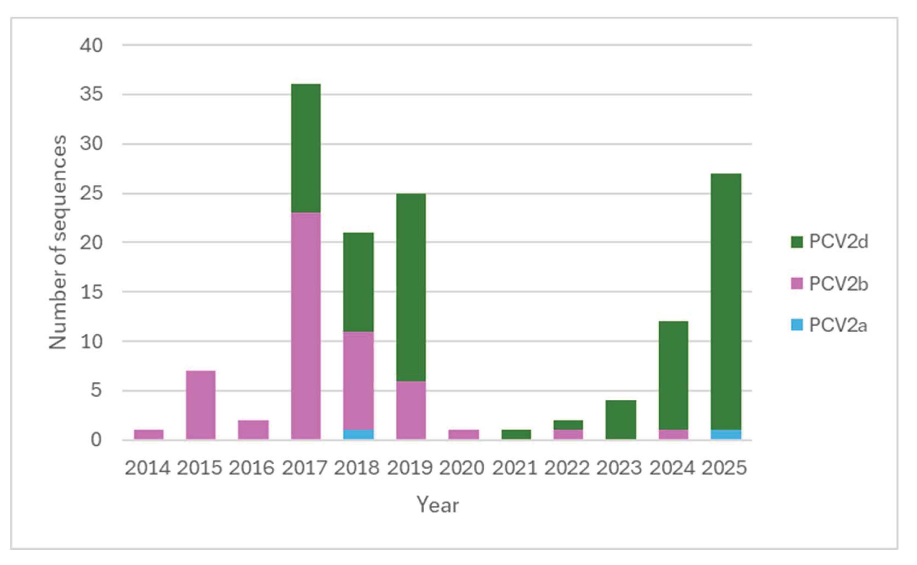
Figure 2.
Maximum-likelihood phylogenetic tree (1,000 bootstrap replicates) based on the complete genome sequence of PCV2. Danish sequences identified in this study are shown in blue, previously published Danish sequences are shown in red, and reference strains are shown in black. Bootstrap values <70% are not shown. The scale bar indicates the number of nucleotide substitutions per site.
Figure 2.
Maximum-likelihood phylogenetic tree (1,000 bootstrap replicates) based on the complete genome sequence of PCV2. Danish sequences identified in this study are shown in blue, previously published Danish sequences are shown in red, and reference strains are shown in black. Bootstrap values <70% are not shown. The scale bar indicates the number of nucleotide substitutions per site.
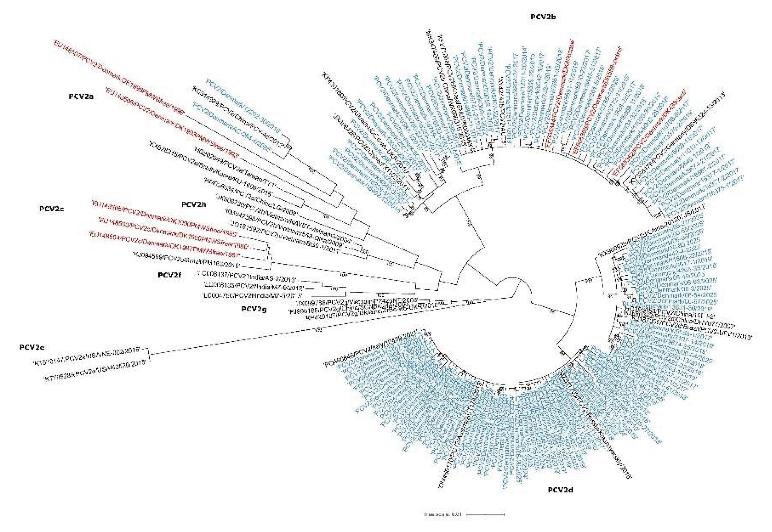
Figure 3.
Maximum-likelihood phylogenetic tree (1,000 bootstrap replicates) based on the ORF2 gene of PCV2. Danish sequences identified in this study are shown in blue, previously published Danish sequences are shown in red, and reference strains are shown in black. Bootstrap values <70% are not shown. The scale bar indicates the number of nucleotide substitutions per site.
Figure 3.
Maximum-likelihood phylogenetic tree (1,000 bootstrap replicates) based on the ORF2 gene of PCV2. Danish sequences identified in this study are shown in blue, previously published Danish sequences are shown in red, and reference strains are shown in black. Bootstrap values <70% are not shown. The scale bar indicates the number of nucleotide substitutions per site.
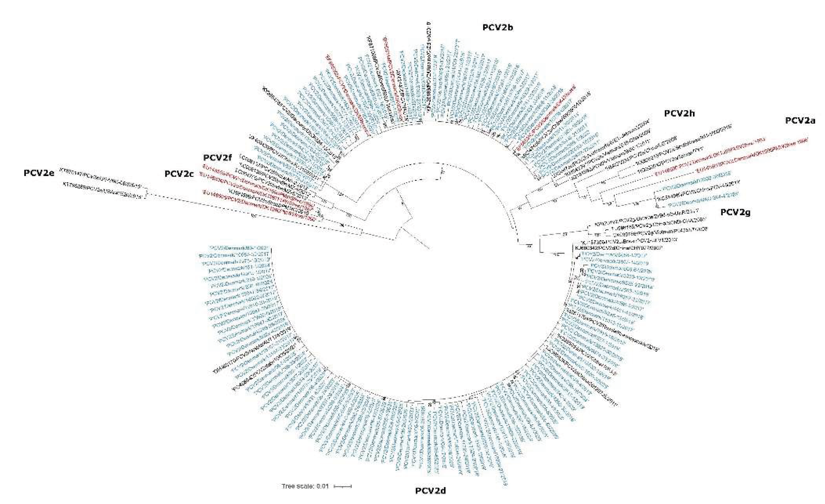
Figure 4.
Maximum-likelihood phylogenetic tree (1,000 bootstrap replicates) based on the complete genome sequence of PCV3. Danish sequences identified in this study are shown in blue, previously published Danish sequences are shown in red, and reference strains are shown in black. Bootstrap values <70% are not shown. The scale bar indicates the number of nucleotide substitutions per site.
Figure 4.
Maximum-likelihood phylogenetic tree (1,000 bootstrap replicates) based on the complete genome sequence of PCV3. Danish sequences identified in this study are shown in blue, previously published Danish sequences are shown in red, and reference strains are shown in black. Bootstrap values <70% are not shown. The scale bar indicates the number of nucleotide substitutions per site.
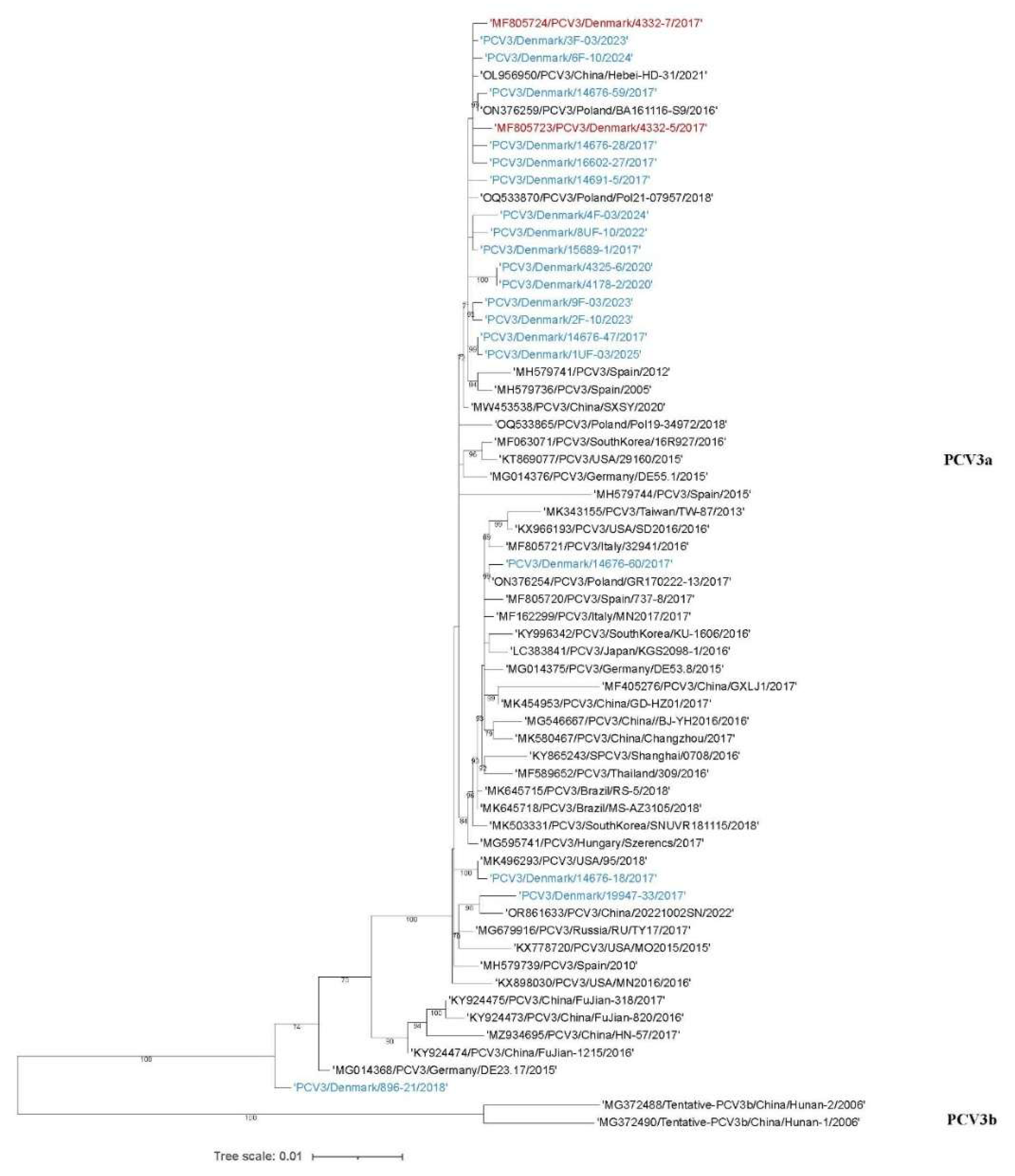
Figure 5.
Maximum-likelihood phylogenetic tree (1,000 bootstrap replicates) based on the ORF2 gene of PCV2. Danish sequences identified in this study are shown in blue, previously published Danish sequences are shown in red, and reference strains are shown in black. Bootstrap values <70% are not shown. The scale bar indicates the number of nucleotide substitutions per site.
Figure 5.
Maximum-likelihood phylogenetic tree (1,000 bootstrap replicates) based on the ORF2 gene of PCV2. Danish sequences identified in this study are shown in blue, previously published Danish sequences are shown in red, and reference strains are shown in black. Bootstrap values <70% are not shown. The scale bar indicates the number of nucleotide substitutions per site.
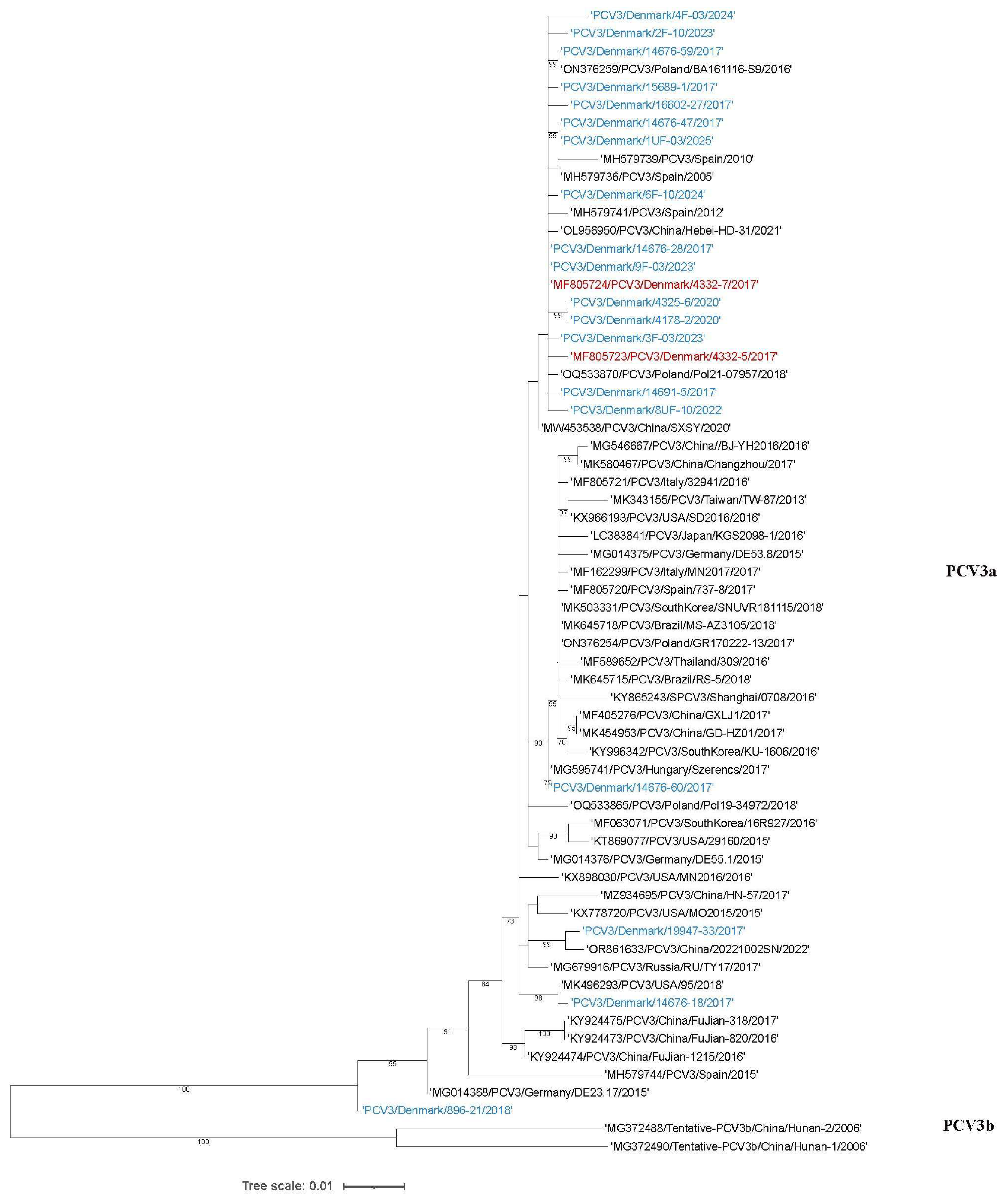

Table 1.
Primer and probe sequences used for qPCR and conventional PCR analyses of PCV2 and PCV3.
| Primer/probe | Sequence (5´-3´) | Product size (bp) | Reference |
| PCV2 qPCR | |||
| PCV2 ORF1-Fw | GATGATCTACTGAGACTGTGTGA | 152 | [36] |
| FAM-PCV2 ORF1-Rv | 6-FAM-AGAGCTTCTACAGCTGGGACA | ||
| PCV2-P | TCAGACCCCGTTGGAATGGTACTCCTC-Cy5 | ||
| PCV2 PCR | |||
| PCV2-1for | CTTCCGAAGACGAGCGC | 792 | [14] |
| PCV2-1rev | TAGCATTCTTCCAAAATACCAAG | ||
| PCV2-2for | GTTTACATAGGGGTCATAGG | 715 | |
| PCV2-2rev | TGCTTCTTCACAAAATTAGCG | ||
| PCV2-3for | TAGAGACTAAAGGTGGAACTGTA | 780 | |
| PCV2-3rev | TCCTGGGCGGTGGACATG | ||
| JK4-Fw (Test 1) | CGGAGGATTACTTCCTTGG | 980 | This study |
| JK2-RC-Rv (Test 1) | GGTTGGGGTCCGCTTCTTCC | ||
| JK1-Fw (Test 12) | CCAGCGCACTTCGGCAGC | 956 | |
| JK5-RC-Rv (Test 12) | GGGAAAGGGTGACGAACTGG | ||
| PCV3 qPCR | |||
| PCV3-Fw | AGT GCT CCC CAT TGA ACG | 135 | [23] |
| PCV3-Rv | ACA CAG CCG TTA CTT CAC | ||
| PCV3-P | FAM-ACCCCATGGCTCAACACATATGACC-BHQ1 | ||
| PCV3 PCR | |||
| PCV3 74-Fw | CACCGTGTGAGTGGATATAC | 854 | [23] |
| PCV3 1-Rv | CCAAACCCACCCTTAACAG | Modified from [23] | |
| PCV3 1303-Fw | ACCGGAGGGGTCAGATTTAT | 1258 | [37] |
| PCV3 541-Rv | GAGCTGCTGCTTGAAGATCC | ||
| PCV3 3-Fw | CGGGAATTGCAGTTGTATTTC | 909 | This study |
| PCV3 1720-Rv | CTCAGCCCTGTAATTTCTCC | ||
| PCV3 817-Fw | GTTATAATGGGGAGGGTGCT | 831 | [37] |
| PCV3 1647-Rv | GCCTGGACCACAAACACT | ||
Table 2.
Characterization of amino acid substitutions in Danish PCV2 ORF2 sequences for the PCV2a, PCV2b, and PCV2d genotypes and for PCV3. Numbers in parentheses indicate how many sequences each substitution was observed in.
Table 2.
Characterization of amino acid substitutions in Danish PCV2 ORF2 sequences for the PCV2a, PCV2b, and PCV2d genotypes and for PCV3. Numbers in parentheses indicate how many sequences each substitution was observed in.
| ORF2 position | PCV2a | PCV2b | PCV2d | PCV3 |
| 5 | R→M (1) | A→P (1) | ||
| 8 | F→Y (3) -1996/2018/2025 | |||
| 13 | H→N (1) - 1996 | H→N (1) H→P (1) |
||
| 20 | G→S (1) | |||
| 24 | A→P (1) | |||
| 27 | R→K (3) | |||
| 33 | R→S (1) | |||
| 34 | H→Q (1) | |||
| 36 | Y→N (1) | |||
| 44 | I→M (1) | |||
| 46 | N→S (1) - 2025 | |||
| 47 | T→A (2) -2018/2025 | |||
| 59 | A→R (1) - 1996 | |||
| 63 | R→T (1) - 1996 R→S (2) -2018/2025 |
K→R (8) | ||
| 64 | T→P (1) - 1996 | |||
| 68 | A→S (2) -2018/2025 | |||
| 75 | K→N (3) - 1996/2018/2025 | |||
| 77 | S→N (1) | |||
| 78 | D→N (1) -1996 | |||
| 85 | G→F (1) | |||
| 101 | I→V (3) -1996/2018/2025 | |||
| 104 | Y→F (4) | |||
| 118 | V→I (1) – 1996 | |||
| 123 | V→I (3) – 1996/2018/2025 | |||
| 131 | P→M (2) -2018/2025 | |||
| 133 | A→V (3) – 1996/2018/2025 | |||
| 134 | T→G (1) - 1996 T→P (2) -2018/2025 |
|||
| 135 | A→N (1) – 1996 | |||
| 136 | L→Q (3) – 1996/2018/2025 | |||
| 150 | I→L (1) | |||
| 151 | T→P (3) - 1996/2018/2025 | |||
| 152 | Q→H (1) | |||
| 157 | A→S (1) | |||
| 160 | Y→D (1) | |||
| 169 | G→R (1) - 1996 G→S (2) -2018/2025 |
R→G (30) |
||
| 177 | N→H (1) | |||
| 178 | N→S (1) | |||
| 180 | R→K (1) – 1996 | |||
| 185 | L→M (2) -2018/2025 | |||
| 190 | T→S (3) – 1996/2018/2025 | A→T (19) | ||
| 191 | G→R (2) -2018/2025 | |||
| 200 | T→I (1) | |||
| 201 | A→S (1) | |||
| 232 | K→H (1) - 1996 | N→K(1) |
Table 3.
Occurrence of PCV3 in Danish pigs based on archived samples from seven research projects (Study 1–7).
Table 3.
Occurrence of PCV3 in Danish pigs based on archived samples from seven research projects (Study 1–7).
| Study no. | Study description | No. herds | Sample material (no.) | % PCV3 positive |
| 1 | Sows with high farrowing rate | 6 | Lymph nodes pool (18) | 100% - herd level |
| Sows with low farrowing rate | 6 | Lymph nodes pool (20) | 100% - herd level | |
| 2 | Piglets submitted for necropsy due to clinical disease issues | 60 | Inguinal and ileal lymph nodes pool (60) | 22% - herd level |
| 3 | Nursery unit* – three monthly tests | 6 | Oral fluid (106) and fecal sock (108) | 100% - herd level OF: 39-83% - pen level FS: 6-22% - pen level |
| 4 | Finishing unit* – three monthly tests | 3 | Oral fluid (54) and fecal sock (54) | 100% - herd level OF: 28-78% - pen level FS: 17-50% - pen level |
| 5 | Case herds - submitted by practicing veterinarians | 13 | Serum pool (42), oral fluid (9), tissue (3) | 46% - herd level |
| 6 | Case herds - suspected PCV2/PMWS during autopsy | 9 | Lymph nodes (24) | 22% - herd level |
| 7 | High health herds | 10 | Serum pool (40) | 90% - herd level |
OF: oral fluid sample. FS: fecal sock sample. *Units from studies 3 and 4 are described in Goecke et al. (2020) [35].
Disclaimer/Publisher’s Note: The statements, opinions and data contained in all publications are solely those of the individual author(s) and contributor(s) and not of MDPI and/or the editor(s). MDPI and/or the editor(s) disclaim responsibility for any injury to people or property resulting from any ideas, methods, instructions or products referred to in the content. |
© 2026 by the authors. Licensee MDPI, Basel, Switzerland. This article is an open access article distributed under the terms and conditions of the Creative Commons Attribution (CC BY) license (http://creativecommons.org/licenses/by/4.0/).
Copyright: This open access article is published under a Creative Commons CC BY 4.0 license, which permit the free download, distribution, and reuse, provided that the author and preprint are cited in any reuse.