
Submitted:
17 November 2026
Posted:
18 November 2026
You are already at the latest version
Abstract
The harsh environmental conditions in the Antarctic have given rise to a large number of novel and unique microorganisms, but they have not yet been fully isolated, identified and described. Here, two novel bacterial strains, designated EDAd-1aT and ER825-2aT, were isolated from the seawater of the deep chlorophyll maximum layers of the Weddell Sea and Ross Sea in Antarctica, respectively. Both strains are Gram-stain-negative, rod-shaped, and non-motile and grow at 4–45 °C, pH 6.0–10.0, in the presence of 0.5–7.0% (w/v) NaCl. Phylogenetic analysis based on the 16S rRNA gene sequences or 120 conserved bacterial concatenated proteins all indicated that they belong to the genus Alcanivorax. The 16S rRNA gene sequence identity of strains EDAd-1aT and ER825-2aT was 99.71%. They separately shared 99.49–99.64% and 97.90–98.04% 16S rRNA se-quence similarity to A. borkumensis SKT and A. jadensis T9T, respectively, but less than 97.00% sim-ilarity to all the other type strains of the genus Alcanivorax. The digital DNA‒DNA hybridization (dDDH) and average nucleotide identity (ANI) values between strains EDAd-1aT and ER825-2aT were 48.4% and 93.2%, respectively, whereas the corresponding dDDH and ANI values between them and all the type strains of the genus Alcanivorax were only 22.0–26.0% and 80.8–83.2%, re-spectively. The predominant cellular fatty acids of EDAd-1aT were summed feature 8 (C18:1 ω7c or C18:1 ω6c), C16:0, summed feature 3 (C16:1 ω7c/C16:1 ω6c or C16:1 ω6c/C16:1 ω7c), and C12:0 3-OH; the predominant cellular fatty acids of ER825-2aT were summed feature 3 (C16:1 ω7c/C16:1 ω6c or C16:1 ω6c/C16:1 ω7c), summed feature 8 (C18:1 ω7c or C18:1 ω6c), C16:0, and C12:0 3-OH. Diphosphatidylglyc-erol, phosphatidylglycerol, phosphatidylethanolamine, phospholipid, aminophospholipid, aminolipid and unknown polar lipid were present in both strains. They also contained similar respiratory quinones, Q-11 and Q-10. On the basis of their phenotypic and genotypic characteris-tics, the two strains represent two novel species within the genus Alcanivorax, for which the names Alcanivorax antarcticus sp. nov. and Alcanivorax rossensis sp. nov. are proposed. The type strains are EDAd-1aT (=MCCC 1A21351T=KCTC 8606T) and ER825-2aT (=MCCC 1A21352T=KCTC 8607T).
Keywords:
Alcanivorax antarcticus sp. nov.
; Alcanivorax rossensis sp. nov.
; deep chlorophyll maximum layer
; seawater
; Weddell Sea
; Ross Sea
; Antarctica
; polyphasic taxonomy
1. Introduction
The microorganisms in the polar regions, both in terms of quantity and species, are dominant in the polar ecosystem. They are crucial for maintaining the healthy operation of polar ecosystems. In harsh and isolated polar environments, they are expected to evolve unique environmental adaptation mechanisms and physiological and biochemical characteristics. They have irreplaceable value in the fields of biomedicine, industrial enzymes, extreme environmental protection and restoration [1,2]. With the deepening of international polar scientific exploration activities and the continuous improvement in logistical support capabilities, the exploration and research of microbial resources in polar regions, especially Antarctica, have become popular. Moreover, with rapid changes in the global climate and Antarctic environment [3], anthropogenic activities such as scientific exploration [4], fishing [5], and commercial tourism [6] are significantly increasing. Frequent anthropogenic activities have brought increasingly serious exogenous pollutants to the Antarctic [7,8], thereby posing a persistent threat to the health of the Antarctic ecosystem and the maintenance of biodiversity.
Bacteria belonging to the Alcanivorax clade are crucial for maintaining the health of marine ecosystems and driving the biogeochemical cycles of marine elements. This is mainly because they can efficiently degrade marine hydrocarbon pollutants and participate extensively in the marine carbon cycle. They belong to the family Alcanivoracaceae in the order Oceanospirillales within the class Gammaproteobacteria. Their first type species, A. borkumensis SK2T, was proposed in 1998 [9]. In 2023, on the basis of whole-genome phylogenetic analysis, two novel genera, Alloalcanivorax and Isoalcanivorax, were separated from the Alcanivorax genus [10]. To date, the Alcanivorax clade consists of three genera and 20 validly or not validly published species (Figure 1). Our previous review indicated that these species were isolated primarily from various marine habitats and several highly saline terrestrial habitats [11]. Moreover, on the basis of our unpublished research, numerous novel marine-originated species in the Alcanivorax clade have not yet been obtained and described.
With respect to the ecological functions of Alcanivorax bacteria, hydrocarbon degradation is the most well-known label. They not only efficiently degrade marine petroleum hydrocarbons caused by anthropogenic activities [12] but are also the main drivers of the “marine cryptic hydrocarbon cycle” mediated by marine cyanobacteria [13,14]. Furthermore, in recent years, an increasing number of studies have shown that they also participate in the degradation of emerging marine plastic pollutants [11,15]. Therefore, Alcanivorax clade bacteria play a very important role in maintaining the health of marine ecosystems and promoting the cycling of marine elements.
With rapid changes in the global climate, anthropogenic activities are intensifying the hydrocarbon pollution into the Antarctic frequently, especially in the Antarctic marine environment. Owing to the restrictions on exotic species imposed by the Antarctic Treaty [16], isolating, studying and evaluating the hydrocarbon degradation characteristics and potential of indigenous microorganisms can provide valuable references for the biological remediation of hydrocarbon-contaminated Antarctic marine ecosystems. During the investigation of marine hydrocarbon-degrading bacterial biodiversity in the marginal seas around the Antarctic, strains EDAd-1aT and ER825-2aT were separately isolated from the seawater of the deep chlorophyll maximum (DCM) layers of the Weddell Sea and Ross Sea. The results of the phylogenetic analysis and overall genomic relatedness indices (OGRIs) indicated that the two strains were closely related to the genus Alcanivorax and may separately represent two novel species. Therefore, This study aimed to determine the taxonomic status of strains EDAd-1aT and ER825-2aT using a polyphasic approach.
2. Materials and Methods
2.1. Sample Collection, Strain Isolation and Cultivation
To obtain candidate hydrocarbon-degrading bacteria, different layers of seawater samples from station DA-05 (60.84° S, 53.71° W, bottom depth -701 m) in the Weddell Sea and station R1-08 (75.00° S, 177.00° W, -358 m) in the Ross Sea were collected using Niskin bottles attached to a conductivity‒temperature‒depth rosette of a SBE 911 plus system (Sea-Bird Scientific, Bellevue, WA, USA) during the 39th Chinese Antarctic Research Expedition of XUE LONG and XUE LONG 2 Icebreakers in 2022–2023. On a shipboard laboratory clean bench, approximately 10 ml of each layer of seawater was added to 100 ml of ONR7a medium [17] in a 250 ml Corning® 75 cm2 cell culture flask with a vent cap. Afterward, 0.5 ml of alkane mixture (C15H32:C17H36 = 9:1, v/v), which simulated the photosynthetic hydrocarbons produced by microscopic photosynthetic planktonic marine algae, was supplied as the sole carbon and energy source. These alkane-enriched consortia were maintained onboard at 4 °C and kept in the dark until they were transferred to the land-based laboratory. In the laboratory, 10-fold dilutions of the enriched culture were spread onto ONR7a medium plates supplemented with 0.5% (v/v) of the abovementioned alkane mixture and incubated under aerobic conditions at 15 °C for 14 days in the dark. Two pure clones were subsequently selected and designated as EDAd-1aT and ER825-2aT. Finally, these strains were routinely maintained at −80 °C in 25% (v/v) glycerol for subsequent polyphasic taxonomy analysis.
For comparative purposes, the two most closely related type strains, Alcanivorax borkumensis DSM 11573T and Alcanivorax jadensis DSM 12178T, were obtained from the German Collection of Microorganisms and Cell Cultures GmbH (DSMZ). Unless otherwise specified, for all tests of the morphological, physiological, and chemotaxonomic characteristics, the novel and type strains were cultivated at 28 °C in MANA medium [18].
2.2. 16S rRNA Gene Sequencing and Phylogenetic Analysis
The 16S rRNA gene sequences of strains EDAd-1aT and ER825-2aT were amplified and sequenced using the universal primers 27F (5′-AGAGTTTGATCCTGGCTCAG-3′) and 1492R (5′-TACCTTGTTACGACTT-3′) [19]. Moreover, the whole-length 16S rRNA gene sequences of the strains were also obtained from their genomes on the basis of the annotation. For phylogenetic analysis, the 16S rRNA gene sequences of all validly or not validly published type strains of the genera Alcanivorax, Alloalcanivorax and Isoalcanivorax were obtained from the National Center for Biotechnology Information (NCBI) GenBank and used as references. A maximum-likelihood (ML) phylogenetic tree was constructed using IQ-TREE 2 [20]. The support of each evolutionary branch was assessed using the SH-aLRT [21] method, which is based on 1000 replications. The tree was visualized using the Interactive Tree of Life (iTOL, v.7.0) online [22]. Furthermore, two other phylogenetic trees were generated using the neighbor-joining (NJ) [23] and minimum evolution (ME) [24] algorithms in MEGA-X (v. 10.0.5) [25]. Genetic distances for these analyses were calculated using Kimura’s two-parameter model [26], and all these analyses used 1,000 bootstrap resampled datasets to estimate node robustness.
2.3. Whole Genome Sequencing and Analysis
The genomic DNA of strains EDAd-1aT and ER825-2aT was extracted using an SBS extraction kit (SBS Genetech, Beijing, China) according to the manufacturer’s manual. The genomes were sequenced using a paired-end (PE) 2×150 bp sequencing strategy on an Illumina NovaSeq 6000 platform. The high-quality paired-end reads were assembled into contigs using SPAdes (v.3.15.5) [27] with kmer values of 21, 33, 45, 55, 77, 99, and 121 and a -careful flag. The qualities of the genomes were estimated by CheckM2 (v.1.0.0) [28]. The genomic DNA G+C content was calculated on the basis of the genome sequence. Genome annotation of the two strains was performed using the Rapid Annotation using Subsystem Technology (RAST) online server (http://rast.nmpdr.org/) [29] and the Prokaryotic Genome Annotation Pipeline (PGAP) online service in NCBI [30].
To determine the biological function and metabolic potential of the two strains and compare them against all type strains of the genus Alcanivorax, the genome-scale workflow METABOLIC-G.pl of the software METABOLIC (v.4.0) was used with default parameters [31]. The different functions and KEGG modules among the two strains and the type strains are presented in heatmaps, which were drawn using the online tool ChiPlot (https://www.chiplot.online/) (accessed on October 2026). Additionally, the two strains were subjected to genome screening of genes related to hydrocarbon degradation on the basis of the PGAP annotation results.
2.4 Overall genomic relatedness indices and Phylogenomic Analysis
Two OGRIs, digital DNA‒DNA hybridization (dDDH) and average nucleotide identity (ANI), were analyzed using the TYGS online service (https://tygs.dsmz.de/) [32] and the software fastANI (v. 1.3.0) [33] under default parameters, respectively. Moreover, a phylogenomic tree based on the genome sequences of the two strains and all the abovementioned validly or not validly published type strains was constructed on the basis of 120 conserved bacterial single-copy genes using GTDB-Tk v2 (v2.4.0) [34]. Similarly, the phylogenomic tree was also visualized using the iTOL v7 online service.
2.5. Morphological, Physiological and Biochemical Analyses
The cell morphology and flagellum of the strains EDAd-1aT and ER825-2aT was observed by transmission electron microscopy (HT7800, Hitachi). Gram-staining was performed using a Gram-stain kit (Thermo Scientific™ Remel™, USA). The hydrolysis of casein, starch, tributyrin or Tween-20, -40, -60 or -80 was tested on MANA plates supplemented with 0.1% (w/v) skim milk, 0.1% (w/v) soluble starch, 0.75% (v/v) tributyrin or 0.8% (v/v) Tween-20, -40, -60 or -80 at 15 °C for 7 days. Oxidase activity was evaluated with oxidase detection strips supplied by Oxoid (Oxoid Limited, UK), and catalase activity was determined by the application of 1% (v/v) hydrogen peroxide. The growth temperature range was determined by incubating cultures at 4, 10, 15, 20, 25, 28, 34, 37 and 45 °C for 7 days on MANA plates. Growth at different pH values was determined using MANA medium adjusted to pH 3–12 at 1 pH interval with the buffer systems described in a previous study [18]. NaCl tolerance was tested using modified MANA with NaCl concentrations of 0, 0.5, 1, 1.5, 2, 3, 4, 5, 6, 7, 8, 9, 10, 11, 12, or 15% (w/v) at 28 °C for 14 days. The optimum temperatures, pH and NaCl tolerance concentration were confirmed according to the growth status of colonies on MANA plates or optical density values at 600 nm from cultures in MANA liquid medium. Anaerobic growth was tested on MANA plates in an anaerobic jar with the Anoxomat Mark II Anaerobic System (Mart Microbiology B.V., Netherlands) at 28 °C for 14 days. The details of this experimental procedure are described in a previous study [35]. Other biochemical tests were performed using API 20NE, API 20E, and API ZYM strips (bioMérieux, France) according to the manufacturer’s manuals, with the modification of adjusting the NaCl concentration to 3.0%.
The ability of the two strains to utilize hydrocarbons was examined in ONR7a medium supplemented with 0.1% (v/v) alkanes of various carbon chain lengths as the sole carbon and energy source at 28 °C for up to 3 weeks in the dark, as described in a previous study [35]. Moreover, the oxygen consumption of the two strains along with A. borkumensis SK2T and A. jadensis T9T during the degradation of photosynthetic hydrocarbons (C15H32:C17H36 = 9:1, v/v) was quantified using a spirometer (Gas Endeavour, BPC Instruments, Lund, Sweden) to further assess their hydrocarbon-utilizing ability. The experimental details were described in our previous study [36].
2.6. Chemotaxonomic Characterization
The fatty acid profiles of strains EDAd-1aT, ER825-2aT, A. borkumensis SK2T and A. jadensis T9T were determined using cells from the third quadrant grown on MANA plates at 28 °C for 72 h. Cellular fatty acids were saponified, methylated and extracted according to the standard MIDI (Sherlock Microbial Identification System, version 6.0B) protocol. The fatty acids were then analyzed by gas chromatography (Agilent Technologies 6850) and identified using the TSBA6.0 database of the Microbial Identification System [37]. To analyze the quinone and polar lipid composition, the cells of strains EDAd-1aT and ER825-2aT cultivated in MANA medium at 28 °C for 72 h were harvested and subjected to freeze‒drying. Two 200 mg samples of freeze‒dried biomass were separately used for the analysis of quinones and polar lipids according to previously described methods [38,39].
3. Results and Discussion
3.1. Phylogenetic Characteristics
The 16S rRNA gene sequences of strains EDAd-1aT (1,380 bp) and ER825-2aT (1,379 bp) showed 99.71% identity. Additionally, the full-length 16S rRNA gene sequences of strains EDAd-1aT (1,547 bp) and ER825-2aT (1,547 bp) were also extracted from their genomes and were identical to the abovementioned partial 16S rRNA gene sequences obtained by Sanger sequencing. The MJ tree showed that the two strains belong to the genus Alcanivorax and formed two separate branches (Figure 1). The 16S rRNA sequences of strains EDAd-1aT and ER825-2aT were 99.49–99.64% and 97.90–98.04% similar to those of A. borkumensis SKT and A. jadensis T9T, respectively, but less than 97.00% similar to those of all the other type strains in the Alcanivorax clade, which included the genera Alcanivorax, Alloalcanivorax and Isoalcanivorax. Moreover, the NJ (Figure S1) and ME (Figure S2) trees both showed similar topologies to the MJ tree.
3.2. Genomic Features, OGRIs and Phylogenomics
The assembled draft genomes of strains EDAd-1aT and ER825-2aT were 3,785,771 bp and 3,698,081 bp in length and consisted of 52 and 24 contigs, respectively (Table S1). The genomes of EDAd-1aT and ER825-2aT were both 99.98% complete and separately possessed 0.05% and 0.10% contamination, indicating their high quality. The genomic DNA G+C contents of strains EDAd-1aT and ER825-2aT were 58.3% and 58.0%, respectively, which were within the range of the G+C content of the type strain of the genus Alcanivorax (54.70%–60.72%) (Table 1). The detailed genomic features of the two strains and their closest type strains are summarized in Table S1. The dDDH value between the two strains was 48.4%, and that between the type strains of the Alcanivorax clade ranged from 19.0–26.0% (Table S2). The ANI value between the two strains was 93.2%, and that between the type strains ranged from 77.1–83.2% (Table S3). Considering that a DDH value of ≥70% [40] and an ANI value of ≥95–96% [41] are recognized as the species boundaries between two bacterial strains, it is clear that the two strains separately represent two novel species of the genus Alcanivorax.
As shown in Figure 2, phylogenomic analysis on the basis of 120 conserved bacterial single-copy genes also place strains EDAd-1aT and ER825-2aT into the genus Alcanivorax cluster. They form two separate branches near the strain A. borkumensis SK2T. These results further confirmed that they belong to the genus Alcanivorax and represent two novel species of this genus.
3.3. Functional Genomics
The metabolic potential of strains EDAd-1aT and ER825-2aT was decoded at both the biological function and metabolic pathway levels on the basis of their genomes. At the functional level, except for a few iterms, the biological functions of the two strains are similar to those of the other type strains in the genus Alcanivorax. (Figure 3). In detail, only the two strains possessed a methanol oxidation function, suggesting that they might have the potential to metabolize C1 compounds. Additionally, only the two strains have the complete ethanol fermentation function of converting acetate into ethanol. Furthermore, several significant functional differences were detected between the two strains and their closest relative SK2T. For instance, they possessed the 4-aminobutyrate aminotransferase required for amino acid utilization and the 4-hydroxybutyrate-CoA ligase required for the 3HP/4HB carbon fixation pathway, but SK2T lacked them (Figure 3). However, strain SK2T had the propionyl-CoA synthetase required for the 3-hydroxypropionate cycle carbon fixation pathway, the periplasmic nitrate reductase required for nitrate reduction, and the urease required for urea utilization, which were absent in both strains. Coincidentally, the API 20NE and 20E test results also confirmed that the two strains could not reduce nitrate and had negative urease activity.
At the KEGG module level, only 25 differences (Figure 4) were identified from all 764 detected modules, further confirming that the species within the genus Alcanivorax have very similar metabolic potential. Overall, the main differences between the two strains and their close relatives were manifested in the following aspects. For instance, the two strains possessed the adhesin protein transport system module in the bacterial secretion system and the thiosulfate oxidation module mediated by the SOX complex in the sulfur metabolism, which were both not presented in all other type strains (Figure 4). Additionally, in addition to A. hongdengensis A-11-3T, only the sulfate transport system module and the multidrug resistance module mediated by the efflux pump EmrAB were present in the two strains. Furthermore, the two strains differed significantly from their closest relative SK2T in several modules. These included the acyl-CoA synthesis module necessary for fatty acid beta-oxidation, the two-component regulatory system module that regulates phosphate metabolism mediated by CreC-CreB, and the multidrug resistance module mediated by the efflux pump MexEF-OprN. However, strain SK2T lacked all of these. Nevertheless, SK2T possessed a Type IV secretion system, dissimilatory nitrate reduction, dTDP-L-rhamnose biosynthesis, and nicotine degradation modules, which were absent in the strains EDAd-1aT and ER825-2aT. Notably, there were also significant differences between the two strains in the following two modules. Strain EDAd-1aT possessed a glycolysis module for converting glucose into pyruvate and a methionine degradation module, while strain ER825-2aT lacked both. Therefore, to a certain extent, these two KEGG modules could serve as genetic markers for distinguishing the two strains.
Collectively, all of the abovementioned results further supported the discrepancy observed in their OGRIs and indicated that they separately represent two different novel species of the genus Alcanivorax.
The PGAP annotation results indicated that both the strains EDAd-1aT and ER825-2aT possessed multiple different types of alkane initial terminal monooxygenase genes, such as alkane 1-monooxygenase (alkB), FAD-dependent monooxygenase (almA), and cytochrome P450 (p450) genes. They mainly introduce oxygen to the terminal or secondary terminal of the alkanes to form corresponding alcohol products, thereby initiating the degradation process of the alkanes [47,48]. In the genome of strain EDAd-1aT, one alkB, one almA, and four p450 genes were found. Additionally, twelve flavin-containing monooxygenase genes were also found and are thought to be involved in initial alkane terminal oxidation. Similarly, one alkB, two almA, four p450 and thirteen flavin-containing monooxygenase genes were detected in the genome of strain ER825-2aT. Collectively, the detection of such a wide variety of initial hydrocarbon degradation genes in their genomes was consistent with the results of subsequent tests of hydrocarbon utilization ability.
3.4. Phenotypic Characteristics
Strains EDAd-1aT and ER825-2aT were Gram-staining-negative, strictly aerobic, nonmotile and rod-shaped, with cells 0.57–0.61 µm in width and 2.16–2.19 µm in length (Figure S3). After cultivation on MANA medium plates for 3 days at 28 °C, the colonies of the two strains were off white, smooth, circular, slightly convex and 1.0–2.0 mm in diameter. The growth of the two strains occurred at 4–45 °C (optimum 25–28 °C), at pH 6.0–10.0 (7.0) and in the presence of 0.5–7.0% (2.0%, w/v) NaCl. The two strains were found to be positive for the hydrolysis of tributyrin, Tween-20, -40, -60 and -80 but negative for casein, starch, aesculin, and gelatin. Their oxidase and catalase activities were both positive. The detailed morphological, physiological and biochemical characteristics of the two strains and their closely related type strains in the genus Alcanivorax are summarized in Table 1 and the species description.
The utilization results of different carbon chain lengths of normal alkanes revealed that both strains could utilize C14-C17 alkanes well and C24-C28 alkanes weakly but could not utilize C8 and C32 alkanes. Furthermore, an oxygen consumption experiment based on respiratory activity further quantified the ability of the two strains to degrade hydrocarbons. As shown in Figure 5, they could degrade photosynthetic hydrocarbons (C15H32:C17H36 = 9:1, v/v) well and consumed 282.2±22.2 mL and 229.1±1.2 mL of oxygen within 3 weeks, respectively. These results suggested that they might be involved in the degradation of hydrocarbons synthesized by photosynthetic phytoplankton in the euphotic zone of the Antarctic Ocean. After all, they all originated from the deep chlorophyll maximum layers.
3.5. Chemotaxonomic Characteristics
The predominant cellular fatty acids (>5% of the total) of the two strains were mainly consistent with those of the reference type strains (Table S4) but varied in their proportions. In detail, the predominant cellular fatty acids of strain EDAd-1aT were summed feature 8 (C18:1 ω7c or C18:1 ω6c) (28.0%), C16:0 (26.1%), summed feature 3 (C16:1 ω7c/C16:1 ω6c or C16:1 ω6c/C16:1 ω7c) (16.3%), and C12:0 3-OH (10.8%). The predominant cellular fatty acids of strain ER825-2aT were summed feature 3 (C16:1 ω7c/C16:1 ω6c or C16:1 ω6c/C16:1 ω7c) (28.7%), summed feature 8 (C18:1 ω7c or C18:1 ω6c) (28.1%), C16:0 (16.5%), and C12:0 3-OH (8.7%). Notably, there were significant differences in the proportions of the two kinds of fatty acids, summed feature 3 and C16:0, between the two strains. Specifically, the proportion of Summed feature 3 in strain EDAd-1aT is only 56.8% of that in strain ER825-2aT, but its C16:0 proportion is actually 58.2% greater than that of strain ER825-2aT. Therefore, to a certain extent, the significant difference in the proportions of these two types of fatty acids could be used as indicators to distinguish between the two potential novel species.
The polar lipids for the two strains were diphosphatidylglycerol, phosphatidylglycerol, phosphatidylethanolamine, phospholipid, aminophospholipid, aminolipid and unknown polar lipids (Figure S4-S5), which was consistent with the main polar lipids of members of the genus Alcanivorax, with the exception of the higher content of diphosphatidylglycerol observed in strain DAd-1aT (Figure S4). Both strains contained Q-11 (86–87% of the total), with minor amounts of Q-10 (13–14%) as the primary isoprenoid quinones.
4. Conclusions
In summary, phylogenetic analyses based on 16S rRNA gene sequences or on 120 conserved bacterial concatenated proteins all indicated that strains EDAd-1aT and ER825-2aT belong to the genus Alcanivorax. However, the two strains could be readily distinguished from other known Alcanivorax species by a combination of several genetic, biochemical, and physiological features given in Table 1 and Table S4, such as discrepant morphological traits, enzyme activities, and use of carbon sources. Furthermore, the low dDDH and ANI values, distinctive genetic functions and metabolic potential compared with those of other closely related species clearly suggested that strains EDAd-1aT and ER825-2aT separately represent two novel species of the genus Alcanivorax, for which the names Alcanivorax antarcticus sp. nov. and Alcanivorax rossensis sp. nov. are proposed.
Description of Alcanivorax antarcticus sp. nov.
Alcanivorax antarcticus (ant.arc’ti.cus. L. masc. adj. antarcticus, southern, pertaining to the Antarctic, where the type strain was isolated).
Cells are Gram-negative, rod shaped, 0.59–0.61 μm in width and 2.16–2.19 μm in length, nonmotile, and aerobic. After 3 days of incubation on MANA plates at 28 °C, the colonies were off white, smooth, circular with regular margins, slightly convex and 1.0–3.0 mm in diameter. Growth occurs at 4–45 °C (optimum 25–28 °C), at pH 6.0–10.0 (pH 7.0) and in the presence of 0.5–7.0% NaCl (w/v) (2.0%). Cells are positive for catalase, oxidase and the hydrolysis of tributyrin, Tween-20, -40, -60 and -80 but negative for the hydrolysis of casein, starch, aesculin, and gelatin. According to the API ZYM tests, the cells are positive for the activities of alkaline phosphatase, esterase (C4), esterase lipase (C8), lipase (C14), leucine aminopeptidase, valine aminopeptidase, α-chymotrypsin, acid phosphatase and naphtol-AS-Bl-phosphoamidase, weakly positive for the activities of cystine aminopeptidase and trypsin, but negative for all the other enzymes. In the API 20NE tests, the cells are positive for the assimilation of N-acetylglucosamine, potassium gluconate and malic acid; weakly positive for the assimilation of D-glucose and adipic acid; but negative for all other tests. In the API 20E tests, the cells are positive for Voges–Proskauer and weakly positive for tryptophan decarboxylase activity, but negative for all the other tests. The principal fatty acids (≥5%) are summed feature 8 (C18:1 ω7c or C18:1 ω6c), C16:0, summed feature 3 (C16:1 ω7c/C16:1 ω6c or C16:1 ω6c/C16:1 ω7c), and C12:0 3-OH. The predominant respiratory quinone is Q-11, with a minor Q-10. The polar lipids are diphosphatidylglycerol, phosphatidylglycerol, phosphatidylethanolamine, phospholipid, aminophospholipid, aminolipid and unknown polar lipid.
The type strain, EDAd-1aT (=MCCC 1A21351T =KCTC 8606T), was isolated from the seawater of the deep chlorophyll maximum layer of the Weddell Sea in Antarctica. The genomic DNA G+C content of the type strain is 58.28 mol%, as calculated from the whole-genome sequence. The GenBank/EMBL/DDBJ accession numbers for the 16S rRNA gene and genome sequence of strain EDAd-1aT are PX884092 and JCGGLW000000000, respectively.
Description of Alcanivorax rossensis sp. nov.
Alcanivorax rossensis (ros.sen’sis. N.L. fem. adj. rossensis, from the Ross Sea, Antarctica, where the type strain was isolated).
Cells are Gram-negative, rod shaped, 0.57–0.59 μm in width and 2.17–2.19 μm in length, nonmotile, and aerobic. After 3 days of incubation on MANA plates at 28 °C, the colonies were off white, smooth, circular with regular margins, slightly convex and 1.0–3.0 mm in diameter. Growth occurs at 4–45 °C (optimum 25–28 °C), at pH 6.0–10.0 (pH 7.0) and in the presence of 0.5–7.0% NaCl (w/v) (2.0%). Cells are positive for catalase, oxidase and the hydrolysis of tributyrin, Tween-20, -40, -60 and -80 but negative for the hydrolysis of casein, starch, aesculin, and gelatin. According to the API ZYM tests, the cells are positive for the activities of alkaline phosphatase, esterase (C4), esterase lipase (C8), lipase (C14), leucine aminopeptidase, valine aminopeptidase, α-chymotrypsin, acid phosphatase and naphtol-AS-Bl-phosphoamidase, weakly positive for the activities of cystine aminopeptidase and trypsin, but negative for all the other enzymes. In the API 20NE tests, the cells are positive for the assimilation of N-acetylglucosamine, potassium gluconate and malic acid; weakly positive for the assimilation of D-glucose; but negative for all other tests. In the API 20E tests, the cells are positive for Voges–Proskauer and weakly positive for tryptophan decarboxylase activity, but negative for all other tests. The principal fatty acids (≥5%) are summed feature 3 (C16:1 ω7c/C16:1 ω6c or C16:1 ω6c/C16:1 ω7c), summed feature 8 (C18:1 ω7c or C18:1 ω6c), C16:0, and C12:0 3-OH. The predominant respiratory quinone is Q-11, with a minor Q-10. The polar lipids are diphosphatidylglycerol, phosphatidylglycerol, phosphatidylethanolamine, phospholipid, aminophospholipid, aminolipid and unknown polar lipid.
The type strain, ER825-2aT (=MCCC 1A21352T =KCTC 8607T), was isolated from the seawater of the deep chlorophyll maximum layer of the Ross Sea in Antarctica. The genomic DNA G+C content of the type strain is 58.29 mol%, as calculated from the whole-genome sequence. The GenBank/EMBL/DDBJ accession numbers for the 16S rRNA gene and genome sequence of strain ER825-2aT are PX884093 and JCGGLX000000000, respectively.
Supplementary Materials
The following supporting information can be downloaded at the website of this paper posted on Preprints.org, Figure S1: Neighbor-joining tree showing phylogenetic relationships based on the 16S rRNA gene sequences of strains EDAd-1aT, ER825-2aT and all type strains of the genera Alcanivorax, Alloalcanivorax and Isoalcanivorax. Bootstrap values (> 50%) based on 1000 resamplings are given at the nodes. Bar, 0.01 substitutions per nucleotide position. The type strain Ketobacter alkanivorans GI5T (KY303768) was used as the outgroup; Figure S2: Minimum-evolution tree showing phylogenetic relationships based on the 16S rRNA gene sequences of strains EDAd-1aT, ER825-2aT and all type strains of the genera Alcanivorax, Alloalcanivorax and Isoalcanivorax. Bootstrap values (> 50%) based on 1000 resamplings are given at the nodes. Bar, 0.01 substitutions per nucleotide position. The type strain Ketobacter alkanivorans GI5T (KY303768) was used as the outgroup; Figure S3: Transmission electron micrographs of negatively stained cells of strains EDAd-1aT (a) and ER825-2aT (b) grown on MANA plates at 28 °C for 3 days; Figure S4: Polar lipid profiles of strain EDAd-1aT stained with 10% ethanolic molybdatophosphoric acid for all lipids (a), ninhydrin for aminolipids and phosphatidylethanolamine (b), molybdenum blue for phospholipids (c), and the α-naphthol-sulphuric acid reagent for glycolipids (d). Figure S5: Polar lipid profiles of strain ER825-2aT stained with 10% ethanolic molybdatophosphoric acid for all lipids (a), ninhydrin for aminolipids and phosphatidylethanolamine (b), molybdenum blue for phospholipids (c), and the α-naphthol-sulphuric acid reagent for glycolipids (d); Table S1: Genome characteristics of the strains Alcanivorax sp. EDAd-1aT, ER825-2aT and their closest relatives; Table S2: dDDH values among strains EDAd-1aT, ER825-2aT and their closest relatives of the genera Alcanivorax, Alloalcanivorax and Isoalcanivorax; Table S3: ANI values among strains EDAd-1aT, ER825-2aT and their closest relatives of the genera Alcanivorax, Alloalcanivorax and Isoalcanivorax; Table S4: Comparison of the cellular fatty acid contents of the strains EDAd-1aT, ER825-2aT and their closest relatives.
Author Contributions
Conceptualization, C.D.; methodology, J.W. and C.D.; investigation, Z.H. and J.W.; resources, W.X. and J.L.; data curation, J.W., C.Y. and C.D.; writing—original draft preparation, C.D.; writing—review and editing, C.D.; visualization, C.D. and C.Y.; supervision, C.D.; project administration, C.D.; funding acquisition, C.D.. All authors have read and agreed to the published version of the manuscript.
Funding
This work was financially supported by the National Key Research and Development Program (2022YFC2807501) and the National Natural Science Foundation of China (41976107).
Institutional Review Board Statement
Not applicable.
Informed Consent Statement
Not applicable.
Data Availability Statement
The GenBank/EMBL/DDBJ accession numbers for the partial-length 16S rRNA gene sequences of strains EDAd-1aT and ER825-2aT are PX884092 and PX884093, respectively. The draft genome sequences of strains EDAd-1aT and ER825-2aT have been deposited in GenBank/EMBL/DDBJ under accession numbers JCGGLW000000000 and JCGGLX000000000, respectively.
Acknowledgments
We greatly thank the whole teams of the 39th Antarctic Research Expedition of XUE LONG and XUE LONG 2 Icebreakers and the Chinese Arctic and Antarctic Administration. We also appreciate the efforts of Dr. Ruolin Cheng to maintain the bioinformation analysis server.
Conflicts of Interest
The authors declare no conflicts of interest.
References
- Doytchinov, V.V.; Dimov, S.G. Microbial Community Composition of the Antarctic Ecosystems: Review of the Bacteria, Fungi, and Archaea Identified through an NGS-Based Metagenomics Approach. Life 2022, 12, 916. [Google Scholar] [CrossRef] [PubMed]
- Medeiros, W.; Kralova, S.; Oliveira, V.; Ziemert, N.; Sehnal, L. Antarctic bacterial natural products: from genomic insights to drug discovery. Nat. Prod. Rep. 2025, 42, 774–787. [Google Scholar] [CrossRef] [PubMed]
- Abram, N.J.; Purich, A.; England, M.H.; McCormack, F.S.; Strugnell, J.M.; Bergstrom, D.M.; Vance, T.R.; Stål, T.; Wienecke, B.; Heil, P.; et al. Emerging evidence of abrupt changes in the Antarctic environment. Nature 2025, 644, 621–633. [Google Scholar] [CrossRef] [PubMed]
- Siegert, M.; Bracegirdle, T.J.; Convey, P.; Hendry, K.; Holmes, C.; Hughes, K.A. Antarctic science operations must account for climate change and extreme environmental events. Commun. Earth Environ. 2026, 7, 445. [Google Scholar] [CrossRef] [PubMed]
- Meyer, B.; Arata, J.A.; Atkinson, A.; Bahlburg, D.; Bernard, K.; Cárdenas, C.A.; Grant, S.M.; Hill, S.L.; Hüppe, L.; Ichii, T.; et al. Adjusting the management of the Antarctic krill fishery to meet the challenges of the 21st century. Proc. Natl. Acad. Sci. USA 2025, 122, e2412624122. [Google Scholar] [CrossRef] [PubMed]
- Tejedo, P.; Benayas, J.; Cajiao, D.; Leung, Y.F.; De Filippo, D.; Liggett, D. What are the real environmental impacts of Antarctic tourism? Unveiling their importance through a comprehensive meta-analysis. J. Environ. Manag. 2022, 308, 114634. [Google Scholar] [CrossRef] [PubMed]
- Goh, W.X.; Tae, R.S.; Lim, Z.S.; Wong, C.Y.; Ahmad, S.A.; Kok, Y.Y.; Convey, P. Hydrocarbon and heavy metal pollution in Antarctica: ecological impacts and remediation strategies. Environ. Sci. Pollut. Res. Int. 2026, 33, 9858–9876. [Google Scholar] [CrossRef] [PubMed]
- Roberts, S.J.; Hughes, K.A.; Li, C.; Peel, R.H.; Saunders, K.M.; Schneider, L.; Waller, C.L. Pollution in Antarctica. In Antarctica and the Earth System, 1 ed.; Meredith, M.P., Melbourne-Thomas, J., Naveira Garabato, A.C., Raphael, M., Eds.; Routledge: London, 2025; pp. 164–185. [Google Scholar]
- Yakimov, M.M.; Golyshin, P.N.; Lang, S.; Moore, E.R.; Abraham, W.R.; Lunsdorf, H.; Timmis, K.N. Alcanivorax borkumensis gen. nov., sp. nov., a new, hydrocarbon-degrading and surfactant-producing marine bacterium. Int. J. Syst. Bacteriol. 1998, 48, 339–348. [Google Scholar] [CrossRef] [PubMed]
- Rai, A.; Suresh, G.; Ria, B.; L, V.; PK, S.; Ipsita, S.; Sasikala, C.; Venkata Ramana, C. Phylogenomic analysis of the genus Alcanivorax: proposal for division of this genus into the emended genus Alcanivorax and two novel genera Alloalcanivorax gen. nov. and Isoalcanivorax gen. nov. Int. J. Syst. Evol. Microbiol. 2023, 73. [Google Scholar] [CrossRef] [PubMed]
- Dong, C.; Wang, Z.; Shao, Z. Degradation from hydrocarbons to synthetic plastics: the roles and biotechnological potential of the versatile Alcanivorax in the marine blue circular economy. Blue Biotechnol. 2024, 1, 14. [Google Scholar] [CrossRef]
- Varjani, S.J. Microbial degradation of petroleum hydrocarbons. Bioresour. Technol. 2017, 223, 277–286. [Google Scholar] [CrossRef]
- McGenity, T.J.; McKew, B.A.; Lea-Smith, D.J. Cryptic microbial hydrocarbon cycling. Nat. Microbiol. 2021, 6, 419–420. [Google Scholar] [CrossRef] [PubMed]
- Love, C.R.; Arrington, E.C.; Gosselin, K.M.; Reddy, C.M.; Van Mooy, B.A.S.; Nelson, R.K.; Valentine, D.L. Microbial production and consumption of hydrocarbons in the global ocean. Nat. Microbiol. 2021, 489–498. [Google Scholar] [CrossRef] [PubMed]
- Koike, H.; Miyamoto, K.; Teramoto, M. Alcanivorax bacteria as important polypropylene degraders in mesopelagic environments. Appl. Environ. Microbiol. 2023, 89, e01365-01323. [Google Scholar] [CrossRef] [PubMed]
- Aislabie, J.; McLeod, M.; Fraser, R. Potential for biodegradation of hydrocarbons in soil from the Ross Dependency, Antarctica. Appl. Microbiol. Biotechnol. 1998, 49, 210–214. [Google Scholar] [CrossRef]
- Dyksterhouse, S.E.; Gray, J.P.; Herwig, R.P.; Lara, J.C.; Staley, J.T. Cycloclasticus pugetii gen. nov., sp. nov., an Aromatic Hydrocarbon-Degrading Bacterium from Marine Sediments. Int. J. Syst. Bacteriol. 1995, 45, 116–123. [Google Scholar] [CrossRef] [PubMed]
- Wei, L.; Wang, J.; Liu, X.; Lai, Q.; Li, G.; Fu, X.; Li, G.; Shao, Z.; Dong, C. Galbibacter pacificus sp. nov., isolated from surface seawater of the western Pacific Ocean and transfer of Joostella marina to the genus Galbibacter as Galbibacter orientalis nom. nov. and emended description of the genus Galbibacter. Int. J. Syst. Evol. Microbiol. 2023, 73. [Google Scholar] [CrossRef] [PubMed]
- Lane, D.J.; Pace, B.; Olsen, G.J.; Stahl, D.A.; Sogin, M.L.; Pace, N.R. Rapid determination of 16S ribosomal RNA sequences for phylogenetic analyses. Proc. Natl. Acad. Sci. USA 1985, 82, 6955–6959. [Google Scholar] [CrossRef] [PubMed]
- Minh, B.Q.; Schmidt, H.A.; Chernomor, O.; Schrempf, D.; Woodhams, M.D.; von Haeseler, A.; Lanfear, R. IQ-TREE 2: New Models and Efficient Methods for Phylogenetic Inference in the Genomic Era. Mol. Biol. Evol. 2020, 37, 1530–1534. [Google Scholar] [CrossRef] [PubMed]
- Guindon, S.; Dufayard, J.F.; Lefort, V.; Anisimova, M.; Hordijk, W.; Gascuel, O. New algorithms and methods to estimate maximum-likelihood phylogenies: assessing the performance of PhyML 3.0. Syst. Biol. 2010, 59, 307–321. [Google Scholar] [CrossRef] [PubMed]
- Letunic, I.; Bork, P. Interactive Tree of Life (iTOL) v6: recent updates to the phylogenetic tree display and annotation tool. Nucleic Acids Res. 2024, 52, W78–w82. [Google Scholar] [CrossRef] [PubMed]
- Saitou, N.; Nei, M. The Neighbor-joining Method: a New Method for Reconstructing Phylogenetic Trees. Mol. Biol. Evol. 1987, 4, 406–425. [Google Scholar] [CrossRef] [PubMed]
- Rzhetsky, A.; Nei, M. A Simple Method for Estimating and Testing Minimum-Evolution Trees. Mol. Biol. Evol. 1992, 9, 945–967. [Google Scholar] [CrossRef]
- Kumar, S.; Stecher, G.; Li, M.; Knyaz, C.; Tamura, K. MEGA X: Molecular Evolutionary Genetics Analysis across Computing Platforms. Mol. Biol. Evol. 2018, 35, 1547–1549. [Google Scholar] [CrossRef] [PubMed]
- Kimura, M. A Simple Method for Estimating Evolutionary Rates of Base Substitutions Through Comparative Studies of Nucleotide Sequences. J. Mol. Evol. 1980, 16, 111–120. [Google Scholar] [CrossRef] [PubMed]
- Prjibelski, A.; Antipov, D.; Meleshko, D.; Lapidus, A.; Korobeynikov, A. Using SPAdes De Novo Assembler. Curr. Protoc. Bioinform. 2020, 70, e102. [Google Scholar] [CrossRef] [PubMed]
- Chklovski, A.; Parks, D.H.; Woodcroft, B.J.; Tyson, G.W. CheckM2: a rapid, scalable and accurate tool for assessing microbial genome quality using machine learning. Nat. Methods 2023, 20, 1203–1212. [Google Scholar] [CrossRef] [PubMed]
- Aziz, R.K.; Bartels, D.; Best, A.A.; DeJongh, M.; Disz, T.; Edwards, R.A.; Formsma, K.; Gerdes, S.; Glass, E.M.; Kubal, M.; et al. The RAST Server: rapid annotations using subsystems technology. BMC Genom. 2008, 9, 75. [Google Scholar] [CrossRef] [PubMed]
- Li, W.; O’Neill, K.R.; Haft, D.H.; DiCuccio, M.; Chetvernin, V.; Badretdin, A.; Coulouris, G.; Chitsaz, F.; Derbyshire, Myra K.; Durkin, A.S.; et al. RefSeq: expanding the Prokaryotic Genome Annotation Pipeline reach with protein family model curation. Nucleic Acids Res. 2021, 49, D1020–D1028. [Google Scholar] [CrossRef] [PubMed]
- Zhou, Z.; Tran, P.Q.; Breister, A.M.; Liu, Y.; Kieft, K.; Cowley, E.S.; Karaoz, U.; Anantharaman, K. METABOLIC: high-throughput profiling of microbial genomes for functional traits, metabolism, biogeochemistry, and community-scale functional networks. Microbiome 2022, 10, 33. [Google Scholar] [CrossRef] [PubMed]
- Meier-Kolthoff, J.P.; Göker, M. TYGS is an automated high-throughput platform for state-of-the-art genome-based taxonomy. Nat. Commun. 2019, 10, 2182. [Google Scholar] [CrossRef] [PubMed]
- Jain, C.; Rodriguez, R.L.; Phillippy, A.M.; Konstantinidis, K.T.; Aluru, S. High throughput ANI analysis of 90K prokaryotic genomes reveals clear species boundaries. Nat. Commun. 2018, 9, 5114. [Google Scholar] [CrossRef] [PubMed]
- Chaumeil, P.A.; Mussig, A.J.; Hugenholtz, P.; Parks, D.H. GTDB-Tk v2: memory friendly classification with the genome taxonomy database. Bioinformatics 2022, 38, 5315–5316. [Google Scholar] [CrossRef] [PubMed]
- Dong, C.; Lai, Q.; Liu, X.; Gu, L.; Zhang, Y.; Xie, Z.; Wang, D.; Shao, Z. Alcanivorax profundimaris sp. nov., a Novel Marine Hydrocarbonoclastic Bacterium Isolated from Seawater and Deep-Sea Sediment. Curr. Microbiol. 2021, 78, 1053–1060. [Google Scholar] [CrossRef] [PubMed]
- Zhang, J.; Wang, J.; Lai, Q.; Liu, X.; Li, G.; Pan, P.; He, Z.; Shao, Z.; Dong, C. Tistrella arctica sp. nov., isolated from the surface seawater of the Bering Sea and Chukchi Sea and emended description of the genus Tistrella. Int. J. Syst. Evol. Microbiol. 2025, 75. [Google Scholar] [CrossRef] [PubMed]
- Sasser, M. Identification of Bacteria by Gas Chromatography of Cellular Fatty Acids. In MIDI Technical Note 101; MIDI Inc.: North Newark, Del, USA, 1990. [Google Scholar]
- Collins, M.D. Isoprenoid quinone analyses in bacterial classification and identification. In Chemical Methods in Bacterial Systematics; Goodfellow, M., Minnikin, D.E., Eds.; Academic Press: London, 1985; pp. 267–287. [Google Scholar]
- Kates, M. Techniques of lipidology:Isolation, Analysis, and Identification of Lipids, 2nd rev. ed.; Elsevier: Amsterdam, 1986. [Google Scholar]
- Wayne, L.G.; Brenner, D.J.; Colwell, R.R.; Grimont, P.A.D.; Kandler, O.; Krichevsky, M.I.; Moore, L.H.; Moore, W.E.C.; Murray, R.G.E.; Stackebrandt, E.; et al. Report of the Ad Hoc Committee on Reconciliation of Approaches to Bacterial Systematics. Int. J. Syst. Bacteriol. 1987, 37, 463–464. [Google Scholar] [CrossRef]
- Richter, M.; Rosselló-Móra, R. Shifting the genomic gold standard for the prokaryotic species definition. Proc. Natl. Acad. Sci. USA 2009, 106, 19126–19131. [Google Scholar] [CrossRef] [PubMed]
- Liao, X.; Lai, Q.; Yang, J.; Dong, C.; Li, D.; Shao, Z. Alcanivorax sediminis sp. nov., isolated from deep-sea sediment of the Pacific Ocean. Int. J. Syst. Evol. Microbiol. 2020, 70, 4280–4284. [Google Scholar] [CrossRef] [PubMed]
- Bruns, A.; Berthe-Corti, L. Fundibacter jadensis gen. nov., sp. nov., a new slightly halophilic bacterium, isolated from intertidal sediment. Int. J. Syst. Bacteriol. 1999, 49 Pt 2, 441–448. [Google Scholar] [CrossRef] [PubMed]
- Lai, Q.; Zhou, Z.; Li, G.; Li, G.; Shao, Z. Alcanivorax nanhaiticus sp. nov., isolated from deep sea sediment. Int. J. Syst. Evol. Microbiol. 2016, 66, 3651–3655. [Google Scholar] [CrossRef] [PubMed]
- Liu, J.; Ren, Q.; Zhang, Y.; Li, Y.; Tian, X.; Wu, Y.; Tian, J.; Zhang, X.H. Alcanivorax profundi sp. nov., isolated from deep seawater of the Mariana Trench. Int. J. Syst. Evol. Microbiol. 2018, 69, 371–376. [Google Scholar] [CrossRef] [PubMed]
- Wu, Y.; Lai, Q.; Zhou, Z.; Qiao, N.; Liu, C.; Shao, Z. Alcanivorax hongdengensis sp. nov., an alkane-degrading bacterium isolated from surface seawater of the straits of Malacca and Singapore, producing a lipopeptide as its biosurfactant. Int. J. Syst. Evol. Microbiol. 2009, 59, 1474–1479. [Google Scholar] [CrossRef] [PubMed]
- Hara, A.; Baik, S.H.; Syutsubo, K.; Misawa, N.; Smits, T.H.; van Beilen, J.B.; Harayama, S. Cloning and functional analysis of alkB genes in Alcanivorax borkumensis SK2. Environ. Microbiol. 2004, 6, 191–197. [Google Scholar] [CrossRef] [PubMed]
- Liu, C.; Wang, W.; Wu, Y.; Zhou, Z.; Lai, Q.; Shao, Z. Multiple alkane hydroxylase systems in a marine alkane degrader, Alcanivorax dieselolei B-5. Environ. Microbiol. 2011, 13, 1168–1178. [Google Scholar] [CrossRef] [PubMed]
Figure 1.
Maximum likelihood tree showing phylogenetic relationships based on the 16S rRNA gene sequences of strains EDAd-1aT, ER825-2aT and all type strains of the genera Alcanivorax, Alloalcanivorax and Isoalcanivorax. The bootstrap values (SH-aLRT, >50%) indicated at each node are based on 1,000 re-samplings. Bar, 0.01 substitutions per nucleotide position. The type strain Ketobacter alkanivorans GI5T (KY303768) was used as the outgroup.
Figure 1.
Maximum likelihood tree showing phylogenetic relationships based on the 16S rRNA gene sequences of strains EDAd-1aT, ER825-2aT and all type strains of the genera Alcanivorax, Alloalcanivorax and Isoalcanivorax. The bootstrap values (SH-aLRT, >50%) indicated at each node are based on 1,000 re-samplings. Bar, 0.01 substitutions per nucleotide position. The type strain Ketobacter alkanivorans GI5T (KY303768) was used as the outgroup.
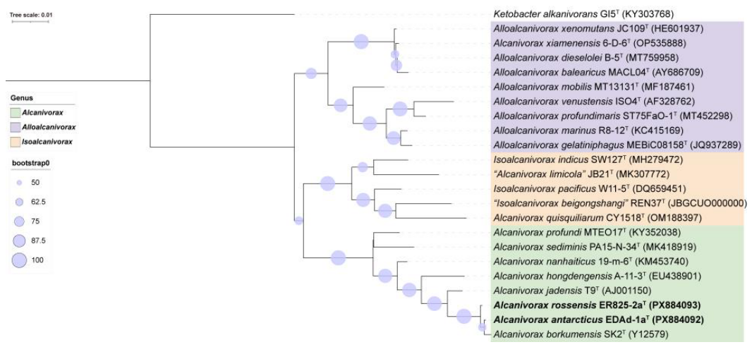
Figure 2.
Phylogenomic tree constructed on the basis of 120 conserved bacterial concatenated proteins showing the relationships between strains EDAd-1aT, ER825-2aT and all type strains of the genera Alcanivorax, Alloalcanivorax and Isoalcanivorax. The tree was rooted by the type strain Ketobacter alkanivorans GI5T (CP022684). The bootstrap values of nodes greater than 0.93 are displayed. The 0.1 bar indicates the number of amino acid substitutions per position.
Figure 2.
Phylogenomic tree constructed on the basis of 120 conserved bacterial concatenated proteins showing the relationships between strains EDAd-1aT, ER825-2aT and all type strains of the genera Alcanivorax, Alloalcanivorax and Isoalcanivorax. The tree was rooted by the type strain Ketobacter alkanivorans GI5T (CP022684). The bootstrap values of nodes greater than 0.93 are displayed. The 0.1 bar indicates the number of amino acid substitutions per position.
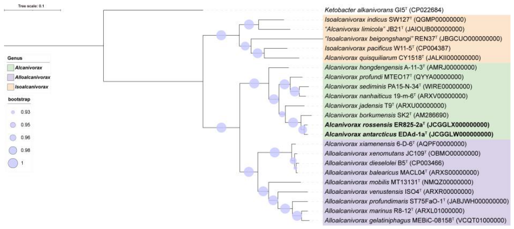
Figure 3.
General biological function profiles of the strains EDAd-1aT, ER825-2aT and their closest relatives based on genome analysis using the METABOLIC genome-scale workflow. The names of the functions in red represent the detected differences among the strains.
Figure 3.
General biological function profiles of the strains EDAd-1aT, ER825-2aT and their closest relatives based on genome analysis using the METABOLIC genome-scale workflow. The names of the functions in red represent the detected differences among the strains.
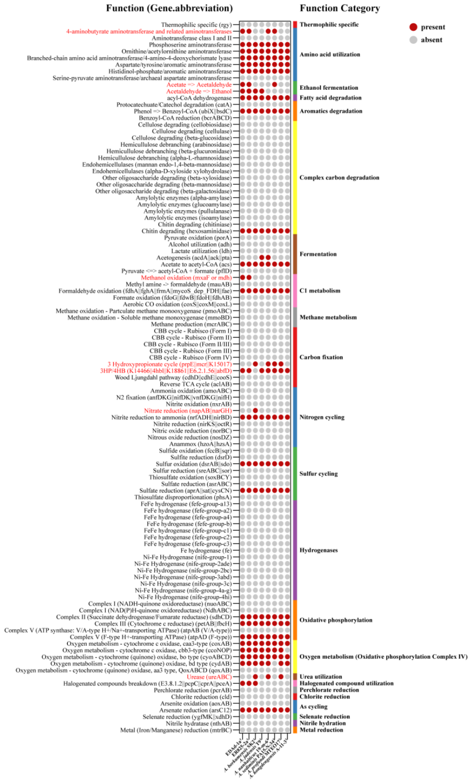
Figure 4.
Different KEGG modules detected among the strains EDAd-1aT, ER825-2aT and their closest relatives on the basis of genome analysis using the METABOLIC genome-scale workflow. The names of the KEGG module in red represent the detected differences among the strains.
Figure 4.
Different KEGG modules detected among the strains EDAd-1aT, ER825-2aT and their closest relatives on the basis of genome analysis using the METABOLIC genome-scale workflow. The names of the KEGG module in red represent the detected differences among the strains.
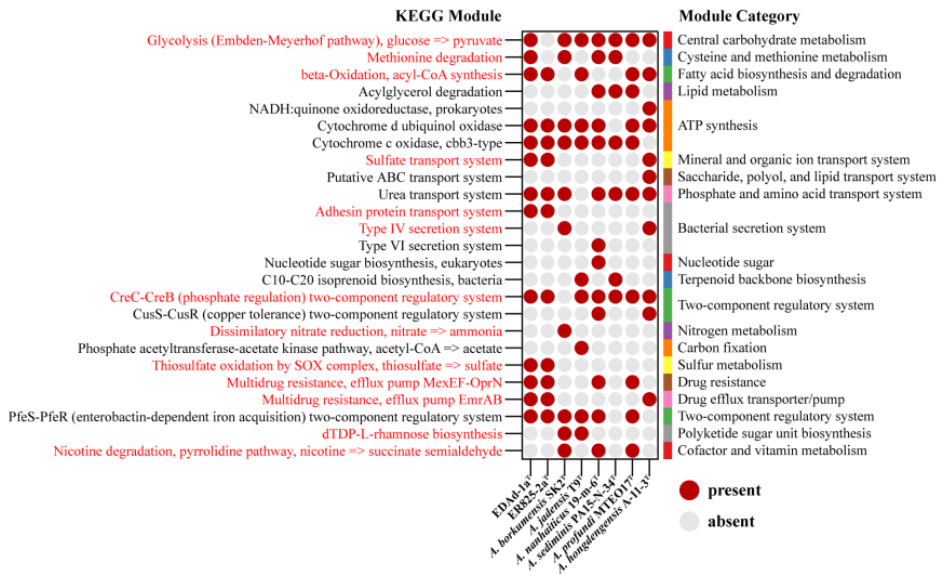
Figure 5.
Oxygen consumption of strains EDAd-1aT, ER825-2aT and their two closest relatives during the degradation of photosynthetic hydrocarbons at 28 °C for 3 weeks.
Figure 5.
Oxygen consumption of strains EDAd-1aT, ER825-2aT and their two closest relatives during the degradation of photosynthetic hydrocarbons at 28 °C for 3 weeks.
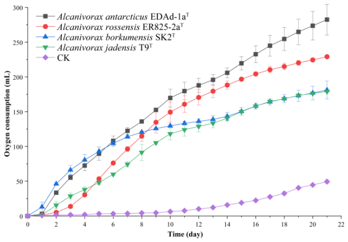

Table 1.
Phenotypic characteristics that differentiate Alcanivorax antarcticus sp. nov. (strain EDAd-1aT) and Alcanivorax rossensis sp. nov. (ER825-2aT) from closely related species.
Table 1.
Phenotypic characteristics that differentiate Alcanivorax antarcticus sp. nov. (strain EDAd-1aT) and Alcanivorax rossensis sp. nov. (ER825-2aT) from closely related species.
| Characteristic | 1 | 2 | 3 | 4 | 5 | 6 | 7 | 8 |
| Isolation source | upper seawater of the Weddell Sea, Antarctica | upper seawater of the Ross Sea, Antarctica | sea water/sediment near the Isle of Borkum North Sea |
intertidal sediment of the North Sea | deep-sea sediment of the South China Sea | deep-sea sediment of the Pacific Ocean | deep seawater of the Mariana Trench | surface seawater of the Straits of Malacca |
| Temperature for growth (oC) | ` | |||||||
| Range | 4–45 | 4–45 | 4–35 | 10–40 | 4–41 | 10–45 | 10–45 | 4–42 |
| Optimum | 25–28 | 25–28 | 25–30 | 30 | 28 | 28–30 | 37 | 18–35 |
| pH for growth | ||||||||
| Range | 6.0–10.0 | 6.0–10.0 | nd | 6.0–8.5 | 6.0–12.0 | 6.0–10.0 | 6.0–10.0 | 4.0–10.0 |
| Optimum | 7.0 | 7.0 | nd | 7.0 | 6.0–7.0 | 7.0 | 7.0–8.0 | 7.0–8.0 |
| NaCl for growth (w/v, %) | ||||||||
| Range | 0.5–7.0 | 0.5–7.0 | 1.0–12.5 | 0.5–15.0 | 0.5–15.0 | 0–15.0 | 0–12.0 | 0.5–15.0 |
| Optimum | 2.0 | 2.0 | 3.0–10.0 | 3.0 | 3.0 | 3.0–5.0 | 3.0 | 3.0–8.0 |
| Oxygen requirement | aerobic | aerobic | aerobic | aerobic or anaerobic | aerobic | aerobic | aerobic | aerobic |
| Reduction of nitrate | - | - | + | + | - | - | - | + |
| Hydrolysis of | ||||||||
| Aesculin | - | - | - | - | - | - | + | - |
| Gelatin | - | - | - | - | - | + | + | + |
| Enzyme activities | ||||||||
| Catalase | + | + | + | + | + | + | - | - |
| Oxidase | + | + | + | + | + | + | + | - |
| Alkaline phosphatase | + | + | + | + | + | + | + | - |
| Lipase (C14) | + | + | + | + | + | + | - | + |
| Leucine aminopeptidase | + | + | + | + | - | + | + | + |
| Cystine aminopeptidase | w | w | + | w | - | w | - | - |
| Trypsin | w | w | w | - | - | - | - | - |
| α-Chymotrypsin | + | + | - | - | - | - | - | - |
| Naphtol-AS-Bl-phosphoamidase | + | + | w | + | + | - | + | w |
| Tryptophan decarboxylase | w | w | - | - | - | - | - | - |
| Gelatinase | - | - | - | + | + | + | + | + |
| Utilization | ||||||||
| N-acetylglucosamine | + | + | - | - | - | - | - | - |
| Potassium gluconate | + | + | - | - | w | - | - | - |
| Capric acid | - | - | - | - | + | - | - | - |
| Adipic acid | w | - | - | - | - | - | - | - |
| Malic acid | + | + | - | - | - | - | - | - |
| Polar lipids | DPG, PG, PE, PL, Al, L | PG, PE, DPG, PL, AL, L | PE, PG, DPG, PL, L | nd | PG, PE, AL,PL, GL, APL | PE, PG, DPG, AL, L | PE, PG, PL, L | nd |
| DNA G+C content (mol%)# | 58.28 | 58.29 | 54.70 | 58.73 | 56.44 | 57.15 | 57.48 | 60.72 |
Strains: 1, EDAd-1aT; 2, ER825-2aT; 3, A. borkumensis SK2T [9,42]; 4, A. jadensis T9T [42,43]; 5, A. nanhaiticus 19-m-6T [42,44]; 6, A. sediminis PA15-N-34T [42]; 7, A. profundi MTEO17T [42,45]; 8, A. hongdengensis A-11-3T [42,46]. The API 20NE, API 20E and API ZYM data for the first four strains were examined in this study. Characteristics are scored as follows: +, positive; –, negative; w, weakly positive; DPG, diphosphatidylglycerol; PG, phosphatidylglycerol; PE, phosphatidylethanolamine; PL, phospholipid; GL, glycolipid; APGL, aminophosphoglycolipid; AL, aminolipid; L, unknown polar lipid; nd, no data. #: data calculated on the basis of the genome sequences.
Disclaimer/Publisher’s Note: The statements, opinions and data contained in all publications are solely those of the individual author(s) and contributor(s) and not of MDPI and/or the editor(s). MDPI and/or the editor(s) disclaim responsibility for any injury to people or property resulting from any ideas, methods, instructions or products referred to in the content. |
© 2026 by the authors. Licensee MDPI, Basel, Switzerland. This article is an open access article distributed under the terms and conditions of the Creative Commons Attribution (CC BY) license (http://creativecommons.org/licenses/by/4.0/).
Copyright: This open access article is published under a Creative Commons CC BY 4.0 license, which permit the free download, distribution, and reuse, provided that the author and preprint are cited in any reuse.