
Submitted:
17 November 2026
Posted:
18 November 2026
You are already at the latest version
Abstract
Huntington’s disease (HD) is a progressive neurodegenerative disorder caused by a CAG repeat expansion in the huntingtin (HTT) gene. Although the genetic cause is well established, the mechanisms linking mutant huntingtin to neurodegeneration remain incompletely understood. Evidence from human studies and experimental models suggests that urea, ammonia, and related nitrogen metabolites are altered in HD. This review summarizes current evidence for cerebral urea accumulation, altered circulating urea cycle metabolites, and changes in ammonia homeo-stasis. We discuss possible explanations for these observations including altered cerebral nitrogen handling and changes in hepatic urea cycle regulation. However, the relative contribution of cen-tral and peripheral processes remains uncertain, and current data does not establish causality. Together, the available evidence suggests that altered nitrogen metabolism warrants further in-vestigation. Future studies should define the origin, timing, and biological significance of these changes to determine whether they contribute to HD pathophysiology and whether they repre-sent biomarkers or potential therapeutic targets.
Keywords:
Huntington’s disease
; nitrogen metabolism
; urea
; ammonia
; neurodegeneration
1. Introduction
Huntington’s disease (HD) is the most common monogenic neurodegenerative disorder [1,2]. It is inherited in an autosomal dominant manner and is caused by a CAG trinucleotide repeat expansion in exon one of the huntingtin (HTT) gene [3], resulting in expression of a toxic, polyglutamine-expanded mutant huntingtin protein (mHTT). Clinically, HD is characterized by progressive motor, cognitive and psychiatric decline, and neuropathologically by pronounced dysfunction and loss of striatal medium spiny neurons, with variable cortical and subcortical degeneration [2,4,5].
Although the genetic cause of HD has been known for over three decades, the molecular mechanisms through which mHTT drives neurodegeneration remain incompletely understood [6]. Glutamate excitotoxicity [7,8,9], mitochondrial dysfunction [2,10], and neuroinflammation [11,12] have all been implicated, yet therapeutic strategies targeting these broad processes have not yielded effective disease-modifying treatments [13,14,15]. A challenge is separating the primary effects of mHTT that initiate cell dysfunction, from the downstream cascades of events that result in cell death. Translation of potential therapies to the clinic is further complicated by practical factors such as timing of treatment relative to disease stage, availability of reliable end points and the statistical power of trials. The emergence of quantifiable biomarkers such as neurofilament light and mHTT levels in cerebrospinal fluid and plasma, together with huntingtin-lowering approaches have been significant advances [16, 17, 2[. Positive topline results from several recent clinical trials, most notably AMT-130, SKY-0515, votoplam and others [18] are providing renewed optimism in the HD community for a disease-modifying treatment. Despite this progress, ongoing challenges of delivery, efficacy and long-term safety mean there also remains a need to identify alternative therapies [16]. In addition to mHTT itself, good targets could include molecules and pathways directly regulated by mHTT which could cause pathogenic effects from an early disease stage. In this review we specifically examine evidence for altered function of the urea cycle, and its key metabolites urea and ammonia, as an early and pathogenic contributor to HD.
2. Urea Cycle Dysfunction and Its Neurological Manifestations
While not a clinical feature of the disease, altered levels of urea and urea cycle dysfunction have been reported in HD literature over several decades. Our laboratory and others have identified cerebral urea accumulation in the post-mortem HD brain, [19,20,21], altered levels of urea in patient biofluids [22], and evidence of impaired urea cycle function in patient tissues and experimental models of HD [23,24,25,26,27,28,29,30]. This evidence will be described in section 4.
Urea is a normal waste product of nitrogen metabolism and is synthesized in the body through a series of enzymatic reactions termed the urea cycle. The urea cycle functions to convert nitrogen in the form of ammonia into urea for excretion [31]. Nitrogen metabolism underpins many essential cellular processes, particularly protein synthesis and catabolism, and is fundamental to life [32]. In excess, however, both urea and ammonia are neurotoxic [33,34]. The acute effects of this neurotoxicity are seen clinically in uremic encephalopathy (UE), urea cycle disorders (UCDs) and hepatic encephalopathy (HE).
UE occurs when urea and other retained uremic solutes accumulate in the brain, typically due to acute or chronic kidney failure [35]. This can cause cerebral oedema, white matter damage, and neurochemical and vascular changes. Meanwhile UCDs and HE occur due to failure of processes in the liver, resulting in the accumulation of ammonia in the blood and brain [31,36]. UCD’s are caused by inherited partial or complete loss-of-function mutations in one of the six enzymes or two transporters constituting the urea cycle [31]. In contrast, HE represents a spectrum of neuropsychiatric disturbances secondary to hepatic dysfunction of diverse etiologies [36]. Despite their distinct origins, UE, UCDs and HE are unified by either elevated urea or hyperammonemia and impaired urea cycle activity [37,38,39], providing a useful comparative framework for understanding the potential neurodegenerative impact of similar metabolic alterations in HD (Table 1).
Notably, the neuropathology of UCDs and HE bear a striking resemblance to Huntington’s disease. Post-mortem analyses of HE and UCD patients reveal pronounced basal ganglia atrophy, including selective loss of medium spiny neurons, ventricular enlargement, and cortical thinning, while other brain regions remain relatively spared [40,41,42,43,44]. Neuroimaging and electrophysiological studies similarly identify morphological and functional abnormalities localized predominantly to the basal ganglia [36,37,44,45]. This regional vulnerability suggests that the basal ganglia are highly susceptible to the neurotoxic effects of ammonia and urea [36,44].
UCDs, HE, and HD also have similar clinical manifestations. Like in HD, patients with UCDs and HE commonly exhibit motor and psychiatric disturbances, including tremor, asterixis, depression, seizures, mood instability, sleep – wake disruption, and impaired cognitive and executive function [1,2,34,36,39,46,47].
These similarities extend to cellular mechanisms implicated in HD pathogenesis. Experimental studies of hyperammonemia and liver failure implicate impaired astrocytic glutamate clearance and excessive NMDA receptor activation in ammonia neurotoxicity [48,49], paralleling reduced astrocytic glutamate transport and increased extra synaptic NMDA receptor signaling in HD models [50,51]. Excess ammonia can also impair mitochondrial function and promote oxidative stress [52,53], while chronic hyperammonemia induces microglial activation and neuroinflammation in experimental rodent models [54]. These effects overlap with the bioenergetic deficits and inflammatory responses associated with mHTT [55,56], suggesting potential convergence on pathways that increase neuronal vulnerability. Urea may exert additional effects on synaptic function: experimental cerebral urea accumulation has been linked to carbamylation-mediated suppression of mTOR signaling, impaired synaptic plasticity and synaptic loss [33]. Together, these findings provide a mechanistic rationale for investigating whether altered ammonia and urea metabolism could exacerbate neuronal dysfunction in HD.
Importantly, safe and affordable therapies exist that effectively modulate urea and ammonia metabolism and are in clinical use for uremia, UCDs and HE [32,47,57]. With this in mind, we examine here the evidence for dysregulated urea and ammonia metabolism in HD, evaluate its potential origins and connection to mHTT, and consider the feasibility of targeting these pathways therapeutically.
3. Physiological Metabolism of Ammonia and Urea
To explore the dysregulation of ammonia and urea metabolism in HD, it is important to understand physiological ammonia and urea metabolism. In mammals, ammonia (NH3) occupies a central position within nitrogen metabolism, serving as both a substrate and product in more than 20 metabolic reactions [57,58,59]. Additional exogenous sources of ammonia arise from dietary protein breakdown and gut microbial urease activity [32,37]. Mammals have evolved several detoxification mechanisms to maintain ammonia within a narrow physiological range [32,58], with some differences in these processes between the brain and peripheral tissues as described in sections 3.1 - 3.3.
3.1. Peripheral Metabolism of Ammonia
The detoxification of circulating and enterically absorbed ammonia primarily occurs in the liver [34] via two interconnected pathways: the urea cycle and the hepatic glutamine-glutamate cycle (Figure 1).
The urea cycle (Figure 1) operates largely within periportal hepatocytes [60] and is a low-affinity, high-flux enzymatic pathway [61]. Ammonia enters the cycle through the rate-limiting enzyme carbamyl phosphate synthetase I (CPS1) and requires N-acetylglutamate as an essential cofactor [60,62]. N-acetylglutamate is synthesized from glutamate by the enzyme N-acetylglutamate synthetase (NAGS). Subsequent reactions are sequentially catalyzed by ornithine transcarbamylase (OTC), argininosuccinate synthetase (ASS1), argininosuccinate lyase (ASL), and arginase I (ARG1). In the final step arginine is metabolized to urea and ornithine by ARG1 [62]. Reactions catalyzed by CPS1, OTC, and NAGS occur within the mitochondria, requiring the transport proteins ornithine translocase 1 (ORNT1) and aspartate-glutamate carrier (AGC) [32,59].
Ammonia not captured by periportal detoxification is removed by glutamine synthetase (GLUL) in a subset of pericentral hepatocytes, through a high-affinity, low-flux system [61] known as the hepatic glutamine-glutamate cycle (Figure 1). The enzyme glutamine synthetase (GLUL) catalyzes the conversion of ammonia and glutamate into glutamine. Glutamine may then be exported from the cell via glutamine transporters, or the reverse reaction catalyzed by glutaminase (GLS) which hydrolyses glutamine back into ammonia and glutamate.
A third reaction which influences activity of the glutamine-glutamate cycle within pericentral hepatocytes is the reversible interconversion of glutamate and α-ketoglutarate catalyzed by glutamate dehydrogenase (GLUD) with concomitant ammonia release or fixation [34,63]. Outside the liver, ammonia detoxification also occurs through GLUL activity in extrahepatic tissues, particularly skeletal muscle, and through direct renal excretion [32,61].
3.2. Cerebral Ammonia Metabolism
At least fourteen metabolic reactions are known to occur in the brain that either use or produce ammonia [64]. Ammonia readily crosses the blood-brain barrier (BBB) by passive diffusion and via several transporters [38,39,46,65]. The canonical urea cycle is largely absent in the brain due to low expression of CPS1 and OTC, and thus the brain uses other pathways [59,60,66].
Cerebral ammonia detoxification primarily occurs through glutamine-glutamate cycling (Figure 2). GLUL combines ammonia and glutamate to form glutamine, with cerebral GLUL expression being largely restricted to astrocytes [34]. Glutamine produced is exported to neurons, where GLS hydrolyses it back to glutamate and ammonia [46]. Additional ammonia metabolism occurs through GLUD activity, which typically favors the production of α-ketoglutarate and ammonia from glutamate, but can reverse to consume ammonia when concentrations rise [64,67].
Glutamate produced within neurons plays a central role in neuronal activity as the predominant excitatory neurotransmitter [64]. It also serves as a precursor for γ-aminobutyric acid (GABA), the major inhibitory neurotransmitter [67], and for α-ketoglutarate which initiates the tricarboxylic acid (TCA) cycle, linking neurotransmission with energy metabolism [34]. GABA and glutamate released into the synapse by neurons are subsequently taken up by astrocytes and converted back into glutamine, completing the glutamine-glutamate cycle [67]. This cycle therefore functions not only in ammonia detoxification, but also as a key regulator of neurotransmission and cerebral metabolism [67].
3.3. Cerebral Urea Production
Although the brain appears to lack a canonical urea cycle under normal physiological conditions, non-cyclic urea metabolism does occur, primarily via mitochondrial arginase II [59]. Arginase II (ARG2) generates ornithine and urea from arginine and has enriched expression in striatal GABAergic medium spiny neurons, which notably are the most vulnerable to neurodegeneration in HD [6,70]. Arginine may be produced within the brain through local protein catabolism or may enter from the periphery via protein catabolism or dietary sources. Urea can cross the BBB by passive diffusion [71] or be transported down its concentration gradient via the urea transporter UT-B [60,62].
Arginine in the brain may also be metabolized through the citrulline-nitric oxide (NO) cycle [44]. Here, the enzyme nitric oxide synthase (NOS) converts arginine to NO and citrulline, with citrulline then recycled back to arginine by ASS1 and ASL [62]. The NO generated in this process contributes to multiple neural functions, including synaptic plasticity and intercellular signaling [44].
4. Aberrant Metabolism of Urea and Ammonia in the HD Brain
Under normal physiological conditions the brain maintains extensive ammonia and urea metabolism despite lacking a complete urea cycle. These processes not only regulate cerebral ammonia levels, but also neurotransmitter cycling and other essential neural functions. This section reviews the evidence that these pathways are compromised in HD. A summary of findings from HD patients and animal models is provided in Figure 3.
4.1. Cerebral Urea Accumulation as a Hallmark of HD
In 2015 the widespread accumulation of urea in postmortem HD brain was discovered. Comparing mass spectrometry profiles from nine HD cases (Vonsattel neuropathological grades 1-4) with nine non-HD controls, urea levels were found to be approximately 3-fold higher on average in HD cases across all eleven brain regions studied [19]. Since urea is a byproduct of protein catabolism, it is reasonable to consider that the cerebral urea accumulation observed in the HD cases may have arisen simply because of cell loss and subsequent protein breakdown [19,58]. Indeed, elevated cerebral urea levels have also been identified in post-mortem brain tissue from other neurodegenerative conditions including Alzheimer’s disease (AD), Parkinson’s disease (PD), dementia with Lewy bodies, vascular dementia, frontal temporal dementia and amyotrophic lateral sclerosis [72,73,74,75,76] demonstrating that this is a phenomenon of brain tissue in end-stage neurodegenerative disease. Interestingly however, when we quantified urea in post-mortem brain tissue (cerebellum, super frontal gyrus) from individuals with minimal neurodegeneration (Vonsattel neuropathological grade 0/1) urea was still observed to be ~2 fold higher in HD cases compared to controls [21].
Additionally, in the OVT73 sheep model which expresses a full-length human huntingtin transgene with a 73-unit polyglutamine-coding repeat [77] striatal urea levels were found to be 1.5-fold higher on average than controls [21]. This is notable because the OVT73 sheep studied were prodromal and showed no overt neuronal loss. Urea accumulation in the OVT73 brain was also restricted to the striatum and not observed in other tissues studied (cerebellum, motor cortex, hippocampus, liver, blood) [21]. Together these observations of elevated urea in brain tissue with minimal cell loss (neuropathological grade 0/1 HD patients and an animal model) suggest that cerebral urea accumulation in HD may not solely be a consequence of protein catabolism following cell loss. The findings from the OVT73 sheep further suggest that urea accumulation may begin early in the disease and support the striatum as the most vulnerable region [21].
4.2. Elevated Cerebral Urea: Increased Production or Failure to Clear?
If cerebral urea does accumulate from an early disease stage an important question is what is its source? One possibility is defective clearance of urea from the brain. Urea transport across the blood brain barrier occurs by facilitated diffusion down its concentration gradient through the urea transporter UT-B [78,79,80]. In mammals UT-B is primarily expressed in erythrocytes, bladder, and kidney, but is also present in the brain where it is enriched in astrocytes [81,82]. UT-B specifically transports urea, water, and analogues of urea [80]. Upregulation of UT-B (encoded by SLC14A1) has been reported in the HD brain alongside urea accumulation [21]. SLC14A1 was also the most significantly differentially expressed gene in a seminal microarray study of HD patient caudate nucleus [83]. The relationship between UT-B expression and urea levels in the brain has not yet been fully elucidated so it is unclear whether upregulation of UTB in the HD brain is a response to elevated cerebral urea. In animal models, however, brain UT-B expression is reported to increase in response to osmotic pressure [82], and in peripheral tissues it has been shown to respond directly to dietary nitrogen and urea load [84,85].
Alternatively, cerebral urea accumulation may reflect its increased production. The brain produces urea through a non-canonical urea cycle focused on the metabolism of arginine to urea and ornithine, catalyzed by Arginase II (refer to section 3.3). Relatively few studies to date have reported ornithine or arginine levels in brain tissue or CSF from HD patients, and results are inconsistent. In a GC-MS metabolite profiling study of post-mortem HD patient brain tissue Patassini [20] found reduced levels of ornithine which were statistically significant in three of the eleven brain regions studied (putamen, globus pallidus, hippocampus). Interestingly this reduction in ornithine was identified in the same HD cases where urea was elevated [19]. Elevated urea alongside reduced ornithine could support defective clearance of urea from the brain as a mechanism. The authors note however that ornithine is rapidly utilized in polyamine synthesis pathways and thus an increase in production is unlikely to be detected [20]. By contrast, an independent LC-MS metabolite profiling study of post-mortem striatum and frontal pole from HD cases (n=14) and non-HD controls (n=14) found no difference in ornithine levels [86]. An earlier NMR-analysis of the same cases also found significantly lower urea levels in the striatum of HD cases compared to controls [87]. These studies by Graham [86,87] and Patassini [19,20] used tissue from HD cases with similar neuropathological grade and an appropriate sample size, thus the difference in findings may be methodological or reflect individual variation.
As the immediate precursor to urea in the brain, determining cerebral arginine levels and arginase activity would be informative, but to our best knowledge these have not been assessed in HD patient cases. Interestingly, polyglutamine expansion has been shown to increase arginase activity in a length-dependent manner [88]. Cerebral arginase activity is also known to be highest in the striatum, the brain region most affected in HD. This is thought to reflect striatal enrichment of manganese, a known arginase cofactor [30]. Transcription of the arginine transporter SLC7A2 is also reported to be upregulated in brain tissue from HD patients and animal models [89]. While not yet reported for brain tissue, arginine has been evaluated in CSF from HD patients, which can be considered to represent a generalized status of the brain. One study compared the LC-MS metabolite profile of HD patients at different disease stages and found that arginine levels in CSF increased nominally with worsening disease [27]. However, a recent LC-MS study of CSF from the CHDI HD Clarity cohort found no differences in arginine or ornithine in HD participants compared to controls (16 pre-manifest, 16 manifest and 16 Late-manifest HD participants, and 12 control participants). The discrepancy in results between these LC-MS studies [22,27] may reflect differences in the study group sizes where McGarry [27] reported a pilot study with fewer participants (3-4 participants at each of stages I-III, and 1 participant at stage IV), while the larger number of participants assessed by Pfalzer and colleagues [26] may have captured more population variability. Interestingly, Pfalzer [22] reported an elevated CSF urea in this study, but this was only detected in the late-manifest cohort.
Acknowledging the challenges of studying post-mortem HD patient brain tissue and the inherent variability of HD, perhaps the strongest evidence of altered urea synthesis in the HD brain to date comes from mouse models. Citrulline, ornithine and arginine were all elevated in the striatum of the HD model YAC128 mice at a prodromal age (12 weeks) when assessed by LC-MS. Arginase II activity was also reduced in YAC128 mice compared to controls, but only at an advanced disease timepoint (42 weeks) [30]. Unfortunately, urea was not measured in these mice. However, an extensive mass spectrometry analysis of whole brain from R6/2 mice, an exon 1 only expressing model, revealed increased levels of arginine, ornithine and arginosuccinate in brain tissue from symptomatic mice (10 weeks of age), with no difference in urea or citrulline levels compared to wild-type controls [28]. These independent studies of two animal models which express distinct transgenes and disease progressions support that, under controlled conditions, mutant HTT can cause an upregulation of urea cycle metabolites in the mammalian brain. Elevated arginine provides a plausible source for locally elevated urea in the brain. Like urea, altered homeostasis of arginine and arginase activity is also reported for Alzheimer’s, Parkinsons’ disease and ALS, suggesting this is a shared feature of neurodegeneration, and may have potential as a broad therapeutic target (see [90] for recent review).
4.3. Cerebral Ammonia in HD
To date there have been no reports of ammonia levels in HD brain tissue or for any neurodegenerative disease, although one study reports elevated ammonia levels in CSF of individuals with AD [91]. A challenge is that ammonia is volatile and rapidly metabolized, meaning it must be measured promptly after tissue collection [37,64,92]. Several studies provide indirect evidence that cerebral ammonia may be elevated in HD, through changes observed in the astrocytic glutamine-glutamate cycling which is the primary method for ammonia metabolism in the brain (refer to section 3.2). This includes elevation of cerebral glutamine, which is known to correlate positively with cerebral ammonia levels, downregulation of GLS, and multiple glutamate transporters [93,94,95,96].
In addition, recent in vitro evidence indicates that astrocytes can upregulate CPS1 and OTC under neurodegenerative stress to establish a canonical urea cycle [97,98]. This phenomenon was observed in a model where amyloid-β fragments were introduced into astrocytic cell culture. In this instance up-regulation of CPS1 and OTC was proposed to be compensatory, mitigating ammonia accumulation secondary to amyloid-β–induced toxicity [97]. Understanding whether this theoretical mechanism also occurs in HD where toxic mutant HTT accumulates would be of value. Given its neurotoxic potential, direct measurement of ammonia in brain tissue and biofluids from HD patients is another important avenue for future research.
5. A Peripheral Urea Cycle Deficit in HD?
With the evidence for perturbation in urea metabolism in the CNS of HD patients and knowledge that urea is produced primarily in the liver, it is interesting to consider the possibility of mHTT exerting an effect on the urea cycle in peripheral tissue. In 2007, Chiang and colleagues [23] reported elevated circulating ammonia and citrulline in R6/2 and HdhCAG150 transgenic mouse models of HD, and were the first to suggest that ammonia toxicity may contribute to neurological deficits in HD. They also found that plasma citrulline levels were higher in symptomatic HD patients (n= 21) compared to healthy controls, providing early clinical evidence of peripheral urea cycle dysfunction. Subsequent mass spectrometry-based metabolite profiling has revealed elevated circulating citrulline and arginine across multiple animal models of HD, including prodromal ovine [29,99] and symptomatic rodent systems [28]. Metabolite profiling studies in patients have shown more variability, with several studies finding no statistically significant difference in levels of circulating urea cycle metabolites including citrulline, arginine, ornithine or urea between HD patients and non-HD controls [100,101,102,103]. See Figure 3 for a summary of findings from peripheral tissues and brain.
There is some evidence to suggest that changes in urea cycle metabolites may relate to HD stage. In a pilot study which used multiple metabolomic platforms to quantify metabolite profiles in plasma and CSF in individuals with manifest HD stage (I – IV), plasma arginine and citrulline levels, were reported to correlate positively with measures of advancing disease severity, while arginine levels in CSF were negatively correlated [27]. Subsequently the same laboratory compared plasma metabolite profiles of fast- and slow-progressing patients who were participants in the 2CARE clinical trial. In this study circulating levels of glutamine and urea-cycle intermediates could distinguish the two patient groups [26]. These two studies had small patient numbers and did not include non-HD controls so did not capture the same population variation of earlier studies.
Metabolite levels in plasma are affected by many factors such as diet, physical activity and circadian rhythm, so variability in HD patient plasma metabolite profiles is not unexpected [104,105,106,107]. Some studies have attempted to account for this variation through environmental control and the use of animal models [25,29,102]. Skene [29] performed LC-MS metabolomics on blood sampled hourly from OVT73 sheep under controlled diet and lighting conditions. Over a 25-hour period this study detected elevated arginine, citrulline and urea in OVT73 compared to wild-type [29]. By contrast, in a separate single-timepoint study no difference in plasma urea levels were found between OVT73 and wild type [21]. Another study examined levels of ammonia and citrulline specifically in thirty HD patients under dietary control in a hospital setting [25]. This study confirmed the author’s earlier findings [23] of elevated circulating citrulline in HD patients on a standardized protein diet for five days. To assess urea cycle function the participants were then given a high protein diet for five days. This resulted in a small increase in plasma citrulline in patients, while ammonia levels did not change leading the authors to conclude that the urea cycle was functionally intact in patients. To our knowledge this is the only report of ammonia levels in any tissue from HD patients. Intriguingly, whilst ammonia levels did not differ on average compared to non-HD controls or after the dietary change, five of the thirty patients were found to have circulating ammonia levels that were higher than the clinical reference used (>65ug/dL) [25]. The levels reported for these individuals may fit with mild-hyperammonemia [108,109].
6. mHTT: Driver of Hepatic Transcriptional Dysregulation?
Alterations in peripheral nitrogen metabolism can cause neurological dysfunction, as evidenced by the effects of hyperammonemia in Hepatic encephalopathy and urea cycle disorders (refer to section 2). Although the changes in peripheral nitrogen metabolism observed in HD patients and animal models do not appear to cause the same acute manifestations, a question is whether subclinical alterations in ammonia handling over the decades-long course of HD could contribute to or even initiate neuronal toxicity. The cause of peripheral changes in nitrogen metabolism in HD warrants further investigation. Generalized metabolic dysfunction provides one possible explanation. Cellular energy deficits, weight loss beginning before motor onset, and altered glucose, fatty acid and cholesterol utilization have been well described in HD [55,110,111,112,113,114]. Increased reliance on protein catabolism following these energy deficits [114] could therefore contribute to altered nitrogen metabolism in the brain and periphery.
mHTT may also exert specific effects on the pathways in the liver that are primarily responsible for ammonia clearance from the body. The liver is affected pathologically in HD. Reported abnormalities include steatosis in juvenile HD [115], impaired hepatic mitochondrial function [116,117], and altered circulating bile acid profiles [118]. Evidence of progressive mitochondrial dysfunction in premanifest mutation carriers suggests that hepatic involvement need not be restricted to advanced disease [117]. Moreover, the liver displays several molecular features relevant to HD pathogenesis. Hepatocytes taken from mouse models and liver tissue from patients both show substantial somatic instability, which correlates well with age of onset [119,120]. Profiling of isogenic human embryonic stem cell-derived hepatocytes identified CAG-length-dependent transcriptional changes, including changes in pathways associated with mitochondrial function and energy metabolism [121]. However, while these observations provide a broader context for a potential role for mHTT in driving hepatic dysfunction, they do not provide a causal link between the mutation and impaired ureagenesis.
More direct evidence connecting mHTT to hepatic nitrogen metabolism comes from studies of urea cycle regulation. In HD mouse models, reduced expression of hepatic urea cycle enzymes was associated with disruption of CCAAT/enhancer binding protein α (C/EBPα), a transcription factor regulating their expression [23,24]. Investigators identified reduced C/EBPα abundance and sequestration of the remaining protein within mHTT aggregates. Reporter assays in the human hepatic cell line HepG2 further demonstrated that mHTT-mediated suppression of ASL transcription depended on an intact C/EBPα-binding motif in the ASL promoter [23]. These findings support a mechanism whereby mHTT interferes with hepatic transcriptional regulation, reducing urea cycle enzyme expression and compromising ammonia disposal. Related work identified reduced expression and sequestration of PPARγ in R6/2 liver, extending this mechanism to a broader network of hepatic metabolic regulation [122].
Together, these observations support hepatic dysfunction as a plausible contributor to disturbed nitrogen metabolism in HD. Whether this contribution is sufficient to alter cerebral ammonia exposure or explain the accumulation of urea in the brain remains unresolved.
7. Potential Therapeutic Targeting of Ammonia and Urea Metabolism
Discovering the mechanisms through which up-regulation of urea and potentially ammonia metabolism occurs in HD, and their contribution to pathogenesis across the disease course is an area of active research. It is clear however that excess of either metabolite is neurotoxic. Enhancing their clearance could therefore be beneficial for HD, and potentially other neurodegenerative diseases where urea accumulation has also been observed including Alzheimer’s disease (see [72] for recent review).
Several FDA approved therapies are used clinically to modulate ammonia and urea metabolism in UCDs and HE, with the aim of reducing ammonia production and/or enhancing its detoxification and excretion [47,57]. The main treatment option involves nitrogen scavengers, like sodium benzoate, sodium phenylbutyrate, and sodium phenylacetate [38,47]. These compounds conjugate glycine or glutamine to sequester nitrogen, thereby reducing its availability for ammonia production and thus lowering systemic ammonia levels [32]. Other approaches include reducing ammonia-producing gut bacteria through antibiotics or non-absorbable disaccharides, supplementing deficient amino acids in UCDs, and performing liver transplantation in severe hyperammonemia [32,39,46,47,57]. Notably, most of these treatments act peripherally [32,47], circumventing the delivery challenges imposed by the BBB which is a significant limitation for many potential HD therapies currently in development [123].
Intriguingly, several nitrogen-scavenging agents have already been investigated as therapies in HD models, albeit for alternative reasons. Sodium butyrate [124], phenylbutyrate [125] and glycerol tribenzoate [126] have each been tested in mice because of their additional properties as histone deacetylase inhibitors (HDACi’s), as histone acetylation is reduced in HD [127]. As such, their effects on ammonia metabolism were not assessed in this research. However, all three compounds ameliorated disease pathology, reducing brain atrophy and neuroinflammation, while improving survival and motor function [124,125,126]. Taken together this implies that nitrogen scavengers may influence HD progression.
Investigation into the clinical translation of nitrogen scavengers in HD patients has thus far been limited. A phase I dose-finding trial demonstrated that sodium phenylbutyrate was well tolerated in HD patients [128], leading to the phase II PHEND-HD study to further assess safety and tolerability [127,129]. However, to our knowledge, results from this trial were never published. Progress in this area may have been hindered by the disappointing outcomes of other HDACi’s, such as valproate, which failed to produce clinical benefit in HD [127]. Notably, valproate lacks nitrogen-scavenging properties, leaving open the possibility that therapies capable of modulating peripheral ammonia and urea metabolism could still hold therapeutic potential.
8. Conclusions and Future Directions
A growing body of evidence indicates that urea, ammonia, and related nitrogen metabolites are altered in Huntington’s disease. Among the most consistent observations are cerebral urea accumulation and changes in circulating urea cycle metabolites, identified across human studies and multiple experimental models. Together, these findings suggest that altered nitrogen metabolism is a reproducible feature of HD biology. However, the extent, timing and mechanistic basis of these metabolic disturbances remain incompletely understood. An important gap in the field is the direct measurement of ammonia within the HD brain, which has been difficult to address because of ammonia’s volatility and rapid turnover.
The available evidence raises the possibility that altered ammonia and urea metabolism may contribute to HD pathophysiology rather than simply reflecting end-stage neurodegeneration. Comparisons with urea cycle disorders and hepatic encephalopathy, together with observations from HD patient tissues, biofluids and animal models, support this hypothesis but do not yet establish causality. Multiple mechanisms are plausible, including altered cerebral nitrogen metabolism, hepatic transcriptional dysregulation, impaired peripheral ammonia handling, or combinations of these processes. Determining the relative contribution of central and peripheral mechanisms will require carefully designed experimental studies.
Several key questions therefore emerge. First, whether ammonia is elevated within the HD brain and at what stage of disease this occurs. Second, whether peripheral abnormalities in nitrogen metabolism precede or follow cerebral metabolic changes. Third, whether liver-specific correction of urea cycle dysfunction or ammonia metabolism is sufficient to modify cerebral metabolite profiles, neuropathology or disease progression in experimental HD models. Finally, it will be important to determine whether alterations in urea and ammonia metabolism represent common downstream consequences of neurodegeneration or are more directly linked to mutant huntingtin biology.
Collectively, the evidence reviewed here suggests that altered nitrogen metabolism deserves greater attention as a component of HD pathophysiology. Importantly, pharmacological and dietary interventions capable of modifying ammonia and urea metabolism are already available and have established clinical safety in other disorders. Whether these approaches offer therapeutic benefit in HD remains unknown, but their availability provides an opportunity to test the biological significance of this pathway. Future studies integrating metabolomics, direct measurements of ammonia, genetically informative animal models, and interventions targeting peripheral nitrogen metabolism will be essential to determine whether altered urea and ammonia metabolism represents a modifiable contributor to disease progression or simply a biomarker of the underlying disease process.
Author Contributions
Conceptualization and investigation, J.T.H, A.J., R.G.S, and R.R.H.; writing—original draft preparation, J.T.H.; writing—review and editing, A.J. R.GS, and R.RH.; supervision, A.J., R.GS. and R.R.H. All authors have read and agreed to the published version of the manuscript.
Funding
This research received no external funding.
Institutional Review Board Statement
Not applicable.
Informed Consent Statement
Not applicable.
Data Availability Statement
No new data were created or analyzed in this study. Data sharing is not applicable to this article.
Acknowledgments
The authors would like to acknowledge the generous support of the University of Auckland Medical and Health Sciences Foundation (Leo Nilon Huntington’s disease Fellowship - through the Hudson-Nilon Medical Research Foundation), the Health Research Council of New Zealand (Explorer Grant 21/759); and support from the Maurice and Phyllis Paykel Trust, Auckland Medical Research Foundation, Freemasons Foundation and Freemasons of New Zealand, and the CHDI Foundation Inc.
Conflicts of Interest
The authors declare no conflicts of interest.
Abbreviations
The following abbreviations are used in this manuscript:
| HD | Huntington’s disease |
| mHTT | Mutant huntingtin protein |
| UE | Uremic encephalopathy |
| UCD | Urea cycle disorder |
| HE | Hepatic Encephalopathy |
| CSF | Cerebrospinal fluid |
| LC-MS | Liquid chromatography-mass spectrometry |
| GC-MS | Gas chromatography-mass spectrometry |
| AD | Alzheimer’s disease |
References
- Bates, G. P.; Dorsey, R.; Gusella, J. F.; Hayden, M. R.; Kay, C.; Leavitt, B. R.; Nance, M.; Ross, C. A.; Scahill, R. I.; Wetzel, R.; Wild, E. J.; Tabrizi, S. J. Huntington Disease. Nat. Rev. Dis. Primers 2015, 1(1), 15005. [Google Scholar] [CrossRef] [PubMed]
- Tabrizi, S. J.; Flower, M. D.; Ross, C. A.; Wild, E. J. Huntington Disease: New Insights into Molecular Pathogenesis and Therapeutic Opportunities. Nat. Rev. Neurol. 2020, 16(10), 529–546. [Google Scholar] [CrossRef] [PubMed]
- The Huntingon’s Disease Collaborative Research Group. A Novel Gene Containing a Trinucleotide Repeat That Is Expanded and Unstable on Huntington’s Disease Chromosomes. Cell 1993, 72(6), 971–983. [Google Scholar] [CrossRef] [PubMed]
- Rosas, H. D.; Salat, D. H.; Lee, S. Y.; Zaleta, A. K.; Pappu, V.; Fischl, B.; Greve, D.; Hevelone, N.; Hersch, S. M. Cerebral Cortex and the Clinical Expression of Huntington’s Disease: Complexity and Heterogeneity. Brain 2008, 131(4), 1057–1068. [Google Scholar] [CrossRef] [PubMed]
- Reiner, A.; Dragatsis, I.; Dietrich, P. Genetics and Neuropathology of Huntington’s Disease. In Pathophysiology, Pharmacology, and Biochemistry of Dyskinesia; International Review of Neurobiology; Brotchie, J., Bezard, E., Jenner, P., Eds.; Academic Press, 2011; Vol. 98, pp. 325–372. [Google Scholar] [CrossRef] [PubMed]
- Jiang, A.; Handley, R. R.; Lehnert, K.; Snell, R. G. From Pathogenesis to Therapeutics: A Review of 150 Years of Huntington’s Disease Research. Int. J. Mol. Sci. 2023, 24(16), 13021. [Google Scholar] [CrossRef] [PubMed]
- Albin, R. L.; Young, A. B.; Penney, J. B.; Handelin, B.; Balfour, R.; Anderson, K. D.; Markel, D. S.; Tourtellotte, W. W.; Reiner, A. Abnormalities of Striatal Projection Neurons and N-Methyl-D-Aspartate Receptors in Presymptomatic Huntington’s Disease. N. Engl. J. Med. 1990, 322(18), 1293–1298. [Google Scholar] [CrossRef] [PubMed]
- Beal, M. F.; Swartz, K. J.; Hyman, B. T.; Storey, E.; Finn, S. F.; Koroshetz, W. Aminooxyacetic Acid Results in Excitotoxin Lesions by a Novel Indirect Mechanism. J. Neurochem. 1991, 57(3), 1068–1073. [Google Scholar] [CrossRef] [PubMed]
- Jurcau, A. Molecular Pathophysiological Mechanisms in Huntington’s Disease. Biomedicines 2022, 10(6), 1432. [Google Scholar] [CrossRef] [PubMed]
- Verma, M.; Lizama, B. N.; Chu, C. T. Excitotoxicity, Calcium and Mitochondria: A Triad in Synaptic Neurodegeneration. Transl. Neurodegener. 2022, 11(1), 3. [Google Scholar] [CrossRef] [PubMed]
- Crotti, A.; Glass, C. K. The Choreography of Neuroinflammation in Huntington’s Disease. Trends Immunol. 2015, 36(6), 364–373. [Google Scholar] [CrossRef] [PubMed]
- Rocha, N. P.; Charron, O.; Latham, L. B.; Colpo, G. D.; Zanotti-Fregonara, P.; Yu, M.; Freeman, L.; Furr Stimming, E.; Teixeira, A. L. Microglia Activation in Basal Ganglia Is a Late Event in Huntington Disease Pathophysiology. Neurol. Neuroimmunol. Neuroinflammation 2021, 8(3), e984. [Google Scholar] [CrossRef] [PubMed]
- Ondo, W. G.; Mejia, N. I.; Hunter, C. B. A Pilot Study of the Clinical Efficacy and Safety of Memantine for Huntington’s Disease. Park. Relat. Disord. 2007, 13(7), 453–454. [Google Scholar] [CrossRef] [PubMed]
- van Diemen, M. P. J.; Hart, E. P.; Abbruscato, A.; Mead, L.; van Beelen, I.; Bergheanu, S. C.; Hameeteman, P. W.; Coppen, E.; Winder, J. Y.; Moerland, M.; Kan, H.; van der Grond, J.; Webb, A.; Roos, R. A. C.; Groeneveld, G. J. Safety, Pharmacokinetics and Pharmacodynamics of SBT-020 in Patients with Early Stage Huntington’s Disease, a 2-Part Study. Br. J. Clin. Pharmacol. 2021, 87(5), 2290–2302. [Google Scholar] [CrossRef] [PubMed]
- Roussakis, A.-A.; Gennaro, M.; Gordon, M. F.; Reilmann, R.; Borowsky, B.; Rynkowski, G.; Lao-Kaim, N. P.; Papoutsou, Z.; Savola, J.-M.; Hayden, M. R.; Owen, D. R.; Kalk, N.; Lingford-Hughes, A.; Gunn, R. N.; Searle, G.; Tabrizi, S. J.; Piccini, P. A PET-CT Study on Neuroinflammation in Huntington’s Disease Patients Participating in a Randomized Trial with Laquinimod. Brain Commun. 2023, 5(2), fcad084. [Google Scholar] [CrossRef] [PubMed]
- Tabrizi, S. J.; Estevez-Fraga, C.; van Roon-Mom, W. M. C.; Flower, M. D.; Scahill, R. I.; Wild, E. J.; Muñoz-Sanjuan, I.; Sampaio, C.; Rosser, A. E.; Leavitt, B. R. Potential Disease-Modifying Therapies for Huntington’s Disease: Lessons Learned and Future Opportunities. Lancet Neurol. 2022, 21(7), 645–658. [Google Scholar] [CrossRef] [PubMed]
- Tabrizi, S. J.; Ghosh, R.; Leavitt, B. R. Huntingtin Lowering Strategies for Disease Modification in Huntington’s Disease. Neuron 2019, 101(5), 801–819. [Google Scholar] [CrossRef] [PubMed]
- Farag, M.; Tabrizi, S. J.; Wild, E. J. Huntington’s Disease Clinical Trials Update: October 2025. J. Huntingt. Dis. 2025, 15(1), 156–166. [Google Scholar] [CrossRef] [PubMed]
- Patassini, S.; Begley, P.; Reid, S. J.; Xu, J.; Church, S. J.; Curtis, M.; Dragunow, M.; Waldvogel, H. J.; Unwin, R. D.; Snell, R. G.; Faull, R. L. M.; Cooper, G. J. S. Identification of Elevated Urea as a Severe, Ubiquitous Metabolic Defect in the Brain of Patients with Huntington’s Disease. Biochem. Biophys. Res. Commun. 2015, 468(1), 161–166. [Google Scholar] [CrossRef] [PubMed]
- Patassini, S.; Begley, P.; Xu, J.; Church, S. J.; Reid, S. J.; Kim, E. H.; Curtis, M. A.; Dragunow, M.; Waldvogel, H. J.; Snell, R. G.; Unwin, R. D.; Faull, R. L. M.; Cooper, G. J. S. Metabolite Mapping Reveals Severe Widespread Perturbation of Multiple Metabolic Processes in Huntington’s Disease Human Brain. Biochim. Et. Biophys. Acta (BBA) -Mol. Basis Dis. 2016, 1862(9), 1650–1662. [Google Scholar] [CrossRef] [PubMed]
- Handley, R. R.; Reid, S. J.; Brauning, R.; Maclean, P.; Mears, E. R.; Fourie, I.; Patassini, S.; Cooper, G. J. S.; Rudiger, S. R.; McLaughlan, C. J.; Verma, P. J.; Gusella, J. F.; MacDonald, M. E.; Waldvogel, H. J.; Bawden, C. S.; Faull, R. L. M.; Snell, R. G. Brain Urea Increase Is an Early Huntington’s Disease Pathogenic Event Observed in a Prodromal Transgenic Sheep Model and HD Cases. Proc. Natl. Acad. Sci. 2017, 114(52), E11293–E11302. [Google Scholar] [CrossRef] [PubMed]
- Pfalzer, A. C.; Shiino, S.; Silverman, J.; Codreanu, S. G.; Sherrod, S. D.; McLean, J. A.; Claassen, D. O. Alterations in Cerebrospinal Fluid Urea Occur in Late Manifest Huntington’s Disease. J. Huntington’s Dis. 2024, 13(1), 103–111. [Google Scholar] [CrossRef] [PubMed]
- Chiang, M.-C.; Chen, H.-M.; Lee, Y.-H.; Chang, H.-H.; Wu, Y.-C.; Soong, B.-W.; Chen, C.-M.; Wu, Y.-R.; Liu, C.-S.; Niu, D.-M.; Wu, J.-Y.; Chen, Y.-T.; Chern, Y. Dysregulation of C/EBPα by Mutant Huntingtin Causes the Urea Cycle Deficiency in Huntington’s Disease. Hum. Mol. Genet 2007, 16(5), 483–498. [Google Scholar] [CrossRef] [PubMed]
- Chiang, M.-C.; Chen, H.-M.; Lai, H.-L.; Chen, H.-W.; Chou, S.-Y.; Chen, C.-M.; Tsai, F.-J.; Chern, Y. The A2A Adenosine Receptor Rescues the Urea Cycle Deficiency of Huntington’s Disease by Enhancing the Activity of the Ubiquitin–Proteasome System. Hum. Mol. Genet 2009, 18(16), 2929–2942. [Google Scholar] [CrossRef] [PubMed]
- Chen, C.-M.; Lin, Y.-S.; Wu, Y.-R.; Chen, P.; Tsai, F.-J.; Yang, C.-L.; Tsao, Y.-T.; Chang, W.; Hsieh, I.-S.; Chern, Y.; Soong, B.-W. High Protein Diet and Huntington’s Disease. PLoS ONE 2015, 10(5), e0127654. [Google Scholar] [CrossRef] [PubMed]
- McGarry, A.; Hunter, K.; Gaughan, J.; Auinger, P.; Ferraro, T. N.; Pradhan, B.; Ferrucci, L.; Egan, J. M.; Moaddel, R. An Exploratory Metabolomic Comparison of Participants with Fast or Absent Functional Progression from 2CARE, a Randomized, Double-Blind Clinical Trial in Huntington’s Disease. Sci. Rep. 2024, 14(1), 1101. [Google Scholar] [CrossRef] [PubMed]
- McGarry, A.; Gaughan, J.; Hackmyer, C.; Lovett, J.; Khadeer, M.; Shaikh, H.; Pradhan, B.; Ferraro, T. N.; Wainer, I. W.; Moaddel, R. Cross-Sectional Analysis of Plasma and CSF Metabolomic Markers in Huntington’s Disease for Participants of Varying Functional Disability: A Pilot Study. Sci. Rep. 2020, 10(1), 20490. [Google Scholar] [CrossRef] [PubMed]
- Hashimoto, M.; Watanabe, K.; Miyoshi, K.; Koyanagi, Y.; Tadano, J.; Miyawaki, I. Multiplatform Metabolomic Analysis of the R6/2 Mouse Model of Huntington’s Disease. FEBS Open Bio 2021, 11(10), 2807–2818. [Google Scholar] [CrossRef] [PubMed]
- Skene, D. J.; Middleton, B.; Fraser, C. K.; Pennings, J. L. A.; Kuchel, T. R.; Rudiger, S. R.; Bawden, C. S.; Morton, A. J. Metabolic Profiling of Presymptomatic Huntington’s Disease Sheep Reveals Novel Biomarkers. Sci. Rep. 2017, 7(1), 43030. [Google Scholar] [CrossRef] [PubMed]
- Bichell, T. J. V.; Wegrzynowicz, M.; Tipps, K. G.; Bradley, E. M.; Uhouse, M. A.; Bryan, M.; Horning, K.; Fisher, N.; Dudek, K.; Halbesma, T.; Umashanker, P.; Stubbs, A. D.; Holt, H. K.; Kwakye, G. F.; Tidball, A. M.; Colbran, R. J.; Aschner, M.; Neely, M. D.; Di Pardo, A.; Maglione, V.; Osmand, A.; Bowman, A. B. Reduced Bioavailable Manganese Causes Striatal Urea Cycle Pathology in Huntington’s Disease Mouse Model. Biochim. Et. Biophys. Acta (BBA) -Mol. Basis Dis. 2017, 1863(6), 1596–1604. [Google Scholar] [CrossRef] [PubMed]
- Summar, M. L.; Mew, N. A. Inborn Errors of Metabolism with Hyperammonemia: Urea Cycle Defects and Related Disorders. Pediatr. Clin. N. Am. 2018, 65(2), 231–246. [Google Scholar] [CrossRef] [PubMed]
- Moedas, M. F.; Simões, R. J. M.; Silva, M. F. B. Mitochondrial Targets in Hyperammonemia: Addressing Urea Cycle Function to Improve Drug Therapies. Biochem. Pharmacol. 2024, 222, 116034. [Google Scholar] [CrossRef] [PubMed]
- Wang, H.; Huang, B.; Wang, W.; Li, J.; Chen, Y.; Flynn, T.; Zhao, M.; Zhou, Z.; Lin, X.; Zhang, Y.; Xu, M.; Li, K.; Tian, K.; Yuan, D.; Zhou, P.; Hu, L.; Zhong, D.; Zhu, S.; Li, J.; Chen, D.; Wang, K.; Liang, J.; He, Q.; Sun, J.; Shi, J.; Yan, L.; Sands, J. M.; Xie, Z.; Lian, X.; Xu, D.; Ran, J.; Yang, B. High Urea Induces Depression and LTP Impairment through mTOR Signalling Suppression Caused by Carbamylation. EBioMedicine 2019, 48, 478–490. [Google Scholar] [CrossRef] [PubMed]
- Limón, I. D.; Angulo-Cruz, I.; Sánchez-Abdon, L.; Patricio-Martínez, A. Disturbance of the Glutamate-Glutamine Cycle, Secondary to Hepatic Damage, Compromises Memory Function. Front. Neurosci. 2021, 15. [Google Scholar] [CrossRef] [PubMed]
- Rosner, M. H.; Husain-Syed, F.; Reis, T.; Ronco, C.; Vanholder, R. Uremic Encephalopathy. Kidney Int. 2022, 101(2), 227–241. [Google Scholar] [CrossRef] [PubMed]
- Rovira, A.; Alonso, J.; Córdoba, J. MR Imaging Findings in Hepatic Encephalopathy. Am. J. Neuroradiol. 2008, 29(9), 1612–1621. [Google Scholar] [CrossRef] [PubMed]
- Rose, C. F.; Amodio, P.; Bajaj, J. S.; Dhiman, R. K.; Montagnese, S.; Taylor-Robinson, S. D.; Vilstrup, H.; Jalan, R. Hepatic Encephalopathy: Novel Insights into Classification, Pathophysiology and Therapy. J. Hepatol. 2020, 73(6), 1526–1547. [Google Scholar] [CrossRef] [PubMed]
- Redant, S.; Empain, A.; Mugisha, A.; Kamgang, P.; Attou, R.; Honoré, P. M.; De Bels, D. Management of Late Onset Urea Cycle Disorders—a Remaining Challenge for the Intensivist? Ann. Intensive Care 2021, 11(1), 2. [Google Scholar] [CrossRef] [PubMed]
- Kido, J.; Nakamura, K. Hyperammonemia in Urea Cycle Disorders: A Toxic Metabolite for the Brain. Pediatr. Int. 2025, 67(1), e70121. [Google Scholar] [CrossRef] [PubMed]
- Dolman, C. L.; Clasen, R. A.; Dorovini-Zis, K. Severe Cerebral Damage in Ornithine Transcarbamylase Deficiency. Clin. Neuropathol. 1988, 7(1), 10–15. [Google Scholar] [PubMed]
- Taylor-Robinson, S. D.; Oatridge, A.; Hajnal, J. V.; Burroughs, A. K.; McIntyre, N.; deSouza, N. M. MR Imaging of the Basal Ganglia in Chronic Liver Disease: Correlation of T1-Weighted and Magnetisation Transfer Contrast Measurements with Liver Dysfunction and Neuropsychiatric Status. Metab. Brain Dis. 1995, 10(2), 175–188. [Google Scholar] [CrossRef] [PubMed]
- Takeoka, M.; Soman, T. B.; Shih, V. E.; Caviness, V. S.; Krishnamoorthy, K. S. Carbamyl Phosphate Synthetase 1 Deficiency: A Destructive Encephalopathy. Pediatr. Neurol. 2001, 24(3), 193–199. [Google Scholar] [CrossRef] [PubMed]
- Yamanouchi, H.; Yokoo, H.; Yuhara, Y.; Maruyama, K.; Sasaki, A.; Hirato, J.; Nakazato, Y. An Autopsy Case of Ornithine Transcarbamylase Deficiency. Brain Dev. 2002, 24(2), 91–94. [Google Scholar] [CrossRef] [PubMed]
- Cagnon, L.; Braissant, O. Hyperammonemia-Induced Toxicity for the Developing Central Nervous System. Brain Res. Rev. 2007, 56(1), 183–197. [Google Scholar] [CrossRef] [PubMed]
- Takanashi, J.; Barkovich, A. J.; Cheng, S. F.; Weisiger, K.; Zlatunich, C. O.; Mudge, C.; Rosenthal, P.; Tuchman, M.; Packman, S. Brain MR Imaging in Neonatal Hyperammonemic Encephalopathy Resulting from Proximal Urea Cycle Disorders. Am. J. Neuroradiol. 2003, 24(6), 1184–1187. [Google Scholar] [PubMed]
- Ochoa-Sanchez, R.; Tamnanloo, F.; Rose, C. F. Hepatic Encephalopathy: From Metabolic to Neurodegenerative. Neurochem Res. 2021, 46(10), 2612–2625. [Google Scholar] [CrossRef] [PubMed]
- Sen, K.; Whitehead, M.; Castillo Pinto, C.; Caldovic, L.; Gropman, A. Fifteen Years of Urea Cycle Disorders Brain Research: Looking Back, Looking Forward. Anal. Biochem 2022, 636, 114343. [Google Scholar] [CrossRef] [PubMed]
- Hermenegildo, C.; Marcaida, G.; Montoliu, C.; Grisolía, S.; Miñana, M. D.; Felipo, V. NMDA Receptor Antagonists Prevent Acute Ammonia Toxicity in Mice. Neurochem Res. 1996, 21(10), 1237–1244. [Google Scholar] [CrossRef] [PubMed]
- Knecht, K.; Michalak, A.; Rose, C.; Rothstein, J. D.; Butterworth, R. F. Decreased Glutamate Transporter (GLT-1) Expression in Frontal Cortex of Rats with Acute Liver Failure. Neurosci. Lett. 1997, 229(3), 201–203. [Google Scholar] [CrossRef] [PubMed]
- Faideau, M.; Kim, J.; Cormier, K.; Gilmore, R.; Welch, M.; Auregan, G.; Dufour, N.; Guillermier, M.; Brouillet, E.; Hantraye, P.; Déglon, N.; Ferrante, R. J.; Bonvento, G. In Vivo Expression of Polyglutamine-Expanded Huntingtin by Mouse Striatal Astrocytes Impairs Glutamate Transport: A Correlation with Huntington’s Disease Subjects. Hum. Mol. Genet 2010, 19(15), 3053–3067. [Google Scholar] [CrossRef] [PubMed]
- Milnerwood, A. J.; Gladding, C. M.; Pouladi, M. A.; Kaufman, A. M.; Hines, R. M.; Boyd, J. D.; Ko, R. W. Y.; Vasuta, O. C.; Graham, R. K.; Hayden, M. R.; Murphy, T. H.; Raymond, L. A. Early Increase in Extrasynaptic NMDA Receptor Signaling and Expression Contributes to Phenotype Onset in Huntington’s Disease Mice. Neuron 2010, 65(2), 178–190. [Google Scholar] [CrossRef] [PubMed]
- Kosenko, E.; Felipo, V.; Montoliu, C.; Grisolía, S.; Kaminsky, Y. Effects of Acute Hyperammonemia in Vivo on Oxidative Metabolism in Nonsynaptic Rat Brain Mitochondria. Metab. Brain Dis. 1997, 12(1), 69–82. [Google Scholar] [CrossRef] [PubMed]
- Bai, G.; Rama Rao, K. V.; Murthy, C. R.; Panickar, K. S.; Jayakumar, A. R.; Norenberg, M. D. Ammonia Induces the Mitochondrial Permeability Transition in Primary Cultures of Rat Astrocytes. J. Neurosci. Res. 2001, 66(5), 981–991. [Google Scholar] [CrossRef] [PubMed]
- Rodrigo, R.; Cauli, O.; Gomez-Pinedo, U.; Agusti, A.; Hernandez-Rabaza, V.; Garcia-Verdugo, J.-M.; Felipo, V. Hyperammonemia Induces Neuroinflammation That Contributes to Cognitive Impairment in Rats with Hepatic Encephalopathy. Gastroenterology 2010, 139(2), 675–684. [Google Scholar] [CrossRef] [PubMed]
- Seong, I. S.; Ivanova, E.; Lee, J.-M.; Choo, Y. S.; Fossale, E.; Anderson, M.; Gusella, J. F.; Laramie, J. M.; Myers, R. H.; Lesort, M.; MacDonald, M. E. HD CAG Repeat Implicates a Dominant Property of Huntingtin in Mitochondrial Energy Metabolism. Hum. Mol. Genet 2005, 14(19), 2871–2880. [Google Scholar] [CrossRef] [PubMed]
- Crotti, A.; Benner, C.; Kerman, B. E.; Gosselin, D.; Lagier-Tourenne, C.; Zuccato, C.; Cattaneo, E.; Gage, F. H.; Cleveland, D. W.; Glass, C. K. Mutant Huntingtin Promotes Autonomous Microglia Activation via Myeloid Lineage-Determining Factors. Nat. Neurosci. 2014, 17(4), 513–521. [Google Scholar] [CrossRef] [PubMed]
- Ribas, G. S.; Lopes, F. F.; Deon, M.; Vargas, C. R. Hyperammonemia in Inherited Metabolic Diseases. Cell Mol. Neurobiol. 2022, 42(8), 2593–2610. [Google Scholar] [CrossRef] [PubMed]
- Natesan, V.; Mani, R.; Arumugam, R. Clinical Aspects of Urea Cycle Dysfunction and Altered Brain Energy Metabolism on Modulation of Glutamate Receptors and Transporters in Acute and Chronic Hyperammonemia. Biomed. Pharmacother. 2016, 81, 192–202. [Google Scholar] [CrossRef] [PubMed]
- Griffin, J. W. D.; Bradshaw, P. C. Amino Acid Catabolism in Alzheimer’s Disease Brain: Friend or Foe? Oxidative Med. Cell. Longev. 2017, 2017(1), 5472792. [Google Scholar] [CrossRef] [PubMed]
- Wang, H.; Ran, J.; Jiang, T. Urea. Subcell. Biochem 2014, 73, 7–29. [Google Scholar] [CrossRef] [PubMed]
- Hakvoort, T. B. M.; He, Y.; Kulik, W.; Vermeulen, J. L. M.; Duijst, S.; Ruijter, J. M.; Runge, J. H.; Deutz, N. E. P.; Koehler, S. E.; Lamers, W. H. Pivotal Role of Glutamine Synthetase in Ammonia Detoxification. Hepatology 2017, 65(1), 281–293. [Google Scholar] [CrossRef] [PubMed]
- Morris, S. M., Jr. Regulation of Enzymes of the Urea Cycle and Arginine Metabolism. Annu. Rev. Nutr. 2002, 22(1), 87–105. [Google Scholar] [CrossRef] [PubMed]
- Zhou, Y.; Eid, T.; Hassel, B.; Danbolt, N. C. Novel Aspects of Glutamine Synthetase in Ammonia Homeostasis. Neurochem. Int. 2020, 140, 104809. [Google Scholar] [CrossRef] [PubMed]
- Cooper, A. J. L. The Role of Glutamine Synthetase and Glutamate Dehydrogenase in Cerebral Ammonia Homeostasis. Neurochem Res. 2012, 37(11), 2439–2455. [Google Scholar] [CrossRef] [PubMed]
- Sørensen, M. Update on Cerebral Uptake of Blood Ammonia. Metab. Brain Dis. 2013, 28(2), 155–159. [Google Scholar] [CrossRef] [PubMed]
- Buniatian, H. Ch.; Davtian, M. A. Urea Synthesis in Brain. J. Neurochem. 1966, 13(8), 743–753. [Google Scholar] [CrossRef] [PubMed]
- Andersen, J. V. The Glutamate/GABA-Glutamine Cycle: Insights, Updates, and Advances. J. Neurochem. 2025, 169(3), e70029. [Google Scholar] [CrossRef] [PubMed]
- Baek, J. H.; Park, H.; Kang, H.; Kim, R.; Kang, J. S.; Kim, H. J. The Role of Glutamine Homeostasis in Emotional and Cognitive Functions. Int. J. Mol. Sci. 2024, 25(2), 1302. [Google Scholar] [CrossRef] [PubMed]
- Parkin, G. M.; Udawela, M.; Gibbons, A.; Dean, B. Glutamate Transporters, EAAT1 and EAAT2, Are Potentially Important in the Pathophysiology and Treatment of Schizophrenia and Affective Disorders. World J. Psychiatry 2018, 8(2), 51–63. [Google Scholar] [CrossRef] [PubMed]
- Nance, M. A. Huntington Disease: Clinical, Genetic, and Social Aspects. J. Geriatr. Psychiatry Neurol. 1998, 11(2), 61–70. [Google Scholar] [CrossRef] [PubMed]
- Sterns, R. H.; Silver, S. M.; Hix, J. K. Urea for Hyponatremia? Kidney Int. 2015, 87(2), 268–270. [Google Scholar] [CrossRef] [PubMed]
- Al-Thani, N. A.; Stewart, G. S.; Costello, D. A. The Role of the Urea Cycle in the Alzheimer’s Disease Brain. J. Neurochem 2025, 169(3), e70033. [Google Scholar] [CrossRef] [PubMed]
- Philbert, S. A.; Church, S. J.; Unwin, R. D.; Cooper, G. J. S. Elevated Urea Levels in Human Frontotemporal Dementia and Amyotrophic Lateral Sclerosis Post-Mortem Brain Tissue: Evidence of a Multi-Dementia Pathogenic Mechanism. Mol. Omics 2026, 22(4), aaiag019. [Google Scholar] [CrossRef] [PubMed]
- Scholefield, M.; Church, S. J.; Xu, J.; Patassini, S.; Roncaroli, F.; Hooper, N. M.; Unwin, R. D.; Cooper, G. J. S. Severe and Regionally Widespread Increases in Tissue Urea in the Human Brain Represent a Novel Finding of Pathogenic Potential in Parkinson’s Disease Dementia. Front. Mol. Neurosci. 2021, 14. [Google Scholar] [CrossRef] [PubMed]
- Scholefield, M.; Church, S. J.; Philbert, S.; Xu, J.; Patassini, S.; Cooper, G. J. S. Human Dementia with Lewy Bodies Brain Shows Widespread Urea Elevations. Park. Relat. Disord. 2024, 124, 107017. [Google Scholar] [CrossRef] [PubMed]
- Xu, J.; Begley, P.; Church, S. J.; Patassini, S.; Hollywood, K. A.; Jüllig, M.; Curtis, M. A.; Waldvogel, H. J.; Faull, R. L. M.; Unwin, R. D.; Cooper, G. J. S. Graded Perturbations of Metabolism in Multiple Regions of Human Brain in Alzheimer’s Disease: Snapshot of a Pervasive Metabolic Disorder. Biochim Biophys. Acta 2016, 1862(6), 1084–1092. [Google Scholar] [CrossRef] [PubMed]
- Jacobsen, J. C.; Bawden, C. S.; Rudiger, S. R.; McLaughlan, C. J.; Reid, S. J.; Waldvogel, H. J.; MacDonald, M. E.; Gusella, J. F.; Walker, S. K.; Kelly, J. M.; Webb, G. C.; Faull, R. L. M.; Rees, M. I.; Snell, R. G. An Ovine Transgenic Huntington’s Disease Model. Hum. Mol. Genet 2010, 19(10), 1873–1882. [Google Scholar] [CrossRef] [PubMed]
- Fröhlich, O.; Macey, R. I.; Edwards-Moulds, J.; Gargus, J. J.; Gunn, R. B. Urea Transport Deficiency in Jk(a-b-) Erythrocytes. Am. J. Physiol. 1991, 260 4 Pt 1, C778–783. [Google Scholar] [CrossRef] [PubMed]
- Sands, J. M.; Gargus, J. J.; Fröhlich, O.; Gunn, R. B.; Kokko, J. P. Urinary Concentrating Ability in Patients with Jk(a-b-) Blood Type Who Lack Carrier-Mediated Urea Transport. J. Am. Soc. Nephrol. 1992, 2(12), 1689–1696. [Google Scholar] [CrossRef] [PubMed]
- Yu, L.; Liu, T.; Fu, S.; Li, L.; Meng, X.; Su, X.; Xie, Z.; Ren, J.; Meng, Y.; Lv, X.; Du, Y. Physiological Functions of Urea Transporter B. Pflug. Arch.-Eur. J. Physiol. 2019, 471(11), 1359–1368. [Google Scholar] [CrossRef] [PubMed]
- Zhang, Y.; Chen, K.; Sloan, S. A.; Bennett, M. L.; Scholze, A. R.; O’Keeffe, S.; Phatnani, H. P.; Guarnieri, P.; Caneda, C.; Ruderisch, N.; Deng, S.; Liddelow, S. A.; Zhang, C.; Daneman, R.; Maniatis, T.; Barres, B. A.; Wu, J. Q. An RNA-Sequencing Transcriptome and Splicing Database of Glia, Neurons, and Vascular Cells of the Cerebral Cortex. J. Neurosci. 2014, 34(36), 11929–11947. [Google Scholar] [CrossRef] [PubMed]
- Huang, B.; Wang, H.; Zhong, D.; Meng, J.; Li, M.; Yang, B.; Ran, J. Expression of Urea Transporter B in Normal and Injured Brain. Front Neuroanat. 2021, 15, 591726. [Google Scholar] [CrossRef] [PubMed]
- Hodges, A.; Strand, A. D.; Aragaki, A. K.; Kuhn, A.; Sengstag, T.; Hughes, G.; Elliston, L. A.; Hartog, C.; Goldstein, D. R.; Thu, D.; Hollingsworth, Z. R.; Collin, F.; Synek, B.; Holmans, P. A.; Young, A. B.; Wexler, N. S.; Delorenzi, M.; Kooperberg, C.; Augood, S. J.; Faull, R. L. M.; Olson, J. M.; Jones, L.; Luthi-Carter, R. Regional and Cellular Gene Expression Changes in Human Huntington’s Disease Brain. Hum. Mol. Genet 2006, 15(6), 965–977. [Google Scholar] [CrossRef] [PubMed]
- Inoue, H.; Kozlowski, S. D.; Klein, J. D.; Bailey, J. L.; Sands, J. M.; Bagnasco, S. M. Regulated Expression of Renal and Intestinal UT-B Urea Transporter in Response to Varying Urea Load. Am. J. Physiol.-Ren. Physiol. 2005, 289(2), F451–F458. [Google Scholar] [CrossRef] [PubMed]
- Simmons, N. L.; Chaudhry, A. S.; Graham, C.; Scriven, E. S.; Thistlethwaite, A.; Smith, C. P.; Stewart, G. S. Dietary Regulation of Ruminal Bovine UT-B Urea Transporter Expression and Localization. J. Anim. Sci. 2009, 87(10), 3288–3299. [Google Scholar] [CrossRef] [PubMed]
- Graham, S. F.; Pan, X.; Yilmaz, A.; Macias, S.; Robinson, A.; Mann, D.; Green, B. D. Targeted Biochemical Profiling of Brain from Huntington’s Disease Patients Reveals Novel Metabolic Pathways of Interest. Biochim Biophys. Acta Mol. Basis Dis. 2018, 1864(7), 2430–2437. [Google Scholar] [CrossRef] [PubMed]
- Graham, S. F.; Kumar, P.; Bahado-Singh, R. O.; Robinson, A.; Mann, D.; Green, B. D. Novel Metabolite Biomarkers of Huntington’s Disease As Detected by High-Resolution Mass Spectrometry. J. Proteome Res. 2016, 15(5), 1592–1601. [Google Scholar] [CrossRef] [PubMed]
- Colton, C. A.; Xu, Q.; Burke, J. R.; Bae, S. Y.; Wakefield, J. K.; Nair, A.; Strittmatter, W. J.; Vitek, M. P. Disrupted Spermine Homeostasis: A Novel Mechanism in Polyglutamine-Mediated Aggregation and Cell Death. J. Neurosci. 2004, 24(32), 7118–7127. [Google Scholar] [CrossRef] [PubMed]
- Gaudet, I. D.; Xu, H.; Gordon, E.; Cannestro, G. A.; Lu, M. L.; Wei, J. Elevated SLC7A2 Expression Is Associated with an Abnormal Neuroinflammatory Response and Nitrosative Stress in Huntington’s Disease. J. Neuroinflammation 2024, 21(1), 59. [Google Scholar] [CrossRef] [PubMed]
- Nalepa, M.; Skweres, A.; Węgrzynowicz, M. Dysregulation of Arginase and Arginine Pathways in Neurodegenerative Diseases: Metabolic and Cellular Dysfunction and Therapeutic Implications. Free Radic. Biol. Med. 2026, 252, 559–579. [Google Scholar] [CrossRef] [PubMed]
- Kaiser, E.; Schoenknecht, P.; Kassner, S.; Hildebrandt, W.; Kinscherf, R.; Schroeder, J. Cerebrospinal Fluid Concentrations of Functionally Important Amino Acids and Metabolic Compounds in Patients with Mild Cognitive Impairment and Alzheimer’s Disease. Neurodegener. Dis. 2010, 7(4), 251–259. [Google Scholar] [CrossRef] [PubMed]
- Cooper, A. J.; McDonald, J. M.; Gelbard, A. S.; Gledhill, R. F.; Duffy, T. E. The Metabolic Fate of 13N-Labeled Ammonia in Rat Brain. J. Biol. Chem. 1979, 254(12), 4982–4992. [Google Scholar] [CrossRef]
- Jenkins, B. G.; Klivenyi, P.; Kustermann, E.; Andreassen, O. A.; Ferrante, R. J.; Rosen, B. R.; Beal, M. F. Nonlinear Decrease over Time in N-Acetyl Aspartate Levels in the Absence of Neuronal Loss and Increases in Glutamine and Glucose in Transgenic Huntington’s Disease Mice. J. Neurochem 2000, 74(5), 2108–2119. [Google Scholar] [CrossRef] [PubMed]
- Behrens, P. F.; Franz, P.; Woodman, B.; Lindenberg, K. S.; Landwehrmeyer, G. B. Impaired Glutamate Transport and Glutamate–Glutamine Cycling: Downstream Effects of the Huntington Mutation. Brain 2002, 125(8), 1908–1922. [Google Scholar] [CrossRef] [PubMed]
- Jiang, R.; Diaz-Castro, B.; Looger, L. L.; Khakh, B. S. Dysfunctional Calcium and Glutamate Signaling in Striatal Astrocytes from Huntington’s Disease Model Mice. J. Neurosci. 2016, 36(12), 3453–3470. [Google Scholar] [CrossRef] [PubMed]
- Rebec, G. V. Corticostriatal Network Dysfunction in Huntington’s Disease: Deficits in Neural Processing, Glutamate Transport, and Ascorbate Release. CNS Neurosci. Ther. 2018, 24(4), 281–291. [Google Scholar] [CrossRef] [PubMed]
- Ju, Y. H.; Bhalla, M.; Hyeon, S. J.; Oh, J. E.; Yoo, S.; Chae, U.; Kwon, J.; Koh, W.; Lim, J.; Park, Y. M.; Lee, J.; Cho, I.-J.; Lee, H.; Ryu, H.; Lee, C. J. Astrocytic Urea Cycle Detoxifies Aβ-Derived Ammonia While Impairing Memory in Alzheimer’s Disease. Cell Metab. 2022, 34(8), 1104–1120.e8. [Google Scholar] [CrossRef] [PubMed]
- Zhang, S.; Wan, G.; Qiu, Y.; Zhang, M.; Deng, H.; Wang, Q.; Hu, J.; Gui, J.; Chen, D.; Huang, B.; Ran, J. Urea Cycle Dysregulation Drives Metabolic Stress and Neurodegeneration in Parkinson’s Disease. npj Park. Dis. 2025, 11(1), 237. [Google Scholar] [CrossRef] [PubMed]
- Spick, M.; Hancox, T. P. M.; Chowdhury, N. R.; Middleton, B.; Skene, D. J.; Morton, A. J. Metabolomic Analysis of Plasma in Huntington’s Disease Transgenic Sheep (Ovis Aries) Reveals Progressive Circadian Rhythm Dysregulation. J. Huntingt. Dis. 2023, 12(1), 31–42. [Google Scholar] [CrossRef] [PubMed]
- Underwood, B. R.; Broadhurst, D.; Dunn, W. B.; Ellis, D. I.; Michell, A. W.; Vacher, C.; Mosedale, D. E.; Kell, D. B.; Barker, R. A.; Grainger, D. J.; Rubinsztein, D. C. Huntington Disease Patients and Transgenic Mice Have Similar Pro-Catabolic Serum Metabolite Profiles. Brain 2006, 129(4), 877–886. [Google Scholar] [CrossRef] [PubMed]
- Gruber, B.; Kłaczkow, G.; Jaworska, M.; Krzysztoń-Russjan, J.; Anuszewska, E. L.; Zielonka, D.; Klimberg, A.; Marcinkowski, J. T. Huntington’ Disease – Imbalance of Amino Acid Levels in Plasma of Patients and Mutation Carriers. Ann. Agric. Environ. Med. 2013, 20(4), 779–783. [Google Scholar] [PubMed]
- Cheng, M.-L.; Chang, K.-H.; Wu, Y.-R.; Chen, C.-M. Metabolic Disturbances in Plasma as Biomarkers for Huntington’s Disease. J. Nutr. Biochem. 2016, 31, 38–44. [Google Scholar] [CrossRef] [PubMed]
- Mastrokolias, A.; Pool, R.; Mina, E.; Hettne, K. M.; van Duijn, E.; van der Mast, R. C.; van Ommen, G.; ‘t Hoen, P. A. C.; Prehn, C.; Adamski, J.; van Roon-Mom, W. Integration of Targeted Metabolomics and Transcriptomics Identifies Deregulation of Phosphatidylcholine Metabolism in Huntington’s Disease Peripheral Blood Samples. Metabolomics 2016, 12, 137. [Google Scholar] [CrossRef] [PubMed]
- Suchak, T.; Chowdhury, N. R.; Revell, V. L.; Isherwood, C.; Raynaud, F. I.; Veen; van der, D. R.; Geifman, N.; Skene, D. J.; Spick, M. Impact of Daily Rhythms and Postprandial Responses on the Plasma Metabolome. Int. J. Mol. Sci. 2026, 27(13), 5669. [Google Scholar] [CrossRef] [PubMed]
- Sato, S.; Parr, E. B.; Devlin, B. L.; Hawley, J. A.; Sassone-Corsi, P. Human Metabolomics Reveal Daily Variations under Nutritional Challenges Specific to Serum and Skeletal Muscle. Mol. Metab. 2018, 16, 1–11. [Google Scholar] [CrossRef] [PubMed]
- Kojouri, M.; Pinto, R.; Mustafa, R.; Huang, J.; Gao, H.; Elliott, P.; Tzoulaki, I.; Dehghan, A. Metabolome-Wide Association Study on Physical Activity. Sci. Rep. 2023, 13(1), 2374. [Google Scholar] [CrossRef] [PubMed]
- Tian, Q.; Corkum, A. E.; Moaddel, R.; Ferrucci, L. Metabolomic Profiles of Being Physically Active and Less Sedentary: A Critical Review. Metabolomics 2021, 17(7), 68. [Google Scholar] [CrossRef] [PubMed]
- Nimmana, B. K.; Rout, P. Hyperammonemia. In StatPearls; StatPearls Publishing: Treasure Island (FL), 2025. [Google Scholar]
- Ezgu, F. Chapter Seven - Inborn Errors of Metabolism. In Advances in Clinical Chemistry; Makowski, G. S., Ed.; Elsevier, 2016; Vol. 73, pp. 195–250. [Google Scholar] [CrossRef] [PubMed]
- Mochel, F.; Haller, R. G. Energy Deficit in Huntington Disease: Why It Matters. J. Clin. Invest 2011, 121(2), 493–499. [Google Scholar] [CrossRef] [PubMed]
- Singh, A.; Agrawal, N. Metabolism in Huntington’s Disease: A Major Contributor to Pathology. Metab. Brain Dis. 2022, 37(6), 1757–1771. [Google Scholar] [CrossRef] [PubMed]
- Antonini, A.; Leenders, K. L.; Spiegel, R.; Meier, D.; Vontobel, P.; Weigell-Weber, M.; Sanchez-Pernaute, R.; de Yébenez, J. G.; Boesiger, P.; Weindl, A.; Maguire, R. P. Striatal Glucose Metabolism and Dopamine D2 Receptor Binding in Asymptomatic Gene Carriers and Patients with Huntington’s Disease. Brain 1996, 119(6), 2085–2095. [Google Scholar] [CrossRef] [PubMed]
- Valenza, M.; Rigamonti, D.; Goffredo, D.; Zuccato, C.; Fenu, S.; Jamot, L.; Strand, A.; Tarditi, A.; Woodman, B.; Racchi, M.; Mariotti, C.; Donato, S. D.; Corsini, A.; Bates, G.; Pruss, R.; Olson, J. M.; Sipione, S.; Tartari, M.; Cattaneo, E. Dysfunction of the Cholesterol Biosynthetic Pathway in Huntington’s Disease. J. Neurosci. 2005, 25(43), 9932–9939. [Google Scholar] [CrossRef] [PubMed]
- Mochel, F.; Charles, P.; Seguin, F.; Barritault, J.; Coussieu, C.; Perin, L.; Bouc, Y. L.; Gervais, C.; Carcelain, G.; Vassault, A.; Feingold, J.; Rabier, D.; Durr, A. Early Energy Deficit in Huntington Disease: Identification of a Plasma Biomarker Traceable during Disease Progression. PLoS ONE 2007, 2(7), e647. [Google Scholar] [CrossRef] [PubMed]
- Squitieri, F.; Monti, L.; Graziola, F.; Colafati, G. S.; Sabatini, U. Early Liver Steatosis in Children with Pediatric Huntington Disease and Highly Expanded CAG Mutations. Park. Relat. Disord. 2022, 103, 102–104. [Google Scholar] [CrossRef] [PubMed]
- Stüwe, S. H.; Goetze, O.; Lukas, C.; Klotz, P.; Hoffmann, R.; Banasch, M.; Orth, M.; Schmidt, W. E.; Gold, R.; Saft, C. Hepatic Mitochondrial Dysfunction in Manifest and Premanifest Huntington Disease. Neurology 2013, 80(8), 743–746. [Google Scholar] [CrossRef] [PubMed]
- Hoffmann, R.; Stüwe, S. H.; Goetze, O.; Banasch, M.; Klotz, P.; Lukas, C.; Tegenthoff, M.; Beste, C.; Orth, M.; Saft, C. Progressive Hepatic Mitochondrial Dysfunction in Premanifest Huntington’s Disease. Mov. Disord. 2014, 29(6), 831–834. [Google Scholar] [CrossRef] [PubMed]
- Chiang, P.-I.; Chang, K.-H.; Tang, H.-Y.; Wu, Y.-R.; Cheng, M.-L.; Chen, C.-M. Diagnostic Potential of Alternations of Bile Acid Profiles in the Plasma of Patients with Huntington’s Disease. Metabolites 2024, 14(7), 394. [Google Scholar] [CrossRef]
- Lee, J.-M.; Pinto, R. M.; Gillis, T.; St Claire, J. C.; Wheeler, V. C. Quantification of Age-Dependent Somatic CAG Repeat Instability in Hdh CAG Knock-in Mice Reveals Different Expansion Dynamics in Striatum and Liver. PLoS ONE 2011, 6(8), e23647. [Google Scholar] [CrossRef] [PubMed]
- Mouro Pinto, R. M.; Arning, L.; Giordano, J. V.; Razghandi, P.; Andrew, M. A.; Gillis, T.; Correia, K.; Mysore, J. S.; Grote Urtubey, D.-M.; Parwez, C. R.; von Hein, S. M.; Clark, H. B.; Nguyen, H. P.; Förster, E.; Beller, A.; Jayadaev, S.; Keene, C. D.; Bird, T. D.; Lucente, D.; Vonsattel, J.-P.; Orr, H.; Saft, C.; Petrasch-Parwez, E.; Wheeler, V. C. Patterns of CAG Repeat Instability in the Central Nervous System and Periphery in Huntington’s Disease and in Spinocerebellar Ataxia Type 1. Hum. Mol. Genet 2020, 29(15), 2551–2567. [Google Scholar] [CrossRef] [PubMed]
- Ooi, J.; Langley, S. R.; Xu, X.; Utami, K. H.; Sim, B.; Huang, Y.; Harmston, N. P.; Tay, Y. L.; Ziaei, A.; Zeng, R.; Low, D.; Aminkeng, F.; Sobota, R. M.; Ginhoux, F.; Petretto, E.; Pouladi, M. A. Unbiased Profiling of Isogenic Huntington Disease hPSC-Derived CNS and Peripheral Cells Reveals Strong Cell-Type Specificity of CAG Length Effects. Cell Rep. 2019, 26(9), 2494–2508.e7. [Google Scholar] [CrossRef] [PubMed]
- Chiang, M.-C.; Chern, Y.; Juo, C.-G. The Dysfunction of Hepatic Transcriptional Factors in Mice with Huntington’s Disease. Biochim. Et. Biophys. Acta (BBA) -Mol. Basis Dis. 2011, 1812(9), 1111–1120. [Google Scholar] [CrossRef] [PubMed]
- Saade, J.; Mestre, T. A. Huntington’s Disease: Latest Frontiers in Therapeutics. Curr. Neurol. Neurosci. Rep. 2024, 24(8), 255–264. [Google Scholar] [CrossRef] [PubMed]
- Ferrante, R. J.; Kubilus, J. K.; Lee, J.; Ryu, H.; Beesen, A.; Zucker, B.; Smith, K.; Kowall, N. W.; Ratan, R. R.; Luthi-Carter, R.; Hersch, S. M. Histone Deacetylase Inhibition by Sodium Butyrate Chemotherapy Ameliorates the Neurodegenerative Phenotype in Huntington’s Disease Mice. J. Neurosci. 2003, 23(28), 9418–9427. [Google Scholar] [CrossRef] [PubMed]
- Gardian, G.; Browne, S. E.; Choi, D.-K.; Klivenyi, P.; Gregorio, J.; Kubilus, J. K.; Ryu, H.; Langley, B.; Ratan, R. R.; Ferrante, R. J.; Beal, M. F. Neuroprotective Effects of Phenylbutyrate in the N171-82Q Transgenic Mouse Model of Huntington’s Disease*. J. Biol. Chem. 2005, 280(1), 556–563. [Google Scholar] [CrossRef] [PubMed]
- Dutta, D.; Majumder, M.; Paidi, R. K.; Pahan, K. Alleviation of Huntington Pathology in Mice by Oral Administration of Food Additive Glyceryl Tribenzoate. Neurobiol. Dis. 2021, 153, 105318. [Google Scholar] [CrossRef] [PubMed]
- Gray, S. G. Targeting Huntington’s Disease through Histone Deacetylases. Clin. Epigenet. 2011, 2(2), 257–277. [Google Scholar] [CrossRef] [PubMed]
- Hogarth, P.; Lovrecic, L.; Krainc, D. Sodium Phenylbutyrate in Huntington’s Disease: A Dose-Finding Study. Mov. Disord. 2007, 22(13), 1962–1964. [Google Scholar] [CrossRef] [PubMed]
- Ebbel, E. N.; Leymarie, N.; Schiavo, S.; Sharma, S.; Gevorkian, S.; Hersch, S.; Matson, W. R.; Costello, C. E. Identification of Phenylbutyrate-Generated Metabolites in Huntington Disease Patients Using Parallel Liquid Chromatography/Electrochemical Array/Mass Spectrometry and off-Line Tandem Mass Spectrometry. Anal. Biochem. 2010, 399(2), 152–161. [Google Scholar] [CrossRef] [PubMed]
Figure 1.
Schematic of hepatic urea and glutamine-glutamate cycles. In periportal hepatocytes (A), ammonia enters the cell via passive diffusion and ammonia transporters or may be generated from glutamine and glutamate by glutaminase (GLS) and glutamate dehydrogenase (GLUD1). Ammonia enters the urea cycle through carbamoyl-phosphate synthetase I (CPS1), with subsequent steps catalyzed by ornithine transcarbamylase (OTC), argininosuccinate synthase I (ASS1), argininosuccinate lyase (ASL), and arginase I (ARG1) to form urea. Arginase II (ARG2) provides an additional non-urea cycle source of urea. Urea is exported via passive diffusion or urea transporters. In pericentral hepatocytes (B), residual ammonia is detoxified by glutamine synthetase (GLUL). Together, GLS, GLUD1, and GLUL form the glutamine-glutamate cycle. N-acetylglutamate synthetase (NAGS), GLS, ARG2, OTC, and GLUD1 localize to the mitochondria. Created in Biorender.com.
Figure 1.
Schematic of hepatic urea and glutamine-glutamate cycles. In periportal hepatocytes (A), ammonia enters the cell via passive diffusion and ammonia transporters or may be generated from glutamine and glutamate by glutaminase (GLS) and glutamate dehydrogenase (GLUD1). Ammonia enters the urea cycle through carbamoyl-phosphate synthetase I (CPS1), with subsequent steps catalyzed by ornithine transcarbamylase (OTC), argininosuccinate synthase I (ASS1), argininosuccinate lyase (ASL), and arginase I (ARG1) to form urea. Arginase II (ARG2) provides an additional non-urea cycle source of urea. Urea is exported via passive diffusion or urea transporters. In pericentral hepatocytes (B), residual ammonia is detoxified by glutamine synthetase (GLUL). Together, GLS, GLUD1, and GLUL form the glutamine-glutamate cycle. N-acetylglutamate synthetase (NAGS), GLS, ARG2, OTC, and GLUD1 localize to the mitochondria. Created in Biorender.com.
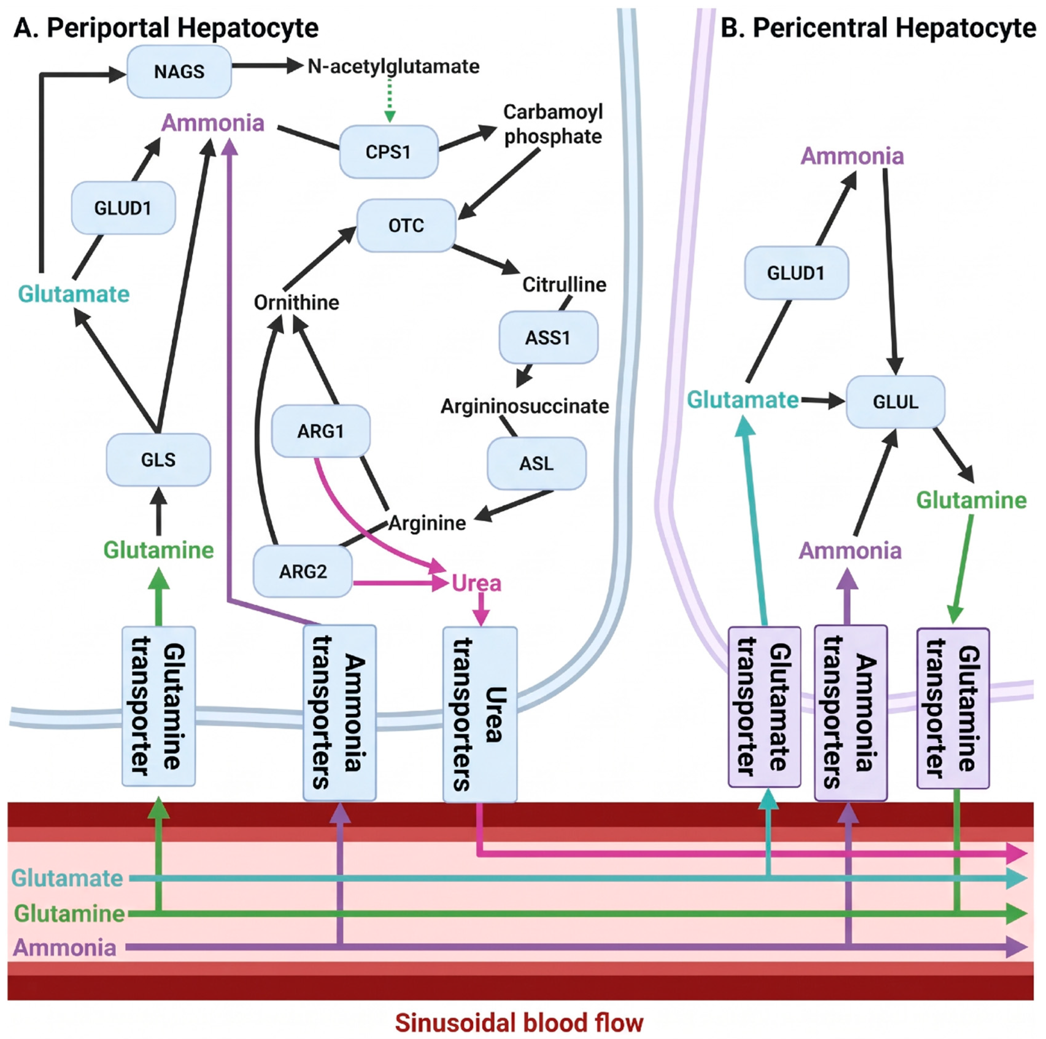
Figure 2.
Cerebral glutamine/GABA-glutamate cycle. Astrocytes detoxify cerebral ammonia by converting glutamate and ammonia into glutamine via glutamine synthetase (GLUL). Glutamine is exported from astrocytes by the sodium-coupled neutral amino acid transporters SNAT3 and SNAT5 and imported into neurons by SNAT1 and SNAT2 (68). In glutamatergic neurons, glutamine is converted to glutamate by glutaminase (GLS), supporting excitatory neurotransmission. Synaptically released glutamate is cleared primarily by astrocytic excitatory amino acid transporters EAAT1 and EAAT2 (69). In GABAergic neurons, glutamate generated by GLS is converted into γ-aminobutyric acid (GABA) by glutamate decarboxylase (GAD) (67). Excess GABA is taken up into GABA transporters GAT1 and GAT3. In all involved cell types, glutamate is reversibly interconverted into ammonia and α-ketoglutarate (α-KG) by glutamate dehydrogenase (GLUD), providing an additional layer of ammonia regulation. Both α-KG and GABA can also feed into the tricarboxylic acid (TCA) cycle, which supports the regeneration of glutamine, completing the cycle. Created in BioRender. Hyde, J. (2026). BioRender.com/j0bl04j.
Figure 2.
Cerebral glutamine/GABA-glutamate cycle. Astrocytes detoxify cerebral ammonia by converting glutamate and ammonia into glutamine via glutamine synthetase (GLUL). Glutamine is exported from astrocytes by the sodium-coupled neutral amino acid transporters SNAT3 and SNAT5 and imported into neurons by SNAT1 and SNAT2 (68). In glutamatergic neurons, glutamine is converted to glutamate by glutaminase (GLS), supporting excitatory neurotransmission. Synaptically released glutamate is cleared primarily by astrocytic excitatory amino acid transporters EAAT1 and EAAT2 (69). In GABAergic neurons, glutamate generated by GLS is converted into γ-aminobutyric acid (GABA) by glutamate decarboxylase (GAD) (67). Excess GABA is taken up into GABA transporters GAT1 and GAT3. In all involved cell types, glutamate is reversibly interconverted into ammonia and α-ketoglutarate (α-KG) by glutamate dehydrogenase (GLUD), providing an additional layer of ammonia regulation. Both α-KG and GABA can also feed into the tricarboxylic acid (TCA) cycle, which supports the regeneration of glutamine, completing the cycle. Created in BioRender. Hyde, J. (2026). BioRender.com/j0bl04j.
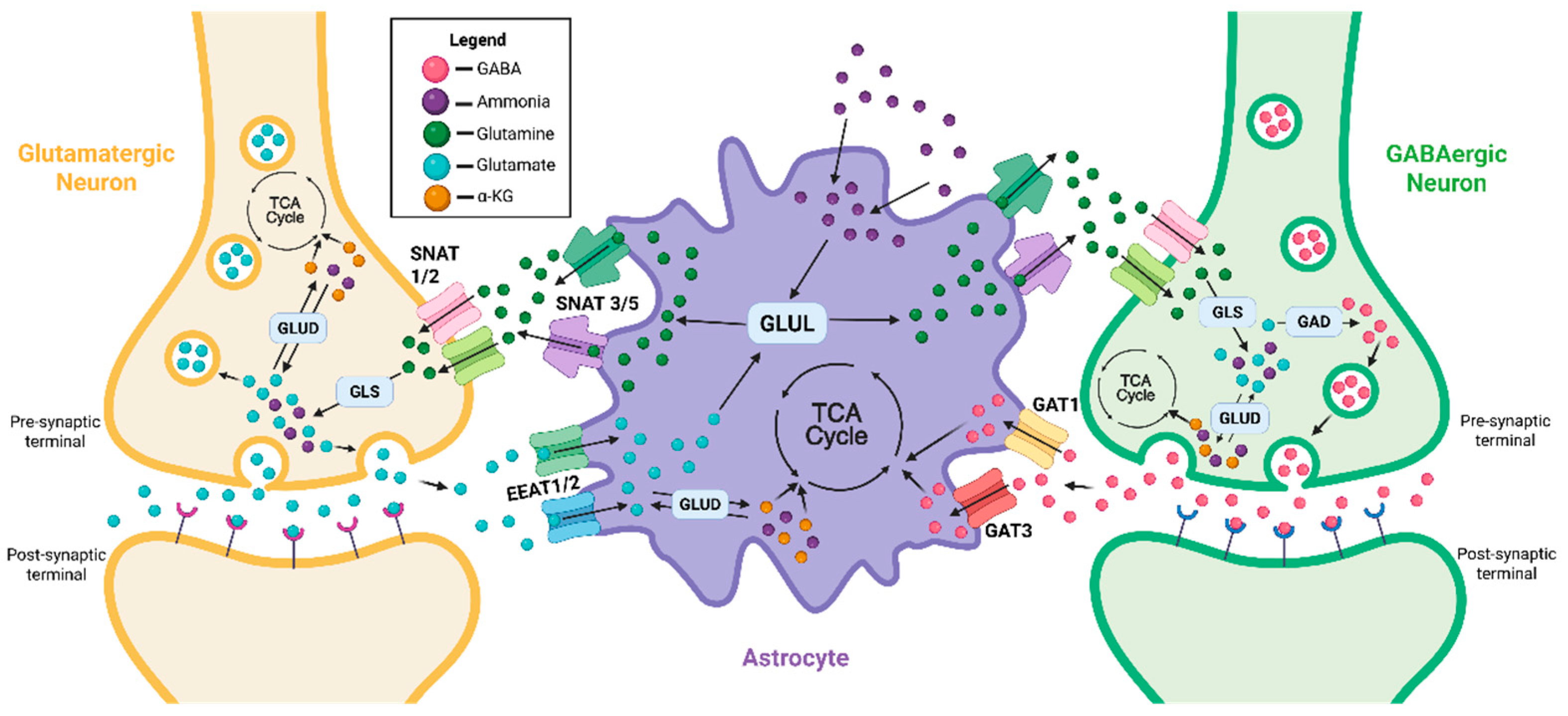
Figure 3.
Summary of reported changes to ammonia, urea, and related metabolites observed in HD patients and animal models. Shared features include an accumulation of urea within the brains of HD patients and OVT73 sheep, elevated circulating arginine and citrulline, which are indicative of a urea cycle deficit. Altered glutamine-glutamate metabolism is also commonly observed. Created in BioRender. Hyde, J. (2026). BioRender.com/qq5t9vy.
Figure 3.
Summary of reported changes to ammonia, urea, and related metabolites observed in HD patients and animal models. Shared features include an accumulation of urea within the brains of HD patients and OVT73 sheep, elevated circulating arginine and citrulline, which are indicative of a urea cycle deficit. Altered glutamine-glutamate metabolism is also commonly observed. Created in BioRender. Hyde, J. (2026). BioRender.com/qq5t9vy.
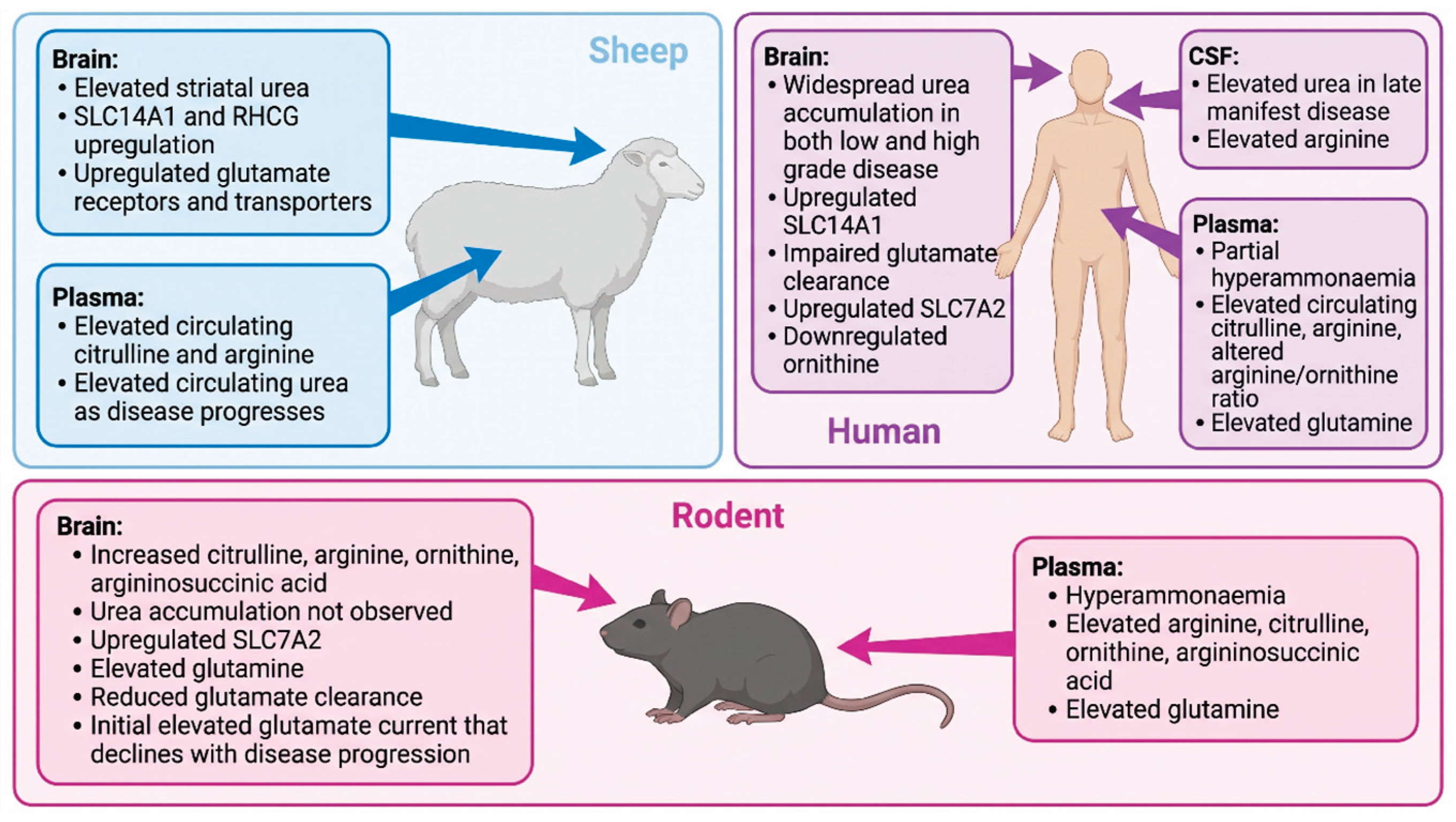

Table 1.
Comparison of Huntington’s disease (HD), urea cycle disorders (UCDs) and hepatic encephalopathy (HE).
Table 1.
Comparison of Huntington’s disease (HD), urea cycle disorders (UCDs) and hepatic encephalopathy (HE).
| HD | UCD | HE | |
| Cause | Expanded polyglutamine coding repeat in exon one of HTT | Inherited deficiency in urea cycle enzyme or transporters | Impaired nitrogen metabolism following liver failure (various etiologies) |
| Age-of-onset | 35 – 40 years old is typical onset, but highly variable depending on CAG repeat length | Complete UCD: Neonatal onset Partial UCD: Anywhere from one month to six decades after birth depending on severity of mutation and enzyme involved |
Typically, adult as most liver failure occurs in adults. Can vary depending on if liver failure is chronic or acute |
| Neuropathology | Progressive neurodegeneration originating in medium spiny neurons of the striatum that then spreads cortically | Basal ganglia lesions and atrophy with cortical thinning, dilated ventricles. Other brain regions spared. Complete UCD show more extensive cerebral oedema and Alzheimer type II astrocytes | Overt HE: Alzheimer Type II astrocytes, cerebral oedema, cortical atrophy Mild HE: impaired EEG function including within basal ganglia |
| Symptom profile | Involuntary movements (e.g. chorea, rigidity) and impaired voluntary movements (akinesia, dysarthria). Cognitive issues including impaired executive decision making, impaired verbal learning and visuospatial complications Neuropsychiatric defects, including anxiety, depression, irritability |
Complete UCD: Lethargy, anorexia, hypothermia, seizures, neurological posturing, coma, severe intellectual disability Partial UCD: Behavioral abnormalities, moderate impairments for executive function, working memory, attention and fine motor skills, hyperreflexia, sleep disorders |
Overt HE: Hypothermia, lethargy, coma, seizures, abnormal posture, mental retardation, asterixis Minimal HE: Appetite loss, impaired motor control, behavioral abnormalities, cyclic vomiting, lethargy, mild cognitive impairment, sleep and mood disorders |
Disclaimer/Publisher’s Note: The statements, opinions and data contained in all publications are solely those of the individual author(s) and contributor(s) and not of MDPI and/or the editor(s). MDPI and/or the editor(s) disclaim responsibility for any injury to people or property resulting from any ideas, methods, instructions or products referred to in the content. |
© 2026 by the authors. Licensee MDPI, Basel, Switzerland. This article is an open access article distributed under the terms and conditions of the Creative Commons Attribution (CC BY) license (http://creativecommons.org/licenses/by/4.0/).
Copyright: This open access article is published under a Creative Commons CC BY 4.0 license, which permit the free download, distribution, and reuse, provided that the author and preprint are cited in any reuse.