
Submitted:
17 November 2026
Posted:
18 November 2026
You are already at the latest version
Abstract
Background/Objectives: Esophageal squamous cell carcinoma (ESCC) is a predominant subtype of esophageal cancer in East Asia and is associated with high cancer-related mortality. CCAAT/enhancer-binding protein beta (CEBPB) is implicated in tumor progression and chemoresistance; however, its functional role and therapeutic potential in ESCC remain poorly understood. This study aimed to investigate the role of CEBPB in ESCC progression and cisplatin resistance and to explore its potential as a therapeutic target. Methods: CEBPB expression and its clinical significance were evaluated using public datasets and human ESCC tissue arrays. A human reflux model, esophageal organoids, and ESCC cell lines were used to investigate the effects of CEBPB on epithelial proliferation, differentiation, and cisplatin sensitivity. CEBPB gain- and loss-of-function experiments, apoptosis assays, pathway gene profiling and luciferase reporter assays were performed to assess its biological functions and transcriptional activity. The effects of combined cisplatin and itraconazole treatment were evaluated in ESCC cells. Statistical analyses were performed using unpaired t-tests or one-way ANOVA, and survival was analyzed using the Kaplan–Meier method and log-rank test. Results: In non-neoplastic esophageal tissues, increased CEBPB expression was associated with reflux-induced basal cell hyperplasia. Organoid experiments demonstrated that CEBPB promoted cell proliferation while suppressing basal cell differentiation. CEBPB expression was significantly elevated during ESCC progression, and higher expression was associated with poorer overall survival (P < 0.05). In cultured ESCC cells, CEBPB enhanced cell growth and survival by directly regulating the transcription of the anti-apoptotic gene MCL1. Elevated CEBPB expression conferred resistance to cisplatin, whereas CEBPB knockdown increased apoptosis and reduced MCL1 expression in cisplatin-resistant ESCC cells. Furthermore, combined treatment with cisplatin and itraconazole promoted apoptosis in cisplatin resistant and naïve ESCC cells compared with cisplatin treatment alone. Conclusions: CEBPB promotes ESCC progression and contributes to cisplatin resistance, at least in part, through transcriptional regulation of MCL1. Targeting CEBPB in combination with cisplatin may represent a potential therapeutic strategy for overcoming chemoresistance in ESCC and may have broader relevance across multiple cancer types in which CEBPB contributes to disease progression and drug resistance.
Keywords:
ESCC
; anti-apoptosis
; MCL-1
; chemo-resistance
; itraconazole
1. Introduction
Esophageal squamous cell carcinoma (ESCC) originates from the squamous cells lining the esophagus and is dominant form of esophageal malignancy the East Asia and parts of Africa. China accounts for more than 50% of global cases of esophageal cancer [1]. About 85% of esophageal cancers in China are ESCC. Treatment options for ESCC are stage-dependent. Early-stage cancers may be treated with endoscopic resection or surgery, while locally advanced cancers often involve neoadjuvant chemoradiotherapy followed by surgery. For advanced or metastatic ESCC, chemotherapy and immunotherapy are the primary treatments, with combination therapies showing promise[2]. Today, chemoresistance is a significant clinical challenge that limits the effectiveness of chemotherapy, particularly in advanced stages of all cancers include ESCC [3].
CCAAT enhancer binding protein beta (CEBPB) is a transcription factor that plays a crucial role in regulating gene expression involved in immune responses and inflammation in human diseases of different organs including esophagus [4]. In non-neoplastic esophagus, CEBPB is upregulated along with a panel of inflammatory molecules in response to food allergens in eosinophilic esophagitis [5]. In esophageal malignancy, the increased stromal expression of CEBPB is linked to the progression from Barrett’s esophagus to esophageal adenocarcinoma [6]. The abnormal CEBPB expression is accompanied with the increased expression of five stromal signature genes which can predict the progression of metaplastic esophagus.
In ESCC, although there is no study has been addressed to the initiation role of CEBPB in early cancer, CEBPB has been found involved in cancer progression and invasive growth through promoting metalloprotease 3 expression[7]. CEBPB also mediates a pro-metastatic signaling pathway in ESCC. Specifically, it works with the ATAD2 protein, to promote TGF-beta downstream signaling cascade that drives metastasis [8]. In addition, CEBPB can regulate the expression of various genes involved in cancer progression and drug resistance[9]. CEBPB can also influence the tumor microenvironment to support tumor growth and drug resistance. Its overexpression can lead to an increase in pro-inflammatory cytokines and other factors that create a supportive niche for cancer cells, further contributing to their survival under chemotherapy [10]. However, the direct relation between CEBPB and chemoresistance in ESCC is not studied.
Here, we explored the roles of CEBPB in mediating abnormal cell growth in pre-neoplastic squamous esophagus. We further demonstrated that the CEBPB expression was linked to cancer progression and overall survival in ESCC. In vitro studies revealed that CEBPB mediated post-cisplatin treatment survival and chemoresistance. Finally, we investigated the potential of reducing post-cisplatin treatment survival by targeting CEBPB in ESCC.
2. Materials and Methods
2.1. Acquisition and Analysis of TCGA Datasets
RNA-sequencing and microarray expression data and corresponding clinical information for esophageal squamous cell carcinoma (ESCC) were obtained from The Cancer Genome Atlas (TCGA) database via The University of Alabama at Birmingham Cancer data analysis Portal (https://ualcan.path.uab.edu/analysis.html). Differentially expressed genes (DEGs) were identified with thresholds set at |log2 fold change| ≥ 1 and adjusted P value < 0.05 (Benjamini–Hochberg correction). Survival analyses were performed using the Kaplan–Meier method and log-rank test (https://kmplot.com/analysis/index.php?p=service).
2.2. Tissue Array and Immunohistochemistry
Paraffin-embedded human ESCC/EAC tissue arrays (cat# T022 and ES781) were obtained from US Biomax (currently TissueArray.com) and processed for immunohistochemical (IHC) analysis. For evaluation of esophageal basal cell proliferation under inflammatory condition, we used tissue sections from our previously published esophagitis reflux model [11,12] (Supplementary Materials and Methods and Supplementary Table 1).
2.3. Cell Lines and Culture Conditions
Normal esophageal squamous cell line NES-G4 was previously developed in our laboratory [13]. The culture media for NES-G4 is described elsewhere [13]. Human ESCC cell lines (KYSE510, 520, 180, and 450) were obtained from Cells Online LLC (San Jose, CA). KYSE-350 was from JCRB Cell Bank, distributed by FUJIFILM Irvine Scientific, Inc. (Richmond, VA) (Supplementary Materials and Methods).
2.4. Organoid and Tumoroid Culture
See Supplementary Materials and Methods.
2.5. Total Protein Extraction and Immunoblotting
Cells or organoids were lysed in RIPA buffer (cat #9806, Cell signaling technology, Danvers, MA) supplemented with protease and phosphatase inhibitor cocktails (Roche). Lysates were clarified by centrifugation and concentrations were determined using the BCA Protein Assay Kit (Thermo Scientific) (Supplementary Materials and Methods and Supplementary Table 1).
2.6. Cell Counting by Coulter Counter
Cell numbers were determined using a Beckman Z1 Coulter Particle Counter (Indianapolis, IN). Cells were trypsinized, resuspended in isotonic solution, and counted according to the manufacturer’s protocol.
2.7. Generation of Stable Knockdown and Overexpression ESCC Clones
Guide RNA (gRNA) sequences targeting CEBPB using primers 5’-CACCG CGCTTACCTCGGCTACCAGG-3’ and 5’-AAAC CCTGGTAGCCGAGGTAAGCG C-3’ were cloned into the pSpCas9 (BB)-2A-puro vector. Scrambled control gRNA was included as a control. For stable overexpression, CEBPB-Lip (plasmid #15713, Addgene, Watertown, MA) and CEBPB-Lap2 (Addgene plasmid# 15740) isoform full-length cDNA clones were obtained as gifts from Joan Massague. Retroviral particles were produced by co-transfecting 293T cells with packaging plasmids (Gag-Pol and VSV-G) and CEBPB cDNA plasmid using Lipofectamine 2000 (Thermo Fisher Scientific, Waltham, MA). Viral supernatants were collected 48 h post-transfection, filtered, and concentrated for subsequent transduction of ESCC cells in the presence of 8 μg/mL polybrene. Stable cell lines were generated by selection with puromycin (2 μg/mL).
2.8. Cell Viability and Cell Apoptosis Assay
ESCC cells were treated with cisplatin (cat# S1166, Selleckchem.com) at the indicated concentrations for 24–72 h. Cell viability was assessed using the MTT assay (cat#CT02, Millipore sigma, Burlington, MA). Apoptosis was measured using Cell Death Detection ELISA (cat# 11-544-675-001, Millipore-Sigma) or the Annexin V–FITC/PI Apoptosis Detection Kit (cat# 556570, BD Biosciences, Franklin Lakes, NJ) according to the manufacturer’s instructions (Supplementary Materials and Methods).
2.9. Generation of Cisplatin-Resistant ESCC Cells
Parental ESCC cells were exposed to an increasing concentration of cisplatin from 2.5, 5, 10, 20, to 40 µM, each for 7 days. Surviving cells of each concentration were treated with next level of cisplatin for another 7 days, which was repeated once. Total treatment time was three weeks. Cells surviving from the final treatment were maintained in medium containing cisplatin (2.5 μM) for subsequent experiments.
2.10. Immunofluorescent Staining for ESCC Cells and Whole-Mount Organoids
See Supplementary Materials and Methods and Supplementary Table 1.
2.11. RNA Extraction and RT–PCR
Total mRNA was isolated by RNAeasy mini kit from Qiagen (Valencia, CA) for cultured cells. For tissue and organoid culture, Trizol reagent was used for initial lysis followed by chloroform extraction. The integrity of total RNA was examined by ethidium bromide-stained formamide RNA gel (Supplementary Materials and Methods, Supplementary Table 2).
2.12. MCL-1 Promoter Construct and Reporter Gene Assay
A fragment of 312 nucleotides (nt) was subcloned from an Addgene plasmid (#19132), a gift from Wafik El-Deiry, into pGL3 luciferase vector. Point mutation was introduced at a putative CEBPB binding (-110 to -96) using the Quick-Change Site-Directed Mutagenesis Kit (Agilent, Santa Clara, CA) and primers listed in Supplementary Table 2 per the manufacturer’s instructions (Supplementary materials and Methods).
2.13. Combined Treatment with Cisplatin and Itraconazole
Cells were treated with 5 uM of cisplatin, or 1 uM of itraconazole (Cat# I6657, Millipore-Sigma) alone or in combination with DMSO included as a control. For combination treatments, drugs were added simultaneously at the indicated concentrations, and cell apoptosis assays were performed after 48 h.
2.14. Data Analysis
See supplementary Materials and Methods.
3. Results
3.1. Overexpression of CEBPB Promoted Abnormal Growth in Basal Esophageal Squamous Epithelial Cells Under Inflammatory Condition
We first assessed esophageal CEBPB protein expression in a human reflux model. We found that CEBPB was rarely detectable when patients were on a proton-pump inhibitor (PPI); at two-week off PPI, CEBPB expression was significantly increased in the nuclei of basal epithelial cells (Figure 1a lower panels, Figure 1c). The increased expression of CEBPB was associated with significantly increased basal cell proliferation as shown by Ki 67 staining (Figure 1 upper panels, Figure 1b). To confirm the role of CEBPB in basal esophageal epithelial cells, we established an organoid culture using patient derived immortalized esophageal squamous cell NESG4. In the control NESG4 organoids, we confirmed that CEBPB was expressed in the basal layer of stratified organoids (Figure 2a). In the NESG4 cells with overexpressed of CEBPB, most organoids were not differentiated after 10-day of culture. Instead, the over-grown organoids had developed into cauliflower-like branching structure (Figure 2b). Multi-layers of basal cells were shown in the H&E section of branching organoids (arrowhead, Figure 2b lower right). By immunoblotting, we found that CEBPB overexpression had led to reduced involucrin (a marker of cell differentiation) and increased PCNA expression (a marker of cell proliferation) in NESG4 cells. These data suggest that CEBPB promoted abnormal esophageal basal cell propagation.
3.2. Upregulation of CEBPB Was Associated with the Progression and Poorer Overall Survival in ESCC
To evaluate the role of CEBPB in the progression of ESCC, we evaluated TCGA public ESCC data sets through UALCAM data analysis portal [14]. Our analysis showed that CEBPB mRNA expression was increased in esophageal malignancies but only significantly upregulated in ESCC (Figure 3a, P<0.001), not esophageal adenocarcinoma (data not shown). By immunohistochemical staining, we confirmed that CEBPB protein expression generally elevated in ESCC, especially in advanced stages (Figure 3b and 3c). To determine whether CEBPB expression was associated with clinical outcome, we analyzed survival data of 81 ESCC patients from the TCGA database. The high CEBPB expression was linked to reduced overall survival (p=0.022) (Figure 3d). These data suggest that CEBPB may be involved in the late stage of tumor development and post cancer treatment survival.
3.3. High-CEBPB Expressing ESCC Cells Were More Proliferative In Vitro
Since CEBPB was overexpressed in advanced ESCC, we next explored the roles of CEBPB in the progression of ESCC. CEBPB protein was expressed at different levels in five selected ESCC cells; high-expression in KYSE 510 and 520 cells, and low-expression in KYSE 180, 350 and 450 cells (Figure 4a). During 4-days culture period, the high-expression KYSE510 and 520 cells grew significantly faster than low-expression KYSE 180, 350 and 450 cell (Figure 4b). CEBPB can be translated into at least two distinct protein isoforms—Lip and Lap2. We then overexpressed CEBPB-Lip and CEBPB-Lap2 separately into KYSE 180 and 450 cells (Figure 4c). The forced expression of CEBPB had promoted cell growth in KYSE 180 (Figure 4d) and KYSE-450 (Figure 4e) cells. Next, we knocked down CEBPB in KYSE 510 and 520 cells (Figure 4f. and 4h), which led to reduced monolayer cell and tumoroid growth (Figure 4g and 4i and Supplementary Figure 1).
3.4. CEBPB Mediated ESCC Cell Viability to Cisplatin Treatment, Directly Through Transcriptional Regulation of Anti-Apoptotic MCL-1
CEBPB has been found to be required for platinum drug resistance in ovarian cancer[15]. To explore if CEBPB is involved in the chemotherapeutic drug resistance in ESCC, we treated KYSE 510, 520 and 180 cells with cisplatin 5 µM overnight. Cisplatin treatment decreased CEBPB expression in all three cell lines (Figure 5a). We then treated these three cells with a series of cisplatin concentrations for 24 hours followed by measuring cell viability, we found that high-CEBPB expressing cells KYSE 510 and 520 had IC50 of 17.5 µM and 18 µM while the low CEBPB expressing KYSE180 had IC50 12 µM. We repeated the cisplatin treatment using cells with CEBPB knockdown or overexpression. Knocking down CEBP led to decreased cell viability in response to cisplatin treatment in KYSE 510 and 520 (Figure 5b), while overexpressing CEBPB led to increased cell viability in response to cisplatin treatment in KYSE180 cells (Figure 5c).
By cell death Elisa, we found that the reduced cell viability by cisplatin (at 5 µM) was linked to increased cell apoptosis in all three ESCC cell lines. (Figure 6a). To define the role of CEBPB in cisplatin-mediated cell apoptosis in ESCC, we overexpressed CEBPB in KYSE 510 cells. The over-expression of CEBPB suppressed apoptosis induced by cisplatin in KYSE 510 cells (Figure 6b). We then examined the impact of CEBPB on entire apoptosis pathway by RT-PCR gene profiling. Compared to control, cisplatin generally upregulated apoptosis gene profiles (shown by red dots) (Figure 6c) while overexpression of CEBPB diminished this effect (Figure 6d). Of the genes affected by CEBPB overexpression, MCL-1, an anti-apoptotic molecule, was the most significant one (Figure 6e, shown by arrow).
In KYSE 510 cell, MCL-1 mRNA was significantly reduced with CEBPB knock-down (Figure 7a). To explore if CEBPB regulates MCL-1 at the transcriptional level, we performed gene reporter assays, comparing the wild-type with CEBPB putative binding site and the mutated MCL-1 promoter (Figure 7b). Knockdown of endogenous CEBPB had significantly reduced luciferase activity with WT promoter but that did not affect luciferase activity with CEBPB site mutated promoter (Figure 7c). Similarly, cisplatin treatment significantly reduced WT promoter activity but not that CEBPB-site mutated promoter (Figure 7d). These results suggested that CEBPB suppressed anti-apoptotic MCL-1 gene expression directly through transcriptional regulation.
3.5. CEBPB Mediated ESCC Cell Apoptosis in Cisplatin Resistant Cells
To assess if CEBPB is associated with drug resistance in ESCC, we generated cisplatin-resistant (CR) KYSE 510 and 520 cells using the procedure shown in Supplementary Figure 2. The KYSE 510 and 520 CR cells were generally larger in cell bodies, with a few so-called giant cells per field (Figure 8a). We then examined if the CR cells had acquired stemness molecular features. We examined CD44, CDH2 and cytokeratin 7 expression in these cells. The CR cells expressed higher CD44 (Figure 8b) protein, and higher CDH2 and CK7 mRNA (Figure 8c). Furthermore, the CR cells derived tumoroids grew significantly larger in size than those from their parental cells (Figure 8d).
To explore if CEBPB was involved in cisplatin drug resistance in ESCC, we examined the two CR cells against their parental cells. Both KYSE 510 and 520 CR cells had upregulated expression of CEBPB protein compared to their parental cells (Figure 9a). To evaluate the role of CEBPB in cell apoptosis in the CR cells, we knocked down the CEBPB in CR cells (CRD) (Supplementary Figure 3). Cisplatin treatment increased cell apoptosis in parental cells (P) but not increased in the CR cells (Figure 9b). The knock-down of CEBPB had increased cell apoptosis in CR cells which was not further increased by cisplatin treatment (Figure 9b). In association with cell apoptosis, the anti-apoptotic gene MCL-1 mRNA and promoter activity were upregulated in CR cells, which was suppressed by the knockdown of CEBPB in these cells (Figure 9c to 9f.).
3.6. Synergistic Treatment effect on ESCC Cell Apoptosis by Cisplatin and Itraconazole
Itraconazole (Itra), an FDA-approved antifungal agent, is reported to suppress colorectal cancer growth through a CEBPB-mediated pathway [16]. We also reported that Itra can exert anti-tumor effects in esophageal cancer [17]. To explore if itra can overcome drug resistance in the KYSE 510- and 520-CR cells, we treated these cells with cisplatin alone, Itra alone or a combination of the two. The combined treatment significantly increased apoptosis in CR cells, which was not seen in Itra or cisplatin treatment alone (Figure 9e).
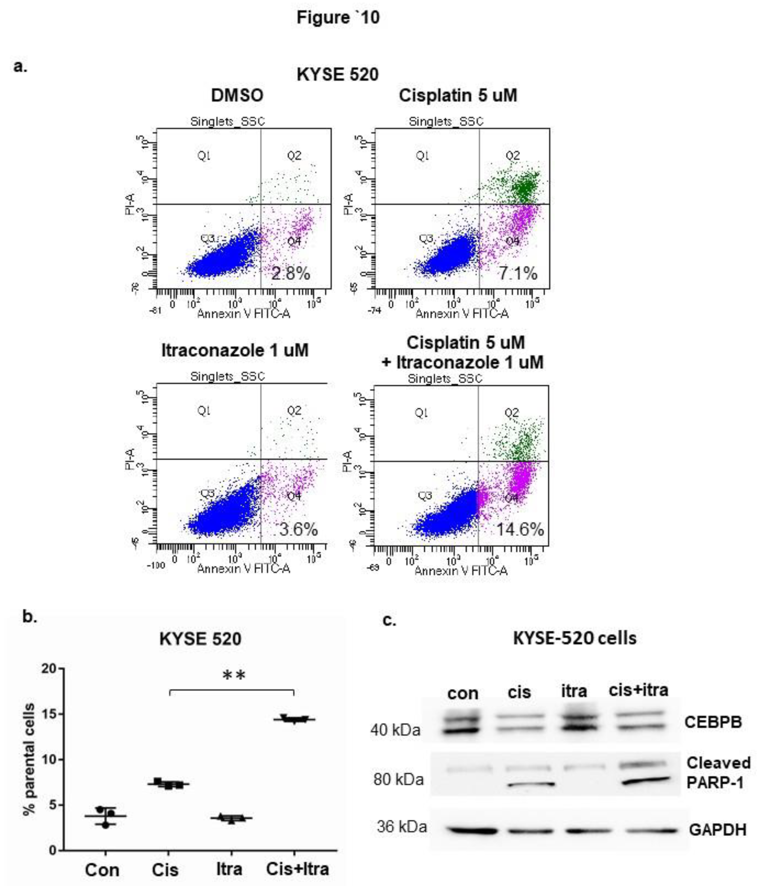
To explore if itra can enhance treatment effect in cisplatin-naïve cells, we repeated the experiments with KYSE 510 and 520 cells. As expected, cisplatin alone increased early cell apoptosis from 2.8 to 7.1% in KYSE 520 (P<0.05) but Itra alone did not (Figure 10a and 10b). Adding Itra to cisplatin treatment further increased averaged early apoptosis to 14.2% (P<0.05). The combined treatment increased cleaved PARP-1 protein expression, confirming the increased cell apoptosis at molecular level (Figure 10c). However, adding Itra did not further reduce CEBPB expression compared to treatment with cisplatin alone (Figure 10c). Similar results were found in KYSE 510 cells (Supplementary Figure 4)
4. Discussion
Summary of findings- CEBPB mediated basal esophageal epithelial cell proliferation in non-neoplastic esophagus and esophageal organoids from GERD patient. In ESCC, CEBPB was upregulated in advanced stages which was associated poorer overall survival. In vitro, high-CEBPB expressing ESCC cells were found to be more proliferative and less sensitive to cisplatin treatment. CEBPB negatively regulated cell apoptosis in ESCC cells, which was linked to the expression of anti-apoptotic genes such as MCL1 at transcriptional level. CEBPB was upregulated in cisplatin-resistant ESCC cells (CR). Knockdown of CEBPB increased cell apoptosis in association with MCL-1 mRNA transcription in the CR ESCC cells. The combined treatment with cisplatin and anti-CEBPB agent itraconazole promoted the apoptosis in CR and cisplatin-naïve ESCC cells.
Our findings in ESCC are consistent with reports in several other malignancies where CEBPB functions as an oncogenic transcription factor. In breast cancer, upregulation of CEBPB and KIFC1 together enhance the aggressive characteristics and poor prognosis of androgen receptor-negative triple-negative breast cancer (AR-TNBC). That study suggests that targeting the interaction of CEBPB and KIFC1 could be a viable therapeutic approach for patients with AR-TNBC [18]. In glioblastoma, CEBPB limits Temozolomide treatment efficacy by inducing P4HA2 expression which promotes cell proliferation in glioma cells [19]. Similarly, In lung cancer, CEBPB coregulated with NRF2 in drug metabolism. The resistance of NRF2-activated lung cancers is achieved by the cooperative function of NRF2 and CEBPB overexpression which is associated with chemoresistance and poor clinical outcome [20].
Interestingly, we observed that cisplatin treatment downregulated CEBPB and thereby facilitated apoptosis in ESCC cells; whereas a subpopulation of cisplatin-resistant (CR) cells displayed increased expression of CEBPB. These findings align with reports from other tumor models in which CEBPB promotes chemotherapy recurrence. For example, in ovarian cancer, CEBPB recruits the histone methyltransferase DOT1L to sustain open chromatin and drive expression of resistance genes[15]. Similarly, CEBPB has been shown to suppress p53-dependent apoptosis[21], upregulate survival mediators and activate homologous recombination repair programs[22]. Together, these mechanisms provide a biologically plausible explanation for the persistence of high CEBPB expression in survival cells following cisplatin treatment.
Synergistic effect of cisplatin with an agent against CEBPB target is novel and remains to be further explored. Itraconazole is a promising repurposed agent in oncology, with multiple mechanisms of action and preclinical & early clinical evidence of benefit, often in combination settings. Its effects on angiogenesis, mTOR/AKT signaling, Hedgehog signaling, and autophagy give it multiple “hooks” to synergize with other anticancer treatments [17,23]. Here we showed that itraconazole can not only promote cell apoptosis in CR ESCC cells, but also amplify anti-apoptotic effect of cisplatin in naïve cells. The mechanism behind was not through downregulating CEBPB expression but perhaps through negating its transcriptional activity. The combination of cisplatin and itraconazole can reduce “off-target” ESCC cells and increase the efficacy of chemo drugs such as cisplatin in first-line treatment.
A key limitation of our study is that we did not distinguish between CEBPB isoforms such as Lip and Lap2, which may exert divergent effects on ESCC cell survival. Future work should therefore dissect the isoform-specific contributions and evaluate whether pharmacologic inhibition of CEBPB or its cofactors (e.g., DOT1L, STAT3) can enhance cisplatin sensitivity in ESCC models.
5. Conclusions
CEBPB promotes ESCC progression and contributes to cisplatin resistance, at least in part, through transcriptional regulation of MCL1. Targeting CEBPB in combination with cisplatin may represent a potential therapeutic strategy for overcoming chemoresistance in ESCC and may have broader relevance across multiple cancer types in which CEBPB contributes to disease progression and drug resistance.
Supplementary Materials
The following supporting information can be downloaded at the website of this paper posted on Preprints.org: Supplementary Materials and Methods; Supplementary Tables 1 and 2; Supplementary Figures 1–4.
Author Contributions
J. Li: technical and material support; drafting of manuscript; analysis and interpretation of data; important intellectual content. P. Liu: study design; technical and material support; drafting of manuscript; important intellectual content. W. Zhang: technical and material support; important intellectual content. Q. Zhang: study design; technical and material support; analysis and interpretation of data; critical revision of manuscript; important intellectual content. All authors have read and agreed to the published version of the manuscript.
Funding
This work was supported in part by the Department of Defense (CDMRP CA220069 subaward HT94252310385 to Q.Z.) and Baylor Scott and White Research Institute. [APC funding source: The APC was funded by the authors.].
Institutional Review Board Statement
The study used commercially available and previously collected human tissue specimens. Ethical approval was obtained or waived as applicable.
Informed Consent Statement
Informed consent was obtained or waived as applicable to the original tissue collection.
Data Availability Statement
The data presented in this study are available from the corresponding author upon reasonable request.
Conflicts of Interest
The authors declare no conflict of interest.
Abbreviations
The following abbreviations are used in this manuscript:
| Esophageal squamous cell carcinoma | ESCC |
| CCAAT enhancer-binding protein beta | CEBPB |
| cisplatin resistant | CR |
| CR cells with CEBPB knockdown | CRD |
| Reverse transcription polymerase chain reaction | RT-PCR |
| enzyme-linked immunosorbent assay | ELISA |
References
- Zhu, H.; et al. Esophageal cancer in China: Practice and research in the new era. Int. J. Cancer 2023, 152(9), 1741–1751. [Google Scholar] [CrossRef] [PubMed]
- Obermannová, R.; et al. Oesophageal cancer: ESMO Clinical Practice Guideline for diagnosis, treatment and follow-up☆. Ann. Oncol. 2022, 33(10), 992–1004. [Google Scholar] [CrossRef] [PubMed]
- Lohan-Codeco, M.; et al. Molecular mechanisms associated with chemoresistance in esophageal cancer. Cell Mol. Life Sci. 2022, 79(2), 116. [Google Scholar] [CrossRef] [PubMed]
- Ren, Q.; et al. C/EBPbeta: The structure, regulation, and its roles in inflammation-related diseases. BioMed Pharmacother. 2023, 169, 115938. [Google Scholar] [CrossRef] [PubMed]
- Kleuskens, M.T.A.; et al. Transcriptomic profiling of the acute mucosal response to local food injections in adults with eosinophilic esophagitis. J. Allergy Clin. Immunol. 2024, 153(3), 780–792. [Google Scholar] [CrossRef] [PubMed]
- Saadi, A.; et al. Stromal genes discriminate preinvasive from invasive disease, predict outcome, and highlight inflammatory pathways in digestive cancers. Proc. Natl. Acad. Sci. U S A 2010, 107(5), 2177–82. [Google Scholar] [CrossRef] [PubMed]
- Li, H.; et al. CCAAT/Enhancer Binding Protein beta-Mediated MMP3 Upregulation Promotes Esophageal Squamous Cell Cancer Invasion In Vitro and Is Associated with Metastasis in Human Patients. Genet Test. Mol. Biomark. 2019, 23(5), 304–309. [Google Scholar] [CrossRef] [PubMed]
- Cao, L.J.; et al. ATAD2 interacts with C/EBPbeta to promote esophageal squamous cell carcinoma metastasis via TGF-beta1/Smad3 signaling. J. Exp. Clin. Cancer Res. 2021, 40(1), 109. [Google Scholar] [CrossRef] [PubMed]
- Tan, J.; et al. CEBPB activates NRF2 to regulate the MAPK pathway through DUSP1 to promote proliferation and antioxidant capacity in ovarian cancer cells. Cell Cycle 2025, 24(5-8), 122–140. [Google Scholar] [CrossRef] [PubMed]
- Allen, B.; et al. A CEBPB/IL-1beta/TNF-alpha feedback loop drives drug resistance to venetoclax and MDM2 inhibitors in monocytic leukemia. Blood 2025, 145(21), 2488–2506. [Google Scholar] [CrossRef] [PubMed]
- Dunbar, K.B.; et al. Association of Acute Gastroesophageal Reflux Disease With Esophageal Histologic Changes. JAMA 2016, 315(19), 2104–12. [Google Scholar] [CrossRef] [PubMed]
- Huo, X.; et al. Hypoxia-inducible factor-2alpha plays a role in mediating oesophagitis in GORD. Gut 2017, 66(9), 1542–1554. [Google Scholar] [CrossRef] [PubMed]
- Huo, X.; et al. In oesophageal squamous cells exposed to acidic bile salt medium, omeprazole inhibits IL-8 expression through effects on nuclear factor-kappaB and activator protein-1. Gut 2014, 63(7), 1042–52. [Google Scholar] [CrossRef] [PubMed]
- Chandrashekar, D.S.; et al. UALCAN: An update to the integrated cancer data analysis platform. Neoplasia 2022, 25, 18–27. [Google Scholar] [CrossRef] [PubMed]
- Liu, D.; et al. C/EBPbeta enhances platinum resistance of ovarian cancer cells by reprogramming H3K79 methylation. Nat. Commun. 2018, 9(1), 1739. [Google Scholar] [CrossRef] [PubMed]
- Zhang, Y.; et al. Itraconazole inhibits tumor growth via CEBPB-mediated glycolysis in colorectal cancer. Cancer Sci. 2024, 115(4), 1154–1169. [Google Scholar] [CrossRef] [PubMed]
- Zhang, W.; et al. Itraconazole Exerts Its Antitumor Effect in Esophageal Cancer By Suppressing the HER2/AKT Signaling Pathway. Mol. Cancer Ther. 2021, 20(10), 1904–1915. [Google Scholar] [CrossRef] [PubMed]
- Joshi, S.; et al. C/EBPbeta increases tumor aggressiveness by enhancing KIFC1 expression in androgen receptor negative triple negative breast cancer. Cell Commun. Signal 2025, 23(1), 255. [Google Scholar] [CrossRef] [PubMed]
- Wang, S.; et al. CEBPB upregulates P4HA2 to promote the malignant biological behavior in IDH1 wildtype glioma. FASEB J. 2023, 37(4), e22848. [Google Scholar] [CrossRef] [PubMed]
- Okazaki, K.; et al. CEBPB is required for NRF2-mediated drug resistance in NRF2-activated non-small cell lung cancer cells. J. Biochem 2022, 171(5), 567–578. [Google Scholar] [CrossRef] [PubMed]
- Tam, H.W.; et al. C/EBPbeta suppresses keratinocyte autonomous type 1 IFN response and p53 to increase cell survival and susceptibility to UVB-induced skin cancer. Carcinogenesis 2019, 40(9), 1099–1109. [Google Scholar] [CrossRef] [PubMed]
- Tan, J.; et al. C/EBPbeta promotes poly(ADP-ribose) polymerase inhibitor resistance by enhancing homologous recombination repair in high-grade serous ovarian cancer. Oncogene 2021, 40(22), 3845–3858. [Google Scholar] [CrossRef] [PubMed]
- Tsubamoto, H.; et al. Itraconazole Inhibits AKT/mTOR Signaling and Proliferation in Endometrial Cancer Cells. Anticancer Res. 2017, 37(2), 515–519. [Google Scholar] [CrossRef] [PubMed]
Figure 1.
The upregulation of CEBPB was associated with basal epithelial cell proliferation in human esophagus. a. In a human esophagitis reflux model [12], the basal epithelial cells were stained for ki67 and CEBPB by immunochemistry. b. and c. The ki67 and CEBPB positive cells were increased and positively associated from “on PPI” to two-weeks “off-PPI” in human esophagus. Biopsies from three patients (N=3) were used in this study. Bars= 200 um. ** P<0.01.
Figure 1.
The upregulation of CEBPB was associated with basal epithelial cell proliferation in human esophagus. a. In a human esophagitis reflux model [12], the basal epithelial cells were stained for ki67 and CEBPB by immunochemistry. b. and c. The ki67 and CEBPB positive cells were increased and positively associated from “on PPI” to two-weeks “off-PPI” in human esophagus. Biopsies from three patients (N=3) were used in this study. Bars= 200 um. ** P<0.01.
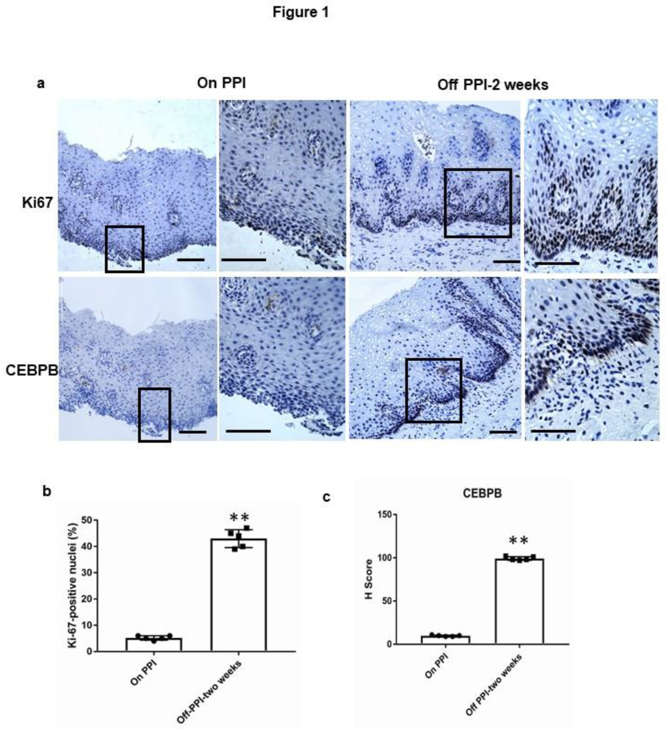
Figure 2.
The overexpression of CEBPB was associated with abnormal basal cell proliferation in human normal esophageal squamous cell (NESG4)-derived organoids. a. CEBPB was expressed in basal cells of NESG4 cell derived squamous organoids. Whole-mount organoids were stained for CEBPB by immunofluorescent staining. Bars=50 um. b. Over-expression of CEBPB had caused abnormal growth and branching in NESG4 organoids. Bars=200 um. c. The overexpression of CEBPB led to decreased expression of involucrin (a differentiation maker) and increased expression of PCNA (a proliferation marker) in NESG4 organoids, shown by the representative immunoblots. GAPDH was included as a loading control.
Figure 2.
The overexpression of CEBPB was associated with abnormal basal cell proliferation in human normal esophageal squamous cell (NESG4)-derived organoids. a. CEBPB was expressed in basal cells of NESG4 cell derived squamous organoids. Whole-mount organoids were stained for CEBPB by immunofluorescent staining. Bars=50 um. b. Over-expression of CEBPB had caused abnormal growth and branching in NESG4 organoids. Bars=200 um. c. The overexpression of CEBPB led to decreased expression of involucrin (a differentiation maker) and increased expression of PCNA (a proliferation marker) in NESG4 organoids, shown by the representative immunoblots. GAPDH was included as a loading control.
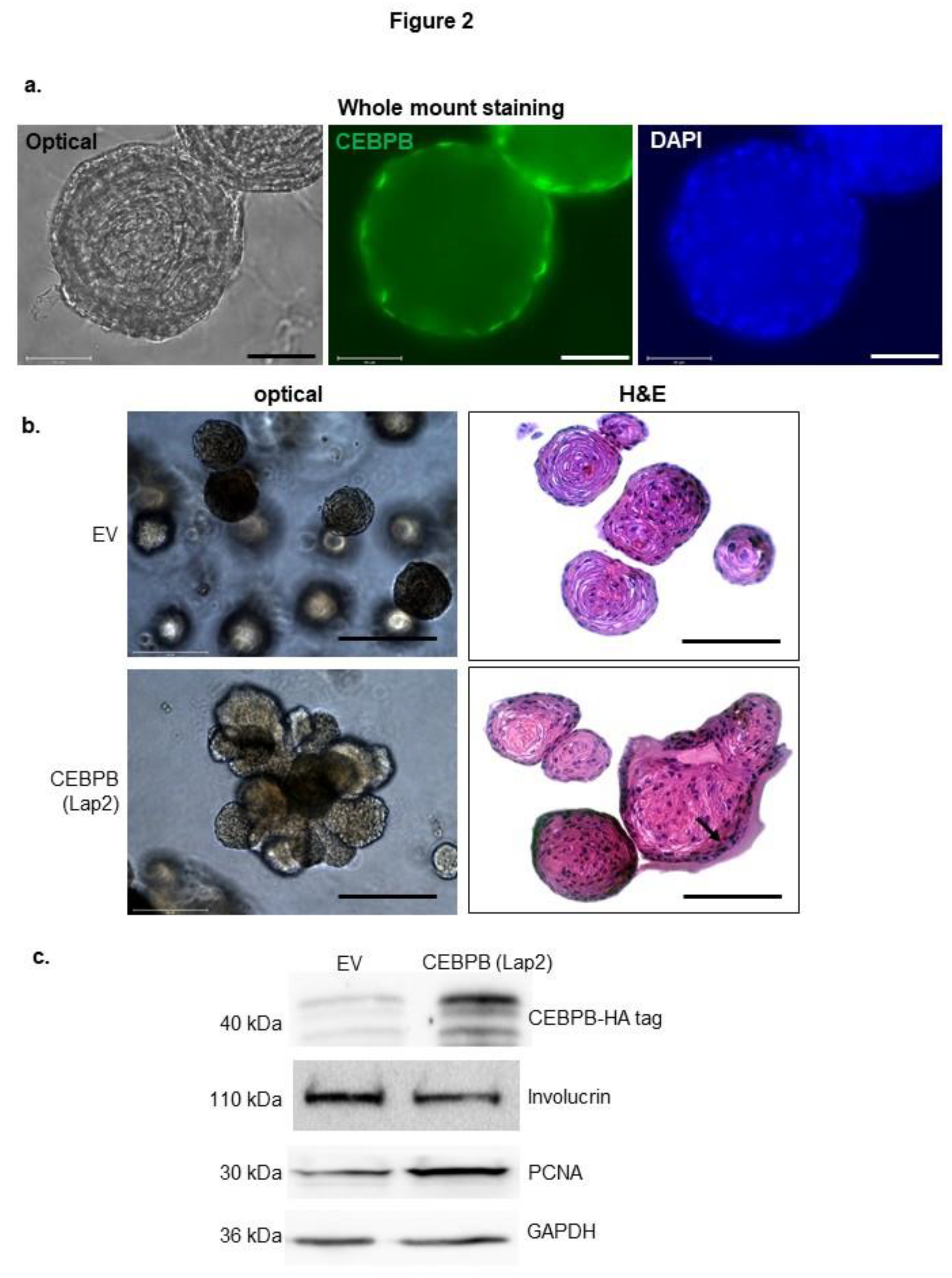
Figure 3.
CEBPB was upregulated in advanced esophageal squamous carcinoma and was associated with poorer overall survival. a. mRNA expression was significantly higher in ESCC (N=95) compared to normal control (N=11) based on TCGA data. b and c. CEBPB protein was significantly higher in the advanced stages of ESCC shown by immunohistochemistry on tissue arrays (cat# T022 and ES781, TissueArray.com). Bars= 200 um. * P<0.05, **P<0.01. d. The higher expression of CEBPB was associated with poorer overall survival based on TGCC data (P=0.022). .
Figure 3.
CEBPB was upregulated in advanced esophageal squamous carcinoma and was associated with poorer overall survival. a. mRNA expression was significantly higher in ESCC (N=95) compared to normal control (N=11) based on TCGA data. b and c. CEBPB protein was significantly higher in the advanced stages of ESCC shown by immunohistochemistry on tissue arrays (cat# T022 and ES781, TissueArray.com). Bars= 200 um. * P<0.05, **P<0.01. d. The higher expression of CEBPB was associated with poorer overall survival based on TGCC data (P=0.022). .
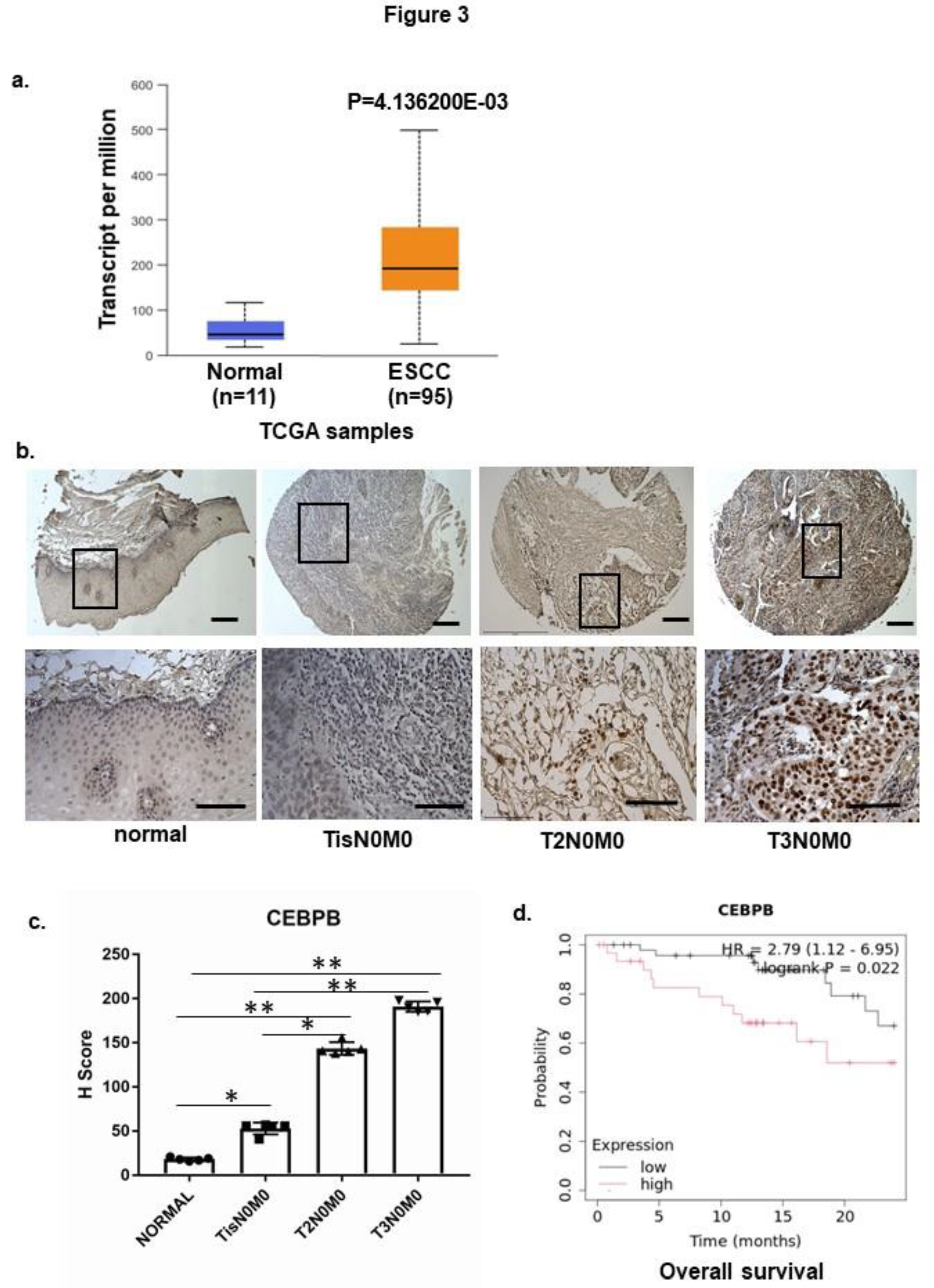
Figure 4.
The CEBPB expression was associated with cell growth in EACC. a. The CEBPB protein expression in five ESCC cell lines (KySE510, 520, 180, 350 and 450). b. The CEBPB expression level was positively associated with cell growth in the five ESCC cell lines. c. d. and e. Over-expression of Lip or Lap2 increased cell growth in low CEBPB-expressing KYSE 180 and 450 cells. f. g. h. and i. Knockdown of CEBPB decreased cell growth in high CEBPB-expressing KYSE 510 and 520 cells. Representative immunoblots showed CEBPB, Lap2-HA, and Lip-HA protein expression with GAPDH included as loading control. Cell growth was determined by cell counting with a coulter counter. *P<0.05, **P<0.01 compared to empty vector or control shRNA.
Figure 4.
The CEBPB expression was associated with cell growth in EACC. a. The CEBPB protein expression in five ESCC cell lines (KySE510, 520, 180, 350 and 450). b. The CEBPB expression level was positively associated with cell growth in the five ESCC cell lines. c. d. and e. Over-expression of Lip or Lap2 increased cell growth in low CEBPB-expressing KYSE 180 and 450 cells. f. g. h. and i. Knockdown of CEBPB decreased cell growth in high CEBPB-expressing KYSE 510 and 520 cells. Representative immunoblots showed CEBPB, Lap2-HA, and Lip-HA protein expression with GAPDH included as loading control. Cell growth was determined by cell counting with a coulter counter. *P<0.05, **P<0.01 compared to empty vector or control shRNA.
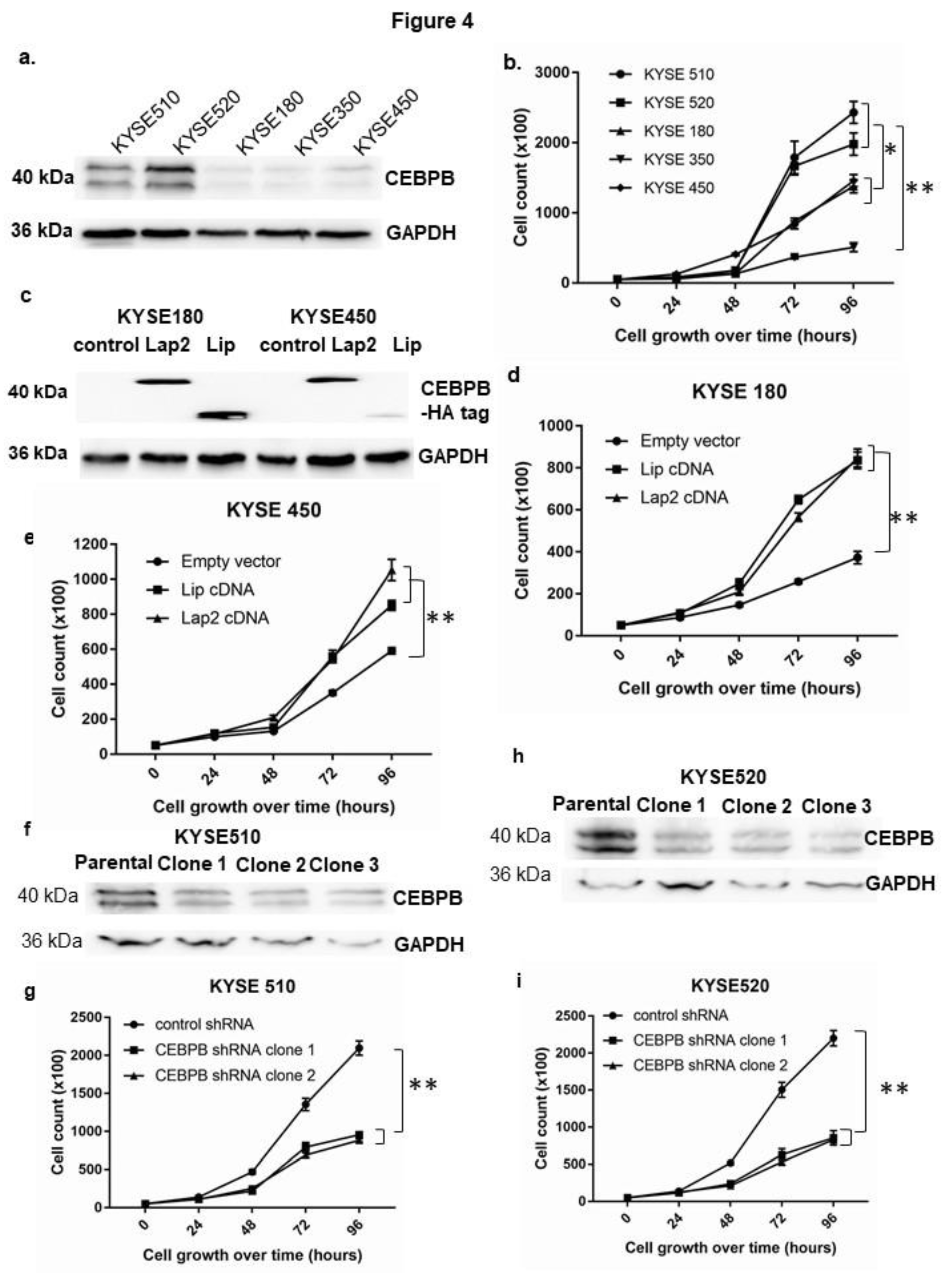
Figure 5.
CEBPB mediated cell viability in cisplatin-treated ESCC cells. a. Cisplatin treatment reduced CEBPB protein expression in ESCC cells. b. In high CEBPB-expressing KYSE 510 and 520 cells, knockdown of CEBPB reduced cell viability which can be further enhanced by cisplatin treatment. c. In low CEBPB-expressing KYSE 180 cells, forced expression of CEBPB decreased sensitivity to cisplatin treatment. Protein expression was determined by immunoblots, and cell viability by a MTT assay kit. ** P<0.01, *P<0.05 vs. corresponding empty vector.
Figure 5.
CEBPB mediated cell viability in cisplatin-treated ESCC cells. a. Cisplatin treatment reduced CEBPB protein expression in ESCC cells. b. In high CEBPB-expressing KYSE 510 and 520 cells, knockdown of CEBPB reduced cell viability which can be further enhanced by cisplatin treatment. c. In low CEBPB-expressing KYSE 180 cells, forced expression of CEBPB decreased sensitivity to cisplatin treatment. Protein expression was determined by immunoblots, and cell viability by a MTT assay kit. ** P<0.01, *P<0.05 vs. corresponding empty vector.
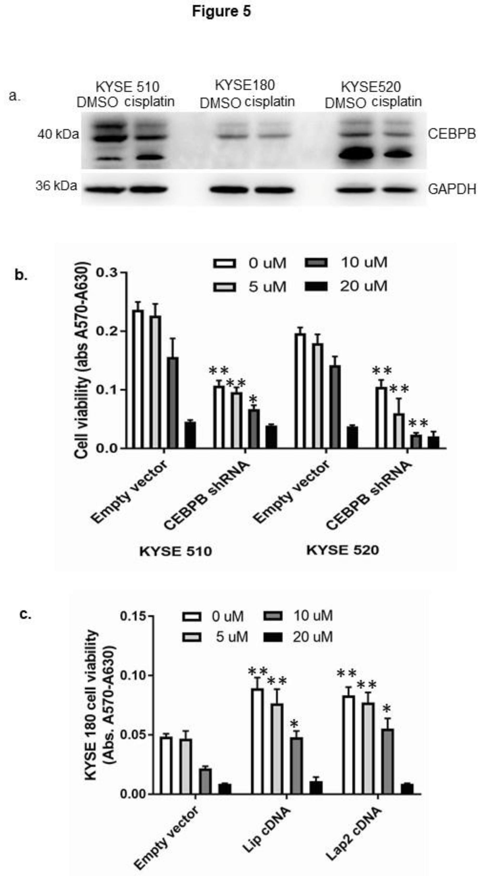
Figure 6.
CEBPB directly mediated anti-apoptosis pathway in ESCC cells. a. Cisplatin treatment increased apoptosis in ESCC cells. In KYSE 510 cells: b. Overexpression of CEBPB (Lap2) decreased cell apoptosis induced by cisplatin treatment. c. Cisplatin induced apoptotic pathway gene (red dots) expression. d. Overexpression of CEBPB (Lap2) diminished cisplatin induced apoptotic pathway gene expression. e. Heat map of apoptotic pathway genes compared among control, cisplatin-treated, CEBPB cDNA overexpression, and CEBPB cDNA overexpression followed by cisplatin treatment. Cell apoptosis was analyzed by cell death Elisa, and apoptosis pathway gene profile analyzed by Rt2 Profiler TM PCR array (Qiagen). ** P<0.01.
Figure 6.
CEBPB directly mediated anti-apoptosis pathway in ESCC cells. a. Cisplatin treatment increased apoptosis in ESCC cells. In KYSE 510 cells: b. Overexpression of CEBPB (Lap2) decreased cell apoptosis induced by cisplatin treatment. c. Cisplatin induced apoptotic pathway gene (red dots) expression. d. Overexpression of CEBPB (Lap2) diminished cisplatin induced apoptotic pathway gene expression. e. Heat map of apoptotic pathway genes compared among control, cisplatin-treated, CEBPB cDNA overexpression, and CEBPB cDNA overexpression followed by cisplatin treatment. Cell apoptosis was analyzed by cell death Elisa, and apoptosis pathway gene profile analyzed by Rt2 Profiler TM PCR array (Qiagen). ** P<0.01.
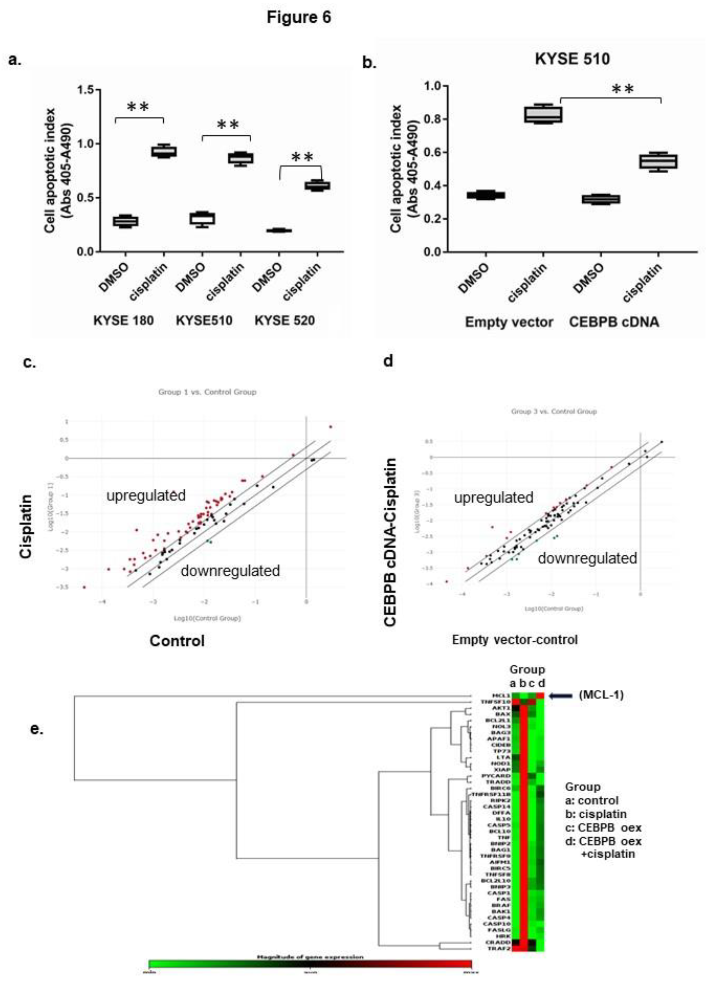
Figure 7.
CEBPB directly mediated anti-apoptotic MCL-1 mRNA transcription. In KYSE 510 cells: a. Knockdown of CEBPB decreased MCL-1 mRNA expression. b.: diagram of wild-type (wt) and putative CEBPB site mutated (mutCEBPB) of MCL-1 gene reporter constructs. c. Stable knockdown of CEBPB reduced wt reporter activity but that was diminished in the mutCEBPB reporter. d. Cisplatin treatment reduced wt reporter activity that was diminished in the mutCEBPB reporter. mRNA expression was assessed by real-time PCR and promoter activity by gene reporter assay. ** P<0.01.
Figure 7.
CEBPB directly mediated anti-apoptotic MCL-1 mRNA transcription. In KYSE 510 cells: a. Knockdown of CEBPB decreased MCL-1 mRNA expression. b.: diagram of wild-type (wt) and putative CEBPB site mutated (mutCEBPB) of MCL-1 gene reporter constructs. c. Stable knockdown of CEBPB reduced wt reporter activity but that was diminished in the mutCEBPB reporter. d. Cisplatin treatment reduced wt reporter activity that was diminished in the mutCEBPB reporter. mRNA expression was assessed by real-time PCR and promoter activity by gene reporter assay. ** P<0.01.
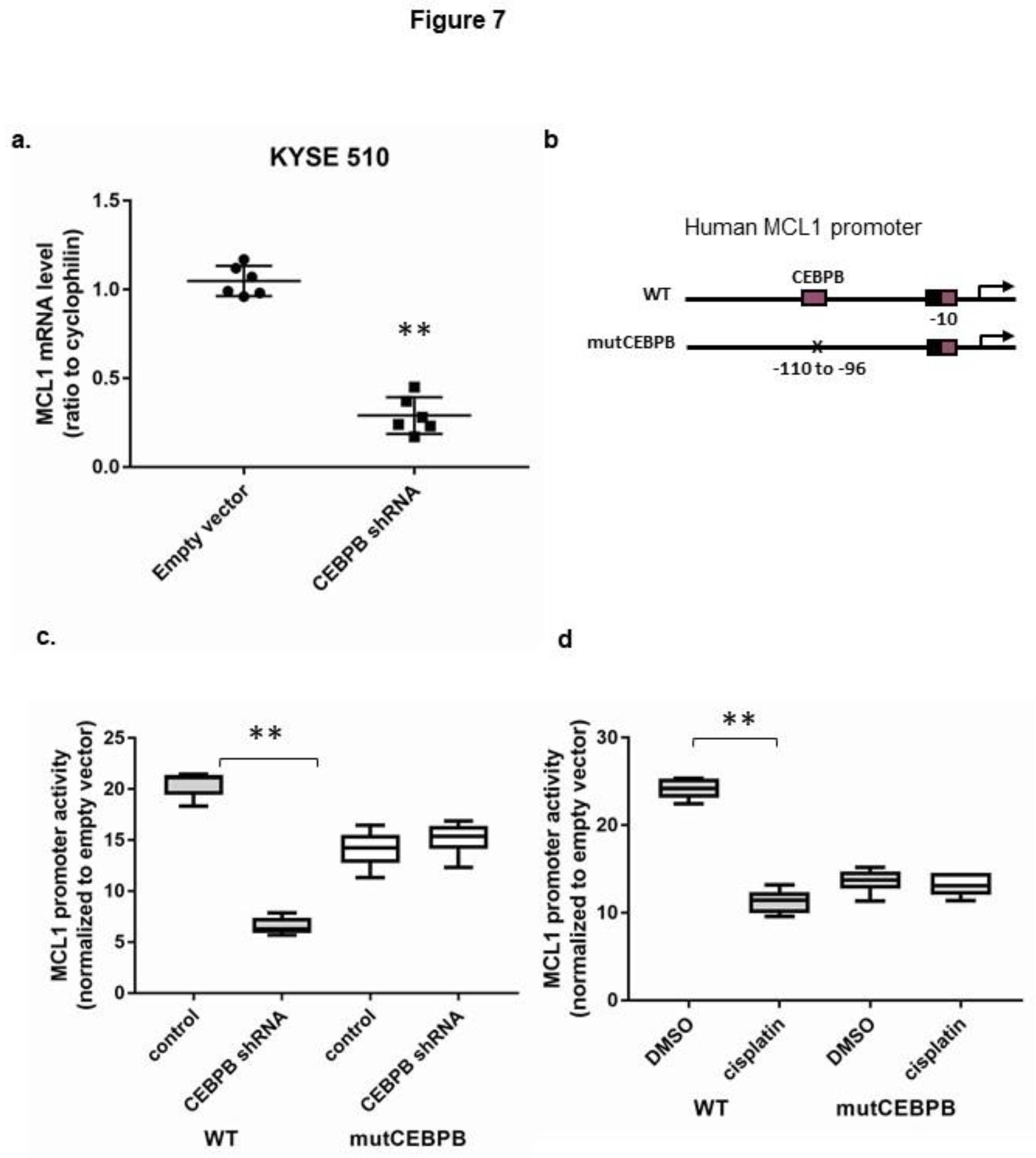
Figure 8.
Features of cisplatin drug resistant KYSE 510 and 520 cells. a. CR cells had larger cell bodies compared to parental cells. bars=275 um. b. CR cells expressed higher level of CD44 protein. bars=50 um. CR cells expressed higher level of CDH2 (c) and cytokeratin 7 (CK7) mRNA (d). e. Tumoroid cultures of CR cells out-performed over their parental cells. CD44 expression was analyzed by fluorescent staining and CDH2 and CK7 by real-time PCR. P=parental, CR=cisplatin resistant. *p<0.05, ** p<0.01.
Figure 8.
Features of cisplatin drug resistant KYSE 510 and 520 cells. a. CR cells had larger cell bodies compared to parental cells. bars=275 um. b. CR cells expressed higher level of CD44 protein. bars=50 um. CR cells expressed higher level of CDH2 (c) and cytokeratin 7 (CK7) mRNA (d). e. Tumoroid cultures of CR cells out-performed over their parental cells. CD44 expression was analyzed by fluorescent staining and CDH2 and CK7 by real-time PCR. P=parental, CR=cisplatin resistant. *p<0.05, ** p<0.01.
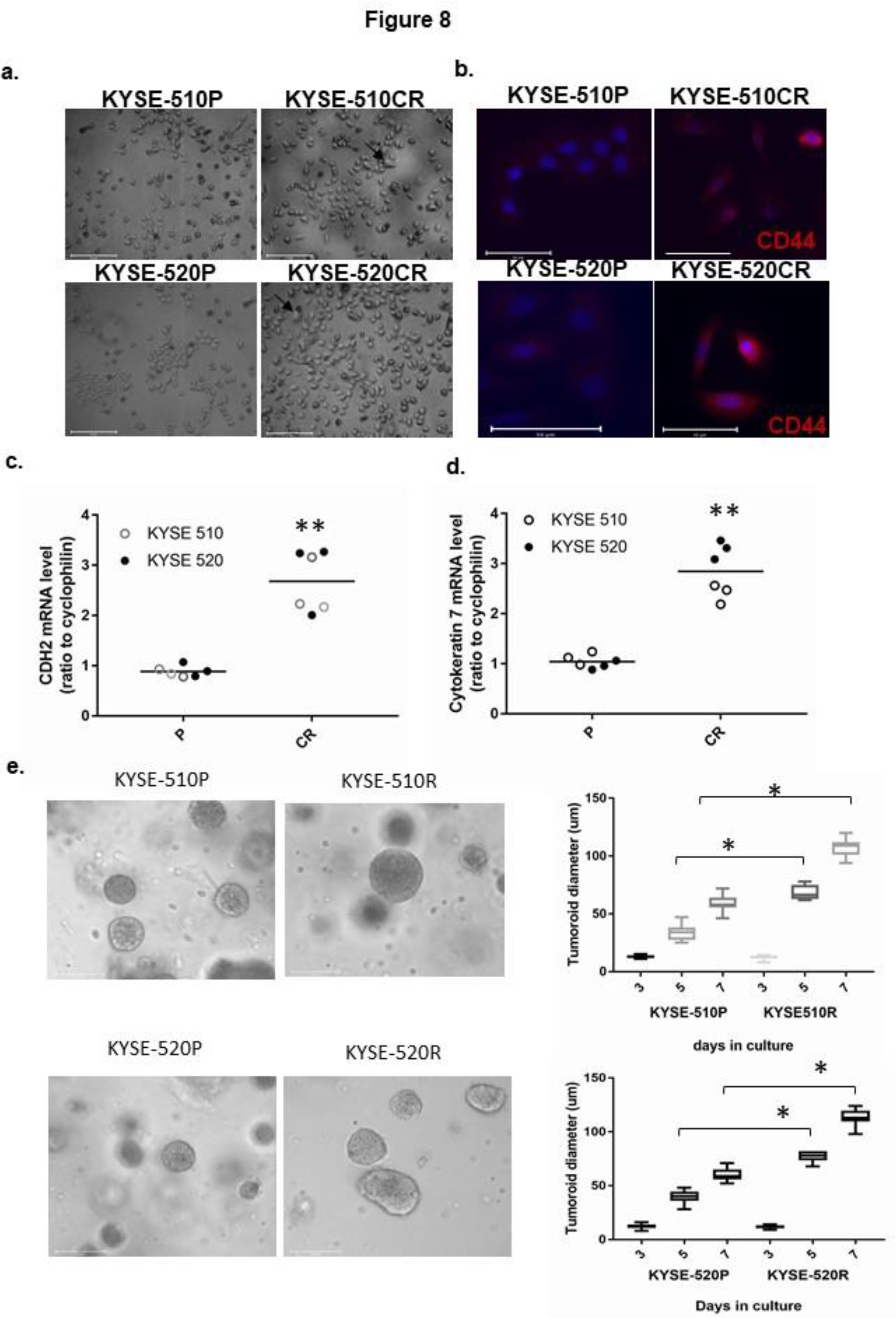
Figure 9.
CEBPB mediated cisplatin drug resistance through an anti-apoptotic pathway. a. CEBPB protein was upregulated in CR cells. b. CEBPB mediated apoptosis in CR cells. c. d. e. and f. CEBPB mediated anti-apoptotic gene MCL-1 promoter activity and mRNA expression in CR cells. g. Combined treatment by Itraconazole and cisplatin promoted apoptosis in CR cells. Protein expression was determined by immunoblot with GAPDH as a loading control, cell apoptosis by cell death Elisa, promoter activity by gene reporter assay, and mRNA expression by RT-PCR. P=parental, CR=cisplatin resistant, CRD=CR cells with CEBPB knockdown. *<p<0.05, **p<0.01.
Figure 9.
CEBPB mediated cisplatin drug resistance through an anti-apoptotic pathway. a. CEBPB protein was upregulated in CR cells. b. CEBPB mediated apoptosis in CR cells. c. d. e. and f. CEBPB mediated anti-apoptotic gene MCL-1 promoter activity and mRNA expression in CR cells. g. Combined treatment by Itraconazole and cisplatin promoted apoptosis in CR cells. Protein expression was determined by immunoblot with GAPDH as a loading control, cell apoptosis by cell death Elisa, promoter activity by gene reporter assay, and mRNA expression by RT-PCR. P=parental, CR=cisplatin resistant, CRD=CR cells with CEBPB knockdown. *<p<0.05, **p<0.01.
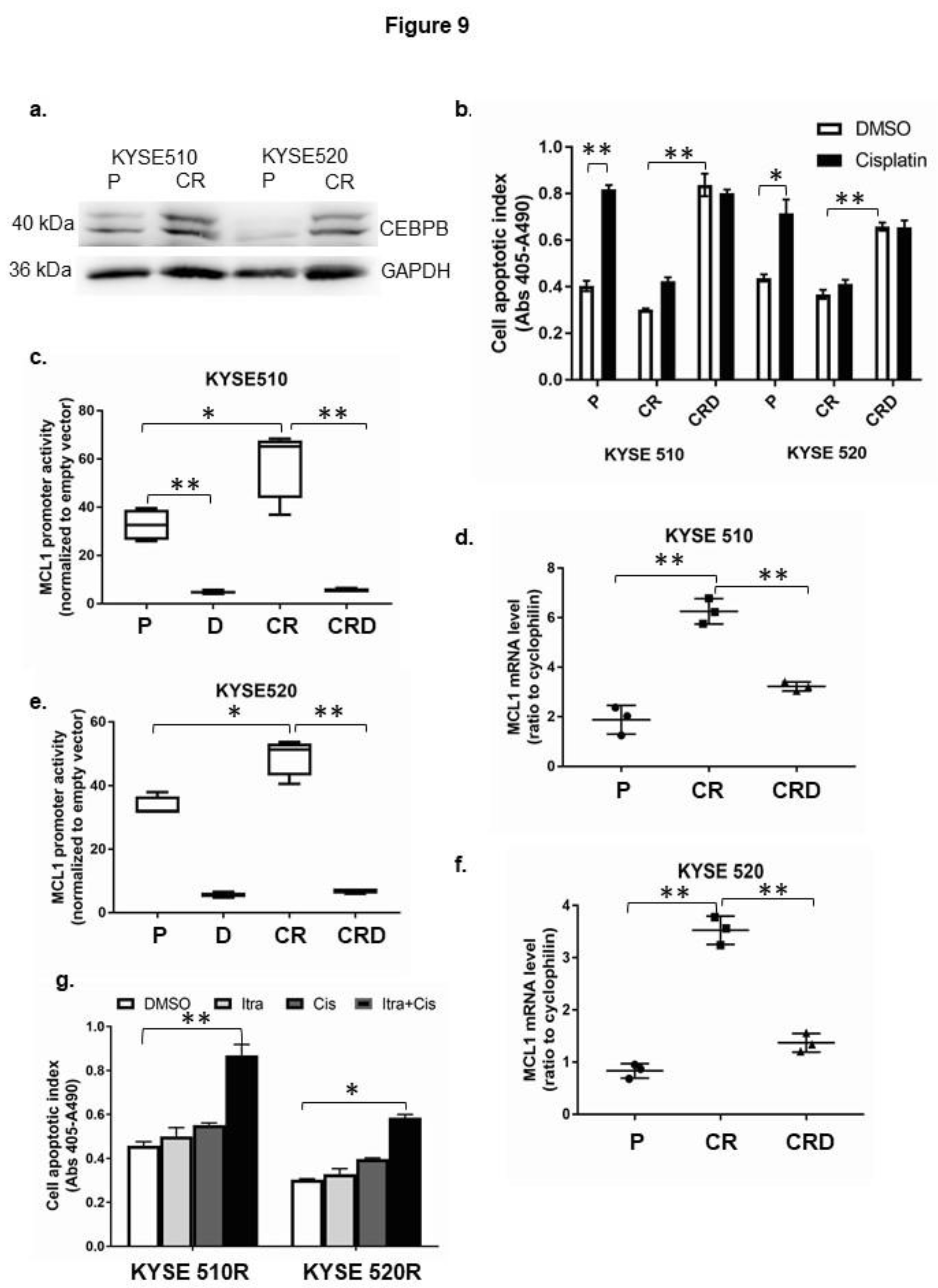
Figure 10.
Combined cisplatin and itraconazole treatment increased early cell apoptosis in KYSE 520 cells. a. Representative plot showed cell death by flow cytometry analysis. b. Dot plot showed averaged early apoptosis from three independent experiments. c. Representative immunoblots showed CEBPB and cleaved parp1 protein expression with GAPDH included as loading control. Con=control, Cis=cisplatin, itra=itraconazole. **p<0.05.
Figure 10.
Combined cisplatin and itraconazole treatment increased early cell apoptosis in KYSE 520 cells. a. Representative plot showed cell death by flow cytometry analysis. b. Dot plot showed averaged early apoptosis from three independent experiments. c. Representative immunoblots showed CEBPB and cleaved parp1 protein expression with GAPDH included as loading control. Con=control, Cis=cisplatin, itra=itraconazole. **p<0.05.
Disclaimer/Publisher’s Note: The statements, opinions and data contained in all publications are solely those of the individual author(s) and contributor(s) and not of MDPI and/or the editor(s). MDPI and/or the editor(s) disclaim responsibility for any injury to people or property resulting from any ideas, methods, instructions or products referred to in the content. |
© 2026 by the authors. Licensee MDPI, Basel, Switzerland. This article is an open access article distributed under the terms and conditions of the Creative Commons Attribution (CC BY) license (http://creativecommons.org/licenses/by/4.0/).
Copyright: This open access article is published under a Creative Commons CC BY 4.0 license, which permit the free download, distribution, and reuse, provided that the author and preprint are cited in any reuse.