
Submitted:
16 November 2026
Posted:
18 November 2026
You are already at the latest version
Abstract
Operative neural damage in pelvic and perineal surgery may affect bladder or sexual function, with retention of urine or impotence. Sexual dysfunction is common following pelvic surgery and urinary dysfunction commonly follows perineal surgery. A better understanding of the anatomy and physiology of urinary and sexual function together with refinement of surgery for both benign and malignant pelvic and perineal disease has led to a decrease in the incidence of urogenital dysfunction. The anatomical and physiological basis of autonomic dysfunction during pelvic and perineal surgery was reviewed. Structured education of surgeons with regard to pelvic neuroanatomy and systemic registration of identified nerves intra-operatively may improve functional outcome of patients undergoing pelvic surgery. At-risk patients should have their functional status evaluated before and after surgery.
Keywords:
pelvic anatomy
; total mesorectal excision (TME)
; autonomic dysfunction
; coloproctology
Introduction
Colorectal cancer is a major health problem being the second commonest cancer in the Western world. The mean age of occurrence is 60 years but in a younger age with the wild type in the developing world [1,2,3]. The 5- year survival rate in 1971 was 20% but 50% in 2019 due to early diagnosis, better staging and peri-operative management, and effective multimodal therapy [4]. This is enhanced by the understanding and application of surgical oncological principles. However, the quality of life issues are very important in rectal cancer surgery. These include (i) the preservation of continence, (ii) the preservation of reasonable bowel frequency and (iii) the avoidance of permanent sexual and urinary disturbance [5]. The anatomical basis for autonomic dysfunction in pelvic surgery is discussed with regard to the latter. Urogenital dysfunction remain a common problem after rectal cancer surgery with a greater than 50% deterioration in sexual function (i.e. men having ejaculatory problems and impotence, women having vaginal dryness and dyspareunia) and greater than 30% urinary dysfunction [6]. Surgical damage is the main cause. Radiotherapy seems to have a role in the development of sexual dysfunction without affecting urinary function [7]. Although the pelvic autonomic nerves supply the blood vessels of the female internal genitalia and are involved in the neural control of the lubrication and swelling response sexual problems in women after rectal surgery seem to be predominantly due to a local mechanical problem[8]. Acute urinary retention is a common post operative complication following surgery on benign anorectal diseases with an incidence ranging from 1-52% and increases hospital stay [9,10,11]. It is most common after haemorrhoidectomy and a 30% incidence after lateral internal sphincterotomy [11,12]. Gender-specific pathologies such as benign prostatic hyperplasia among others give a higher incidence of post-operative urinary retention in males (4.7%) than in women (2.9%) [11]. The risk increasing by 2.5 times in patients over 50 years of age due to age-related progressive neuronal degeneration leading to bladder dysfunction [8,11]. The incidence of urinary dysfunction also varies with the type of surgery; general surgical procedures (3.8%), hip joint arthroplasty (10.7-84%), and after hernia repair (5.9%-48%) [11].
Methods
A critical review of autonomic nervous system dysfunction was performed. Literature was identified through systematic searches of MEDLINE (PubMed) database, Scopus, Web of Science, and Google Scholar. Search terms combined controlled vocabulary and free text words, including pelvic neuroanatomy,” AND “total mesorectal excision” AND “surgical coloproctology” and “sexual” AND “urinary dysfunction” OR “implications and management of autonomic dysfunction”. Additional sources were retrieved by hand- searching reference lists of key and historic articles to ensure coverage of seminal and emerging contributions. Priority was given to peer-reviewed original research and meta-analyses with clear methodological rigor and reproducibility. Cases of low anterior resection syndrome (LARS) and non-English articles were excluded. A narrative synthesis was employed to integrate evidence from the included studies and presented as perspectives in thematic subsections.
Anatomy of the Pelvic Autonomic Nervous System
The autonomic nervous system is an involuntary motor control system of the viscera. It consists of the parasympathetic nervous system (PSN) whose functions are involved in ‘rest and digest’, i.e. pupil constriction, stimulation of salivary glands, bronchoconstriction, decrease heart rate and contractility, increase gut motility and relax sphincters, stimulate gut glandular secretions and bladder contraction. It however has specific (independent) functions such as vasodilatation of the genital organs thus stimulating erection and vaginal lubrication. The sympathetic nervous system is involved in ‘emergency’ (fight, fright and flight) conditions. Although its functions are mostly complementary to the PSN function i.e. pupil dilatation, inhibition of salivary secretions, increase heart rate and contractility, bronchodilatation, decrease gut motility and contracting sphincters, and inhibition of bladder contraction, it also has specific (independent) functions. These include stimulation of the adrenal glands, the homeostatatic temperature regulation (via sweating, pilomotor vasomotor mechanisms) and haemodynamic selective vasoconstriction in the splanchnics (viscera) when required, contraction of the bladder neck, seminal vesicles and prostate in males for ejaculation and orgasm (contraction of the bulbospongiosus) in females and vasoconstriction of the genital organs. The independent function of stimulating ejaculation and contracting the bladder neck during that process is to prevent retrograde ejaculation in the male. The parasympathetic output is cranio-caudal. The cranial components are cranial nerves III, VII, IX and X (vagus). The vagus nerve is the true parasympathetic nerve arising directly from the parasympathetic nuclei in the medulla oblongata. The pre-ganglionic parasympathetic nerves to the foregut and midgut (up to proximal two-thirds of the transverse colon) travel in the vagus nerve. The hindgut and the pelvic organs receive their pre-ganglionic parasympathetic nerve fibres from the pelvic parasympathetic splanchnic nerves (nervi erigentes) which arise within the sacral plexus (S2, S3, S4). The parasympathetic supply are difficult to see as it is situated deep in the pelvis and distributed to the pelvic organs in the endopelvic fascia on the surface of the pelvic floor. The parasympathetic nerves that supply the erectile components of the penis and clitoris synapse in the tiny ganglia in pelvic plexuses and then run on as post-ganglionic nerve fibres which pierce the pelvic diaphragm and perineal membrane. From here they innervate the erectile tissue of crura and bulb of penis or clitoris directly (Figure 1 and Figure 2). The sympathetic nerves (thoracic splanchnic nerves) arises from both sides of the thoracic spinal cord (T5-T12), pierce the diaphragmatic crura, flow down infront of the aorta and the root of the mesentery and enter the pelvis in front of the promontory of the sacrum as the hypogastric nerves. These pre-aortic nerves turn just beyond the aortic bifurcation into the superior hypogastric pelvic plexus of nerves. The sympathetic nerves are strengthened by the lumbar and sacral sympathetic nerves as they descend down the abdomen into the pelvis (Figure 3). They divide into two trunks, which pass to form the pelvic plexus (the inferior hypogastric plexus) on the side wall of the pelvis to be distributed to the pelvic organs by branches passing with the blood vessels, or with the parasympathetic nervi erigentes (pelvic splanchnic nerves) arising from the sacral segments (S2, S3, S4) of the spinal cord. The autonomic fibres penetrate levator ani and the perineal membrane to supply the deep and superficial perineal spaces, and the parasympathetic fibres have the independent function of stimulating erection of the penis/clitoris (Figure 4) [13,14,15]. Pelvic autonomic nerves innervate the blood vessels of the female internal genitalia and are involved in the neural control of the lubrication and swelling response [1,6,14,15,16].
The functional implications are that as the bladder and genital tract are supplied by autonomic nerves that are in close relationship with the rectum these would be at risk during rectal mobilization (Figure 2). Thus, pelvic autonomic nerves are especially at risk in cases of low rectal cancer and during abdomino-perineal excision [8]. Damage to the superior hypogastric plexus and hypogastric nerves will lead to retrograde ejaculation but bladder detrusor function will not be affected. However, damage to the inferior hypogastric plexus will lead to erectile dysfunction (impotence) and detrusor failure [4,6,8].
Pelvic Surgery
The Concept of TME in Rectal Cancer
The concept of total mesorectal excision in rectal cancer whereby the autonomic pelvic nerves in close contact to the visceral pelvic fascia that surrounds the mesorectum (fascia propria) is freed and dropped back without tearing or damaging the mesorectum has led to substantial improvement of autonomic nerve preservation and oncological clearance. This entails sharp dissection under direct vision along pre-existing anatomical planes between visceral and parietal structures thereby preserving the autonomic nerves required for the maintenance of sexual function and urinary voiding (Figure 5 and Figure 6) [17].
Finding the Correct Plane (‘Holy Plane’)
This is done by finding the plane behind the root of the ‘pedicle package’, i.e. the root of the hind gut mesentery (Figure 7 [18]. The visceral structure is carefully dissected away from the parietal structures of the posterior abdominal wall. The inferior mesenteric artery is ligated about 1-2cm from the aorta itself, just far enough to preserve the pre-aortic sympathetic nerve mini-trunks. The main sympathetic trunks are further back, but the mini-trunks are still worth preserving. The plane is most readily reviewed by looking at the things which cross it. These include the left common iliac artery, the gonadal vessels, the ureter and most subtly the superior hyopgastric plexus of sympathetic nerves. Stripping the plexus off the aorta as part of lympho-vascular (oncological) clearance adds nothing to the cure of the cancer but does mean the patient loses the ability to ejaculate and in various other subtle ways the autonomic nerve function is impaired. Nerve preservation in principle does not compromise resectability unless infiltrated with tumour. Finding a plane infront of these nerves, in a circumferential manner will lead down into the correct plane in the pelvis called the’ Holy plane’ which is between the mesorectum- the integral visceral mesentery of the hind gut and the surrounding nerves on which sexual function depends. This is easiest done at the back by first identifying the two superior hypogastric nerves that come from the superior hypogastric plexus and run down the pelvic side wall. Using St Mark’s retractor, the mesorectum is lifted forward and the nearly avascular cleavage plane around the lateral pelvic wall developed to a point of adherence between the mesorectum and the inferior hypogastric plexuses low down laterally on the pelvic wall (Figure 8). These are were the parasympathetic and sympathetic nerves meet often called the lateral ligaments which links the prerectal and retrorectal parts of the field of dissection. At this point it is easy to taper in the mesorectum with consequent bleeding. This is avoided by cutting at the adherence slightly inside the point where they swing away from the parietal fascia at the site of the planned rectal excision. Thus, cutting the nervi erigentes that innervate only that aspect of the rectum which is being excised (Figure 9)[19].
Anterior Dissection
In the anterior dissection, incorporating the peritoneal reflection as part of the specimen creates a dissection plane (cul-de-sac) behind the top and back of the seminal vesicles that goes down onto the fascia of Denonvillier. Although the antero-lateral lying neurovascular bundles of Walsh that supply the seminal vesicles may be injured, this is extremely unusual owing to the presence and protection of Denonvillier’s fascia (Figure 10) [20,21]. This white fascia layer is useful because it is fairly firmly attached to the anterior mesorectal fat and closely applied to the prostate than the rectum. It lies just anterior to the fascia propria and would give the appropriate plane of dissection that would lead down to its adherence to the back of the prostate. By not excising Denonvillier’s fascia postoperative sexual function can be minimized without compromising the oncological outcome (Figure 11) [20,21,22]. A ‘U’ shape incision on Denonvillier’s fascia may also prevent damage to the antero-lateral nerves [22,23,24]. This is a difficult and challenging part of the dissection in low rectal tumours with regard to avoiding damage to the back of the prostate and consequent bleeding. Bladder injury may occur during rectal excision for an adherent rectal carcinoma especially low down on the posterior abdominal wall or trigone. It is desirable to open the bladder in difficult dissections so that the orientation becomes easier and an injury to the trigone is avoided. In addition to a surgical training programme, intraoperative neuromonitoring (Figure 12), stereotactic 3-D navigation and magnetic resonance neuroimaging may promote the preservation of pelvic autonomic nerves in TME [25,26,27,28,29].
Advantages and Limitations of TME
The en bloc excision of perirectal lymphatics and fatty tissue of the mesorectum down to the pelvic floor in TME gives a low local recurrence rate of 4-5% and improved survival [30]. The recurrence may be due to (1) lateral pelvic wall lymph nodes occurring in 10-30% of patients and (2) ‘coning’ in on the distance clearance margin may leave some of the mesorectum behind on the pelvic wall, thus increasing the chance of local recurrence. Using the international prostate symptom score and international index of erectile function, TME with pelvic autonomic nerve preservation showed relative safety in preserving sexual and voiding function [31]. It reduced the problem of accidental bladder denervation from 50-60% with conventional rectal cancer surgery to <20% with TME, and the problem of post operative impotence from 70-100% to <30% (Table 1) [31,32]. Thus, although severe urinary dysfunction is rare, sexual impairment still remains a serious concern after rectal resection with TME. Laparoscopic approach for TME allows similarly favourable results with regards to postoperative urogenital function at best for tumours situated in the middle and upper third of rectum compared with open surgery [32]. The risk factors are low rectal cancers, anterioly-encroaching tumours, the narrow male pelvis and abdomino-perineal excision (APE). Therefore, experience with, and the learning curve in the TME procedure play a major role with both autonomic nerve preservation and the oncological surgical outcome [33]. Lateral pelvic lymph nodes are usually associated with locally advanced disease and sexual function particularly ejaculation was often damaged after an associated radical pelvic lateral node dissection [34]. Toritani et al [35] recently demonstrated that following TME and lateral pelvic wall lymphadenectomy more than 20% of patients had urinary dysfunction and the preoperative risk factors were tumour location, tumour size and diabetes mellitus. They concluded that high risk patients with more than two of the above risk factors should be informed of the risk of urinary dysfunction following lateral pelvic wall lymphadenectomy. High resolution magnetic resonance imaging (MRI) have enabled more selective application of lateral lymph node dissection (LLND). The use of the enhanced visualization of laparoscopic or robotic surgery may further decrease local recurrence rate even in patients with enlarged pelvic lymph nodes, minimize bleeding and postoperative complications [36]. LLND is not popular in the West, because of poor functional result as most patients had sexual and urinary disturbance, lengthy operation times, excessive blood loss and the perceived success of TME [37]. In addition, a Swedish study demonstrated only 2 of 33 pelvic recurrences attributed to lateral pelvic side wall lymph node involvement [38], and in the current era of neoadjuvant chemoradiotherapy for rectal cancer LLND may not be necessary [39,40].
Transanal TME
Transanal TME (taTME) is the ‘bottom-up’ approach to TME. The utilization of carbon dioxide (CO2) for insufflation show the areolar plane for mesorectal dissection from below. Following a rectal purse-string closure an antero-lateral and posterior mesorectal dissection is performed to meet the abdominal dissection and mobilization of the descending and sigmoid colon above (Figure 13). However CO2 insufflation might also show areolar planes beyond the scope of dissection leading the surgeon off plane to the presacral planes with risk of inadvertent injury to the pelvic side wall autonomic nerves and sacral venous plexus posteriorly resulting in haemorrhage. There is an increased risk of urethral injury early in the learning curve especially at the level of the post prostatic urethra in the setting of anterior tumours and prior radiation There is also a steep learning curve for taTME and single site surgery platforms a to facilitate docking and transanal access are awaited [41].Thus, an advantage of the robotic TME procedure over taTME in reducing the steep learning curve and thus reducing these risks [42,43,44].The advantages of taTME over the standard TME are tremenduous: (1) There is better vision and dissection, (2) the distal margin of the tumour is easily assessed, (3) improves resection quality, (4) improves sphincter preserving rate and decrease APE rate, (5) facilitates anastomosis, thus a restorative procedure, (6) facilitates preservation of the autonomic pelvic nerves, (7) facilitates dissection of the Denonvillier’s fascia especially for anterior tumours with a high risk of a positive circumferential resection margin (CRM ) and minimizes injury to prostate, seminal vesicles and vagina. (8) facilitates rectal cancer excision in the narrow male pelvis, obese, prior radiotherapy and lower third cancers where open/ laparoscopic pelvic dissection would be difficult [45].
Inflammatory Bowel Disease
Since the presacral nerves are situated well posterior or lateral to the rectum, it should not be difficult to avoid damage when operating for inflammatory bowel disease as the dissection stays close to the rectum. As the lateral ligaments are divided close to the rectum in inflammatory bowel disease bladder and sexual dysfunction is kept to a minimum. Impotence is rare after rectal excision for inflammatory bowel disease and fashioning of ileo-anal pouch but cases of retrograde ejaculation have been recorded [46]. Although the risk of impotence is low, warnings are vital as these patients are often young, and impotence will be a particularly severe disaster. Some women have dyspareunia (pain on sexual intercourse) after rectal excision due to the vaginal wall falling back to the sacrum [47]. Dyspareunia has been reported in 7-28% of females after ileo pouch-anal anastomosis [6,46]. This can be prevented or minimized by by transporting the omentum or other tissue into the area in front of the rectum.
Perineal Surgery
Abdomino-Perineal Excision of Rectal Cancer
The surgical innovation in the abdominal-perineal excision for rectal/ anal cancer by Miles in 1908 [48] was inspired by Halstead radical oncological surgery based on pathological lymph node spread. The distal spread was important to Miles which Dukes later found to be limited [49]. However, the procedure is indicated for anal cancer or low rectal cancer involving the sphincter. Unfortunately, there is a higher positive circumferential resection margin (CRM) with this procedure for technical and oncological reasons. There is a tendency of ‘coning’ of the specimen at the level of the levators and these low tumours are usually locally advanced (T4) with nodal spread [50]. Thus the oncological importance of excising widely at the level of the pelvic floor (extralevator excision) and a TME in the abdominal phase (Figure 14). The prone position in performing the perineal part of the operation facilitates the wide pelvic floor excision with improved circumferential resection margin [51,52]. The key to the perineal excision is exposure and intra-operative perforation of the surgical specimen is common (8-26%) [53] rendering the operation palliative. In the male the perineal dissection is among the most difficult in colorectal surgery. Having freed the rectum posteriorly and laterally, the retroprostatic plane may be difficult to enter but guided by the landmark of the superficial border of the superficial transverse perineal muscles the plane will gradually become apparent [51,52,53].
Autonomic Dysfunction in Perineal Surgery
Acute urinary retention is either as a result of an atonic bladder or a frank bladder outflow obstruction from an underlying benign prostatic hyperplasia. Post-operative pain, rectal distension and anal dilatation increase bladder neck sympathetic tone through a sympathetic reflex with the afferents being the somatic and visceral sensory nerves [54] In either sex, the sympathetic response to pain may lead to abnormal bladder sphincteric activity (autonomic dysreflexia) causing difficulty in voiding and thence urinary retention [54,55,56]. Disease severity, amount of analgesia required, simultaneous operations i.e. sphincterotomy, haemorrhoidectomy, fistulotomy, incision and drainage of abscess etc, were risk factors of post-operative urinary retention [57]. An inserted, haemostatic anal pack usually cause post-operative pain and thus the insertion of an indwelling urinary catheter for 24 hrs may avoid acute urinary retention [12,58,59]. The frequency of acute urinary retention is greater in males than females because of benign prostatic hyperplasia providing a ‘weak’ bladder with no pushing power [56]. Excessive fluid intravenous administration may cause over distension of the bladder with the sequelae of inhibiting the detrusor and decreasing bladder compliance. A bladder volume greater than 270 mls has a 2-3 fold risk of post operative urinary retention [54,55,56,57,58,59,60]. Spinal anaesthesia blocks afferent and efferent nerves causing the lack of sensation of bladder-filling and would thus exacerbate the over- distention of the bladder. General anaesthesia interfere with ANS function precipitating bladder atony [61,62]. Systemic opioid, both by intravenous and intramuscular routes inhibit the release of acetycholine from the parasympathetic sacral neurons that control detrusor contractility and increase the incidence of acute urinary retention [63]. This is because the axons of pre-ganglionic parasympathetic fibres also contain enkephalins that are transported to the parasympathetic ganglia that have an inhibitory modulatory effect on the release of acetyicholine [54]. Patients greater than 50 year old have age-related neuronal degeneration. Urodynamic investigations will distinguish bladder atonia from obstruction if not clinically deduced [64]. If atonia, urethral catheterization will allow recovery but a prostatectomy is definitive treatment for obstruction from BPH [56]. The neuroanatomical basis for autonomic dysfunction in surgical coloproctology is summarized in Table 2, and the autonomic dysfunctions of surgical coloproctological procedures in Table 3.
Clinical Implications of Autonomic Dysfunction in Surgical Coloproctology
Parasympathetic nerve dysfunction occurs through the mechanisms of (a) neuropraxia, (b) intentional division ( tumour extension), or (c) an unintentional division. This is corroborated by the study which showed that pelvic nerves (S2, S3, S4) were identified in only 5 of 19 patients [64]. Bladder dysfunction is as a result of a lower motor neurone lesion from injury to motor PSN to detrusor causing a hypotonic bladder or urinary retention. The treatment options are the insertion of a suprapubic catheter, intermittent self catheterization or urethral catheterisation. The parasympathomimetic agent, bethanecol, in a dose of 10mg subcutaneously significantly lowers the incidence of postoperative urinary catheterization and, should be considered as initial treatment of postoperative urinary retention following anorectal surgery [65,66]. It seldom works on the detrusor that is exhausted by obstruction or rendered unstable from whatever cause [11]. Further studies would establish the role of alpha antagonists in the prevention of postoperative urinary retention among patients undergoing anorectal surgery [67]. Postoperative urinary retention in ambulatory surgery (low risk patients) is usually treated with in-out catheterization, but for anorectal surgery most authors suggest 5 days with a range between 3-10 days [68,69]. The incidence of urinary tract infection adter anorectal surgery and 5 days of catheterization ranges between 42% and 60%, thus the need for good sterile technique at urethral catheterization and prophylactic antibiotics [70]. For major complicated surgery and with extensive perineal and rectal dissection, bladder catheterization is required for a longer period of time according to clinical conditions [71,72].
The mechanisms of sympathetic nerve dysfunction are (1) pelvic plexus (SHP & IHP) damage or (2) vasculogenic from internal iliac artery, rarely pudendal artery or bilateral arterial artery embolozation. The management of male impotence depends upon if it is neurogenic and psychogenic which requires pharmacotherapy, or vasculogenic requiring a vacuum device or a penile prosthesis [73]. The parasympathetic nerve-mediated sexual function cause erection of the penis and clitoris and in addition genital lubrication and engorgement in females. Banbrick et al [74] reported significant changes in sexual function in 262 women 3 months following restorative proctocolectomy for ulcerative colitis. These included vaginal dryness (22%), dyspareunia (26%), pain interfering with ability to feel sexual pleasure (20%) probably due to a local mechanical problem and fear of stool leakage (17%). Van Driel et al [75] reported 5 series of female sexual functioning after radical surgical treatment of rectal and bladder cancer with follow-up at 3 months to 23 years. 20-78% had decreased libido, 21-50% had severe dyspareunia and 38-65% had a disappearance of coitus post-operation. The treatment of female sexual dysfunction include viagra which increase genital blood flow, libido, sexual interests, vaginal elasticity and sensation after hysterectomy, and Eros (urometrics) which is a battery driven vacuum device [76]. Therefore, preoperative counseling in surgical coloproctology is important in addition to attention to the meticulous details of the operative techniques mentioned above. Patients who still have voiding and/ or erectile dysfunction at 3 months should be referred to the urologist.
Conclusions
Urogenital dysfunction remains a common problem in surgical coloproctology. Sexual dysfunction is a common post-operative complication following pelvic surgery and urinary dysfunction following perineal surgery. At-risk patients should have their functional status evaluated before and after surgery. Structured education of surgeons with regard to pelvic neuroanatomy and systemic registration of identified nerves intra-operatively using ‘intraoperative nerve stimulation’ may reduce autonomic dysfunction in patients undergoing pelvic surgery.
Author contributions
EPW is the main author, EN contributed to literature search.
Conflict of Interest
No potential conflict of interest relevant to this article was reported
References
- Sung, H.; ferlay, J.; Siegel, R.L.; Laversanne, M.; Soerjomataram, I.; Jemal, A.; et al. Global cancer statistics 2020: GLOBOCAN estimates of incidence and mortality worldwide for 36 cancers in 85 countries. CA Cancer J. Clin. 2021, 71(3), 209–49. [Google Scholar] [CrossRef] [PubMed]
- Myers, E.A.; Feingold, D.L.; Forde, K.A.; Arnell, T.; Jang, J.H.; Whelan, R.L. Colorectal cancer in patients under 50 years of age: A retrospective analysis of two institutions’ experience. World J. Gastroentrol 2013, 19(34), 5851–5657. [Google Scholar] [CrossRef] [PubMed]
- Gullickson, C.; Goodman, M.; Joko-Fru, Y.W.; Gnangnon, F.H.R.; N’Da, G.; Woldegeorgis, M.A.; et al. Colorectal cancer survival in sub-Saharan Africa by age, stage at diagnosis and Human Development Index. A population- based registry study. Int. J. Cancer 2021, 149(8), 1553–63. [Google Scholar] [CrossRef] [PubMed]
- Dulskas, A.; Gaizauskas, V.; Kilusiene, I.; Samalavivius, N.E.; Smailyte, G. Improvement of survival over time for Colorectal cancer patients. A population-based study. J. Clin. Med. 2020, 9(12), 4038. [Google Scholar] [CrossRef] [PubMed]
- Giglia, M.; Stein, S. Overlooked Long-Term Complications of Colorectal Surgery. Clin. Colon Rectal Surg. 2019, 3, 204–211. [Google Scholar] [CrossRef] [PubMed]
- Lange, M.M.; van de Velde, C.J. Urinary and sexual dysfunction after rectal cancer treatment. Nat. Rev. Urol. 2011, 1, 51–7. [Google Scholar] [CrossRef] [PubMed]
- Saito, S.; Fujita, S.; Mizusawa, J.; Kanemitsu, Y.; Saito, N.; Kinugas, Y.; et al. Male sexual dysfunction after rectal cancer surgery: Results of a randomized trial comparing mesorectal excision with and without lateral lymph node dissection for patients with lower rectal cancer: Japan Clinical Oncology Group Study JCOG0212. Eur. J. Surg.. Oncol. 2019, 42, 1851–1858. [Google Scholar]
- Scholfield, P.F. Iatrogenic disease. In: Nicholls JR, Dozois RR, editors. Surgery of the Colon and Rectum. Churchill livingstone ; 1999 p.839-63.
- Glimelius, B. Neo-adjuvant radiotherapy in rectal cancer. World J. Gastroenterol. 2013, 19, 8489–8501. [Google Scholar] [CrossRef] [PubMed]
- Banarjee, A.K. Sexual dysfunction after surgery for rectal cancer. Lancet 1999, 353, 1900–1901. [Google Scholar] [CrossRef] [PubMed]
- Toyonaga, T.; Matsushima, M.; Sogawa, N.; et al. Postoperative urinary retention after surgery for benign anorectal diseas: Potential risk factors and strategy for prevention. Int. J. Colorectal Dis. 2006, 21, 676–82. [Google Scholar] [CrossRef] [PubMed]
- Keita, H.; Diouf, E.; Tubach, F.; et al. Predictive factors of early postoperative urinary retention in the post anaesthesia care unit. Anesth. Analg. 2005, 101, 592–6. [Google Scholar] [CrossRef] [PubMed]
- Baldini, G.; Bagry, H.; Aprikian, A.; Carli, F. Post operative urinary retention. Anaesthesiology 2009, 110, 1139–57. [Google Scholar]
- Weledji, E.P.; Eyongeta, D.E. Post operative acute urinary retention following lateral internal sphincterotomy. Surg. Curr. Reseaech 2014, 5, 1. [Google Scholar]
- Sagar, P.M.; Pemberton, J.H. Topographic anatomy. In: Nicholls JR, Dozois RR, editors. Surgery of the colon and rectum. Churchill Livingstone; 1997. p. 1-17.
- Sienkiewicz-Zawilińska, J.; Zawiliński, J.; Kaythampilla, L.N.; Jankiel, M.; Urbaniak, J.; Bereza, T.; et al. Autonomic nervous system of the pelvis—General overview. Folia Med. Cracov 2018, 58, 21–44. [Google Scholar] [PubMed]
- Williams, P.I.; Warwick, R. Gray’s anatomy. Edinburgh, Churchill Livingstone; 1980. p. 1356.
- Maas, K.; Moriya, Y.; Kenter, G.; et al. A plea for preservation of the pelvic autonomic nerves. The Lancet 1999, 354, 772–773. [Google Scholar] [CrossRef] [PubMed]
- Heald, R.J.; Moran, B.J. Embryology and anatomy of the rectum. Semin. Surg. Oncol. 1998, 15, 66–71. [Google Scholar] [CrossRef]
- Heald, R.J.; Moran, B.J.; Ryall, R.D.H.; Sexton, R.; McFarlane, J.K. Rectal cancer. The Basingstoke Experience of Total Mesorectal Excision, 1978-1997. Arch. Surg. 1998, 133, 894–8. [Google Scholar] [PubMed]
- Lindsey, T.; Warren, B.; Mortensen, N. Denonvillier’s fascia lies anterior to fascia propria and rectal dissection plane in total mesorectal excision. Dis. Colon Rectum 2005, 48(1), 37–42. [Google Scholar] [CrossRef] [PubMed]
- Ghareeb, W.M.; Wang, X.; Chi, P.; Wang, W. The ‘multilayer’ theory of Denonvillier’s fascia: anatomical dissection of cadavers with the aim to improve neurovascular bundle preservation during rectal mobilization. Colorectal Dis. 2020, 22, (3). [Google Scholar] [CrossRef] [PubMed]
- Heald, R.J.; Moran, B.J.; Brown, G.; et al. Optimal total mesorectal excision for rectal cancer is by dissection in front of Denonvillier’s fascia. Br. J. Surg. 2004, 91, 121–3. [Google Scholar] [CrossRef] [PubMed]
- Fang, J.; Wei, B.; Zheng, Z.; Xiao, J.; Han, F.; Huang, M.; et al. Preservation versus resection of Denonvilliers’ fascia in total mesorectal excision for male rectal cancer: Follow-up analysis of the randomized PUF-01 trial. Nat. Commun. 2023, 14, 6667. [Google Scholar] [CrossRef] [PubMed]
- Lehander, A.; Holm, T.; Rutqvist, L.E.; Moran, V.J.; Heald, R.J.; Cedemark, B. Effect of a surgical training programme on the outcome of rectal cancer in the county of Stockholm. Lancet 2000, 356, 93–96. [Google Scholar] [CrossRef] [PubMed]
- Kneist, W.; Kauff, D.W.; Juhre, V.; Hoffmann, K.P.; Lang, H. Is intraoperative neuromonitoring associated with better functional outcome in patients undergoing open TME? Eur. J. Surg. Oncol. 2013, 39, 994–999. [Google Scholar] [CrossRef] [PubMed]
- M de Silva, G.; Zmora, O.; Borjesson, I.; et al. The efficacy of a nerve stimulator (Cavermap) to enhance autonomic nerve identification and confirm nerve preservation during total mesorectal excision. Dis. Colon Rectum 2005, 48, 2354–61. [Google Scholar] [CrossRef] [PubMed]
- Wijsmuller, A.R.; Giraudeau, C.; Leroy, J.; Kleinrensink, G.J.; Rociu, E.; Romagnolo, L.G.; et al. A step towards stereotactic navigation during pelvic surgery: 3D nerve toporaphy. Surg. Endosc. 2018, 32, 3582–3591. [Google Scholar] [CrossRef] [PubMed]
- Zhong, G.; Yang, B.; Zhong, J.; Zhong, Y.; Zhi, S.; Shen, J.; et al. Magnetic resonance neuroimaging promotes the preservation of pelvic autonomic nerves in laparoscopic total mesorectal excision: A comparative study. Ann. Transl. Med. 2021, 1756. [Google Scholar] [CrossRef] [PubMed]
- Cecil, T.D.; Sexton, R.; Moran, B.J.; Heald, R.J. Total mesorectal excision results in low recurrence rates in lymph node positive rectal cancer. Dis. Colon Rectum 2004, 47(7), 1145–50. [Google Scholar] [CrossRef] [PubMed]
- Kim, N.K.; Acha, T.W.; Park, J.W.; et al. Assessment of sexual and voiding function after total mesorectal excision with autonomic nerve presentation in males with rectal cancer. Dis. Colon Rectum 2002, 15, 1178–85. [Google Scholar] [CrossRef] [PubMed]
- Maurer, C.A. Urinary and sexual function after total mesorectal excision. Recent Results Cancer Res. 2005, 165, 196–201. [Google Scholar] [CrossRef] [PubMed]
- Evano, C.; Lambin, A.; Mariette, C.; Poccid, M. Sexual and urinary dysfunction after protectomy for rectal cancer. J. Visc. Surg. 2010, 1, 21–30. [Google Scholar] [CrossRef] [PubMed]
- Sartoi, C.A.; Sartori, A.; Vigne, S.; et al. Urinary and sexual function after laparoscopic total mesorectal excision for rectal cancer in males. J. Gastrointest. Surg. 2011, 4, 637–43. [Google Scholar]
- Toritani, K.; WatanabeJ; Suwa, Y.; Suzuki, S.; Nakagawa, K.; Suwa, H.; et al. The risk factors for urinary dysfunction after autonomic nerve- preserving rectal cancer surgery: a multicenter retrospective study at Tokohama Clinical Oncology Group (YCOG1307). Int. J. Colorectal Dis. 201934, 1697–1703. [Google Scholar]
- Kyo, K.; Sameshima, S.; Takahashi, M.; et al. Impact of autonomic nerve preservation and lateral node dissection on male urogenital function after total mesorectal excision for lower rectal cancer. World J. Surg. 2006, 6, 1014–9. [Google Scholar] [CrossRef] [PubMed]
- Kim, M.J.; Oh, J.H. Lateral lymph node dissection with the focus on indications, functional outcomes, and minimally invasive surgery. Ann. Coloproctol. 2018, 34(5), 229–233. [Google Scholar] [CrossRef] [PubMed]
- Enker, W.E.; Philipshen, S.J.; Heilwell, M.L.; et al. En bloc pelvic lymphadenectomy and sphincter preservation in the surgical management of rectal cancer. Ann. Surg. 1986, 293, 426–433. [Google Scholar] [CrossRef] [PubMed]
- Syke, E.; Torkzard, M.R.; Blomqvist, L.; et al. Radiological findings do not support lateral residual tumour as a major cause of local recurrence of rectal cancer. Br. J. Surg. 2006, 93, 113–119. [Google Scholar] [CrossRef] [PubMed]
- Kundagulwar, G.K.; Pai, V.D.; Saklani, A.P. Is there a role of lateral pelvic lymph node dissection in the current era of neoadjuvant chemoradiotherapy for rectal cancer? J. Gastrointest. Dig. Dyst 2016, 6, 473. [Google Scholar]
- Hompes, R.; Guy, R.; Jones, O.; Lindsey, I.; Mortensen, N.; Cunningham, C. Transanal total mesorectal excision with side-to-end stapled anastomosis- a video vignette. Colorectal Dis. 2014, 16, 567. [Google Scholar] [CrossRef] [PubMed]
- Atallah, S.; Martin-Oerez, B.; Parra-Davila, E.; deBeche-Adams, T.; Nassif, G.; Albert, M.; et al. Robotic transanal surgery for local excision, transanal total mesorectal excision, and repair of complex fistulae: clinical experience with the first 18 cases at a single institution. Tech Coloproctol. 2015, 19, 401–410. [Google Scholar] [CrossRef] [PubMed]
- Balk, S.H.; Ko, Y.T.; Kang, C.M.; Lee, W.J.; Kim, N.K.; Sohn, S.K.; et al. Robotic tumour specific mesorectal excision of rectal cancer: short-term outcome of a pilot randomized trial. Surg. Endosc. 2008, 22, 1601–1608. [Google Scholar] [CrossRef] [PubMed]
- Halabi, W.J.; Kang, C.Y.; Jafari, M.D.; Nguyen, V.Q.; Carmichael, J.C.; Mills, S.; et al. Robotic-assisted colorectal surgery in the United States: a nationwide analysis of trends and outcomes. World J. Surg. 2013, 37, 2782–2790. [Google Scholar] [CrossRef] [PubMed]
- Buchs, N.S.; Nicholson, G.A.; Ris, F.; Mortensen, N.J.; Hompes, R. Transanal total mesorectal excision: A valid option for rectal cancer? World J. Gastroenterol. 2015, 21, 11700–11708. [Google Scholar] [CrossRef] [PubMed]
- Counihan, T.C.; Roberts, P.C.; Schoetz, D.J. Fertility and sexual and gynaecological function after ileal pouch-anal anastomosis. Dis. Colon Rectum 1994, 37, 1126–1129. [Google Scholar] [CrossRef] [PubMed]
- Heppell, J.; Kelly, K.A.; Dozois, R.R. Surgery for ulceratice colitis. In: Surgery of the Colon and Rectum. Edited by R. John Nicholls. Roger R. Dozois 1997, Churchill Livingstone.
- Miles, W.E. A method of performing abdomino-perineal excision for carcinoma of the rectum and of the terminal portion of the pelvic colon. Lancet 1908, 2, 1812–1813. [Google Scholar] [CrossRef]
- Dukes, C.E. The classification of cancer of the rectum. J. Pathol. Bacteriol. 1932, 35, 323–332. [Google Scholar] [CrossRef]
- Phillips, R.K.S. Rectal cancer. In: Phillips KS, editor, Colorectal surgery. A companion to specialist surgical practice. 4<sup>th</sup> ed. London, Elsevier Saunders; 2009. p.67-81.
- Hanif, Z.; Bradley, A.; Hammad, A.; Mukherjee, A. Extralevator abdominoperineal excision (Elape): A retrospective cohort study. Ann. Med. Surg. (Lond) 2016, 10, 32–35. [Google Scholar] [CrossRef] [PubMed]
- Mulsow, J.; Winter, D.C. Extralevator abdominoperineal resection for low rectal cancer: new direction or miles behind? Arch.surg. 2010, 145, 811–813. [Google Scholar] [PubMed]
- Eriksen, M.T.; Wibe, A.; Syse, A.; et al. Inadvertent perforation during rectal cancer resection in Norway. Br. J. Surg. 2004, 91, 210–216. [Google Scholar] [CrossRef] [PubMed]
- Wehrwein, E.A.; Orer, H.S.; Barman, S.M. Overview of the Anatomy, Physiology, and Pharmacology of the Autonomic Nervous System. Compr. Physiol. 2016, 6, 1239–1278. [Google Scholar] [CrossRef]
- Weledji, E.P.; Eyongeta, D.; Ngounou, E. The anatomy of urination: What every physician should Know. Clin. Anat. 2019, 32(1), 60–67. [Google Scholar] [CrossRef] [PubMed]
- Blandy, J. Disorders of Micturition. In: Blandy J, Kaisary AV. Editors. Urology. 6th ed. London, Wiley Blackwell; 2009.p.177-190.
- Petros, J.G.; Bradley, T.M. Factors influencing postoperative urinary retention in patients undergoing surgery for benign anorectal disease. Am. J. Surg. 1990, 159, 374–6. [Google Scholar] [CrossRef] [PubMed]
- Burgos, F.J.; Romero, J.; Fernandez, E.; et al. Risk factors for developing voiding dysfunction after abdominoperineal resection for adenocarcinoma of the rectum. Dis. Colon Rectum 1988, 31, 532–9. [Google Scholar] [CrossRef] [PubMed]
- Scoma, J.A. Catheterization in anorectal surgery. Arch. Surg. 1975, 110, 1506. [Google Scholar] [CrossRef] [PubMed]
- Bailey, H.R.; Ferguson, J.A. Prevention of urinary retention by fluid restriction following anorectal operations. Dis. Coln Rectum 1976, 19, 250–2. [Google Scholar] [CrossRef] [PubMed]
- Weiss, H.; Badlani, G. Effects of anesthesia on micurition and urodynamics. Int. Anaesthesiol. Clin. 1993, 31, 1–24. [Google Scholar] [CrossRef] [PubMed]
- Bankenahally, R.; Krovvidi, H. Autonomic nervous system: Anatomy, physiology, and relevance in anaesthesia and critical care medicine. BJA Educ. 2016, 16, 381–387. [Google Scholar] [CrossRef]
- Dray, A.; Metsch, R. Inhibition of urinary bladder contractions by a spinal action of morphine and other opioids. J. Pharmacol. Exp. Ther. 1984, 231, 254–6. [Google Scholar] [CrossRef]
- Gerstenberg, T.C.; Nielsen, M.L.; Clausen, S.; et al. Bladder function after abdominoperineal resection of the rectum for anorectal cancer. Urodynamic investigation after operation in a consecutive series. Ann. Surg. 1980, 191, 816. [Google Scholar]
- Petros, J.G.; Bradley, T.M. Factors influencing postoperative urinary retention in patients undergoing surgery for benign anorectal disease. Am. J. Surg. 1990, 159, 374–6. [Google Scholar] [CrossRef] [PubMed]
- Leventhal, A.; Pfau, A. Pharmacological management of postoperative over distention of the bladder. Surg. Gynecol. Obstet. 1978, 146, 347–8. [Google Scholar] [PubMed]
- Cataldo, P.A.; Senagore, A.J. Does alpha sympathetic blockade prevent urinary retention following anorectal surgery? Dis. Colon Rectum 1991, 34, 1113–6. [Google Scholar] [CrossRef] [PubMed]
- Lau, H.; Lam, B. Management of postoperative urinary retention: A randomized trial of in-out versus overnight catheterization. ANZ J. Surg. 2004, 74, 658–61. [Google Scholar] [CrossRef] [PubMed]
- Mulroy, M.F.; Salinas, F.V.; Larkin, K.L.; Polissar, N.L. Ambulatory surgery patients may be discharged before voiding after short-acting spinal and epidural anesthesia. Anesthesiology 2002, 97, 315–9. [Google Scholar] [CrossRef] [PubMed]
- Martinez, O.V.; Civesta, M.; Anderson, K.; et al. Bacteriura in the catheterized surgical intensive care patient. Crit. Care Med. 1986, 14, 186–91. [Google Scholar] [CrossRef] [PubMed]
- Benoist, S.; Panis, Y.; Denet, C. Optimal duration of urinary drainage after rectal resection: A randomized controlled trial. Surgery 1999, 125, 135–41. [Google Scholar] [CrossRef]
- Leveckis, J.; Boucher, N.R.; Parys, B.T.; et al. Bladder and erectile dysfunction before and after rectal surgery for cancer. Br. J. Urol. 1995, 76, 752–756. [Google Scholar] [CrossRef] [PubMed]
- Ghignone, F.; Hernandez, P.; Mahmoud, N.N.; Ugolini, G. Functional recovery in senior adults undergoing surgery for colorectal cancer: Assessment tools and strategies to preserve functional status. EUR J. Surg. Oncol. 2020, 387–393. [Google Scholar] [CrossRef] [PubMed]
- Banbrick, M.; Fazio, V.W.; Hull, T.L.; et al. Sexual function following restorative proctocolectomy in women. Dis. Colon Rectum 1996, 39, 610–614. [Google Scholar] [CrossRef] [PubMed]
- Van Driel, M. F.; Schultz Weymar, W.C.; van de Weil, H.B.; Hahn, D.E.; Mensink, H.J.F.; et al. Female sexual functioning after radical surgical treatment of rectal and bladder cancer. Eur. J. Surg. Oncol. 1993, 19(2), 183–7. [Google Scholar] [PubMed]
- Murray, F.T.; Murphy, T.C.; Greisser, M. Evaluation and treatment of erectile dysfunction. Am. J. Med. Sci. 1995, 109(2), 99–109. [Google Scholar] [CrossRef] [PubMed]
Figure 1.
Schematic diagram of the sympathetic vs parasympathetic output.
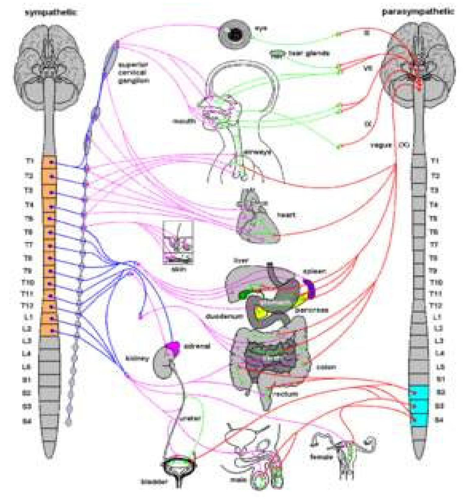
Figure 2.
Right sympathetic trunk and its connexions with abdominal and pelvic plexuses 15 (with permission).
Figure 2.
Right sympathetic trunk and its connexions with abdominal and pelvic plexuses 15 (with permission).
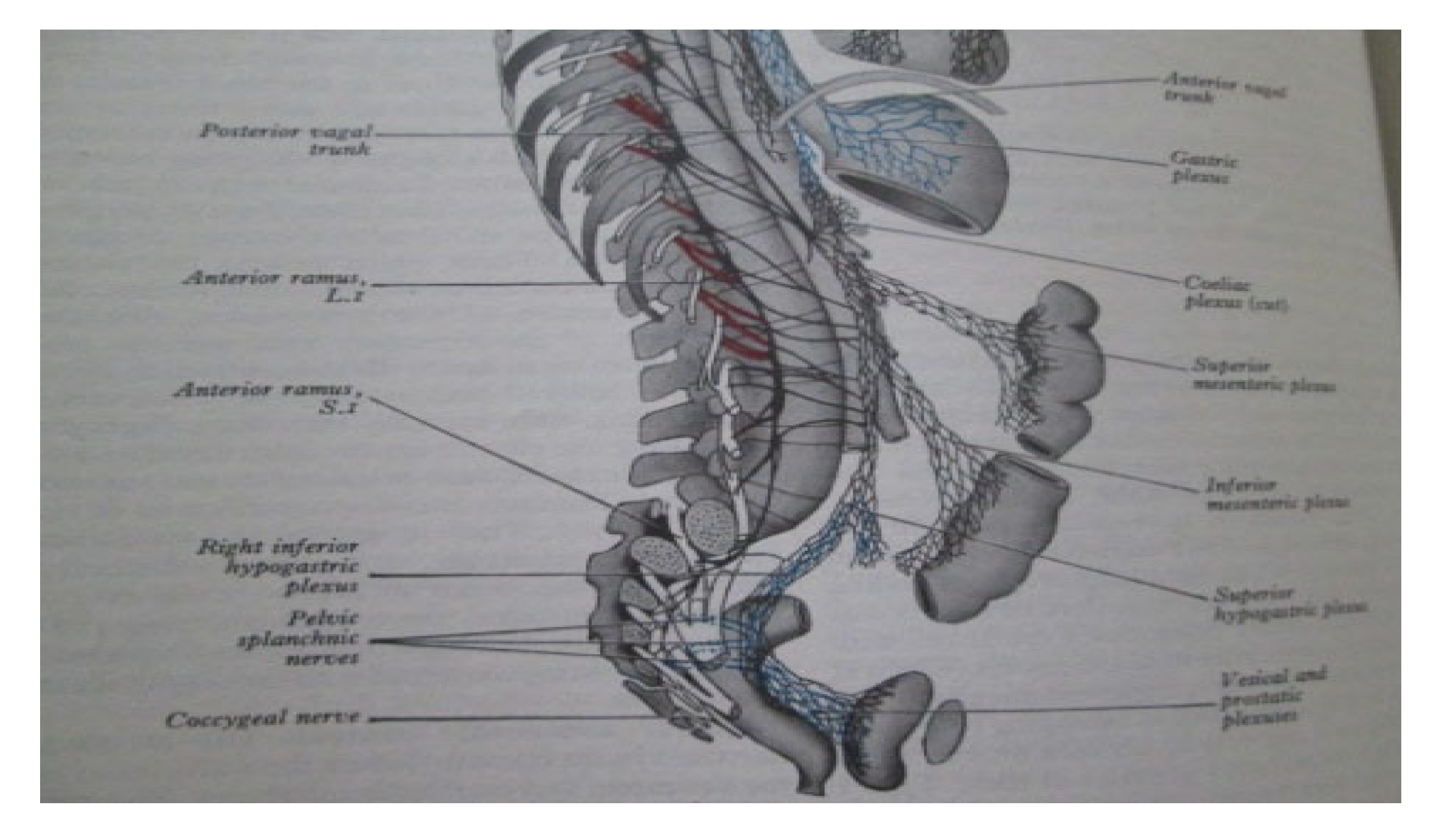
Figure 3.
Schematic diagram of the abdominal and pelvic autonomic nervous pathways 14 (with permission).
Figure 3.
Schematic diagram of the abdominal and pelvic autonomic nervous pathways 14 (with permission).
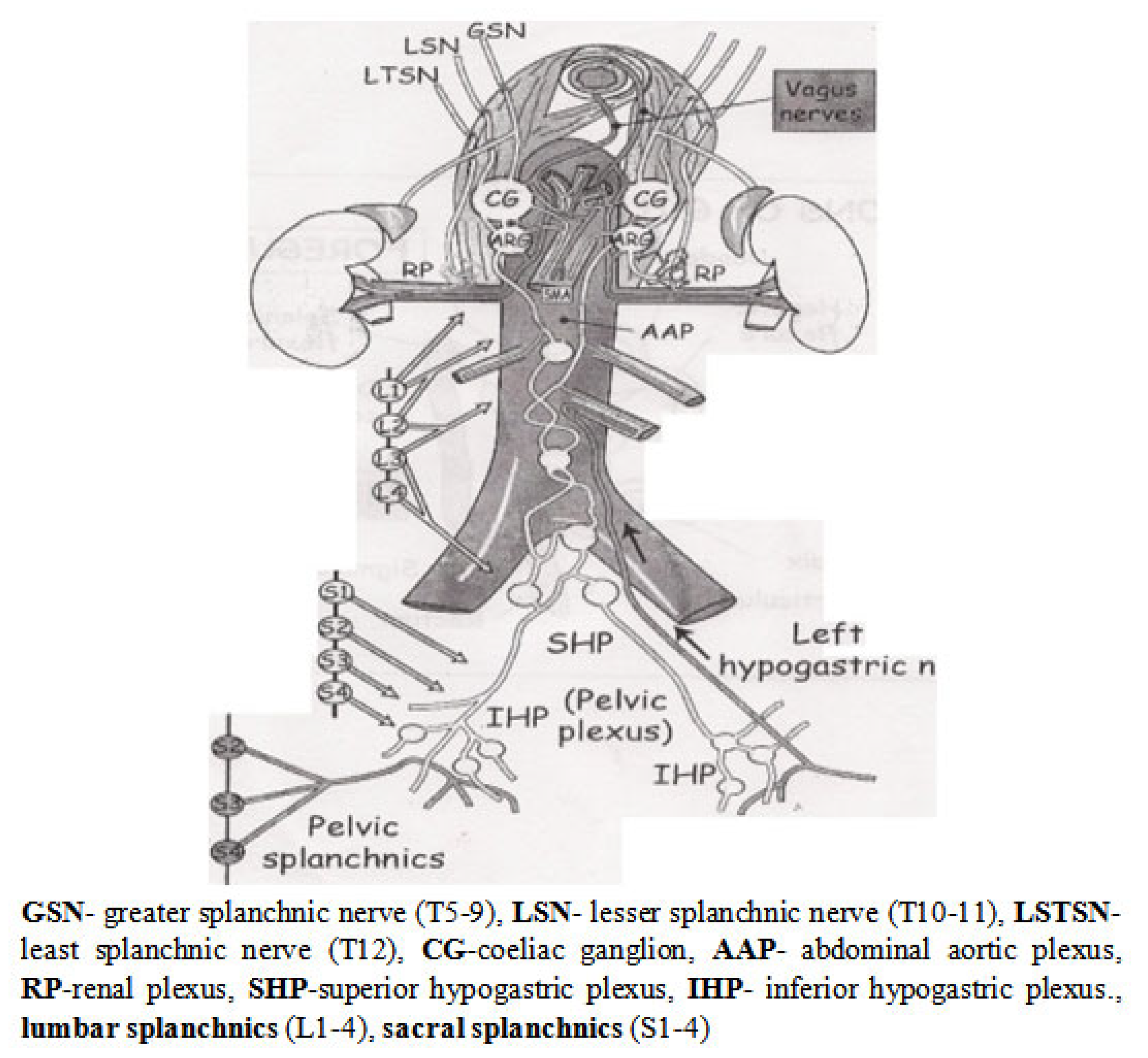
Figure 4.
Schematic diagram of the innervations of the rectum, anal canal and pelvic floor 13.
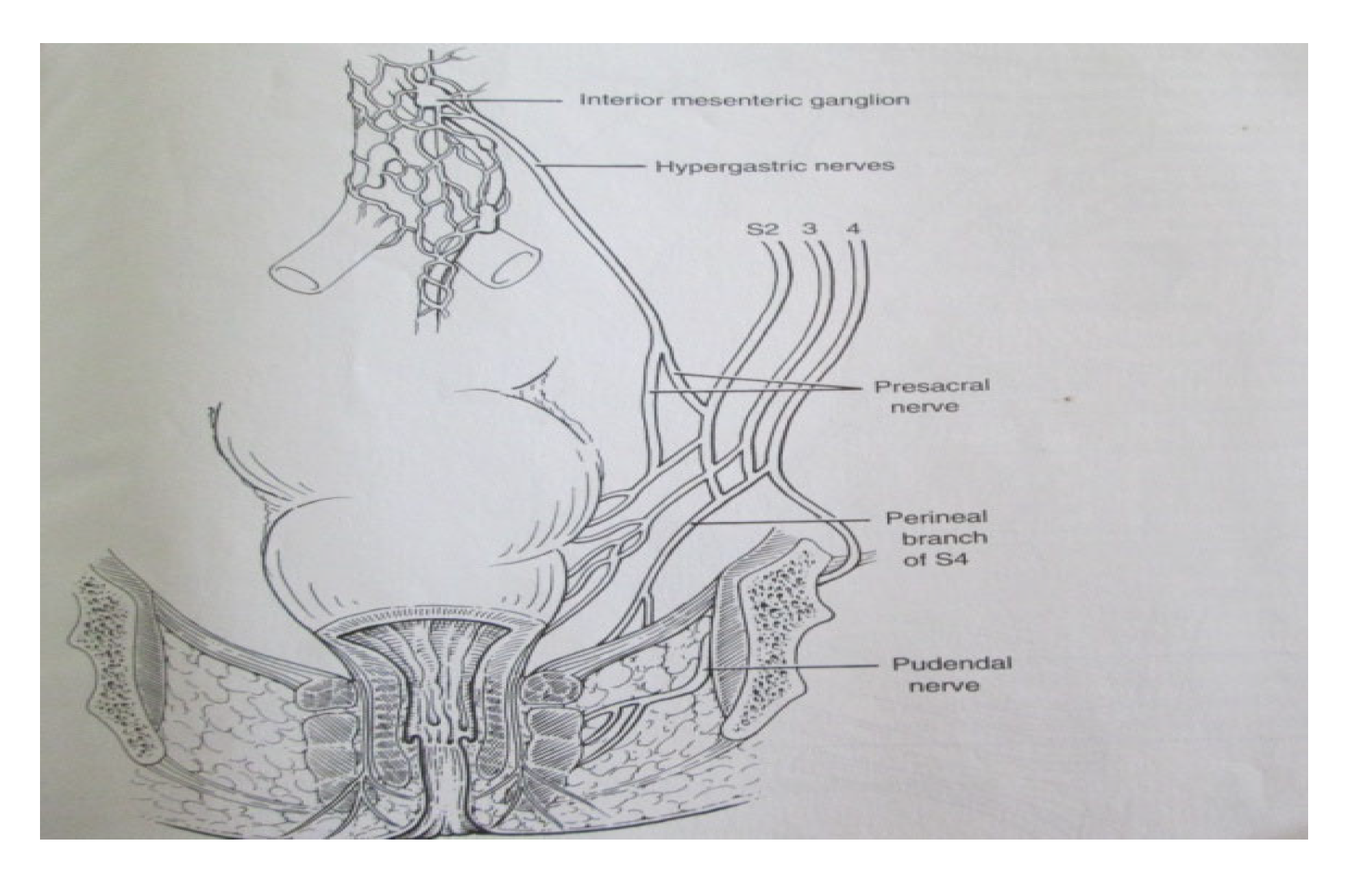
Figure 5.
Schematic diagram of line of total meserectal excision which includes tumour and satellite deposits.
Figure 5.
Schematic diagram of line of total meserectal excision which includes tumour and satellite deposits.
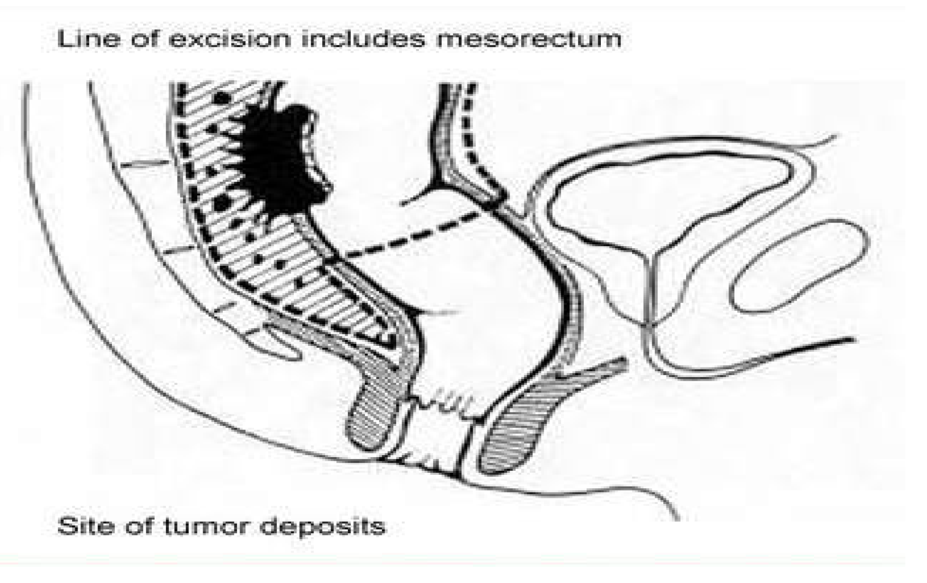
Figure 6.
An ideal total mesorectal excision specimen with an unbreached mesorectum.
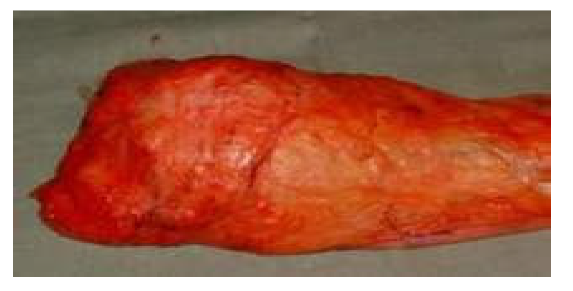
Figure 7.
Schematic diagram illustrating the anterior resection (sphincter preserving procedure) starting behind the root of the hind gut mesentery before leading to the correct plane, the ‘holy plane’ in the pelvis.
Figure 7.
Schematic diagram illustrating the anterior resection (sphincter preserving procedure) starting behind the root of the hind gut mesentery before leading to the correct plane, the ‘holy plane’ in the pelvis.
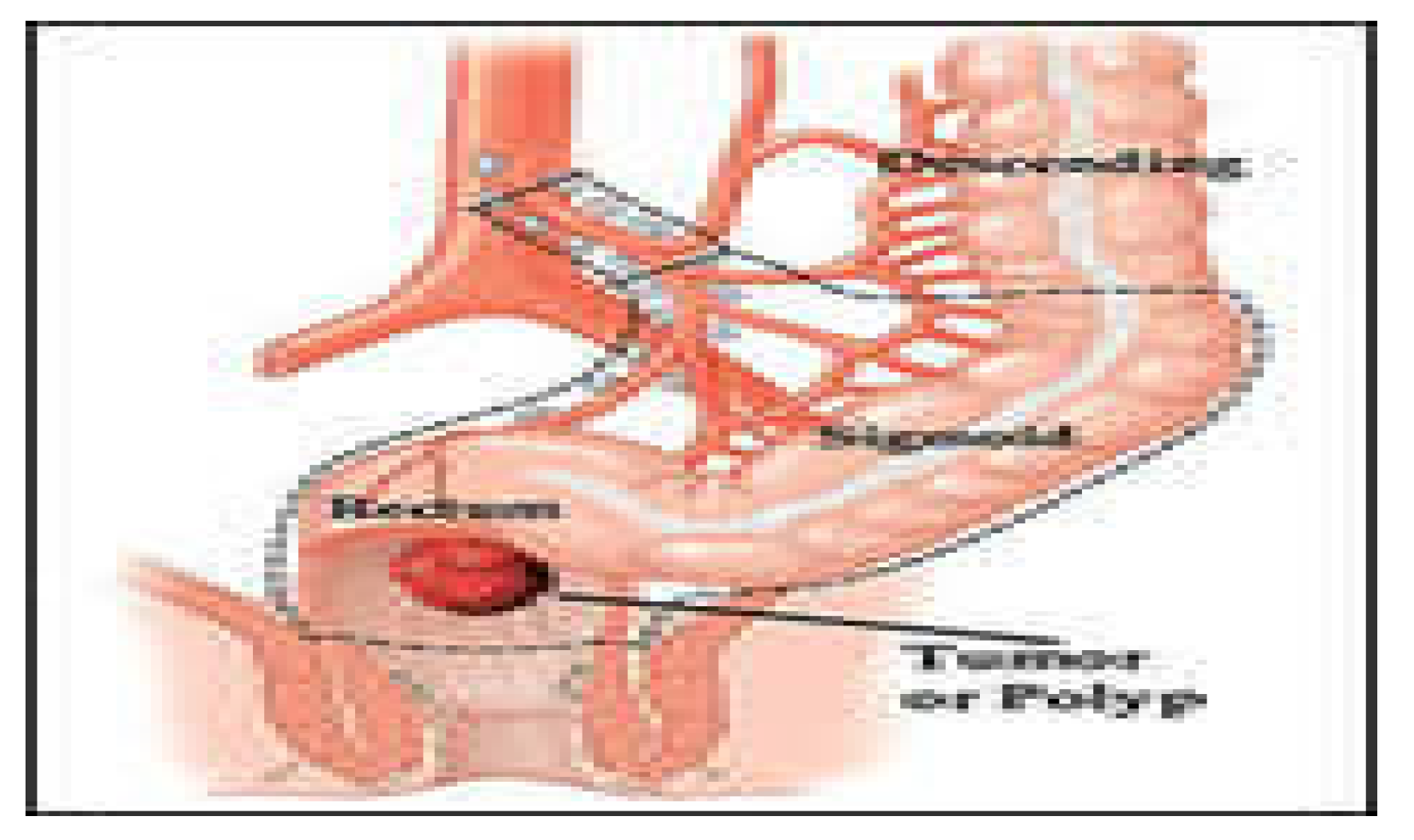
Figure 8.
Intraoperative view of the superior hypogastric plexus, the hypogastric nerves and pelvic inferior hypogastic plexus on the lateral pelvic wall after total mesorectal excision with nerve preservation..
Figure 8.
Intraoperative view of the superior hypogastric plexus, the hypogastric nerves and pelvic inferior hypogastic plexus on the lateral pelvic wall after total mesorectal excision with nerve preservation..
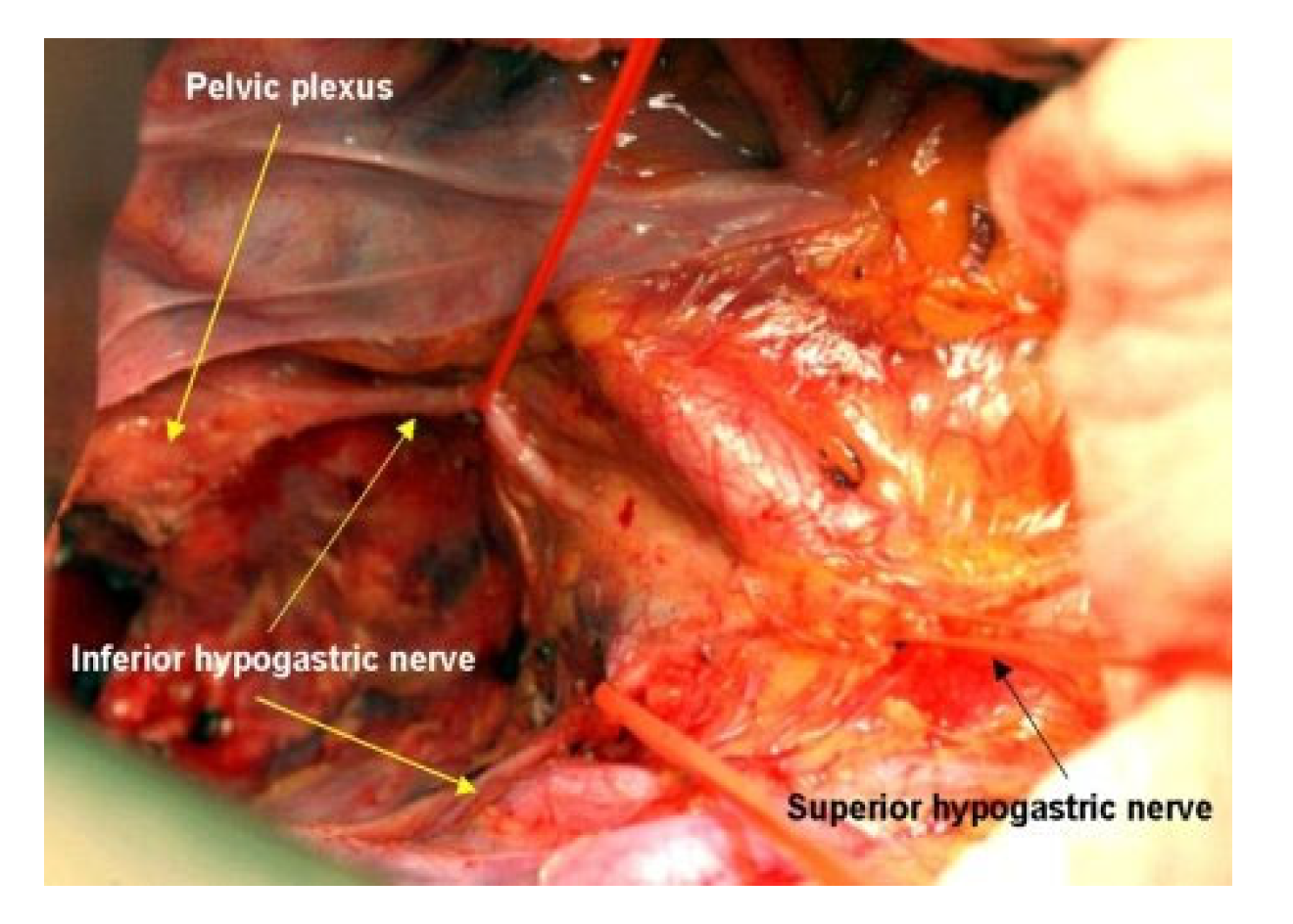
Figure 9.
Robotic view with medial-to- lateral dissection approach of Inferior hypogastric nerve with branches to the rectum. Note the tenting of the inferior hypogastric nerve with medial traction and risk of damage.
Figure 9.
Robotic view with medial-to- lateral dissection approach of Inferior hypogastric nerve with branches to the rectum. Note the tenting of the inferior hypogastric nerve with medial traction and risk of damage.
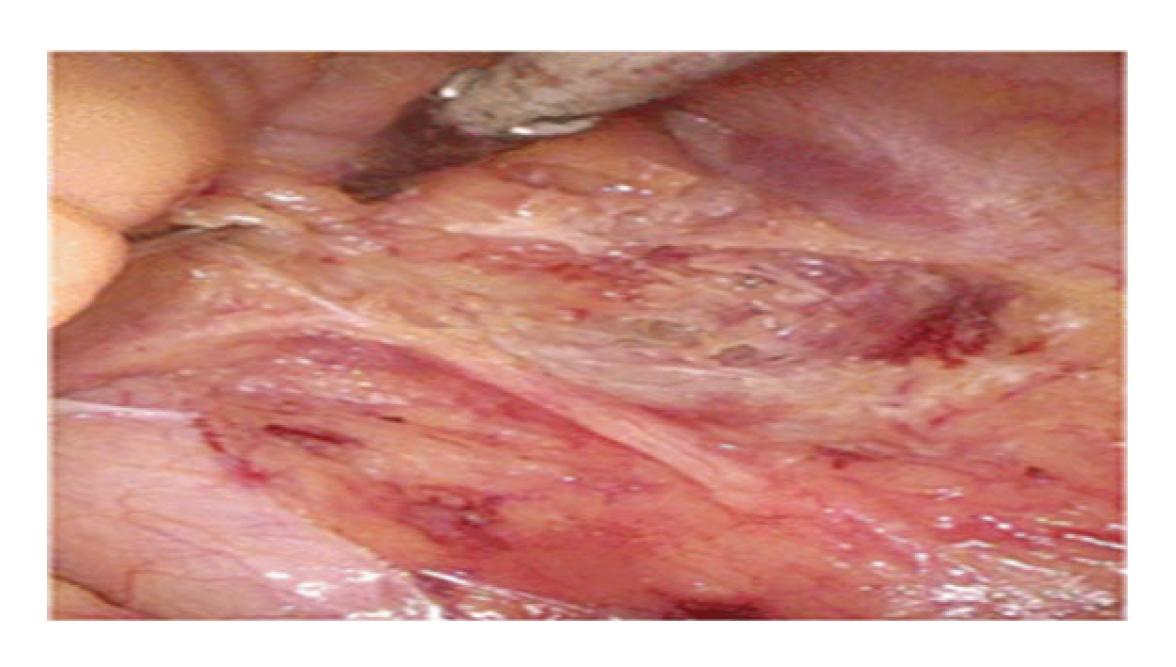
Figure 10.
Schematic diagram of the mesorectum, Denonvillier’s fascia and the antero-lateral neurovascular bundle of Walsh anterior to the fascia propria at the back of the seminal vesicles.
Figure 10.
Schematic diagram of the mesorectum, Denonvillier’s fascia and the antero-lateral neurovascular bundle of Walsh anterior to the fascia propria at the back of the seminal vesicles.
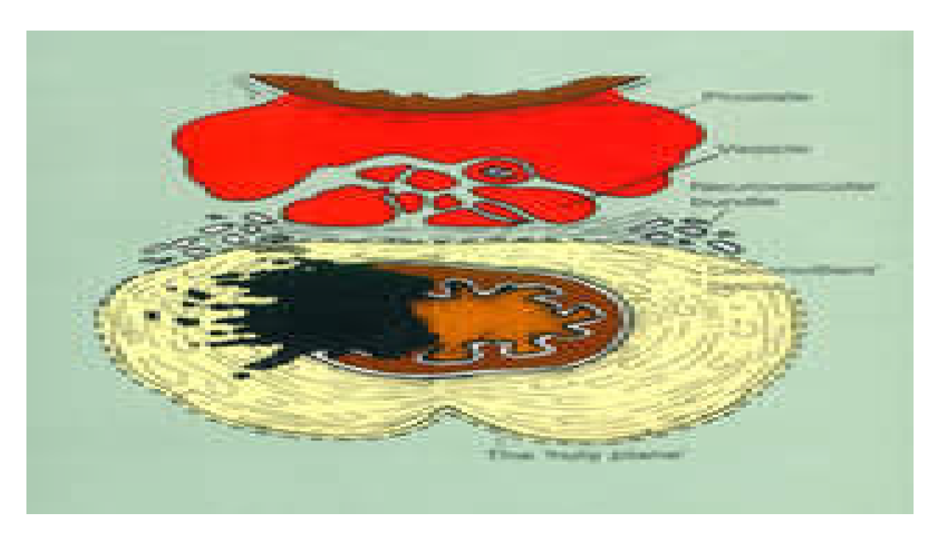
Figure 11.
Preservation of the neurovascular bundles during anterior dissection. The red arrows indicate the correct dissection plane behind the recto-prostatic or recto-vaginal septum, protecting the neurovascular bundles located at 2 and 10 o’clock on the anterior rectal surface. (26).
Figure 11.
Preservation of the neurovascular bundles during anterior dissection. The red arrows indicate the correct dissection plane behind the recto-prostatic or recto-vaginal septum, protecting the neurovascular bundles located at 2 and 10 o’clock on the anterior rectal surface. (26).
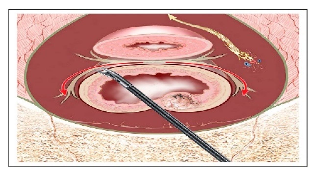
Figure 12.
Intraoperative autonomic nerve monitoring.
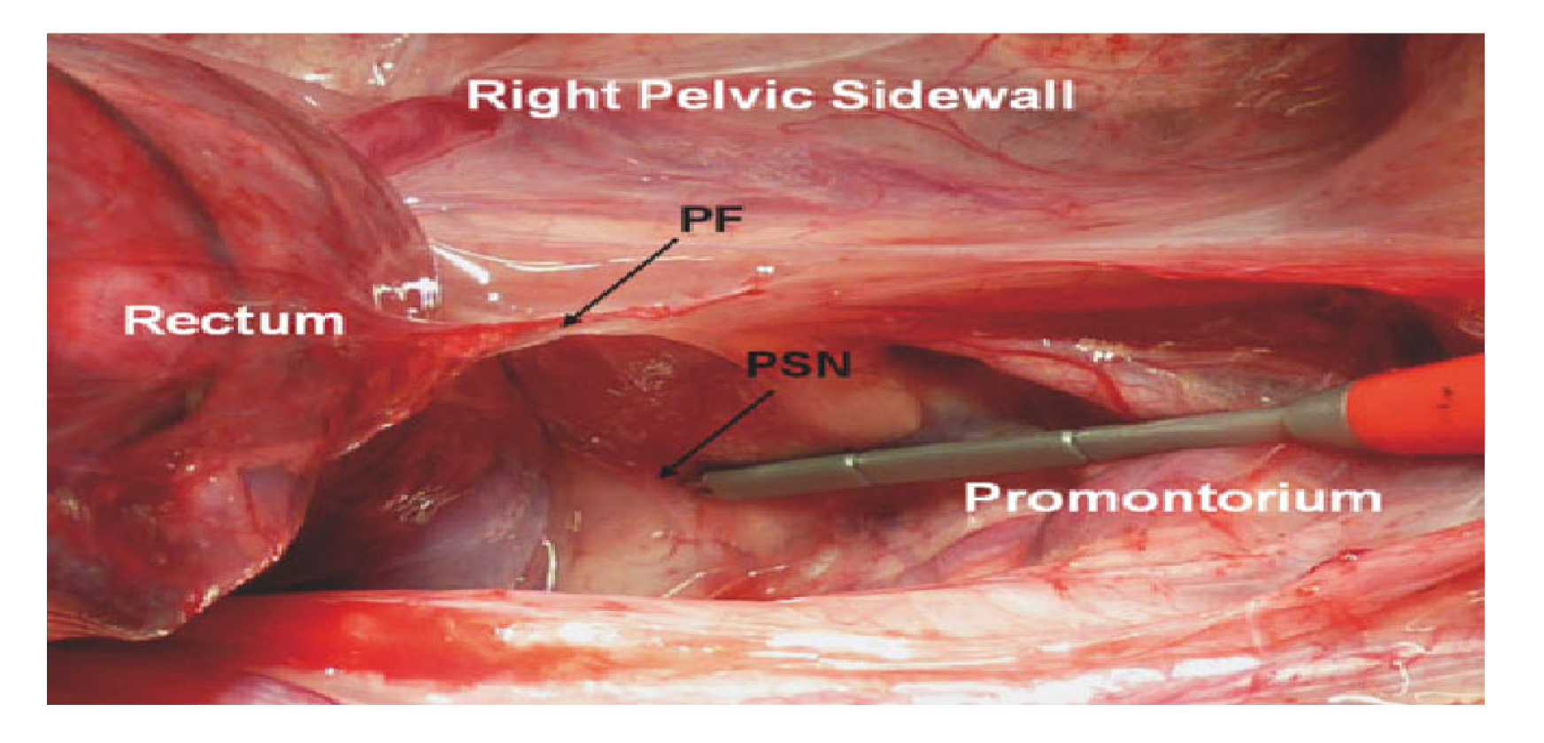
Figure 13.
Transanal TME: Rectal purse -string & antero-lateral mesorectal dissection.
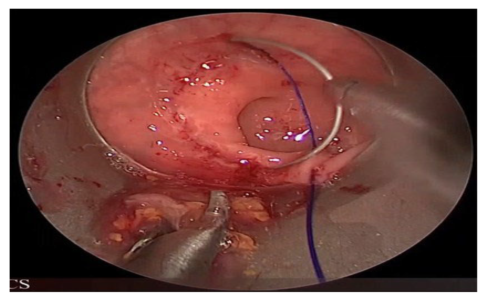

Table 1.
Autonomic dysfunction: conventional rectal cancer surgery vs total mesorectal excision.
| Conventional Rectal Cancer Surgery | Total Mesorectal Excision | |
|---|---|---|
| Accidental bladder denervation | 50-60% | <20% |
| Post-operative impotence | 70-100% | <30% |
Table 2.
Summary of autonomic nervous system pathways and function.
| Sympathetic pathway | Parasympathetic pathway | |
|---|---|---|
| Pre-ganglionic: | anterior roots of T11-T12 ( least splanchnic n) pass via paravertebral sympathetic chain | ant roots of S2-4 cross medial surface of levator ani (pelvic splanchnic nerves) |
| Ganglionic | superior hypogastric plexus | pelvic plexus (inf hypogastric) |
| Post-ganglionic | L& R hypogastric nerves to pelvic plexus | cavervous nerves and vesical branches |
| Function | Ejaculation | voiding and sexual function (erection) |
Table 3.
Summary of autonomic dysfunctions in surgical coloproctology.
| Sexual dysfunction | Urinary dysfunction | |
|---|---|---|
|
Pelvic surgery TME for rectal cancer4,-6, 8-11 Proctocolectomy for UC6, 40, 41 Restorative proctocolectomy40, 41, 60 Colectomy6 |
+++ + + + |
+ + + + |
|
Perineal surgery Haemorrhoidectomy6 ,9, 51, 53 Internal sphincterotomy6, 54, 60 Abdomino-perineal excision for anal/rectal cancer6, 52, 57 |
+ + +++ |
++ ++ + |
+++= very common, ++= common, += not common, UC= Ulcerative colitis.
Disclaimer/Publisher’s Note: The statements, opinions and data contained in all publications are solely those of the individual author(s) and contributor(s) and not of MDPI and/or the editor(s). MDPI and/or the editor(s) disclaim responsibility for any injury to people or property resulting from any ideas, methods, instructions or products referred to in the content. |
© 2026 by the authors. Licensee MDPI, Basel, Switzerland. This article is an open access article distributed under the terms and conditions of the Creative Commons Attribution (CC BY) license (http://creativecommons.org/licenses/by/4.0/).
Copyright: This open access article is published under a Creative Commons CC BY 4.0 license, which permit the free download, distribution, and reuse, provided that the author and preprint are cited in any reuse.