

Submitted:
15 November 2026
Posted:
17 November 2026
You are already at the latest version
Abstract
Despite advances in surgery, radiotherapy, chemotherapy and targeted treatments, resistance remains a major obstacle in nervous system (NS) tumours. Treatment failure reflects not only individual oncogenic alterations but also adaptive regulatory networks that enable tumour cells to survive therapeutic stress, maintain plasticity and adopt therapy-tolerant phenotypes. Within these networks, long non-coding RNAs (lncRNAs) have emerged as potential therapeutic targets because of their spatiotemporal expression patterns, context-dependent functions and ability to coordinate gene-expression programmes. Advances in RNA therapeutics provide tools to exploit these properties by targeting disease-associated transcripts and RNA-dependent interactions that are difficult to address with conventional small molecules or antibodies. Studies in NS tumour models have implicated lncRNAs as determinants of therapeutic responses, including DNA repair, apoptotic thresholds, stem-like states, mesenchymal-like plasticity, invasion, metabolic adaptation and stress responses, as well as intercellular communication within the tumour microenvironment. Targeting oncogenic lncRNAs may weaken resistance programmes and enhance the activity of partner treatments, providing a rationale for combinations that could maintain antitumour efficacy at lower drug doses. In this Review, we discuss the rationale for targeting lncRNAs to influence therapeutic response in NS tumours. The evidence is concentrated in gliomas, particularly glioblastoma, with additional studies in other entities. We summarise preclinical evidence for combining lncRNA-directed interventions with chemotherapy or radiotherapy, distinguishing changes in treatment sensitivity and resistance from quantitatively demonstrated synergy. We discuss targeting strategies, including antisense oligonucleotides, siRNAs and interaction-disrupting approaches, and consider translational challenges related to target selection, context-dependent biology, tumour heterogeneity, intracellular accessibility, delivery to tumours and potential toxicity to normal tissues. Collectively, these findings support lncRNAs as potentially actionable regulatory nodes and provide a foundation for developing more effective and better-tolerated combinatorial strategies in NS tumours.
Keywords:
long non-coding RNAs
; RNA therapeutics
; nervous system tumours
; combination therapy
; precision medicine
; antisense oligonucleotides
; neuro-oncology
1. Introduction: From RNA Biology to RNA Therapy
Recent decades have broadened the range of molecular targets and processes considered amenable to therapeutic intervention. In cancer, including nervous system (NS) tumours, treatment failure can reflect the ability of tumour cells to reorganise regulatory circuits, preserve plasticity and survive therapeutic stress. These adaptive properties highlight the therapeutic relevance of the regulatory layers that shape cellular states and treatment response. RNA, including non-coding RNA, contributes to these processes, and RNA therapeutics have advanced in parallel. Together, the two developments make a disease-associated transcript, or an RNA-dependent mechanism, something a drug can now address directly. This Review examines this transition and its implications for combining long non-coding RNA (lncRNA) targeting with conventional neuro-oncological treatments, drawing primarily on evidence from gliomas, particularly glioblastoma (GBM), with additional studies in medulloblastoma (MB), neuroblastoma (NB), pituitary tumours (PT) and retinoblastoma (RB).
Within the framework of the central dogma, RNA mediates the transfer of genetic information from DNA to proteins (Crick, 1970). Beyond this role, RNA also participates directly in the regulation of gene expression. Complementary base pairing, demonstrated in early studies of RNA duplex formation (Rich & Davies, 1956), provides a molecular basis for many of these regulatory interactions. The same principle can be exploited therapeutically: oligonucleotides can be designed to bind selected transcripts and modulate their abundance or function, extending therapeutic intervention to RNA alongside established protein-directed approaches.
An early proof of concept for transcript-directed therapy emerged in the late 1970s, when antisense oligonucleotides (ASOs) were shown to inhibit viral replication through sequence-specific hybridisation by base complementarity (Stephenson & Zamecnik, 1978; Zamecnik & Stephenson, 1978). Translating this approach into therapeutics, however, was initially constrained by the susceptibility of unmodified oligonucleotides to degradation. Efforts to overcome this limitation led to the adoption of phosphorothioate backbones and subsequent sugar modifications, improving nuclease resistance while also enabling optimisation of pharmacokinetics and target affinity (Y. K. Kim, 2022).
A seminal advance in RNA biology came with the discovery that small RNAs can direct sequence-specific gene silencing. Early silencing observations in plants and fungi (Napoli et al., 1990; Romano & Macino, 1992) were followed by the identification of the first microRNA (miRNA), lin-4, which represses lin-14 translation in C. elegans through sequence complementarity (R. C. Lee et al., 1993; Wightman et al., 1993). RNA interference (RNAi) was subsequently demonstrated in the same organism, where double-stranded RNA induced potent, sequence-specific gene silencing (Fire et al., 1998). Subsequent work defined the machinery. Dicer, an RNase III enzyme, cuts double-stranded RNA into small interfering RNAs (siRNAs). One strand of each siRNA then guides the RNA-induced silencing complex (RISC) to a complementary transcript. In mammalian cells, Argonaute 2 cleaves that transcript when the match is close enough (Ipsaro & Joshua-Tor, 2015). These discoveries established RNA-guided silencing as a fundamental mechanism of gene regulation and provided the basis for its therapeutic exploitation, positioning RNAi alongside antisense as a complementary approach to modulating disease-associated transcripts (Y. K. Kim, 2022). Fomivirsen, approved in 1998 for cytomegalovirus retinitis, became the first ASO approved by the U.S. Food and Drug Administration (FDA), validating transcript-directed therapy despite its later commercial discontinuation (Piascik, 1999; European Medicines Agency, 2002). RNA medicines also diversified beyond transcript degradation. Pegaptanib, approved in 2004 for neovascular age-related macular degeneration, demonstrated high-affinity protein targeting by structured RNA aptamers (B. Zhou & Wang, 2006). Steric-blocking ASOs, including splice-switching agents, extended intervention to RNA processing (S. Chen et al., 2024), allowing restoration of productive gene expression (Y. K. Kim, 2022). Nusinersen, approved in 2016 for spinal muscular atrophy, demonstrated clinically effective splice modulation within the central nervous system (CNS) following intrathecal administration (Finkel et al., 2017), establishing the feasibility of RNA intervention in a pharmacologically challenging tissue. For RNAi, improved chemistry and delivery enabled approval of patisiran in 2018 as the first siRNA medicine (Jadhav et al., 2024), validating therapeutic RNAi after development had been constrained by instability, immune activation, off-target effects and delivery barriers (Y. K. Kim, 2022). Across these modalities, refined sugar modifications, constrained chemistries, hepatocyte-targeting N-acetylgalactosamine (GalNAc) conjugates and lipid nanoparticles improved stability and biodistribution, with potential benefits for the therapeutic index (Das et al., 2022; Haque et al., 2024). These advances expanded local and systemic applications of RNA medicines for muscular (Stein & Castanotto, 2017), neurological (McDowall et al., 2024), metabolic (S. Guo et al., 2024) and rare genetic disorders (A. Singh et al., 2026).
Nucleoside modification and lipid nanoparticles also advanced messenger RNA (mRNA) therapeutics by reducing innate immune sensing and improving intracellular availability. The rapid development and global deployment of COVID-19 mRNA vaccines demonstrated manufacturing scalability and clinical efficacy (Gote et al., 2023; Shi et al., 2024). Within RNA medicine, mRNA vaccines encode antigens to elicit adaptive immunity for prophylactic or therapeutic purposes, whereas non-vaccine approaches modulate disease-associated RNAs, bind protein targets or deliver mRNAs encoding therapeutic proteins.
Regulatory approvals now span ASOs, including splice-switching agents, siRNAs, aptamers and mRNA vaccines (Tani, 2024). By the end of 2025, 24 oligonucleotide medicines had received FDA approval, including 23 RNA therapeutics, encompassing RNA-targeting agents and RNA aptamers (AlShaer et al., 2026). The approval of the ASO zilganersen in September 2026 increased the cumulative count to 25 oligonucleotide medicines (U.S. Food and Drug Administration, 2026). These counts exclude mRNA vaccines. Collectively, RNA medicines enable transcript degradation, RNA-processing modulation, interaction blockade or delivery of genetic information (Teng et al., 2024). Their sequence- and structure-dependent activities expand access to targets poorly served by conventional drugs (Kole et al., 2012), providing a technological basis for investigating lncRNAs as therapeutic targets (Figure 1).
2. LncRNAs: Therapeutic Implications from Biological Features
Since the early 2000s, transcriptome-wide studies have further expanded the landscape of potential RNA targets by showing that mammalian genomes are transcribed far beyond canonical protein-coding genes. The long-standing observation that genome size correlates poorly with organismal complexity had already highlighted the limitations of equating genomic content with biological complexity (Thomas, 1971). Large-scale initiatives such as Functional ANnotation Of the Mammalian genome (FANTOM) and the Encyclopedia of DNA Elements (ENCODE) revealed extensive transcription of non-coding regions across cellular contexts, greatly broadening the catalogue of expressed transcripts (Abugessaisa et al., 2017; Carninci et al., 2005; Derrien et al., 2012; Djebali et al., 2012). These advances brought the non-coding transcriptome into focus as a source of potential regulatory functions, including roles in tissue development and regeneration, and of therapeutic opportunities (Ahmad & Carninci, 2026; Pagano et al., 2020; Taft et al., 2007). However, transcriptional detection alone establishes neither biological function nor druggability: individual candidates require annotation, contextual validation and mechanistic dissection.
Within this expanded landscape, lncRNAs have emerged as a heterogeneous class of transcripts (Figure 2), recently redefined as RNAs longer than 500 nucleotides with little or no protein-coding potential (Mattick et al., 2023; Rinn & Chang, 2012). Many lncRNAs share structural and biosynthetic features with mRNAs, including RNA polymerase II-dependent transcription, 5′ capping, splicing and polyadenylation (Guttman et al., 2009), yet this biogenesis do not imply a common mechanism of action.
Genomic organisation provides a useful descriptive framework, based on position and orientation relative to protein-coding genes (Figure 3). Long intergenic and antisense non-coding RNAs are major components of human lncRNA catalogues and include extensively investigated candidates for therapeutic intervention (Ballarino et al., 2023; Palma et al., 2025). Functionally, lncRNAs have also been described as signals reflecting specific cellular states, decoys that sequester regulatory factors, guides that direct molecular partners to their targets, or scaffolds that support the assembly of molecular complexes. These categories are not mutually exclusive, and individual transcripts may operate through more than one modality (Statello et al., 2021; K. C. Wang & Chang, 2011). Such classifications organise biological diversity, but neither genomic position nor a functional label is sufficient to identify the activity that should be perturbed; this requires consideration of context-dependent expression patterns, subcellular localisation and molecular interactions.
A first distinction concerns the functional entity itself. At some loci, regulatory output depends on promoter or enhancer-like activity, transcription, RNA processing or local chromatin effects rather than on the mature lncRNA product (Engreitz et al., 2016). Accordingly, a phenotype produced by genetic disruption of a locus cannot automatically be attributed to the lack of its RNA or taken to predict the effects of RNA depletion. A further distinction concerns whether a lncRNA acts in cis or in trans, through interactions with genomic regions, RNAs or proteins. This point helps define the relevant mechanisms and downstream readouts. Subcellular localisation connects mechanism to target accessibility, affecting the efficiency of RNA-targeting approaches and informing the choice of intervention (Lennox & Behlke, 2016). Nuclear lncRNAs can regulate chromatin architecture, transcription and splicing, whereas cytoplasmic lncRNAs can influence mRNA stability, translation and signalling (Figure 3, panels 1-12). Some cytoplasmic lncRNAs can bind microRNAs and thereby reduce microRNA-mediated repression of other transcripts. This competing endogenous RNA (ceRNA) mechanism is widely invoked in oncology. Effective competition depends on the relative abundance, interaction strength, and colocalisation of the lncRNA with the relevant microRNAs and their other targets. Finer partitioning, including enrichment in nuclear ribonucleoprotein compartments, adds further specificity (Statello et al., 2021; K. C. Wang & Chang, 2011). Paraspeckles, for example, are phase-separated nuclear bodies assembled around the architectural lncRNA NEAT1_2 (Statello et al., 2021). More broadly, subcellular localisation can influence the efficiency of lncRNA silencing by RNA-targeting reagents (Lennox & Behlke, 2016). The intervention must therefore reach the compartment in which the relevant RNA activity occurs.
Beyond subcellular localisation, transcript levels, expression specificity and evolutionary conservation also shape the design and evaluation of lncRNA-directed interventions. Compared with mRNAs, lncRNAs are generally less abundant, more tissue- and cell-state-specific, and less conserved in primary sequence. Conservation can nevertheless persist at promoters and splice sites, in short sequence motifs or structural modules, and in syntenic positions. Evolutionary constraint therefore does not require long stretches of identical sequences across the lncRNA body (Cabili et al., 2011; Mattick et al., 2023; Rinn & Chang, 2012). Overall, these heterogeneous properties shape therapeutic development, linking lncRNA biology to target selection, intervention choice and experimental-model suitability.
3. LncRNAs in Cancer
In cancer, the above-discussed lncRNAs’ properties create opportunities and constraints. Bulk and single-cell transcriptomic studies can help identify lncRNAs with disease- and tissue-type-restricted expression, which may favour selective therapeutic intervention, whereas inter-individual variability can inform the selection of relevant patient populations (Coan et al., 2024; Lavorgna et al., 2016). Low abundance, incomplete annotation and repetitive sequences can complicate transcript quantification and sequence-specific intervention, while limited sequence conservation and the scarcity of recognisable orthologues constrain the predictive value of non-human preclinical models (Hezroni et al., 2015; Necsulea et al., 2014). In cancer, lncRNA expression and function are shaped by aberrant transcriptional programmes, somatic mutations, copy-number changes and epigenetic alterations (Esposito et al., 2023; Ramnarayanan et al., 2026; Zhou et al., 2023). By contributing to the regulatory programmes that sustain malignant cell identity and adaptation, lncRNAs may expose vulnerabilities that complement those associated with protein-coding drivers (Huarte, 2015; Schmitt & Chang, 2016). However, individual lncRNAs can support oncogenic or tumour-suppressive functions depending on context (Mello et al., 2017; Adriaens et al., 2016). Target prioritisation therefore requires not simply a catalogue of dysregulated lncRNAs but also evidence that perturbing the relevant RNA activity alters a malignant phenotype, alongside assessment of its functions in normal cells (Coan et al., 2024).
Several lncRNAs that helped establish the field illustrate these principles. H19, one of the first identified imprinted lncRNAs, participates in developmental regulation at the IGF2/H19 locus and is aberrantly re-expressed in numerous malignancies. Its reported contributions to tumour progression and therapeutic resistance involve ceRNA and epigenetic mechanisms (L. Li et al., 2024). H19 also gives rise to miR-675, which can reduce retinoblastoma protein (pRB) expression and promote growth in colorectal cancer cells (Tsang et al., 2010). The contributions of the full-length RNA and its processed products should therefore be distinguished when selecting the target and interpreting the effects of inhibition.
XIST, originally characterised for its role in X-chromosome inactivation, is also implicated in tumour progression and treatment response through chromatin regulation, immune responses and ceRNA networks (J. Yang et al., 2021). Its physiological role raises a specific issue for target selection: deletion of Xist in the haematopoietic compartment of female mice induced haematological malignancies by reactivating the inactive X chromosome and raising X-linked gene dosage (Yildirim et al., 2013). This genetic evidence makes tissue context and preservation of normal X-linked gene dosage relevant considerations when evaluating XIST inhibition. HOTAIR provided an early link between lncRNA-mediated chromatin regulation and breast-cancer metastasis through Polycomb-associated epigenetic reprogramming (Gupta et al., 2010). Biochemical studies identified distinct RNA domains interacting with Polycomb repressive complex 2 (PRC2) and lysine-specific demethylase 1 (LSD1)-containing complexes (Tsai et al., 2010), while subsequent work linked HOTAIR to stem-like properties, invasion and resistance to chemotherapy (Zhu et al., 2022). Similarly, HOTTIP links chromatin activation to developmental gene regulation: it binds the adaptor protein WDR5 and recruits WDR5/MLL histone methyltransferase complexes to promote histone H3 lysine 4 (H3K4) trimethylation and HOXA gene expression (K. C. Wang et al., 2011). It has also been linked to proliferation, metastatic dissemination and poor prognosis in solid tumours (Lian et al., 2016) and leukaemogenesis in haematological malignancies. Such mechanisms make RNA depletion and disruption of specific RNA-protein interactions distinct design options, whose effects should be assessed against the relevant transcriptional programme.
NEAT1 deregulation has been linked to tumour progression, chemoresistance and radioresistance (K. Li et al., 2021; Long et al., 2024), but the contributions of its isoforms need to be distinguished. The long isoform NEAT1_2 provides the architectural scaffold for paraspeckles, whereas selective loss of the overlapping short isoform NEAT1_1, accompanied by increased NEAT1_2 expression, did not reproduce the phenotypes of complete NEAT1 loss in the models examined (Adriaens et al., 2019). The shared 5′ sequence affects the isoform specificity of RNA-targeting reagents and target-engagement assays.
MALAT1 illustrates why the direction of intervention also requires validation. Although overexpressed in numerous cancers and implicated in treatment response (Bhat et al., 2024), MALAT1 suppressed metastasis in experimental breast-cancer models by binding TEAD transcription factors and limiting their association with the transcriptional coactivator YAP; genetic restoration reversed the metastatic phenotype caused by MALAT1 loss (J. Kim et al., 2018). These findings support using loss-of-function and RNA re-expression experiments in the relevant disease model to establish whether inhibition is appropriate, rather than inferring benefit from elevated expression alone.
Beyond individual examples, lncRNAs can affect coordinated gene-expression programmes. Large-scale loss-of-function profiling has connected lncRNA depletion to transcriptomic changes and cellular phenotypes (Ramilowski et al., 2020). In cancer, this provides a rationale for investigating whether targeting a single lncRNA may modulate multiple disease-relevant pathways. This rationale is particularly relevant where durable responses are limited by adaptive plasticity and treatment resistance. Evidence implicating lncRNAs in responses to chemotherapy and radiotherapy (Cheng et al., 2024), as well as immunotherapy (Arshi et al., 2024; Pan et al., 2023), supports investigating whether their modulation can alter therapeutic response, provided that the relevant mechanism is experimentally established and can be selectively perturbed, as discussed below for NS tumours.
4. LncRNAs in NS Tumours
4.1. The NS Tumour Context
The NS, and particularly the brain, is characterised by exceptional cellular and molecular complexity and requires finely tuned gene-expression control across development and function, from regional patterning, lineage specification and neuronal or glial differentiation to circuit formation, synaptic plasticity and activity-dependent responses (L. Wang et al., 2025; Sugo et al., 2025). These processes depend on regulatory layers with strong spatial, temporal and cell-type specificity. LncRNAs are well suited to this biology (Coan et al., 2024), as their spatially restricted expression and regulatory activities connect gene-expression control with processes ranging from cellular differentiation to subcellular organisation (Mercer et al., 2008; Ng et al., 2012; Sunwoo et al., 2009; Y. He et al., 2025). This is also relevant to NS tumours, which can co-opt developmental programmes and adopt lineage-associated or plastic cellular states (Hovestadt et al., 2019; Neftel et al., 2019; Vladoiu et al., 2019; Williamson et al., 2022). LncRNAs associated with these programmes may also be retained or repurposed in malignant cells, potentially contributing to the maintenance of tumour-cell state and adaptation to therapeutic stress.
The 2021 World Health Organization (WHO) classification of CNS tumours is based on integrated histological and molecular diagnosis and distinguishes biologically distinct tumour groups and molecularly defined entities with diagnostic, prognostic and, in some cases, therapeutic implications. These include adult-type diffuse gliomas, paediatric-type diffuse low- and high-grade gliomas, circumscribed astrocytic gliomas, glioneuronal and neuronal tumours, ependymal tumours, CNS embryonal tumours, meningiomas, mesenchymal tumours, cranial and paraspinal nerve tumours, pineal tumours and tumours of the sellar region, among others (Louis et al., 2021). Within this heterogeneous framework, the disease-specific evidence reviewed here is concentrated in adult-type diffuse gliomas, especially GBM, with additional studies in embryonal tumours such as MB (Laneve et al., 2019; Laneve & Caffarelli, 2020). These settings are particularly informative because tumour subtype, developmental identity, cell-state plasticity and treatment resistance are closely intertwined. In MB, molecular subgroup identity, oncogenic programmes and heterogeneity within subgroups (Cavalli et al., 2017; Northcott et al., 2012) provide a framework for investigating how subgroup-associated lncRNAs relate to tumour identity and potential therapeutic dependencies. In GBM, single-cell analyses have identified coexisting neural-progenitor-like, oligodendrocyte-progenitor-like, astrocyte-like and mesenchymal-like malignant cell states, whose relative abundance and interconversion link genetic alterations to intratumoural heterogeneity and plasticity (Neftel et al., 2019).
This tumour diversity is also reflected in lncRNA expression profiles, which have been associated with molecular subtypes and patient prognosis (Katsushima et al., 2021; Reon et al., 2016; Huang et al., 2021).
Further examples from other NS tumours are also discussed below, including NB, a tumour of the peripheral NS in which lineage-associated transcriptional programmes and plasticity between adrenergic and mesenchymal states link developmental identity to treatment response (Boeva et al., 2017; van Groningen et al., 2017). The intensity of studies in these contexts may partly reflect the convergence of established treatment regimens, unmet needs related to resistance or treatment-associated toxicity, and the availability of experimental models for functional perturbation and treatment-response assessment. Moreover, the lncRNAs discussed in this review are primarily expressed by NS tumour cells or cells of the associated microenvironment. Their intercellular transfer, including through extracellular vesicles (section 5.8), should be distinguished from transport of peripherally produced RNAs across the blood-brain barrier (BBB), for which the studies reviewed here provide no evidence.
4.2. Combinatorial Targeting: Biological Rationale and Treatment Design
Combination treatment integrates two or more therapeutic interventions, administered concurrently or sequentially, to increase the magnitude or durability of response, address tumour heterogeneity, overcome or delay resistance, and potentially maintain efficacy at lower treatment doses. It encompasses associations between drugs or between different modalities, including RNA-directed interventions, chemotherapy, radiotherapy and immunotherapy.
Within this framework, lncRNAs represent potential therapeutic entry points alongside established treatment modalities. In NS tumours, their relevance extends beyond direct cytotoxicity to changes in the cellular response to therapeutic stress. LncRNA targeting may act as a priming or sensitising intervention, as discussed below, lowering the threshold (or restoring the dose) at which radiotherapy, temozolomide (TMZ), cisplatin, vincristine or other agents convert DNA damage, mitotic stress or metabolic pressure into irreversible loss of fitness or cell death. This is particularly pertinent to GBM, where DNA-repair capacity and adaptive cellular programmes influence the response to standard chemoradiotherapy (Hegi et al., 2005; Neftel et al., 2019; Stupp et al., 2005). A functional screen in glioma illustrates this approach (S. J. Liu et al., 2020). CRISPR interference (CRISPRi), which represses transcription at targeted loci, identified lncRNAs whose perturbation altered radiosensitivity. Subsequent ASO-mediated depletion of lncGRS-1 enhanced the response to radiation in a human brain organoid model containing glioma cells. Although the transcript was also expressed in nonmalignant neural cells, its depletion did not compromise their viability in the models examined (S. J. Liu et al., 2020, see section 5.2). This example nicely illustrates that selective antitumour activity can arise from a preferential tumour dependency without requiring tumour-restricted expression.
Mechanistically, lncRNA-directed interventions may reinforce inhibition at multiple levels within the same regulatory pathway (vertical targeting) or impair parallel and compensatory programmes (horizontal targeting), including survival signalling, stem-like persistence, mesenchymal-like plasticity, metabolic adaptation and cytoprotective stress responses. Combinations can also be designed to address distinct tumour-cell populations or interactions between malignant cells and their microenvironment. In glioma, pharmacological combinations provide relevant precedents for coupling treatment-induced damage with disruption of cellular defence mechanisms. Poly(ADP-ribose) polymerase 1 (PARP-1) inhibition increased TMZ sensitivity in resistant GBM cell lines, illustrating the potential of interfering with DNA repair to enhance the effects of an alkylating agent (Montaldi et al., 2020). A complementary strategy exploits lipid-metabolic dependencies: fluoxetine-mediated inhibition of acid sphingomyelinase (SMPD1) perturbed sphingolipid metabolism, suppressed epidermal growth factor receptor (EGFR) signalling and induced lysosomal stress, enhancing the antitumour activity of TMZ in GBM models (Bi et al., 2021). These pharmacological examples illustrate how disruption of repair or adaptive programmes can enhance the activity of a partner treatment, providing a mechanistic rationale that also informs lncRNA-directed combinations.
Genetic interactions also inform combinatorial strategies, with synthetic lethality providing a specific biological rationale. It describes an interaction in which disruption of either of two genes individually is compatible with cell survival, whereas their combined disruption is lethal. Therapeutically, this relationship can be exploited by targeting a dependency created by a pre-existing tumour alteration, as exemplified by PARP inhibition in BRCA1/2-deficient cells, or by designing combinations that disrupt both functions (Bryant et al., 2005; Farmer et al., 2005). For lncRNA-directed strategies in brain tumours, this suggests testing whether inhibition of a lncRNA-dependent repair or survival programme creates a dependency on a complementary, druggable pathway.
Transcript depletion with ASOs, including gapmers, or siRNAs, and functional interference with steric-blocking oligonucleotides or other interaction-disrupting approaches provide different means of testing these possibilities, according to the relevant RNA function, subcellular localisation and the intended mode of inhibition (Arun et al., 2018; Lennox & Behlke, 2016). Their integration with other treatments also has a temporal dimension. Priming describes an initial intervention that changes the cellular state and thereby modifies the response to a subsequent treatment. Sequential and concurrent administration can consequently produce different outcomes (M. J. Lee et al., 2012). For lncRNA targeting, this makes the onset and persistence of RNA depletion or functional inhibition relevant to the timing of the accompanying treatment. The translational potential of these approaches also depends on achieving effective modulation of the relevant RNA function in the appropriate cell populations within heterogeneous tumours, at tolerable exposures. Intracranial tumours pose additional CNS-specific delivery constraints, as discussed below.
4.3. Combinatorial Targeting: Interpretation of Effects and Therapeutic Benefit
The effects of lncRNA-directed combinations encompass several related biological outcomes. Sensitisation denotes increased responsiveness to a treatment following lncRNA modulation, without requiring pre-existing resistance. In models with intrinsic resistance, present before treatment, or acquired resistance, developing during treatment or following an initial response, lncRNA modulation may attenuate the resistant phenotype and, where sensitivity has been lost, restore responsiveness (resensitisation). H19 and MALAT1 depletion in TMZ-resistant glioma cells illustrate this possibility (P. Jiang et al., 2016; W. Chen et al., 2017). Resensitisation can be partial, and concerns recovered responsiveness to the partner treatment, beyond the cytotoxic contribution of lncRNA inhibition itself. Preventing or delaying the emergence of resistance represents a complementary objective, extending the potential value of combinations from response magnitude to durability. These biological outcomes are related to, but distinct from, the quantitative interaction between treatments. Additivity describes a combined effect consistent with the expectation of a specified non-interaction model; synergy and antagonism describe antitumour effects greater or smaller than this expectation, respectively. The additive expectation is therefore not necessarily the arithmetic sum of the individual effects, and a combination can outperform either intervention alone while remaining additive. Sensitisation and resensitisation may accompany different interaction classifications. Several analytical approaches capture these relationships. Loewe additivity is based on dose equivalence, comparing doses in combination with those producing the same effect individually, whereas Bliss independence predicts the combined response from the individual fractional effects under an assumption of independent action (Demidenko & Miller, 2019; Ianevski et al., 2022). In Chou-Talalay analysis, combination index (CI) values below, equal to or above 1 indicate synergy, additivity or antagonism, respectively (Chou, 2010). These different assumptions, together with variation in dose, schedule, endpoint and biological context, explain why interaction classifications can differ across analyses and experimental settings (Ianevski et al., 2022). A shift in the half-maximal inhibitory concentration (IC₅₀) captures a change in apparent drug sensitivity, whereas synergy concerns how the combined response compares with the model-predicted effect. For lncRNA-directed approaches, interpretation also depends on the extent and duration of RNA perturbation achieved through genetic inhibition, oligonucleotide transfection or therapeutic delivery. Stable genetic depletion does not provide a conventional dose-response relationship for the RNA-directed intervention, limiting the applicability of Loewe and Chou-Talalay analyses. However, interaction can still be assessed using a Bliss framework when the individual and combined effects are quantified against a common reference (Demidenko & Miller, 2019). This methodological constraint is relevant to the interpretation of studies in Section 5 that use pre-implantation genetic manipulation. The therapeutic relevance of a combination extends beyond its interaction classification. Additive combinations may provide useful antitumour activity, while population-level benefit can also arise through independent drug action when different patients respond to different components of a regimen (Palmer & Sorger, 2017). Synergy is therefore neither necessary nor sufficient for clinical value. Another potential advantage is dose sparing: maintenance of a given antitumour effect at lower doses of one or more components, a relationship that can be quantified by the dose-reduction index (Chou, 2010). Whether enhanced activity or dose sparing translates into an improved therapeutic index depends on the accompanying toxicity profile, including effects on normal tissues, both neural and non-neural, and those introduced by the RNA-targeting intervention or its carrier. The following sections examine lncRNA-regulated pathways involved in treatment response and the strategies available to reach and target them, highlighting the biological rationale and the experimental evidence supporting combinations in NS tumour models.
5. LncRNA-Mediated Pathways Shaping Treatment Response in NS Tumours
5.1. A Framework for Reading the Evidence
The pathways discussed below focus on lncRNAs for which experimental perturbation has been linked to altered treatment response in NS tumour models (Figure 4). Across cancer types, lncRNAs intersect with core pathways governing therapeutic sensitivity and resistance (Grillone et al., 2024; Mitra et al., 2017), providing potential points of intervention in tumours characterised by cellular plasticity and adaptive survival programmes, particularly high-grade gliomas (Neftel et al., 2019; Singh Tomar et al., 2021). This provides a rationale for combining lncRNA-directed interventions with existing treatments to enhance antitumour activity and counter resistance.
Here we focused on diffuse gliomas, particularly GBM, with additional studies in MB and NB and more limited reports in pituitary neuroendocrine tumours and RB. Examples from NB, pituitary neuroendocrine tumours and RB are discussed in a dedicated subsection (Section 5.10), considering their distinct biological and therapeutic contexts, anatomical barriers and delivery approaches. Given the heterogeneity of experimental designs and pharmacological characterisation, each example is assessed using the criteria summarised in Table 1. Each example is assigned to one of seven ascending evidence tiers: (E1) descriptive association only, based on differential expression or prognostic correlation; (E2) mechanistic validation in vitro, supported by interaction assays and rescue experiments; (E3) modulation of drug or radiation response in vitro only; (E4) modulation of treatment response in vivo, but using tumour cells in which the lncRNA was genetically manipulated before implantation (an asterisk marks contextual E4 entries supported by in vivo target validation but with combination activity demonstrated only in vitro, as specified in Table 1 and Figure 5.); (E5) administration of an RNA-targeted agent to an established tumour, in combination with the partner treatment; (E6) as E5, with demonstrated target engagement, pharmacodynamic readout and formal safety assessment in a clinically relevant model; and (E7) clinical investigation in patients. The distribution of the reported examples across these tiers is shown in Figure 5 and in Table 1.
5.2. DNA-Damage Response and Repair
TMZ is an alkylating agent central to the treatment of high-grade gliomas, typically in combination with radiotherapy, and exerts its cytotoxic effect by inducing DNA damage that triggers cell-cycle arrest and cell death when repair mechanisms fail. Mechanistically, TMZ methylates DNA at several purine sites, the O6-methylguanine lesion being particularly relevant: when not directly repaired by O6-methylguanine-DNA methyltransferase (MGMT), it mispairs with thymine during replication and engages mismatch-repair-dependent processing, promoting replication-associated DNA damage and cytotoxic stress responses (Aasland et al., 2019).
Several lncRNAs strengthen the tumour DNA-repair capacity or DNA-damage tolerance, thereby weakening drug effects. Specifically, the species discussed below act on distinct nodes of this axis, such as MGMT transcription, MGMT mRNA export and protein availability, double-strand break repair, replication-stress tolerance, nucleotide supply and cytoprotective autophagy.
The lncRNA HOTAIR regulates chromatin programmes that support survival and cell-cycle progression. HOTAIR expression is markedly upregulated in TMZ-resistant GBM cells, while its lentiviral shRNA-mediated depletion inhibited proliferation, migration, invasion and mesenchymal features and reduced TMZ resistance in flank xenografts receiving daily TMZ. Mechanistically, HOTAIR acts through a miR-519a-3p/RRM1 axis. It is transferred between cells through exosomes and has been proposed as an accessible circulating biomarker for exosomal lncRNAs (Yuan et al., 2020). In U251 cells, CRISPR-Cas9-mediated deletion of HOTAIR regulatory element locus increased TMZ sensitivity through long-range regulation of CALCOCO1 and ZC3H10, identified by combining RNA-seq, Capture Hi-C and 3C analysis (L. Zhang et al., 2020). Independently, siRNA-mediated HOTAIR silencing restored TMZ responsiveness in vitro through the miR-214/β-catenin/MGMT axis (Lan et al., 2024).
Other lncRNAs converge on MGMT itself, and the two examples below illustrate regulation at the transcriptional and post-transcriptional levels respectively. LINC00473 is induced downstream of CREB activation and promotes chemoresistance by binding CEBPα and thereby enhancing MGMT transcription. Its subcellular distribution also varies with the resistant state: LINC00473 is predominantly cytoplasmic in TMZ-sensitive U251 cells but relocates to the nucleus in TMZ-resistant derivatives, consistent with the acquisition of a transcriptional function. Exosomal LINC00473 also transferred chemoresistance to adjacent TMZ-sensitive cells, a process validated in vivo (L. Y. Jiang et al., 2024).
LINC01956 provides an advanced example of lncRNA-associated DNA-repair modulation investigated through ASO administration to established intracranial tumours. LINC01956 drives TMZ resistance even in MGMT promoter-methylated GBM, a context that would ordinarily be expected to confer lower MGMT expression and greater TMZ sensitivity. Mechanistically, TMZ-activated CHK1 phosphorylates the RNA helicase DDX46 at T886, producing a dense-to-loose conformational change that increases its unwinding activity; DDX46-mediated tertiary remodelling of LINC01956 exposes binding motifs to the 3’UTR of MGMT, accelerating recruitment of MGMT mRNA to the nuclear export machinery, including NXF1, ALYREF, THOC1 and ZC3H11A, and increasing cytoplasmic abundance of MGMT mRNA and MGMT protein.
Three therapeutic strategies were tested against this axis (Liao et al., 2024). First, ASOs targeting LINC01956 were administered to mice bearing established intracranial tumours from patient-derived glioma cells, together with TMZ at 50 mg/kg five days per week, starting once bioluminescence reached a defined threshold: LINC01956 ASO alone did not affect tumour growth, whereas the combination reduced growth, increased TMZ-induced apoptosis and O6-methylguanine accumulation, reduced MGMT expression and prolonged survival. Second, two steric-blocking ASOs designed to disrupt the LINC01956-MGMT mRNA interaction, rather than to degrade the transcript, abolished TMZ-induced nuclear export of MGMT mRNA, illustrating interaction-focused targeting of a defined RNA-RNA interface. Third, the CHK1 inhibitor SRA737 abolished the remodelling event upstream; combined SRA737 and TMZ treatment was analysed with CalcuSyn and produced an enhanced antitumour effect in LINC01956-transduced but not LINC01956-low tumours, indicating target-dependent synergy (Liao et al., 2024).
DNA-repair-associated resistance can also be propagated between tumour cells through extracellular vesicles. In GBM, ZEB1-induced SBF2-AS1 acts through a miR-151a-3p/XRCC4 regulatory axis that supports DNA double-strand break repair, and its depletion delayed the repair of TMZ-induced DNA damage (Z. Zhang et al., 2019). Lentiviral shRNA-mediated SBF2-AS1 depletion increased TMZ responsiveness in resistant cells and in subcutaneous xenografts established from depleted cells. SBF2-AS1 was itself transferred in exosomes from TMZ-resistant donor cells to initially sensitive recipient cells, and intracranial experiments using recipient cells preincubated with these vesicles supported the contribution of exosomal SBF2-AS1 to resistance in vivo (Z. Zhang et al., 2019).
A related cell-extrinsic mechanism involves lnc-TALC, a transcript originally identified in TMZ-resistant GBM cells, where it sustains MGMT expression by competitively binding miR-20b-3p and activating the c-Met/Stat3/p300 axis, thereby promoting acetylation of H3K9, H3K27 and H3K36 at the MGMT promoter (P. Wu et al., 2019). Beyond this tumour-cell-intrinsic function, lnc-TALC is packaged into GBM-derived exosomes and transferred to microglia and tumour-associated macrophages, in which it promotes M2-like polarisation. Mechanistically, exosomal lnc-TALC binds ENO1 and promotes phosphorylation of p38 MAPK in recipient microglia, thereby enhancing MEF2C-dependent transcription of complement C5, as supported by chromatin immunoprecipitation, luciferase-reporter and MEF2C-knockdown experiments. Microglia-derived C5a in turn increased pATM, pATR and RAD51 and reduced γH2AX in GBM cells subsequently exposed to TMZ, indicating accelerated resolution of drug-induced DNA damage, while comet and apoptosis assays confirmed reduced TMZ-induced damage and cell death; a C5a receptor antagonist (C5aRa) reversed these effects in vitro (Z. Li et al., 2021). In an orthotopic, immunocompetent C57BL/6 model established with GL261 cells overexpressing lnc-TALC or with TMZ-resistant 261R cells, systemically administered C5aRa combined with TMZ suppressed tumour growth and prolonged survival relative to TMZ alone, whereas the same combination produced no significant survival benefit in tumours derived from control cells. No pathological changes in visceral organs, haematological toxicity or impairment of hepatic and renal function were observed with C5aRa (Z. Li et al., 2021). This example connects lncRNA-dependent DNA repair to myeloid-cell activity, provides one of the few instances in which benefit is confined to tumours with high activity of the resistance pathway, and pairs TMZ with blockade of a downstream secreted effector rather than with an agent directed against lnc-TALC.
Among the lncRNAs experimentally targeted in this context, MIR155HG is markedly upregulated in glioma compared with normal controls, with higher expression correlating with WHO grade and poorer survival. siRNA-mediated depletion increased glioma sensitivity to TMZ in vitro and in subcutaneous xenografts treated with TMZ at 50 mg/kg/day, an effect reversed by MIR155HG overexpression. Mechanistically, MIR155HG binds polypyrimidine tract binding protein 1 (PTBP1), as demonstrated by RNA immunoprecipitation, and activates Wnt/β-catenin signalling; co-transfection of PTBP1 siRNA into MIR155HG-overexpressing cells reversed the resistant phenotype, establishing mechanistic dependency (X. He et al., 2020).
XLOC013218 (XLOC) is markedly upregulated in TMZ-resistant cells and associated with high glioma grade and poor prognosis in a 73-patient GBM cohort and in the TCGA dataset (J. Zhou et al., 2022; Zhou et al., 2025). Its subcellular distribution also varies with the resistant state: it is mainly cytoplasmic in parental cells but enriched in the nuclei of TMZ-resistant derivatives. Mechanistically, XLOC binds specificity protein 1 (Sp1), shown by RNA immunoprecipitation, and promotes Sp1 occupancy at the PIK3R2 promoter, increasing PIK3R2 expression and activating PI3K/AKT signalling; shRNA-mediated Sp1 knockdown attenuated the effect (J. Zhou et al., 2022; Zhou et al., 2025). Both subcutaneous and intracranial four-arm xenograft experiments were performed, with TMZ given intraperitoneally at 5 mg/kg every three days (J. Zhou et al., 2022; Zhou et al., 2025).
Two other lncRNAs were shown to influence drug tolerance rather than repair enzymes directly. The lncRNA CRNDE has been proposed as a prognostic indicator and candidate biomarker of the TMZ response in GBM. siRNA-mediated depletion in vitro, and shRNA-mediated depletion in subcutaneous xenografts subsequently treated with TMZ at 5 mg/kg, enhanced TMZ sensitivity, reduced tumour growth, impaired TMZ-induced cytoprotective autophagy with a reduced LC3 II/I ratio and lower Beclin 1 and Atg5 levels, modulated PI3K/Akt/mTOR signalling and decreased expression of the multidrug-resistance transporter ABCG2 (Z. Zhao et al., 2021). Another example is MALAT1, which is also upregulated in GBM, particularly in TMZ-resistant tumours, where it suppresses miR-203 and increases thymidylate synthase expression, supporting the nucleotide supply required for DNA replication and repair; plasmid-mediated overexpression increased the TMZ IC₅₀ by 4.60-fold and 5.87-fold in U87 and U251 cells respectively, whereas siRNA-mediated silencing reduced thymidylate synthase expression and resensitised resistant cells in vitro (W. Chen et al., 2017).
A translationally distinct mechanism involves TUG1-dependent resolution of R-loops and tolerance of replication stress (Suzuki et al., 2023). ATR-CHK1 signalling rapidly induces TUG1 under replication stress, with E2F1 recruited to and E2F6 excluded from its promoter. TUG1 resolves R-loops by interacting with phosphorylated replication protein A 32 and the RNA helicase DHX9; 63% of TUG1-sensitive regions contain (CA)n and (TG)n repeats. Depletion with chemically modified chimeric DNA ASOs increased R-loop accumulation, replication stress and apoptosis and enhanced TMZ-induced apoptosis in GBM cells. This is one of the few examples in which the interaction was formally quantified in a tumour type. Combination indices calculated with CompuSyn 1.0 were 0.84, 0.67, 0.50 and 0.53 in LN229 at TMZ concentrations of 1.0 and 2.0 µM, and 0.38, 0.33, 0.31 and 0.25 in U251MG at TMZ concentrations of 250 and 500 µM, in each case combined with TUG1 ASO at 50 and 100 nM. CI values were lower in U251MG under the conditions tested. These cells also had lower MGMT promoter methylation than LN229 (23.2% versus 82.7%, measured by pyrosequencing) and required higher TMZ concentrations. In orthotopic xenografts, ASO treatment begun two weeks after implantation and combined with TMZ suppressed tumour growth and prolonged overall survival more effectively than either treatment alone, with reduced intratumoural TUG1 expression and increased R-loop accumulation (Suzuki et al., 2023). The administration schedules, group sizes, pharmacodynamic assays and subsequent clinical development are described in Section 5.11.
The mechanism also has implications beyond acute cytotoxicity. Although all examined cell lines, including the GBM models, were initially microsatellite stable, continuous TUG1 depletion for more than one week increased instability at the three CA-repeat-containing dinucleotide markers of the Bethesda panel, D2S123, D5S346 and D17S250. This occurred in mismatch repair-proficient cells without reducing MLH1, MSH2, MSH6, PMS1 or PMS2 expression. The authors interpreted these findings as induction of high microsatellite instability and proposed combining TUG1 ASOs with immune checkpoint inhibitors in settings where this phenotype is uncommon (Suzuki et al., 2023). This proposed combination remains distinct from the experimentally evaluated association with TMZ.
The helicase partner DHX9 offers a related intervention point. The inhibitor ATX968 was investigated in mismatch repair-deficient cancers, a setting distinct from the microsatellite-stable GBM models examined here (Castro et al., 2025; Suzuki et al., 2023). Genetic loss of DHX9 has also been reported to be lethal in mice (Suzuki et al., 2026), although this observation does not establish the safety profile of pharmacological inhibition or a comparative safety advantage for TUG1 targeting.
The rationale for increasing R-loop burden is tempered by their protective as well as damaging roles. At stalled replication forks, transiently formed RNA/DNA hybrids are protective rather than damaging, shielding reversed forks from excessive nucleolytic degradation by DNA replication helicase/nuclease 2 until they are resolved by the helicase DDX39A, a step facilitated by RAD51 (Suzuki et al., 2026). The consequences of manipulating R-loop homeostasis are therefore context-dependent, and the assumption that any increase in R-loop burden will be selectively cytotoxic to tumour cells is not yet established. A related point concerns the partner agent: TMZ itself activates ATR-CHK1 signalling in GBM cells, together with p21 and NF-κB, and induces senescence and repression of DNA-repair pathways (Aasland et al., 2019).
Finally, systematic approaches have begun to identify DNA-damage-relevant lncRNA dependencies that become visible only under treatment pressure. A CRISPRi screen of 5,689 lncRNA loci in GBM cells, performed under clinically relevant fractionated radiation delivered in 2 Gy doses every other day, identified 467 growth-modifying hits, of which 33 sensitised cells to radiation and nine were prioritised as lncRNA glioma radiation sensitisers (lncGRS) (S. J. Liu et al., 2020). ASOs targeting lncGRS-1 decreased tumour growth and sensitised glioma cells to radiotherapy, with the combined effect exceeding an explicit additive-effect prediction, and lncGRS-1 was reported to be selectively required by glioma cells rather than by normal neural cells (S. J. Liu et al., 2020).
5.3. Apoptosis Control
Apoptosis is a genetically controlled cell-death programme that contributes to tissue homeostasis and to the elimination of damaged or stressed cells. Its deregulation is a central feature of cancer, where tumour cells frequently acquire the ability to raise the apoptotic threshold and survive oncogenic stress, DNA damage, nutrient limitation, hypoxia and therapeutic injury (Elmore, 2007). In NS tumours, resistance to chemotherapy and radiotherapy can reflect both enhanced repair or adaptive signalling and failure to convert treatment-induced stress into irreversible cell death. Accordingly, lncRNAs that modulate pro- and anti-apoptotic pathways may influence tumour persistence, treatment tolerance and therapeutic sensitivity.
NEAT1 is elevated in recurrent relative to primary gliomas and in TMZ-resistant cells is present in both nuclear and cytoplasmic fractions of U87MG and A172 cells, and acts through a miR-454-3p/Connexin 43 axis, supported by RNA immunoprecipitation and rescue by GJA1/Cx43 overexpression. Three independent siRNAs were used in vitro, and a lentiviral shRNA (short hairpin RNA) was used for stable cellular models. NEAT1-depleted U87MG-Luc cells were implanted intracranially into the striatum of nude mice, which then received TMZ at 20 mg/kg intraperitoneally daily for five days: NEAT1 depletion enhanced TMZ sensitivity and extended survival (J. Liang et al., 2024).
The lncRNA H19 is consistently overexpressed in TMZ-resistant gliomas and has emerged as a regulator of chemoresistance. Concomitant treatment of resistant glioma clones with siRNA targeting H19 and with TMZ decreased the TMZ IC50 and increased apoptotic rates relative to control-siRNA-treated cells, with increased PARP cleavage. Reduced H19 expression also altered major drug-resistance genes including MDR, MRP and ABCG2 at both mRNA and protein levels (P. Jiang et al., 2016). More recently, H19 was shown to act as a molecular decoy for miR-138-5p and miR-22-3p, thereby sustaining BMP2: shRNA-mediated silencing restored TMZ sensitivity in U87TR and LN229TR cells and reduced tumour volume and mass in subcutaneous xenografts from mice receiving oral TMZ at 66 mg/kg/day for five days, whereas BMP2 overexpression abolished the effect (Q. Chen et al., 2025).
In Group 3 MB, where MYC acts as a major oncogenic driver (Rea et al., 2021), the MYC-dependent lncRNA lncMB3 provides a disease-relevant example of how lncRNA-mediated apoptotic control can influence tumour-cell sensitivity to cytotoxic stress. Mechanistically, lncMB3 sustains tumour-cell survival through a cytoplasmic RNA-RNA interaction with the HMGN5 transcript, as confirmed by native RNA pulldown after subcellular fractionation, resulting in translational activation of HMGN5, modulation of TGF-β-dependent signalling and reinforcement of anti-apoptotic programmes. Inhibition with an LNA-based ASO (GapmeR), verified with a second, independent GapmeR directed to the 3’ end of the transcript, lowers the apoptotic threshold and sensitises tumour cells to cytotoxic agents. This effect is particularly relevant in combination with cisplatin, a DNA-damaging agent that forms platinum-DNA adducts and crosslinks, leading to replication stress and apoptosis (Grandioso et al., 2025). Interaction analysis using the Chou-Talalay method confirmed a synergistic effect (combination index below unity) with cisplatin and an additive effect (combination index of approximately unity) with vincristine, a microtubule-disrupting agent used in MB regimens. The same study characterises a ferritin-based nanocarrier (HFt-HIS-PASE) for delivery of the anti-lncMB3 GapmeR, linking target biology to a delivery approach, although the combination data remain in vitro (Grandioso et al., 2025). This is a clear current demonstration in a paediatric CNS tumour that mechanistically complementary routes produce genuinely distinct interaction profiles with diverse partner drugs, whose differences are measurable.
5.4. Stemness and Cellular Plasticity
Cancer stem-like cells (CSCs) are functionally defined tumour subpopulations contributing to intratumoural heterogeneity, tumour propagation and recurrence (Chu et al., 2024) due to their self-renewal capacity and ability to generate more differentiated progeny. These properties resemble core features of normal stem cells, whose behaviour is regulated by developmental pathways such as those Wnt-, Notch- or Hedgehog-dependent, which can be co-opted by tumour cells to sustain stemness, plasticity and survival (X. Fan et al., 2006). In NS tumours, stem-like states are particularly relevant because they may persist after chemotherapy and radiotherapy, contribute to relapse and limit durable therapeutic efficacy. Several lncRNAs have emerged as regulators of CSC-associated programmes, influencing self-renewal, differentiation state, signalling-pathway activity and treatment tolerance (Hu et al., 2022). LncRNA TP73-AS1 is significantly overexpressed in GBM, particularly in GBM stem-like cell cultures, where it contributes to aggressiveness and TMZ resistance. TP73-AS1 is linked to metabolism-related transcriptional programmes and to the expression of ALDH1A1, a stemness-associated detoxifying enzyme that protects stem-like cells from TMZ-induced cytotoxicity. TP73-AS1 repression, which was achieved by CRISPRi, using a guide RNA directed to a catalytically inactive Cas9 fused to a transcriptional repressor domain, caused increased TMZ sensitivity and cell death in treated cultures (Mazor et al., 2019). Although described as synergy in the original report, the combination therapy effect is classified here as in vitro sensitisation because no combination index or other reference-model analysis was reported. Nevertheless, this example is informative for modality selection, since it demonstrates that transcriptional silencing at the locus is sufficient to reverse a drug-tolerant state.
In the same biological compartment, another example is provided by GSCAR, a cytoplasmic-enriched, glioma stem-like-cell-associated lncRNA that sustains self-renewal and TMZ resistance by stabilising SOX2 mRNA by promoting complex formation between DHX9 and IGF2BP2. LncRNA loss-of-function was achieved with both shRNA- and ASO-based strategies. GSCAR shRNA-mediated depletion impaired self-renewal in tumour sphere assays and reduced xenograft formation, while ASOs targeting inhibited tumour growth in vivo both alone and in combination with TMZ (X. Jiang et al., 2023). Another example is represented by NEAT1 which contributes to GBM stem-cell maintenance and chemoresistance through the let-7g-5p/MAP3K1 axis, with serum NEAT1 raised as a possible circulating biomarker (C. L. Bi et al., 2020).
In MB, the lncRNA RBM5-AS1 links stem-like phenotypes to radiation response. RBM5-AS1 shRNA-mediated depletion reduced sphere formation and stemness-associated markers in DAOY cells and, in an acquired radioresistant derivative, diminished the surviving fraction after irradiation and enhanced radiation-induced apoptosis. Mechanistically, RBM5-AS1 interacted with and stabilised SIRT6 protein, and SIRT6 re-expression counteracted the radiosensitising effect of depletion, supporting a functional contribution of this interaction. In a four-arm subcutaneous xenograft experiment, RBM5-AS1-depleted DAOY cells exposed to a single 10 Gy dose produced smaller tumours than irradiated control cells (C. Zhu et al., 2021). These findings support preclinical radiosensitisation, without establishing that stemness loss causes the altered radiation response. The evidence derives from cells genetically modified before implantation, ectopic tumours and single-dose irradiation (C. Zhu et al., 2021).
5.5. Invasion and Infiltrative Growth
Among the biological processes that limit therapeutic efficacy in NS tumours, invasion is particularly relevant, because it promotes diffuse tumour spread, incomplete surgical control, recurrence and adaptive resistance. This is especially evident in GBM, where infiltrative growth into the surrounding brain parenchyma is associated with aggressive behaviour and poor outcome. In this context, lncRNAs are emerging as regulators of invasive behaviour, extracellular-matrix remodelling and cytoskeletal dynamics, making them candidate adjunctive targets to complement current standard-of-care treatments (Neftel et al., 2019).
HMMR-AS1 is an antisense lncRNA upregulated in GBM that stabilises the HMMR transcript, which encodes the hyaluronan-mediated motility receptor RHAMM, thereby sustaining signalling that supports proliferation, migration, invasion and mesenchymal-like phenotypes (J. Li et al., 2018). Both siRNA-mediated transient depletion and lentiviral shRNA-mediated stable depletion were used; depletion reduced HMMR expression and suppressed migratory and invasive behaviour, mesenchymal traits and tumour growth. Importantly, cells transfected with HMMR-AS1 shRNA were more sensitive to X-ray irradiation, and its depletion reduced expression of DNA-repair-associated proteins including ATM, RAD51 and BMI1. This is one of the few examples in this section evaluated in an orthotopic irradiated setting: U87-luc cells were transplanted into the right striatum of nude mice, which received whole-brain radiotherapy of 12 Gy delivered as 3 Gy on four consecutive days. The intervention remains a pre-implantation genetic manipulation (J. Li et al., 2018).
TALNEC2 is an E2F1-regulated lncRNA highly expressed in GBM, particularly in glioma stem-like cultures and in tumours from patients with poor prognosis; its cytoplasmic distribution was established by fractionation in U2OS, MCF-7 and H1299 cells and was not measured in any glioma line. siRNA-mediated silencing inhibited proliferation, induced G1/S arrest, reduced self-renewal and mesenchymal features, and increased the sensitivity of stem-like cells to γ-irradiation in vitro. In vivo, silencing prolonged median survival of mice bearing stem-cell-derived xenografts by 12 days, although no irradiated arm was included in the animal experiment, with radiosensitisation supported only in vitro, as the animal experiment did not include irradiation. The perturbation was a transient siRNA transfection performed two days before implantation rather than a stable genetic manipulation. Mechanistically, silencing altered miRNA networks involved in proliferation, cell-cycle regulation and mesenchymal transition; miR-21 and miR-191 were downregulated after depletion, and rescue experiments indicated that both miRNAs mediate part of the TALNEC2-dependent effects, while a direct molecular interaction remains unestablished (Brodie et al., 2017).
5.6. Proneural-to-Mesenchymal Transition
Mesenchymal transition is a key manifestation of GBM plasticity and is closely linked to invasiveness, stem-like states and resistance to radiotherapy and chemotherapy (Leung et al., 2023). Proneural-to-mesenchymal transition (PMT) may represent its GBM equivalent, and it is associated with other aggressive cancers. LINC01057 is a lncRNA overexpressed in GBM, particularly in the mesenchymal subtype, where it promotes PMT, proliferation, invasion and radioresistance through activation of NF-κB signalling. Nuclear-cytoplasmic fractionation showed that the transcript is localised in both compartments, and RNA immunoprecipitation demonstrated an endogenous interaction with IKKα. Mechanistically, LINC01057 maintains IKKα nuclear localisation and thereby increases chromatin accessibility at NF-κB-responsive promoters through histone-modification-dependent mechanisms, with phosphorylation of p65 reduced upon depletion while total p65 remained unchanged. In vivo, intracranial injection of LN229-CD44+ cells carrying LINC01057 shRNA vectors reduced tumour growth, as measured by bioluminescence at three weeks, and prolonged survival. Radiosensitisation was demonstrated in vitro, while the animal experiment did not include irradiation (Tang et al., 2021).
MIR222HG acts in a related but more complex mesenchymal programme. This nuclear lncRNA is overexpressed in mesenchymal GBM tissues and stem-like cells and is transcriptionally induced by SPI1/PU.1. MIR222HG promotes PMT, self-renewal, tumourigenicity and radioresistance by anchoring to histone H4 and binding the YWHAE/HDAC5 complex, leading to H4K16 deacetylation and consequent activation of STAT3 and ERK-MAPK signalling. ASO-mediated depletion was used as the primary loss-of-function approach, complemented by lentiviral shRNA for stable models; depletion enhanced the radiotherapy response, attenuated tumourigenesis and prolonged survival in tumour-bearing mice receiving four 2.5 Gy fractions. The BRAF inhibitor PLX-4720 further increased DNA damage, apoptosis and G2/M arrest when combined with radiotherapy in SPI1-high mesenchymal models (Y. Fan et al., 2023).
In GBM, MALAT1 is amongst the best-characterised oncogenic lncRNAs. A particularly informative study investigating its role in treatment response showed that TMZ treatment itself induces MALAT1 expression in an NF-κB- and p53-co-dependent manner, through κB- and p53-binding sites located in the proximal MALAT1 transcribed region. Specifically, TMZ reduces p50 recruitment in a Ser329 phosphorylation manner while increasing p53 recruitment. The use of siRNAs against MALAT1 encapsulated in a nanoparticle vector was delivered by direct intracranial injection on days 4 and 7 after tumour inoculation, with intraperitoneal TMZ, sensitised patient-derived GBM cells to TMZ; the combination increased survival relative to all other groups (P < 0.02), with a comparable effect in hindlimb xenografts. The reported approximately 50% knockdown was measured in U87 cells in vitro; in vivo target-engagement data were obtained from the hindlimb, rather than the intracranial, model. The same study found that MALAT1 expression within GBM tissue was not prognostic of overall survival by in situ hybridisation, in TCGA or in REMBRANDT, although it was higher in recurrent than in primary gliomas (Voce et al., 2019).
A distinct transcript contributes to the same mesenchymal programme. TGF-β-regulated lncRNA-MUF, which is upregulated in GBM, promotes Snail1 expression by acting as a competing endogenous RNA for miR-34a, while its depletion impairs TGF-β-induced SMAD2/3 phosphorylation. The resulting programme drives expression of mesenchymal markers and enhances migration, invasion and proliferation. Two independent siRNAs against lncRNA-MUF suppressed these phenotypes, and depletion at a low dose of 20 nM combined with TMZ at 600 μM increased caspase 3/7 activity approximately five-fold in T98G and LN229 cells relative to control-transfected cells treated with TMZ alone (Shree et al., 2022).
Finally, the lncRNA DLEU1 provides an example of how lncRNAs can link invasive behaviour to adaptive chemoresistance. Its overexpression promotes glioma-cell proliferation, migration and invasion through regulation of mesenchymal factors including ZEB1, Snail, β-catenin and N-cadherin. Conversely, siRNA-mediated DLEU1 depletion attenuates these malignant phenotypes while enhancing TMZ-induced apoptosis, in part by inhibiting TMZ-induced cytoprotective autophagy (Lv et al., 2020).
5.7. Metabolic Adaptation
Growing evidence indicates that metabolic adaptation is not merely a consequence of tumour progression but an active determinant of therapeutic response. Metabolic plasticity enables tumour cells to survive hypoxia, nutrient limitation, oxidative stress and treatment-induced injury by rewiring bioenergetic and stress-response pathways. In GBM, altered glucose metabolism, mitochondrial plasticity and adaptive stress signalling contribute to resistance to chemotherapy and radiotherapy, providing a rationale for targeting metabolic vulnerabilities in combination with standard treatments (Chung et al., 2025; Marin-Valencia et al., 2012). In this direction, HOTAIR provides one of the clearest examples. The HOTAIR/miR-125 axis increases hexokinase 2 (HK2) expression, supporting glycolytic activity, GBM proliferation and chemoresistance. HK2 depletion reduced the TMZ median lethal concentration (LC50) and enhanced TMZ-induced apoptosis. Moreover, HK2-mediated glycolysis and mitochondrial permeability transition pore opening assessed by VDAC binding and cytochrome c release, were both associated with the chemoresistant phenotype. In this case, the in vivo evidence is stronger than in most metabolic examples: in addition to subcutaneous xenografts, orthotopic intracranial U87 tumours were established and mice received TMZ at 25 mg/kg intraperitoneally three times weekly from day nine, with HOTAIR depletion reducing intracranial tumour volume and extending survival (J. Zhang et al., 2020). Broader HOTAIR-associated networks have also been connected to metabolic fitness, invasive behaviour and drug resistance.
ZBED3-AS1 illustrates the opposite direction of modulation. This lncRNA, predicted as nuclear in LncACTdb 3.0 and validated by biochemical fractionation, is downregulated in TMZ-resistant GBM tissues and cell lines, including samples from 61 patients with relapsed GBM. ZBED3-AS1 binds SPI1 blocking SPI1-mediated transcriptional activation of thrombomodulin (THBD), thereby limiting glycolytic activity. Lentiviral shRNA-mediated ZBED3-AS1 depletion promoted, and lentiviral overexpression suppressed, TMZ resistance, viability, migration and glycolysis.
ZBED3-AS1 restoration also reduced endothelial tube formation by HUVECs cultured in GBM-conditioned medium, linking the axis to angiogenesis. In vivo, U87/TMZ cells carrying sh-ZBED3-AS1 or ZBED3-AS1 overexpression were injected into both orthotopic and subcutaneous sites, with TMZ given at 5 mg/kg intraperitoneally, and restoration suppressed tumour growth and the proliferation markers, KI67 and PCNA (Dong et al., 2023).
Metabolism-associated chemosensitisation has also been reported in MB. siRNA-mediated NEAT1 depletion, at 25 nM, increased cisplatin sensitivity in DAOY, D341 and UW228 cells, with increased drug-induced apoptosis in DAOY and D341 cells. Acquired cisplatin-resistant DAOY derivatives exhibited increased NEAT1 expression, glutamine uptake and glutaminase activity, linking resistance to altered glutamine metabolism, and limiting glutamine supply increased their cisplatin sensitivity. RNA pulldown, combined with reporter and rescue experiments, supported a NEAT1/miR-23a-3p/glutaminase axis (Ge et al., 2022). The evidence remains confined to cell models, without therapeutic RNA delivery or formal interaction analysis.
5.8. Non-Cell-Autonomous Regulation Within the Treatment-Response Ecosystem
The examples discussed so far are largely tumour-cell intrinsic (Figure 3, panels 13 and 14). Therapeutic response in NS tumours, however, is increasingly understood as an emergent property of the entire treatment-response ecosystem, in which dynamic interactions among tumour cells, microglia, tumour-associated macrophages, endothelial cells, astrocytes, neurons, the extracellular matrix and the neurovascular niche collectively shape treatment sensitivity, adaptive resistance and long-term disease evolution. Treatment itself was shown to remodel this ecosystem: in GBM, spatial profiling after oncolytic virotherapy has described tumour-immune interactions that persist once the virus has been cleared and that track with clinical outcome (Ma et al., 2026).
Spatial transcriptomics and multiplex imaging are revealing how these interactions are organised and how they evolve following treatment. Specifically, spatial analyses of recurrent GBM have identified organised perivascular niches containing endothelial cells, fibroblasts and immunosuppressive macrophages (Onubogu et al., 2024). Following oncolytic virus therapy, spatial profiling documented persistent T-cell engagement with viable GBM tissue, with proximity between cytotoxic T cells and apoptotic tumour cells associated with longer progression-free survival (Meylan et al., 2026; Ma et al., 2026).
Several of the transcripts discussed above act not only within the malignant cell but also as transmissible signals or as regulators of intercellular communication and immune adaptation and are considered here for their effects on the tumour ecosystem (Figure 3, panels 13 and 14). The best-documented class is horizontal transfer of a resistance-conferring lncRNA between tumour cells. Exosomes from TMZ-resistant GBM cells carry high levels of SBF2-AS1 and spread resistance to chemoresponsive cells, with depletion in donor cells reducing the resistance-promoting activity of their vesicles (Z. Zhang et al., 2019); exosomal transfer of HOTAIR regulates TMZ resistance through the miR-519a-3p/RRM1 axis, and serum exosomal HOTAIR was proposed as a stable circulating biomarker (Yuan et al., 2020); and LINC00473 packaged in exosomes transferred chemoresistance to adjacent TMZ-sensitive cells, with the effect also demonstrated in vivo (L. Y. Jiang et al., 2024). From a pharmacological perspective, this suggests that an agent capable of depleting the transcript in the cells it reaches might also limit the spread of resistance to cells it does not reach, a possibility that warrants further investigation. The second class involves lncRNA-dependent modulation of the immune compartment. The lnc-TALC study, described in Section 5.2, provides a particularly comprehensive example, tracing a mechanistic pathway from a tumour-derived exosomal lncRNA through microglial p38 MAPK and MEF2C to complement C5 secretion, and ultimately to altered DNA-repair capacity in tumour cells. Notably, the study also identifies the myeloid effector as a potential therapeutic target (Z. Li et al., 2021). INCR1 provides a complementary example directly involving immunotherapy. This IFNγ-inducible lncRNA regulates programmed death-ligand 1 (PD-L1) and JAK2 expression through binding of its primary transcript to HNRNPH1. Stable INCR1 knockdown before implantation enhanced the activity of EGFRvIII-directed chimeric antigen receptor (CAR) T cells against subcutaneous GBM xenografts (Mineo et al., 2020). INCR1 expression also increased in GBM specimens following interleukin-12 (IL-12) gene therapy, while shRNA-mediated depletion enhanced IL-12-stimulated immune-cell killing of U251 spheroids in co-culture. In separate experiments, INCR1-directed ASOs reduced immunosuppressive gene expression in patient-derived GBM cells (Saini et al., 2025). These findings connect treatment-associated immune activation with lncRNA-dependent immune evasion and provide a rationale for combining INCR1 targeting with immunotherapy. The MIR222HG locus provides a second route, since its co-transcribed miR-221/222 are delivered to macrophages in stem-cell-derived exosomes and promote immunosuppressive polarisation by targeting SOCS3 (Y. Fan et al., 2023). In peripheral NB, NUTM2A-AS1 limits ubiquitin-proteasome-mediated degradation of the checkpoint molecule B7-H3, demonstrated by cycloheximide-chase experiments, and its siRNA-mediated depletion both improved cisplatin sensitivity and increased NK-cell-mediated cytotoxicity, with B7-H3 overexpression partially rescuing the effect; NUTM2A-AS1 is itself transactivated by NR1D1 (Xiang et al., 2024). These observations suggest that lncRNA targeting may act simultaneously on chemosensitivity and on immune recognition, although both datasets remain in vitro.
The third class concerns vascular programmes. Restoration of ZBED3-AS1 reduced endothelial tube formation in GBM-conditioned medium through the SPI1/THBD axis, with THBD recognised as a regulator of angiogenesis (Dong et al., 2023).
A fourth, conceptually distinct application concerns the barrier that limits drug access rather than the death threshold of the tumour cell. Using an in vitro blood-tumour barrier (BTB) model based on co-culture of hCMEC/D3 endothelial and glioma cells, MIAT was found to be upregulated and to contribute to the regulation of barrier integrity. Plasmid-mediated MIAT overexpression reduced transendothelial electrical resistance, increased horseradish peroxidase flux and lowered expression of the tight-junction proteins ZO-1, occludin and claudin-5, whereas shRNA-mediated silencing produced the opposite effect. Mechanistically, MIAT acts as a ceRNA for miR-140-3p, relieving repression of its target ZAK. ZAK-dependent phosphorylation of NF-κB-p65 then negatively regulates tight-junction protein expression and increases barrier permeability. In this model, MIAT overexpression combined with miR-140-3p inhibition in endothelial cells increased the passage of doxorubicin across the BTB and the apoptotic rate of U87 glioma cells, and the dual manipulation was more effective than either intervention alone (J. He et al., 2020). In this case, the therapeutic logic is inverted, because the intervention is a gain-of-function in the non-malignant endothelium rather than inhibition of a transcript in tumour cells.
5.9. Other Emerging Pathways and Experimental Treatment Combinations
Ferroptosis provides a further context for lncRNA-mediated sensitisation, illustrating a therapeutic rationale based on restoration rather than inhibition. SNAI3-AS1 is an epigenetically silenced lncRNA whose restoration enhanced erastin-induced ferroptosis in glioma cells. Mechanistically, SNAI3-AS1 competitively binds SND1 and perturbs the m6A-dependent recognition of the Nrf2 mRNA 3’ untranslated region, reducing Nrf2 transcript stability and weakening antioxidant defence. Lentiviral overexpression increased erastin sensitivity in U87MG and U251 cells, whereas the ferroptosis inhibitor ferrostatin-1 attenuated the associated cell death, confirming the ferroptotic nature of the effect. In an orthotopic model generated by intracranial injection of genetically pre-modified U87MG cells expressing firefly luciferase, SNAI3-AS1 overexpression combined with erastin prolonged survival, and SND1 re-expression counteracted this effect (Zheng et al., 2023). This example represents a proof-of-concept combination with an experimental ferroptosis inducer rather than validation alongside standard glioma chemotherapy or radiotherapy, with formal interaction analysis and therapeutic delivery to established tumours remaining unexplored. Two further observations contextualise R-loop biology: abnormal R-loop accumulation can cause DNA damage, including double-strand breaks, and genomic instability. To the best of our knowledge, TUG1 remains the only lncRNA with an experimentally validated role in R-loop regulation in cancer (Deguchi et al., 2025), making it the most advanced candidate among those discussed here in terms of mechanistic evidence. Although Lnc530 was also shown to suppress R-loop accumulation in mouse embryonic stem cells through cooperative interaction with DDX5 and TDP-43 (Gong et al., 2023), its relevance to NS tumours is at present unknown. Separately, TUG1-associated chemosensitisation has also been observed outside the NS: in pancreatic cancer, cancer-specific TUG1 targeting enhanced the effects of chemotherapy (Tasaki et al., 2021), and in ovarian cancer, where TUG1 promotes cisplatin resistance (Sonobe et al., 2024).
5.10. Evidence Beyond Glioma and Medulloblastoma
The studies discussed below extend the analysis to tumours with distinct developmental origins and anatomical settings, in the central or peripheral NS. Pituitary neuroendocrine tumours arise in the adenohypophysis and are included by the WHO classification of CNS tumours, among tumours of the sellar region (Louis et al., 2021). NB originates from neural crest-derived sympathoadrenal precursors and affects the peripheral sympathetic nervous system. RB develops in the neural retina, a tissue of CNS origin, but occupies an intraocular rather than an intracranial compartment (Heavner & Pevny, 2012; London et al., 2013). These settings broaden the pharmacological context of lncRNA-directed combinations, encompassing dopamine-agonist treatment in prolactinoma and cytotoxic treatments in NB and RB. Delivery requirements also vary with anatomical site, from distribution within extracranial tumours to access across ocular barriers, including the blood-retina barrier. The following examples examine lncRNA-associated treatment responses within these specific settings; partner treatments, administration routes and experimental models are summarised in Table 1.
5.10.1. Pituitary Tumours: H19 Restoration and Dopamine-Agonist Response
Prolactinomas (PLs) are prolactin-secreting pituitary neuroendocrine tumours for which dopamine agonists, including cabergoline and bromocriptine, represent established pharmacological treatments. These agents should be distinguished from conventional cytotoxic chemotherapy, and the combination logic is correspondingly different: the objective is to enhance the antiproliferative effect of a receptor-directed drug rather than to lower the threshold for cytotoxic cell death. In contrast to the resistance-promoting role of H19 in glioma (Section 5.3), studies in pituitary tumour models support a growth-suppressive and treatment-sensitising role, illustrating the importance of disease context when selecting the direction of lncRNA modulation (Gonzaga et al., 2018).
Adenovirus-mediated H19 overexpression in GH3 pituitary tumour cells generated H19-enriched exosomes that were taken up by recipient cells and were associated with reduced phosphorylation of the mTORC1 substrate 4E-BP1, through inhibition of its association with Raptor, without a comparable effect on S6K1 phosphorylation. Separately, intravenous administration of isolated H19-enriched exosomes, at 100 µg of total protein twice weekly by tail vein from day 7 after subcutaneous implantation, reduced the growth of flank GH3 xenografts (mean tumour mass 0.093 ± 0.05 g versus 0.51 ± 0.18 g in animals given control exosomes, P < 0.0001). In a separate contralateral-implantation experiment, in which H19 was overexpressed before implantation rather than delivered as a treatment, the mass of untransduced GH3 tumours implanted in the opposite flank was reduced by 72.77% (Y. Zhang et al., 2019).
A subsequent study investigated an H19/miR-93a/ATG7 mechanism, reporting that H19 promotes ATG7 expression by inhibiting miR-93a, which targets ATG7 mRNA. Lentiviral H19 overexpression enhanced cabergoline- and bromocriptine-induced cell death in GH3 cells, whereas siRNA-mediated H19 depletion attenuated drug responsiveness, and miR-93a re-expression counteracted the H19-associated effect. The authors described an enhanced response to dopamine agonists in vitro and in vivo; however, the absence of a drug-free arm and combination-index analysis supports treatment sensitisation rather than a formally demonstrated synergistic interaction. In the subcutaneous xenograft experiment, H19-manipulated tumours were randomised into four groups, all receiving cabergoline at 0.5 mg/kg every three days by gavage. Without a drug-free arm or combination-index analysis, these findings support treatment sensitisation rather than a quantitative interaction classification (Wu et al., 2020). Both studies relied on a rat pituitary tumour cell model.
5.10.2. Examples from Peripheral Neuroblastoma
NB is an embryonal malignancy of the developing peripheral sympathetic NS. Its extracranial location means that the BBB and BTB constraints governing intracranial drug and oligonucleotide delivery do not apply in the same way, allowing lncRNA-associated modulation of chemotherapy and radiotherapy responses to be examined with delivery as a less restrictive variable.
Cisplatin resistance has been the most frequently studied context. SNHG16 was increased in cisplatin-resistant NB models, and siRNA-mediated depletion enhanced cisplatin sensitivity with a reduction in IC50, acting through a miR-338-3p/PLK4 axis with downstream effects on PI3K-AKT signalling. In vivo, SK-N-AS-R cells stably infected with lentiviral sh-SNHG16 were inoculated subcutaneously and mice received intravenous cisplatin at 3 mg/kg every seven days, with improved tumour control in the depleted arm (Z. Xu et al., 2020). Since the lncRNA was depleted genetically before implantation, this is an E4 design without reference-model interaction analysis. SNHG7 represents a second cisplatin-associated example: siRNA-mediated silencing reduced cisplatin resistance and suppressed cisplatin-induced autophagy through a miR-329-3p/MYO10 pathway, with rescue experiments supporting the axis, with evidence confined to in vitro transfection experiments, without in vivo validation, pharmacological delivery or formal interaction analysis (S. Y. Wang et al., 2020).
ZNF674-AS1 is a cytoplasmic lncRNA that is upregulated in chemotherapy non-responsive NB samples. Lentiviral shRNA-mediated silencing significantly enhanced the susceptibility of NB cells to cisplatin-induced cell death in both MYCN-amplified SK-N-BE and non-amplified SH-SY5Y cellular models, partly by restoring caspase-3/GSDME-mediated pyroptosis. Mechanistically, ZNF674-AS1 bridges IGF2BP3 and CA9 mRNA, strengthening their interaction and stabilising the CA9 transcript to maintain a pro-survival state, with accelerated CA9 mRNA decay upon depletion providing a pharmacodynamic readout. Depletion reduced xenograft growth in NSG mice, but the in vivo experiment did not include cisplatin, so the lncRNA-cisplatin combination was validated only in vitro (K. Zhao et al., 2024).
Anthracycline responses have also been examined: NORAD depletion, achieved with three independent siRNAs in vitro and lentiviral shRNA in vivo, increased doxorubicin sensitivity in SK-N-SH and IMR-32 cells through a miR-144-3p/HDAC8 axis, accompanied by increased apoptosis and autophagic cell death. Rescue experiments support causality. Stable depletion reduced flank xenograft growth; the combination evidence is confined to cell models because doxorubicin was not administered in vivo. Formal interaction analysis and therapeutic anti-NORAD delivery remain unexplored (B. Wang et al., 2020).
For the tumour-suppressive lncRNA NBAT1, restoration is the relevant intervention, and the study provides the clearest illustration of a molecularly defined selection criterion. Stable NBAT1 overexpression increased sensitivity to etoposide and to the MDM2 inhibitor nutlin-3a in TP53-wild-type SH-SY5Y and IMR-32 cells, whereas overexpression in the TP53-mutant lines SK-N-AS and SK-N-BE did not produce comparable sensitisation, identifying p53 functionality as a determinant of response. Depletion, by siRNA and lentiviral shRNA, impaired drug responsiveness in association with altered USP36-dependent regulation of CRM1/XPO1 stability and reduced nuclear and mitochondrial p53 accumulation, with cytoplasmic sequestration of wild-type p53 as the underlying defect. The study then pursued a downstream pharmacological strategy combining the nuclear-export inhibitor selinexor with nutlin-3a, for which Chou-Talalay combination indices were calculated using CompuSyn software and predicted ED50 values, the doses producing 50% of the maximal effect, were tested experimentally. In NSG mice bearing subcutaneously engrafted IMR-32 tumours, the combination reduced tumour progression without adverse effects on animal weight. This drug-drug combination involves no therapeutic administration of NBAT1; direct evidence for NBAT1 restoration with conventional chemotherapy is therefore confined to in vitro etoposide sensitisation (Mitra et al., 2021).
A mechanistically distinct study investigated NDM29, a non-coding RNA induced by the small molecule perhexiline maleate. Perhexiline increased NDM29 expression up to 16.7-fold and enhanced the antitumour activity of cisplatin, and the sensitising effect was lost when NDM29 was depleted by siRNA, supporting a causal contribution of the transcript. The interaction was quantified at 48 h using Chou-Talalay analysis. Combination indices were 0.99, 0.60, 0.73 and 0.81 at cisplatin concentrations of 0.5, 5, 50 and 100 µM. The authors interpreted the combination as synergistic at most cisplatin concentrations tested with a combination index of 0.99 at the lowest cisplatin concentration. Those indices were obtained in SH-SY5Y cells, selected because SKNBE2 cells are cisplatin-resistant; interaction analysis and in vivo experiments therefore used different lines. In subcutaneous SK-N-BE xenografts, with cisplatin given intraperitoneally weekly and perhexiline by daily gavage, mean progression-free survival was 12 days in controls versus 11, 22.6 and 35.5 days with perhexiline, cisplatin and the combination, respectively. The combination was significantly superior to control and to perhexiline alone (P = 0.0414 and P = 0.0168, respectively), whereas its comparison with cisplatin alone did not reach significance (P = 0.1746), leaving the incremental in vivo benefit over cisplatin alone unresolved (Vella et al., 2015). Since NDM29 was pharmacologically induced rather than targeted by ASO, RNAi or genome-editing strategies, this is a close non-coding RNA example rather than direct evidence for lncRNA-targeted therapy.
Beyond chemotherapy, LINC01410 has been implicated in NB radioresistance through regulation of glycolytic metabolism. siRNA-mediated depletion in vitro, and lentiviral shRNA-mediated depletion in vivo, reduced clonogenic survival after irradiation in GI-LI-N and SK-N-BE cells, alongside decreased glucose consumption and lactate production. Mechanistic experiments supported a LINC01410/miR-545-3p/HK2 axis, with miR-545-3p inhibition counteracting the effects of the lncRNA depletion and HK2 re-expression reversing the radiosensitisation associated with miR-545-3p restoration. In a four-arm subcutaneous xenograft experiment with weekly 6 Gy irradiation, depletion combined with radiotherapy reduced tumour volume and weight relative to either intervention alone (Mou et al., 2021).
5.10.3. Retinoblastoma: UCA1 Targeting and Carboplatin Response
RB provides a distinct intraocular tumour context in which lncRNA targeting has been investigated alongside conventional chemotherapy. UCA1 expression was increased in carboplatin-resistant derivatives of Y79 and WERI-Rb1 cells. UCA1, present in both compartments but substantially enriched in the cytoplasm, acts as a ceRNA for miR-206, thereby sustaining expression of the receptor tyrosine kinases MET and AXL. Lentiviral shRNA-mediated depletion enhanced carboplatin sensitivity with a reduction in IC₅₀, while miR-206 inhibition and MET/AXL restoration supported the functional involvement of this pathway. The investigators also administered in vivo-grade UCA1-targeting LNA oligonucleotides intravenously at 50 nmol twice weekly for four weeks, together with carboplatin at 40 mg/kg/day, to mice bearing orthotopic intraocular xenografts, reporting restored sensitivity of the xenografts to concurrent carboplatin and prolonged survival (Wang et al., 2022). This constitutes preclinical evidence for an administered oligonucleotide-chemotherapy combination rather than a phenotype induced by genetic manipulation before implantation.
5.11. From Preclinical Sensitisation to Clinical Evaluation: TUG1 as a Reference Case
Established clinical applications of RNA platforms provide a foundation for lncRNA-directed drug development, but do not establish equivalent clinical maturity for interventions against specific lncRNA targets. This depends on evidence for the individual target, formulation and disease setting. In NS tumours, the TUG1 programme illustrates the progression from mechanistic studies and preclinical combinations with TMZ to clinical investigation as monotherapy.
The initial rationale focused on developmental programmes. Notch1 activation in glioma stem-like cells induces TUG1, promoting self-renewal through YY1-dependent Polycomb recruitment and H3K27 methylation at differentiation genes in the nucleus, and miR-145 sponging in the cytoplasm. TUG1-targeting ASOs coupled to cyclic Arg-Gly-Asp (cRGD) peptide-conjugated polymeric micelles were administered intravenously at 1 mg/kg twice weekly for four weeks, beginning thirty days after intracranial implantation. Treatment reduced tumour growth and TUG1 expression relative to control micelles (P = 5.451 × 10⁻⁵) and outperformed the γ-secretase inhibitor RO4929097, given orally at 20 mg/kg five times weekly (P = 9.237 × 10⁻³). Thus, direct RNA depletion was more effective than upstream Notch inhibition in this model. Tumour volume was assessed at four weeks in four animals per arm; the study evaluated monotherapy without a survival endpoint (Katsushima et al., 2016).
The subsequent replication-stress rationale and quantitative synergy with TMZ are detailed in Section 5.2. For translation, the important step was testing a systemic ASO formulation against established intracranial tumours. In orthotopic LN229 xenografts, an ASO coupled to a branched PEGylated poly-(L-ornithine) carrier was administered intravenously at 1 mg/kg every three days for thirty days, starting two weeks after implantation, with intraperitoneal TMZ at 2.5 mg/kg. The combination suppressed tumour growth assessed on day 35 and prolonged overall survival relative to either single agent (P < 0.001 for both survival comparisons, log-rank test). The four arms comprised control ASO (n = 11), anti-TUG1 ASO (n = 13), TMZ (n = 15) and combination treatment (n = 19). Reduced TUG1 expression was demonstrated by single-molecule RNA-FISH on tumour sections, while increased R-loops at TUG1-sensitive loci were detected by DRIP-qPCR on DNA recovered from xenografts after one treatment week (Suzuki et al., 2023). These intratumoural measurements connected treatment activity with target modulation and the expected downstream effect. The programme therefore progressed from targeting stemness to exploiting replication-stress dependence, alongside changes in oligonucleotide chemistry and carrier formulation. The formulation taken into clinical development combines a TUG1-directed LNA gapmer with a branched PEG-cationic polymer, forming approximately 17-nm complexes for intravenous administration. This differs from the cRGD-micelle formulation used in the initial study (Katsushima et al., 2016; Suzuki et al., 2023; Deguchi et al., 2025). The xenograft study reported no apparent adverse effects (Suzuki et al., 2023). Subsequent repeated-dose toxicology in rats and cynomolgus monkeys identified somnolence and increases in alanine aminotransferase, total cholesterol and triglycerides, without reported serious adverse events. At high doses, foam cells were observed in various organs and tissues without reported cellular or tissue damage. These findings informed precautions for liver dysfunction and liver-function monitoring in the clinical protocol (Deguchi et al., 2025).
The first-in-human, investigator-initiated phase I trial, jRCT2041230136, involved two academic hospitals and one national cancer centre in Japan and evaluated TUG1ASO monotherapy. The protocol specified a 3 + 3 design with four weekly intravenous dose levels (0.1, 0.3, 0.6 and 1.0 mg/kg), three to six patients per level and up to 24 patients. Treatment comprised four seven-day cycles unless discontinuation criteria were met. Eligible patients were aged 18-75 years and had histologically diagnosed GBM, including giant cell GBM or gliosarcoma, refractory or intolerant to standard treatment or without an equivalent option. Prior treatment included postoperative concurrent TMZ and radiotherapy (≥54 Gy at age ≤69 years or ≥25 Gy at age ≥70 years), including cases initially diagnosed as WHO grade III, followed by at least two standard-dose maintenance TMZ courses. Karnofsky Performance Status ≥60 within 14 days before registration was required.
Primary objectives were safety, tolerability and maximum tolerated dose; secondary objectives included response rate by Response Assessment in Neuro-Oncology criteria for high-grade gliomas, response duration, progression-free and overall survival, and pharmacokinetics assessed through serial blood concentrations. The published protocol scheduled treatment and observation from January 2024 to March 2029, with enrolment planned to end in March 2026 (Deguchi et al., 2025).
Recently, preliminary phase I findings reported in an AACR 2026 conference abstract included 16 patients at the December 24, 2025 data cutoff and identified a maximum tolerated dose of 0.6 mg/kg. Dose-limiting toxicities included grade 3 hyperuricaemia, cerebral infarction and an allergic reaction; improvement or recovery was reported in all affected cases. Eight patients achieved stable disease, including one with an unconfirmed partial response, and median progression-free survival was 54 days. Drug exposure was non-linear above dose level 3 (Saito et al., 2026). From the initial 2016 study to trial initiation in 2024, the programme integrated mechanism, formulation, combination testing in established tumours, intratumoural pharmacodynamics and regulatory toxicology. The clinical studies concern TUG1ASO monotherapy; the combination with TMZ remains supported by preclinical evidence.
6. Strategies for Targeting lncRNAs in NS Tumours
6.1. General Considerations
The targeting strategies discussed here act at different molecular levels: depletion of the mature transcript, interference with a specific RNA function, or perturbation of the locus and its transcription (Figure 6). The appropriate approach depends on the functional entity and mechanism underlying the biological dependency, linking lncRNA activity to interventions with the ultimate aim of modulating the response to a partner treatment (Coan et al., 2024; Kopp & Mendell, 2018; Mattick et al., 2023; Statello et al., 2021).
These strategies differ in the maturity and context of their supporting evidence, ranging from clinical experience with RNA-based platforms to early clinical investigation in NS tumours, and preclinical proof of concept in NS and other disease models. Although ASOs and siRNAs are clinically established RNA platforms, including in non-malignant neurological disease, this experience does not establish the efficacy of any particular lncRNA-directed intervention. Other approaches, including interaction-directed compounds, aptamers, ribonuclease-targeting chimaeras (RIBOTACs), CRISPRi and Cas13 broaden the available approaches, but their validation varies across cell-based and animal models and across NS and non-NS settings. Table 1 summarises these strategies and their translational status.
Delivery further shapes the applicability of each approach. Where available, the examples below specify the route of administration and model in which activity was demonstrated, while Section 7 examines tumour access, intracellular delivery and tolerability in greater detail. Although the evidence reviewed here predominantly concerns inhibition, tumour-suppressive lncRNAs may instead require restoration of expression or functional replacement. These approaches remain less developed, particularly in combination therapies, and direct RNA replacement presents additional challenges owing to the delivery requirements of long, structured RNAs (Coan et al., 2024).
6.2. Transcript Depletion by RNAi and Degradative ASOs
RNA depletion remains the most widely used approach to (lnc)RNA inhibition. RNAi, including siRNA- and shRNA-based approaches, directs sequence-specific degradation through the RNA-induced silencing complex (Pratt & MacRae, 2009). Degradative ASOs instead recruit RNase H1 to cleave the RNA strand of an RNA-DNA duplex (Arun et al., 2018; Coan et al., 2024). A comparative study found nuclear lncRNAs more susceptible to ASOs and cytoplasmic lncRNAs more susceptible to RNAi (Lennox & Behlke, 2016), but this is a useful tendency rather than a strict compartmental rule. RNAi factors can also mediate nuclear RNA cleavage (Rinn & Chang, 2012; Gagnon et al., 2014), while RNase H1-dependent ASOs can act efficiently in both compartments (X. H. Liang et al., 2017). Subcellular localisation therefore informs, but does not determine, modality selection, which also depends on target accessibility and productive intracellular delivery. Finally, depletion also requires mechanistic interpretation: RNase H-dependent cleavage of nascent transcripts can induce premature transcription termination, so an ASO-induced phenotype does not necessarily distinguish a function of the mature RNA from one associated with transcription at its locus (Lee & Mendell, 2020).
Chemical optimisation has addressed the poor nuclease stability and unfavourable pharmacological properties of early oligonucleotides through successive generations of chemical modifications (Kurreck, 2003; Khvorova & Watts, 2017; Ersöz & Demir-Dora, 2024). The first generation introduced phosphorothioate (PS) linkages, which replace a non-bridging oxygen atom in the phosphate backbone to increase nuclease resistance and enhance plasma protein binding, thereby improving tissue distribution. To overcome the toxicity and lower target affinity of PS oligonucleotides, the second generation incorporated 2′-O-alkyl sugar modifications, specifically 2′-O-methyl (2′-OMe) and 2′-O-methoxyethyl (2′-MOE), as well as 2′-fluoro (2′-F) substitutions. These modifications improve binding affinity and metabolic stability, with 2′-OMe also reducing innate immune stimulation. The third generation further maximises target affinity using conformationally constrained sugars, including locked nucleic acids (LNA) and constrained ethyl (cEt) cyclic analogues. These generational advancements are strategically deployed depending on the therapeutic platform. In ASO gapmers, modified second or third generation “wings” provide high affinity and protection, while a central first generation DNA segment preserves RNase H1 recruitment (Stein & Castanotto, 2017). Conversely, for siRNAs, modification patterns must be carefully balanced to retain compatibility with the RNAi silencing machinery; here, third generation alterations and phosphatase-resistant 5′-phosphate analogues, such as 5′-(E)-vinylphosphonate, improve metabolic stability without disrupting RISC loading and cleavage activity (Khvorova & Watts, 2017).
Overall, these improvements do not eliminate toxicity, which remains sequence-, chemistry- and delivery-dependent. Unwanted effects include inhibition of the intended RNA in normal tissues, cleavage or silencing of unintended transcripts, sequence-independent interactions and immune activation (Coan et al., 2024; Penchovsky et al., 2024). RNase H1-dependent off-target hepatotoxicity has been documented for certain high-affinity gapmers (Burel et al., 2016). Sequence and chemistry selection must therefore balance potency with specificity and tolerability, alongside the selectivity of the biological dependency being targeted.
Clinical development provides progressively more relevant precedents, from protein-coding targets in non-malignant disease to oncology and lncRNA targeting in NS tumours. Patisiran established therapeutic siRNA-mediated silencing of hepatic TTR in transthyretin amyloidosis with polyneuropathy (Adams et al., 2018; Traber & Yu, 2024). Outside the NS, oncology programmes have investigated siRNAs against VEGF and KSP (ALN-VSP02), PKN3 (Atu027) and mutant KRAS (siG12D-LODER), as well as the STAT3 ASO danvatirsen (Tabernero et al., 2013; Strumberg et al., 2012; Golan et al., 2015; Reilley et al., 2018). Among these, siG12D-LODER was evaluated with chemotherapy, providing experience with combination treatment. Within NS tumours, protein-coding targets have been investigated through trabedersen, directed against TGF-β2 in a phase IIb study, and NU-0129, directed against BCL2L12 in a phase 0 trial (Bogdahn et al., 2011; Kumthekar et al., 2021). NU-0129 demonstrated tumour uptake following systemic administration and an associated reduction in BCL2L12 protein expression. TUG1-directed ASOs extend this trajectory to early clinical investigation of lncRNA depletion in recurrent GBM (Deguchi et al., 2025). These programmes provide evidence of platform feasibility, tumour delivery or target modulation at different stages of development but do not yet establish clinical benefit from lncRNA-directed combinations in NS tumours.
The preclinical studies reviewed in Section 5 illustrate how depletion can modify treatment response. Representative examples include RNAi-mediated suppression of H19 in TMZ-resistant glioma cells and ASO-mediated depletion of lncMB3 in combination with cisplatin in Group 3 MB cells (P. Jiang et al., 2016; Grandioso et al., 2025; see Section 5.3 for lncMB3). Their translation depends on reproducing sufficient depletion in the relevant tumour-cell populations, with an onset and duration compatible with the partner treatment.
Delivery studies illustrate both progress and remaining constraints. MALAT1 siRNA nanoparticles were administered locally in glioma models, bypassing the requirement for delivery across the BBB and BTB (Voce et al., 2019). In MB models, focused ultrasound enhanced brain accumulation of systemically administered extracellular vesicles carrying LOXL1-AS1 siRNA (Do et al., 2025). TUG1 ASOs have been delivered using polymeric carriers in preclinical models and clinical development, whereas ferritin-mediated delivery of lncMB3 ASOs has been investigated in cultured MB cells (Katsushima et al., 2016; Deguchi et al., 2025; Grandioso et al., 2025). These examples establish delivery under specific experimental conditions rather than general feasibility across NS tumours. Tumour access, intracellular delivery and carrier-associated tolerability are examined in Section 7.
Another observation is that not all lncRNA-directed strategies are inhibitory. Section 5 includes five examples in which the intervention is restoration or gain of function: ZBED3-AS1, whose re-expression suppressed TMZ resistance in vivo (Dong et al., 2023); SNAI3-AS1, whose restoration enhanced erastin-induced ferroptosis (Zheng et al., 2023); NBAT1, whose restoration increased sensitivity to etoposide and nutlin-3a in a p53-dependent manner (Mitra et al., 2021); H19 in pituitary tumour models, where restoration enhanced the dopamine-agonist response (Wu et al., 2020; Y. Zhang et al., 2019); and MIAT, where increased expression in non-malignant endothelium increased doxorubicin passage across an experimental barrier (J. He et al., 2020). These studies support a biological rationale for increasing lncRNA expression, although most relied on genetic overexpression rather than therapeutic delivery. An exception is the intravenous administration of H19-enriched exosomes, which reduced the growth of established subcutaneous pituitary tumour xenografts; however, that in vivo delivery experiment did not evaluate combination treatment with a dopamine agonist (Y. Zhang et al., 2019). The direction of modulation is itself disease-dependent, as the opposing roles of H19 in glioma and in pituitary tumours illustrate (Section 5.3 and Section 5.10.1).
In conclusion, depletion reduces the transcript and potentially all functions it supports. When the therapeutic objective is to disrupt a particular interaction or activity while preserving others, non-degradative targeting offers an alternative, as considered below (Coan et al., 2024; Crooke et al., 2021).
6.3. Non-Degradative Steric and Competitive Interference
Non-degradative strategies constitute a distinct and complementary approach to transcript depletion. Rather than eliminating the target lncRNA, steric-blocking ASOs (SB-ASOs) mask defined RNA sequences to modulate RNA processing, localisation or molecular interactions without recruiting RNase H1. Nusinersen provides a clinical precedent by promoting SMN2 exon 7 inclusion through blockade of an intronic splicing silencer (Paton, 2017; S. Chen et al., 2024). Although translation to oncogenic lncRNA targeting remains less mature (Y. K. Kim, 2022; Kurreck, 2003), interaction-focused interference offers a distinct therapeutic rationale. Multifunctional lncRNAs may contain discrete pathological modules, and complete transcript depletion could remove unrelated or even protective activities. In such cases, SB-ASOs, competitor oligonucleotides or decoy RNAs may disrupt a defined RNA-protein or RNA-RNA interface, potentially sparing other activities of the transcript (Coan et al., 2024; Taniguchi et al., 2022). This functional selectivity requires experimental validation, including assessment of effects on RNA abundance and processing. The clinical precedent for steric blockade therefore concerns splice modulation in a non-malignant disease, whereas interaction-focused targeting of lncRNAs in NS tumours remains preclinical. No therapeutic lncRNA-directed steric-blocking ASO delivery to NS tumours was established in the studies reviewed here.
Interaction-directed small molecules provide examples in GBM. EPIC-0628 disrupts the HOTAIR-EZH2 interaction and enhances TMZ efficacy by promoting ATF3 expression, reducing MGMT-associated resistance and impairing DNA-damage repair (E. Yang et al., 2024). Similarly, EPIC-0307 disrupts the PRADX-EZH2 interaction and increases TMZ sensitivity through effects on MGMT and DNA-repair pathways (L. Xin et al., 2023). For EPIC-0307, disruption of the interaction occurred without reducing PRADX abundance, supporting function interference rather than transcript depletion. Both studies used cellular interaction assays to support the proposed mechanism.
In established orthotopic GBM models, oral administration of either compound together with TMZ reduced tumour burden and prolonged survival relative to either single agent. EPIC-0307 also showed synergy with TMZ in vitro, assessed using dose matrices and the Bliss model (L. Xin et al., 2023). For EPIC-0628, the reported enhancement of TMZ activity was not evaluated using a reference-model analysis of drug interaction (E. Yang et al., 2024). Orally administered EPIC-0307 reaches tumour and normal brain, with higher levels in the tumour (L. Xin et al., 2023). EPIC-0628 tissue-distribution analyses detected the compound in brain samples with and without tumours. Repeated administration of EPIC-0628, TMZ or the combination produced no appreciable change in body weight, and blood counts, hepatic and renal function tests and histopathology of the major organs showed no evident damage under the tested conditions (E. Yang et al., 2024). EPIC-0307 was also evaluated through repeated-dose tolerability assessments, although partial hepatocyte oedema was reported (L. Xin et al., 2023). These findings provide preclinical support for targeting lncRNA-protein interfaces in combination with TMZ, with complementary evidence from interaction assays in cultured cells and downstream pharmacodynamic measurements in treated tumours. Further investigation of interface disruption within the tumour could clarify the relationship between drug exposure and the proposed mechanism.
6.4. RNA-RNA Interfaces and Aptamer-Based Approaches
A parallel form of interference applies to RNA-RNA mechanisms. For lncRNAs acting through miRNA sequestration or direct RNA pairing, depletion, restoration of miRNA activity and selective blockade of an interaction represent distinct interventions. Selective blockade requires identification of the functional binding region and demonstration that it is accessible to the interfering agent. In glioma, MALAT1 and NEAT1 illustrate how lncRNA-miRNA interactions involving miR-101 and miR-454-3p, respectively, can influence TMZ response, as supported by studies employing lncRNA knockdown (T. Cai et al., 2018; J. Liang et al., 2024). In MB, the CRNDE-miR-29c-3p axis links ceRNA regulation to chemosensitivity (Sun et al., 2020), whereas lncMB3-HMGN5 connects direct lncRNA-mRNA pairing to cisplatin response in MYC-driven tumour cells (Grandioso et al., 2025). These depletion studies provide a rationale for investigating selective interference with the corresponding interfaces, but do not themselves establish the efficacy of interface-blocking combinations.
Aptamers can also contribute to interaction-focused approaches. These structured DNA or RNA ligands are selected to bind molecular targets with high affinity and specificity, typically through systematic evolution of ligands by exponential enrichment (SELEX) (Germer et al., 2013; Stoltenburg et al., 2007; Thiel & Giangrande, 2009). In principle, aptamers selected against RNA-binding proteins, chromatin regulators or other components of lncRNA-containing complexes could act as competitive inhibitors of pathological scaffolds. In GBM, however, aptamers have mainly been explored as tumour-recognition or therapeutic-delivery ligands, rather than as direct inhibitors of intracellular lncRNA functions (Cesarini et al., 2020; Driscoll et al., 2025). For example, GL21.T and Gint4.T aptamer conjugates carrying miRNA-based cargoes were evaluated in vitro (Esposito et al., 2016). They crossed a BBB model, with receptor-mediated transport proposed as an explanation; that study did not establish in vivo delivery or delivery of lncRNA-targeting cargoes.
6.5. Structure-Directed RNA Binders and RNA-Targeted Degraders
Structure-directed small molecules recognise RNA motifs and can alter folding, stability or RNA-protein interactions without requiring a complementary oligonucleotide. MALAT1 triple-helix binders (Abulwerdi et al., 2019; Donlic et al., 2018) illustrate the feasibility of targeting structured lncRNA elements, complementing the interaction-directed approaches described above (Childs-Disney et al., 2022). RIBOTACs extend this principle by linking an RNA-binding module to a ribonuclease-recruiting moiety (Costales et al., 2018; Meyer et al., 2020). The foundational examples target microRNA precursors rather than lncRNAs: the in vivo study cited here targeted pre-miR-21 in a mouse model of metastatic breast cancer (Costales et al., 2020). The compound was administered intraperitoneally, and the study did not establish BBB/BTB penetration. These findings provide precedents for RNA recognition and degradation although their applicability to lncRNA-directed combination therapies in NS tumours remains to be established.
6.6. Transcriptional Repression, Genome Editing and RNA-Targeting CRISPR Systems
CRISPRi uses catalytically inactive Cas9, often fused to repressive domains, to silence transcription without DNA cleavage (Larson et al., 2013). It has been used to interrogate lncRNA loci and regulatory DNA elements, including enhancer-promoter connections (Fulco et al., 2019; S. J. Liu et al., 2017). In GBM stem cells, CRISPRi-mediated repression of TP73-AS1 reduced TMZ resistance (Mazor et al., 2019); radiation-modifier screening likewise identified lncRNA loci that influence response to fractionated irradiation (S. J. Liu et al., 2020). The radiation-modifier screen was performed in GBM cells engineered to stably express the CRISPRi machinery. It therefore supports target identification and validation, without establishing therapeutic delivery of CRISPRi to NS tumours.
Genome editing by CRISPR-Cas9 complements CRISPRi by enabling guide RNA-directed cleavage at selected genomic sites, followed by endogenous DNA repair that can generate local indels or, with a suitable repair template, defined edits (Mahato et al., 2024; Mali et al., 2013). This allows perturbation of promoters, exons, splice sites or other regulatory elements. Such interventions help dissect locus function when compared with transcript-directed approaches, but their phenotypes do not by themselves identify the mature RNA as the relevant target (Engreitz et al., 2016). For lncRNA-directed therapeutic development, permanent editing is therefore particularly useful for target and mechanism validation. Effects on overlapping genes or regulatory DNA may differ from those of RNA depletion, even when both perturbations reduce the same transcript. Translation also depends on delivery of the editing machinery to the relevant tumour cells (Khoshandam et al., 2024).
RNA-targeting CRISPR systems, including Cas13, enable guide-directed transcript depletion without editing DNA. The duration of their effects depends on how the effector is delivered and expressed (Abudayyeh et al., 2016; Konermann et al., 2018) while their activity on guide design and target accessibility (Wessels et al., 2020).
A study of very long intergenic non-coding RNAs demonstrated greater specificity than RNAi and identified drug-response dependencies but was conducted in K562 leukaemia cells rather than an NS tumour model (Xu et al., 2020). Specificity requires assessment for each system, since target-activated collateral RNA cleavage can occur. In this direction, engineered Cas13 variants with reduced collateral activity have been developed (Tong et al., 2023).
The size of many single-effector CRISPR enzymes can complicate viral-vector packaging, whereas compact Cas13d effectors are compatible with adeno-associated virus (AAV) delivery. Catalytically inactive Cas13 derivatives can also support RNA editing or splice modulation rather than cleavage (Cox et al., 2017; Konermann et al., 2018). The neuronal example used AAV-delivered, catalytically inactive dCasRx to modulate MAPT splicing in cultured human iPSC-derived neurons (Konermann et al., 2018), whereas Xu et al. used lentiviral expression in K562 cells. Neither study established in vivo delivery to NS tumours or BBB/BTB penetration. These systems thus support discovery and validation of RNA-dependent vulnerabilities, while their therapeutic application to lncRNA-directed combinations in NS tumours remains to be established.
7. Delivery of RNA Therapeutics to NS Tumours
The strategies discussed in Section 6 face a common translational barrier: reaching the tumour, distributing within it, entering the relevant cells and achieving productive intracellular delivery with acceptable tolerability (Figure 7).
In intracranial tumours this also involves overcoming the BBB and BTB, which are the main focus of this section. The constraint is not uniform across the instances of this review: tumours of the peripheral nervous system do not pose the same challenge, and RB occupies a distinct intraocular but extracranial compartment with its own delivery barriers.
These limitations have prompted complementary approaches to improve anatomical access, cellular uptake and intracellular availability. The discussion below focuses on non-viral delivery platforms; viral vectors used to express RNA effectors or gene-regulatory machinery are considered in Section 6.
7.1. An Overview of Delivery Barriers and Strategies
Oligonucleotide-based therapeutics provide direct pharmacological means of transcript targeting, including lncRNA loss-of-function approaches, but their delivery is constrained by intrinsic physicochemical and pharmacokinetic properties (Hammond et al., 2021; H. J. Lee et al., 2026). Their relatively large size and anionic character limit passive membrane permeability, making cellular entry largely dependent on endocytic uptake. Backbone chemistry, protein binding and tissue retention further influence distribution and clearance; systemically administered, unconjugated ASOs generally achieve greater exposure in peripheral organs, particularly the liver and kidney, than in the brain (Hammond et al., 2021). The TUG1ASO phase I protocol identifies inefficient delivery to lesions and off-target effects as major challenges for lncRNA-directed therapy (Deguchi et al., 2025).
In intracranial tumours, these molecular constraints are compounded by the organisation of neurovascular barriers. The BBB preserves neural homeostasis by tightly regulating molecular exchange between the circulation and the CNS; tumour growth can remodel this interface into a BTB with spatially heterogeneous permeability rather than uniformly increased access. Tumour-core regions may be more accessible, whereas infiltrative margins can retain more intact barrier properties and remain poorly exposed to treatment (Sarkaria et al., 2018). Bulk tumour accumulation therefore does not necessarily reflect exposure throughout the lesion. This is particularly relevant in GBM, where heterogeneous cellular states and radioresistant stem-like populations can contribute to treatment failure (Bao et al., 2006; Neftel et al., 2019).
One way to address these anatomical constraints is to bypass the vascular barrier. Local and regional approaches deliver the therapeutic directly into tumour tissue or into the cerebrospinal fluid through intrathecal or intraventricular administration. Intrathecal nusinersen provides a clinical precedent in spinal muscular atrophy (Finkel et al., 2017), whereas trabedersen/AP 12009, a TGF-β2-directed ASO, was administered intratumourally by convection-enhanced delivery (CED) in a randomised phase IIb study of high-grade glioma (Bogdahn et al., 2011). CED uses pressure-driven infusion through intracerebral catheters to promote tissue distribution, making catheter placement and infusate distribution central to its application (Kunwar et al., 2010). Nevertheless, bypassing the BBB does not ensure homogeneous exposure throughout bulky, infiltrative or disseminated tumours; intrathecal ASO studies likewise show regional differences in distribution (Krichevsky & Uhlmann, 2019; Mazur et al., 2019; Monine et al., 2021).
Local administration also allows the use of hydrogel depots, hydrated polymer networks that retain a formulation and support sustained release. In a postoperative GBM model, a hydrogel loaded with polyamidoamine (PAMAM)-based complexes carrying a CD47-targeting shRNA expression plasmid prolonged survival when combined with TMZ (Song et al., 2021).
As a less invasive alternative to direct CNS dosing, intranasal administration has been explored for nanocarrier-mediated delivery to the brain. Its efficiency remains formulation-dependent (Kanazawa et al., 2013; Marrocco et al., 2024).
Systemic dosing avoids direct CNS administration but places greater demands on stability in the circulation, barrier passage and tumour-cell uptake. At the vascular interface, physical approaches can facilitate access: ultrasound combined with microbubbles can transiently and regionally increase BBB/BTB permeability, with enhanced drug transport demonstrated preclinically in brain metastases (Arvanitis et al., 2018). Clinical studies in brain tumours have assessed non-invasive focused ultrasound and implantable ultrasound devices alongside chemotherapy (Mainprize et al., 2019; Sonabend et al., 2023). These studies support the feasibility of controlled barrier opening, while lncRNA-directed applications remain preclinical, as discussed below.
Chemical modification, bioconjugation and nanocarriers complement these anatomical approaches by protecting oligonucleotides or redirecting uptake (Karlsson et al., 2021; Shirmast et al., 2024). Bioconjugation can attach a targeting ligand directly to the oligonucleotide, as established for hepatocyte-directed GalNAc ASOs (Prakash et al., 2014), although CNS targeting presents different anatomical constraints.
Carrier-based systems instead encapsulate or complex the cargo with a delivery structure. Lipid nanoparticles can efficiently encapsulate nucleic acids, but many systemic formulations favour hepatic and other peripheral uptake (Jayaraman et al., 2012; R. Zhang et al., 2021). Polymeric systems offer alternative architectures for electrostatic association with oligonucleotides, ranging from micelles to unit polyion complexes, small assemblies of individual oligonucleotides and charged block polymers (Katsushima et al., 2016; Watanabe et al., 2019). These systems can also incorporate targeting ligands to influence cellular uptake and biodistribution.
Beyond lipid and polymeric assemblies, inorganic carriers offer distinct physical properties, including the magnetism of iron oxide nanoparticles. Their behaviour remains sensitive to material composition and surface coating, with consequences for toxicity and clearance (Feng et al., 2018).
Biologically derived carriers provide another way to combine cargo protection with cellular recognition. Cell-derived extracellular vesicles and self-assembling protein nanocages can exploit intrinsic or engineered recognition properties (Do et al., 2025; M. Liang et al., 2014). A related biomimetic strategy coats synthetic nanoparticles with cellular membranes. For example, Angiopep-2-functionalised erythrocyte membrane-coated nanocomplexes enabled systemic siRNA delivery and antitumour activity in an orthotopic GBM model (Y. Liu et al., 2020).
Despite these differences in route and formulation, productive delivery ultimately requires the oligonucleotide to become available at its intracellular site of action. Receptor-mediated endocytosis can facilitate uptake (Ming et al., 2010), but it also places the cargo within membrane-bound compartments from which release may be inefficient. Endosomal sequestration therefore remains a major limitation, and only a fraction of internalised cargo may reach its intracellular target (Gilleron et al., 2013; Juliano et al., 2009; Wittrup et al., 2015). As discussed in Section 6, subcellular localisation informs modality selection without restricting ASO or RNAi activity to specific compartments (Coan et al., 2024; Gagnon et al., 2014; Lennox & Behlke, 2016).
The contrast with extracranial tumours is instructive. In RB, in vivo-grade LNA oligonucleotides were administered intravenously to established intraocular tumours, with carboplatin given concurrently (Wang et al., 2022). In the NB studies, however, lncRNA modulation was largely achieved in cultured cells or before implantation, rather than through therapeutic delivery to established tumours (Section 5.10). The absence of a BBB constraint therefore does not eliminate the need for adequate tumour distribution and productive intracellular delivery, and carrier optimisation is not exclusive to intracranial applications.
For combination treatment, productive delivery also has a temporal dimension: the onset and persistence of RNA inhibition determine whether the intended perturbation coincides with, or precedes, exposure to the partner treatment. The agents may be delivered separately or within a shared formulation, taking into account whether the intended combination involves activity within the same tumour cells or coordinated effects on distinct cellular populations.
7.2. Delivery Platforms and Evidence in Neuro-Oncology
Studies in neuro-oncology illustrate how target biology and delivery architecture can be aligned, providing both evidence of effective delivery of lncRNA-targeting agents and examples of their use in combination treatment. The TUG1 programme in GBM illustrates the evolution of carrier-assisted lncRNA targeting. Intravenously administered cyclic RGD peptide-conjugated polymeric micelles carrying TUG1 ASOs accumulated in intracranial xenografts, reduced TUG1 expression, promoted glioma stem-cell differentiation and suppressed tumour growth (Katsushima et al., 2016). A distinct platform using charge-matched Y-shaped block catiomers to form unit polyion complexes also enabled systemic TUG1 ASO delivery, target suppression and antitumour activity in an orthotopic glioma model (Watanabe et al., 2019). The subsequent first-in-human phase I protocol describes intravenous administration of a locked nucleic acid gapmer formulated with a Y-shaped block-copolymer delivery system in recurrent GBM (Deguchi et al., 2025). Tumour-tissue target engagement is not a specified endpoint in the published protocol. The clinical stage of this programme is discussed in Section 5.11.
Carrier-based delivery can also be combined with transient barrier opening, as illustrated in MB. Extracellular vesicles displaying the tumour-targeting peptide E1-3 and carrying LOXL1-AS1 siRNA were administered intravenously with microbubble-enhanced focused ultrasound. This approach increased brain accumulation and, in an orthotopic metastatic MB model, reduced LOXL1-AS1 expression, suppressed metastatic dissemination and improved survival (Do et al., 2025). In a related study with a protein-coding target, siRNA-loaded lipid-polymer nanoparticles and microbubble-enhanced ultrasound increased delivery in both glioma and SHH MB models; SMO suppression and increased apoptosis were demonstrated in the SMO-driven MB model (Y. Guo et al., 2021).
Ferritin nanocages offer a protein-based alternative architecture, favouring uptake in transferrin receptor 1/CD71-expressing tumour cells (M. Liang et al., 2014). Intranasal or intravenous ferritin nanovectors reduced tumour growth and improved survival in an orthotopic glioma model, although the payload was the small-molecule topoisomerase inhibitor Genz-644282, not an oligonucleotide (Marrocco et al., 2024). In Group 3 MB cells, ferritin-mediated delivery of ASOs against lncMB3 achieved knockdown and enhanced cisplatin-associated cytotoxicity (Grandioso et al., 2025). For lncMB3, this carrier-specific evidence is restricted to cultured cells and does not establish BBB/BTB passage. The formal synergy analysis in that study concerned transfected ASOs, whereas the ferritin formulation enhanced cisplatin sensitivity without formal quantification of synergy.
Other carriers have enabled lncRNA inhibition in GBM models. Reduction-responsive nanoparticles carrying siRNA against NONHSAT159592.1 suppressed tumour progression and prolonged survival in an orthotopic model (R. Wang et al., 2024). A dual-functional PAMAM system combined siLSINCT5 with a BBB-targeting peptide and an anti-NKG2A antibody, coupling lncRNA silencing with immune-checkpoint blockade within a single delivery system (Jin et al., 2021).
For MALAT1, locally administered siRNA nanoparticles enhanced TMZ activity and improved survival in an orthotopic GBM model, with direct intracranial injection bypassing the BBB/BTB (Voce et al., 2019). Nanoparticle-mediated knockdown was measured in cultured cells (approximately 50%) and verified in vivo in subcutaneous tumours, rather than in the orthotopic tumours used for survival analysis. A different approach used glucose-coated polymeric nanocarriers for systemic ASO delivery: intravenous administration under experimental glycaemic control reduced MALAT1 expression in several brain regions in non-tumour-bearing mice (Min et al., 2020). These findings demonstrate brain delivery and lncRNA knockdown, but do not establish tumour-selective exposure or antitumour efficacy.
Studies targeting other RNAs provide additional precedents. In recurrent GBM, intravenous NU-0129, a gold-core spherical nucleic acid carrying siRNA against the protein-coding transcript BCL2L12, showed tumour accumulation and reduced BCL2L12 protein expression in a phase 0 trial (Kumthekar et al., 2021). This represents clinical delivery and pharmacodynamic evidence, rather than a demonstration of therapeutic efficacy. Nanoparticle-delivered antisense inhibitors of miR-21 and miR-10b enhanced TMZ activity in subcutaneous GBM xenografts (Malhotra et al., 2018), whereas intranasal peptide-targeted antisense co-micelles inhibited miR-21 and reduced tumour growth in an intracranial glioma model (Y. Lee et al., 2023). These studies support carrier-assisted RNA intervention, with distinct implications for barrier passage and combination treatment.
Receptor-directed approaches offer further possibilities for systemic delivery. The interaction between Angiopep-2 and LRP1 has been investigated computationally (di Polidoro et al., 2022), while transferrin-receptor-directed antibody-oligonucleotide conjugates achieved CNS splice modulation in mouse models relevant to spinal muscular atrophy (Hammond et al., 2022). Transport studies also show that endothelial carrier accumulation and subsequent delivery of cargo to brain parenchyma are distinct events (Johnsen et al., 2017), an important consideration when evaluating receptor-directed delivery systems.
Some lncRNAs may also regulate the vascular interface itself. MALAT1 (Ma et al., 2016), TUG1 (H. Cai et al., 2015), NEAT1 (J. Guo et al., 2017), MIAT (J. He et al., 2020), FBXL19-AS1 (X. Liu et al., 2020) and Lnc00462717 (C. Zhang et al., 2020) have been implicated in the regulation of BTB permeability in experimental models through effects on endothelial tight junctions. The direction of intervention matters: MIAT upregulation increased permeability, whereas the other cited studies used lncRNA inhibition to weaken barrier properties. Several of these interventions also enhanced doxorubicin access or activity in experimental models (J. He et al., 2020; X. Liu et al., 2020; C. Zhang et al., 2020). This extends the combination rationale beyond tumour-cell sensitisation to modulation of drug access to the tumour. Its translational value depends on whether increased tumour exposure can be achieved without compromising normal neurovascular integrity.
8. Conclusions and Perspectives
The perspective developed in this review brings together two fields that have evolved largely in parallel: neuro-oncology, which increasingly recognises treatment resistance as an emergent property of interacting cellular programmes rather than a consequence of isolated molecular alterations, and RNA therapeutics, which have transformed transcript-directed intervention into an increasingly tangible therapeutic possibility. Bridging these fields may open new avenues for overcoming treatment resistance by targeting the regulatory RNA networks that sustain it, bringing the prospect of lncRNA-directed combination therapies closer to experimental and, ultimately, clinical evaluation. In this direction, lncRNAs are emerging as key points of intersection. Their expression is often context-restricted, their mechanisms are diverse, and their activities can connect developmental identity and gene regulation with cancer hallmarks and treatment tolerance within the same malignant programme. Their therapeutic interest therefore extends beyond enlarging the catalogue of cancer targets: selected lncRNA functions may offer ways to interfere with the processes through which tumour cells adapt to therapeutic stress, repair damage or evade treatment effects.
In NS tumours, developmental programmes offer a plausible explanation for the involvement of specific lncRNAs. Tumours that retain or reactivate lineage-associated programmes may recruit the corresponding RNAs and molecular partners involved, while oncogenic signalling and therapeutic stress can alter their expression, localisation and interactions, rendering an otherwise dispensable RNA functionally important. This interpretation conncts the developmental states described in glioma, MB and NB with the treatment-associated lncRNA activities discussed here, without implying that every dysregulated transcript drives resistance (Neftel et al., 2019; Vladoiu et al., 2019; Boeva et al., 2017). Evidence remains concentrated in gliomas, with additional findings in MB, NB, pituitary tumours and RB. Although these studies raise related questions about regulatory dependence, their targets, treatment partners and delivery requirements differ across tumour settings.
The E1-E7 hierarchy introduced in this review (Figure 5; Table 1) distinguishes the biological importance of a target from the extent of therapeutic validation. Its lower tiers separate descriptive association (E1), mechanistic validation (E2) and modulation of treatment response in vitro (E3). The transition from E4 to E5 concerns how the intervention is tested in vivo: manipulating cells before implantation establishes a role for the perturbed system, whereas administering an RNA-targeted agent to established tumours alongside a partner treatment tests whether that role can be exploited pharmacologically. At E6, these experiments are supported by target engagement, downstream pharmacodynamics and formal safety assessment in a clinically relevant model. E7 identifies entry into clinical investigation, not established therapeutic benefit.
The therapeutic appeal of lncRNAs lies in their contribution to tumour cell state and the possibility of targeting the underlying regulatory functions directly. Their expression patterns, localisation and molecular partners can confer selectivity beyond sequence recognition alone (Section 2, Section 3 and Section 4). This is particularly relevant where malignant cells depend on a function that remains dispensable in neighbouring tissue, as illustrated by radiosensitisation following lncGRS-1 depletion (S. J. Liu et al., 2020). Such dependencies can be therapeutically relevant even when expression is not tumour exclusive. Combining transcriptomic maps with functional perturbation can help identify these dependencies in the cellular populations that sustain disease.
The importance of combination treatment follows from this biology. DNA repair, apoptotic control, plasticity and metabolic adaptation, examined in Section 5, support survival under therapeutic pressure. Targeting the lncRNAs involved could weaken these responses and complement treatments that induce cellular stress. LINC01956 targeting, which improved TMZ-associated tumour control despite limited activity alone, illustrates the treatment-dependent nature of some of these vulnerabilities (Liao et al., 2024). Studying resistant and residual populations could reveal opportunities to restore sensitivity or prolong response, alongside those aimed at increasing initial tumour killing.
The value of combination treatment extends beyond pharmacological synergy (Section 4.2 and Section 4.3). The different effects of lncMB3 targeting with cisplatin and vincristine illustrate the importance of the treatment partner, while additive activity can also have therapeutic value (Grandioso et al., 2025; Palmer & Sorger, 2017). The timing and duration of RNA modulation may also influence benefit: weakening protective programmes before chemotherapy or limiting adaptive recovery between treatment cycles may be more useful than maximising simultaneous exposure. Such schedules could prolong response or allow dose reduction, depending on tumour control and the toxicity of the combined regimen (M. J. Lee et al., 2012).
Mechanistic understanding becomes especially valuable as targeting approaches extend beyond transcript depletion (Section 6). Locus activity, functions of the mature transcript and individual molecular interactions offer different points of intervention. Degradative ASOs and RNAi target transcript-dependent functions, whereas interface-directed agents may modify selected activities without removing the entire RNA. Orally administered EPIC compounds combined with TMZ in orthotopic models provide pharmacological support for interaction-directed targeting, complemented by the RNA-pairing mechanisms described for LINC01956 (L. Xin et al., 2023; E. Yang et al., 2024; Liao et al., 2024). The contrasting roles of H19 in glioma and pituitary tumours show how the desired direction of modulation can differ: restoring a sensitising function can be as relevant biologically as suppressing a resistance-associated one (P. Jiang et al., 2016; Wu et al., 2020).
The treatment-response ecosystem extends this reasoning beyond malignant cells (Section 5.8). LncRNA-dependent communication can link tumour survival to myeloid activity, immune evasion and vascular organisation, creating opportunities for combinations that act across cellular compartments. Exosomal transfer of SBF2-AS1 between tumour cells suggests that lncRNA targeting could also limit the propagation of resistance, extending its potential impact beyond the response of directly targeted cells (Z. Zhang et al., 2019). Pharmacological intervention downstream of lnc-TALC and the immunotherapy-related findings for INCR1 provide complementary examples (Z. Li et al., 2021; Mineo et al., 2020; Saini et al., 2025). Spatial transcriptomics and multiplex imaging can help identify where such dependencies operate and how they evolve under treatment. Connecting these observations to functional intervention could extend lncRNA-directed strategies from sensitising isolated tumour cells to disrupting the interactions that sustain resistant disease.
Delivery is not simply a subsequent formulation problem but part of the biological strategy, closely connected to target selection (Section 7) because activity depends on exposure in the relevant cells and subcellular compartments. In intracranial disease, BBB/BTB passage, access to infiltrative populations and intracellular availability can each constrain that exposure; extracranial tumours pose different anatomical challenges without eliminating the need for intracellular delivery. Chemical optimisation, carrier engineering and local or systemic administration offer complementary approaches. Local delivery of MALAT1 siRNA nanoparticles with TMZ in GBM and ultrasound-assisted systemic delivery of LOXL1-AS1 siRNA in MB illustrate how different delivery approaches can support lncRNA-directed intervention in vivo (Voce et al., 2019; Do et al., 2025). Mechanism-matched pharmacodynamic measurements could link the distribution of these agents to modulation of RNA function and treatment response. This is particularly relevant for combinations whose components act in different populations or over different timescales.
Tumour heterogeneity remains a major challenge in translating these approaches. Expression in resistant cellular states, reproducible dependency across independent models and the integrity of downstream response pathways can inform target prioritisation and patient selection beyond prognostic association alone. NBAT1-associated sensitisation, for example, depends on p53 functionality (Mitra et al., 2021). Pharmacological tractability also depends on whether the relevant RNA function can be modulated at tolerable exposure and monitored through a mechanism-linked pharmacodynamic readout. Bulk, single-cell and spatial analyses, combined with patient-derived models, can support the development of predictive biomarkers. Human neural systems and models retaining relevant immune interactions provide complementary information on selectivity and safety, particularly where limited lncRNA conservation restricts what xenografts reveal about normal-tissue effects (Hezroni et al., 2015; Necsulea et al., 2014). Safety also depends on the physiological functions of the target and on sequence-, chemistry- and carrier-related liabilities, including off-target and immune effects (Coan et al., 2024; Burel et al., 2016). In neuro-oncology, especially paediatric disease, the interest in dose reduction lies in preserving tumour control with less treatment-associated injury.
The TUG1 programme brings several of these considerations together, linking mechanistic work, formulation, treatment of established tumours and toxicology to early clinical investigation (Section 5.11; Katsushima et al., 2016; Suzuki et al., 2023; Deguchi et al., 2025). Clinical evaluation concerns monotherapy, while the TMZ combination remains preclinical. The programme illustrates how experience with established RNA modalities can support the development of a specific lncRNA intervention, while preserving the distinction between platform maturity and target-specific evidence. The E1-E7 framework places this progress in the context of the wider evidence base. Important questions remain about therapeutic combinations in models of established resistant disease, the relationship between intratumoural pharmacodynamics and response durability, and the effects of treatment sequence on efficacy and toxicity. UCA1-targeting LNA oligonucleotides administered with carboplatin in established intraocular RB xenografts provide a further preclinical precedent beyond glioma (Section 5.10.3; Wang et al., 2022). Comparing dependencies across tumour lineages and treatment states could extend these approaches beyond the glioma-dominated literature, revealing whether similar therapeutic effects arise from shared RNAs or distinct context-specific regulators. LncRNA-directed therapy may therefore improve the efficacy and durability of established treatments by targeting the regulatory dependencies that allow tumours to survive and adapt.
Funding
This work was supported by grants from the European Union - Next Generation EU, within the MUR National Recovery and Resilience Plan (NRRP): a) M4C2 - Action 1.4 - Call “Potenziamento strutture di ricerca e di campioni nazionali di R&S”, Project CN3 “National Center for Gene Therapy and Drugs based on RNA Technology”, no. CN00000041 (Spoke #6 “RNA Drug Development”, CUP B83C22002860006 to Pietro Laneve, and Spoke 3 “Neurodegeneration”, CUP B83C22002870006 to Monica Ballarino); b) M4C2 - Action 1.1, Call “PRIN 2022” (id. 2022BYB33L, CUP: B53D23016090006 to Monica Ballarino and Pietro Laneve); c) M4C2, Action. 1.1, Call “PRIN 2022 PNRR” - id. P2022FFEWN RNA2FUN, CUP Master J53D23014960001, CUP B53D23026140001 to Monica Ballarino. Further support from: Sapienza University of Rome (RM12117A5DE7A45B and RM123188F6B80CE4) to Monica Ballarino and Avvio alla Ricerca Type 2 (Prot. AR22419075AEEB4D) to Alessia Grandioso; from Ministero della Salute 2024 Ricerca Finalizzata (RF 2024 RF-2024-12377182) to Monica Ballarino; from European Commission-NextGenerationEU, Project ELIXIRxNextGenIT “Consolidation of the Italian Infrastructure for Omics Data and Bioinformatics” Area ESFRI “Health and Food” Cod N° IR0000010 (Agreement 32A) to Monica Ballarino; from the National Research Council of Italy (projects DBA.AD005.225-NUTRAGE-FOE2021 and DSB.AD006.371-InvAt-FOE2022) to Pietro Laneve; from PR Lazio FESR 2021/2027 - OS 1-1 (Programma Technology Transfer Lazio) to Pietro Laneve.
Data Availability
Not applicable.
Ethics Statement
Not applicable.
Declaration of generative: AI and AI-assisted technologies in the manuscript preparation process. The authors used ChatGPT for improving the language and readability of the text. The authors reviewed and edited the output as needed and take full responsibility for the content of the published article.
Declaration of Competing Interests
The authors declare no competing interests.
CRediT Authorship Contribution Statement
Paolo Tollis: Conceptualisation, Investigation, Data Curation, Visualisation, Writing - original draft, Writing - review & editing. Alessia Grandioso: Conceptualisation, Investigation, Visualisation, Writing - original draft, Writing - review & editing. Alessandro Belvedere: Investigation, Visualisation, Writing - original draft, Writing - review & editing. Monica Ballarino: Supervision, Funding acquisition, Writing - original draft. Pietro Laneve: Project administration, Conceptualisation, Funding acquisition, Supervision, Writing - original draft, Writing - review & editing.
Acknowledgments
Alessia Grandioso was supported by a research fellowship from Project CN3 “National Center for Gene Therapy and Drugs based on RNA Technology”, no. CN00000041 (Spoke #6 “RNA Drug Development”).
Informed Consent
The manuscript is approved by all authors for publication.
Abbreviations
2′-F, 2′-fluoro; 2′-MOE, 2′-O-methoxyethyl; 2′-OMe, 2′-O-methyl; 3C, chromosome conformation capture; AAV, adeno-associated virus; AACR, American Association for Cancer Research; AKT, protein kinase B; ASO, antisense oligonucleotide; BBB, blood-brain barrier; BTB, blood-tumour barrier; CAR, chimeric antigen receptor; CED, convection-enhanced delivery; ceRNA, competing endogenous RNA; cEt, constrained ethyl; CI, combination index; CNS, central nervous system; CRISPR, clustered regularly interspaced short palindromic repeats; CRISPRa, CRISPR activation; CRISPRi, CRISPR interference; CSC, cancer stem-like cell; DSB, double-strand break; ECM, extracellular matrix; ED50, half-maximal effective dose; EGFR, epidermal growth factor receptor; ENCODE, Encyclopedia of DNA Elements; ERK-MAPK, extracellular signal-regulated kinase–mitogen-activated protein kinase; FANTOM, Functional Annotation of the Mammalian Genome; FDA, Food and Drug Administration; FISH, fluorescence in situ hybridisation; GalNAc, N-acetylgalactosamine; GBM, glioblastoma; H3K4, histone H3 lysine 4; H3K9, histone H3 lysine 9; H3K27, histone H3 lysine 27; H3K36, histone H3 lysine 36; H4K16, histone H4 lysine 16; Hi-C, high-throughput chromosome conformation capture; IC50, half-maximal inhibitory concentration; IFN-γ, interferon-γ; IL-12, interleukin-12; LC50, median lethal concentration; LNA, locked nucleic acid; lncRNA, long non-coding RNA; LSD1, lysine-specific demethylase 1; MB, medulloblastoma; MGMT, O6-methylguanine-DNA methyltransferase; miRNA, microRNA; mRNA, messenger RNA; mTOR, mechanistic target of rapamycin; NB, neuroblastoma; NF-κB, nuclear factor κB; NKG2A, natural killer group 2A; NS, nervous system; PAMAM, polyamidoamine; PARP-1, poly(ADP-ribose) polymerase 1; PD-L1, programmed death-ligand 1; PEG, polyethylene glycol; PI3K, phosphoinositide 3-kinase; PMT, proneural-to-mesenchymal transition; PRC2, Polycomb repressive complex 2; PS, phosphorothioate; pRB, retinoblastoma protein; RB, retinoblastoma; REMBRANDT, Repository for Molecular Brain Neoplasia DaTa; RIBOTAC, ribonuclease-targeting chimera; RISC, RNA-induced silencing complex; RNAi, RNA interference; RNase H, ribonuclease H; SB-ASO, steric-blocking antisense oligonucleotide; SELEX, systematic evolution of ligands by exponential enrichment; SHH, sonic hedgehog; shRNA, short hairpin RNA; siRNA, small interfering RNA; TCGA, The Cancer Genome Atlas; TGF-β, transforming growth factor β; TMZ, temozolomide; VEGF, vascular endothelial growth factor; WHO, World Health Organization.
References
- Aasland, D., Gotzinger, L., Hauck, L., Berte, N., Meyer, J., Effenberger, M., Schneider, S., Reuber, E. E., Roos, W. P., Tomicic, M. T., Kaina, B., & Christmann, M. (2019). Temozolomide Induces Senescence and Repression of DNA Repair Pathways in Glioblastoma Cells via Activation of ATR-CHK1, p21, and NF-κB. Cancer Research, 79(1), 99–113. [CrossRef]
- Abudayyeh, O. O., Gootenberg, J. S., Konermann, S., Joung, J., Slaymaker, I. M., Cox, D. B. T., Shmakov, S., Makarova, K. S., Semenova, E., Minakhin, L., Severinov, K., Regev, A., Lander, E. S., Koonin, E. V., & Zhang, F. (2016). C2c2 is a single-component programmable RNA-guided RNA-targeting CRISPR effector. Science, 353(6299). [CrossRef]
- Abugessaisa, I., Noguchi, S., Hasegawa, A., Harshbarger, J., Kondo, A., Lizio, M., Severin, J., Carninci, P., Kawaji, H., & Kasukawa, T. (2017). FANTOM5 CAGE profiles of human and mouse reprocessed for GRCh38 and GRCm38 genome assemblies. Scientific Data, 4(1), Article 170107. [CrossRef]
- Abulwerdi, F. A., Xu, W., Ageeli, A. A., Yonkunas, M. J., Arun, G., Nam, H., Schneekloth, J. S., Dayie, T. K., Spector, D., Baird, N., & Le Grice, S. F. J. (2019). Selective Small-Molecule Targeting of a Triple Helix Encoded by the Long Noncoding RNA, MALAT1. ACS Chemical Biology, 14(2), 223–235. [CrossRef]
- Adams, D., Gonzalez-Duarte, A., O’Riordan, W. D., Yang, C.-C., Ueda, M., Kristen, A. V., Tournev, I., Schmidt, H. H., Coelho, T., Berk, J. L., Lin, K.-P., Vita, G., Attarian, S., Planté-Bordeneuve, V., Mezei, M. M., Campistol, J. M., Buades, J., Brannagan, T. H., III, Kim, B. J., … Suhr, O. B. (2018). Patisiran, an RNAi therapeutic, for hereditary transthyretin amyloidosis. The New England Journal of Medicine, 379(1), 11–21. [CrossRef]
- Adriaens, C., Rambow, F., Bervoets, G., Silla, T., Mito, M., Chiba, T., Asahara, H., Hirose, T., Nakagawa, S., Jensen, T. H., & Marine, J.-C. (2019). The long noncoding RNA NEAT1_1 is seemingly dispensable for normal tissue homeostasis and cancer cell growth. RNA, 25(12), 1681–1695. [CrossRef]
- Adriaens, C., Standaert, L., Barra, J., Latil, M., Verfaillie, A., Kalev, P., Boeckx, B., Wijnhoven, P. W. G., Radaelli, E., Vermi, W., Leucci, E., Lapouge, G., Beck, B., van den Oord, J., Nakagawa, S., Hirose, T., Sablina, A. A., Lambrechts, D., Aerts, S., … Marine, J.-C. (2016). p53 induces formation of NEAT1 lncRNA-containing paraspeckles that modulate replication stress response and chemosensitivity. Nature Medicine, 22(8), 861–868. [CrossRef]
- Ahmad, Z., & Carninci, P. (2026). Decoding the regulatory grammar of human gene promoters. Cell Research, 36(7). [CrossRef]
- AlShaer, D., Al Musaimi, O., Albericio, F., & de la Torre, B. G. (2026). 2025 FDA TIDES (peptides and oligonucleotides) harvest. Pharmaceuticals, 19(2), Article 244. [CrossRef]
- Arshi, A., Mahmoudi, E., Raeisi, F., Dehghan Tezerjani, M., Bahramian, E., Ahmed, Y., & Peng, C. (2024). Exploring potential roles of long non-coding RNAs in cancer immunotherapy: a comprehensive review. Frontiers in Immunology, 15, Article 1446937. [CrossRef]
- Arun, G., Diermeier, S. D., & Spector, D. L. (2018). Therapeutic Targeting of Long Non-Coding RNAs in Cancer. Trends in Molecular Medicine, 24(3), 257–277. [CrossRef]
- Arvanitis, C. D., Askoxylakis, V., Guo, Y., Datta, M., Kloepper, J., Ferraro, G. B., Bernabeu, M. O., Fukumura, D., McDannold, N., & Jain, R. K. (2018). Mechanisms of enhanced drug delivery in brain metastases with focused ultrasound-induced blood–tumor barrier disruption. Proceedings of the National Academy of Sciences, 115(37), E8717–E8726. [CrossRef]
- Ballarino, M., Pepe, G., Helmer-Citterich, M., & Palma, A. (2023). Exploring the landscape of tools and resources for the analysis of long non-coding RNAs. Computational and Structural Biotechnology Journal, 21, 4706–4716. [CrossRef]
- Bao, S., Wu, Q., McLendon, R. E., Hao, Y., Shi, Q., Hjelmeland, A. B., Dewhirst, M. W., Bigner, D. D., & Rich, J. N. (2006). Glioma stem cells promote radioresistance by preferential activation of the DNA damage response. Nature, 444(7120), 756–760. [CrossRef]
- Bhat, A. A., Afzal, O., Afzal, M., Gupta, G., Thapa, R., Ali, H., Hassan almalki, W., Kazmi, I., Alzarea, S. I., Saleem, S., Samuel, V. P., Gubbiyappa, S. K., & Subramaniyan, V. (2024). MALAT1: A key regulator in lung cancer pathogenesis and therapeutic targeting. Pathology Research and Practice, 253. [CrossRef]
- Bi, C. L., Liu, J. F., Zhang, M. Y., Lan, S., Yang, Z. Y., & Fang, J. S. (2020). LncRNA NEAT1 promotes malignant phenotypes and TMZ resistance in glioblastoma stem cells by regulating let-7g-5p/MAP3K1 axis. Bioscience Reports, 40(10). [CrossRef]
- Bi, J., Khan, A., Tang, J., Armando, A. M., Wu, S., Zhang, W., Gimple, R. C., Reed, A., Jing, H., Koga, T., Wong, I. T. Lo, Gu, Y., Miki, S., Yang, H., Prager, B., Curtis, E. J., Wainwright, D. A., Furnari, F. B., Rich, J. N., … Mischel, P. S. (2021). Targeting glioblastoma signaling and metabolism with a re-purposed brain-penetrant drug. Cell Reports, 37(5), Article 109957. [CrossRef]
- Boeva, V., Louis-Brennetot, C., Peltier, A., Durand, S., Pierre-Eugène, C., Raynal, V., Etchevers, H. C., Thomas, S., Lermine, A., Daudigeos-Dubus, E., Geoerger, B., Orth, M. F., Grünewald, T. G. P., Diaz, E., Ducos, B., Surdez, D., Carcaboso, A. M., Medvedeva, I., Deller, T., … Janoueix-Lerosey, I. (2017). Heterogeneity of neuroblastoma cell identity defined by transcriptional circuitries. Nature Genetics, 49(9), 1408–1413. [CrossRef]
- Bogdahn, U., Hau, P., Stockhammer, G., Venkataramana, N. K., Mahapatra, A. K., Suri, A., Balasubramaniam, A., Nair, S., Oliushine, V., Parfenov, V., Poverennova, I., Zaaroor, M., Jachimczak, P., Ludwig, S., Schmaus, S., Heinrichs, H., & Schlingensiepen, K. H. (2011). Targeted therapy for high-grade glioma with the TGF-β2 inhibitor trabedersen: results of a randomized and controlled phase IIb study. Neuro-Oncology, 13(1), 132–142. [CrossRef]
- Brodie, S., Lee, H. K., Jiang, W., Cazacu, S., Xiang, C., Poisson, L. M., Datta, I., Kalkanis, S., Ginsberg, D., & Brodie, C. (2017). The novel long non-coding RNA TALNEC2, regulates tumor cell growth and the stemness and radiation response of glioma stem cells. Oncotarget, 8(19), 31785. [CrossRef]
- Bryant, H. E., Schultz, N., Thomas, H. D., Parker, K. M., Flower, D., Lopez, E., Kyle, S., Meuth, M., Curtin, N. J., & Helleday, T. (2005). Specific killing of BRCA2-deficient tumours with inhibitors of poly(ADP-ribose) polymerase. Nature, 434(7035), 913–917. [CrossRef]
- Burel, S. A., Hart, C. E., Cauntay, P., Hsiao, J., Machemer, T., Katz, M., Watt, A., Bui, H., Younis, H., Sabripour, M., Freier, S. M., Hung, G., Dan, A., Prakash, T. P., Seth, P. P., Swayze, E. E., Bennett, C. F., Crooke, S. T., & Henry, S. P. (2016). Hepatotoxicity of high affinity gapmer antisense oligonucleotides is mediated by RNase H1 dependent promiscuous reduction of very long pre-mRNA transcripts. Nucleic Acids Research, 44(5), 2093–2109. [CrossRef]
- Cabili, M., Trapnell, C., Goff, L., Koziol, M., Tazon-Vega, B., Regev, A., & Rinn, J. L. (2011). Integrative annotation of human large intergenic noncoding RNAs reveals global properties and specific subclasses. Genes & Development, 25(18), 1915–1927. [CrossRef]
- Cai, H., Xue, Y., Wang, P., Wang, Z., Li, Z., Hu, Y., Li, Z., Shang, X., & Liu, Y. (2015). The long noncoding RNA TUG1 regulates blood-tumor barrier permeability by targeting miR-144. Oncotarget, 6(23), 19759–19779. [CrossRef]
- Cai, T., Liu, Y., & Xiao, J. (2018). Long noncoding RNA MALAT1 knockdown reverses chemoresistance to temozolomide via promoting microRNA-101 in glioblastoma. Cancer Medicine, 7(4), 1404–1415. [CrossRef]
- Carninci, P., Kasukawa, T., Katayama, S., Gough, J., Frith, M. C., Maeda, N., Oyama, R., Ravasi, T., Lenhard, B., Wells, C., Kodzius, R., Shimokawa, K., Bajic, V. B., Brenner, S. E., Batalov, S., Forrest, A. R. R., Zavolan, M., Davis, M. J., Wilming, L. G., … Hayashizaki, Y. (2005). Molecular biology: The transcriptional landscape of the mammalian genome. Science, 309(5740), 1559–1563. [CrossRef]
- Castro, J., Daniels, M. H., Brennan, D., Johnston, B., Gotur, D., Lee, Y.-T., Knockenhauer, K. E., Lu, C., Wu, J., Nayak, S., Collins, C., Bansal, R., Buker, S. M., Case, A., Liu, J., Yao, S., Sparling, B. A., Sickmier, E. A., Silver, S. J., … Copeland, R. A. (2025). A potent, selective, small-molecule inhibitor of DHX9 abrogates proliferation of microsatellite instable cancers with deficient mismatch repair. Cancer Research, 85(4), 758–776. [CrossRef]
- Cavalli, F. M. G., Remke, M., Rampasek, L., Peacock, J., Shih, D. J. H., Luu, B., Garzia, L., Torchia, J., Nor, C., Morrissy, A. S., Agnihotri, S., Thompson, Y. Y., Kuzan-Fischer, C. M., Farooq, H., Isaev, K., Daniels, C., Cho, B. K., Kim, S. K., Wang, K. C., … Taylor, M. D. (2017). Intertumoral Heterogeneity within Medulloblastoma Subgroups. Cancer Cell, 31(6), 737–754.e6. [CrossRef]
- Cesarini, V., Scopa, C., Silvestris, D. A., Scafidi, A., Petrera, V., Del Baldo, G., & Gallo, A. (2020). Aptamer-Based In Vivo Therapeutic Targeting of Glioblastoma. Molecules, 25(18), Article 4267. [CrossRef]
- Chen, Q., Zhuang, S., Chen, S., Wu, B., Zhou, Q., & Wang, W. (2025). Targeting the dual miRNA/BMP2 network: LncRNA H19-mediated temozolomide resistance unveils novel therapeutic strategies in glioblastoma. Frontiers in Oncology, 15. [CrossRef]
- Chen, S., Heendeniya, S. N., Le, B. T., Rahimizadeh, K., Rabiee, N., Zahra, Q. ul ain, & Veedu, R. N. (2024). Splice-Modulating Antisense Oligonucleotides as Therapeutics for Inherited Metabolic Diseases. BioDrugs, 38(2), 177. [CrossRef]
- Chen, W., Xu, X. K., Li, J. L., Kong, K. K., Li, H., Chen, C., He, J., Wang, F., Li, P., Ge, X. S., & Li, F. C. (2017). MALAT1 is a prognostic factor in glioblastoma multiforme and induces chemoresistance to temozolomide through suppressing miR-203 and promoting thymidylate synthase expression. Oncotarget, 8(14), 22783–22799. [CrossRef]
- Cheng, H., Kahlert, U. D., & Shi, W. (2024). Editorial: lncRNAs: application in immunotherapy, radiotherapy, and chemotherapy. Frontiers in Cell and Developmental Biology, 12, Article 1438773. [CrossRef]
- Childs-Disney, J. L., Yang, X., Gibaut, Q. M. R., Tong, Y., Batey, R. T., & Disney, M. D. (2022). Targeting RNA structures with small molecules. Nature Reviews Drug Discovery, 21(10), 736–762. [CrossRef]
- Chou, T. C. (2010). Drug combination studies and their synergy quantification using the Chou-Talalay method. Cancer Research, 70(2), 440–446. [CrossRef]
- Chu, X., Tian, W., Ning, J., Xiao, G., Zhou, Y., Wang, Z., Zhai, Z., Tanzhu, G., Yang, J., & Zhou, R. (2024). Cancer stem cells: advances in knowledge and implications for cancer therapy. Signal Transduction and Targeted Therapy, 9(1), Article 170. [CrossRef]
- Coan, M., Haefliger, S., Ounzain, S., & Johnson, R. (2024). Targeting and engineering long non-coding RNAs for cancer therapy. Nature Reviews Genetics, 25(8), 578–595. [CrossRef]
- Costales, M. G., Aikawa, H., Li, Y., Childs-Disney, J. L., Abegg, D., Hoch, D. G., Velagapudi, S. P., Nakai, Y., Khan, T., Wang, K. W., Yildirim, I., Adibekian, A., Wang, E. T., & Disney, M. D. (2020). Small-molecule targeted recruitment of a nuclease to cleave an oncogenic RNA in a mouse model of metastatic cancer. Proceedings of the National Academy of Sciences of the United States of America, 117(5), 2406–2411. [CrossRef]
- Costales, M. G., Matsumoto, Y., Velagapudi, S. P., & Disney, M. D. (2018). Small Molecule Targeted Recruitment of a Nuclease to RNA. Journal of the American Chemical Society, 140(22), 6741–6744. [CrossRef]
- Cox, D. B. T., Gootenberg, J. S., Abudayyeh, O. O., Franklin, B., Kellner, M. J., Joung, J., & Zhang, F. (2017). RNA editing with CRISPR-Cas13. Science, 358(6366), 1019–1027. [CrossRef]
- Crick, F. (1970). Central dogma of molecular biology. Nature, 227(5258), 561–563. [CrossRef]
- Crooke, S. T., Liang, X. H., Baker, B. F., & Crooke, R. M. (2021). Antisense technology: A review. Journal of Biological Chemistry, 296, Article 100416. [CrossRef]
- Das, G., Harikrishna, S., & Gore, K. R. (2022). Influence of Sugar Modifications on the Nucleoside Conformation and Oligonucleotide Stability: A Critical Review. The Chemical Record, 22(12). [CrossRef]
- Deguchi, S., Ohka, F., Shiba, Y., Yamaguchi, J., Sato, A., Shinjo, K., Arakawa, Y., Narita, Y., Kondo, Y., & Saito, R. (2025). Investigator-initiated phase I trial of an oligonucleotide therapeutic targeting long noncoding RNA TUG 1 for recurrent glioblastoma. BMC Cancer, 25(1). [CrossRef]
- Demidenko, E., & Miller, T. W. (2019). Statistical determination of synergy based on Bliss definition of drugs independence. PLoS ONE, 14(11), Article e0224137. [CrossRef]
- Derrien, T., Johnson, R., Bussotti, G., Tanzer, A., Djebali, S., Tilgner, H., Guernec, G., Martin, D., Merkel, A., Knowles, D. G., Lagarde, J., Veeravalli, L., Ruan, X., Ruan, Y., Lassmann, T., Carninci, P., Brown, J. B., Lipovich, L., Gonzalez, J. M., … Guigó, R. (2012). The GENCODE v7 catalog of human long noncoding RNAs: Analysis of their gene structure, evolution, and expression. Genome Research, 22(9), 1775–1789. [CrossRef]
- di Polidoro, A. C., Cafarchio, A., Vecchione, D., Donato, P., De Nola, F., & Torino, E. (2022). Revealing Angiopep-2/LRP1 Molecular Interaction for Optimal Delivery to Glioblastoma (GBM). Molecules, 27(19). [CrossRef]
- Djebali, S., Davis, C. A., Merkel, A., Dobin, A., Lassmann, T., Mortazavi, A., Tanzer, A., Lagarde, J., Lin, W., Schlesinger, F., Xue, C., Marinov, G. K., Khatun, J., Williams, B. A., Zaleski, C., Rozowsky, J., Röder, M., Kokocinski, F., Abdelhamid, R. F., … Gingeras, T. R. (2012). Landscape of transcription in human cells. Nature, 489(7414), 101–108. [CrossRef]
- Do, A. D., Chu, S. S., Giang, L. H., Huang, P. L., Lin, Y. L., Chang, C. C., Wong, T. T., Sung, S. Y., & Hsieh, C. L. (2025). Delivery of LOXL1-AS1-siRNAs using targeting peptide-engineered extracellular vesicles with focused ultrasound to suppress medulloblastoma metastasis. Journal of Nanobiotechnology, 23(1), Article 460. [CrossRef]
- Dong, J., Peng, Y., Zhong, M., Xie, Z., Jiang, Z., Wang, K., & Wu, Y. (2023). Implication of lncRNA ZBED3-AS1 downregulation in acquired resistance to Temozolomide and glycolysis in glioblastoma. European Journal of Pharmacology, 938, Article 175444. [CrossRef]
- Donlic, A., Morgan, B. S., Xu, J. L., Liu, A., Roble, C., & Hargrove, A. E. (2018). Discovery of Small Molecule Ligands for MALAT1 by Tuning an RNA-Binding Scaffold. Angewandte Chemie International Edition, 57(40), 13242–13247. [CrossRef]
- Driscoll, J., Gondaliya, P., Zinn, D. A., Jain, R., Yan, I. K., Dong, H., & Patel, T. (2025). Using aptamers for targeted delivery of RNA therapies. Molecular Therapy, 33(4), 1344–1367. [CrossRef]
- Elmore, S. (2007). Apoptosis: a review of programmed cell death. Toxicologic Pathology, 35(4), 495–516. [CrossRef]
- Engreitz, J. M., Ollikainen, N., & Guttman, M. (2016). Long non-coding RNAs: Spatial amplifiers that control nuclear structure and gene expression. Nature Reviews Molecular Cell Biology, 17(12), 756–770. [CrossRef]
- Ersöz, E., & Demir-Dora, D. (2024). Unveiling the potential of antisense oligonucleotides: Mechanisms, therapies, and safety insights. Drug Development Research, 85(4). [CrossRef]
- Esposito, C. L., Nuzzo, S., Kumar, S. A., Rienzo, A., Lawrence, C. L., Pallini, R., Shaw, L., Alder, J. E., Ricci-Vitiani, L., Catuogno, S., & de Franciscis, V. (2016). A combined microRNA-based targeted therapeutic approach to eradicate glioblastoma stem-like cells. Journal of Controlled Release, 238, 43–57. [CrossRef]
- Esposito, R., Lanzós, A., Uroda, T., Ramnarayanan, S., Büchi, I., Polidori, T., Guillen-Ramirez, H., Mihaljevic, A., Merlin, B. M., Mela, L., Zoni, E., Hovhannisyan, L., McCluggage, F., Medo, M., Basile, G., Meise, D. F., Zwyssig, S., Wenger, C., Schwarz, K., … Johnson, R. (2023). Tumour mutations in long noncoding RNAs enhance cell fitness. Nature Communications, 14(1). [CrossRef]
- European Medicines Agency. (2002). Public statement on Vitravene (fomivirsen): withdrawal of the marketing authorisation in the European Union. EMEA/12382/02. Retrieved from https://www.ema.europa.eu/en/documents/public-statement/public-statement-vitravene-fomivirsen-withdrawal-marketing-authorisation-european-union_en.pdf. Accessed October 13, 2026.
- Fan, X., Matsui, W., Khaki, L., Stearns, D., Chun, J., Li, Y. M., & Eberhart, C. G. (2006). Notch pathway inhibition depletes stem-like cells and blocks engraftment in embryonal brain tumors. Cancer Research, 66(15), 7445–7452. [CrossRef]
- Fan, Y., Gao, Z., Xu, J., Wang, H., Guo, Q., Li, B., Li, M., Xu, H., Qi, Y., Zhao, S., Qiu, W., Pan, Z., Wang, Q., Xue, H., Zhao, R., Guo, X., & Li, G. (2023). SPI1-mediated MIR222HG transcription promotes proneural-to-mesenchymal transition of glioma stem cells and immunosuppressive polarization of macrophages. Theranostics, 13(10), 3310–3329. [CrossRef]
- Farmer, H., McCabe, N., Lord, C. J., Tutt, A. N. J., Johnson, D. A., Richardson, T. B., Santarosa, M., Dillon, K. J., Hickson, I., Knights, C., Martin, N. M. B., Jackson, S. P., Smith, G. C. M., & Ashworth, A. (2005). Targeting the DNA repair defect in BRCA mutant cells as a therapeutic strategy. Nature, 434(7035), 917–921. [CrossRef]
- Feng, Q., Liu, Y., Huang, J., Chen, K., Huang, J., & Xiao, K. (2018). Uptake, distribution, clearance, and toxicity of iron oxide nanoparticles with different sizes and coatings. Scientific Reports, 8(1), Article 2082. [CrossRef]
- Finkel, R. S., Mercuri, E., Darras, B. T., Connolly, A. M., Kuntz, N. L., Kirschner, J., Chiriboga, C. A., Saito, K., Servais, L., Tizzano, E., Topaloglu, H., Tulinius, M., Montes, J., Glanzman, A. M., Bishop, K., Zhong, Z. J., Gheuens, S., Bennett, C. F., Schneider, E., … De Vivo, D. C. (2017). Nusinersen versus Sham Control in Infantile-Onset Spinal Muscular Atrophy. The New England Journal of Medicine, 377(18), 1723–1732. [CrossRef]
- Fire, A., Xu, S., Montgomery, M. K., Kostas, S. A., Driver, S. E., & Mello, C. C. (1998). Potent and specific genetic interference by double-stranded RNA in caenorhabditis elegans. Nature, 391(6669), 806–811. [CrossRef]
- Fulco, C. P., Nasser, J., Jones, T. R., Munson, G., Bergman, D. T., Subramanian, V., Grossman, S. R., Anyoha, R., Doughty, B. R., Patwardhan, T. A., Nguyen, T. H., Kane, M., Perez, E. M., Durand, N. C., Lareau, C. A., Stamenova, E. K., Aiden, E. L., Lander, E. S., & Engreitz, J. M. (2019). Activity-by-contact model of enhancer-promoter regulation from thousands of CRISPR perturbations. Nature Genetics, 51(12), 1664–1669. [CrossRef]
- Gagnon, K. T., Li, L., Chu, Y., Janowski, B. A., & Corey, D. R. (2014). RNAi factors are present and active in human cell nuclei. Cell Reports, 6(1), 211–221. [CrossRef]
- Ge, J., Wang, B., Zhao, S., & Xu, J. (2022). Inhibition of lncRNA NEAT1 sensitizes medulloblastoma cells to cisplatin through modulating the miR-23a-3p-glutaminase (GLS) axis. Bioengineered, 13(3), 7670–7682. [CrossRef]
- Germer, K., Leonard, M., & Zhang, X. (2013). RNA aptamers and their therapeutic and diagnostic applications. International Journal of Biochemistry and Molecular Biology, 4(1), 27. https://pmc.ncbi.nlm.nih.gov/articles/PMC3627066/.
- Gilleron, J., Querbes, W., Zeigerer, A., Borodovsky, A., Marsico, G., Schubert, U., Manygoats, K., Seifert, S., Andree, C., Stöter, M., Epstein-Barash, H., Zhang, L., Koteliansky, V., Fitzgerald, K., Fava, E., Bickle, M., Kalaidzidis, Y., Akinc, A., Maier, M., & Zerial, M. (2013). Image-based analysis of lipid nanoparticle-mediated siRNA delivery, intracellular trafficking and endosomal escape. Nature Biotechnology, 31(7), 638–646. [CrossRef]
- Golan, T., Khvalevsky, E. Z., Hubert, A., Gabai, R. M., Hen, N., Segal, A., Domb, A., Harari, G., David, E. Ben, Raskin, S., Goldes, Y., Goldin, E., Eliakim, R., Lahav, M., Kopleman, Y., Dancour, A., Shemi, A., Galun, E., Golan, T., … Galun, E. (2015). RNAi therapy targeting KRAS in combination with chemotherapy for locally advanced pancreatic cancer patients. Oncotarget, 6(27), 24560–24570. [CrossRef]
- Gong, D., Wang, L., Zhou, H., Gao, J., Zhang, W., & Zheng, P. (2023). Long noncoding RNA Lnc530 localizes on R-loops and regulates R-loop formation and genomic stability in mouse embryonic stem cells. Stem Cell Reports, 18(4), 952–968. [CrossRef]
- Gonzaga, M. de F. de M., de Castro, L. F., Naves, L. A., Mendonça, J. L., Oton de Lima, B., Kessler, I., & Casulari, L. A. (2018). Prolactinomas resistant to treatment with dopamine agonists: Long-term follow-up of six cases. Frontiers in Endocrinology, 9, Article 625. [CrossRef]
- Gote, V., Bolla, P. K., Kommineni, N., Butreddy, A., Nukala, P. K., Palakurthi, S. S., & Khan, W. (2023). A Comprehensive Review of mRNA Vaccines. International Journal of Molecular Sciences, 24(3), Article 2700. [CrossRef]
- Grandioso, A., Tollis, P., Pellegrini, F. R., Falvo, E., Palma, A., Migliaccio, F., Belvedere, A., Rea, J., Tisci, G., Carissimo, A., Bozzoni, I., Trisciuoglio, D., Ballarino, M., Ceci, P., & Laneve, P. (2025). The MYC-dependent lncRNA MB3 inhibits apoptosis in Group 3 Medulloblastoma by regulating the TGF-β pathway via HMGN5. Cell Death & Disease, 16(1), Article 800. [CrossRef]
- Grillone, K., Ascrizzi, S., Cremaschi, P., Amato, J., Polerà, N., Croci, O., Rocca, R., Riillo, C., Conforti, F., Graziano, R., Brancaccio, D., Caracciolo, D., Alcaro, S., Pagano, B., Randazzo, A., Tagliaferri, P., Iorio, F., & Tassone, P. (2024). An unbiased lncRNA dropout CRISPR-Cas9 screen reveals RP11-350G8.5 as a novel therapeutic target for multiple myeloma. Blood, 144(16), 1705–1721. [CrossRef]
- Guo, J., Cai, H., Zheng, J., Liu, X., Liu, Y., Ma, J., Que, Z., Gong, W., Gao, Y., Tao, W., & Xue, Y. (2017). Long non-coding RNA NEAT1 regulates permeability of the blood-tumor barrier via miR-181d-5p-mediated expression changes in ZO-1, occludin, and claudin-5. Biochimica et Biophysica Acta - Molecular Basis of Disease, 1863(9), 2240–2254. [CrossRef]
- Guo, S., Zhang, M., & Huang, Y. (2024). Three ‘E’ challenges for siRNA drug development. Trends in Molecular Medicine, 30(1), 13–24. [CrossRef]
- Guo, Y., Lee, H., Fang, Z., Velalopoulou, A., Kim, J., Thomas, M. Ben, Liu, J., Abramowitz, R. G., Kim, Y. T., Coskun, A. F., Krummel, D. P., Sengupta, S., MacDonald, T. J., & Arvanitis, C. (2021). Single-cell analysis reveals effective siRNA delivery in brain tumors with microbubble-enhanced ultrasound and cationic nanoparticles. Science Advances, 7(18), Article eabf7390. [CrossRef]
- Gupta, R. A., Shah, N., Wang, K. C., Kim, J., Horlings, H. M., Wong, D. J., Tsai, M. C., Hung, T., Argani, P., Rinn, J. L., Wang, Y., Brzoska, P., Kong, B., Li, R., West, R. B., Van De Vijver, M. J., Sukumar, S., & Chang, H. Y. (2010). Long non-coding RNA HOTAIR reprograms chromatin state to promote cancer metastasis. Nature, 464(7291), 1071–1076. [CrossRef]
- Guttman, M., Amit, I., Garber, M., French, C., Lin, M. F., Feldser, D., Huarte, M., Zuk, O., Carey, B. W., Cassady, J. P., Cabili, M. N., Jaenisch, R., Mikkelsen, T. S., Jacks, T., Hacohen, N., Bernstein, B. E., Kellis, M., Regev, A., Rinn, J. L., & Lander, E. S. (2009). Chromatin signature reveals over a thousand highly conserved large non-coding RNAs in mammals. Nature, 458(7235), 223–227. [CrossRef]
- Hammond, S. M., Aartsma-Rus, A., Alves, S., Borgos, S. E., Buijsen, R. A. M., Collin, R. W. J., Covello, G., Denti, M. A., Desviat, L. R., Echevarría, L., Foged, C., Gaina, G., Garanto, A., Goyenvalle, A. T., Guzowska, M., Holodnuka, I., Jones, D. R., Krause, S., Lehto, T., … Arechavala-Gomeza, V. (2021). Delivery of oligonucleotide-based therapeutics: challenges and opportunities. EMBO Molecular Medicine, 13(4), Article EMMM202013243. [CrossRef]
- Hammond, S. M., Abendroth, F., Goli, L., Stoodley, J., Burrell, M., Thom, G., Gurrell, I., Ahlskog, N., Gait, M. J., Wood, M. J. A., & Webster, C. I. (2022). Antibody-oligonucleotide conjugate achieves CNS delivery in animal models for spinal muscular atrophy. JCI Insight, 7(24). [CrossRef]
- Haque, M. A., Shrestha, A., Mikelis, C. M., & Mattheolabakis, G. (2024). Comprehensive analysis of lipid nanoparticle formulation and preparation for RNA delivery. International Journal of Pharmaceutics: X, 8. [CrossRef]
- He, J., Xue, Y., Wang, Q., Zhou, X., Liu, L., Zhang, T., Shang, C., Ma, J., & Ma, T. (2020). Long non-coding RNA MIAT regulates blood tumor barrier permeability by functioning as a competing endogenous RNA. Cell Death & Disease, 11(10), Article 936. [CrossRef]
- He, X., Sheng, J., Yu, W., Wang, K., Zhu, S., & Liu, Q. (2020). LncRNA MIR155HG Promotes Temozolomide Resistance by Activating the Wnt/β-Catenin Pathway Via Binding to PTBP1 in Glioma. Cellular and Molecular Neurobiology, 41(6), 1271. [CrossRef]
- He, Y., Tang, Y., Tan, P., Huang, D., Wang, Y., Wen, T., Huang, L., Wang, J., Shao, L., Cai, Q., Li, Z., Wang, Y., Liu, T., & Han, Z. (2025). Genetic regulation of lncRNA expression in whole human brain and their contribution to CNS disorders. Briefings in Bioinformatics, 26(3). [CrossRef]
- Heavner, W., & Pevny, L. (2012). Eye development and retinogenesis. Cold Spring Harbor Perspectives in Biology, 4(12), Article a008391. [CrossRef]
- Hegi, M. E., Diserens, A.-C., Gorlia, T., Hamou, M.-F., de Tribolet, N., Weller, M., Kros, J. M., Hainfellner, J. A., Mason, W., Mariani, L., Bromberg, J. E. C., Hau, P., Mirimanoff, R. O., Cairncross, J. G., Janzer, R. C., & Stupp, R. (2005). MGMT gene silencing and benefit from temozolomide in glioblastoma. The New England Journal of Medicine, 352(10), 997–1003. [CrossRef]
- Hezroni, H., Koppstein, D., Schwartz, M. G., Avrutin, A., Bartel, D. P., & Ulitsky, I. (2015). Principles of long noncoding RNA evolution derived from direct comparison of transcriptomes in 17 species. Cell Reports, 11(7), 1110–1122. [CrossRef]
- Hovestadt, V., Smith, K. S., Bihannic, L., Filbin, M. G., Shaw, M. K. L., Baumgartner, A., DeWitt, J. C., Groves, A., Mayr, L., Weisman, H. R., Richman, A. R., Shore, M. E., Goumnerova, L., Rosencrance, C., Carter, R. A., Phoenix, T. N., Hadley, J. L., Tong, Y., Houston, J., … Northcott, P. A. (2019). Resolving medulloblastoma cellular architecture by single-cell genomics. Nature, 572(7767), 74–79. [CrossRef]
- Hu, Q., Ma, H., Chen, H., Zhang, Z., & Xue, Q. (2022). LncRNA in tumorigenesis of non-small-cell lung cancer: From bench to bedside. Cell Death Discovery, 8(1), Article 359. [CrossRef]
- Huang, Y., Ling, A., Pareek, S., & Huang, R. S. (2021). Oncogene or tumor suppressor? Long noncoding RNAs role in patient’s prognosis varies depending on disease type. Translational Research, 230, 98–110. [CrossRef]
- Huarte, M. (2015). The emerging role of lncRNAs in cancer. Nature Medicine, 21(11), 1253–1261. [CrossRef]
- Ianevski, A., Giri, A. K., & Aittokallio, T. (2022). SynergyFinder 3.0: an interactive analysis and consensus interpretation of multi-drug synergies across multiple samples. Nucleic Acids Research, 50(W1), W739–W743. [CrossRef]
- Ipsaro, J. J., & Joshua-Tor, L. (2015). From guide to target: molecular insights into eukaryotic RNA-interference machinery. Nature Structural & Molecular Biology, 22(1), 20–28. [CrossRef]
- Jadhav, V., Vaishnaw, A., Fitzgerald, K., & Maier, M. A. (2024). RNA interference in the era of nucleic acid therapeutics. Nature Biotechnology, 42(3), 394–405. [CrossRef]
- Jayaraman, M., Ansell, S. M., Mui, B. L., Tam, Y. K., Chen, J., Du, X., Butler, D., Eltepu, L., Matsuda, S., Narayanannair, J. K., Rajeev, K. G., Hafez, I. M., Akinc, A., Maier, M. A., Tracy, M. A., Cullis, P. R., Madden, T. D., Manoharan, M., & Hope, M. J. (2012). Maximizing the potency of siRNA lipid nanoparticles for hepatic gene silencing in vivo. Angewandte Chemie International Edition, 51(34), 8529–8533. [CrossRef]
- Jiang, L. Y., Wang, G. H., Xu, J. J., Li, X. L., Lin, X. Y., Fang, X., Zhang, H. X., Feng, M., & Jiang, C. M. (2024). CREB-induced LINC00473 promotes chemoresistance to TMZ in glioblastoma by regulating O6-methylguanine-DNA-methyltransferase expression via CEBPα binding. Neuropharmacology, 243. [CrossRef]
- Jiang, P., Wang, P., Sun, X., Yuan, Z., Zhan, R., Ma, X., & Li, W. (2016). Knockdown of long noncoding RNA H19 sensitizes human glioma cells to temozolomide therapy. OncoTargets and Therapy, 9, 3501. [CrossRef]
- Jiang, X., Zhang, Y., Yuan, Y., Jin, Z., Zhai, H., Liu, B., Li, Y., Zhang, C., Chen, M., Shi, Y., Yan, D., Pu, J., Chen, Y., & Yang, C. (2023). LncRNA GSCAR promotes glioma stem cell maintenance via stabilizing SOX2 expression. International Journal of Biological Sciences, 19(6), 1681–1697. [CrossRef]
- Jin, Z., Piao, L., Sun, G., Lv, C., Jing, Y., & Jin, R. (2021). Dual functional nanoparticles efficiently across the blood-brain barrier to combat glioblastoma via simultaneously inhibit the PI3K pathway and NKG2A axis. Journal of Drug Targeting, 29(3), 323–335. [CrossRef]
- Johnsen, K. B., Burkhart, A., Melander, F., Kempen, P. J., Vejlebo, J. B., Siupka, P., Nielsen, M. S., Andresen, T. L., & Moos, T. (2017). Targeting transferrin receptors at the blood-brain barrier improves the uptake of immunoliposomes and subsequent cargo transport into the brain parenchyma. Scientific Reports, 7(1), Article 10396. [CrossRef]
- Juliano, R., Bauman, J., Kang, H., & Ming, X. (2009). Biological Barriers to Therapy with Antisense and siRNA Oligonucleotides. Molecular Pharmaceutics, 6(3), 686. [CrossRef]
- Kanazawa, T., Akiyama, F., Kakizaki, S., Takashima, Y., & Seta, Y. (2013). Delivery of siRNA to the brain using a combination of nose-to-brain delivery and cell-penetrating peptide-modified nano-micelles. Biomaterials, 34(36), 9220–9226. [CrossRef]
- Karlsson, J., Luly, K. M., Tzeng, S. Y., & Green, J. J. (2021). Nanoparticle designs for delivery of nucleic acid therapeutics as brain cancer therapies. Advanced Drug Delivery Reviews, 179. [CrossRef]
- Katsushima, K., Jallo, G., Eberhart, C. G., & Perera, R. J. (2021). Long non-coding RNAs in brain tumors. NAR Cancer, 3(1), Article zcaa041. [CrossRef]
- Katsushima, K., Natsume, A., Ohka, F., Shinjo, K., Hatanaka, A., Ichimura, N., Sato, S., Takahashi, S., Kimura, H., Totoki, Y., Shibata, T., Naito, M., Kim, H. J., Miyata, K., Kataoka, K., & Kondo, Y. (2016). Targeting the Notch-regulated non-coding RNA TUG1 for glioma treatment. Nature Communications, 7(1), Article 13616. [CrossRef]
- Khoshandam, M., Soltaninejad, H., Mousazadeh, M., Hamidieh, A. A., & Hosseinkhani, S. (2024). Clinical applications of the CRISPR/Cas9 genome-editing system: Delivery options and challenges in precision medicine. Genes and Diseases, 11(1), 268–282. [CrossRef]
- Khvorova, A., & Watts, J. K. (2017). The chemical evolution of oligonucleotide therapies of clinical utility. Nature Biotechnology, 35(3), 238–248. [CrossRef]
- Kim, J., Piao, H. L., Kim, B. J., Yao, F., Han, Z., Wang, Y., Xiao, Z., Siverly, A. N., Lawhon, S. E., Ton, B. N., Lee, H., Zhou, Z., Gan, B., Nakagawa, S., Ellis, M. J., Liang, H., Hung, M. C., You, M. J., Sun, Y., & Ma, L. (2018). Long noncoding RNA MALAT1 suppresses breast cancer metastasis. Nature Genetics, 50(12), 1705–1715. [CrossRef]
- Kim, Y. K. (2022). RNA therapy: rich history, various applications and unlimited future prospects. Experimental & Molecular Medicine, 54(4), 455–465. [CrossRef]
- Kole, R., Krainer, A. R., & Altman, S. (2012). RNA therapeutics: Beyond RNA interference and antisense oligonucleotides. Nature Reviews Drug Discovery, 11(2), 125. [CrossRef]
- Konermann, S., Lotfy, P., Brideau, N. J., Oki, J., Shokhirev, M. N., & Hsu, P. D. (2018). Transcriptome Engineering with RNA-Targeting Type VI-D CRISPR Effectors. Cell, 173(3), 665–676.e14. [CrossRef]
- Kopp, F., & Mendell, J. T. (2018). Functional Classification and Experimental Dissection of Long Noncoding RNAs. Cell, 172(3), 393–407. [CrossRef]
- Krichevsky, A. M., & Uhlmann, E. J. (2019). Oligonucleotide Therapeutics as a New Class of Drugs for Malignant Brain Tumors: Targeting mRNAs, Regulatory RNAs, Mutations, Combinations, and Beyond. Neurotherapeutics, 16(2), 319. [CrossRef]
- Kumthekar, P., Ko, C. H., Paunesku, T., Dixit, K., Sonabend, A. M., Bloch, O., Tate, M., Schwartz, M., Zuckerman, L., Lezon, R., Lukas, R. V., Jovanovic, B., McCortney, K., Colman, H., Chen, S., Lai, B., Antipova, O., Deng, J., Li, L., … Stegh, A. H. (2021). A first-in-human phase 0 clinical study of RNA interference-based spherical nucleic acids in patients with recurrent glioblastoma. Science Translational Medicine, 13(584). [CrossRef]
- Kunwar, S., Chang, S., Westphal, M., Vogelbaum, M., Sampson, J., Barnett, G., Shaffrey, M., Ram, Z., Piepmeier, J., Prados, M., Croteau, D., Pedain, C., Leland, P., Husain, S. R., Joshi, B. H., & Puri, R. K. (2010). Phase III randomized trial of CED of IL13-PE38QQR vs Gliadel wafers for recurrent glioblastoma. Neuro-Oncology, 12(8), 871. [CrossRef]
- Kurreck, J. (2003). Antisense technologies. Improvement through novel chemical modifications. European Journal of Biochemistry, 270(8), 1628–1644. [CrossRef]
- Lan, T., Quan, W., Yu, D. H., Chen, X., Wang, Z. F., & Li, Z. Q. (2024). High expression of LncRNA HOTAIR is a risk factor for temozolomide resistance in glioblastoma via activation of the miR-214/β-catenin/MGMT pathway. Scientific Reports, 14(1). [CrossRef]
- Laneve, P., & Caffarelli, E. (2020). The Non-coding Side of Medulloblastoma. Frontiers in Cell and Developmental Biology, 8. [CrossRef]
- Laneve, P., Rea, J., & Caffarelli, E. (2019). Long noncoding RNAs: Emerging players in medulloblastoma. Frontiers in Pediatrics, 7(MAR), Article 67. [CrossRef]
- Larson, M. H., Gilbert, L. A., Wang, X., Lim, W. A., Weissman, J. S., & Qi, L. S. (2013). CRISPR interference (CRISPRi) for sequence-specific control of gene expression. Nature Protocols, 8(11), 2180. [CrossRef]
- Lavorgna, G., Vago, R., Sarmini, M., Montorsi, F., Salonia, A., & Bellone, M. (2016). Long non-coding RNAs as novel therapeutic targets in cancer. Pharmacological Research, 110, 131–138. [CrossRef]
- Lee, H. J., Xie, Y., Greineder, C. F., & Tessier, P. M. (2026). Bioconjugates for improved delivery of oligonucleotide therapeutics to the central nervous system. Advanced Drug Delivery Reviews, 230, Article 115778. [CrossRef]
- Lee, J.-S., & Mendell, J. T. (2020). Antisense-mediated transcript knockdown triggers premature transcription termination. Molecular Cell, 77(5), 1044–1054.e3. [CrossRef]
- Lee, M. J., Ye, A. S., Gardino, A. K., Heijink, A. M., Sorger, P. K., MacBeath, G., & Yaffe, M. B. (2012). Sequential application of anticancer drugs enhances cell death by rewiring apoptotic signaling networks. Cell, 149(4), 780–794. [CrossRef]
- Lee, R. C., Feinbaum, R. L., & Ambros, V. (1993). The C. elegans heterochronic gene lin-4 encodes small RNAs with antisense complementarity to lin-14. Cell, 75(5), 843–854. [CrossRef]
- Lee, Y., Ha, J., Kim, M., Kang, S., Kang, M., & Lee, M. (2023). Antisense-oligonucleotide co-micelles with tumor targeting peptides elicit therapeutic effects by inhibiting microRNA-21 in the glioblastoma animal models. Journal of Advanced Research, 53, 249–260. [CrossRef]
- Lennox, K. A., & Behlke, M. A. (2016). Cellular localization of long non-coding RNAs affects silencing by RNAi more than by antisense oligonucleotides. Nucleic Acids Research, 44(2), 863–877. [CrossRef]
- Leung, D. H. L., Phon, B. W. S., Sivalingam, M., Radhakrishnan, A. K., & Kamarudin, M. N. A. (2023). Regulation of EMT Markers, Extracellular Matrix, and Associated Signalling Pathways by Long Non-Coding RNAs in Glioblastoma Mesenchymal Transition: A Scoping Review. Biology, 12(6). [CrossRef]
- Li, J., Ji, X., & Wang, H. (2018). Targeting Long Noncoding RNA HMMR-AS1 Suppresses and Radiosensitizes Glioblastoma. Neoplasia, 20(5), 456–466. [CrossRef]
- Li, K., Yao, T., Zhang, Y., Li, W., & Wang, Z. (2021). NEAT1 as a competing endogenous RNA in tumorigenesis of various cancers: Role, mechanism and therapeutic potential. International Journal of Biological Sciences, 17(13), 3428. [CrossRef]
- Li, L., Gao, Y., Yu, B., Zhang, J., Ma, G., & Jin, X. (2024). Role of LncRNA H19 in tumor progression and treatment. Molecular and Cellular Probes, 75. [CrossRef]
- Li, Z., Meng, X., Wu, P., Zha, C., Han, B., Li, L., Sun, N., Qi, T., Qin, J., Zhang, Y., Tian, K., Li, S., Yang, C., Ren, L., Ming, J., Wang, P., Song, Y., Jiang, C., & Cai, J. (2021). Glioblastoma cell-derived lncRNA-containing exosomes induce microglia to produce complement C5, promoting chemotherapy resistance. Cancer Immunology Research, 9(12), 1383–1399. [CrossRef]
- Lian, Y., Cai, Z., Gong, H., Xue, S., Wu, D., & Wang, K. (2016). HOTTIP: a critical oncogenic long non-coding RNA in human cancers. Molecular BioSystems, 12(11), 3247–3253. [CrossRef]
- Liang, J., Xie, J. xiu, He, J., Li, Y., Wei, D., Zhou, R., Wei, G., Liu, X., Chen, Q., & Li, D. (2024). Inhibiting lncRNA NEAT1 Increases Glioblastoma Response to TMZ by Reducing Connexin 43 Expression. Cancer Reports, 7(10), Article e70031. [CrossRef]
- Liang, M., Fan, K., Zhou, M., Duan, D., Zheng, J., Yang, D., Feng, J., & Yan, X. (2014). H-ferritin-nanocaged doxorubicin nanoparticles specifically target and kill tumors with a single-dose injection. Proceedings of the National Academy of Sciences of the United States of America, 111(41), 14900–14905. [CrossRef]
- Liang, X. H., Sun, H., Nichols, J. G., & Crooke, S. T. (2017). RNase H1-Dependent Antisense Oligonucleotides Are Robustly Active in Directing RNA Cleavage in Both the Cytoplasm and the Nucleus. Molecular Therapy, 25(9), 2075–2092. [CrossRef]
- Liao, X., Zhang, S., Li, X., Qian, W., Li, M., Chen, S., Wu, X., Yu, X., Li, Z., Tang, M., Xu, Y., Yu, R., Zhang, Q., Wu, G., Zhang, N., Song, L., & Li, J. (2024). Dynamic structural remodeling of LINC01956 enhances temozolomide resistance in MGMT-methylated glioblastoma. Science Translational Medicine, 16(767), Article eado1573. [CrossRef]
- Liu, S. J., Horlbeck, M. A., Cho, S. W., Birk, H. S., Malatesta, M., He, D., Attenello, F. J., Villalta, J. E., Cho, M. Y., Chen, Y., Mandegar, M. A., Olvera, M. P., Gilbert, L. A., Conklin, B. R., Chang, H. Y., Weissman, J. S., & Lim, D. A. (2017). CRISPRi-based genome-scale identification of functional long noncoding RNA loci in human cells. Science, 355(6320). [CrossRef]
- Liu, X., Wu, P., Su, R., Xue, Y., Yang, C., Wang, D., Ruan, X., Zheng, J., Yang, Y., Li, Z., & Liu, Y. (2020). IGF2BP2 stabilized FBXL19-AS1 regulates the blood-tumour barrier permeability by negatively regulating ZNF765 by STAU1-mediated mRNA decay. RNA Biology, 17(12), 1777–1788. [CrossRef]
- London, A., Benhar, I., & Schwartz, M. (2013). The retina as a window to the brain—from eye research to CNS disorders. Nature Reviews Neurology, 9(1), 44–53. [CrossRef]
- Long, F., Li, X., Pan, J., Ye, H., Di, C., Huang, Y., Li, J., Zhou, X., Yi, H., Huang, Q., & Si, J. (2024). The role of lncRNA NEAT1 in human cancer chemoresistance. Cancer Cell International, 24(1). [CrossRef]
- Louis, D. N., Perry, A., Wesseling, P., Brat, D. J., Cree, I. A., Figarella-Branger, D., Hawkins, C., Ng, H. K., Pfister, S. M., Reifenberger, G., Soffietti, R., Von Deimling, A., & Ellison, D. W. (2021). The 2021 WHO Classification of Tumors of the Central Nervous System: a summary. Neuro-Oncology, 23(8), 1231. [CrossRef]
- Lv, Q. L., Wang, L. C., Li, D. C., Lin, Q. X., Shen, X. L., Liu, H. Y., Li, M., Ji, Y. L., Qin, C. Z., & Chen, S. H. (2020). Knockdown lncRNA DLEU1 Inhibits Gliomas Progression and Promotes Temozolomide Chemosensitivity by Regulating Autophagy. Frontiers in Pharmacology, 11. [CrossRef]
- Ma, J., Wang, P., Yao, Y., Liu, Y., Li, Z., Liu, X., Li, Z., Zhao, X., Xi, Z., Teng, H., Liu, J., & Xue, Y. (2016). Knockdown of long non-coding RNA MALAT1 increases the blood-tumor barrier permeability by up-regulating miR-140. Biochimica et Biophysica Acta - Gene Regulatory Mechanisms, 1859(2), 324–338. [CrossRef]
- Ma, W., Sposito, T., Wirtjes, K., & Jamieson, C. (2026). Persistent spatial immune responses in glioblastoma following oncolytic virus therapy. Cytokine & Growth Factor Reviews, 89, 1–4. [CrossRef]
- Mahato, R. K., Bhattacharya, S., Khullar, N., Sidhu, I. S., Reddy, P. H., Bhatti, G. K., & Bhatti, J. S. (2024). Targeting long non-coding RNAs in cancer therapy using CRISPR-Cas9 technology: A novel paradigm for precision oncology. Journal of Biotechnology, 379, 98–119. [CrossRef]
- Mainprize, T., Lipsman, N., Huang, Y., Meng, Y., Bethune, A., Ironside, S., Heyn, C., Alkins, R., Trudeau, M., Sahgal, A., Perry, J., & Hynynen, K. (2019). Blood-Brain Barrier Opening in Primary Brain Tumors with Non-invasive MR-Guided Focused Ultrasound: A Clinical Safety and Feasibility Study. Scientific Reports, 9(1), Article 321. [CrossRef]
- Malhotra, M., Sekar, T. V., Ananta, J. S., Devulapally, R., Afjei, R., Babikir, H. A., Paulmurugan, R., & Massoud, T. F. (2018). Targeted nanoparticle delivery of therapeutic antisense microRNAs presensitizes glioblastoma cells to lower effective doses of temozolomide in vitro and in a mouse model. Oncotarget, 9(30), 21478–21494. [CrossRef]
- Mali, P., Yang, L., Esvelt, K. M., Aach, J., Guell, M., DiCarlo, J. E., Norville, J. E., & Church, G. M. (2013). RNA-guided human genome engineering via Cas9. Science, 339(6121), 823–826. [CrossRef]
- Marrocco, F., Falvo, E., Mosca, L., Tisci, G., Arcovito, A., Reccagni, A., Limatola, C., Bernardini, R., Ceci, P., D’Alessandro, G., & Colotti, G. (2024). Nose-to-brain selective drug delivery to glioma via ferritin-based nanovectors reduces tumor growth and improves survival rate. Cell Death & Disease, 15(4). [CrossRef]
- Mattick, J. S., Amaral, P. P., Carninci, P., Carpenter, S., Chang, H. Y., Chen, L.-L., Chen, R., Dean, C., Dinger, M. E., Fitzgerald, K. A., Gingeras, T. R., Guttman, M., Hirose, T., Huarte, M., Johnson, R., Kanduri, C., Kapranov, P., Lawrence, J. B., Lee, J. T., … Wu, M. (2023). Long non-coding RNAs: definitions, functions, challenges and recommendations. Nature Reviews Molecular Cell Biology, 24(6), 430–447. [CrossRef]
- Mazor, G., Levin, L., Picard, D., Ahmadov, U., Carén, H., Borkhardt, A., Reifenberger, G., Leprivier, G., Remke, M., & Rotblat, B. (2019). The lncRNA TP73-AS1 is linked to aggressiveness in glioblastoma and promotes temozolomide resistance in glioblastoma cancer stem cells. Cell Death & Disease, 10(3). [CrossRef]
- Mazur, C., Powers, B., Zasadny, K., Sullivan, J. M., Dimant, H., Kamme, F., Hesterman, J., Matson, J., Oestergaard, M., Seaman, M., Holt, R. W., Qutaish, M., Polyak, I., Coelho, R., Gottumukkala, V., Gaut, C. M., Berridge, M., Albargothy, N. J., Kelly, L., … Verma, A. (2019). Brain pharmacology of intrathecal antisense oligonucleotides revealed through multimodal imaging. JCI Insight, 4(20). [CrossRef]
- McDowall, S., Aung-Htut, M., Wilton, S., & Li, D. (2024). Antisense oligonucleotides and their applications in rare neurological diseases. Frontiers in Neuroscience, 18. [CrossRef]
- Mello, S. S., Sinow, C., Raj, N., Mazur, P. K., Bieging-Rolett, K., Kenzelmann Broz, D., Conklin Imam, J. F., Vogel, H., Wood, L. D., Sage, J., Hirose, T., Nakagawa, S., Rinn, J., & Attardi, L. D. (2017). Neat1 is a p53-inducible lincRNA essential for transformation suppression. Genes & Development, 31(11), 1095–1108. [CrossRef]
- Mercer, T. R., Dinger, M. E., Sunkin, S. M., Mehler, M. F., & Mattick, J. S. (2008). Specific expression of long noncoding RNAs in the mouse brain. Proceedings of the National Academy of Sciences of the United States of America, 105(2), 716–721. [CrossRef]
- Meyer, S. M., Williams, C. C., Akahori, Y., Tanaka, T., Aikawa, H., Tong, Y., Childs-Disney, J. L., & Disney, M. D. (2020). Small molecule recognition of disease-relevant RNA structures. Chemical Society Reviews, 49(19), 7167. [CrossRef]
- Meylan, M., Tian, Y., Wu, L., Ling, A. L., Kovarsky, D., Barlow, G. L., Nguyen, L. D., Pyrdol, J., Marx, S., Westphal, L., Michel, J., Gonzalez Castro, L. N., Dumont, S., Santos, A., Tirosh, I., Suvà, M. L., Chiocca, E. A., & Wucherpfennig, K. W. (2026). Persistent T cell activation and cytotoxicity against glioblastoma following single oncolytic virus treatment in a clinical trial. Cell, 189(5), 1287–1304. [CrossRef]
- Min, H. S., Kim, H. J., Naito, M., Ogura, S., Toh, K., Hayashi, K., Kim, B. S., Fukushima, S., Anraku, Y., Miyata, K., & Kataoka, K. (2020). Systemic Brain Delivery of Antisense Oligonucleotides across the Blood-Brain Barrier with a Glucose-Coated Polymeric Nanocarrier. Angewandte Chemie International Edition, 59(21), 8173–8180. [CrossRef]
- Mineo, M., Lyons, S. M., Zdioruk, M., von Spreckelsen, N., Ferrer-Luna, R., Ito, H., Alayo, Q. A., Kharel, P., Larsen, A. G., Fan, W. Y., Auduong, S., Grauwet, K., Passaro, C., Khalsa, J. K., Shah, K., Reardon, D. A., Ligon, K. L., Beroukhim, R., Nakashima, H., Ivanov, P., Anderson, P. J., Lawler, S. E., & Chiocca, E. A. (2020). Tumor interferon signaling is regulated by a lncRNA INCR1 transcribed from the PD-L1 locus. Molecular Cell, 78(6), 1207–1223.e8. [CrossRef]
- Ming, X., Alam, M. R., Fisher, M., Yan, Y., Chen, X., & Juliano, R. L. (2010). Intracellular delivery of an antisense oligonucleotide via endocytosis of a G protein-coupled receptor. Nucleic Acids Research, 38(19), 6567–6576. [CrossRef]
- Mitra, R., Chen, X., Greenawalt, E. J., Maulik, U., Jiang, W., Zhao, Z., & Eischen, C. M. (2017). Decoding critical long non-coding RNA in ovarian cancer epithelial-to-mesenchymal transition. Nature Communications, 8(1), Article 1604. [CrossRef]
- Mitra, S., Veppil Muralidharan, S., Di Marco, M., Juvvuna, P. K., Kosalai, S. T., Reischl, S., Jachimowicz, D., Subhash, S., Raimondi, I., Kurian, L., Huarte, M., Kogner, P., Fischer, M., Johnsen, J. I., Mondal, T., & Kanduri, C. (2021). Subcellular distribution of p53 by the p53-responsive lncRNA NBAT1 determines chemotherapeutic response in neuroblastoma. Cancer Research, 81(6), 1457–1471. [CrossRef]
- Monine, M., Norris, D., Wang, Y., & Nestorov, I. (2021). A physiologically-based pharmacokinetic model to describe antisense oligonucleotide distribution after intrathecal administration. Journal of Pharmacokinetics and Pharmacodynamics, 48(5), 639–654. [CrossRef]
- Montaldi, A. P., LIMA, S. C. G., GODOY, P. R. D. V., XAVIER, D. J., & SAKAMOTO-HOJO, E. T. (2020). PARP-1 inhibition sensitizes temozolomide-treated glioblastoma cell lines and decreases drug resistance independent of MGMT activity and PTEN proficiency. Oncology Reports, 44(5), 2275–2287. [CrossRef]
- Mou, L., Wang, L., Zhang, S., & Wang, Q. (2021). Long noncoding RNA LINC01410 suppresses tumorigenesis and enhances radiosensitivity in neuroblastoma cells through regulating miR-545-3p/HK2 axis. OncoTargets and Therapy, 14, 3225–3238. [CrossRef]
- Napoli, C., Lemieux, C., & Jorgensen, R. (1990). Introduction of a chimeric chalcone synthase gene into petunia results in reversible co-suppression of homologous genes in trans. The Plant Cell, 2(4), 279–289. [CrossRef]
- Necsulea, A., Soumillon, M., Warnefors, M., Liechti, A., Daish, T., Zeller, U., Baker, J. C., Grützner, F., & Kaessmann, H. (2014). The evolution of lncRNA repertoires and expression patterns in tetrapods. Nature, 505(7485), 635–640. [CrossRef]
- Neftel, C., Laffy, J., Filbin, M. G., Hara, T., Shore, M. E., Rahme, G. J., Richman, A. R., Silverbush, D., Shaw, M. L., Hebert, C. M., Dewitt, J., Gritsch, S., Perez, E. M., Gonzalez Castro, L. N., Lan, X., Druck, N., Rodman, C., Dionne, D., Kaplan, A., … Suvà, M. L. (2019). An Integrative Model of Cellular States, Plasticity, and Genetics for Glioblastoma. Cell, 178(4), 835–849.e21. [CrossRef]
- Ng, S. Y., Johnson, R., & Stanton, L. W. (2012). Human long non-coding RNAs promote pluripotency and neuronal differentiation by association with chromatin modifiers and transcription factors. EMBO Journal, 31(3), 522–533. [CrossRef]
- Northcott, P. A., Jones, D. T. W., Kool, M., Robinson, G. W., Gilbertson, R. J., Cho, Y. J., Pomeroy, S. L., Korshunov, A., Lichter, P., Taylor, M. D., & Pfister, S. M. (2012). Medulloblastomics: The end of the beginning. Nature Reviews Cancer, 12(12), 818–834. [CrossRef]
- Onubogu, U., Gatenbee, C. D., Prabhakaran, S., Wolfe, K. L., Oakes, B., Salatino, R., Vaubel, R., Szentirmai, O., Anderson, A. R. A., & Janiszewska, M. (2024). Spatial analysis of recurrent glioblastoma reveals perivascular niche organization. JCI Insight, 9(12), Article e179853. [CrossRef]
- Pagano, F., Calicchio, A., Picchio, V., & Ballarino, M. (2020). The Noncoding Side of Cardiac Differentiation and Regeneration. Current Stem Cell Research & Therapy, 15(8), 723–738. [CrossRef]
- Palma, A., Buonaiuto, G., Ballarino, M., & Laneve, P. (2025). Genome biology of long non-coding RNAs in humans: A virtual karyotype. Computational and Structural Biotechnology Journal, 27, 575–584. [CrossRef]
- Palmer, A. C., & Sorger, P. K. (2017). Combination cancer therapy can confer benefit via patient-to-patient variability without drug additivity or synergy. Cell, 171(7), 1678–1691. [CrossRef]
- Pan, X., Li, C., & Feng, J. (2023). The role of LncRNAs in tumor immunotherapy. Cancer Cell International, 23(1). [CrossRef]
- Paton, D. M. (2017). Nusinersen: antisense oligonucleotide to increase SMN protein production in spinal muscular atrophy. Drugs of Today, 53(6), 327–337. [CrossRef]
- Penchovsky, R., Georgieva, A. V., Dyakova, V., Traykovska, M., & Pavlova, N. (2024). Antisense and Functional Nucleic Acids in Rational Drug Development. Antibiotics, 13(3). [CrossRef]
- Piascik, P. (1999). Fomiversen sodium approved to treat CMV retinitis. Journal of the American Pharmaceutical Association, 39(1), 84–85. [CrossRef]
- Prakash, T. P., Graham, M. J., Yu, J., Carty, R., Low, A., Chappell, A., Schmidt, K., Zhao, C., Aghajan, M., Murray, H. F., Riney, S., Booten, S. L., Murray, S. F., Gaus, H., Crosby, J., Lima, W. F., Guo, S., Monia, B. P., Swayze, E. E., & Seth, P. P. (2014). Targeted delivery of antisense oligonucleotides to hepatocytes using triantennary N-acetyl galactosamine improves potency 10-fold in mice. Nucleic Acids Research, 42(13), 8796–8807. [CrossRef]
- Pratt, A. J., & MacRae, I. J. (2009). The RNA-induced silencing complex: A versatile gene-silencing machine. Journal of Biological Chemistry, 284(27), 17897–17901. [CrossRef]
- Ramilowski, J. A., Yip, C. W., Agrawal, S., Chang, J. C., Ciani, Y., Kulakovskiy, I. V., Mendez, M., Ooi, J. L. C., Ouyang, J. F., Parkinson, N., Petri, A., Roos, L., Severin, J., Yasuzawa, K., Abugessaisa, I., Akalin, A., Antonov, I. V., Arner, E., Bonetti, A., … Carninci, P. (2020). Functional annotation of human long noncoding RNAs via molecular phenotyping. Genome Research, 30(7), 1060–1072. [CrossRef]
- Ramnarayanan, S., Koya, R., Soszynska-Jozwiak, M., Szabat, M., Roban, J., Bhutada, S., Dhaka, B., Schaefer, T., Tham, A. M., Ramirez, H. A. G., Alessandroni, F., Coan, M., Baranovskii, A., Moyon, L., Marsico, A., Ryan, C., Houlston, R. S., Kierzek, R., Kierzek, E., & Johnson, R. (2026). Pan-cancer discovery of driver mutations in long noncoding RNAs reveals widespread functional rewiring of RNA regulatory elements. bioRxiv, 2026.06.10.730876. [CrossRef]
- Rea, J., Carissimo, A., Trisciuoglio, D., Illi, B., Picard, D., Remke, M., Laneve, P., & Caffarelli, E. (2021). Identification and Functional Characterization of Novel MYC-Regulated Long Noncoding RNAs in Group 3 Medulloblastoma. Cancers, 13(15). [CrossRef]
- Reilley, M. J., McCoon, P., Cook, C., Lyne, P., Kurzrock, R., Kim, Y., Woessner, R., Younes, A., Nemunaitis, J., Fowler, N., Curran, M., Liu, Q., Zhou, T., Schmidt, J., Jo, M., Lee, S. J., Yamashita, M., Hughes, S. G., Fayad, L., … Hong, D. S. (2018). STAT3 antisense oligonucleotide AZD9150 in a subset of patients with heavily pretreated lymphoma: results of a phase 1b trial. Journal for Immunotherapy of Cancer, 6(1). [CrossRef]
- Reon, B. J., Anaya, J., Zhang, Y., Mandell, J., Purow, B., Abounader, R., & Dutta, A. (2016). Expression of lncRNAs in Low-Grade Gliomas and Glioblastoma Multiforme: An In Silico Analysis. PLoS Medicine, 13(12). [CrossRef]
- Rich, A., & Davies, D. R. (1956). A new two stranded helical structure: polyadenylic acid and polyuridylic acid. Journal of the American Chemical Society, 78(14), 3548–3549. [CrossRef]
- Rinn, J. L., & Chang, H. Y. (2012). Genome regulation by long noncoding RNAs. Annual Review of Biochemistry, 81, 145–166. [CrossRef]
- Romano, N., & Macino, G. (1992). Quelling: transient inactivation of gene expression in Neurospora crassa by transformation with homologous sequences. Molecular Microbiology, 6(22), 3343–3353. [CrossRef]
- Saini, S., Gadet, J. A. M. A., Freeman, G. J., Chiocca, E. A., & Mineo, M. (2025). Improving IL12 immunotherapy in glioblastoma by targeting the long noncoding RNA INCR1. Journal of Neuro-Oncology, 173(1), 205–216. [CrossRef]
- Saito, R., Ohka, F., Mineharu, Y., Ohno, M., Suzuki, M., Shinjo, K., Deguichi, S., Shiba, Y., Sato, A., Arakawa, Y., Narita, Y., Akinaga, S., & Kondo, Y. (2026). Investigator-initiated phase I trial of an oligonucleotide therapeutic targeting long noncoding RNA TUG1 for recurrent glioblastoma [abstract]. In Proceedings of the American Association for Cancer Research Annual Meeting 2026; Part 2 (Late-Breaking, Clinical Trial, and Invited Abstracts); 2026 Apr 17-22; San Diego, CA. Philadelphia (PA): AACR; Cancer Research, 86(8_Supplement), Abstract CT041. [CrossRef]
- Sarkaria, J. N., Hu, L. S., Parney, I. F., Pafundi, D. H., Brinkmann, D. H., Laack, N. N., Giannini, C., Burns, T. C., Kizilbash, S. H., Laramy, J. K., Swanson, K. R., Kaufmann, T. J., Brown, P. D., Agar, N. Y. R., Galanis, E., Buckner, J. C., & Elmquist, W. F. (2018). Is the blood-brain barrier really disrupted in all glioblastomas? A critical assessment of existing clinical data. Neuro-Oncology, 20(2), 184–191. [CrossRef]
- Schmitt, A. M., & Chang, H. Y. (2016). Long Noncoding RNAs in Cancer Pathways. Cancer Cell, 29(4), 452–463. [CrossRef]
- Shi, Y., Shi, M., Wang, Y., & You, J. (2024). Progress and prospects of mRNA-based drugs in pre-clinical and clinical applications. Signal Transduction and Targeted Therapy, 9(1), Article 322. [CrossRef]
- Shirmast, P., Shahri, M. A., Brent, A., Idris, A., & McMillan, N. A. J. (2024). Delivering therapeutic RNA into the brain using extracellular vesicles. Molecular Therapy Nucleic Acids, 35(4). [CrossRef]
- Shree, B., Tripathi, S., & Sharma, V. (2022). Transforming Growth Factor-Beta-Regulated LncRNA-MUF Promotes Invasion by Modulating the miR-34a Snail1 Axis in Glioblastoma Multiforme. Frontiers in Oncology, 11. [CrossRef]
- Singh Tomar, M., Kumar, A., Srivastava, C., & Shrivastava, A. (2021). Elucidating the mechanisms of Temozolomide resistance in gliomas and the strategies to overcome the resistance. BBA-Reviews on Cancer, 1876, Article 188616. [CrossRef]
- Singh, A., Tekade, M., Nagaraja, S., Bharti, A., Rakesh, & Tekade, K. (2026). Advancements in RNA-based therapies from bench to bedside. npj Drug Discovery, 3(1), Article 4. [CrossRef]
- Sonabend, A. M., Gould, A., Amidei, C., Ward, R., Schmidt, K. A., Zhang, D. Y., Gomez, C., Bebawy, J. F., Liu, B. P., Bouchoux, G., Desseaux, C., Helenowski, I. B., Lukas, R. V., Dixit, K., Kumthekar, P., Arrieta, V. A., Lesniak, M. S., Carpentier, A., Zhang, H., … Stupp, R. (2023). Repeated blood-brain barrier opening with an implantable ultrasound device for delivery of albumin-bound paclitaxel in patients with recurrent glioblastoma: a phase 1 trial. The Lancet. Oncology, 24(5), 509–522. [CrossRef]
- Song, J., Zhang, H., Wang, D., Wang, J., Zhou, J., Zhang, Z., Wang, J., Hu, Y., Xu, Q., Xie, C., Lu, W., & Liu, M. (2021). Hydrogel loading functionalized PAMAM/shRNA complex for postsurgical glioblastoma treatment. Journal of Controlled Release, 338, 583–592. [CrossRef]
- Sonobe, R., Yang, P., Suzuki, M. M., Shinjo, K., Iijima, K., Nishiyama, N., Miyata, K., Kataoka, K., Kajiyama, H., & Kondo, Y. (2024). Long noncoding RNA TUG1 promotes cisplatin resistance in ovarian cancer via upregulation of DNA polymerase eta. Cancer Science, 115(6), 1910–1923. [CrossRef]
- Statello, L., Guo, C. J., Chen, L. L., & Huarte, M. (2021). Gene regulation by long non-coding RNAs and its biological functions. Nature Reviews Molecular Cell Biology, 22(2), 96–118. [CrossRef]
- Stein, C. A., & Castanotto, D. (2017). FDA-Approved Oligonucleotide Therapies in 2017. Molecular Therapy, 25(5), 1069–1075. [CrossRef]
- Stephenson, M. L., & Zamecnik, P. C. (1978). Inhibition of Rous sarcoma viral RNA translation by a specific oligodeoxyribonucleotide. Proceedings of the National Academy of Sciences of the United States of America, 75(1), 285–288. [CrossRef]
- Stoltenburg, R., Reinemann, C., & Strehlitz, B. (2007). SELEX-A (r)evolutionary method to generate high-affinity nucleic acid ligands. Biomolecular Engineering, 24(4), 381–403. [CrossRef]
- Strumberg, D., Schultheis, B., Traugott, U., Vank, C., Santel, A., Keil, O., Giese, K., Kaufmann, J., & Drevs, J. (2012). Phase I clinical development of Atu027, a siRNA formulation targeting PKN3 in patients with advanced solid tumors. International Journal of Clinical Pharmacology and Therapeutics, 50(1), 76–78. [CrossRef]
- Stupp, R., Mason, W. P., van den Bent, M. J., Weller, M., Fisher, B., Taphoorn, M. J. B., Belanger, K., Brandes, A. A., Marosi, C., Bogdahn, U., Curschmann, J., Janzer, R. C., Ludwin, S. K., Gorlia, T., Allgeier, A., Lacombe, D., Cairncross, J. G., Eisenhauer, E., & Mirimanoff, R. O. (2005). Radiotherapy plus concomitant and adjuvant temozolomide for glioblastoma. The New England Journal of Medicine, 352(10), 987–996. [CrossRef]
- Sugo, N., Atsumi, Y., & Yamamoto, N. (2025). Transcription and epigenetic factor dynamics in neuronal activity-dependent gene regulation. Trends in Genetics, 41(5), 425–436. [CrossRef]
- Sun, X. H., Fan, W. J., An, Z. J., & Sun, Y. (2020). Inhibition of Long Noncoding RNA CRNDE Increases Chemosensitivity of Medulloblastoma Cells by Targeting miR-29c-3p. Oncology Research, 28(1), 95. [CrossRef]
- Sunwoo, H., Dinger, M. E., Wilusz, J. E., Amaral, P. P., Mattick, J. S., & Spector, D. L. (2009). MEN ε/β nuclear-retained non-coding RNAs are up-regulated upon muscle differentiation and are essential components of paraspeckles. Genome Research, 19(3), 347. [CrossRef]
- Suzuki, M. M., Iijima, K., Ogami, K., Shinjo, K., Murofushi, Y., Xie, J., Wang, X., Kitano, Y., Mamiya, A., Kibe, Y., Nishimura, T., Ohka, F., Saito, R., Sato, S., Kobayashi, J., Yao, R., Miyata, K., Kataoka, K., Suzuki, H. I., & Kondo, Y. (2023). TUG1-mediated R-loop resolution at microsatellite loci as a prerequisite for cancer cell proliferation. Nature Communications, 14, Article 4521. [CrossRef]
- Suzuki, M. M., Shinjo, K., Nishimura, T., & Kondo, Y. (2026). Regulation of R-loop dynamics by proteins and long noncoding RNAs: an emerging paradigm for cancer treatment. Cancer Science, 117(2), 297–307. [CrossRef]
- Tabernero, J., Shapiro, G. I., LoRusso, P. M., Cervantes, A., Schwartz, G. K., Weiss, G. J., Paz-Ares, L., Cho, D. C., Infante, J. R., Alsina, M., Gounder, M. M., Falzone, R., Harrop, J., White, A. C. S., Toudjarska, I., Bumcrot, D., Meyers, R. E., Hinkle, G., Svrzikapa, N., … Burris, H. A. (2013). First-in-humans trial of an RNA interference therapeutic targeting VEGF and KSP in cancer patients with liver involvement. Cancer Discovery, 3(4), 406–417. [CrossRef]
- Taft, R. J., Pheasant, M., & Mattick, J. S. (2007). The relationship between non-protein-coding DNA and eukaryotic complexity. BioEssays, 29(3), 288–299. [CrossRef]
- Tang, G., Luo, L., Zhang, J., Zhai, D., Huang, D., Yin, J., Zhou, Q., Zhang, Q., & Zheng, G. (2021). lncRNA LINC01057 promotes mesenchymal differentiation by activating NF-κB signaling in glioblastoma. Cancer Letters, 498, 152–164. [CrossRef]
- Tani, H. (2024). Recent Advances and Prospects in RNA Drug Development. International Journal of Molecular Sciences, 25(22), Article 12284. [CrossRef]
- Taniguchi, H., Suzuki, Y., Imai, K., & Adachi, Y. (2022). Antitumoral RNA-targeted oligonucleotide therapeutics: The third pillar after small molecule inhibitors and antibodies. Cancer Science, 113(9), 2952–2961. [CrossRef]
- Tasaki, Y., Suzuki, M., Katsushima, K., Shinjo, K., Iijima, K., Murofushi, Y., Naiki-Ito, A., Hayashi, K., Qiu, C., Takahashi, A., Tanaka, Y., Kawaguchi, T., Sugawara, M., Kataoka, T., Naito, M., Miyata, K., Kataoka, K., Noda, T., Gao, W., … Kondo, Y. (2021). Cancer-specific targeting of taurine-upregulated gene 1 enhances the effects of chemotherapy in pancreatic cancer. Cancer Research, 81(7), 1654–1666. [CrossRef]
- Teng, M., Xia, Z. J., Lo, N., Daud, K., & He, H. H. (2024). Assembling the RNA therapeutics toolbox. Medical Review, 4(2), 110–128. [CrossRef]
- Thiel, K. W., & Giangrande, P. H. (2009). Therapeutic Applications of DNA and RNA Aptamers. Oligonucleotides, 19(3), 209. [CrossRef]
- Thomas, C. A. (1971). The genetic organization of chromosomes. Annual Review of Genetics, 5, 237–256. [CrossRef]
- Tong, H., Huang, J., Xiao, Q., He, B., Dong, X., Liu, Y., Yang, X., Han, D., Wang, Z., Wang, X., Ying, W., Zhang, R., Wei, Y., Xu, C., Zhou, Y., Li, Y., Cai, M., Wang, Q., Xue, M., … Yang, H. (2023). High-fidelity Cas13 variants for targeted RNA degradation with minimal collateral effects. Nature Biotechnology, 41(1), 108–119. [CrossRef]
- Traber, G. M., & Yu, A. M. (2024). The Growing Class of Novel RNAi Therapeutics. Molecular Pharmacology, 106(1), 13–20. [CrossRef]
- Tsai, M.-C., Manor, O., Wan, Y., Mosammaparast, N., Wang, J. K., Lan, F., Shi, Y., Segal, E., & Chang, H. Y. (2010). Long noncoding RNA as modular scaffold of histone modification complexes. Science, 329(5992), 689–693. [CrossRef]
- Tsang, W. P., Ng, E. K. O., Ng, S. S. M., Jin, H., Yu, J., Sung, J. J. Y., & Kwok, T. T. (2010). Oncofetal H19-derived miR-675 regulates tumor suppressor RB in human colorectal cancer. Carcinogenesis, 31(3), 350–358. [CrossRef]
- U.S. Food and Drug Administration. (2026, September 3). FDA approves first drug to treat Alexander disease [Press announcement]. Retrieved from https://www.fda.gov/news-events/press-announcements/fda-approves-first-drug-treat-alexander-disease. Accessed October 13, 2026.
- van Groningen, T., Koster, J., Valentijn, L. J., Zwijnenburg, D. A., Akogul, N., Hasselt, N. E., Broekmans, M., Haneveld, F., Nowakowska, N. E., Bras, J., van Noesel, C. J. M., Jongejan, A., van Kampen, A. H., Koster, L., Baas, F., van Dijk-Kerkhoven, L., Huizer-Smit, M., Lecca, M. C., Chan, A., … Versteeg, R. (2017). Neuroblastoma is composed of two super-enhancer-associated differentiation states. Nature Genetics, 49(8), 1261–1266. [CrossRef]
- Vella, S., Penna, I., Longo, L., Pioggia, G., Garbati, P., Florio, T., Rossi, F., & Pagano, A. (2015). Perhexiline maleate enhances antitumor efficacy of cisplatin in neuroblastoma by inducing over-expression of NDM29 ncRNA. Scientific Reports, 5, Article 18144. [CrossRef]
- Vladoiu, M. C., El-Hamamy, I., Donovan, L. K., Farooq, H., Holgado, B. L., Sundaravadanam, Y., Ramaswamy, V., Hendrikse, L. D., Kumar, S., Mack, S. C., Lee, J. J. Y., Fong, V., Juraschka, K., Przelicki, D., Michealraj, A., Skowron, P., Luu, B., Suzuki, H., Morrissy, A. S., … Taylor, M. D. (2019). Childhood cerebellar tumours mirror conserved fetal transcriptional programs. Nature, 572(7767), 67–73. [CrossRef]
- Voce, D. J., Bernal, G. M., Wu, L., Crawley, C. D., Zhang, W., Mansour, N. M., Cahill, K. E., Szymura, S. J., Uppal, A., Raleigh, D. R., Spretz, R., Nunez, L., Larsen, G., Khodarev, N. N., Weichselbaum, R. R., & Yamini, B. (2019). Temozolomide Treatment Induces lncRNA MALAT1 in an NF-κB and p53 Codependent Manner in Glioblastoma. Cancer Research, 79(10), 2536–2548. [CrossRef]
- Wang, B., Xu, L., Zhang, J., Cheng, X., Xu, Q., Wang, J., & Mao, F. (2020). LncRNA NORAD accelerates the progression and doxorubicin resistance of neuroblastoma through up-regulating HDAC8 via sponging miR-144-3p. Biomedicine & Pharmacotherapy, 129, Article 110268. [CrossRef]
- Wang, K. C., & Chang, H. Y. (2011). Molecular Mechanisms of Long Noncoding RNAs. Molecular Cell, 43(6), 904–914. [CrossRef]
- Wang, L., Wang, C., Moriano, J. A., Chen, S., Zuo, G., Cebrián-Silla, A., Zhang, S., Mukhtar, T., Wang, S., Song, M., de Oliveira, L. G., Bi, Q., Augustin, J. J., Ge, X., Paredes, M. F., Huang, E. J., Alvarez-Buylla, A., Duan, X., Li, J., & Kriegstein, A. R. (2025). Molecular and cellular dynamics of the developing human neocortex. Nature, 647(8088), 169–178. [CrossRef]
- Wang, N., Fan, H., Fu, S., Li, S., Zhou, B., Jin, Q., & You, Z. (2022). Long noncoding RNA UCA1 promotes carboplatin resistance in retinoblastoma cells by acting as a ceRNA of miR-206. American Journal of Cancer Research, 12(5), 2160–2172.
- Wang, R., Li, Q., Chu, X., Li, N., Liang, H., & He, F. (2024). Nanoparticles (NPs)-meditated si-lncRNA NONHSAT159592.1 inhibits glioblastoma progression and invasion through targeting the ITGA3/FAK/PI3K/AKT pathway. Metabolic Brain Disease, 40(1). [CrossRef]
- Wang, S. Y., Wang, X., & Zhang, C. Y. (2020). LncRNA SNHG7 enhances chemoresistance in neuroblastoma through cisplatin-induced autophagy by regulating miR-329-3p/MYO10 axis. European Review for Medical and Pharmacological Sciences, 24(7), 3805–3817. [CrossRef]
- Watanabe, S., Hayashi, K., Toh, K., Kim, H. J., Liu, X., Chaya, H., Fukushima, S., Katsushima, K., Kondo, Y., Uchida, S., Ogura, S., Nomoto, T., Takemoto, H., Cabral, H., Kinoh, H., Tanaka, H. Y., Kano, M. R., Matsumoto, Y., Fukuhara, H., … Kataoka, K. (2019). In vivo rendezvous of small nucleic acid drugs with charge-matched block catiomers to target cancers. Nature Communications, 10(1), Article 1894. [CrossRef]
- Wessels, H. H., Méndez-Mancilla, A., Guo, X., Legut, M., Daniloski, Z., & Sanjana, N. E. (2020). Massively parallel Cas13 screens reveal principles for guide RNA design. Nature Biotechnology, 38(6), 722–727. [CrossRef]
- Wightman, B., Ha, I., & Ruvkun, G. (1993). Posttranscriptional regulation of the heterochronic gene lin-14 by lin-4 mediates temporal pattern formation in C. elegans. Cell, 75(5), 855–862. [CrossRef]
- Williamson, D., Schwalbe, E. C., Hicks, D., Aldinger, K. A., Lindsey, J. C., Crosier, S., Richardson, S., Goddard, J., Hill, R. M., Castle, J., Grabovska, Y., Hacking, J., Pizer, B., Wharton, S. B., Jacques, T. S., Joshi, A., Bailey, S., & Clifford, S. C. (2022). Medulloblastoma group 3 and 4 tumors comprise a clinically and biologically significant expression continuum reflecting human cerebellar development. Cell Reports, 40(5). [CrossRef]
- Wittrup, A., Ai, A., Liu, X., Hamar, P., Trifonova, R., Charisse, K., Manoharan, M., Kirchhausen, T., & Lieberman, J. (2015). Visualizing lipid-formulated siRNA release from endosomes and target gene knockdown. Nature Biotechnology, 33(8), 870–876. [CrossRef]
- Wu, P., Cai, J., Chen, Q., Han, B., Meng, X., Li, Y., Li, Z., Wang, R., Lin, L., Duan, C., Kang, C., & Jiang, C. (2019). Lnc-TALC promotes O6-methylguanine-DNA methyltransferase expression via regulating the c-Met pathway by competitively binding with miR-20b-3p. Nature Communications, 10, Article 2045. [CrossRef]
- Wu, Z., Zheng, Y., Xie, W., Li, Q., Zhang, Y., Ren, B., Cai, L., Cheng, Y., Tang, H., Su, Z., & Wu, Z. B. (2020). The long noncoding RNA-H19/miRNA-93a/ATG7 axis regulates the sensitivity of pituitary adenomas to dopamine agonists. Molecular and Cellular Endocrinology, 518, Article 111033. [CrossRef]
- Xiang, T., Li, Y., Liu, G., & Li, X. (2024). NR1D1-transactivated lncRNA NUTM2A-AS1 promotes chemoresistance and immune evasion in neuroblastoma via inhibiting B7-H3 degradation. Journal of Cellular and Molecular Medicine, 28(10), Article e18360. [CrossRef]
- Xin, L., Tan, Y., Zhu, Y., Cui, X., Wang, Q., Zhao, J., Tian, S., Xu, C., Xiao, M., Hong, B., Xu, J., Yuan, X., Wang, C., Kang, C., & Fang, C. (2023). EPIC-0307-mediated selective disruption of PRADX-EZH2 interaction and enhancement of temozolomide sensitivity to glioblastoma via inhibiting DNA repair and MGMT. Neuro-Oncology, 25(11), 1976–1988. [CrossRef]
- Xu, D., Cai, Y., Tang, L., Han, X., Gao, F., Cao, H., Qi, F., & Kapranov, P. (2020). A CRISPR/Cas13-based approach demonstrates biological relevance of vlinc class of long non-coding RNAs in anticancer drug response. Scientific Reports, 10(1), Article 1794. [CrossRef]
- Yang, E., Hong, B., Wang, Y., Wang, Q., Zhao, J., Cui, X., Wu, Y., Yang, S., Su, D., Liu, X., & Kang, C. (2024). EPIC-0628 abrogates HOTAIR/EZH2 interaction and enhances the temozolomide efficacy via promoting ATF3 expression and inhibiting DNA damage repair in glioblastoma. Cancer Letters, 588. [CrossRef]
- Yang, J., Qi, M., Fei, X., Wang, X., & Wang, K. (2021). Long non-coding RNA XIST: a novel oncogene in multiple cancers. Molecular Medicine, 27(1), Article 159. [CrossRef]
- Yildirim, E., Kirby, J. E., Brown, D. E., Mercier, F. E., Sadreyev, R. I., Scadden, D. T., & Lee, J. T. (2013). Xist RNA is a potent suppressor of hematologic cancer in mice. Cell, 152(4), 727–742. [CrossRef]
- Yuan, Z., Yang, Z., Li, W., Wu, A., Su, Z., & Jiang, B. (2020). Exosome-Mediated Transfer of Long Noncoding RNA HOTAIR Regulates Temozolomide Resistance by miR-519a-3p/RRM1 Axis in Glioblastoma. Cancer Biotherapy & Radiopharmaceuticals. [CrossRef]
- Zamecnik, P. C., & Stephenson, M. L. (1978). Inhibition of Rous sarcoma virus replication and cell transformation by a specific oligodeoxynucleotide. Proceedings of the National Academy of Sciences of the United States of America, 75(1), 280–284. [CrossRef]
- Zhang, C., Zhang, X., Wang, J., Di, F., Xue, Y., Lin, X., Zhang, Y., Zhang, H., Zhang, Z., & Gu, Y. (2020). Lnc00462717 regulates the permeability of the blood-brain tumor barrier through interaction with PTBP1 to inhibit the miR-186-5p/Occludin signaling pathway. FASEB Journal, 34(8), 9941–9958. [CrossRef]
- Zhang, J., Chen, G., Gao, Y., & Liang, H. (2020). HOTAIR/miR-125 axis-mediated Hexokinase 2 expression promotes chemoresistance in human glioblastoma. Journal of Cellular and Molecular Medicine, 24(10), 5707–5717. [CrossRef]
- Zhang, L., He, A., Chen, B., Bi, J., Chen, J., Guo, D., Qian, Y., Wang, W., Shi, T., Zhao, Z., Shi, J., An, W., Attenello, F., & Lu, W. (2020). A HOTAIR regulatory element modulates glioma cell sensitivity to temozolomide through long-range regulation of multiple target genes. Genome Research, 30(2), 155–163. [CrossRef]
- Zhang, R., El-Mayta, R., Murdoch, T. J., Warzecha, C. C., Billingsley, M. M., Shepherd, S. J., Gong, N., Wang, L., Wilson, J. M., Lee, D., & Mitchell, M. J. (2021). Helper Lipid Structure Influences Protein Adsorption and Delivery of Lipid Nanoparticles to Spleen and Liver. Biomaterials Science, 9(4), 1449. [CrossRef]
- Zhang, Y., Liu, Y. T., Tang, H., Xie, W. Q., Yao, H., Gu, W. T., Zheng, Y. Z., Shang, H. B., Wang, Y., Wei, Y. X., Wu, Z. R., & Wu, Z. B. (2019). Exosome-transmitted lncRNA H19 inhibits the growth of pituitary adenoma. The Journal of Clinical Endocrinology & Metabolism, 104(12), 6345–6356. [CrossRef]
- Zhang, Z., Yin, J., Lu, C., Wei, Y., Zeng, A., & You, Y. (2019). Exosomal transfer of long non-coding RNA SBF2-AS1 enhances chemoresistance to temozolomide in glioblastoma. Journal of Experimental & Clinical Cancer Research, 38, Article 166. [CrossRef]
- Zhao, K., Wang, X., Jin, Y., Zhu, X., Zhou, T., Yu, Y., Ji, X., Chang, Y., Luo, J., Ni, X., Guo, Y., & Yu, D. (2024). LncRNA ZNF674-AS1 drives cell growth and inhibits cisplatin-induced pyroptosis via up-regulating CA9 in neuroblastoma. Cell Death & Disease, 15, Article 5. [CrossRef]
- Zhao, Z., Liu, M., Long, W., Yuan, J., Li, H., Zhang, C., Tang, G., Jiang, W., Yuan, X., Wu, M., & Liu, Q. (2021). Knockdown lncRNA CRNDE enhances temozolomide chemosensitivity by regulating autophagy in glioblastoma. Cancer Cell International, 21(1), Article 456. [CrossRef]
- Zheng, J., Zhang, Q., Zhao, Z., Qiu, Y., Zhou, Y., Wu, Z., Jiang, C., Wang, X., & Jiang, X. (2023). Epigenetically silenced lncRNA SNAI3-AS1 promotes ferroptosis in glioma via perturbing the m6A-dependent recognition of Nrf2 mRNA mediated by SND1. Journal of Experimental & Clinical Cancer Research, 42, Article 127. [CrossRef]
- Zhou, B., & Wang, B. (2006). Pegaptanib for the treatment of age-related macular degeneration. Experimental Eye Research, 83(3), 615–619. [CrossRef]
- Zhou, H., Hao, X., Zhang, P., & He, S. (2023). Noncoding RNA mutations in cancer. Wiley Interdisciplinary Reviews: RNA, 14(6), Article e1812. [CrossRef]
- Zhou, J., Xu, N., Liu, B., Wang, C., He, Z., Lenahan, C., Tang, W., Zeng, H., & Guo, H. (2022). lncRNA XLOC013218 promotes cell proliferation and TMZ resistance by targeting the PIK3R2-mediated PI3K/AKT pathway in glioma. Cancer Science, 113(8), 2681. [CrossRef]
- Zhou, J., Xu, N., Liu, B., Wang, C., He, Z., Lenahan, C., Tang, W., Zeng, H., & Guo, H. (2025). Correction to “lncRNA XLOC013218 promotes cell proliferation and TMZ resistance by targeting the PIK3R2-mediated PI3K/AKT pathway in glioma”. Cancer Science, 116(9), 2618–2619. [CrossRef]
- Zhu, C., Li, K., Jiang, M., & Chen, S. (2021). RBM5-AS1 promotes radioresistance in medulloblastoma through stabilization of SIRT6 protein. Acta Neuropathologica Communications, 9, Article 123. [CrossRef]
- Zhu, C., Wang, X., Wang, Y., & Wang, K. (2022). Functions and underlying mechanisms of lncRNA HOTAIR in cancer chemotherapy resistance. Cell Death Discovery, 8(1), Article 383. [CrossRef]
Figure 1.
Key milestones in RNA therapeutics. Five decades of discovery have established RNA as a programmable therapeutic modality, unlocking the full potential of the transcriptome, including lncRNAs. The timeline summarises the major conceptual, technological and clinical advances that have shaped the evolution of RNA therapeutics over the past five decades. Beginning with the discovery that antisense oligonucleotides could selectively inhibit gene expression through sequence-specific hybridisation, the field expanded rapidly following the identification of microRNAs and RNA interference, establishing endogenous RNA-mediated regulation as a fundamental biological principle. Continued advances in oligonucleotide chemistry, RNA engineering and delivery technologies enabled the clinical translation of multiple RNA-based platforms, culminating in the approval of antisense oligonucleotides, RNA aptamers, siRNAs and mRNA vaccines. In parallel, sequencing-based initiatives, such as ENCODE and FANTOM, revealed the pervasive transcription of the human genome and greatly expanded the catalogue of lncRNAs available for functional investigation. Collectively, these milestones transformed RNA from a biological intermediary into a programmable therapeutic modality and laid the scientific foundations for the development of lncRNA-targeted therapies aimed at advancing precision medicine and combination treatment strategies in cancer.
Figure 1.
Key milestones in RNA therapeutics. Five decades of discovery have established RNA as a programmable therapeutic modality, unlocking the full potential of the transcriptome, including lncRNAs. The timeline summarises the major conceptual, technological and clinical advances that have shaped the evolution of RNA therapeutics over the past five decades. Beginning with the discovery that antisense oligonucleotides could selectively inhibit gene expression through sequence-specific hybridisation, the field expanded rapidly following the identification of microRNAs and RNA interference, establishing endogenous RNA-mediated regulation as a fundamental biological principle. Continued advances in oligonucleotide chemistry, RNA engineering and delivery technologies enabled the clinical translation of multiple RNA-based platforms, culminating in the approval of antisense oligonucleotides, RNA aptamers, siRNAs and mRNA vaccines. In parallel, sequencing-based initiatives, such as ENCODE and FANTOM, revealed the pervasive transcription of the human genome and greatly expanded the catalogue of lncRNAs available for functional investigation. Collectively, these milestones transformed RNA from a biological intermediary into a programmable therapeutic modality and laid the scientific foundations for the development of lncRNA-targeted therapies aimed at advancing precision medicine and combination treatment strategies in cancer.
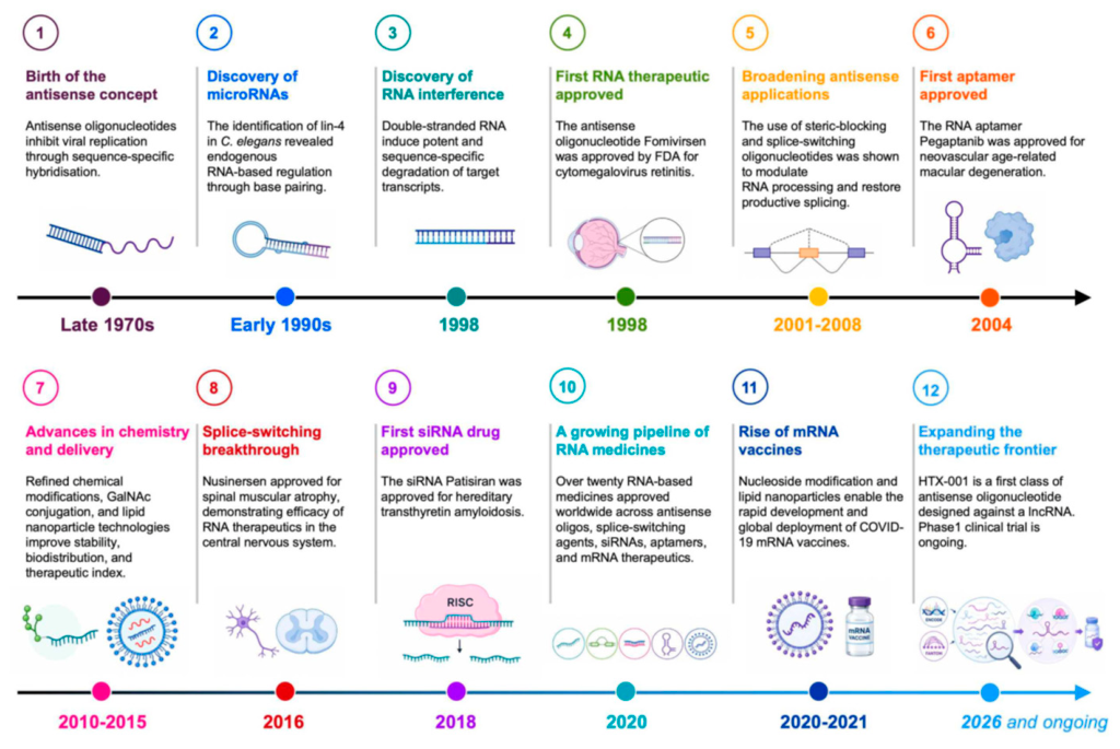
Figure 2.
From RNA biology to the clinical investigation of lncRNA-targeted therapeutics. The timeline follows the transition of long non-coding RNAs (lncRNAs) from largely uncharacterised transcripts to candidate therapeutic targets, in three phases. Foundations: the recognition that RNA is capable of sequence-specific base pairing (1956) and the formulation of the central dogma (1958) established RNA both as an intermediary between genes and proteins and as a programmable recognition element. Key lncRNAs and the non-coding genome: the characterisation of H19 (1990), an oncofetal imprinted transcript re-expressed in cancer; XIST (1991), which initiates X-chromosome inactivation; MALAT1 (2003), implicated in splicing regulation and metastasis; HOTAIR (2007), which recruits PRC2 to silence target genes; NEAT1 (2009), an essential scaffold of nuclear paraspeckles; and HOTTIP (2011), which activates HOXA gene expression in cis, together defined the principal regulatory modalities of the class. In parallel, FANTOM (2005) and ENCODE (2012) revealed pervasive transcription of the human genome and greatly expanded the catalogue of lncRNAs available for functional investigation. Clinical translation: systemically delivered TUG1-targeting antisense oligonucleotides suppressed glioma growth in mice (2016); the small molecules EPIC-0307 (2023) and EPIC-0628 (2024), which disrupt lncRNA-protein interactions, enhanced temozolomide sensitivity in GBM models; and the protocol of the first-in-human phase I trial of TUG1ASO in recurrent GBM was published in 2025. The arrow indicates the progressive therapeutic exploitation of RNA biology and of lncRNA targeting.
Figure 2.
From RNA biology to the clinical investigation of lncRNA-targeted therapeutics. The timeline follows the transition of long non-coding RNAs (lncRNAs) from largely uncharacterised transcripts to candidate therapeutic targets, in three phases. Foundations: the recognition that RNA is capable of sequence-specific base pairing (1956) and the formulation of the central dogma (1958) established RNA both as an intermediary between genes and proteins and as a programmable recognition element. Key lncRNAs and the non-coding genome: the characterisation of H19 (1990), an oncofetal imprinted transcript re-expressed in cancer; XIST (1991), which initiates X-chromosome inactivation; MALAT1 (2003), implicated in splicing regulation and metastasis; HOTAIR (2007), which recruits PRC2 to silence target genes; NEAT1 (2009), an essential scaffold of nuclear paraspeckles; and HOTTIP (2011), which activates HOXA gene expression in cis, together defined the principal regulatory modalities of the class. In parallel, FANTOM (2005) and ENCODE (2012) revealed pervasive transcription of the human genome and greatly expanded the catalogue of lncRNAs available for functional investigation. Clinical translation: systemically delivered TUG1-targeting antisense oligonucleotides suppressed glioma growth in mice (2016); the small molecules EPIC-0307 (2023) and EPIC-0628 (2024), which disrupt lncRNA-protein interactions, enhanced temozolomide sensitivity in GBM models; and the protocol of the first-in-human phase I trial of TUG1ASO in recurrent GBM was published in 2025. The arrow indicates the progressive therapeutic exploitation of RNA biology and of lncRNA targeting.
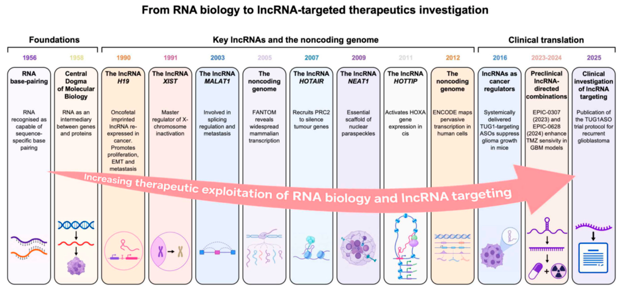
Figure 3.
Genomic organisation, subcellular functions and non-cell-autonomous effects of long non-coding RNAs. LncRNA function is shaped by the interplay between genomic context, subcellular localisation and molecular partners. In the nucleus, lncRNAs regulate gene expression through several, often overlapping, mechanisms. (1) LncRNAs can modulate chromatin architecture and chromatin state by interacting with chromatin-associated proteins and regulatory complexes, thereby altering the accessibility, spatial organisation or transcriptional activity of specific genomic regions. (2) Through guide activity, lncRNAs can recruit transcription factors, RNA polymerase II, chromatin modifiers or other regulatory proteins to defined genomic loci, thereby promoting or repressing transcription. (3) Nuclear lncRNAs may influence pre-mRNA processing by modulating spliceosome assembly, splice-site selection or alternative splicing. (4) By simultaneously binding multiple molecular partners, lncRNAs can act as scaffolds that promote the assembly, stabilisation and spatial organisation of regulatory ribonucleoprotein complexes and nuclear bodies, including nuclear speckles and paraspeckles. (5) Conversely, decoy lncRNAs can sequester transcription factors, chromatin regulators or RNA-binding proteins, thereby limiting their availability for alternative substrates or genomic targets. (6) Cis-acting lncRNAs regulate neighbouring genes or local chromatin domains, frequently through mechanisms linked to the lncRNA promoter, the act of transcription, RNA processing or the mature RNA molecule. (7) Trans-acting lncRNAs interact with distant genomic regions, RNAs or proteins to regulate spatially separated targets. The inset summarises the principal genomic configurations of lncRNA loci. Antisense lncRNAs are transcribed from the DNA strand opposite an overlapping gene; overlapping lncRNAs overlap another annotated gene, on the same or the opposite strand depending on locus organisation; bidirectional lncRNAs originate near a shared or closely positioned promoter and are transcribed in the direction opposite to that of a neighbouring gene; and intergenic lncRNAs arise from genomic regions located between annotated protein-coding genes. These categories describe genomic organisation but do not, by themselves, define the molecular mechanism or the biological function of the resulting transcript. In the cytoplasm, lncRNAs primarily regulate post-transcriptional processes, RNA localisation and the organisation of ribonucleoprotein assemblies. (8) They may alter mRNA stability by recruiting, displacing or modulating RNA-binding proteins, microRNAs or RNA-degradation machinery, thereby promoting transcript stabilisation or decay. (9) Cytoplasmic decoy lncRNAs can sequester translation factors or other regulatory proteins, preventing their interaction with target mRNAs and indirectly affecting protein synthesis. (10) LncRNAs may contribute to intracellular RNA transport by acting as molecular adaptors within ribonucleoprotein complexes that couple RNA cargoes to motor proteins and cytoskeletal networks, including microtubules. (11) Some lncRNAs have been proposed to participate in competing endogenous RNA networks by binding shared microRNAs and modifying microRNA-dependent repression of target transcripts. (12) LncRNAs can additionally function as scaffolds for the assembly, stabilisation or spatial organisation of cytoplasmic ribonucleoprotein condensates and RNA granules. LncRNAs can also generate extracellular and non-cell-autonomous effects through two mechanistically distinct routes. (13) Direct extracellular lncRNA transfer: lncRNAs may be released from donor cells within extracellular vesicles or in association with other extracellular carriers; following uptake by recipient cells, transferred lncRNAs may interact with cellular RNAs, proteins or chromatin-regulatory machinery and thereby alter gene expression or cell behaviour. (14) Indirect regulation through soluble mediators: a lncRNA may remain entirely within the donor cell while regulating transcriptional or signalling pathways that alter the production and secretion of soluble factors; these bind receptors on neighbouring or distant cells and modify their gene expression and phenotype, so that the lncRNA influences the behaviour of cells it never enters. Both routes are relevant to treatment response in nervous system tumours and are discussed in Section 5.8.
Figure 3.
Genomic organisation, subcellular functions and non-cell-autonomous effects of long non-coding RNAs. LncRNA function is shaped by the interplay between genomic context, subcellular localisation and molecular partners. In the nucleus, lncRNAs regulate gene expression through several, often overlapping, mechanisms. (1) LncRNAs can modulate chromatin architecture and chromatin state by interacting with chromatin-associated proteins and regulatory complexes, thereby altering the accessibility, spatial organisation or transcriptional activity of specific genomic regions. (2) Through guide activity, lncRNAs can recruit transcription factors, RNA polymerase II, chromatin modifiers or other regulatory proteins to defined genomic loci, thereby promoting or repressing transcription. (3) Nuclear lncRNAs may influence pre-mRNA processing by modulating spliceosome assembly, splice-site selection or alternative splicing. (4) By simultaneously binding multiple molecular partners, lncRNAs can act as scaffolds that promote the assembly, stabilisation and spatial organisation of regulatory ribonucleoprotein complexes and nuclear bodies, including nuclear speckles and paraspeckles. (5) Conversely, decoy lncRNAs can sequester transcription factors, chromatin regulators or RNA-binding proteins, thereby limiting their availability for alternative substrates or genomic targets. (6) Cis-acting lncRNAs regulate neighbouring genes or local chromatin domains, frequently through mechanisms linked to the lncRNA promoter, the act of transcription, RNA processing or the mature RNA molecule. (7) Trans-acting lncRNAs interact with distant genomic regions, RNAs or proteins to regulate spatially separated targets. The inset summarises the principal genomic configurations of lncRNA loci. Antisense lncRNAs are transcribed from the DNA strand opposite an overlapping gene; overlapping lncRNAs overlap another annotated gene, on the same or the opposite strand depending on locus organisation; bidirectional lncRNAs originate near a shared or closely positioned promoter and are transcribed in the direction opposite to that of a neighbouring gene; and intergenic lncRNAs arise from genomic regions located between annotated protein-coding genes. These categories describe genomic organisation but do not, by themselves, define the molecular mechanism or the biological function of the resulting transcript. In the cytoplasm, lncRNAs primarily regulate post-transcriptional processes, RNA localisation and the organisation of ribonucleoprotein assemblies. (8) They may alter mRNA stability by recruiting, displacing or modulating RNA-binding proteins, microRNAs or RNA-degradation machinery, thereby promoting transcript stabilisation or decay. (9) Cytoplasmic decoy lncRNAs can sequester translation factors or other regulatory proteins, preventing their interaction with target mRNAs and indirectly affecting protein synthesis. (10) LncRNAs may contribute to intracellular RNA transport by acting as molecular adaptors within ribonucleoprotein complexes that couple RNA cargoes to motor proteins and cytoskeletal networks, including microtubules. (11) Some lncRNAs have been proposed to participate in competing endogenous RNA networks by binding shared microRNAs and modifying microRNA-dependent repression of target transcripts. (12) LncRNAs can additionally function as scaffolds for the assembly, stabilisation or spatial organisation of cytoplasmic ribonucleoprotein condensates and RNA granules. LncRNAs can also generate extracellular and non-cell-autonomous effects through two mechanistically distinct routes. (13) Direct extracellular lncRNA transfer: lncRNAs may be released from donor cells within extracellular vesicles or in association with other extracellular carriers; following uptake by recipient cells, transferred lncRNAs may interact with cellular RNAs, proteins or chromatin-regulatory machinery and thereby alter gene expression or cell behaviour. (14) Indirect regulation through soluble mediators: a lncRNA may remain entirely within the donor cell while regulating transcriptional or signalling pathways that alter the production and secretion of soluble factors; these bind receptors on neighbouring or distant cells and modify their gene expression and phenotype, so that the lncRNA influences the behaviour of cells it never enters. Both routes are relevant to treatment response in nervous system tumours and are discussed in Section 5.8.
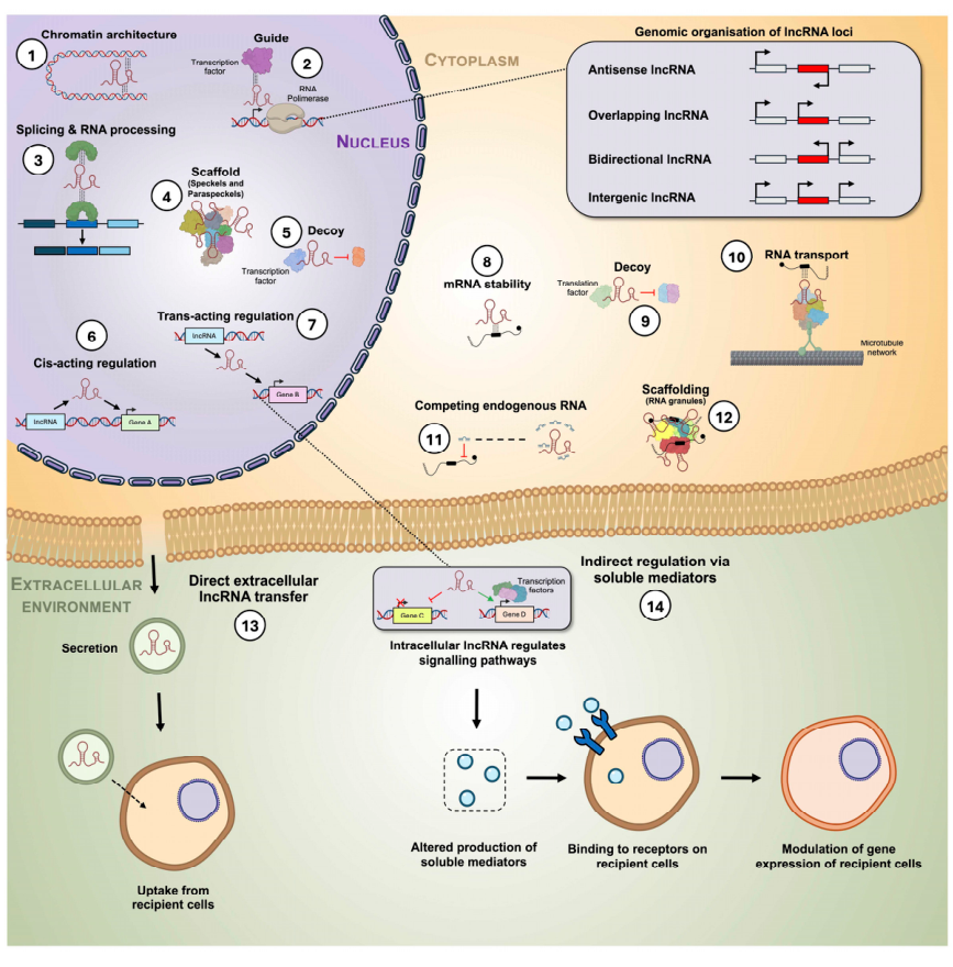
Figure 4.
Biological processes through which lncRNAs modulate therapeutic response in nervous system tumours. The schematic groups the examples discussed in this review according to the biological processes implicated in treatment response in specific tumour models. (1) DNA-damage and radiation response: HOTAIR, LINC00473, LINC01956, MALAT1, SBF2-AS1, lnc-TALC, TUG1 and lncGRS-1, together with perturbation of a regulatory element within the HOTAIR locus and pharmacological disruption of the PRADX–EZH2 and HOTAIR–EZH2 interactions by EPIC-0307 and EPIC-0628, respectively. (2) Survival signalling and apoptosis: NEAT1, H19, lncMB3, NBAT1, NORAD, MIR155HG, XLOC013218, SNHG16 and UCA1. (3) Stemness, differentiation and cellular plasticity: TP73-AS1, GSCAR, TUG1, RBM5-AS1, TALNEC2 and NDM29. (4) Invasion and infiltrative growth: HMMR-AS1, lncRNA-MUF and NEAT1. (5) Mesenchymal programmes and plasticity: LINC01057, MIR222HG, lncRNA-MUF and DLEU1. (6) Metabolic adaptation: HOTAIR, ZBED3-AS1, NEAT1, LINC01410 and TP73-AS1. (7) Microenvironment and emerging pathways: exosomal transfer of resistance (HOTAIR, LINC00473 and SBF2-AS1); myeloid signalling and immune evasion (lnc-TALC, the MIR222HG locus through miR-221/222, INCR1 and NUTM2A-AS1); stress and cell-cycle regulation (MALAT1 and NEAT1); blood–tumour barrier permeability (MIAT, studied in endothelial cells in vitro); ferroptosis (SNAI3-AS1, investigated with the experimental inducer erastin) and pyroptosis (ZNF674-AS1); and cytoprotective autophagy (CRNDE, DLEU1 and SNHG7).
Figure 4.
Biological processes through which lncRNAs modulate therapeutic response in nervous system tumours. The schematic groups the examples discussed in this review according to the biological processes implicated in treatment response in specific tumour models. (1) DNA-damage and radiation response: HOTAIR, LINC00473, LINC01956, MALAT1, SBF2-AS1, lnc-TALC, TUG1 and lncGRS-1, together with perturbation of a regulatory element within the HOTAIR locus and pharmacological disruption of the PRADX–EZH2 and HOTAIR–EZH2 interactions by EPIC-0307 and EPIC-0628, respectively. (2) Survival signalling and apoptosis: NEAT1, H19, lncMB3, NBAT1, NORAD, MIR155HG, XLOC013218, SNHG16 and UCA1. (3) Stemness, differentiation and cellular plasticity: TP73-AS1, GSCAR, TUG1, RBM5-AS1, TALNEC2 and NDM29. (4) Invasion and infiltrative growth: HMMR-AS1, lncRNA-MUF and NEAT1. (5) Mesenchymal programmes and plasticity: LINC01057, MIR222HG, lncRNA-MUF and DLEU1. (6) Metabolic adaptation: HOTAIR, ZBED3-AS1, NEAT1, LINC01410 and TP73-AS1. (7) Microenvironment and emerging pathways: exosomal transfer of resistance (HOTAIR, LINC00473 and SBF2-AS1); myeloid signalling and immune evasion (lnc-TALC, the MIR222HG locus through miR-221/222, INCR1 and NUTM2A-AS1); stress and cell-cycle regulation (MALAT1 and NEAT1); blood–tumour barrier permeability (MIAT, studied in endothelial cells in vitro); ferroptosis (SNAI3-AS1, investigated with the experimental inducer erastin) and pyroptosis (ZNF674-AS1); and cytoprotective autophagy (CRNDE, DLEU1 and SNHG7).
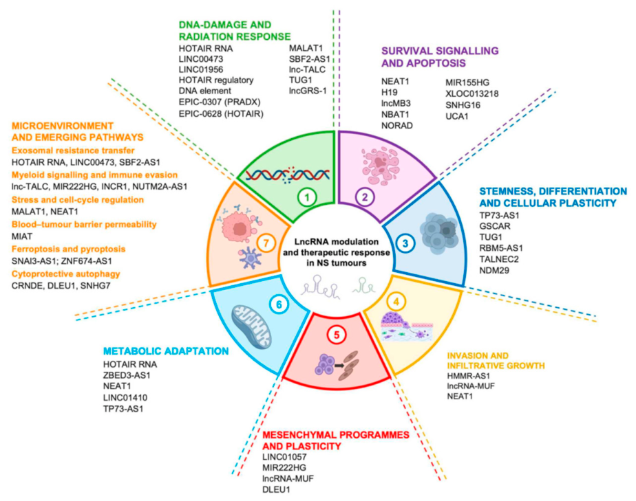
Figure 5.
Evidence hierarchy and translational maturity of lncRNA-associated therapeutic strategies in nervous system tumours. The 50 study-level entries summarised in Table 1 are positioned on the seven-tier framework described in Section 5.1. Vertical position and a single-hue colour scale indicate evidence level. Unless explicitly qualified, tier assignment refers to the specified intervention and experimental setting. E1 and E2 contain no entries because descriptive association or mechanistic evidence alone was insufficient for inclusion in the combination-focused evidence set. NORAD is retained at E4*, with the asterisk distinguishing in vivo target validation from in vivo combination evidence: depletion reduced xenograft growth, whereas doxorubicin sensitisation was demonstrated only in vitro, and the animal experiment did not include doxorubicin. Its position therefore reflects the in vivo biological validation of the target, not demonstration of the combination in animals. Monotherapy and indirect pharmacological evidence are also distinguished from direct lncRNA-targeted combinations. The TUG1 study by Katsushima et al. represents administered lncRNA-targeted monotherapy at E5; lnc-TALC/C5aRa and NDM29/perhexiline represent downstream pathway inhibition and pharmacological transcript induction, respectively. For TUG1, E7 reflects the overall translational maturity of the programme, including clinical investigation as monotherapy; the combination evidence remains preclinical. Several lncRNAs appear at more than one tier because separate studies examined different interventions or experimental settings. Mechanistic support, treatment-interaction analyses, target-engagement and pharmacodynamic readouts, and safety findings are detailed in Table 1. Tier position alone does not establish pharmacological synergy, clinical efficacy or safety.
Figure 5.
Evidence hierarchy and translational maturity of lncRNA-associated therapeutic strategies in nervous system tumours. The 50 study-level entries summarised in Table 1 are positioned on the seven-tier framework described in Section 5.1. Vertical position and a single-hue colour scale indicate evidence level. Unless explicitly qualified, tier assignment refers to the specified intervention and experimental setting. E1 and E2 contain no entries because descriptive association or mechanistic evidence alone was insufficient for inclusion in the combination-focused evidence set. NORAD is retained at E4*, with the asterisk distinguishing in vivo target validation from in vivo combination evidence: depletion reduced xenograft growth, whereas doxorubicin sensitisation was demonstrated only in vitro, and the animal experiment did not include doxorubicin. Its position therefore reflects the in vivo biological validation of the target, not demonstration of the combination in animals. Monotherapy and indirect pharmacological evidence are also distinguished from direct lncRNA-targeted combinations. The TUG1 study by Katsushima et al. represents administered lncRNA-targeted monotherapy at E5; lnc-TALC/C5aRa and NDM29/perhexiline represent downstream pathway inhibition and pharmacological transcript induction, respectively. For TUG1, E7 reflects the overall translational maturity of the programme, including clinical investigation as monotherapy; the combination evidence remains preclinical. Several lncRNAs appear at more than one tier because separate studies examined different interventions or experimental settings. Mechanistic support, treatment-interaction analyses, target-engagement and pharmacodynamic readouts, and safety findings are detailed in Table 1. Tier position alone does not establish pharmacological synergy, clinical efficacy or safety.
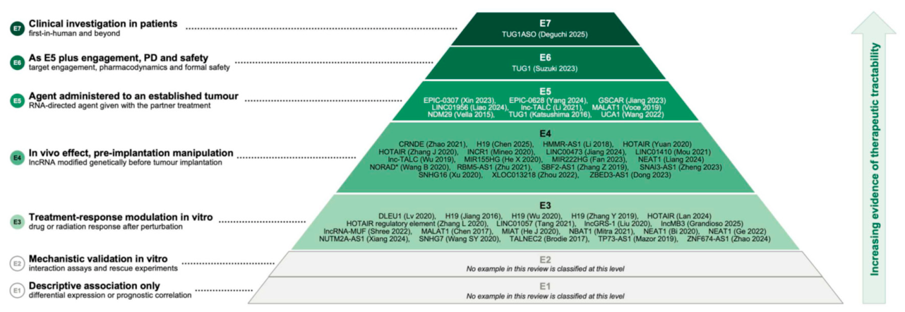
Figure 6.
Therapeutic strategies for targeting long non-coding RNAs. The available approaches are grouped according to their principal mode of action and their current level of translational maturity. (1) Transcript depletion can be achieved with antisense oligonucleotides (ASOs), including gapmers, which promote ribonuclease H-dependent degradation of nuclear or cytoplasmic RNAs, or through RNA interference using small interfering RNAs (siRNAs) and short hairpin RNAs (shRNAs), which preferentially act on cytoplasmic transcripts through the RNA-induced silencing complex (RISC). (2) Functional blockade is obtained without degrading the target RNA: steric-blocking ASOs interfere with RNA processing, splicing or RNA-protein interactions, whereas RNA aptamers competitively bind selected protein partners. (3) Genome and transcriptional editing approaches act at the DNA locus and include CRISPR-Cas9-mediated gene knockout, CRISPR interference (CRISPRi) for transcriptional repression and CRISPR activation (CRISPRa) for transcriptional induction. (4) Messenger RNA (mRNA) therapeutics provide a complementary strategy based on protein replacement or expression rather than on direct lncRNA inhibition. The labels indicate the approximate clinical maturity of each modality, from experimental or preclinical application to clinically established and FDA-approved therapeutic classes. Maturity refers to the platform and not to its use against a lncRNA: clinical evidence for lncRNA-directed treatment of nervous system tumours remains confined to a single phase I study, as set out in Section 5.11. ASO, antisense oligonucleotide; CRISPRa, CRISPR activation; CRISPRi, CRISPR interference; FDA, US Food and Drug Administration; lncRNA, long non-coding RNA; mRNA, messenger RNA; RISC, RNA-induced silencing complex; shRNA, short hairpin RNA; siRNA, small interfering RNA.
Figure 6.
Therapeutic strategies for targeting long non-coding RNAs. The available approaches are grouped according to their principal mode of action and their current level of translational maturity. (1) Transcript depletion can be achieved with antisense oligonucleotides (ASOs), including gapmers, which promote ribonuclease H-dependent degradation of nuclear or cytoplasmic RNAs, or through RNA interference using small interfering RNAs (siRNAs) and short hairpin RNAs (shRNAs), which preferentially act on cytoplasmic transcripts through the RNA-induced silencing complex (RISC). (2) Functional blockade is obtained without degrading the target RNA: steric-blocking ASOs interfere with RNA processing, splicing or RNA-protein interactions, whereas RNA aptamers competitively bind selected protein partners. (3) Genome and transcriptional editing approaches act at the DNA locus and include CRISPR-Cas9-mediated gene knockout, CRISPR interference (CRISPRi) for transcriptional repression and CRISPR activation (CRISPRa) for transcriptional induction. (4) Messenger RNA (mRNA) therapeutics provide a complementary strategy based on protein replacement or expression rather than on direct lncRNA inhibition. The labels indicate the approximate clinical maturity of each modality, from experimental or preclinical application to clinically established and FDA-approved therapeutic classes. Maturity refers to the platform and not to its use against a lncRNA: clinical evidence for lncRNA-directed treatment of nervous system tumours remains confined to a single phase I study, as set out in Section 5.11. ASO, antisense oligonucleotide; CRISPRa, CRISPR activation; CRISPRi, CRISPR interference; FDA, US Food and Drug Administration; lncRNA, long non-coding RNA; mRNA, messenger RNA; RISC, RNA-induced silencing complex; shRNA, short hairpin RNA; siRNA, small interfering RNA.
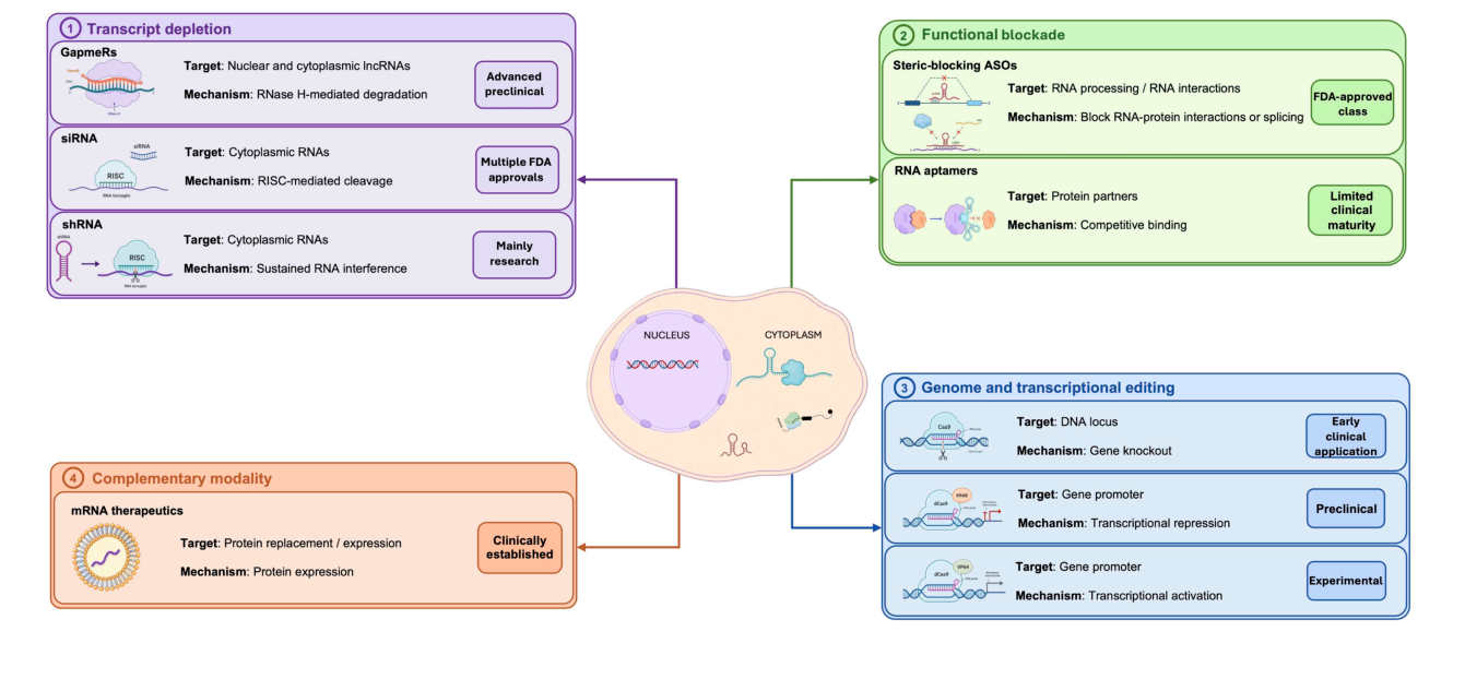
Figure 7.
Delivery of RNA therapeutics to nervous system tumours. The constraints on delivery are grouped into four stages, each of which must be satisfied for an RNA-directed agent to reach and act on its target. (1) Delivery platforms. Non-viral carriers used to protect oligonucleotide cargoes and to direct their biodistribution include lipid nanoparticles, polymeric micelles, unit polyion complexes, inorganic nanoparticles, ferritin nanocages and cell-derived extracellular vesicles; they differ in encapsulation capacity, surface functionalisation and tissue tropism, and several can carry targeting ligands. (2) Brain access. Routes to the intracranial compartment comprise systemic intravenous administration, which requires passage across the blood-brain and blood-tumour barriers (BBB and BTB); intrathecal and intraventricular administration into the cerebrospinal fluid; intratumoural infusion, including convection-enhanced delivery (CED); and intranasal administration. Focused ultrasound combined with microbubbles can transiently and regionally increase barrier permeability. These routes are requirements of the intracranial setting: tumours of the peripheral nervous system are reachable by systemic administration without barrier engineering, and RB occupies an intermediate position, being intraocular but extracranial. (3) Tumour distribution. Within the lesion, a dense extracellular matrix, uneven penetration from a heterogeneous vasculature, infiltrative margins that retain more intact barrier properties, and the presence of stem-like cell populations together determine whether the agent reaches all the cells in which the target is active; bulk tumour accumulation therefore does not establish exposure throughout the lesion. (4) Intracellular delivery. Cellular uptake occurs largely by endocytosis, after which endosomal escape and the avoidance of lysosomal degradation govern how much of the internalised cargo becomes available at its intracellular site of action. BBB, blood-brain barrier; BTB, blood-tumour barrier; CED, convection-enhanced delivery; ECM, extracellular matrix.
Figure 7.
Delivery of RNA therapeutics to nervous system tumours. The constraints on delivery are grouped into four stages, each of which must be satisfied for an RNA-directed agent to reach and act on its target. (1) Delivery platforms. Non-viral carriers used to protect oligonucleotide cargoes and to direct their biodistribution include lipid nanoparticles, polymeric micelles, unit polyion complexes, inorganic nanoparticles, ferritin nanocages and cell-derived extracellular vesicles; they differ in encapsulation capacity, surface functionalisation and tissue tropism, and several can carry targeting ligands. (2) Brain access. Routes to the intracranial compartment comprise systemic intravenous administration, which requires passage across the blood-brain and blood-tumour barriers (BBB and BTB); intrathecal and intraventricular administration into the cerebrospinal fluid; intratumoural infusion, including convection-enhanced delivery (CED); and intranasal administration. Focused ultrasound combined with microbubbles can transiently and regionally increase barrier permeability. These routes are requirements of the intracranial setting: tumours of the peripheral nervous system are reachable by systemic administration without barrier engineering, and RB occupies an intermediate position, being intraocular but extracranial. (3) Tumour distribution. Within the lesion, a dense extracellular matrix, uneven penetration from a heterogeneous vasculature, infiltrative margins that retain more intact barrier properties, and the presence of stem-like cell populations together determine whether the agent reaches all the cells in which the target is active; bulk tumour accumulation therefore does not establish exposure throughout the lesion. (4) Intracellular delivery. Cellular uptake occurs largely by endocytosis, after which endosomal escape and the avoidance of lysosomal degradation govern how much of the internalised cargo becomes available at its intracellular site of action. BBB, blood-brain barrier; BTB, blood-tumour barrier; CED, convection-enhanced delivery; ECM, extracellular matrix.
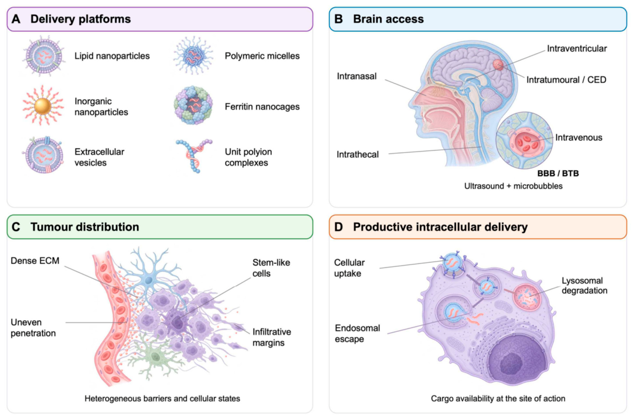
Disclaimer/Publisher’s Note: The statements, opinions and data contained in all publications are solely those of the individual author(s) and contributor(s) and not of MDPI and/or the editor(s). MDPI and/or the editor(s) disclaim responsibility for any injury to people or property resulting from any ideas, methods, instructions or products referred to in the content. |
© 2026 by the authors. Licensee MDPI, Basel, Switzerland. This article is an open access article distributed under the terms and conditions of the Creative Commons Attribution (CC BY) license (http://creativecommons.org/licenses/by/4.0/).
Copyright: This open access article is published under a Creative Commons CC BY 4.0 license, which permit the free download, distribution, and reuse, provided that the author and preprint are cited in any reuse.