
Submitted:
15 November 2026
Posted:
17 November 2026
You are already at the latest version
Abstract
Tumor cells remodel their membrane lipids, and the extracellular vesicles (EVs) they release carry the result: EV lipid profiles differ from those of the parent cells because certain membrane components are selectively enriched. Headgroup chemistry, acyl-chain composition and cholesterol set how tightly a membrane packs and how easily it bends, and local anionic lipids recruit trafficking proteins. Two routes link these properties to exosome biogenesis: ARF6–PLD2–syntenin–ALIX, and ceramide production by neutral sphingomyelinase 2. Once vesicles are released, their surface lipids affect clearance and immune recognition. Phosphatidylserine promotes macrophage uptake and can suppress T cells, and vesicle-bound autotaxin delivers lysophosphatidic acid to receptors on recipient cells. In tumors these activities have been linked to angiogenesis and immune suppression, and in some settings to antitumor immunity. We review how lipid classes act in these processes, keeping changes in vesicle production apart from changes in cargo and signaling, and we outline the measurements needed to tie a lipid perturbation to an EV-mediated tumor phenotype. Therapeutic options that follow from this work include targeting phosphatidylserine-dependent recognition and lipid-mediated immune activation. EV lipid profiles are being explored as biomarkers, but their clinical value depends on the biofluid, the separation method and the intended diagnostic use.
Keywords:
membrane lipids
; exosomes
; extracellular vesicles
; lipid metabolism
; tumor microenvironment
; membrane biophysics
Introduction
Tumor-derived extracellular vesicles (EVs) deliver signals to stromal and immune cells. Exosomal epidermal growth factor receptor alters the liver microenvironment in gastric cancer, and exosomal programmed death-ligand 1 (PD-L1) suppresses T-cell activity in melanoma models (1, 2). Lipids take part in this communication in several ways: they make up the vesicle membrane, act as recognition signals and, in some cases, activate receptors directly. They matter at every stage, from the formation of a vesicle to its distribution and activity after release.
Exosomes originate as intraluminal vesicles (ILVs) inside multivesicular bodies (MVBs) and are released when MVBs fuse with the plasma membrane (3). Small EV preparations, however, also contain vesicles that bud from the cell surface, and these overlap with exosomes in size and markers (4, 5). We follow MISEV2023 (6): “EVs” or “small EVs” refers to preparations whose endosomal origin has not been established, and “exosomes” is reserved for defined endosomal processes or used where the original study did so.
Lipidomics of cells and their vesicles shows that EV membranes are not a random sample of the donor membrane. In PC-3 prostate cancer cells, Llorente et al. quantified about 280 lipid species in cells and exosomes, and the exosomes were enriched in sphingomyelin, cholesterol, glycosphingolipids and phosphatidylserine (7). A wider panel of cancer and non-cancer cell lines gave the same picture: EV lipid profiles clustered apart from cellular ones and differed by cancer type (8). Vesicle types also affect lipid distribution. The lipid enrichment of exosomes and larger vesicles is different, and the specific difference depends on the cell type (9).
In this paper, the relationship between lipid composition changes and membrane structure, vesicle biogenesis and tumor signal transduction was analyzed by combining model membrane experiment, lipidomics and cell and animal intervention research.
Membrane Organization and Vesicle Formation
Lipid Packing and Membrane Mechanics
The order of fatty acyl chain, transverse fluidity and bending stiffness reflect the chain arrangement, molecular movement speed and the ability of membrane to resist bending deformation respectively. In phosphatidylcholine bilayer, the length of acyl chain and the degree of polyunsaturated have obvious influence on bending stiffness, but have little effect on area tensile modulus (10). The head-base structure will also change the membrane properties. The results of solid-state nuclear magnetic resonance (NMR) show that the acyl chain arrangement of dipalmitoyl phosphatidylethanolamine is more orderly, and the calculated bending stiffness is higher than that of the corresponding phosphatidylcholine bilayer (11).
Cholesterol can regulate these properties together, but the mode of action is affected by the composition of surrounding lipids. It condenses phospholipid bilayers and alters chain ordering (12, 13). Yet in simulations of unsaturated membranes, cholesterol redistributed between leaflets as the membrane bent, and the membrane softened even though it became thicker and the lipids more compressed (14). A fluidity probe and a bending-rigidity measurement can therefore disagree after lipid remodeling.
Proteins add forces of their own. In reconstituted systems, crowding of membrane-bound proteins is enough to bend a membrane (15). Lipid remodeling can thus change vesiculation by altering the bilayer itself and by altering which proteins assemble on it.
Curvature and Recruitment of Budding Machinery
Spontaneous monolayer curvature depends on lipid structure and on the molecular environment. For raft-forming lipids, phosphatidylethanolamines have a much more negative spontaneous curvature than most phosphatidylcholines (16). Phosphatidic acid (PA) and lysophosphatidic acid (LPA) behave differently depending on pH and divalent cations, which change headgroup interactions and phase preference (17). Which leaflet a lipid sits in matters too: in cellular transport experiments, flipping phosphatidylserine across the bilayer changed both curvature and surface charge (18).
Anionic lipids also help position the budding machinery. Phosphatidylserine (PS) shapes membrane electrostatics and the localization of positively charged proteins (19). In phase-separated giant vesicles, the endosomal sorting complexes required for transport (ESCRT), ESCRT-II and ESCRT-III, made more ILVs when PS was concentrated in liquid-disordered domains, yet the ILVs that formed were enriched in liquid-ordered lipids (20). Where the machinery is recruited and which lipids end up in the bud are therefore different questions, and this gives selective vesicle assembly in a heterogeneous membrane a physical basis.
Lipid Classes in Exosome Biogenesis and Signaling
Phosphatidylcholine and Phosphatidylethanolamine
Phosphatidylcholine (PC) and phosphatidylethanolamine (PE) make up much of the cellular glycerophospholipid pool. The relative content of PE and PC affects the accumulation mode of lipid head groups in the membrane, and the fatty acyl chain structure is related to the order and flexibility of the membrane. Because the head base of PE is smaller, its tendency to form negative spontaneous curvature is usually stronger than that of PC(16). Dawaliby et al. found that PE can reduce the fluidity of liposomes, but in cells, although the change of cholesterol level changes the ratio of PE to PC, the membrane fluidity is basically stable, indicating that phospholipids and sterols participate in the regulation (21). In addition, the study of model membrane shows that monounsaturated PE and PE containing docosahexaenoic acid behave differently when mixed with sphingomyelin and cholesterol, which further reflects the importance of fatty acyl chain composition (22).
The fatty acid desaturation of cancer cells will affect the composition of membrane lipids. 134 tissues of six cancers were imaged by mass spectrometry, and it was found that monounsaturated fatty acids and phosphatidylcholine in cancerous areas were more abundant than the corresponding polyunsaturated lipids (23). Tumor matrix also plays a role: breast cancer-related fibroblasts can promote the expression of stearoyl-CoA desaturase 1(SCD1) in MCF-7 and MDA-MB-231 cells, while inhibiting SCD1 can weaken the cell migration ability (24). In glucose-deprived lung adenocarcinoma cells, oleate from fibroblasts promoted SCD expression and stemness (25). All of this concerns the lipid environment of the donor cell. Whether the released EVs change accordingly has to be measured directly.
At the level of individual molecular species, EV lipidomics shows finer differences. Elmallah et al. found higher PC 34:1, PE 36:2 and SM d18:1/16:0 than in controls in vesicles from nonmetastatic colorectal cancer cells and in patient plasma preparations. After cancer metastasis, the levels of these lipids decreased, but Cer d18:1/24:1 increased instead (26). There are also differences in the composition of vesicles in prostate cancer cell lines. For example, vesicles derived from LNCaP contain more PC, PE and phosphatidylinositol than mother cells (27). The discovery of colorectal cancer shows that the change direction of lipid index is influenced by metastasis, so the biomarker analysis must consider the disease progression stage.
The relationship between different cell membranes is also reflected in the metabolism of PC and PE. PS synthase generates PS by replacing the head group of PC or PE, mitochondrial PS decarboxylase converts PS into PE, and phosphatidylethanolamine N- methyltransferase further converts PE into PC(Figure 1). Lack of PS decarboxylase will lead to mitochondrial abnormality and embryo death in mice, which highlights the importance of phospholipid synthesis position (28). Therefore, when studying EV, we should not only rely on the whole cell PC:PE ratio, but also analyze the related membrane components and released vesicles simultaneously.
A, PTDSS1 converts PC to PS, and PTDSS2 converts PE to PS through headgroup exchange (29, 30). PISD converts PS to PE (28), and PEMT converts PE to PC (31). B, phospholipase A₂ generates LPC from PC (32); ATX/ENPP2 hydrolyzes LPC to LPA and choline (33). PLD generates PA from PC (34), and DAG kinases generate PA from DAG (35). C, nSMase2 hydrolyzes SM to ceramide (36); sphingomyelin synthases transfer phosphocholine from PC to ceramide, producing SM and DAG (37). PTDSS, phosphatidylserine synthase; PISD, phosphatidylserine decarboxylase; PEMT, phosphatidylethanolamine N-methyltransferase; SMS, sphingomyelin synthase.
Phosphatidic Acid and Ceramide
PA is produced by phospholipase D (PLD)-mediated hydrolysis of PC and by phosphorylation of diacylglycerol (DAG) (34, 35). Its small anionic headgroup interacts with basic protein residues, and an electrostatic/hydrogen-bond switch has been described for PA binding to proteins (38). In model membranes, PA accumulated in negatively curved regions and promoted tubulation (39).
The clearest connection to exosome production involves the small GTPase ADP-ribosylation factor 6 (ARF6) and PLD2. Ghossoub et al. showed that both control ILV budding and syntenin-associated exosome production (40). This route overlaps with the syndecan–syntenin–ALIX pathway, in which syntenin binds ALIX through LYPX(n)L motifs and exosome formation depends on syndecans and ESCRT components (41).
Ceramide, produced by sphingomyelin hydrolysis, offers a second way to drive budding. Trajkovic et al. described an ESCRT-independent, ceramide-dependent route in an oligodendroglial model (36). Kosaka et al. later tied neutral sphingomyelinase 2 (nSMase2) to cancer progression. In their work nSMase2 regulated exosomal miRNA secretion, which promoted angiogenesis and metastasis, and transferred miR-210 changed target-gene expression in endothelial cells (42).
Ceramide metabolism also affects what happens to MVBs. In HeLa cells, inhibiting nSMase2 increased vacuolar H⁺-ATPase (V-ATPase) assembly and MVB acidification and reduced small EV secretion, whereas stimulating nSMase2 with TNF increased it (43). So nSMase2 can change EV output at a step after budding as well.
Not every system agrees. In PC-3 cells, inhibiting nSMase2 or de novo ceramide formation did not reduce exosome secretion (44), and genetic Smpd3 deficiency left EV release unchanged in the cell and mouse systems Burgelman et al. examined (45). Menck et al. saw a shift between export routes instead: nSMase inhibition lowered the exosome-enriched fraction but increased plasma-membrane-derived vesicles, including vesicles carrying Wnt proteins (46).
The load composition of EV is also affected by enzyme activity. In MCF-7 cells, after the inhibition of neutral sphingomyelinase, the load of EV nanoparticles, their RNA and protein decreased. Inhibition of acid sphingomyelinase can increase the content of specific RNA binding proteins and related RNA, and the generated vesicles can also promote the migration and translation activities of MCF10A cells (47). Therefore, the difference in the results of nSMase2 research does not necessarily mean contradiction, but shows that the total amount of particles can not fully reflect the influence of inhibitors. Even if the release amount remains the same, the secretory pathway or load of vesicles may change (46, 47). The final effect depends on the cell model and the enzyme targeted.
Phosphatidylserine
PS acts inside the donor cell and on the surface of released vesicles. Within the cell it contributes to the electrostatic identity of the plasma membrane and endocytic compartments (19, 48), and imaging has located it on the cytoplasmic leaflet of the endoplasmic reticulum, from where TMEM16K can redistribute(49-51). In pancreatic cancer models, stronger oncogenic KRAS signaling went with higher EV secretion and a more negative surface potential, to which PS and extraluminal DNA contributed (52).
Exposed PS is a recognition signal. TIM4 binds PS directly, and milk fat globule-EGF factor 8 (MFG-E8) bridges PS to phagocytic cells (53, 54). Ligands of the Tyro3/Axl/Mer (TAM) receptor family bridge in a similar way, with signaling depending on receptor, ligand and PS together: Gas6 activates Axl, whereas Gas6 and Protein S can both activate Tyro3 and Mer (55). How a PS-rich surface is read therefore depends on which receptors and bridging proteins the recipient tissue has.
PS-dependent uptake shapes how long vesicles circulate. Negatively charged PS-containing liposomes competed with intravenously injected B16BL6-derived exosomes for macrophage uptake and delayed their clearance from the blood (56). Matsumoto et al. later identified PS-deficient small EVs that escaped rapid macrophage uptake and, as a result, predominated among circulating somatic-cell-derived vesicles (57). Depleting macrophages confirmed that these cells account for a large share of the clearance of injected B16BL6 vesicles (58).
In the tumor microenvironment, PS can also suppress immune responses. EVs isolated from human ovarian tumor ascites blocked T-cell signaling, and removing PS-positive vesicles or blocking PS reduced the effect (59). Follow-up work showed that the inhibition was reversible and covered several measures of T-cell activation: transcription-factor translocation, cytokine production and proliferation (60). The PS-binding compound ExoBlock reversed the suppressive activity of exosomes from ovarian tumors and melanoma in vitro, and improved T-cell-dependent tumor control in two human tumor xenograft models (61). This evidence is preclinical, but it supports PS as a target for relieving vesicle-associated T-cell suppression.
The surface of a vesicle does not stay as it left the cell. After exposure to plasma, EVs acquired apolipoproteins, complement components and other proteins, became denser, and changed how dendritic cells were activated (62). In a separate study of synthetic anionic liposomes, corona composition varied with lipid composition, and highly anionic particles were less stable in plasma (63). The liposome data are only an analogy for EVs; the EV study is the one that shows a biologically active coating being acquired.
PS is not the only surface lipid with this effect. GD3-positive vesicles from ovarian tumor ascites inhibited T-cell activation, and antibody blockade, depletion of GD3-positive vesicles or enzymatic removal of sialic acid all reduced the inhibition. GD3-bearing liposomes reproduced it, which ties the effect to the ganglioside itself (64).
Sphingomyelin and Cholesterol
Sphingomyelin (SM) and cholesterol organize the membrane laterally. In SM–cholesterol model membranes, ordered domains kept considerable molecular motion, and order and fluidity varied across the thickness of the bilayer (65). Such behavior may help explain how cholesterol-rich EV membranes can be both selectively organized and dynamic.
De Gassart et al. detected several raft-associated proteins in exosomes and placed part of the exosomal major histocompatibility complex (MHC) class II pool in detergent-resistant fractions (66). Lipid domains, though, are not the only sorting mechanism. In an ESCRT RNAi screen, depleting HRS, STAM1 or TSG101 changed secretion of EV-associated CD63 and MHC class II, with different effects on vesicle size and composition. Depleting ALIX increased exosomal MHC class II in HeLa-CIITA cells, but it also raised intracellular MHC class II protein and mRNA (67). This result shows that donor cells and EVs should be detected simultaneously to avoid mistaking the increase of intracellular expression for the increase of vesicle enrichment.
SM metabolism not only affects the formation of membrane structure domain, but also participates in ceramide formation. Model membrane experiments show that the lipid composition of lipid rafts can change the sphingomyelinase activity and the physical function of ceramide at the same time (68). Different goods in the same system may also have different loading mechanisms. For example, in the neuron model, the entry of normal prions into vesicles depends on nSMase2, while the loading of disease-related prions does not depend on this enzyme (69).
Cholesterol not only acts through membrane structure, but also affects secretion through signal pathway. Astrocyte experiments showed that cholesterol accumulation was accompanied by decreased exosomes release and increased PI3K/Akt phosphorylation. After reducing the cholesterol content or inhibiting PI3K, the release amount recovered (70). Because the research object is not malignant cells, the importance of this mechanism in tumor cells and its relative contribution to lipid sorting and ESCRT pathway still need to be verified.
Lysophospholipids and Vesicle-Associated Autotaxin
LPC and LPA participate in membrane remodeling through extracellular receptor signals. Phospholipase A catalyzes PC to produce LPC, and then ATX/ENPP2 hydrolyzes LPC into LPA and choline (32, 33). PH and divalent cations can change the phase behavior of LPA (17). The influence of LPC on the stability of the membrane is concentration-dependent: in the encapsulated lipid bilayer experiment at the droplet interface, low concentration LPC changes the membrane size, and when it reaches the micelle concentration, it destroys the whole bilayer network (71).
Jethwa et al. confirmed that secreted ATX can attach to exosomes, maintain catalytic function and carry LPA produced by it. This vesicle-bound ATX delivered LPA to cell-surface receptors and triggered AKT and MAPK phosphorylation, intracellular Ca²⁺ release and migration (72). The signaling unit here is an enzyme–lipid complex displayed on the vesicle surface.
The pathway has also been implicated in cancer pain. In a fibrosarcoma bone-pain model, cancer exosomes sensitized nociceptive neurons and caused hypersensitivity, and inhibiting ATX or blocking LPA receptors attenuated both (73). Vesicle-associated lipids can thus act on symptoms, not only on tumor-cell behavior.
Localized Lipid Signaling in Endosomes
Phosphoinositides regulate trafficking through small, spatially restricted lipid pools. Jin et al. identified phosphatidylinositol 4-phosphate (PI4P) as the main phosphatidylinositol monophosphate in macrophage-derived small EVs. Early in lipopolysaccharide stimulation, type I interferon lowered PIP5K1γ expression, which raised MVB-associated PI4P and recruited RAB10 to promote vesicle generation. With prolonged stimulation, PI4P and RAB10 were redistributed toward the endoplasmic reticulum in an HSPA5-associated manner, and release was curtailed (74). In these macrophages, the location of the PI4P pool sets the timing of the EV response.
Endosomal lipid remodeling can also produce vesicles with antitumor activity. Dendrogenin A activated an LXRβ-dependent program involving PLD and CLN5 that increased bis(monoacylglycero)phosphate (BMP) in tumor-derived small EVs. Blocking BMP reduced uptake by dendritic cells, dendritic-cell maturation and antitumor immune activity, and BMP-enriched vesicles enhanced anti-PD-1 efficacy in a melanoma model (75). Here the lipid composition of a tumor-derived vesicle pushes the immune response in the opposite direction from the PS- and GD3-associated suppression described above.
Vesicle Activity and Recipient-Cell Responses
Dose and Cargo Abundance
An EV response depends on how many particles are released, how much active cargo they carry and what fraction reaches a responsive cell, and these can vary independently. Chevillet et al. found low average miRNA occupancy in the exosome preparations they examined, and Albanese et al. reported that vesicular miRNAs were inefficiently delivered to target cells in their systems (76, 77). It must be supported by load measurement results to attribute the phenotypic changes of recipient cells to some RNA.
In the intervention experiment, the total output of EV of the same number of donor cells reflects the signal output of the culture system, while the activity comparison of the same EV dose measures the effect of recovering vesicle particles, and the concerns of the two are different. Reporting both, along with donor-cell viability and collection time, shows where a lipid perturbation acts (6). Normalizing by protein also requires a particle count, because co-isolated protein and the protein content per vesicle both change the dose actually given (78). Table 1 lists representative lipid-dependent EV studies with their models, main findings and readouts.
Surface Signaling and Intracellular Delivery
Some EV signals act at the surface of the recipient cell. Exosomal PD-L1 binds PD-1 and suppresses CD8 T-cell activity (2), and ATX–LPA signaling is a lipid example of receptor activation at the vesicle–cell interface (72). Cargo that must act inside the cell needs one more step, delivery. With cargo reporters, Bonsergent et al. measured EV uptake and cytosolic release as separate events, and Joshi et al. localized cargo exposure to endosomes and lysosomes; in both, endosomal acidification contributed to delivery (79, 80).
Uptake data also depend on the label used. Lipophilic dyes can pass to recipient cells from co-isolated proteins and lipoproteins (81), and PKH26 can form fluorescent nanoparticles that enter cells without any EV involved (82). It is impossible to confirm the specific biological process only by fluorescence signal, and it is necessary to set up pure dye and non-vesicle control, and combine it with cargo reporting system or receptor experiment to verify it.
The distribution of EV in the body will also affect its function. Experiments in mice show that the source of vesicles and the mode of administration determine their distribution characteristics (83), and multimodal reporting technology can dynamically track different positioning information (84). PS-rich vesicles may be quickly cleared by macrophages and gathered in related organs, and the role of surface ligands depends on the receptor expression of target cells.
Experimental Design for Lipid-Dependent EV Mechanisms
To study the mechanism of lipid-dependent EV, it is necessary to analyze the lipid changes of donor cells and EV-releasing cells at the same time, and observe the reaction of recipient cells. Targeted lipidomics should use appropriate internal standards to fully explain the extraction, identification and quantification process (85). Lipid sensors can locate intracellular lipids such as PS and PI4P (49, 74), but the recognition range of probes is different. For example, SM probes derived from two toxins can detect different subcellular lipid pools respectively (86).
The way of sample separation will affect the analysis results. EV in blood is often separated from low-density lipoprotein and high-density lipoprotein (87, 88). Combining various separation techniques can improve the purity of plasma EV (89), but it may also change the results of RNA analysis (90). Therefore, the components obtained should be identified by combining morphology, particle characteristics and markers. High resolution separation and immunocapture can further distinguish exosome components, non-vesicular RNA binding proteins and other extracellular substances (91).
The combination of genetic intervention and functional recovery experiment is helpful to explain the role of inhibitors. When regulating lipid metabolism, the number, activity and sampling time of donor cells should be controlled (6). For example, the inhibition of SCD1 will reduce the survival rate of cancer cells, and its effect is affected by serum conditions, and can be alleviated by supplementing unsaturated fatty acids (92, 93). Apoptosis cells may also release vesicles through membrane fragmentation (94). Therefore, the change of cell state may directly affect EV population, and cell activity must be included in the result analysis.
To confirm the functional contribution of EV, it is necessary to compare the components containing EV and removing EV, and add vesicles again according to the prescribed dose (6). Bobrie et al. found in the study of 4T1 breast tumor that the blocking of Rab27a affects both exocrine secretion and non-exocrine MMP9 release, and tumor growth is jointly regulated by vesicles and other secretion factors (95). Therefore, the target lipid or cargo can be tracked in the separation process, and the related pathways of recipient cells can be detected. The surface signal is suitable for receptor blocking verification, and the intracellular delivery can use cytoplasmic reporting system.
Therapeutic and Biomarker Implications
EV therapy intervention can be aimed at vesicle formation, surface recognition and receptor cell signal. The study of nSMase2 shows that the efficacy evaluation should detect both vesicle subtypes and goods, rather than just counting the total amount of particles. Pre-clinical ExoBlock research supports the feasibility of targeting PS-dependent immunosuppression (61), and blocking ATX-LPA pathway can also alleviate hyperalgesia caused by exosomes in fibrosarcoma model (73). Different targets should be evaluated by corresponding functional indicators.
There is some experimental support for combining vesicle-directed approaches with checkpoint therapy. Suppressing exosomal PD-L1 restored antitumor immunity and acted additively with anti-PD-L1 antibodies in mouse models (96), and BMP-enriched vesicles enhanced anti-PD-1 treatment in melanoma (75). For lipid-directed combinations, the key tests are whether the intervention alters the immune activity of the vesicles that are released, and whether that alteration accounts for the benefit of the combination. PS-dependent clearance and uptake by host cells will also affect dose and tissue exposure.
As biomarkers, individual lipid species and their combinations carry more information than total lipid abundance. In a study of 15 prostate cancer patients and 13 healthy controls, a combination of urinary EV lipids separated the two groups with 93% sensitivity and 100% specificity within that cohort, and the urinary profiles differed from those of cell-line-derived exosomes (97). With so few subjects, this is a candidate signature that still needs clinical evaluation. The change direction of lipid in colorectal cancer before and after metastasis is different, which indicates that the combination of biomarkers should be designed according to the disease state (26).
Rani et al. compared the lipid profiles of paired plasma and small EV with different ovarian tumor subtypes, and found that specific lipid indexes combined with CA-125 can improve the classification effect of some borderline and low-grade tumors, including cases with normal CA-125. However, the differential diagnosis and screening results come from synthetic cohort, and the performance data are still simulation results, which need to be verified by independent patient cohort (98).
According to STARD’s suggestion, diagnostic research needs to clarify the detection purpose, sample selection, reference standard, positive threshold and uncertainty of accuracy estimation (99). The detection of EV lipids also needs a repeatable separation and proteomics process, and it is proved that it can provide additional information beyond the existing clinical indicators.
Conclusions
Membrane lipids regulate EV communication through different channels. PA participates in vesicle sprouting, sphingomyelinase affects vesicle formation, endosome fate and load, and PS and GD3 regulate cell recognition and immune response. Vesicle-bound ATX is involved in extracellular lipid synthesis and receptor signal transduction, while BMP-related findings show that tumor lipid remodeling may produce EV with immune stimulation.
Lipid-targeted drugs are expected to regulate EV function, but it is necessary to determine the key lipids and their corresponding vesicle populations, distinguish the secretion quantity from the single vesicle activity, and verify the receptor cell pathway that triggers tumor response. These studies are also the basis for judging whether EV lipid can be a reliable biomarker or an intervention therapy target.
Author Contributions
Yiyu Chen and Jingru Zhang contributed equally to the conceptualization, literature review, writing of the original draft, and critical revision of this review. Both authors read and approved the final manuscript.
Funding
This work was supported by the National Natural Science Foundation of China (grant numbers 32460175, 32160158, and 81460468) and the Natural Science Foundation of Jiangxi Province (grant numbers 20232BAB206112 and 20202BAB206045).
Ethics approval and consent to participate
Not applicable. This review is based on published literature and does not report new studies involving human participants, human data, or animals.
Consent for publication
Not applicable.
Data availability
No new datasets were generated or analysed for this review. All sources are cited in the article.
Acknowledgments
We thank Xiaojuan Hu for guidance and comments on the manuscript, and our colleagues in the research group for discussions during its writing and revision.
Competing interests
The authors declare no competing interests.
Preprint clearance
All authors have read and approved the manuscript and agree to its public posting as a preprint on Preprints.org. The manuscript is not under consideration for publication elsewhere and has not been published previously in the same form.
Copyright and permissions
Figure 1 was created by the authors. No third-party material requiring permission is included.
References
- Zhang, H.; Deng, T.; Liu, R.; Bai, M.; Zhou, L.; Wang, X.; et al. Exosome-delivered EGFR regulates liver microenvironment to promote gastric cancer liver metastasis. Nat. Commun. 2017, 8(1), 15016. [Google Scholar] [CrossRef] [PubMed]
- Chen, G.; Huang, A.C.; Zhang, W.; Zhang, G.; Wu, M.; Xu, W.; et al. Exosomal PD-L1 contributes to immunosuppression and is associated with anti-PD-1 response. Nature 2018, 560(7718), 382–6. [Google Scholar] [CrossRef] [PubMed]
- Ostrowski, M.; Carmo, N.B.; Krumeich, S.; Fanget, I.; Raposo, G.; Savina, A.; et al. Rab27a and Rab27b control different steps of the exosome secretion pathway. Nat. Cell Biol. 2010, 12(1), 19–30. [Google Scholar] [CrossRef] [PubMed]
- Mathieu, M.; Névo, N.; Jouve, M.; Valenzuela, J.I.; Maurin, M.; Verweij, F.J.; et al. Specificities of exosome versus small ectosome secretion revealed by live intracellular tracking of CD63 and CD9. Nat. Commun. 2021, 12(1), 4389. [Google Scholar] [CrossRef] [PubMed]
- Kowal, J.; Arras, G.; Colombo, M.; Jouve, M.; Morath, J.P.; Primdal-Bengtson, B.; et al. Proteomic comparison defines novel markers to characterize heterogeneous populations of extracellular vesicle subtypes. Proc. Natl. Acad. Sci. 2016, 113(8), E968–E77. [Google Scholar] [CrossRef] [PubMed]
- Consortium, M. Minimal information for studies of extracellular vesicles (MISEV2023): From basic to advanced approaches. J. Extracell. Vesicles 2024, 13(2), e12404. [Google Scholar] [CrossRef] [PubMed]
- Llorente, A.; Skotland, T.; Sylvänne, T.; Kauhanen, D.; Róg, T.; Orłowski, A.; et al. Molecular lipidomics of exosomes released by PC-3 prostate cancer cells. Biochim. Et. Biophys. Acta (BBA) -Mol. Cell Biol. Lipids 2013, 1831(7), 1302–9. [Google Scholar] [CrossRef] [PubMed]
- Douanne, N.; Benslimane, Y.; López, R.R.; Bustamante, P.; Tsering, T.; Chen, Y.; et al. Lipidome Analysis of Cancer Cells and Their Extracellular Vesicles Reveals Cancer-Type-Specific Lipid Signatures and Enables the Design of EV-Mimetic Liposomes. J. Extracell. Vesicles 2026, 15(5), e70265. [Google Scholar] [CrossRef] [PubMed]
- Haraszti, R.A.; Didiot, M.-C.; Sapp, E.; Leszyk, J.; Shaffer, S.A.; Rockwell, H.E.; et al. High-resolution proteomic and lipidomic analysis of exosomes and microvesicles from different cell sources. J. Extracell. Vesicles 2016, 5(1), 32570. [Google Scholar] [CrossRef] [PubMed]
- Rawicz, W.; Olbrich, K.C.; McIntosh, T.; Needham, D.; Evans, E. Effect of chain length and unsaturation on elasticity of lipid bilayers. Biophys. journal. 2000, 79(1), 328–39. [Google Scholar] [CrossRef] [PubMed]
- Molugu, T.R.; Thurmond, R.L.; Alam, T.M.; Trouard, T.P.; Brown, M.F. Phospholipid headgroups govern area per lipid and emergent elastic properties of bilayers. Biophys. journal. 2022, 121(21), 4205–20. [Google Scholar] [CrossRef] [PubMed]
- Hung, W.-C.; Lee, M.-T.; Chen, F.-Y.; Huang, H.W. The condensing effect of cholesterol in lipid bilayers. Biophys. journal. 2007, 92(11), 3960–7. [Google Scholar] [CrossRef] [PubMed]
- Keller, F.; Heuer, A. Chain ordering of phospholipids in membranes containing cholesterol: what matters? Soft Matter 2021, 17(25), 6098–108. [Google Scholar] [CrossRef] [PubMed]
- Pöhnl, M.; Trollmann, M.F.; Böckmann, R.A. Nonuniversal impact of cholesterol on membranes mobility, curvature sensing and elasticity. Nat. Commun. 2023, 14(1), 8038. [Google Scholar] [CrossRef] [PubMed]
- Stachowiak, J.C.; Schmid, E.M.; Ryan, C.J.; Ann, H.S.; Sasaki, D.Y.; Sherman, M.B.; et al. Membrane bending by protein–protein crowding. Nat. Cell Biol. 2012, 14(9), 944–9. [Google Scholar] [CrossRef] [PubMed]
- Kollmitzer, B.; Heftberger, P.; Rappolt, M.; Pabst, G. Monolayer spontaneous curvature of raft-forming membrane lipids. Soft Matter 2013, 9(45), 10877–84. [Google Scholar] [CrossRef] [PubMed]
- Kooijman, E.E.; Chupin, V.; de Kruijff, B.; Burger, K.N. Modulation of membrane curvature by phosphatidic acid and lysophosphatidic acid. Traffic 2003, 4(3), 162–74. [Google Scholar] [CrossRef] [PubMed]
- Xu, P.; Baldridge, R.D.; Chi, R.J.; Burd, C.G.; Graham, T.R. Phosphatidylserine flipping enhances membrane curvature and negative charge required for vesicular transport. J. Cell Biol. 2013, 202(6), 875–86. [Google Scholar] [CrossRef] [PubMed]
- Yeung, T.; Gilbert, G.E.; Shi, J.; Silvius, J.; Kapus, A.; Grinstein, S. Membrane phosphatidylserine regulates surface charge and protein localization. Science 2008, 319(5860), 210–3. [Google Scholar] [CrossRef] [PubMed]
- Booth, A.; Marklew, C.J.; Ciani, B.; Beales, P.A. The influence of phosphatidylserine localisation and lipid phase on membrane remodelling by the ESCRT-II/ESCRT-III complex. Faraday Discuss. 2021, 232, 188–202. [Google Scholar] [CrossRef] [PubMed]
- Dawaliby, R.; Trubbia, C.; Delporte, C.; Noyon, C.; Ruysschaert, J.-M.; Van Antwerpen, P.; et al. Phosphatidylethanolamine is a key regulator of membrane fluidity in eukaryotic cells. J. Biol. Chem. 2016, 291(7), 3658–67. [Google Scholar] [CrossRef] [PubMed]
- Shaikh, S.R.; Brzustowicz, M.R.; Gustafson, N.; Stillwell, W.; Wassall, S.R. Monounsaturated PE does not phase-separate from the lipid raft molecules sphingomyelin and cholesterol: role for polyunsaturation? Biochemistry 2002, 41(34), 10593–602. [Google Scholar] [CrossRef] [PubMed]
- Guo, S.; Wang, Y.; Zhou, D.; Li, Z. Significantly increased monounsaturated lipids relative to polyunsaturated lipids in six types of cancer microenvironment are observed by mass spectrometry imaging. Sci. Rep. 2014, 4(1), 5959. [Google Scholar] [CrossRef] [PubMed]
- Angelucci, C.; Maulucci, G.; Colabianchi, A.; Iacopino, F.; D’Alessio, A.; Maiorana, A.; et al. Stearoyl-CoA desaturase 1 and paracrine diffusible signals have a major role in the promotion of breast cancer cell migration induced by cancer-associated fibroblasts. Br. J. Cancer 2015, 112(10), 1675–86. [Google Scholar] [CrossRef] [PubMed]
- Hwang, S.-H.; Yang, Y.; Jung, J.-H.; Kim, Y. Oleic acid from cancer-associated fibroblast promotes cancer cell stemness by stearoyl-CoA desaturase under glucose-deficient condition. Cancer Cell Int. 2022, 22(1), 404. [Google Scholar] [CrossRef] [PubMed]
- Elmallah, M.I.; Ortega-Deballon, P.; Hermite, L.; Pais-De-Barros, J.P.; Gobbo, J.; Garrido, C. Lipidomic profiling of exosomes from colorectal cancer cells and patients reveals potential biomarkers. Mol. Oncol. 2022, 16(14), 2710–8. [Google Scholar] [CrossRef] [PubMed]
- Yi, X.; Li, Y.; Hu, X.; Wang, F.; Liu, T. Changes in phospholipid metabolism in exosomes of hormone-sensitive and hormone-resistant prostate cancer cells. J. Cancer 2021, 12(10), 2893. [Google Scholar] [CrossRef] [PubMed]
- Steenbergen, R.; Nanowski, T.S.; Beigneux, A.; Kulinski, A.; Young, S.G.; Vance, J.E. Disruption of the phosphatidylserine decarboxylase gene in mice causes embryonic lethality and mitochondrial defects. J. Biol. Chem. 2005, 280(48), 40032–40. [Google Scholar] [CrossRef] [PubMed]
- Long, T.; Li, D.; Vale, G.; Jiang, Y.; Schmiege, P.; Yang, Z.J.; et al. Molecular insights into human phosphatidylserine synthase 1 reveal its inhibition promotes LDL uptake. Cell 2024, 187(20), 5665–78. e18. [Google Scholar] [CrossRef] [PubMed]
- Kuge, O.; Saito, K.; Nishijima, M. Cloning of a Chinese hamster ovary (CHO) cDNA encoding phosphatidylserine synthase (PSS) II, overexpression of which suppresses the phosphatidylserine biosynthetic defect of a PSS I-lacking mutant of CHO-K1 cells. J. Biol. Chem. 1997, 272(31), 19133–9. [Google Scholar] [CrossRef] [PubMed]
- Shields, D.J.; Agellon, L.B.; Vance, D.E. Structure, expression profile and alternative processing of the human phosphatidylethanolamine N-methyltransferase (PEMT) gene. Biochim. Et. Biophys. Acta (BBA) -Mol. Cell Biol. Lipids 2001, 1532(1-2), 105–14. [Google Scholar] [CrossRef] [PubMed]
- Dessen, A.; Tang, J.; Schmidt, H.; Stahl, M.; Clark, J.D.; Seehra, J.; et al. Crystal structure of human cytosolic phospholipase A2 reveals a novel topology and catalytic mechanism. Cell 1999, 97(3), 349–60. [Google Scholar] [CrossRef] [PubMed]
- Umezu-Goto, M.; Kishi, Y.; Taira, A.; Hama, K.; Dohmae, N.; Takio, K.; et al. Autotaxin has lysophospholipase D activity leading to tumor cell growth and motility by lysophosphatidic acid production. J. Cell Biol. 2002, 158(2), 227–33. [Google Scholar] [CrossRef] [PubMed]
- Martin, T.W. Formation of diacylglycerol by a phospholipase D-phosphatidate phosphatase pathway specific for phosphatidylcholine in endothelial cells. Biochim. Et. Biophys. Acta (BBA) -Lipids Lipid Metab. 1988, 962(3), 282–96. [Google Scholar] [CrossRef] [PubMed]
- Sakane, F.; Yamada, K.; Kanoh, H.; Yokoyama, C.; Tanabe, T. Porcine diacylglycerol kinase sequence has zinc finger and E–F hand motifs. Nature 1990, 344(6264), 345–8. [Google Scholar] [CrossRef] [PubMed]
- Trajkovic, K.; Hsu, C.; Chiantia, S.; Rajendran, L.; Wenzel, D.; Wieland, F.; et al. Ceramide triggers budding of exosome vesicles into multivesicular endosomes. Science 2008, 319(5867), 1244–7. [Google Scholar] [CrossRef] [PubMed]
- Huitema, K.; Van Den Dikkenberg, J.; Brouwers, J.F.; Holthuis, J. Identification of a family of animal sphingomyelin synthases. EMBO J. 2004, 23(1), 33–44. [Google Scholar] [CrossRef] [PubMed]
- Kooijman, E.E.; Tieleman, D.P.; Testerink, C.; Munnik, T.; Rijkers, D.T.; Burger, K.N.; et al. An electrostatic/hydrogen bond switch as the basis for the specific interaction of phosphatidic acid with proteins. J. Biol. Chem. 2007, 282(15), 11356–64. [Google Scholar] [CrossRef] [PubMed]
- Bills, B.L.; Knowles, M.K. Phosphatidic acid accumulates at areas of curvature in tubulated lipid bilayers and liposomes. Biomolecules 2022, 12(11), 1707. [Google Scholar] [CrossRef] [PubMed]
- Ghossoub, R.; Lembo, F.; Rubio, A.; Gaillard, C.B.; Bouchet, J.; Vitale, N.; et al. Syntenin-ALIX exosome biogenesis and budding into multivesicular bodies are controlled by ARF6 and PLD2. Nat. Commun. 2014, 5(1), 3477. [Google Scholar] [CrossRef] [PubMed]
- Baietti, M.F.; Zhang, Z.; Mortier, E.; Melchior, A.; Degeest, G.; Geeraerts, A.; et al. Syndecan–syntenin–ALIX regulates the biogenesis of exosomes. Nat. Cell Biol. 2012, 14(7), 677–85. [Google Scholar] [CrossRef] [PubMed]
- Kosaka, N.; Iguchi, H.; Hagiwara, K.; Yoshioka, Y.; Takeshita, F.; Ochiya, T. Neutral sphingomyelinase 2 (nSMase2)-dependent exosomal transfer of angiogenic microRNAs regulate cancer cell metastasis. J. Biol. Chem. 2013, 288(15), 10849–59. [Google Scholar] [CrossRef] [PubMed]
- Choezom, D.; Gross, J.C. Neutral sphingomyelinase 2 controls exosome secretion by counteracting V-ATPase-mediated endosome acidification. J. Cell Sci. 2022, 135(5), jcs259324. [Google Scholar] [CrossRef] [PubMed]
- Phuyal, S.; Hessvik, N.P.; Skotland, T.; Sandvig, K.; Llorente, A. Regulation of exosome release by glycosphingolipids and flotillins. FEBS journal. 2014, 281(9), 2214–27. [Google Scholar] [CrossRef] [PubMed]
- Burgelman, M.; Dujardin, P.; Willems, A.; Hochepied, T.; Van Imschoot, G.; Van Wonterghem, E.; et al. Challenging the conventional wisdom: Re-evaluating Smpd3’s role in extracellular vesicle biogenesis. J. Extracell. Biol. 2024, 3(11), e70015. [Google Scholar] [CrossRef] [PubMed]
- Menck, K.; Sönmezer, C.; Worst, T.S.; Schulz, M.; Dihazi, G.H.; Streit, F.; et al. Neutral sphingomyelinases control extracellular vesicles budding from the plasma membrane. J. Extracell. Vesicles 2017, 6(1), 1378056. [Google Scholar] [CrossRef] [PubMed]
- Padilla, J.-C.A.; Barutcu, S.; Boulais, J.; da Silva, J.C.; Chen, Y.; Syed, E.; et al. Inhibition of acid or neutral sphingomyelinases differentially impacts RNA and protein cargo sorting to extracellular vesicles. Iscience 2025, 28(5). [Google Scholar] [CrossRef] [PubMed]
- Yeung, T.; Heit, B.; Dubuisson, J.-F.; Fairn, G.D.; Chiu, B.; Inman, R.; et al. Contribution of phosphatidylserine to membrane surface charge and protein targeting during phagosome maturation. J. Cell Biol. 2009, 185(5), 917–28. [Google Scholar] [CrossRef] [PubMed]
- Tsuji, T.; Cheng, J.; Tatematsu, T.; Ebata, A.; Kamikawa, H.; Fujita, A.; et al. Predominant localization of phosphatidylserine at the cytoplasmic leaflet of the ER, and its TMEM16K-dependent redistribution. Proc. Natl. Acad. Sci. 2019, 116(27), 13368–73. [Google Scholar] [CrossRef] [PubMed]
- Suzuki, J.; Umeda, M.; Sims, P.J.; Nagata, S. Calcium-dependent phospholipid scrambling by TMEM16F. Nature 2010, 468(7325), 834–8. [Google Scholar] [CrossRef] [PubMed]
- Suzuki, J.; Denning, D.P.; Imanishi, E.; Horvitz, H.R.; Nagata, S. Xk-related protein 8 and CED-8 promote phosphatidylserine exposure in apoptotic cells. Science 2013, 341(6144), 403–6. [Google Scholar] [CrossRef] [PubMed]
- Spezza, P.A.; Kapoor, K.S.; Pforr, B.P.; Luo, X.; Church, K.A.; Bell, M.M.; et al. Oncogenic KRAS Regulates Secretion of Extracellular Vesicles and Surface Membrane Charge via Regulation of Phosphatidylserine in Pancreatic Cancer. ACS Nano 2026, 20(36), 24605–20. [Google Scholar] [CrossRef] [PubMed]
- Miyanishi, M.; Tada, K.; Koike, M.; Uchiyama, Y.; Kitamura, T.; Nagata, S. Identification of Tim4 as a phosphatidylserine receptor. Nature 2007, 450(7168), 435–9. [Google Scholar] [CrossRef] [PubMed]
- Hanayama, R.; Tanaka, M.; Miwa, K.; Shinohara, A.; Iwamatsu, A.; Nagata, S. Identification of a factor that links apoptotic cells to phagocytes. Nature 2002, 417(6885), 182–7. [Google Scholar] [CrossRef] [PubMed]
- Lew, E.D.; Oh, J.; Burrola, P.G.; Lax, I.; Zagórska, A.; Través, P.G.; et al. Differential TAM receptor–ligand–phospholipid interactions delimit differential TAM bioactivities. Elife 2014, 3, e03385. [Google Scholar] [CrossRef]
- Matsumoto, A.; Takahashi, Y.; Nishikawa, M.; Sano, K.; Morishita, M.; Charoenviriyakul, C.; et al. Role of phosphatidylserine-derived negative surface charges in the recognition and uptake of intravenously injected B16BL6-derived exosomes by macrophages. J. Pharm. Sci. 2017, 106(1), 168–75. [Google Scholar] [CrossRef] [PubMed]
- Matsumoto, A.; Takahashi, Y.; Ogata, K.; Kitamura, S.; Nakagawa, N.; Yamamoto, A.; et al. Phosphatidylserine-deficient small extracellular vesicle is a major somatic cell-derived sEV subpopulation in blood. Iscience 2021, 24(8). [Google Scholar] [CrossRef] [PubMed]
- Imai, T.; Takahashi, Y.; Nishikawa, M.; Kato, K.; Morishita, M.; Yamashita, T.; et al. Macrophage-dependent clearance of systemically administered B16BL6-derived exosomes from the blood circulation in mice. J. Extracell. Vesicles 2015, 4(1), 26238. [Google Scholar] [CrossRef] [PubMed]
- Kelleher, R.J., Jr.; Balu-Iyer, S.; Loyall, J.; Sacca, A.J.; Shenoy, G.N.; Peng, P.; et al. Extracellular vesicles present in human ovarian tumor microenvironments induce a phosphatidylserine-dependent arrest in the T-cell signaling cascade. Cancer Immunol. Res. 2015, 3(11), 1269–78. [Google Scholar] [CrossRef] [PubMed]
- Shenoy, G.N.; Loyall, J.; Maguire, O.; Iyer, V.; Kelleher, R.J., Jr.; Minderman, H.; et al. Exosomes associated with human ovarian tumors harbor a reversible checkpoint of T-cell responses. Cancer Immunol. Res. 2018, 6(2), 236–47. [Google Scholar] [CrossRef] [PubMed]
- Bhatta, M.; Shenoy, G.N.; Loyall, J.L.; Gray, B.D.; Bapardekar, M.; Conway, A.; et al. Novel phosphatidylserine-binding molecule enhances antitumor T-cell responses by targeting immunosuppressive exosomes in human tumor microenvironments. J. Immunother. Cancer 2021, 9(10), e003148. [Google Scholar] [CrossRef] [PubMed]
- Tóth, E.Á.; Turiák, L.; Visnovitz, T.; Cserép, C.; Mázló, A.; Sódar, B.W.; et al. Formation of a protein corona on the surface of extracellular vesicles in blood plasma. J. Extracell. Vesicles 2021, 10(11), e12140. [Google Scholar] [CrossRef] [PubMed]
- Tretiakova, D.; Kobanenko, M.; Alekseeva, A.; Boldyrev, I.; Khaidukov, S.; Zgoda, V.; et al. Protein corona of anionic fluid-phase liposomes compromises their integrity rather than uptake by cells. Membranes 2023, 13(7), 681. [Google Scholar] [CrossRef] [PubMed]
- Shenoy, G.N.; Loyall, J.; Berenson, C.S.; Kelleher, R.J., Jr.; Iyer, V.; Balu-Iyer, S.V.; et al. Sialic acid–dependent inhibition of t cells by exosomal ganglioside GD3 in ovarian tumor microenvironments. J. Immunol. 2018, 201(12), 3750–8. [Google Scholar] [CrossRef] [PubMed]
- Mainali, L.; Raguz, M.; Subczynski, W.K. Phases and domains in sphingomyelin–cholesterol membranes: structure and properties using EPR spin-labeling methods. Eur. Biophys. journal. 2012, 41(2), 147–59. [Google Scholar] [CrossRef] [PubMed]
- De Gassart, A.; Géminard, C.; Février, B.; Raposo, G.; Vidal, M. Lipid raft-associated protein sorting in exosomes. Blood 2003, 102(13), 4336–44. [Google Scholar] [CrossRef] [PubMed]
- Colombo, M.; Moita, C.; Van Niel, G.; Kowal, J.; Vigneron, J.; Benaroch, P.; et al. Analysis of ESCRT functions in exosome biogenesis, composition and secretion highlights the heterogeneity of extracellular vesicles. J. Cell Sci. 2013, 126(24), 5553–65. [Google Scholar] [CrossRef] [PubMed]
- Silva, L.C.; Futerman, A.H.; Prieto, M. Lipid raft composition modulates sphingomyelinase activity and ceramide-induced membrane physical alterations. Biophys. journal. 2009, 96(8), 3210–22. [Google Scholar] [CrossRef] [PubMed]
- Guo, B.B.; Bellingham, S.A.; Hill, A.F. The neutral sphingomyelinase pathway regulates packaging of the prion protein into exosomes. J. Biol. Chem. 2015, 290(6), 3455–67. [Google Scholar] [CrossRef] [PubMed]
- Abdullah, M.; Nakamura, T.; Ferdous, T.; Gao, Y.; Chen, Y.; Zou, K.; et al. Cholesterol regulates exosome release in cultured astrocytes. Front. Immunol. 2021, 12, 722581. [Google Scholar] [CrossRef] [PubMed]
- Dimitriou, P.; Li, J.; Jamieson, W.D.; Schneider, J.J.; Castell, O.K.; Barrow, D.A. Manipulation of encapsulated artificial phospholipid membranes using sub-micellar lysolipid concentrations. Commun. Chem. 2024, 7(1), 120. [Google Scholar] [CrossRef] [PubMed]
- Jethwa, S.A.; Leah, E.J.; Zhang, Q.; Bright, N.A.; Oxley, D.; Bootman, M.D.; et al. Exosomes bind to autotaxin and act as a physiological delivery mechanism to stimulate LPA receptor signalling in cells. J. Cell Sci. 2016, 129(20), 3948–57. [Google Scholar] [CrossRef] [PubMed]
- Khasabova, I.A.; Khasabov, S.G.; Johns, M.; Juliette, J.; Zheng, A.; Morgan, H.; et al. Exosome-associated lysophosphatidic acid signaling contributes to cancer pain. Pain 2023, 164(12), 2684. [Google Scholar] [CrossRef] [PubMed]
- Jin, X.; Xia, T.; Luo, S.; Zhang, Y.; Xia, Y.; Yin, H. Exosomal lipid PI4P regulates small extracellular vesicle secretion by modulating intraluminal vesicle formation. J. Extracell. Vesicles 2023, 12(4), 12319. [Google Scholar] [CrossRef] [PubMed]
- Buñay, J.; Record, M.; de Medina, P.; Ayadi, S.; Pucheu, L.; Colacios, C.; et al. The phospholipid bis (monoacylglycero) phosphate confers antitumour immunogenicity to exosomes secreted by dendrogenin A, which activates its biosynthesis in tumour cells. J. Extracell. Vesicles 2026, 15(2), e70225. [Google Scholar] [CrossRef] [PubMed]
- Chevillet, J.R.; Kang, Q.; Ruf, I.K.; Briggs, H.A.; Vojtech, L.N.; Hughes, S.M.; et al. Quantitative and stoichiometric analysis of the microRNA content of exosomes. Proc. Natl. Acad. Sci. 2014, 111(41), 14888–93. [Google Scholar] [CrossRef] [PubMed]
- Albanese, M.; Chen, Y.-F.A.; Hüls, C.; Gärtner, K.; Tagawa, T.; Mejias-Perez, E.; et al. MicroRNAs are minor constituents of extracellular vesicles that are rarely delivered to target cells. PLoS Genet. 2021, 17(12), e1009951. [Google Scholar] [CrossRef] [PubMed]
- Webber, J.; Clayton, A. How pure are your vesicles? J. Extracell. Vesicles 2013, 2(1), 19861. [Google Scholar] [CrossRef] [PubMed]
- Bonsergent, E.; Grisard, E.; Buchrieser, J.; Schwartz, O.; Théry, C.; Lavieu, G. Quantitative characterization of extracellular vesicle uptake and content delivery within mammalian cells. Nat. Commun. 2021, 12(1), 1864. [Google Scholar] [CrossRef] [PubMed]
- Joshi, B.S.; de Beer, M.A.; Giepmans, B.N.; Zuhorn, I.S. Endocytosis of extracellular vesicles and release of their cargo from endosomes. ACS Nano 2020, 14(4), 4444. [Google Scholar] [CrossRef] [PubMed]
- Takov, K.; Yellon, D.M.; Davidson, S.M. Confounding factors in vesicle uptake studies using fluorescent lipophilic membrane dyes. J. Extracell. Vesicles 2017, 6(1), 1388731. [Google Scholar] [CrossRef] [PubMed]
- Dominkuš, P.P.; Stenovec, M.; Sitar, S.; Lasič, E.; Zorec, R.; Plemenitaš, A.; et al. PKH26 labeling of extracellular vesicles: Characterization and cellular internalization of contaminating PKH26 nanoparticles. Biochim. Et. Biophys. Acta (BBA) -Biomembranes 2018, 1860(6), 1350–61. [Google Scholar] [CrossRef] [PubMed]
- Wiklander, O.P.; Nordin, J.Z.; O’Loughlin, A.; Gustafsson, Y.; Corso, G.; Mäger, I.; et al. Extracellular vesicle in vivo biodistribution is determined by cell source, route of administration and targeting. J. Extracell. Vesicles 2015, 4(1), 26316. [Google Scholar] [CrossRef] [PubMed]
- Lai, C.P.; Mardini, O.; Ericsson, M.; Prabhakar, S.; Maguire, C.A.; Chen, J.W.; et al. Dynamic biodistribution of extracellular vesicles in vivo using a multimodal imaging reporter. ACS Nano 2014, 8(1), 483–94. [Google Scholar] [CrossRef] [PubMed]
- Kopczynski, D.; Ejsing, C.S.; McDonald, J.G.; Bamba, T.; Baker, E.S.; Bertrand-Michel, J.; et al. The lipidomics reporting checklist a framework for transparency of lipidomic experiments and repurposing resource data. J. Lipid Res. 2024, 65(9). [Google Scholar] [CrossRef] [PubMed]
- Yachi, R.; Uchida, Y.; Balakrishna, B.H.; Anderluh, G.; Kobayashi, T.; Taguchi, T.; et al. Subcellular localization of sphingomyelin revealed by two toxin-based probes in mammalian cells. Genes To Cells 2012, 17(8), 720–7. [Google Scholar] [CrossRef] [PubMed]
- Sódar, B.W.; Kittel, Á.; Pálóczi, K.; Vukman, K.V.; Osteikoetxea, X.; Szabó-Taylor, K.; et al. Low-density lipoprotein mimics blood plasma-derived exosomes and microvesicles during isolation and detection. Sci. Rep. 2016, 6(1), 24316. [Google Scholar] [CrossRef] [PubMed]
- Yuana, Y.; Levels, J.; Grootemaat, A.; Sturk, A.; Nieuwland, R. Co-isolation of extracellular vesicles and high-density lipoproteins using density gradient ultracentrifugation. J. Extracell. Vesicles 2014, 3(1), 23262. [Google Scholar] [CrossRef] [PubMed]
- Karimi, N.; Cvjetkovic, A.; Jang, S.C.; Crescitelli, R.; Hosseinpour Feizi, M.A.; Nieuwland, R.; et al. Detailed analysis of the plasma extracellular vesicle proteome after separation from lipoproteins. Cell. Mol. Life Sci. 2018, 75(15), 2873–86. [Google Scholar] [CrossRef] [PubMed]
- Van Deun, J.; Mestdagh, P.; Sormunen, R.; Cocquyt, V.; Vermaelen, K.; Vandesompele, J.; et al. The impact of disparate isolation methods for extracellular vesicles on downstream RNA profiling. J. Extracell. Vesicles 2014, 3(1), 24858. [Google Scholar] [CrossRef] [PubMed]
- Jeppesen, D.K.; Fenix, A.M.; Franklin, J.L.; Higginbotham, J.N.; Zhang, Q.; Zimmerman, L.J.; et al. Reassessment of exosome composition. Cell 2019, 177(2), 428–45. e18. [Google Scholar] [CrossRef] [PubMed]
- Mason, P.; Liang, B.; Li, L.; Fremgen, T.; Murphy, E.; Quinn, A.; et al. SCD1 inhibition causes cancer cell death by depleting mono-unsaturated fatty acids. PLoS ONE 2012, 7(3), e33823. [Google Scholar] [CrossRef] [PubMed]
- Roongta, U.V.; Pabalan, J.G.; Wang, X.; Ryseck, R.-P.; Fargnoli, J.; Henley, B.J.; et al. Cancer cell dependence on unsaturated fatty acids implicates stearoyl-CoA desaturase as a target for cancer therapy. Mol. Cancer Res. 2011, 9(11), 1551–61. [Google Scholar] [CrossRef] [PubMed]
- Atkin-Smith, G.K.; Tixeira, R.; Paone, S.; Mathivanan, S.; Collins, C.; Liem, M.; et al. A novel mechanism of generating extracellular vesicles during apoptosis via a beads-on-a-string membrane structure. Nat. Commun. 2015, 6(1), 7439. [Google Scholar] [CrossRef] [PubMed]
- Bobrie, A.; Krumeich, S.; Reyal, F.; Recchi, C.; Moita, L.F.; Seabra, M.C.; et al. Rab27a supports exosome-dependent and-independent mechanisms that modify the tumor microenvironment and can promote tumor progression. Cancer Res. 2012, 72(19), 4920–30. [Google Scholar] [CrossRef] [PubMed]
- Poggio, M.; Hu, T.; Pai, C.-C.; Chu, B.; Belair, C.D.; Chang, A.; et al. Suppression of exosomal PD-L1 induces systemic anti-tumor immunity and memory. Cell 2019, 177(2), 414–27. e13. [Google Scholar] [CrossRef] [PubMed]
- Skotland, T.; Ekroos, K.; Kauhanen, D.; Simolin, H.; Seierstad, T.; Berge, V.; et al. Molecular lipid species in urinary exosomes as potential prostate cancer biomarkers. Eur. J. Cancer 2017, 70, 122–32. [Google Scholar] [CrossRef] [PubMed]
- Rani, S.; Guanzon, D.; Lai, A.; Scholz-Romero, K.; Perrin, L.; Lourie, R.; et al. Small Extracellular Vesicle and Total Lipid Profiles Provide a Diagnostic Advantage Over CA-125 Across Ovarian Cancer Subtypes. J. Extracell. Vesicles 2026, 15(9), e70348. [Google Scholar] [CrossRef] [PubMed]
- Bossuyt, P.M.; Reitsma, J.B.; Bruns, D.E.; Gatsonis, C.A.; Glasziou, P.P.; Irwig, L.; et al. STARD 2015: an updated list of essential items for reporting diagnostic accuracy studies. Radiology 2015, 277(3), 826–32. [Google Scholar] [CrossRef] [PubMed]
Figure 1.
Reactions connecting the membrane lipids discussed in this review.
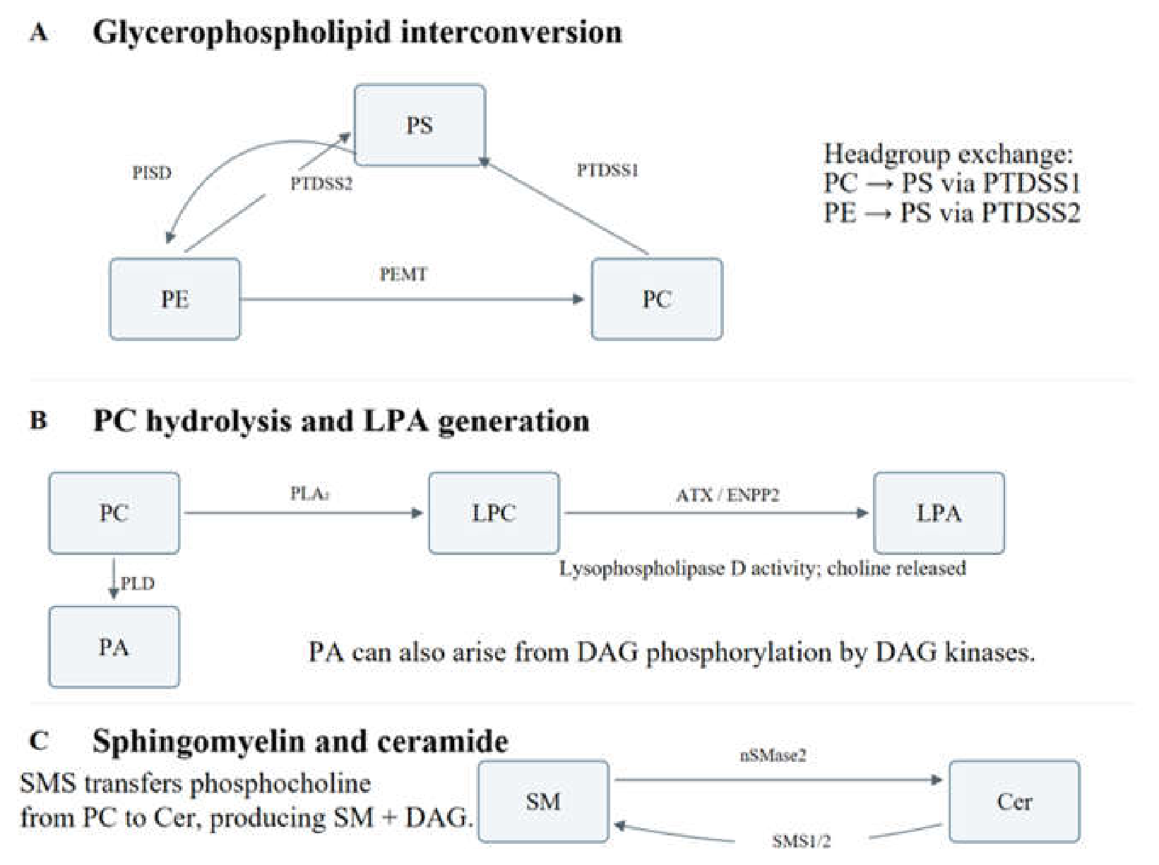

Table 1.
Functional studies of lipid-dependent EV pathways.
| Study | Experimental model | Main finding | Functional readout |
| Ghossoub et al., 2014 (40) | ARF6/PLD2 perturbation and endosomal budding assays. | ARF6 and PLD2 control syntenin-associated ILV formation. | Budding and exosome production. |
| Trajkovic et al., 2008 (36) | Oligodendroglial cells; ceramide-pathway perturbation. | Ceramide supports an ESCRT-independent budding route. | ILV formation and exosome release. |
| Kosaka et al., 2013 (42) | Cancer-cell nSMase2 manipulation; endothelial and tumor assays. | Exosomal miRNA transfer contributes to angiogenesis and metastasis. | Endothelial responses and metastasis. |
| Burgelman et al., 2024 (45) | Genetic Smpd3 deficiency in cell and mouse models. | Smpd3 deficiency leaves EV release unchanged in the systems studied. | In vitro and circulating EV output. |
| Matsumoto et al., 2021 (57) | PS-defined small EV populations; mouse circulation studies. | PS-deficient EVs escape rapid macrophage uptake. | Blood persistence and macrophage uptake. |
| Bhatta et al., 2021 (61) | Human tumor-associated vesicles; T-cell assays and xenografts. | ExoBlock reverses vesicle-associated suppression and improves tumor control. | T-cell activation and tumor growth. |
| Jethwa et al., 2016 (72) | Exosome-bound ATX and recipient-cell signaling assays. | Catalytically active ATX carries and delivers LPA. | Receptor signaling, Ca²⁺ release and migration. |
| Padilla et al., 2025 (47) | MCF7 sphingomyelinase inhibition; EV omics and MCF10A assays. | Acid and neutral enzyme inhibition alter cargo and responses differently. | Recipient-cell translation and migration. |
| Buñay et al., 2026 (75) | Dendrogenin A-treated tumor cells; dendritic cells and melanoma. | BMP-enriched vesicles promote antitumor immunity. | Dendritic-cell activation and anti-PD-1 response. |
Disclaimer/Publisher’s Note: The statements, opinions and data contained in all publications are solely those of the individual author(s) and contributor(s) and not of MDPI and/or the editor(s). MDPI and/or the editor(s) disclaim responsibility for any injury to people or property resulting from any ideas, methods, instructions or products referred to in the content. |
© 2026 by the authors. Licensee MDPI, Basel, Switzerland. This article is an open access article distributed under the terms and conditions of the Creative Commons Attribution (CC BY) license (http://creativecommons.org/licenses/by/4.0/).
Copyright: This open access article is published under a Creative Commons CC BY 4.0 license, which permit the free download, distribution, and reuse, provided that the author and preprint are cited in any reuse.