

Submitted:
13 November 2026
Posted:
17 November 2026
You are already at the latest version
Abstract
The manuscript demonstrates a new approach to improve and evaluate the effect of chitosan (CS) underlayer on the protective properties of ZrO2 sol gel films, deposited on galvanized low carbon steel. In order to obtain more optimized results chitosan was dissolved in citric acid leading to appearance of chitosan-based solution accompanied with crosslinking of the polymer. Two manners of treatment of chitosan underlayer have been applied – drying at ambient temperature or processing at higher temperatures for a definite time interval. Thereafter, the experimental samples with deposited CS layer, were subsequently immersed into zirconium precursor solution and finally treated in air at 180oC. Several methods such as X-ray diffraction (XRD) analysis, Scanning Electron Microscopy (SEM), Atomic Force Microscopy (AFM), water contact angle (CA) and X-ray Photoelectron spectroscopy (XPS) were applied to examine the properties of the newly developed hybrid systems. The corrosion parameters and protective ability of the latter were evaluated by means of potentiodynamic polarization (PDP) curves in a model corrosive medium of 5% NaCl. The results obtained confirmed the positive effect of chitosan on the corrosion resistance of the hybrid protective system in aggressive medium containing chloride ions as corrosion activators.
Keywords:
chitosan
; ZrO2 film
; sol gel
; hybrid coating system
; corrosion
; low-carbon steel
1. Introduction
The reliable protection of metals and steel materials by application of effective hybrid coatings aiming significant extending of their service life is an important and fundamental problem. In industry, especially in engineering, shipbuilding, aerospace, etc., some metals such as iron, aluminium and their alloys are used due to their beneficial physical characteristics: hardness, high strength, heat resistance, etc. Various external factors, such as the presence of solid particles, changes in temperature, different liquids and chemicals, almost always lead to the appearance and development of corrosion processes [1]. Corrosion causes large energy and material losses amounting to billions of dollars annually. This problem requires an application of environmentally friendly protective coatings, monitoring and repair of various infrastructures, as well as costs for the disposal of hazardous waste materials. Although the protection of steel from corrosion has been a subject of permanent scientific research, an ideal method for improved corrosion protection has not been established yet. The protection of different steels in aggressive environment can be achieved by application of various types corrosion-resistant coatings, providing passive protection of the material. This method is particularly effective and economically advantageous. Additional (active) protection is often realized by including in the coating of active components (for example inhibitors) to slow down the rate of the corrosion processes [2,3,4,5]. An example of a good steel corrosion inhibitor is the hexavalent chromium, which, however, forms highly toxic and carcinogenic products and is already banned for application in many countries. This necessitates the search for new and safer inhibitors for nature and humans, as well as methods for their successful inclusion in anti-corrosion coatings. The usage of non-toxic oxide coatings on metal surfaces is a very promising approach for their effective protection against corrosion and extension of their service life [6,7].
Recently, the researchers are focused on studying the effect of different types of inorganic coatings like TiO2, ZrO2, SiO2 and CeO2 on steels in order to increase their corrosion resistance [8,9]. These coatings have a wide range of significant advantages such as good mechanical properties (strength, hardness, wear resistance, etc.), chemical and thermal inertness, high dielectric constant and coefficient of thermal expansion.
Polymers of natural origin (biopolymers) such as hyaluronic acid, chitosan, starch, gelatine, synthetic polypeptides, polylactic acid (PA) and others are preferred as renewable sources. They possess many useful features - non-toxicity, bio-compatibility, acceptable cost, good adhesion to metal substrates and specific solubility. The polysaccharide chitosan (CS) is the most abundant natural polymer after cellulose [10]. Chitosan is a pH-dependent cationic amino polysaccharide [11]. Its structure is composed of randomly distributed D-glucosamine and N-acetyl-D-glucosamine units linked by -(1,14) glycosidic bonds [12]. Chitosan is a weak base and is soluble in acidic aqueous media (pH < 6). The latter protonates the amino groups presented in chitosan’s structure, making the polymer positively charged [13]. Citric acid is a non-toxic weak tricarboxylic acid, which is organic in nature. It is a natural acid with the ability to cross-link and stabilize polysaccharide structures by creating covalent intermolecular di-ester linkages with the hydroxyl groups of the polysaccharide [14]. Chitosan films exhibit some useful characteristics such as high antimicrobial and low oxygen permeability. Various organic acids have been used for the chitosan film preparation: acetic, formic, lactic and tartaric. Such kinds of cross-linking can increase the elasticity and mechanical strength of chitosan films. In the scientific study, Wei et al. have developed one-step strategy to prepare a chitosan-based hydrogel in which citric acid was used simultaneously as a dissolving and crosslinking agent [15]. Sharmin et al. have studied the effect of citric acid cross-linking on the mechanical, rheological and barrier properties of chitosan [16]. They have established, that the values for the elongation at break of the citric acid samples were higher than the acetic acid samples, which kept increasing with an increasing citric acid content due to the plasticizing effect from residual citric acid. Different acid concentrations did not have a large effect on the gelation time. Citric acid containing samples showed higher viscosities as compared to the acetic acid containing solution [16].
In the last decade, the attention of scientists has been directed to application of organic-inorganic composites (hybrids), due to the combination of the useful properties of the individual components [17]. Bulk or surface incorporation of inorganic particles into the organic matrix is being one of the most often used possibilities [18]. However, the data in the available literature on composite coatings consisting of various nano-oxide particles (TiO2, ZnO, etc.) and chitosan layer are relatively scarce [19,20,21,22].
In this article a new approach to improve the corrosion protection properties of low carbon steel structure after deposition of Zn electrochemical layer, chitosan layer, followed by three times ZrO2 films sol gel deposition has been presented and described. Chitosan was dissolved in citric acid with an aim to create chitosan-based solution, as well as for simultaneous dissolution of this compound and crosslinking the polymer.
2. Materials and Methods
2.1. Materials
Medium-molecular-weight chitosan, composed of β-(1–4)-linked d-glucosamine and N-acetyl-d-glucosamine with a deacetylation degree of about 75 – 85%; molecular weight 190 - 310; viscosity 200 - 800 cP at 25 ͦ C and citric acid (CA - C6H8O7 - 99%); molecular weight - 192.12 g/mol were purchased from Sigma–Aldrich®. All solutions were prepared with deionized water and their pH values were measured using a FiveEasy Plus pH meter (Mettler Toledo, Columbus, OH, USA) equipped with LE 410 electrode.
2.2. Preparation of Chitosan
Chitosan (CS - 0.5 g) was dissolved in 3% water solution of citric acid under vigorous stirring for 48 hours. The substrates (low carbon steel with composition presented in Part 2.3. below) were immersed in CS-based solution, and after 30 s were withdrawn at a rate of 30 mm/min. Two series of samples were prepared:
- CS layer dried at ambient room temperature (RT) for one-week period of time – designated as CD;
- CS layer thermally treated in two consecutive steps: 1/. at 100 oC for 30 minutes and 2/. at 180oC for 1 h – designated as CT.
2.3. Electrodeposition of Composite CS/ZrO2 Coatings
Low-carbon steel substrates (3 x 1 x 0.1 cm) with composition (in weight %) as follows: C 0.05–0.12; S ≤ 0.04; P ≤ 0.35; Mn 0.25–0.5; Cr ≤ 0.1; Si ≤ 0.03; Ni ≤ 0.3; Cu ≤ 0.3; As ≤ 0.08; Fe - balance (manufacturer Metalsnab, Sofia, Bulgaria) were galvanically coated with a very thin Zn layer (1 µm) from electrolytic bath at electrodeposition conditions: current density 2 A·dm−2, pH value 5, ambient temperature 25 ◦C, metallurgical (soluble) Zn anodes. The composition of the bath was: ZnSO4·7H2O - 150 g·dm−3; (NH4)2Cl - 30 g·dm−3; H3BO3 - 30 g·dm−3. Prior the electrodeposition, the steel substrates were cleaned with sandpaper, degreaser and thereafter ultrasonically treated in ethanol during 10 min.
The obtained samples with corresponding CS layers (described in Part 2.2.) were immersed into Zr-based precursor solution. The latter was prepared using zirconium butoxide Zr(C4H9O)4 (Alfa Aesar), which was diluted in isopropanol. Thereafter, small amounts of acetyl acetone and ciric acid as complexing agent were added. The samples were treated first at 100 oC for 0.5 h, after which the temperature was increased up to 180 oC for 1 h. The ZrO2 films deposition procedure was repeated 3 times. As already mentioned above the final composite samples containing CS layer, dried at RT, were noted as CD and those containing thermally treated chitosan layer were noted as CT.
2.4. X-Ray Diffraction (XRD) Analyses
An X-ray diffractometer having a PW1830 generator and a PW1050 goniometer (Philips, Almelo, the Netherlands) with CuKα radiation and a generator voltage of 40 kV was applied in order to study the phase composition. The Inorganic Crystal Structure Database (ICSD), Powder Diffraction File™ (PDF-2 2023) and HighScore Plus 3.0 program were used in order to determine the crystalline phases of the bi-layers.
2.5. Atomic Force Microscope (AFM) Studies
Atomic Force microscope (AFM NanoScopeV System, Bruker Inc.) operating in tapping mode in air was used to characterize the surface topography of the coatings. The scanning rate was set at 1 Hz. All images were flattened by means of Nanoscope software. Images from three independent locations on the samples were taken for reproducibility purposes. The roughness analysis gives the value Ra, which is an arithmetic average of the absolute values Zj of the surface height deviations measured from the mean plane, i.e:
while the image Rq is the root mean square average of height deviations taken from the mean image data plane, expressed as
2.6. Contact Angle (CA) Measurements
Contact angle measurements were performed with Ramé-Hart automated goniometer model 290 with DROP image advanced v2.4 (Succasunna, NJ, USA) at room temperature. Drops of 2-5 μL were formed and deposited by Ramé-Hart automatic dispensing system.
2.7. X-Ray Photoelectron Spectroscopy (XPS) Studies
The XPS studies were performed by a VG Escalab II system (East Grinstead, UK), using AlKα radiation with an energy of 1486.6 eV. The chamber pressure was 1.10-9 Torr. The binding energy (BE) of Zn2p at 1021.5 eV for Zn (BE of Zn and ZnO are very close, that is why regardless of the oxidation state of the zinc, their BE can be used as a reliable reference value for calibration) and of Fe2p at 710.8 for Fe3O4 lines were used as internal standards to calibrate the BE of all spectra. The photoelectron spectra were corrected by subtracting a Shirley-type background and were quantified using the peak area and Scofield’s photo-ionization cross-section. The accuracy of BE measurement was ± 0.2 eV. To determine the surface composition of the studied samples and the changes that occurred as a result of the corrosion processes, analyses of as deposited samples and samples subjected to corrosion were performed.
2.8. Potentiodynamic Polarization Investigations (PDP)
The corrosion behavior of the samples in the selected model corrosive medium with chloride ions was investigated under conditions of external polarization (cathodic and anodic). The experiments were performed in a glass tri-electrode electrochemical cell having a volume of 300 ml. A saturated calomel electrode (SCE) was used as a reference electrode and a platinum plate was applied as a polarizing one. The characterization of the test specimens (PDP curves) was performed by “PAR VersaStat 4” equipment at a sufficiently low potential scan rate - 1 mV/s.
2.9. Data Reproducibility and Corrosive Medium
The application of five specimens of every sample type, investigated in the model corrosive test medium (5% NaCl solution) at ambient temperature provided data reproducibility. Тhe error deviations of the individual experiments, according to the methods used, were within 10%.
3. Results
Various reaction elementary steps are likely to occur due to the specific structure of CS, possessing multiple hydroxyl and amine functional groups (Figure 1). The first step is combining of CS template with Zr(OBu)4 precursor in order to achieve higher coordination number of Zr. The complexation, appearing as a result, brings the zirconium butoxide and hydroxyl groups, which leads to the formation of a covalently bound layer. The next reaction step is condensation between Zr-OH and formation of crystal nuclei. The thermal treatment initiates the formation of oxide nanoparticles.
The chemical immobilization is considered to hinder the chemical interaction between neighboring octaedra and to lower the condensation rate, making easier to occur the rearrangement of Zr-O bonds into an ordered lattice.
3.1. XRD Analyses
Only metallic phases belonging to Fe and Zn (originating from the steel substrate and electrochemically deposited zinc layer) were detected by XRD analyses of CT sample (Figure 2). Any crystallographic phase relating to ZrO2 coating was not recorded, because it has an amorphous structure. Practically similar X-Ray difractogram was registered and observed for CD sample.
3.3. AFM Studies
Atomic force microscope was applied to observe the nanostructure of the surface of galvanized steel and chitosan layer dried (CD) as well as chitosan layer treated (CT).
The surfaces of these coatings were compared in Figure 3. AFM images showing the topography of the coatings with both types CS underlayer demonstrate structural difference at a nanoscale level. Although the morphology of the coatings has similar structure, individual CS layers deposited demonstrate difference in their surface densification, which is more pronounced in the case of CT. This statement is supported by the roughness values of CT layer, i.e. Ra and Rq are higher compared to CD layer (Table 1). Therefore, it is obvious that the properties of CS underlayer deposited on the galvanized steel depend on the thermal treatment. The surfaces of CD and CT samples are quite different. The roughness of CD with a scanning area of 5µm x 5µm has a value of Ra = 47.3 nm and Rq = 63 nm, while the roughness of CT with the same scanning area has a value of Ra = 60.9 nm and Rq = 75.6 nm, respectively (Table 1). For comparison, the corresponding parameters of the galvanized steel are: Ra = 52.2 nm and Rq = 65.6 nm.
From the AFM measurements, it was found that the CT samples have higher roughness compared to CD ones. The topography of both coating types distinguish due to different coverage with chitosan on the top of the low carbon steel at different temperature treatments.
3.3. Contact Angle Measurements
Figure 4 (b,d) presents the surface morphology and SEM images of the hybrid CS/ZrO2 coating system with chitosan dried layer (CD) and hybrid CS/ZrO2 coating system with chitosan thermally treated layer (CT) at magnification of 1000 (10 µm). Тhese SEM images are accompanied with the results for water contact angles of the samples (Figure 4 a,c and Table 1). Hybrid systems are homogeneous and the chitosan layer is evenly distributed on the surface. The contact angle of CD sample is 86° and of CT one - 90°, respectively. As seen from Table 1 the contact angle of the galvanized steel is 94, which is a sign for generally comparable hydrophobicity of the layers.
The temperature of the additional treatment does not significantly affect the structure of the layer at a microscopic level. The surface morphology of both CD and CT samples demonstrate similar worm-like surface morphology, typical for sol-gel coatings (Figure 4b, d). Some small cracks on the surface stronger expressed for CT sample can be also observed.
3.3. XPS Studies
To determine the surface composition of the studied samples and the changes that occurred as a result of the corrosion processes, XPS analyses of as deposited samples and samples after corrosion treatment were performed. The chemical composition, calculated from the areas of high resolution core XPS spectra of the respective chemical element is given in Table 2.
The chemical analyses showed that the surface of the samples before corrosion is covered mainly with carbon (59.0 at.% for CT and 48.5 at.% for CD) and oxygen (25.1 at.% for CT and 29.2 at.% for CD). The concentration of zirconium, that forms the top layer, is 4.8 at.% for CT and 6.7 at.% for CD. In the case of CТ this result may indicate the diffusion of Zr into the volume of the sample. Iron is not detected on the surface before corrosion test, which is encouraging sign for densely covered surface with an anti-corrosion coating.
After the electrochemical corrosion, the oxygen concentration increases relative strongly, which demonstrates that the surface was oxidized. Zirconium is not detected in the case of CD but appears in small amount for CT. The zinc concentration decreases for both test samples. Iron was also detected confirming the stripping of the surface during the test. The carbon concentration of CT coating remains relative close to the initial value (as deposited) but in the case of CD it drops sharply to 18.1 at.%. This can be explained having in mind the increased NaCl amount and the greater amount of oxygen on the surface.
To determine the chemical state of the elements, as well as the compounds in which they participate, a peak fitting of the high resolution spectra of the relevant element was performed.
Figure 5 demonstrates the fitted peak of the high resolution XPS spectra of C1s. The spectra of all samples are broad and are divided into three (in the case of the corroded samples into 4) main sub-peaks. The peak positioned at 284.9 eV corresponds to the presence of a C – C bond within a matrix of organic compounds. This BE is also corresponding to the presence of the so-called “adventitious carbon”. The peak at 286.1 eV indicates the presence of a C – OH bond. The peaks at 287.1 – 287.9 eV can be associated with the availability of C = O bonds. Changes in their binding energies can be explained by the changes in the hydrocarbon radical of the organic agents used. The peaks at and around 288.8 eV are a sign for the presence of –COOH – groups. It can be noted that only in the case of fresh CT sample this peak is absent. The peaks at 283.5 eV can be attributed to the presence of carbides. It is known that β-(1,4)-D-glucosamine (chitosan) contains amino groups that make its polymer hydrocarbon chain positively charged [23]. This positive charge could be explained by the presence of peaks with binding energies lower than 284.9 eV.
The deconvoluted high-resolution O1 spectra (Figure 6) show the presence of four main sub-peaks. The first peak, registered at 528.1 eV corresponds to Zr-doped ZnO with electron density shifts. The incorporation of zirconium strongly modifies the local electronic environment. Since Zr⁴⁺ ions replace Zn²⁺ ions, an extra positive charge appear, leading to an intense charge transfer (electron density) towards the neighboring oxygen anions (O²⁻). This increases the screening of the oxygen nucleus, thereby shifting the O1s binding energy down to ~528 eV [24]. The second peak at 529.7 eV corresponds to the presence of O in potential intermediate oxides [25]. This peak possesses the highest intensity in the case of CT samples, subjected to corrosion.
The peak at 530.9 eV corresponds to metal oxides. The BE of the oxygen ions in ZnO are usually between 530.0 – 530.6 eV, and of O in ZrO2 – 530.4 eV, i.e. the peaks of both oxides cannot be separated since they overlap. This peak can also be associated with the presence of a C – O bond. The peak at 532.0 eV corresponds to the presence of OH¯- bonds and adsorbed H2O [26,27]. The peaks from 533.2 eV to 537.0 eV in the corroded samples are most likely due to charges and/or uneven surface, as well as to angular effects.
The peak fitted high-resolution spectra of Zn2p3/2 (Figure 7) are divided into three peaks. The most intense of them is located at 1021.4 eV. This value is more typical for metallic zinc - the binding energies corresponding to metal (1021.5 eV) and oxide (1021.7 eV) are very close. However, considering the shape of the Auger spectra (Figure 8), the absence in it of the characteristic shoulder for metallic zinc at 995 eV, as well as the value of the Modified Auger parameter – 2009.3 eV leads to the conclusion that zinc is mainly in the form of oxide and small amounts of metal. Following this logic, the peaks at 1018.3 and 1019.6 eV can be attributed to metal and non-stoichiometric zinc oxide.
After corrosion treatment the Zn2p3/2 spectra (Figure 7) of the corroded samples become wider. Peak fitting of the spectra demonstrates the formation of four peaks, the latter corresponding to a mixture of metal, non-stoichiometric oxide, ZnO and hydroxides (1022.8 eV and 1024.7 eV, respectively).
The corresponding Auger spectra (Figure 8) are also broad and indicate a mixture of metal, oxides and hydroxides.
Figure 9 presents the Zr3d spectrum of the noncorroded samples. It is divided into three peaks positioned at 181.2, 182.2 and 183.7 eV, respectively - the last peak being present only in sample CT. The most intense peak at 182.2 eV corresponds to ZrO2 [28].
The peak positioned at 181.2 eV is with higher BE than that of metallic zirconium (178.9 eV). Therefore, it can be a sign for the presence of zirconium ions diffused into the zinc matrix lattice and the formation of Zr – O – Zn bonds [29,30]. The presence of non-stoichiometric zirconium suboxides is also possible. In these suboxides zirconium ions penetrate the zinc oxide lattice and compete for oxygen atoms with the zinc ions of the matrix [31]. In such a case, zirconium fails to reach its highest oxidation state (Zr4+) and remains in its intermediate oxidation states - Zr3+ or Zr2+. For CT sample another additional low-intensity peak is formed at 183.7 eV which can be associated with the presence of zirconium hydroxide [32,33].
Considering the peak fitted spectrum of Cl2p of the corroded samples the presence of three main peaks can be registered (Figure 10). They present in both samples and are positioned at 197.9, 199.5 and 201.0 eV, respectively. All three peaks can be associated with the presence of metal chlorides – NaCl [34], as well as zirconium chlorides (199.5 еV) [35]. The third peak at 201.0 eV can be probably attributed to the presence of Zn5(OH)8Cl2.H2O [36].
There is no indication of the presence of NaClO, whose peak is principally placed between 205.0 eV and 206.0 eV and which is an expected by-product of electrolysis in the presence of NaCl. The peak in CD sample at 196.2 eV has a very low binding energy and is most likely due to charging or chlorine adsorbed in the cracks of the sample.
3.4. Potentiodynamic Polarization (PDP) Investigations
Potentiodynamic investigations of low carbon galvanized steel, CD and CT samples are represented in Figure 11. Some of the most important electrochemical parameters registered from the curves in the figure are demonstrated in Table 3. It is well visible from the table and also from the figure that the corrosion potentials of the investigated CS-based hybrid protective systems as well as of the galvanized steel in the test medium are relatively close.
Bearing these results in mind, it can be assumed that all the investigated samples are anodic (sacrificial) to bare low carbon steel (since the potential of the iron is more positive) and all they will dissolve as first during the corrosion attack ensuring enhanced protective action.
According to the data from Figure 11, the anodic curve of the galvanized steel shows increased current density greater than 10-1 A.cm-2 which seems to be a result of the presence of a very thin zinc layer (thickness of 1 µm). Generally, galvanized steel covered with a 12 µm thick zinc layer has much lower anodic current density in that potential area (after the corrosion potential) [37,38]. Most probably, as a result of the thermal treatment a mixed corrosion potential (simultaneous presence of zinc and iron enriched zones) appears on the galvanized steel surface leading to increased anodic current density and accelerated dissolution.
Contrary to this, both CD and CT samples demonstrate lower corrosion currents and also lower anodic current densities of about 10-2 A.cm-2. The current density of these coatings is much lower in the whole anodic zone, but however increases after reaching potential values of about -0.4 V. Since these hybrid systems are more negative compared to the ordinary steel, it could be expected that they will ensure sacrificial protection of the latter in the test medium.
4. Discussion
The experimental results obtained by application of two types chitosan-based ZrO2 containing hybrid systems with and without additional thermal treatment demonstrate their improved corrosion resistance and protective ability toward galvanized steel in a model test medium with chloride ions as corrosion activators.
The corrosion potential values (Ecorr) of both hybrid systems represent close potential values compared to the galvanized steel. In addition, the corrosion current density values of CD and CT, obtained from the potentiodynamic polarization curves, are lower than that of the galvanized steel and their PDP curves are longer. This is a sign that they last longer at conditions of external anodic polarization, i.e. they demonstrate greater resistivity in that medium. Considering the experimental data, it follows that both hybrid protective systems are characterized with very good protective ability and can be successfully applied for the protection of galvanized and low carbon steel in chloride-containing media.
It is known that the presence of amorphous structure is among of the basic factors which influence the corrosion stability of oxide layers and alloys. According to the literature data it has been proved that metallic alloys and glasses possess higher corrosion resistance than the crystalline materials [39,40]. Our previous studies devoted on protective single ZrO2, ZrO2-TiO2 composite coatings and multilayered ZrO2/TiO2 systems on steel/galvanized steel substrate revealed the positive role of amorphous structure on protective anticorrosion properties [9,37,41].
Other research group has also obtained, that the amorphous titania-zirconia composite films on AISI 316L steel exhibited high protection efficiency in NaCl medium. The amorphous structure possesses high chemical homogeneity without crystalline defects such as grain boundaries, precipitates etc. The above mentioned defects play the role of active sites for appearing of corrosion process. The absence of such type of defects on the amorphous surface leads to delayed formation of galvanized cells leading to lower corrosion rate. Additionally, the amorphous structure can reduce the diffusion of the ionic species.
Based on the literature data it is known that the C1c spectrum of chitosan consists of three main sub-peaks: C-C/C-H (≈ 283.9 eV), C-N/C-NH₂ (≈ 285.4 eV), and C-O/C=O (≈ 286.9 eV) [43]. During the peak fitting of the C1c spectrum, peaks at around 285.4 eV, characteristic of the presence of C-N/C-NH2 groups and 286.9 eV were not detected. This, together with the absence of nitrogen in the survey spectra of the samples, indicates that chitosan was not detected on the surface of the samples. A possible reason for this is its ability to deprotonate in environments with pH above 6.5 [44]. As a result of this deprotonation of the amino groups in its molecule, a hydrogel layer is formed. Another possible explanation for the lack of chitosan may be its relatively low thermal stability. Several research groups have showed that in the temperature range of 50–150 °C, dehydration of its molecule occurs [45,46]. In this case, agglomeration processes of the polymer would appear. Agglomeration of chitosan would significantly affect both the results of XPS analyses and its physicochemical properties (for example, its ability to realize bridges between Zn and Zr layers). When the chains are condensed and agglomerated, a large part of free amino groups -NH2 turn out to be masked (buried) in the interior (bulk) of the agglomerate or participate in strong intermolecular hydrogen bonds. Therefore, it is possible that nitrogen is not detected on the surface of these agglomerates.
At the same time, peaks characteristic for the presence of –С – ОН , –С = О and –СООН – groups may increase their intensity due to the “jumping” of these groups on the surface of the agglomerates. During this formation of the hydrogel layer/agglomerates of chitosan, probably a contact between the zinc electroplating of the iron alloy and the zirconium oxide layer occurs. In this case, diffusion has occurred between these two layers until Zr – O – Zn bonds are formed [24]. This process as well as the presence of insoluble zinc oxides and hydroxides have a positive effect on the corrosion current. Their combination with the possible formation of insoluble Zn5(OH)8Cl2.H2O phase makes them promising as layers for corrosion protection.
5. Conclusions
The successful preparation of corrosive resistant CS-based hybrid protective systems with three chitosan underlayer with additional ZrO2 top layer was reported. The application of these systems demonstrates their enhanced protective ability in NaCl medium at external polarization conditions.. Hydrophobicity measurements revealed that both systems have water contact angles of about 90°. The type of the treatment of chitosan underlayer has not significant influence on the anticorrosion ability of the structures. During the treatment of the coatings, the formation of Zr – O – Zn bonds in the volume of the zinc layer occurs. The presence of such metal-oxide bonds and of insoluble zinc oxides and hydroxides have a positive effect on the corrosion resistance and protective ability. The presence of amorphous hydrophobic structure of the coatings also can contribute the good corrosion properties of the multilayer hybrid systems.
Author Contributions
For research articles with several authors, a short paragraph specifying their individual contributions must be provided. The following statements should be used “Conceptualization, I.S. and D.S.; methodology, I.S., D.S., Neli Boshkova (Ne.B), O.D., G.A., Nikolai Boshkov (N.B.); validation, A.T., S.S., N.G., I.S., N.B.; formal analysis, S.S., N.G., G.A., A.T., Ne.B.; investigation, Ne.B., N.B., I.S., D.S.; resources, I.S., D.S.; data curation, O.D., G.A., A.T., S.S., N.G.; writing—original draft preparation, I.S., D.S., A.T., N.B.; writing—I.S., N.B., X.X.; visualization, S.S., I.S.; supervision, I.S., N.B.; project administration, I.S.; funding acquisition, Y.Y. All authors have read and agreed to the published version of the manuscript.” Please turn to the CRediT taxonomy for the term explanation. Authorship must be limited to those who have contributed substantially to the work reported.
Funding
This research received no external funding.
Institutional Review Board Statement
Not applicable.
Informed Consent Statement
Not applicable.
Data Availability Statement
It is mandatory for the authors to include a “Data Availability Statement (DAS)” section in their manuscripts, except for the following article types: Book Review, Editorial, Obituary, Correction, Retraction, Expression of Concern. In this section, authors are required to provide details regarding where data supporting their reported results can be found, including links to publicly archived datasets analyzed or generated during the study. Where no new data were created or where data is unavailable due to privacy, legal, or ethical restrictions, a statement is still required. Suggested Data Availability Statements are available in section “MDPI Research Data Policies” at https://www.mdpi.com/ethics.
Acknowledgments
The authors express their gratitude to the contract “Multicomponent oxide coatings for increased corrosion resistance steels” which is within the Non-currency Equivalent Exchange Bilateral Cooperation between the Bulgarian Academy of Sciences and the Serbian Academy of Sciences and Fine Arts. The research equipment of the distributed research infrastructure INFRAMAT (part of the Bulgarian National roadmap for research infrastructures) was supported by the Bulgarian Ministry of Education and Science was used in this investigation.
Conflicts of Interest
The authors declare no conflicts of interest.
References
- Ekerenam, O.O.; Ikeuba, A.I.; Njoku, C.N.; Njoku, D.I.; Emori, W.; Nwokolo, I.K.; Etim, I.-I.N.; Okonkwo, B.O.; Udoh, I.I.; Daniel, E.F.; Uzoma, P.C.; Awonusi, B.O.; Kolawole, S.K.; Etim, I.P.; Olanrele, O.S. “Advancements in corrosion studies and protective measures for copper and copper-based alloys in varied environmental conditions”. Res. Engin. 26(2025), 105257. [CrossRef]
- Koch, G.; Brongers, M.; Thomson, N.; Virmani, Y.; Payer, J. “Corrosion Cost and Preventive Strategies in the United States”. Mater. Perform. 2002, 65, 1. [Google Scholar]
- Fragata, F.; Salai, R.; Amorin, C.; Almeida, E. “Compatibility and Incompatibility in Anticorrosive Painting - The Particular Case of Maintenance Painting”. Prog. Org. Coat. 2006, 56, 257. [Google Scholar]
- Brooman, E.W. “Modifying Organic coatings to provide Corrosion Resistance-Part I: Back-ground and General Principle”. Metal. Finish. 2002, 100(1), 48. [Google Scholar] [CrossRef]
- Zheludkevich, M.L.; Shchukin, D.G.; Yasakau, K.A.; Moehwald, H.; Ferreira, M.G.S. “Anticorrosion Coatings with Self-Healing Effect Based on Nanocontainers Impregnated with Corrosion Inhibitor”. Chem. Mater. 2007, 19(3), 402. [Google Scholar] [CrossRef]
- Zheludkevich, M.L.; Tedim, J.; Ferreira, M.G.S. “Smart” coatings for active corrosion protection based on multi-functional micro and nanocontainers”. Electrochim. Acta 2012, 82, 314. [Google Scholar] [CrossRef]
- Bachvarov, V.; Peshova, M.; Vitkova, S.; Boshkov, N. “Corrosion properties of Zn-Ni-P alloys in neutral model medium”. Cent. Eur. J. Chem. 2014, 12, 1183. [Google Scholar] [CrossRef]
- Rajendran, P.; Muthuraj, A.; Rajagounder, N. E. Review on CeO2-Based Corrosion Coatings. Trans. Ind. Ceram. Soc. 2022, 81, 158–174. [Google Scholar] [CrossRef]
- Stambolova, I.; Boshkov, N.; Boshkova, N.; Stoyanova, D.; Shipochka, M.; Simeonova, S.; Grozev, N. Environmentally-Friendly Anticorrosive Layered Zirconia/Titania/Low-Carbon Steel Structures”. MDPI proceedings, 2-nd International Online-Conference on Nanomaterials /, 2021; Vol 4. [Google Scholar] [CrossRef]
- Georgieva, V.; Zvezdova, D.; Vlaev, L. “No-isothermal kinetics of thermal degradation of chitosan”. Chem. Centr. J. 2012, 6, 81. [Google Scholar] [CrossRef]
- Pinheiro, A.C.; Bourbon, A.I.; Cerqueira, M.A.; Maricato, É.; Nunes, C.; Coimbra, M.A.; Vicente, A.A. Chitosan/fucoidan multilayer nanocapsules as a vehicle for controlled release of bioactive compounds. Carbohydr. Polym. 2015, 1–9, 115. [Google Scholar] [CrossRef]
- Nataraj, D.; Sakkara, S.; Meghwal, M.; Reddy, N. Crosslinked chitosan films with controllable properties for commercial applications. Int. J. Biol. Macromol. 2018, 120, 1256. [Google Scholar] [CrossRef]
- Moura, M.J.; Figueiredo, M.M.L.; Gil, M.H. Rheological Study of Genipin Cross-Linked Chitosan Hydrogels. Biomacromolecules 2007, 8, 3823. [Google Scholar] [CrossRef]
- Sharmin, N.; Sone, I.; Walsh, J.L.; Sivertsvik, M.; Fernández, E.N. Effect of citric acid and plasma activated water on the functional properties of sodium alginate for potential food packaging applications. Food Packag. Shelf Life 2021, 29, 100733. [Google Scholar] [CrossRef]
- Wei, B.; Zou, J.; Pu, Q.; Shi, K.; Xu, B.; Ma, Y. One-step preparation of hydrogel based on different molecular weights of chitosan with citric acid. J. Sci. Food Agric. 2022, 102, 3826. [Google Scholar] [CrossRef]
- Sharmin, N.; Rosnes, T.J.; Prabhu, L.; Böcker, U.; Sivertsvik, M. Effect of Citric Acid Cross Linking on the Mechanical, Rheological and Barrier Properties of Chitosan. Molecules 2022, 27, 5118. [Google Scholar] [CrossRef]
- Pourhashem, S.; Saba, F.; Duan, J.; Rashidi, A.; Guan, F.; Nezhad, E.G.; Hou, B. Polymer / inorganic nanocomposite coatings with superior corrosion protection performance: A review. J. Ind. Eng. Chem. 2020, 88, 29. [Google Scholar] [CrossRef]
- Kango, S.; Kalia, S.; Celli, A.; Njuguna, J.; Habibi, Y.; Kumar, R. “Surface modification of inorganic nanoparticles for development of organic–inorganic nanocomposites—A review”. Prog. Polym. Sci. 2013, 38 8, 1232. [Google Scholar] [CrossRef]
- Salam, J.A.; Baby, A.M.; Joseph, A. Corrosion inhibition of mild steel using chitosan/TiO2 nanocomposite coatings. Progr. Org. Coat. 2019, 122, 254. [Google Scholar] [CrossRef]
- Sanmugam, A.; Vikraman, D.; Karuppasamy, K.; Lee, J.Y.; Kim, H.-S. Evaluation of the corrosion resistance properties of electroplated chitosan-Zn1-XCuxO composite thin films. Nanomaterials 2017, 7, 432. [Google Scholar] [CrossRef]
- Carneiro, J.; Tedim, J.; Ferreira, M.G.S. Chitosan as a smart coating for corrosion protection of aluminum alloy 2024: A review. Progr. Org. Coat. 2015, 89, 348. [Google Scholar] [CrossRef]
- Cui, L.; Gao, S.; Song, X.; Huang, L.; Dong, H.; Liu, J.; Chen, F.; Yu, S. Preparation and characterization of chitosan membranes. RSC Adv. 2018, 8, 28433. [Google Scholar] [CrossRef]
- Yoo, S.-Ho; Lee, Ji-S.; Park, S. Y.; Kim, Y.-S.; Kim; Chang, P.-Sh.; Lee, H. G. Effects of selective oxidation of chitosan on physical and biological properties. Intern. J. Biol. Macromol. 2005, 35, 27–31. [Google Scholar] [CrossRef]
- Gandla, S.; Gollu, S. R.; Sharma, R.; Sarangi, V.; Gupta, D. Dual role of boron in improving electrical performance and device stability of low temperature solution processed ZnO thin film transistors. appl. phys. Lett. 107 2015, 152102. [Google Scholar] [CrossRef]
- Liu, Y.; Huang, J.; Claypool, J. B.; O’Keefe, M. J. Structure and Corrosion Behavior of Sputter Deposited Ce-Al-O Coating on Al 2O24-T3 Alloy Substrate. J. Electrochem. Soc. 2016, 163(5), C198–C204. [Google Scholar] [CrossRef]
- Hachoun, Z.; Ouerdane, A.; Yahiaoui, I.; Gulakhmadov, A.; Chen, X. Investigating the sensing properties of SnO2 nanoparticles doped with gold. Appl. Phys. A. 2021, 127, 559. [Google Scholar] [CrossRef]
- Graf, A.; Isaacs, M. A.; Morgan, D. J. Insight Notes: Considerations in the XPS Analysis of O1s Spectra for Metal Oxides. Surf. Interface Anal. 0 2025, 1–7. [Google Scholar] [CrossRef]
- Gondal, M.A.; Fasari, T.A.; Baig, U.; Mekki, A. Effects of Oxidizing Media on the Composition, Morphology and Optical Properties of Colloidal Zirconium Oxide Nanoparticles Synthesized via Pulsed Laser Ablation in Liquid Technique. J. Nanosci. Nanotechnol. 2017, 18(6), 1–10. [Google Scholar] [CrossRef]
- Zandaleh, Z.; Bols, M.; Yazdani, P.; Poelman, H.; Saeys, M. Structural contributions of Zn in enhancing CO2 hydrogenation to methanol over ZnxZrOy catalysts. Catal. Sci. Technol. 2024, 15, 563–579. [Google Scholar] [CrossRef]
- Lin, W.-Y.; Chien, F.-T.; Chiu, H.-C.; Sheu, J.-K.; Hsueh, K.-P. Effects of Thermal Annealing on the Properties of Zirconium-Doped MgxZn1−XO Films Obtained through Radio-Frequency Magnetron Sputtering. Membranes 2021, 11, 373. [Google Scholar] [CrossRef]
- Meng, C.; Zhao, G.; Shi, X.-R.; Chen, P.; Liu, Y.; Lu, Y. Oxygen-deficient metal oxides supported nano-intermetallic InNi3C0.5 toward efficient CO2 hydrogenation to methanol. Sci. Adv. 4 2021, 7. [Google Scholar] [CrossRef]
- Huang, C.; Tang, Z.; Zhang, Z. Differences between Zirconium Hydroxide (Zr(OH)4•nH2O) and Hydrous Zirconia (ZrO2•nH2O). J. Amer. Ceram. Soc. 2004, 7, 1637–1638. [Google Scholar] [CrossRef]
- Zhang, Y.; Lan, Y.; Jin, F.; Li, G. Y3+-Stabilized Zirconia (YSZ) Coatings for Protection Against Water Vapor Corrosion. Coatings 2026, 16, 272. [Google Scholar] [CrossRef]
- Wu, K.-H.; Wang, D.-W.; Zeng, Q.; Li, Y.; Gentle, I. Solution phase synthesis of halogenated graphene and the electrocatalytic activity for oxygen reduction reaction. Chin. J. Catal. 2014, 35, 884–890. [Google Scholar] [CrossRef]
- Roy, P. C.; Jeong, H. S.; Doh, W. H.; Kim, C. M. Atomic layer deposition (ALD) of ZrO2 in ultrahigh vacuum (UHV). Bull. Kor. Chem. Soc. 2013, 34, 1221. [Google Scholar] [CrossRef]
- Winiarski, J.; Tylus, W.; Winiarska, K.; Szczygieł, I.; Szczygieł, B. XPS and FT-IR Characterization of Selected Synthetic Corrosion Products of Zinc Expected in Neutral Environment Containing Chloride Ions. J. Spectr. Iss. 1 2018, 2079278. [Google Scholar] [CrossRef]
- Boshkova, N.; Stoyanova, D.; Stambolova, I.; Dimitrov, O.; Simeonova, S.; Avdeev, G.; Peshova, M.; Bachvarov, V.; Smrichkova, S.; Boshkov, N. “Corrosion Efficiency of Zn-Ni/ZrO2 and Zn-Co/ZrO2 Bi-Layer Systems: Impact of Zn-Alloy Sublayer Thickness”. Coatings 2024, 14(7), 792. [Google Scholar] [CrossRef]
- Kamburova, K.; Boshkova, N.; Radeva, Ts.; Shipochka, M.; Boshkov, N. Chitosan-alginate nanocontainers with caffeine as green corrosion inhibitors for protection of galvanized steel. Crystals 2024, 14(7), 660. [Google Scholar] [CrossRef]
- Naka, M.; Hashimoto, K.; Masumoto, T. Corrosion behavior of amorphous and crystalline Cu50Ti50 and Cu50Zr50 alloys. J. Non-cryst. Sol. 1978, 30, 29–36. [Google Scholar] [CrossRef]
- Scully, J.R.; Gebert, A.; Payer, J.H. Corrosion and related mechanical properties of bulk metallic glasses. J. Mater. Res. 2006, 22, 302–313. [Google Scholar] [CrossRef]
- Stambolova, I.; Stoyanova, D.; Shipochka, M.; Boshkova, N.; Eliyas, A.; Simeonova, S.; Grozev, N.; Boshkov, N. Surface Morphological and Chemical Features of Anticorrosion ZrO2–TiO2 Coatings: Impact of Zirconium Precursor. Coatings 2021, 11, 703. [Google Scholar] [CrossRef]
- Ćurković, L.; Ćurković, H.O.; Žmak, I.; Mustafa, M.K.; Gabelica, I. Corrosion Behavior of Amorphous Sol–Gel TiO2–ZrO2 Nano Thickness Film on Stainless Steel. Coatings 2021, 11, 988. [Google Scholar] [CrossRef]
- Daniyal, W.M.E.M.M.; Fen, Y.W.; Saleviter, S.; Chanlek, N.; Nakajima, H.; Abdullah, J.; Yusof, N.A. X-ray Photoelectron Spectroscopy Analysis of Chitosan–Graphene Oxide-Based Composite Thin Films for Potential Optical Sensing Applications. Polymers 2021, 13, 478. [Google Scholar] [CrossRef]
- Zou, Y.; Zhong, Y.; Li, H.; Ding, F.; Shi, X. Electrodeposition of Polysaccharide and Protein Hydrogels for Bio-medical Applications. Curr. Med. Chem. 2020, 27, 1–21. [Google Scholar] [CrossRef]
- Zawadzki, J.; Kaczmarek, H. Thermal treatment of chitosan in various conditions. Carbohydr. Polym. 2010, 80, 394–400. [Google Scholar] [CrossRef]
- Diab, M. A.; El-Sonbati, A. Z.; Al-Halawany, M. M.; Bader, D. M. D. Thermal stability and degradation of chitosan modified by cinnamic acid. Open J. Polym. Chem. 2012, 2(01), 14–20. Available online: http://www.SciRP.org/journal/ojpchem.
Figure 1.
Schematic presentation of the multilayer system, consisting of consecutive ordered chitosan/ZrO2 layers on galvanized steel.
Figure 1.
Schematic presentation of the multilayer system, consisting of consecutive ordered chitosan/ZrO2 layers on galvanized steel.
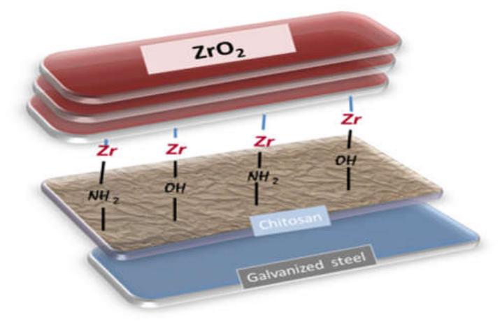
Figure 2.
Typical XRD pattern of hybrid CT (CD sample, respectively) coating systems.
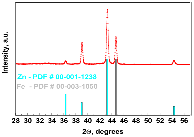
Figure 3.
Topography of AFM images presented in 3D formats of:(a) galvanized steel substrate; (b) with dried chitosan layer - CD; (c) with treated chitosan layer - CT.
Figure 3.
Topography of AFM images presented in 3D formats of:(a) galvanized steel substrate; (b) with dried chitosan layer - CD; (c) with treated chitosan layer - CT.
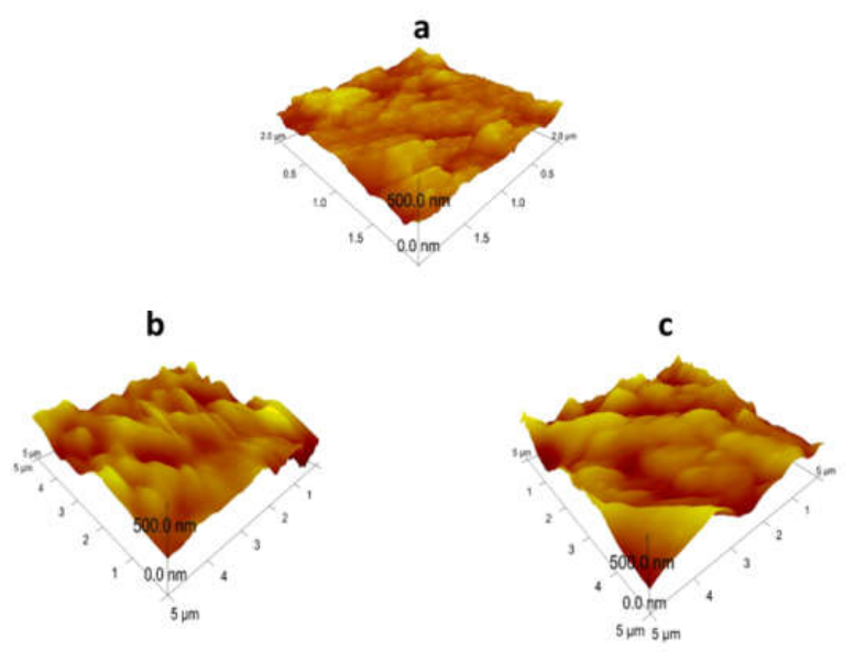
Figure 4.
Water contact angles and morphology of CD (a, b) and CT coatings (c, d).
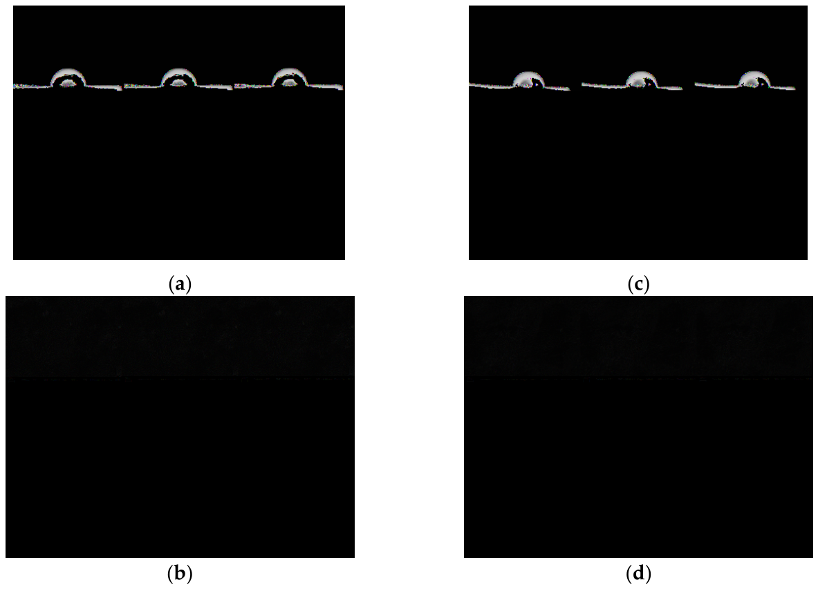
Figure 5.
High-resolution peak fitted XPS spectra of C1s. From bottom to top:CD – as prepared; CT – as prepared; CD – after corrosion; CT – after corrosion. With different colors along the ordinate are labeled the scales for the samples.
Figure 5.
High-resolution peak fitted XPS spectra of C1s. From bottom to top:CD – as prepared; CT – as prepared; CD – after corrosion; CT – after corrosion. With different colors along the ordinate are labeled the scales for the samples.
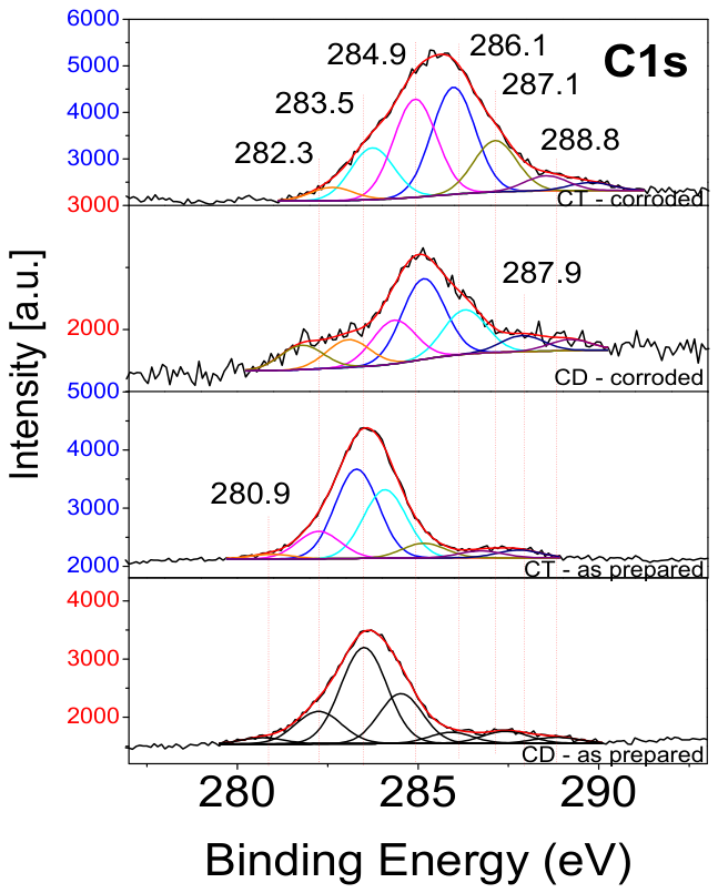
Figure 6.
High-resolution peak fitted XPS spectra of O1s. From bottom to top: CD – as prepared; CT – as prepared; CD – after corrosion; CT – after corrosion. With different colors along the ordinate are labeled the scales for the samples.
Figure 6.
High-resolution peak fitted XPS spectra of O1s. From bottom to top: CD – as prepared; CT – as prepared; CD – after corrosion; CT – after corrosion. With different colors along the ordinate are labeled the scales for the samples.
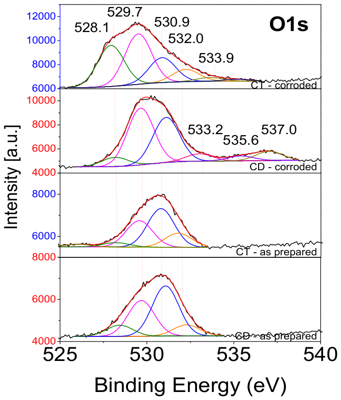
Figure 7.
High-resolution peak fitted XPS spectra of Zn2p. From bottom to top: CD – as prepared; CT – as prepared; CD – after corrosion, CT – after corrosion. With different colors along the ordinate are labeled the scales for the samples.
Figure 7.
High-resolution peak fitted XPS spectra of Zn2p. From bottom to top: CD – as prepared; CT – as prepared; CD – after corrosion, CT – after corrosion. With different colors along the ordinate are labeled the scales for the samples.
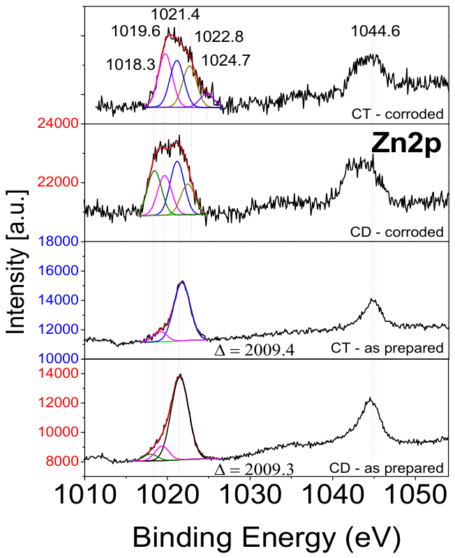
Figure 8.
L3M45M45 Auger electron spectra of Zn.
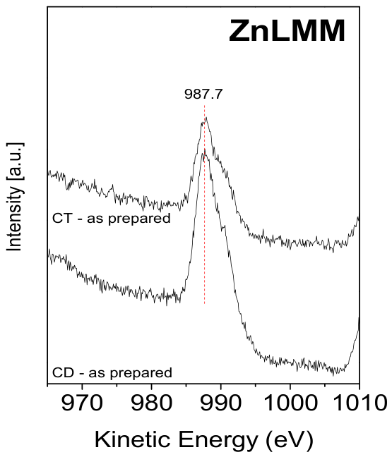
Figure 9.
High-resolution peak fitted XPS spectra of Zr3d.
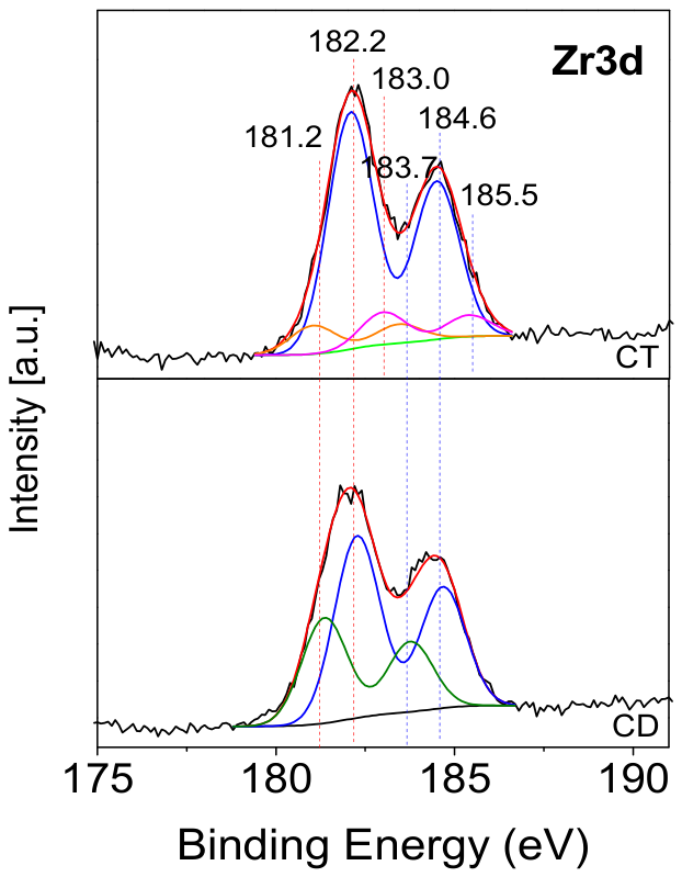
Figure 10.
High-resolution peak fitted XPS spectra of Cl2p. With different colors along the ordinate are labeled the scales for the samples.
Figure 10.
High-resolution peak fitted XPS spectra of Cl2p. With different colors along the ordinate are labeled the scales for the samples.
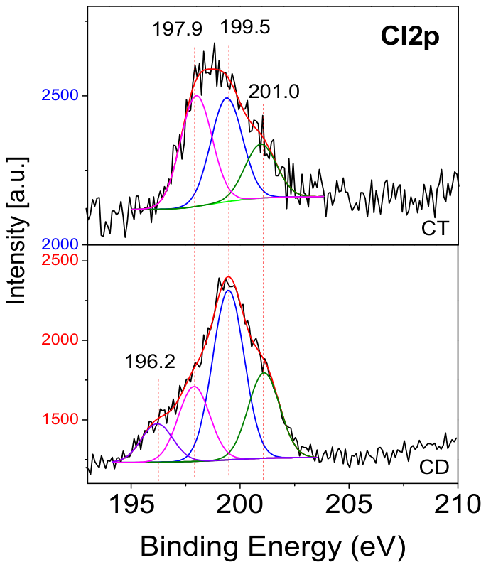
Figure 11.
Potentiodynamic polarization curves in 5% NaCl solution ofgalvanized steel, CD and CT samples, respectively.
Figure 11.
Potentiodynamic polarization curves in 5% NaCl solution ofgalvanized steel, CD and CT samples, respectively.
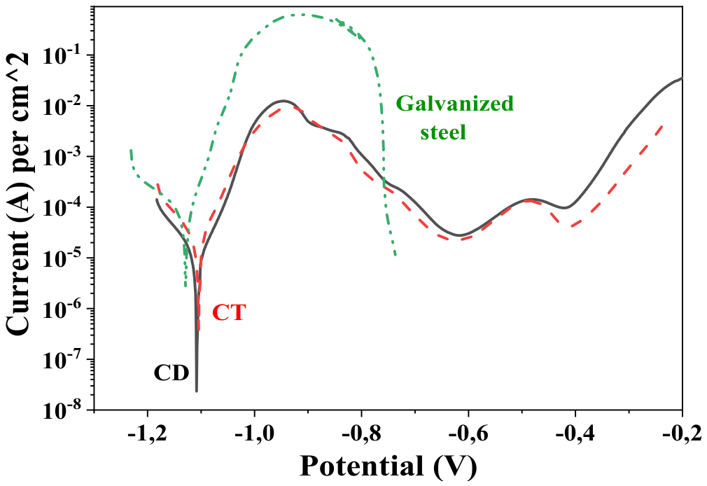

Table 1.
Roughness parameters Rq, Ra and water contact angles (CA) values of bare galvanized steel, CD and CT coatings, respectively (scanning area - 5µm x 5µm).
Table 1.
Roughness parameters Rq, Ra and water contact angles (CA) values of bare galvanized steel, CD and CT coatings, respectively (scanning area - 5µm x 5µm).
| Parameter | Galvanized steel | CD | CT |
| CA |
94 |
86 |
90 |
| Ra | 52.2 | 47.3 | 60.9 |
|
Rq |
65.6 | 63.0 | 75.6 |
Table 2.
Chemical composition of CD and CT coatings before and after corrosion test.
| Sample |
C [at. %] |
O [at. %] |
Zn [at. %] |
Zr [at. %] |
Cl [at. %] |
Fe [at. %] |
Na [at. %] |
| CD – as deposited | 48.5 | 29.2 | 8.4 | 6.7 | 7.2 | 0 | 0 |
| CT - as deposited | 59.0 | 25.1 | 6.5 | 4.8 | 4.6 | 0 | 0 |
| CD – after corrosion | 18.1 | 47.1 | 4.2 | 0 | 12.1 | 8.2 | 10.3 |
| CT – after corrosion | 52.1 | 35.0 | 2.9 | 0.5 | 3.5 | 4.2 | 1.8 |
Table 3.
Electrochemical parameters obtained from Figure 11.
Table 3.
Electrochemical parameters obtained from Figure 11.
|
Sample |
Icorr, A.cm-2 |
Ecorr, V |
Ipass, A.cm-2 |
| Galvanized steel | 6.87.10-5 | -1.131 | - |
| CD | 1.03.10-5 | -1.087 | 2.81.10-5 |
| CT | 2,48.10-5 | -1.065 | 2.13.10-5 |
Disclaimer/Publisher’s Note: The statements, opinions and data contained in all publications are solely those of the individual author(s) and contributor(s) and not of MDPI and/or the editor(s). MDPI and/or the editor(s) disclaim responsibility for any injury to people or property resulting from any ideas, methods, instructions or products referred to in the content. |
© 2026 by the authors. Licensee MDPI, Basel, Switzerland. This article is an open access article distributed under the terms and conditions of the Creative Commons Attribution (CC BY) license.
Copyright: This open access article is published under a Creative Commons CC BY 4.0 license, which permit the free download, distribution, and reuse, provided that the author and preprint are cited in any reuse.