

Submitted:
13 November 2026
Posted:
16 November 2026
You are already at the latest version
Abstract
Dense extracellular matrix deposition, stromal activation and impaired perfusion can create tumour niches that favour lactate accumulation. Lysine lactylation (Kla) provides a molecular link between lactate metabolism, gene expression and protein function, with potential implications for tumour progression and treatment response. Here, we review histone and non-histone lactylation in desmoplastic and stroma-rich tumour contexts, encompassing pancreatic ductal adenocarcinoma and cholangiocarcinoma alongside selected settings in gastric, colorectal, triple-negative breast and hepatocellular cancers. This framework recognises that stromal features vary within and between these malignancies. We synthesise evidence linking lactylation to cancer stemness, fibroblast activation, stromal remodelling and immune suppression, and examine how these processes contribute to therapeutic resistance. Recurrent mechanisms include metabolic adaptation, enhanced DNA repair, suppression of cell death and impaired antitumour immunity. However, lactylation effects depend on the modified protein, lysine residue and cellular context, and are not uniformly tumour-promoting. We critically assess limitations in chemical discrimination, antibody specificity and causal inference, particularly the difficulty of distinguishing Kla-dependent mechanisms from other effects of lactate metabolism. We also discuss therapeutic strategies targeting lactate availability, lactylation enzymes and individual substrates. Most evidence remains preclinical, highlighting the need for quantitative, site-specific and spatially resolved analyses, functional validation and prospective clinical studies. Establishing which lactylation events directly influence tumour behaviour and treatment response will be essential for translating these findings into selective therapies and clinically informative biomarkers.
Keywords:
lactylation
; tumor microenvironment
; post-translational modification
; stroma
; therapy response
1. Introduction
Desmoplasia is a prominent feature of several aggressive solid malignancies. Desmoplastic tumour microenvironments are characterized by the expansion and remodelling of a collagen-rich extracellular matrix (ECM), with activation of cancer-associated fibroblasts (CAFs) or hepatic stellate cells (HSCs), altered tissue mechanics, abnormal vascularization and restricted perfusion. These changes generate hypoxic, nutrient-poor, immune-excluded niches with impaired drug delivery and contribute substantially to tumour progression and treatment failure1. In this review, the terms desmoplastic and stroma-rich are used operationally to denote tumours with a high stromal fraction, increased collagen/ECM, activated fibroblastic or stellate populations, increased stiffness, or a validated stroma-high/mesenchymal classification, without implying uniformity within a histological type or a universal cut-off. Pancreatic ductal adenocarcinoma (PDAC) and cholangiocarcinoma (CCA) are canonical desmoplastic malignancies1,2, whereas gastric cancer (GC) exhibits heterogeneous stroma-rich phenotypes, including a defined mesenchymal-like molecular subtype3. Colorectal cancer (CRC), triple-negative breast cancer (TNBC) and hepatocellular carcinoma (HCC) are considered context-dependent or borderline settings: CRC includes a well-defined mesenchymal, stroma-rich CMS4 subtype4; TNBC displays marked intertumoral variation in stromal and collagen content5; and HCC, although not classically desmoplastic, commonly develops in a fibrotic or cirrhotic liver shaped by activated HSCs and ECM remodelling 6. Across these settings, the stroma functions not merely as a structural scaffold but as an active metabolic and signalling compartment that shapes tumour-cell plasticity, immune regulation and therapeutic response.
A defining feature of these microenvironments is the accumulation and intercellular exchange of lactate. Increased tumour glycolysis, hypoxia and reciprocal tumour-stroma-immune interactions promote the formation of lactate-rich niches, while poor perfusion and dense ECM restrict lactate clearance. Lactate can also be imported and reused as an oxidative carbon source7 and act extracellularly through the Gi-couple receptor HCAR1 to support metabolic adaptation and suppress antigen presentation8,9. In parallel, proton-coupled export drives extracellular acidification that independently alters metabolism, invasion, immune-cell function and therapeutic response10. Tumour-derived lactate can polarize macrophages and impair T- and NK-cell activity, HCAR1 signalling or acidosis rather than lactylation alone11,12. These parallel mechanisms must therefore be distinguished experimentally before a lactate-induced phenotype can be attributed specifically to lactylation.
The discovery of lysine lactylation (Kla) provided a direct link between lactate and the regulation of gene expression and protein function: first described as a metabolically responsive histone modification connecting glycolysis to transcription, it also modifies non-histone proteins involved in transcription, tumour suppression, translation, metabolism and DNA repair. This is particularly relevant to desmoplastic tumours because stromal remodelling and lactate-dependent regulation may form mutually reinforcing circuits. ECM deposition and stromal activation promote hypoxia and lactate retention. In turn, tumour-derived lactate can be imported by CAFs, whereas stromal lactate can induce lactylation in neighbouring cancer and immune cells. The biological consequences of lactylation depend on the modified protein, lysine residue and cellular context. In cancer cells, lactylation has been linked to proliferation, metabolic rewiring, stemness, ferroptosis regulation, DNA repair and adaptation to stress. In stromal and immune cells, it may influence fibroblast activation, ECM remodelling, macrophage polarization, lymphocyte dysfunction and immune-checkpoint responses. Therefore, global increases in Kla may reflect biologically distinct processes depending on the cellular origin and spatial distribution of the modification.
Despite the rapid expansion of the field, evidence is dispersed across tumour types and individual marks, making recurrent mechanisms across desmoplastic tumour contexts difficult to identify. Moreover, changes in glycolysis, lactate production or lactylation enzymes do not alone demonstrate that a phenotype is Kla-dependent. Convincing evidence requires direct measurement of the relevant lactylation site, selective disruption of the modification and functional validation. Interventions targeting glycolysis, lactate dehydrogenase (LDH) activity or transporters should therefore be considered strategies against the broader lactate-lactylation axis. Clinical translation also remains limited, with few studies prospectively evaluating site-specific Kla marks as patient-selection, pharmacodynamic or treatment-response biomarkers.
Rather than presenting lactylation as a collection of isolated molecular events, this review provides an integrated framework for histone and non-histone lactylation in desmoplastic and stroma-rich tumour ecosystems, comparing canonical malignancies (PDAC, CCA) with context-dependent settings (GC, CRC, TNBC, HCC) to identify recurrent links between stromal-tumour metabolic crosstalk, epigenetic and proteomic reprogramming, immune suppression and therapeutic resistance, while distinguishing Kla-dependent mechanisms from broader lactate effects. We first outline the biochemistry and enzymes of Kla, its histone and non-histone roles in stemness, stromal remodelling, immune regulation and treatment resistance, and finally its therapeutic and biomarker potential.
2. Lactylation: Linking Lactate Metabolism to Epigenetic and Proteomic Regulation
Long viewed as a glycolytic by-product, lactate is now recognized as a fuel, an acidifier and a signalling metabolite influencing gene expression, protein function and cell-cell communication13. The discovery study showed that glycolysis-promoting conditions such as hypoxia and macrophage activation increased histone lactylation and enhanced gene expression19, positioning lactate as a substrate for lysine modification that transmits metabolic information to nuclear and non-nuclear regulatory systems. In the tumoral context, lactylation provides a plausible mechanism through which the metabolic stress of hypoxia, glycolysis and stroma-exchanging events is converted into durable phenotypes.
Biochemically, lactylation can occur through distinct but interconnected routes. One use activated lactyl donors such as lactyl-CoA: ACSS2 generates lactyl-CoA and cooperates with KAT2A to promote histone lactylation, linking lactate metabolism to immune-evasion transcriptional programmes20. A second uses ATP-dependent lactyl-AMP intermediates: AARS1 and AARS2 can bind L-lactate and act as both lactate sensors and lactyltransferases, enabling CoA-independent Kla across different cellular compartments21. These findings highlight that lactylation is not a single linear reaction but multiple lactate-dependent processes connecting metabolism to epigenetic and protein regulation.
Lactylation is dynamically regulated by enzymes first characterized for acetylation. p300, the early proposed writer13, has been implicated in glycolysis-associated histone lactylation circuits22, and KAT2/5/7/8, ATAT1 and AARS1/2 also act as lactyltransferases21,23–25. Conversely, lactyl groups can be removed by two distinct eraser classes: zinc-dependent class I HDACs (HDAC1-3)26 and NAD+-dependent sirtuins (SIRT1-3)27. The involvement of both HDAC and sirtuin families support the active and reversible regulation of lactylation, suggesting the two systems act in distinct compartments or conditions, relevant to therapy since class I HDAC inhibitors and sirtuin modulators act through independent mechanisms. Because these enzymes also regulate acetylation and other acylations, their inhibition is an important caveat when attributing effects specifically to lactylation.
Lactylation is also chemically diverse: lysine L-lactylation, D-lactylation and the related N-ε-(carboxyethyl)-lysine modification are difficult to distinguish without appropriate methods. L-lactylation is the predominant glycolysis-responsive, enzyme-dependent and thus regulated mark, whereas D-lactylation arises largely non-enzymatically from methylglyoxal detoxified by GLO1/2 to D-lactate28, and may reflect glycolytic or oxidative stress rather than a regulated epigenetic signal. Given the widespread use of pan-Kla antibodies that may not resolve stereoisomeric form, site specificity or cellular origin, studies should move beyond global Kla to identify the specific lactylation form, lysine site, substrate and cell population involved.
Although first described on histones, lactylation acts as both an epigenetic and a proteomic mechanism, modifying histone and non-histone proteins in transcription, metabolism, signalling, translation and DNA repair, with regulation of metabolic enzymes such as p53, eEF1A2 and NBS1 among documented examples. Therefore, lactate-rich environments influence cells by reshaping chromatin-associated transcription and by directly altering regulatory proteins. This is particularly relevant to desmoplastic tumours, where dense ECM, poor perfusion, hypoxia, activated fibroblasts and immune metabolic rewiring drive lactate accumulation, making lactylation a bridge between metabolic architecture and the stable phenotypes associated with progression, immune suppression, and resistance. Because lactate also signals through non-Kla routes that produce overlapping phenotypes – addressed in Section 5.3. – the following sections explore histone and non-histone lactylation in cancer, emphasizing stemness, the TME, immune regulation and treatment response.
3. Landscape of Lactylation in Desmoplastic Cancer Cells
Histone and non-histone lactylation represent complementary mechanisms through which lactate regulates tumour progression. While histone Kla modulates transcriptional programmes, non-histone Kla alters protein stability, activity and localization. Together, they influence key processes including proliferation, invasion, immune evasion and therapeutic resistance (Figure 1).
3.1. Histone Lysine Lactylation
Histone lactylation links glycolytic activity to transcriptional programmes involved in tumour progression (Table 1). H3K18la is among the most extensively studied marks, with reported roles in metabolic adaptation, proliferation, invasion and interactions with the tumour microenvironment. In GC, H3K18la enrichment at the POM121 and VCAM1 promoters activates PI3K/AKT and AKT/mTOR signalling, supporting invasion, survival and epithelial-mesenchymal transition30,31. Across GC and CRC, several studies describe positive feedback loops in which glycolytic activity promotes H3K18la, which in turn sustains metabolic and oncogenic programmes32–34. In PDAC, H3K18la has been linked to oncogenic mRNA stabilisation, increased lactate production and macrophage polarisation, connecting tumour-cell metabolism with immune suppression22,35,36. Studies in TNBC and HCC further implicate this mark in proliferation, migration and stemness, including through interactions with other protein modifications37–39. Together, these findings suggest that H3K18la can couple metabolic changes to transcriptional responses that support tumour progression, although the downstream targets and functional consequences vary across cellular and tumour contexts.
Beyond H3K18la, other histone lactylation marks have been linked to metastatic progression, metabolic adaptation and cell survival (Table 1). Studies of H3K9la illustrate this functional diversity, implicating it in lymphangiogenesis and metastasis in GC and HCC, as well as lipid metabolism and ferroptosis resistance in PDAC and cholesterol transport in CRC40. H4K12la further illustrates the context-dependent relationship between lactylation and transcription. In CRC, it supports glucose uptake through activation of the LBX2/GFPT2/mTORC1 pathway40, whereas in TNBC, its enrichment at the SLFN5 promoter is associated with transcriptional repression and reduced programmed cell death41. Additional marks have been implicated in glycolytic feedback, adaptation to hypoxia and tumour growth in HCC, including H4K5la, H2BK58la and H3K56la42–44. In CRC, H4K8la has also been linked to invasion and migration in response to bacterial lipopolysaccharide or lactate45. Together, these studies support a role for histone lactylation in linking metabolic changes to transcriptional programmes associated with tumour progression. However, its consequences vary with the histone residue, target locus and cellular context, and distinguishing a direct regulatory role from an association with altered metabolic activity remains essential.
3.2. Non-Histone Lactylation
Non-histone lactylation alters protein stability, enzymatic activity, molecular interactions and subcellular localisation, linking metabolic changes to diverse cellular functions (Table 2). The mechanisms described across the tumour contexts reviewed here involve RNA regulation, metabolic adaptation, oncogenic signalling and resistance to cell death.
RNA processing and epitranscriptomic regulation constitute a prominent group of mechanisms. In intrahepatic cholangiocarcinoma (iCCA) and CRC, lactylation of RNA-associated proteins, including NCL, NOL6 and YBX1, sustains oncogenic signalling through effects on RNA processing, protein stability and transcript regulation46–48. Lactylation of RNA-modifying enzymes also produces distinct functional outcomes. In GC, METTL16 lactylation promotes cuproptosis, whereas NSUN2 lactylation supports glutathione synthesis and ferroptosis resistance49,50. In CRC, NSUN2 lactylation reinforces glycolysis through a positive feedback loop involving ENO151. These findings illustrate how modifications within the same broad regulatory system can either support tumour survival or increase susceptibility to cell death.
Direct modification of metabolic enzymes provides another link between lactylation and metabolic adaptation. In TNBC, lactylation of ACAA2 and ENO1 supports fatty acid oxidation and sustained glycolytic activity, respectively52,53. However, lactylation does not uniformly enhance metabolic flux: modification of PFKP and ALDOA reduces their enzymatic activity in CRC, suggesting a potential feedback mechanism that limits glycolysis54.
Lactylation also sustains tumour-associated signalling by altering protein degradation, localisation and activity. Examples include stabilisation of S100A8 in CRC and PD-L1 in GC and CRC, linking protein persistence to tumour growth and impaired CD8⁺ T-cell function55–57. In HCC, lactylation affects the nuclear localisation and signalling functions of ABCF1 and YAP, promotes Rab7A-dependent exosome secretion and supports c-Myc stability58–61. Other studies implicate lactylation in mitotic progression, cell-cycle regulation and angiogenesis through TPX2, CENPA, Cyclin E2 and ASH2L62–65. Conversely, loss of CA3 lactylation compromises its tumour-suppressive function, demonstrating that delactylation can also favour tumour progression66.
Additional mechanisms involve protein synthesis and protection from cellular stress. In CRC, lactylation of translation elongation factors influences protein synthesis and translational fidelity67,68, while modification of HDAC1 and PRMT5 supports ferroptosis resistance through regulation of RNA methylation pathways69. In iCCA, PSMD14 lactylation stabilises a protective pathway involving ENO1, further linking protein turnover to ferroptosis resistance69. Non-histone lactylation also connects metabolic signals to immune regulation. RIG-I lactylation in macrophages promotes M2 polarisation and CRC metastasis70, whereas ENSA lactylation in PDAC sustains SRC/STAT3 signalling, pro-tumour macrophage recruitment and immunotherapy resistance71.
Together, these studies identify non-histone lactylation as a mechanism through which metabolic conditions can influence protein function and communication between tumour and immune cells. Its biological consequences depend on the substrate and cellular context, encompassing both tumour-supporting effects and mechanisms that restrain metabolism or promote cell death.
3.3. Lactylation in Cancer Stemness, Tumour Microenvironment and Immune Regulation
The consequences of lactylation are not confined to tumour cells. In desmoplastic tumours, lactate secreted by glycolytic cancer cells acts as an intercellular signal that induces lactylation across non-tumour compartments, such as CAFs, cytotoxic T cells, macrophages, dendritic cells and neutrophils, so that a single metabolic event can simultaneously reprogram the stromal and immune architecture of the TME. Because cancer stem cell (CSC) maintenance, CAF activation and immunosuppression (Figure 2) programmes reinforce one another, they are best understood as an integrated ecosystem response whose collective consequence, resistance to multiple modalities, is addressed in Section 3.4.
3.3.1. Cancer Stem-Cell Maintenance
CSCs drive tumour initiation, metastasis and therapeutic resistance72, and in the hypoxic, lactate-rich desmoplastic niche, lactylation sustains stem-cell phenotypes by coupling metabolic rewiring to self-renewal and stress adaptation programmes. It reinforces stem-cell identity through both histone and non-histone routes that frequently converge on developmental signalling. In CRC, H3K18la at the GCLC promoter increased glutathione synthesis and suppressed ferroptosis, preserving the CSC pool73. Comparable programmes operate in HCC, where H3K56la activates OCT4 and ALDOA lactylation at K230/K322 which supports DDX17-dependent SOX2 expression74. In PDAC, α2δ1-driven glycolysis and histone lactylation activate signalling pathways associated with stemness, including NOTCH, Hedgehog, WNT and TGF-β75. An important exception challenges a uniformly oncogenic view. In GC, H3K18la instead promoted transcription of the tumour suppressor METTL14, driving m6A-dependent degradation of ATF5 mRNA and limiting β-catenin signalling, thereby suppressing gastric CSC maintenance76. The same mark that drives stemness in CRC, HCC and PDAC therefore acts in opposite direction in GC depending on the locus it occupies, so global increases in H3K18la cannot be equated with a pro-stemness phenotype without knowledge of the downstream targets in each tumour type.
3.3.2. Stromal Remodelling and Fibroblast Reprogramming
Abundant CAFs and dense ECM are characteristic features of desmoplastic tumours and contribute to tumour growth, invasion, immune exclusion and therapy resistance77,78. Tumour-derived lactate can reinforce this stromal response by promoting histone lactylation in CAFs, coupling tumour glycolysis to fibroblast activation. In GC, MFAP5-LDHA interaction raised global Kla, enhancing CAF self-renewal, angiogenesis and the capacity of CAF-conditioned medium to drive EMT, migration and invasion79; tumour-derived lactate also induced AARS1-dependent H4K8la at the FAP promoter, promoting ECM remodelling, collagen deposition and matrix stiffening that formed barriers to CD8⁺ T-cell infiltration80. In PDAC, H3K18la at the Acta2 and Col1a1 promoters derived fibroblast activation and desmoplastic matrix deposition, while inhibiting tumour UBE2T attenuated CAF lactylation, reduced ECM accumulation and restored CD8⁺ T-cell infiltration81. These proof-of-principle findings remain limited to GC and PDAC (preclinical models); whether analogous CAF reprogramming operates in CCA, HCC, TNBC or CRC are unknown. Critically, whether lactylation drives CAF activation or merely reports high glycolytic flux in an already-activated stroma is unresolved, as no study has shown that site-specific Kla disruption attenuates CAF activation independently of lactate levels.
3.3.3. Immune-Cell Reprogramming and Immune Evasion
Tumour-derived lactate induces lactylation across immune populations, such as CD8+ T cells, tumor-associated macrophages (TAMs), dendritic cells, and neutrophils, each reprogrammed through distinct targets converging on immunosuppression and resistance to checkpoint blockade. In CD8+ T cells, lactylation promotes dysfunction and exhaustion: in CRC, KRAS-driven lactate induced circATXN7 transcription, sequestering NF-κB p65 and increasing activation-induced cell death82, while in GC, H3K18la at the PDCD1 promoter raised PD-1 via p300, an effect reversed by p300 inhibition83. Macrophages are the best-characterized target, with distinct routes converging on M2 polarization. In HCC, NUPR1-dependent lactylation raises PD-L1 and SIRPA, promoting CD8⁺ T-cell exhaustion84–86; in CRC TAMs lactylation repressed RARγ to enhance TRAF6/NF-κB and IL-6, with the IGF2BP2-NRF2 axis also implicated87–89; in PDAC, ENSA K63 lactylation activates SRC/STAT3 to induce ARG1, IL10 and S100A9 to suppress CD8⁺ T-cell proliferation71. Lactylation also impairs antigen presentation: in CRC, GC and PDAC, three distinct targets – CD33, PDIA6 and the YY1/CARD9 axis – suppress DC maturation90–92. Convergence on the same outcome through different mechanisms suggests that impaired antigen presentation is a conserved feature of the desmoplastic TME. Neutrophils constitute a fourth axis: enhanced glycolysis induced neutrophil lactylation, promoting PD-L1 and an immunosuppressive phenotype93, and KRAS-driven H3K18la at the IL10 promoter increased IL-10 and suppressed CD8⁺ T cells, and effect reversed by AKR1B10 inhibition94. Beyond immune cells, lactylation raises the tumour cell’s own immune-evasion set-point, with multiple routes converging on PD-L1. In GC, H3K18la-dependent ASPM activates NCAPG/SRC/STAT3 pathway to induce PD-L195; in HCC, PRMT3 enriches H3K18la at the PD-L1 promoter96 and H3K18la-driven KIF20A stabilizes c-Myc to promote PD-L197. Lactylation can also promote immune evasion by stabilising PD-L1. In NSCLC, AARS1-mediated lactylation of PD-L1 at K280 blocks HUWE1-dependent ubiquitination, increasing PD-L1 stability and impairing CD8⁺ T-cell cytotoxicity. This modification was also associated with poorer survival98. Although demonstrated outside a desmoplastic tumour context, this mechanism warrants investigation in the tumour settings reviewed here. Whether PD-L1 stabilisation contributes to resistance to immune checkpoint blockade remains context dependent. Although increased PD-L1 abundance can reinforce immune evasion, it does not necessarily confer resistance to PD-1/PD-L1 blockade. In some settings, greater dependence on this checkpoint pathway could instead increase susceptibility to its inhibition, although this possibility requires direct experimental validation. We therefore discuss PD-L1 lactylation as a mechanism of immune evasion rather than an established determinant of immunotherapy resistance (Section 3.4.3).
3.4. Lactylation-Mediated Therapy Resistance
Section 3.3 established that lactylation reshapes the desmoplastic TME; section 3.4 asks whether these and additional lactylation events are causative of treatment failure and whether that role is shared across modalities. Two categories are relevant: direct resistance, in which lactylation activates drug efflux, DNA-damage repair or ferroptosis suppression against a specific treatment, and indirect resistance, in which it maintains pre-existing CSCs, stromal or immunosuppressive states. A comparison at the end of the section identifies which adaptive programmes recur and which are modality-specific (Table 3). Radiotherapy is not considered here, as the available lactylation evidence is confined to two HCC studies and is insufficient to support conclusions in desmoplastic tumours.
3.4.1. Chemotherapy Resistance
Maintenance of CSC populations can contribute to chemoresistance. In CRC, CAF-derived lactate sustains CSC phenotypes through ANTXR1 lactylation and activation of RhoC/ROCK1/SMAD5 signalling. Disrupting lactate transfer from CAFs to CSCs restored oxaliplatin sensitivity99. A complementary mechanism involves H4K12la-mediated upregulation of GCLC, which enhances glutathione synthesis and suppresses ferroptosis, supporting CSC survival during chemotherapy73. Lactylation also builds glycolytic feedback loops: in oxaliplatin-resistant CRC, loss of tsRNA-08614 triggered ALDH1A3-dependent glycolysis and EFHD2 lactylation100, and in HCC, H3K14la promoted NEDD4-mediated PTEN degradation, activating PI3K/AKT and sustaining FOLFOX resistance101, circuits that amplify glycolytic flux with each treatment cycle. Ferroptosis suppression is the most consistent chemoresistance mechanism: H3K18la induced ZFP64 in TNBC (doxorubicin)102 and MAPKAP1 in GC (paclitaxel)103, while PCBP2 K115 lactylation with BASP1-AS1-mediated H3K14la stabilised PCBP2 to promote oxaliplatin resistance104; conversely, CD147 inhibition increased H3K9la-dependent ACSL4, inducing ferroptosis and restoring gemcitabine sensitivity in PDAC, showing the direction is reversible upstream105. Lactylation further activates stress-survival programmes: ABC-transporter mediated drug efflux in diapause-like CRC45, HDAC2-mediated METTL3 delactylation enhancing m6A-dependent DNA-repair transcripts and cisplatin resistance in TNBC106, and H3K18la-driven HNRNPC stabilizing TRAF6 to activate cytoprotective autophagy and gemcitabine resistance in PDAC107. Thus, chemoresistance arises less from any single target than from simultaneous reinforcement of stemness, glycolysis and ferroptosis suppression, each compensating for loss of the others, so MCT or LDH inhibitors may require combination with ferroptosis inducers and anti-stemness agents for durable sensitisation.
3.4.2. Immunotherapy Resistance
Lactylation contributes to resistance to immune checkpoint blockade through a multi-front mechanism: it reprograms immune-cell populations and remodels the stromal compartment to restrict immune-cell infiltration; it also elevates tumour-cell PD-L1, although the net effect of that elevation on checkpoint-blockade efficacy is context-dependent. By simultaneously affecting these, Kla can establish a complex immunosuppressive environment that limits immunotherapy efficacy, highlighting its potential as a target for combination strategies aimed at improving immunotherapy responses. In CRC, MNDA-dependent H3K18la activation of CXCR2 recruited PMN-MDSCs, impairing anti-PD-1 efficacy108. In HCC, lactylation of TAMs84 and NUPR1-driven M2 polarization with PD-L1/SIRPA promoted CD8+ exhaustion and anti-PD-1 resistance86. These studies demonstrate that lactylation-driven immune-cell reprograming is not merely associated with an immunosuppressive TME but can directly contribute to immunotherapy efficacy. Stromal remodelling driven by CAF lactylation, described in Section 3.3.2, constitutes another axis of immunotherapy resistance by generating physical and immunological barriers to T-cell infiltration. In PDAC, H3K18la in CAFs drove ECM deposition and immune exclusion that reduced anti-PD-1 responsiveness81. Emerging evidence indicates that lactylation also contributes to chemo-immunotherapy resistance. In iCCA, pPCK1-pLDHA-induced SPRING lactylation promoted resistance to combined treatment and simvastatin-mediated inhibition of this pathway restored sensitivity109. Collectively, lactylation shapes immunotherapy response at the level of immune-cell function, checkpoint regulation, stromal exclusion and cell death. However, their context-dependent effects suggest that single-axis interventions are unlikely to be sufficient and that therapeutic strategies will require careful consideration of the specific lactylation targets and TME.
3.4.3. Targeted Therapy Resistance
Evidence for targeted-therapy resistance centres on HCC, where intrinsic mechanisms converge on metabolic and antioxidant circuits. In lenvatinib-resistant HCC, AKR1B10 lactylation at K173 participates in a positive feedback loop with glycolysis110. Lactylation of IGF2BP3 at K76 provides another adaptive mechanism by stabilising PCK2 and NRF2 transcripts, supporting serine metabolism and antioxidant defence111. In sorafenib-resistant HCC, H3K18la-mediated upregulation of HOXB13 stabilises HIF-1α and alters lipid metabolism112. H3K18la also promotes NRF2 activation through HECTD2-mediated degradation of KEAP1, while ABHD6 lactylation at K245 preserves mitochondrial networks and limits apoptosis113,114. NRF2 recurs as the most consistent and a candidate co-target. Acquired resistance is reinforced by stromal lactate: GPX8⁺ CAFs released lactate that raised H3K18la-dependent BRPF1 in HCC tumour cells during lenvatinib115, so stroma already reprogrammed by prior lactylation (Section 3.3.2) can sustain Kla even when tumour glycolysis is inhibited. In CRC, H3K18la activated RUBCNL/Pacer-driven autophagy, reducing bevacizumab sensitivity116.
Viewed alongside the chemotherapy and immunotherapy studies discussed above, these findings reveal both shared and treatment-specific resistance mechanisms. In the literature reviewed here, stemness maintenance and ferroptosis suppression recur in chemotherapy resistance, while metabolic/antioxidant reprogramming underlies chemo- and targeted-therapy resistance. Immunotherapy resistance stands apart, operating mainly through immune-cell reprogramming, checkpoint upregulation and stromal exclusion. This asymmetry of cytotoxic and targeted therapies sharing a partially overlapping lactylation programme while immunotherapy depends on a distinct, immune-centred one, implies that metabolic or ferroptosis-directed Kla agents will preferentially sensitise to chemo- and targeted therapy, whereas effective immunotherapy combinations will additionally require suppression of the lactate-TME or lactate-T-cell circuit.
4. Therapeutic and Biomarker Potential
Lactylation represents an attractive therapeutic and biomarker axis because it links metabolic flux, stromal signalling, chromatin and protein function and, unlike genetic alterations, is dynamic and potentially reversible. Most available therapeutic interventions act upstream, on glycolysis, lactate production or transport, or on enzymes with broader acyl-transfer functions, and therefore target the lactate-lactylation axis rather than lactylation specifically, unless shown to modify a defined Kla site and its phenotype. Across the six malignancies, actionable nodes include tumour-intrinsic histone and non-histone lactylation, cancer-CAF metabolic exchange, immune-cell reprogramming and resistance pathways (Table 4).
Collectively, the available evidence indicates that the lactate-lactylation axis can be managed at several levels (Figure 4). First, reducing lactate production: in PDAC, LDHA inhibition with FX11 reduced tumour lactate, α-SMA+ CAFs and collagen, while increasing CD3⁺ and CD8⁺ T-cell infiltration17, though such effects cannot be attributed to Kla alone.
Figure 3.
Therapeutic strategies targeting the lactate-lactylation axis at multiple levels in desmoplastic tumours. Because lactylation-dependent resistance programmes are driven by tumour-intrinsic and stromal lactate, effective therapeutic targeting requires intervention at several points along the lactate-Kla axis rather than at any single node. Five interconnected strategies are illustrated, organised broadly from upstream (lactate production) to downstream (substrate-specific) intervention. (1) Reducing lactate production:pharmacological inhibition of glycolysis or LDHA activity, attenuating CAF activation, collagen deposition and immunosuppression while increasing CD3+ and CD8+ T-cell infiltration, demonstrating that upstream glycolytic blockade has downstream TME consequences beyond tumour cell. (2) Blocking lactate transport and stromal-tumour metabolic exchange. (3) Targeting lactylationwriters and erasers: lactyltransferases (p300, HBO1, KAT7-8, AARS1-2) and delactylases (HDAC1-3, SIRT1, SIRT3) represent direct regulatory targets; inhibiting writers or activating erasers could selectively reduce Kla without globally suppressing glycolysis. (4) Targeting lactylated substrates: site-specific lactylation of individual proteins – including ANTXR1, ASPM, METTL3, NEDD4, NBS1, RUBCNL and ZFP64 – represents the most selective therapeutic opportunity, as it would disrupt individual resistance mechanisms without broadly impairing metabolic function. (5) Nanomedicine-based lactate depletion: lactate oxidase-containing nanocapsules deplete extracellular lactate and generate hydrogen peroxide within the TME, restoring T-cell activation and enhancing responses to immune checkpoint blockade; the potential toxicity of local H2O2 accumulation in normal tissue requires further evaluation before clinical translation. An important translational caveat applies across all five strategies: most currently available compounds target the broader lactate-Kla axis rather than Kla itself with selectivity, and the development of site-specific Kla inhibitors remains a critical unmet need. Abbreviations: AARS1-2, alanyl-tRNA synthetase 1-2; ANTXR1, anthrax toxin receptor 1; ASPM, abnormal spindle microtubule assembly; AZD3965, MCT1 inhibitor; CAF, cancer-associated fibroblast; FX11, LDHA inhibitor; H₂O₂, hydrogen peroxide; HDAC, histone deacetylase; HBO1, histone acetyltransferase binding to ORC1; KAT7-8, lysine acetyltransferase 7-8; Kla, lysine lactylation; Lac, lactate; LDHA, lactate dehydrogenase A; MCT, monocarboxylate transporter; METTL3, methyltransferase-like protein 3; NEDD4, neural precursor cell-expressed developmentally downregulated protein 4; NBS1, Nijmegen breakage syndrome protein 1; p300, E1A-binding protein p300; RUBCNL, RUN and cysteine-rich domain-containing beclin-1-interacting protein-like; SIRT, sirtuin; TME, tumour microenvironment; ZFP64, zinc finger protein 64.
Figure 3.
Therapeutic strategies targeting the lactate-lactylation axis at multiple levels in desmoplastic tumours. Because lactylation-dependent resistance programmes are driven by tumour-intrinsic and stromal lactate, effective therapeutic targeting requires intervention at several points along the lactate-Kla axis rather than at any single node. Five interconnected strategies are illustrated, organised broadly from upstream (lactate production) to downstream (substrate-specific) intervention. (1) Reducing lactate production:pharmacological inhibition of glycolysis or LDHA activity, attenuating CAF activation, collagen deposition and immunosuppression while increasing CD3+ and CD8+ T-cell infiltration, demonstrating that upstream glycolytic blockade has downstream TME consequences beyond tumour cell. (2) Blocking lactate transport and stromal-tumour metabolic exchange. (3) Targeting lactylationwriters and erasers: lactyltransferases (p300, HBO1, KAT7-8, AARS1-2) and delactylases (HDAC1-3, SIRT1, SIRT3) represent direct regulatory targets; inhibiting writers or activating erasers could selectively reduce Kla without globally suppressing glycolysis. (4) Targeting lactylated substrates: site-specific lactylation of individual proteins – including ANTXR1, ASPM, METTL3, NEDD4, NBS1, RUBCNL and ZFP64 – represents the most selective therapeutic opportunity, as it would disrupt individual resistance mechanisms without broadly impairing metabolic function. (5) Nanomedicine-based lactate depletion: lactate oxidase-containing nanocapsules deplete extracellular lactate and generate hydrogen peroxide within the TME, restoring T-cell activation and enhancing responses to immune checkpoint blockade; the potential toxicity of local H2O2 accumulation in normal tissue requires further evaluation before clinical translation. An important translational caveat applies across all five strategies: most currently available compounds target the broader lactate-Kla axis rather than Kla itself with selectivity, and the development of site-specific Kla inhibitors remains a critical unmet need. Abbreviations: AARS1-2, alanyl-tRNA synthetase 1-2; ANTXR1, anthrax toxin receptor 1; ASPM, abnormal spindle microtubule assembly; AZD3965, MCT1 inhibitor; CAF, cancer-associated fibroblast; FX11, LDHA inhibitor; H₂O₂, hydrogen peroxide; HDAC, histone deacetylase; HBO1, histone acetyltransferase binding to ORC1; KAT7-8, lysine acetyltransferase 7-8; Kla, lysine lactylation; Lac, lactate; LDHA, lactate dehydrogenase A; MCT, monocarboxylate transporter; METTL3, methyltransferase-like protein 3; NEDD4, neural precursor cell-expressed developmentally downregulated protein 4; NBS1, Nijmegen breakage syndrome protein 1; p300, E1A-binding protein p300; RUBCNL, RUN and cysteine-rich domain-containing beclin-1-interacting protein-like; SIRT, sirtuin; TME, tumour microenvironment; ZFP64, zinc finger protein 64.
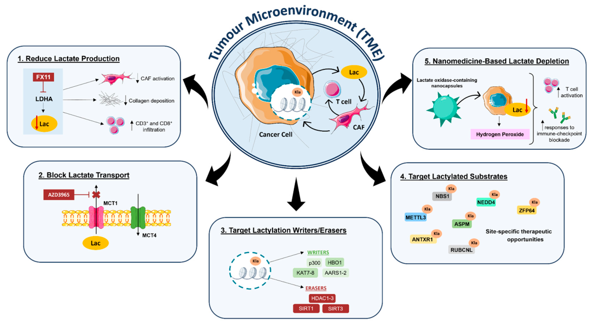
Figure 4.
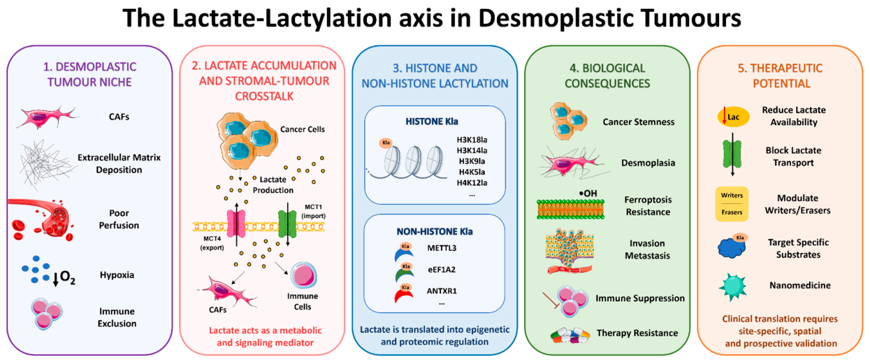
The lactate-lactylation axis in desmoplastic tumours: from niche biology to therapeutic opportunity. This figure presents an integrated left-to-right model of the lactate-lactylation axis, in which five sequential panels depict the causal chain from the desmoplastic tumour niche to therapeutic intervention. Panel 1: CAFs, extensive ECM deposition, poor vascular perfusion, hypoxia and immune exclusion collectively create metabolically restricted microenvironment that favours lactate accumulation and limits drug and immune-cell access. Panel 2: cancer cells, CAFs and immune cells engage in reciprocal metabolic exchange, with MCT4 mediating lactate export and MCT1 mediating import; lactate thereby acts as both a metabolic substrate and an intercellular signalling mediator that coordinates reprogramming across cellular compartments. Panel 3: intracellular lactate is translated into epigenetic and post-translational regulation through site-specific Kla of histone marks (H3K18la, H3K14la, H3K9la, H4K5la, H4K12la and others) and non-histone proteins (including eEf1A2, ANTXR1 and others); this step is the mechanistic link between tumour metabolism and transcriptional and proteomic reprogramming. Panel 4: the resulting Kla-driven programmes promote cancer stemness, desmoplasia, ferroptosis resistance, invasion, metastasis, immune suppression and therapy resistance, constituting the multi-front adaptive plasticity described throughout this review. Panel 5: the lactate-Kla axis can be targeted at five levels – reducing lactate availability, blocking lactate transport, modulating writers and erasers, targeting specific lactylated substrates and nanomedicine-based lactate depletion – through clinical translation requires site-specific, spatially resolved and prospectively validated approaches to achieve selectivity for individual Kla events without broadly impairing metabolic homeostasis. Abbreviations: ANTXR1, anthrax toxin receptor 1; CAF, cancer-associated fibroblast; ECM, extracellular matrix; eEF1A2, eukaryotic translation elongation factor 1 alpha 2; Kla, lysine lactylation; MCT, monocarboxylate transporter; OH, hydroxyl radical.
Figure 4.
The lactate-lactylation axis in desmoplastic tumours: from niche biology to therapeutic opportunity. This figure presents an integrated left-to-right model of the lactate-lactylation axis, in which five sequential panels depict the causal chain from the desmoplastic tumour niche to therapeutic intervention. Panel 1: CAFs, extensive ECM deposition, poor vascular perfusion, hypoxia and immune exclusion collectively create metabolically restricted microenvironment that favours lactate accumulation and limits drug and immune-cell access. Panel 2: cancer cells, CAFs and immune cells engage in reciprocal metabolic exchange, with MCT4 mediating lactate export and MCT1 mediating import; lactate thereby acts as both a metabolic substrate and an intercellular signalling mediator that coordinates reprogramming across cellular compartments. Panel 3: intracellular lactate is translated into epigenetic and post-translational regulation through site-specific Kla of histone marks (H3K18la, H3K14la, H3K9la, H4K5la, H4K12la and others) and non-histone proteins (including eEf1A2, ANTXR1 and others); this step is the mechanistic link between tumour metabolism and transcriptional and proteomic reprogramming. Panel 4: the resulting Kla-driven programmes promote cancer stemness, desmoplasia, ferroptosis resistance, invasion, metastasis, immune suppression and therapy resistance, constituting the multi-front adaptive plasticity described throughout this review. Panel 5: the lactate-Kla axis can be targeted at five levels – reducing lactate availability, blocking lactate transport, modulating writers and erasers, targeting specific lactylated substrates and nanomedicine-based lactate depletion – through clinical translation requires site-specific, spatially resolved and prospectively validated approaches to achieve selectivity for individual Kla events without broadly impairing metabolic homeostasis. Abbreviations: ANTXR1, anthrax toxin receptor 1; CAF, cancer-associated fibroblast; ECM, extracellular matrix; eEF1A2, eukaryotic translation elongation factor 1 alpha 2; Kla, lysine lactylation; MCT, monocarboxylate transporter; OH, hydroxyl radical.
Second, blocking lactate transport: MCT1/4 sustain metabolic exchange, and in PDAC CAFs take up cancer-derived lactate via MCT1, with MCT1 knockdown or AZD3965 impairing lactate-supported CAF proliferation17. Clinically, AZD3965 has undergone first-in-human phase I evaluation in patients with advanced solid tumours and lymphoma, demonstrating the feasibility of pharmacologically targeting lactate transport117. Nevertheless, lactylation marks were not evaluated as pharmacodynamic endpoints in this trial. AZD3965 should therefore be considered clinical evidence for the druggability of lactate transport rather than a lactylation-directed therapy.
Third, targeting the enzymatic machinery: p300 was the first proposed histone lactyltransferase13, with HBO1/KAT7 (H3K9la)118, KAT8 (eEF1A2, promoting translation and colorectal carcinogenesis)67 and AARS1/AARS2 (sensing lactate to modify p53 or YAP-TEAD)119,120 acting as context-dependent writers, while HDAC1-3 remove lactyl groups121. Despite this therapeutic potential, pharmacological targeting of writers and erasers presents substantial specificity challenges. p300/CBP, HBO1, KAT8 and HDACs also regulate acetylation and other lysine acyl modifications. Consequently, inhibition of these enzymes cannot be interpreted as evidence of a lactylation-dependent effect unless it is accompanied by measurements of the relevant Kla site, genetic substitution of the modified lysine residue and functional rescue experiments. The development of substrate-selective inhibitors or approaches that disrupt the interaction between a lactyltransferase and a specific target protein may ultimately provide greater specificity than global inhibition of these enzymes.
Fourth, targeting individual lactylated substrates may help overcome therapy resistance. NBS1 lactylation at K388 promotes homologous recombination and resistance to DNA-damaging therapies. LDHA depletion or treatment with stiripentol reversed this resistance122. Within desmoplastic tumours, examples include NEDD4/PTEN/PI3K-AKT activation driven by H3K14la in multidrug-resistant HCC101, ZFP64-linked ferroptosis resistance in TNBC102, HDAC-METTL3 delactylation in DNA repair106, ASPM/NCAPG programmes in GC anti-PD-1 resistance95, ANTXR1-supported stemness and oxaliplatin resistance in CRC99, and RUBCNL-driven autophagy and bevacizumab resistance116. Together, these studies suggest that individual lactylated proteins and their downstream pathways may represent therapeutically actionable pathways, although most evidence remains preclinical.
Fifth, nanomedicine-based lactate depletion: lactate-oxidase nanocapsules consume tumour lactate and enhance checkpoint-blockade responses preclinically123, but dense ECM may limit penetration and lactate oxidase is not tumour-specific, generating cytotoxic H₂O₂ in normal tissues, highlighting the need for tumour-selective delivery and spatially controlled enzyme activation124,125.
Beyond therapeutic targeting, lactylation marks may have value as tissue biomarkers of metabolic activity, stromal interaction, immune suppression and treatment resistance. In PDAC, perineural-invasion-associated CAFs raise tumour H3K18la and L1CAM/SLIT1 neural invasion programmes, with elevated H3K18la marking severe perineural invasion and poorer clinical outcome126. In breast cancer, H4K5 associates with nodal involvement and Ki-67127, and KCNK1-LDHA-drivenH3K18 promotes proliferation, invasion and metastasis128. In TNBC, H4K12 is a candidate prognostic biomarker. H4K12la was positively associated with Ki-67 expression and negatively associated with overall survival129 that promotes progression via SLFN5 downregulation41. In HCC, histone lactylation-dependent activation of ESM1 has been associated with proliferation, invasion, EMT and metastatic progression130.
Nevertheless, substantial methodological challenges must be addressed before lactylation biomarkers can enter routine clinical use. These include variability in antibody specificity, the need to distinguish L- from D-lactylation and N-ε-(carboxyethyl)-lysine, low occupancy of many Kla sites and cellular heterogeneity of tumour tissues131. Bulk measurements of pan-Kla or H3K18la cannot distinguish signals originating from cancer cells, CAFs or immune populations so, studies need mass spectrometry validation, standardized scoring, independent patient cohorts and spatial methods capable of identifying the cell types carrying the relevant lactylation mark.
Emerging Clinical Evaluation of Lactylation-Associated Mechanisms
Among the tumour types covered in this review, NCT07197151 represents a rare, registered human of a lactylation-associated mechanism in CRC by examining CYFIP1-PKM2 and glycolysis-driven HIF-1α lactylation with pan-Kla and LC-MS/MS site mapping. However, it is observational, not an interventional trial of a lactylation-directed therapy, with no associated peer-reviewed publication identified. An additional clinical proof-of-concept has emerged outside the desmoplastic and stroma-rich tumour contexts selected for this review. NCT06766266 is a single-arm phase I study evaluating liposomal irinotecan combined with intravesical epirubicin in patients with recurrent, anthracycline-resistant non-muscle-invasive urothelial carcinoma. Although this malignancy is not conventionally classified as fibrotic or desmoplastic, the associated original study provides an important example of clinical translation arising from a site-specific non-histone lactylation mechanism. The study identified that AARS1-mediated BLM K24 lactylation reduces BLM ubiquitination and enhances homologous recombination and treatment resistance, which irinotecan inhibited132. Together, these studies illustrate the earliest stages of clinical evaluation of lactylation-associated mechanisms. When considered alongside the expanding mechanistic and preclinical evidence summarized in Table 4, these studies highlight the substantial gap between experimental discovery and clinical validation. Among the registered studies identified, no interventional trial in PDAC, CCA, HCC, TNBC, GC or CRC have prospectively prespecified a defined Kla site as a patient-selection, pharmacodynamic or therapeutic-response biomarker.
5. Current Limitations and Future Perspectives
5.1. Analytical Specificity and Site-Level Resolution
Despite the rapid expansion of mechanistic evidence, major analytical, biological and translational challenges must be addressed before the lactate-lactylation axis can be clinically exploited. Analytically, many studies rely on pan-lactylation antibodies that may not distinguish lysine L- from D-lactylation or N-ε-(carboxyethyl)-lysine and may cross-react with the more abundant acetyl mark. Anti-H3K18la reagents should therefore be validated against H3K18ac-modified peptides using peptide arrays or dot blots, competition assays and orthogonal mass spectrometry13,133. Moreover, global Kla measurements also provide limited information about the modified protein, lysine residue, stoichiometry or cellular source of the signal, arguing for MS-based site mapping with validated site-specific antibodies131.
5.2. Low Stoichiometry and Unresolved Transcriptional Causality
A central unresolved question is whether histone Kla occurs at sufficient occupancy to function as a direct transcriptional regulator. Quantitative mass spectrometry shows it is far less abundant than acetylation, often below 1% occupancy131. This challenges models in which widespread Kla deposition directly remodels chromatin or recruits transcriptional machinery131. Importantly, increases detected by immunoblotting, immunohistochemistry or enrichment-based chromatin assays indicate relative changes in signal but do not establish the absolute proportion of histones modified at a given residue or genomic locus134,135. Associations between Kla enrichment and active transcription may therefore reflect a shared glycolytic state, co-occurring histone acetylation, preferential detection within already accessible chromatin or another lactate-dependent pathway rather than a direct causal effect of Kla13,136.
Low stoichiometry does not, however, exclude biological activity. A rare mark could act if it concentrated at key loci, deposited transiently or recognized by a high-affinity effector131,136. Resolving this requires absolute, site-resolved occupancy, calibrated chromatin profiling and temporal analysis of Kla deposition relative to transcriptional activation and experimental designs that separate loss of lactylation from loss of other modifications at the same lysine residue134. Until such evidence is available, transcriptional changes should be described as Kla-correlated rather than Kla-driven.
5.3. Establishing Kla-Specific Causality
Establishing mechanistic causality represents a further major challenge because altering glycolysis, LDH activity or lactate transport affects multiple metabolic and signalling pathways. Accordingly, phenotypes induced by exogenous lactate or altered lactate transport should not be classified as Kla-dependent without excluding HCAR1/GPR81 signalling, oxidative lactate utilization and extracellular acidification137,138. This requires, as appropriate, receptor perturbation, metabolic tracing and pH-matched or adequately buffered controls. Similarly, as most proposed lactylation writers and erasers, including p300/CBP, HBO1/KAT7, KAT8 and class I HDACs, also regulate acetylation and other lysine acylations13,67,118,121, their inhibition cannot be attributed to Kla without site-specific Kla measurements, lysine substitution, functional rescue and, ideally, disruption of specific enzyme-substrate interactions67,119,122. Because lysine substitution can simultaneously prevent multiple acylations and alter protein structure, such experiments should be interpreted alongside orthogonal approaches capable of distinguishing lactylation from competing modifications. The development of substrate-selective inhibitors may ultimately provide greater therapeutic specificity than global inhibition of writers or erasers.
5.4. Spatial and Cellular Heterogeneity
Biological interpretation is complicated by the spatial and cellular heterogeneity of desmoplastic and stroma-rich tumour contexts tumour. A bulk Kla signal may originate from cancer cells, CAFs, HSCs or immune populations each carrying distinct functional and clinical implications139. Moreover, lactylation is not uniformly oncogenic – effects depend on substrate, lysine residue, cell type and physiological context, and both increased lactylation and inappropriate delactylation may promote tumour progression or treatment resistance101,106. Therefore, future studies should integrate spatial proteomics, multiplex imaging and single-cell approaches with physiologically relevant co-culture, organoid and stromal models to determine where specific Kla events occur and how they influence tumour-stroma interactions.
5.5. Limitations of Preclinical Models
Many reported mechanisms remain confined to reductionist or preclinical models that poorly reproduce the metabolic gradients, ECM and cellular diversity of human desmoplastic and stroma-rich tumours78. Validation in independent patient cohorts, paired tumour and adjacent-tissue samples, and pre- and post-treatment specimens will be required to establish the reproducibility and clinical relevance of candidate lactylation mechanisms. Experimental models should also reproduce the concentrations and spatial gradients of lactate, oxygen availability and extracellular pH documented in human tumours, rather than applying a single high lactate concentration under normoxic and conventionally buffered culture conditions that may not reflect in vivo exposure138,140.
5.6. Clinical Validation and Biomarker Development
Finally, a substantial gap remains between discovery and clinical validation: most relevant trials target upstream metabolism without measuring a defined Kla site. For example, AZD3965 showed druggability of lactate transport but did not assess lactylation endpoints117. Future interventional studies should incorporate defined Kla sites as biomarkers for patient selection, target engagement, pharmacodynamic monitoring and treatment response, with paired pre/post-treatment specimens, mass-spectrometry validation and spatially resolved analyses in independent cohorts to establish whether lactylation is an actionable driver or a surrogate of altered lactate metabolism.
5.7. Integrated Future Priorities
Taken together, progress in the field requires moving from global Kla measurements and upstream metabolic perturbations toward quantitatively defined, cell-resolved, site-specific and functionally validated mechanisms. Future therapeutic strategies should target context-dependent lactylated substrates or their downstream dependencies within defined tumour and stromal compartments rather than universal lactylation inhibition.
6. Conclusion
Kla discovery provides a framework for understanding how the metabolic architecture of desmoplastic tumours can be translated into epigenetic and proteomic regulation. Through histone and non-histone lactylation, lactate-rich niches actively reshape transcription, protein function and intercellular communication rather than merely reflecting metabolic overflow. As summarised in Figure 4, the lactate-lactylation axis connects tumour metabolism to cancer stemness, stromal remodelling, immunosuppression and resistance to chemotherapy, immunotherapy and targeted treatments, with effects that depend strongly on the modified substrate, lysine residue and cellular context, reinforcing the need to move beyond global lactate or pan-Kla measurements.
The dynamic and potentially reversible nature of lactylation creates promising therapeutic and biomarker opportunities. Translating these findings into clinical practice will require site-specific measurements in defined cell populations, combined with mechanistic validation, spatial profiling and prospective clinical studies. These approaches will be essential to distinguish lactylation events that influence tumour behaviour from those that reflect broader metabolic changes. Understanding these mechanisms within the spatial and metabolic complexity of desmoplastic tumour microenvironments could inform patient stratification and guide the development of treatment combinations to overcome resistance in these aggressive malignancies.
Author Contributions
RP and IG performed the full literature review and drafted the manuscript. RP also prepared the figures. MGC provided scientific guidance and reviewed the manuscript. AJ supervised the work, provided overall scientific guidance, and critically revised and validated the final manuscript. RS performed the full literature review, contributed to the conceptual framework and critical synthesis of the evidence on lactylation in desmoplastic tumours, prepared the figures, and critically revised and validated the final manuscript. All authors reviewed and approved the final manuscript.
Funding
This work was supported by Liga Portuguesa Contra o Cancro – Núcleo Regional do Sul (LPCC-NRS) and Jerónimo Martins through their 2025 research grant. Additional support was provided by the iNOVA4Health Programme in Translational Medicine and Fundação para a Ciência e a Tecnologia [UI/BD/154334/2022 and 2025.17953.PRT]. This project is funded by the European Union under Horizon Europe [project 101060346]. However, the views and opinions expressed are those of the authors only and do not necessarily reflect those of the European Union. Neither the European Union nor the granting authority can be held responsible for them. The funders had no role in the preparation of this review or the decision to submit it for publication.
Data Availability Statement
Not applicable. This review does not report individual participant data.
Acknowledgments
The scientific questions addressed in this review were shaped in part by the development of a research project awarded an Oncology Research Grant for the 2026-2028 period, co-funded by Liga Portuguesa Contra o Cancro – Núcleo Regional do Sul (LPCC-NRS) and Jerónimo Martins, whom we gratefully acknowledge for their support. We also thank the NOVA Medical School Library for its assistance in compiling the bibliographic search results and applying the search filters used in this review. We dedicate this work to people living with cancer and to their families and caregivers, whose experiences give purpose and urgency to efforts to understand therapeutic resistance and improve cancer care. Finally, we gratefully acknowledge the support provided by the Research Unit iNOVA4Health and the Associated Laboratory LS4FUTURE.
Conflicts of Interest
The authors declare that they have no commercial or financial relationships that could be construed as a potential conflict of interest.
References
- Sahai, E.; et al. A framework for advancing our understanding of cancer-associated fibroblasts. Nat. Rev. Cancer 2020, 20, 174–186. [Google Scholar] [CrossRef]
- Sirica, A. E.; Gores, G. J. Desmoplastic stroma and cholangiocarcinoma: Clinical implications and therapeutic targeting. Hepatology 2014, 59, 2397–2402. [Google Scholar] [CrossRef]
- Cristescu, R.; et al. Molecular analysis of gastric cancer identifies subtypes associated with distinct clinical outcomes. Nat. Med. 2015, 21, 449–456. [Google Scholar] [CrossRef]
- Guinney, J.; et al. The consensus molecular subtypes of colorectal cancer. Nat. Med. 2015, 21, 1350–1356. [Google Scholar] [CrossRef]
- Karancsi, Z.; et al. Digital Whole Slide Image Analysis of Elevated Stromal Content and Extracellular Matrix Protein Expression Predicts Adverse Prognosis in Triple-Negative Breast Cancer. Int. J. Mol. Sci. 2024, 25, 9445. [Google Scholar] [CrossRef]
- Barry, A. E.; et al. Hepatic Stellate Cells and Hepatocarcinogenesis. Front. Cell Dev. Biol. 2020, 8. [Google Scholar] [CrossRef]
- Kitamura, F.; et al. Cancer-associated fibroblasts reuse cancer-derived lactate to maintain a fibrotic and immunosuppressive microenvironment in pancreatic cancer. JCI Insight 2023, 8. [Google Scholar] [CrossRef]
- Brown, T. P.; et al. The lactate receptor GPR81 promotes breast cancer growth via a paracrine mechanism involving antigen-presenting cells in the tumor microenvironment. Oncogene 2020, 39, 3292–3304. [Google Scholar] [CrossRef]
- Roland, C. L.; et al. Cell Surface Lactate Receptor GPR81 Is Crucial for Cancer Cell Survival. Cancer Res. 2014, 74, 5301–5310. [Google Scholar] [CrossRef]
- Corbet, C.; Feron, O. Tumour acidosis: from the passenger to the driver’s seat. Nat. Rev. Cancer 2017, 17, 577–593. [Google Scholar] [CrossRef]
- Colegio, O. R.; et al. Functional polarization of tumour-associated macrophages by tumour-derived lactic acid. Nature 2014, 513, 559–563. [Google Scholar] [CrossRef]
- Brand, A.; et al. LDHA-Associated Lactic Acid Production Blunts Tumor Immunosurveillance by T and NK Cells. Cell Metab. 2016, 24, 657–671. [Google Scholar] [CrossRef]
- Zhang, D.; et al. Metabolic regulation of gene expression by histone lactylation. Nature 2019, 574, 575–580. [Google Scholar] [CrossRef]
- Chen, H.; et al. NBS1 lactylation is required for efficient DNA repair and chemotherapy resistance. Nature 2024, 631, 663–669. [Google Scholar] [CrossRef]
- Xie, B.; et al. KAT8-catalyzed lactylation promotes eEF1A2-mediated protein synthesis and colorectal carcinogenesis. Proc. Natl. Acad. Sci. 2024, 121. [Google Scholar] [CrossRef]
- Zong, Z.; et al. Alanyl-tRNA synthetase, AARS1, is a lactate sensor and lactyltransferase that lactylates p53 and contributes to tumorigenesis. Cell 2024, 187, 2375–2392.e33. [Google Scholar] [CrossRef]
- Kitamura, F.; et al. Cancer-associated fibroblasts reuse cancer-derived lactate to maintain a fibrotic and immunosuppressive microenvironment in pancreatic cancer. JCI Insight 2023, 8. [Google Scholar] [CrossRef]
- Li, J.; et al. Lactylation in cancer: molecular mechanisms and advances in clinical study. Mol. Cancer 2026, 25, 53. [Google Scholar] [CrossRef]
- Zhang, D.; et al. Metabolic regulation of gene expression by histone lactylation. Nature 2019, 574, 575–580. [Google Scholar] [CrossRef]
- Zhu, R.; et al. ACSS2 acts as a lactyl-CoA synthetase and couples KAT2A to function as a lactyltransferase for histone lactylation and tumor immune evasion. Cell Metab. 2025, 37, 361–376.e7. [Google Scholar] [CrossRef]
- Li, H.; et al. AARS1 and AARS2 sense l-lactate to regulate cGAS as global lysine lactyltransferases. Nature 2024, 634, 1229–1237. [Google Scholar] [CrossRef]
- Li, F.; et al. Positive feedback regulation between glycolysis and histone lactylation drives oncogenesis in pancreatic ductal adenocarcinoma. Mol. Cancer 2024, 23, 90. [Google Scholar] [CrossRef]
- Zong, Z.; et al. Alanyl-tRNA synthetase, AARS1, is a lactate sensor and lactyltransferase that lactylates p53 and contributes to tumorigenesis. Cell 2024, 187, 2375–2392.e33. [Google Scholar] [CrossRef]
- Xie, B.; et al. KAT8-catalyzed lactylation promotes eEF1A2-mediated protein synthesis and colorectal carcinogenesis. Proc. Natl. Acad. Sci. 2024, 121. [Google Scholar] [CrossRef]
- Niu, Z.; et al. HBO1 catalyzes lysine lactylation and mediates histone H3K9la to regulate gene transcription. Nat. Commun. 2024, 15, 3561. [Google Scholar] [CrossRef]
- Moreno-Yruela, C.; et al. Class I histone deacetylases (HDAC1–3) are histone lysine delactylases. Sci. Adv. 2022, 8. [Google Scholar] [CrossRef]
- Fang, C.; et al. Lactate metabolism and lactylation in cancer: from pathogenesis to therapeutic advances. Signal Transduct. Target. Ther. 2026, 11, 190. [Google Scholar] [CrossRef]
- Zhang, D.; et al. Lysine l-lactylation is the dominant lactylation isomer induced by glycolysis. Nat. Chem. Biol. 2025, 21, 91–99. [Google Scholar] [CrossRef]
- Yang, Z.; et al. Lactylome analysis suggests lactylation-dependent mechanisms of metabolic adaptation in hepatocellular carcinoma. Nat. Metab. 2023, 5, 61–79. [Google Scholar] [CrossRef]
- Wang, E.; et al. H3K18 lactylation promotes POM121 transcription and accelerates gastric cancer progression via the PI3K/AKT pathway. Pathol. Res. Pract. 2026, 282, 156427. [Google Scholar] [CrossRef]
- Zhao, Y.; et al. H3K18 lactylation-mediated VCAM1 expression promotes gastric cancer progression and metastasis via AKT-mTOR-CXCL1 axis. Biochem. Pharmacol. 2024, 222, 116120. [Google Scholar] [CrossRef]
- Tsukihara, S.; et al. Delactylase effects of SIRT1 on a positive feedback loop involving the H19-glycolysis-histone lactylation in gastric cancer. Oncogene 2025, 44, 724–738. [Google Scholar] [CrossRef]
- Li, Y.; et al. Histone lactylation-boosted AURKB facilitates colorectal cancer progression by inhibiting HNRNPM-mediated PSAT1 mRNA degradation. J. Exp. Clin. Cancer Res. 2025, 44, 233. [Google Scholar] [CrossRef]
- Wang, W.; et al. Feedback regulation between histone H3 lysine 18 lactylation and TROP2-mediated glycolysis drives metastatic progression of colorectal cancer. Clin. Transl. Med. 2026, 16. [Google Scholar] [CrossRef]
- Liu, Y.; et al. CTCF enhances pancreatic cancer progression via FLG-AS1-dependent epigenetic regulation and macrophage polarization. Cell Death Differ. 2025, 32, 745–762. [Google Scholar] [CrossRef]
- Yang, J.; et al. Histone lactylation-driven feedback loop modulates cholesterol-linked immunosuppression in pancreatic cancer. Gut 2025, 74, 1859–1872. [Google Scholar] [CrossRef]
- Xiao, F.; et al. Histone Lactylation Promotes Proliferation and Migration of TNBC Cells via Upregulating MCM7 Transcription. Tohoku J. Exp. Med. 2026, 268, 2025.J096. [Google Scholar] [CrossRef]
- Yu, J.; et al. A Feedback Loop Driven by H3K18la and ASF1B via the LINC02732-miR-1291 Axis Promotes Hepatocellular Carcinoma Proliferation. Cells 2026, 15, 952. [Google Scholar] [CrossRef]
- Ji, Y.; et al. O-GlcNAcylation of YBX1 drives a glycolysis-histone lactylation feedback loop in hepatocellular carcinoma. Cancer Lett. 2025, 631, 217957. [Google Scholar] [CrossRef]
- Jiang, Y.; et al. LBX2 promotes colorectal cancer progression via the glycosylation and lactylation positive feedback. Cell Death Discov. 2025, 11, 556. [Google Scholar] [CrossRef]
- Li, J.; et al. Histone H4K12 lactylation promotes malignancy progression in triple-negative breast cancer through SLFN5 downregulation. Cell. Signal. 2024, 124, 111468. [Google Scholar] [CrossRef]
- Yamane, M.; et al. H4K5 lactylation - ENO2 loop drives glycolysis and HCC progression. JHEP Rep. 2026, 8, 101786. [Google Scholar] [CrossRef]
- Li, L.; Dong, J.; Xu, C.; Wang, S. Lactate drives senescence-resistant lineages in hepatocellular carcinoma via histone H2B lactylation of NDRG1. Cancer Lett. 2025, 616, 217567. [Google Scholar] [CrossRef]
- Jiang, Z.; et al. PDHX acetylation facilitates tumor progression by disrupting PDC assembly and activating lactylation-mediated gene expression. Protein Cell 2025, 16, 49–63. [Google Scholar] [CrossRef]
- Sun, X.; et al. The diapause-like colorectal cancer cells induced by SMC4 attenuation are characterized by low proliferation and chemotherapy insensitivity. Cell Metab. 2023, 35, 1563–1579.e8. [Google Scholar] [CrossRef]
- Yang, L.; et al. Nucleolin lactylation contributes to intrahepatic cholangiocarcinoma pathogenesis via RNA splicing regulation of MADD. J. Hepatol. 2024, 81, 651–666. [Google Scholar] [CrossRef]
- Zhang, H.; et al. Lactylation-stabilized NOL6 promotes colorectal cancer progression via STAMBP-mediated YY1 deubiquitination and c-Myc transcription upregulation. Cell Rep. 2026, 45, 116774. [Google Scholar] [CrossRef]
- Chen, S.; Wang, X.; Chen, Y.; Qiao, Y.; Hu, X. K92 Lactylation of YBX1 orchestrates m5C RNA recognition to facilitate colorectal Cancer progression. Int. J. Biol. Macromol. 2026, 346, 150686. [Google Scholar] [CrossRef]
- Sun, L.; et al. Lactylation of METTL16 promotes cuproptosis via m6A-modification on FDX1 mRNA in gastric cancer. Nat. Commun. 2023, 14, 6523. [Google Scholar] [CrossRef]
- Niu, K.; et al. NSUN2 lactylation drives cancer cell resistance to ferroptosis through enhancing GCLC-dependent glutathione synthesis. Redox Biol. 2025, 79, 103479. [Google Scholar] [CrossRef]
- Chen, B.; et al. Metabolic Recoding of NSUN2-Mediated m5C Modification Promotes the Progression of Colorectal Cancer via the NSUN2/YBX1/m5C-ENO1 Positive Feedback Loop. Adv. Sci. 2024, 11. [Google Scholar] [CrossRef]
- Cui, Z.; et al. Lactate Dehydrogenase C4 Accelerates Triple-Negative Breast Cancer Progression by Promoting Acetyl-CoA Acyltransferase 2 Lactylation to Increase Free Fatty Acid Accumulation. Adv. Sci. 2025, 12. [Google Scholar] [CrossRef]
- Cheng, S.; et al. A Lamp2a-linked RNA secreted by ADSCs prevents ENO1–lactylation–glycolysis feedback and cell malignant behavior in triple-negative breast cancer. Cell Death Dis. 2026, 17, 288. [Google Scholar] [CrossRef]
- Cheng, Z.; Huang, H.; Li, M.; Chen, Y. Proteomic analysis identifies PFKP lactylation in SW480 colon cancer cells. iScience 2024, 27, 108645. [Google Scholar] [CrossRef]
- Yan, H.-C.; Lai, X.-X.; Liu, Y.; Sheng, L. S100A8 lactylation at K23/K36 reduces RNF5-dependent degradation and promotes colorectal cancer progression. Biochem. Biophys. Res. Commun. 2026, 820, 153827. [Google Scholar] [CrossRef]
- Hu, X.; et al. Claudin-9 (CLDN9) promotes gastric cancer progression by enhancing the glycolysis pathway and facilitating PD-L1 lactylation to suppress CD8+ T cell anti-tumor immunity. Cancer Pathog. Ther. 2025, 3, 253–266. [Google Scholar] [CrossRef]
- Tong, H.; et al. Dual impacts of serine/glycine-free diet in enhancing antitumor immunity and promoting evasion via PD-L1 lactylation. Cell Metab. 2024, 36, 2493–2510.e9. [Google Scholar] [CrossRef]
- Hong, H.; et al. ABCF1-K430-Lactylation promotes HCC malignant progression via transcriptional activation of HIF1 signaling pathway. Cell Death Differ. 2025, 32, 613–631. [Google Scholar] [CrossRef]
- Wei, X.; et al. LDHA-mediated YAP lactylation promotes the tumor progression of hepatocellular carcinoma by inducing YAP dephosphorylation and activation. Biol. Direct 2025, 20, 64. [Google Scholar] [CrossRef]
- Jiang, C.; et al. Lactate accumulation drives hepatocellular carcinoma metastasis through facilitating tumor-derived exosome biogenesis by Rab7A lactylation. Cancer Lett. 2025, 627, 217636. [Google Scholar] [CrossRef]
- Yao, G.; Yang, Z. Glypican-3 knockdown inhibits the cell growth, stemness, and glycolysis development of hepatocellular carcinoma cells under hypoxic microenvironment through lactylation. Arch. Physiol. Biochem. 2023, 1–9. [Google Scholar] [CrossRef]
- Liu, S.; et al. TPX2 lactylation is required for the cell cycle regulation and hepatocellular carcinoma progression. Life Sci. Alliance 2025, 8, e202402978. [Google Scholar] [CrossRef]
- Liao, J.; et al. CENPA functions as a transcriptional regulator to promote hepatocellular carcinoma progression via cooperating with YY1. Int. J. Biol. Sci. 2023, 19, 5218–5232. [Google Scholar] [CrossRef]
- Jin, J.; et al. SIRT3-dependent delactylation of cyclin E2 prevents hepatocellular carcinoma growth. EMBO Rep. 2023, 24, EMBR202256052. [Google Scholar] [CrossRef]
- Han, H.; et al. ASH2L-K312-Lac Stimulates Angiogenesis in Tumors to Expedite the Malignant Progression of Hepatocellular Carcinoma. Adv. Sci. 2025, 12. [Google Scholar] [CrossRef]
- Yan, J.; et al. Delactylation diminished the growth inhibitory role of CA3 by restoring DUOX2 expression in hepatocellular carcinoma. Exp. Cell Res. 2025, 444, 114392. [Google Scholar] [CrossRef]
- Xie, B.; et al. KAT8-catalyzed lactylation promotes eEF1A2-mediated protein synthesis and colorectal carcinogenesis. Proc. Natl. Acad. Sci. 2024, 121. [Google Scholar] [CrossRef]
- Rana, M. N.; Yan, S.; Cao, J.; Wang, Y.; Gao, X.-W. Lactate promotes mRNA translation in colorectal cancer cells through eEF2 lactylation. Biochem. Biophys. Res. Commun. 2026, 796, 153119. [Google Scholar] [CrossRef]
- Cai, D.; et al. L-lactate-driven PSMD14 lactylation and stabilization promote lactate production and ferroptosis resistance via ENO1 in intrahepatic cholangiocarcinoma. Cancer Lett. 2026, 646, 218394. [Google Scholar] [CrossRef]
- Gu, J.; et al. Tumor-resident microbiota contributes to colorectal cancer liver metastasis by lactylation and immune modulation. Oncogene 2024, 43, 2389–2404. [Google Scholar] [CrossRef]
- Sun, K.; et al. Elevated protein lactylation promotes immunosuppressive microenvironment and therapeutic resistance in pancreatic ductal adenocarcinoma. J. Clin. Investig. 2025, 135. [Google Scholar] [CrossRef]
- Batlle, E.; Clevers, H. Cancer stem cells revisited. Nat. Med. 2017, 23, 1124–1134. [Google Scholar] [CrossRef]
- Deng, J.; et al. Histone lactylation enhances GCLC expression and thus promotes chemoresistance of colorectal cancer stem cells through inhibiting ferroptosis. Cell Death Dis. 2025, 16, 193. [Google Scholar] [CrossRef]
- Feng, F.; et al. Lactylome Analysis Unveils Lactylation-Dependent Mechanisms of Stemness Remodeling in the Liver Cancer Stem Cells. Adv. Sci. 2024, 11. [Google Scholar] [CrossRef]
- Lv, M.; et al. SIRT4 Promotes Pancreatic Cancer Stemness by Enhancing Histone Lactylation and Epigenetic Reprogramming Stimulated by Calcium Signaling. Adv. Sci. 2025, 12. [Google Scholar] [CrossRef]
- Zhang, P.; et al. METTL14 attenuates cancer stemness by suppressing ATF5/WDR74/β-catenin axis in gastric cancer. Cancer Sci. 2025, 116, 112–127. [Google Scholar] [CrossRef]
- Kalluri, R. The biology and function of fibroblasts in cancer. Nat. Rev. Cancer 2016, 16, 582–598. [Google Scholar] [CrossRef]
- Sahai, E.; et al. A framework for advancing our understanding of cancer-associated fibroblasts. Nat. Rev. Cancer 2020, 20, 174–186. [Google Scholar] [CrossRef]
- Gao, B.; et al. Identification of cancer-associated fibrolast subtypes and distinctive role of MFAP5 in CT-detected extramural venous invasion in gastric cancer. Transl. Oncol. 2025, 51, 102188. [Google Scholar] [CrossRef]
- Huang, C.; et al. Histone H4K8 lactylation modulated immunosuppressive properties by promoting FAP transcription and ECM remodeling. Gastric Cancer 2026, 29, 70–82. [Google Scholar] [CrossRef]
- Ma, Y.; et al. UBE2T-Driven p53 Degradation Rewires Glycolysis to Orchestrate Lactylation-Mediated CAFs Activation and ECM Deposition in Pancreatic Cancer. Adv. Sci. 2026, 13. [Google Scholar] [CrossRef]
- Zhou, C.; et al. Mutant KRAS-activated circATXN7 fosters tumor immunoescape by sensitizing tumor-specific T cells to activation-induced cell death. Nat. Commun. 2024, 15, 499. [Google Scholar] [CrossRef]
- Xiang, Z.; et al. CD8a antibody-functionalized biomimetic red blood cell membrane ectosomes delivering C646 reverse CD8+ T Cell exhaustion via H3K18la histone delactylation in gastric cardia adenocarcinoma. J. Nanobiotechnology 2026, 24, 156. [Google Scholar] [CrossRef]
- Cai, J.; et al. Targeting SRSF10 might inhibit M2 macrophage polarization and potentiate anti-PD-1 therapy in hepatocellular carcinoma. Cancer Commun. 2024, 44, 1231–1260. [Google Scholar] [CrossRef]
- Gan, C.; et al. PFKFB4 promotes M2 polarization of tumor-associated macrophages through aerobic glycolysis-mediated modification of histone H3K18 lactylation in hepatocellular carcinoma. Cancer Metab. 2026, 14, 15. [Google Scholar] [CrossRef]
- Cai, J.; et al. Lactylation-Driven NUPR1 Promotes Immunosuppression of Tumor-Infiltrating Macrophages in Hepatocellular Carcinoma. Adv. Sci. 2025, 12. [Google Scholar] [CrossRef]
- Li, X.-M.; et al. Histone lactylation inhibits RARγ expression in macrophages to promote colorectal tumorigenesis through activation of TRAF6-IL-6-STAT3 signaling. Cell Rep. 2024, 43, 113688. [Google Scholar] [CrossRef]
- Zhu, J.; et al. Histone lactylation-mediated up-regulation of IGF2BP2 enhances ferroptosis resistance via Nrf2 in colorectal cancer. Clin. Transl. Med. 2025, 15. [Google Scholar] [CrossRef]
- Huang, H.; et al. A multi-dimensional approach to unravel the intricacies of lactylation related signature for prognostic and therapeutic insight in colorectal cancer. J. Transl. Med. 2024, 22, 211. [Google Scholar] [CrossRef]
- Zhang, B.; et al. MPC-mediated lactate production drives histone lactylation in dendritic cells to affect tumor progression and immunotherapy. Cell. Mol. Life Sci. 2025, 82, 371. [Google Scholar] [CrossRef]
- Tu, C.; et al. Endoplasmic reticulum stress-induced histone lactylation mediating immune escape of gastric cancer via PDIA6 overexpression in dendritic cells. Cell Death Dis. 2026, 17, 686. [Google Scholar] [CrossRef]
- Ding, L.; et al. YY1 Lactylation Elicits CARD9 Deficiency in Dendritic Cells Promoting Pancreatic Cancer Immune Escape. Int. J. Biol. Sci. 2026, 22, 5036–5053. [Google Scholar] [CrossRef]
- Liang, X.; et al. Claudin-7 deficiency induces metabolic reprogramming of neutrophils in the colorectal cancer microenvironment. Cell Death Dis. 2025, 16, 728. [Google Scholar] [CrossRef]
- Li, W.; et al. AKR1B10 reprograms neutrophils by histone lactylation to foster immune evasion in KRASG12C mutation colorectal cancer liver metastasis. J. Exp. Clin. Cancer Res. 2026, 45, 61. [Google Scholar] [CrossRef]
- Zhou, S.; et al. CAFs promote immune evasion in gastric cancer through histone lactylation-mediated suppression of NCAPG ubiquitination. J. Transl. Med. 2025, 23, 989. [Google Scholar] [CrossRef]
- Ding, C.-H.; et al. PRMT3 drives PD-L1-mediated immune escape through activating PDHK1-regulated glycolysis in hepatocellular carcinoma. Cell Death Dis. 2025, 16, 158. [Google Scholar] [CrossRef]
- Chen, S.; et al. Lactate drives immune resistance via a pharmaceutically reversible H3K18la-KIF20A-c-Myc-PD-L1 axis in hepatocellular carcinoma. Cancer Biol. Med. 2026, 1–26. [Google Scholar] [CrossRef]
- Liang, L.; et al. Lactylation stabilizes PD-L1 to promote tumor immune evasion and cell growth. Cell Death Dis. 2026, 17, 335. [Google Scholar] [CrossRef]
- He, J.; et al. Cancer associated fibroblasts-derived lactate induces oxaliplatin treatment resistance by promoting cancer stemness via ANTXR1 lactylation in colorectal cancer. Cancer Lett. 2025, 631, 217917. [Google Scholar] [CrossRef]
- Chen, Z.; Zhang, Y.; Yan, F.; Zhao, G.; Wang, Y. tsRNA-08614 inhibits glycolysis and histone lactylation by ALDH1A3 to confer oxaliplatin sensitivity in colorectal cancer. Transl. Oncol. 2025, 58, 102427. [Google Scholar] [CrossRef]
- Zeng, Y.; et al. Histone lactylation promotes multidrug resistance in hepatocellular carcinoma by forming a positive feedback loop with PTEN. Cell Death Dis. 2025, 16, 59. [Google Scholar] [CrossRef]
- Zhang, K.; Guo, L.; Li, X.; Hu, Y.; Luo, N. Cancer-associated fibroblasts promote doxorubicin resistance in triple-negative breast cancer through enhancing ZFP64 histone lactylation to regulate ferroptosis. J. Transl. Med. 2025, 23, 247. [Google Scholar] [CrossRef]
- Sun, Y.; et al. GLUT3 drives paclitaxel resistance in peritoneal metastatic gastric cancer by promoting H3K18 lactylation-mediated MAPKAP1 transcription to suppress ferroptosis. Int. J. Biol. Sci. 2026, 22, 4826–4846. [Google Scholar] [CrossRef]
- Zhao, Y.; et al. LncRNA BASP1-AS1 drives PCBP2 K115 lactylation to suppress ferroptosis and confer oxaliplatin resistance in gastric cancer. Free Radic. Biol. Med. 2025, 240, 717–734. [Google Scholar] [CrossRef]
- Li, J.; et al. A CD147-targeted small-molecule inhibitor potentiates gemcitabine efficacy by triggering ferroptosis in pancreatic ductal adenocarcinoma. Cell Rep. Med. 2025, 6, 102292. [Google Scholar] [CrossRef]
- He, X.; et al. HDAC2-Mediated METTL3 Delactylation Promotes DNA Damage Repair and Chemotherapy Resistance in Triple-Negative Breast Cancer. Adv. Sci. 2025, 12. [Google Scholar] [CrossRef]
- Huang, X.; et al. Positive Feedback Loop of Histone Lactylation-Driven HNRNPC Promotes Autophagy to Confer Pancreatic Ductal Adenocarcinoma Gemcitabine Resistance. Adv. Sci. 2026, 13. [Google Scholar] [CrossRef]
- Zhang, X.; et al. MNDA promotes immunosuppression in microsatellite instability-high colorectal cancer by facilitating PMN-MDSC infiltration via H3K18 lactylation. J. Transl. Med. 2025, 23, 1049. [Google Scholar] [CrossRef]
- Zhu, J.; et al. Simvastatin overcomes the pPCK1-pLDHA-SPRINGlac axis-mediated ferroptosis and chemo-immunotherapy resistance in AKT-hyperactivated intrahepatic cholangiocarcinoma. Cancer Commun. 2025, 45, 1038–1071. [Google Scholar] [CrossRef]
- Liu, Z.; et al. AARS1-mediated AKR1B10 lactylation stabilizes an aerobic glycolysis-positive feedback loop to drive lenvatinib resistance in hepatocellular carcinoma. Clin. Transl. Med. 2026, 16. [Google Scholar] [CrossRef]
- Lu, Y.; et al. Lactylation-Driven IGF2BP3-Mediated Serine Metabolism Reprogramming and RNA m6A—Modification Promotes Lenvatinib Resistance in HCC. Adv. Sci. 2024, 11. [Google Scholar] [CrossRef]
- Xie, Q.; et al. Homeobox B13 activates the hypoxia-inducible factor 1 pathway through histone lactylation thereby reprogramming lipid metabolism and promoting sorafenib resistance in hepatocellular carcinoma. J. Cell Commun. Signal. 2026, 20. [Google Scholar] [CrossRef]
- Dong, R.; et al. Lactylation-Driven HECTD2 Limits the Response of Hepatocellular Carcinoma to Lenvatinib. Adv. Sci. 2025, 12. [Google Scholar] [CrossRef]
- Sun, Y.; et al. Lactylation Converts ABHD6 into a Mitochondrial Regulator That Drives Lenvatinib Resistance in Hepatocellular Carcinoma. Cancer Res. 2026, 86, 2970–2989. [Google Scholar] [CrossRef]
- Wan, Q.; et al. GPX8+ cancer-associated fibroblast-derived lactate contributes to lenvatinib resistance by facilitating BRPF1 expression through histone H3 lysine 18 lactylation in hepatocellular carcinoma. Oncogene 2026, 45, 2491–2505. [Google Scholar] [CrossRef]
- Li, W.; et al. Tumor-derived lactate promotes resistance to bevacizumab treatment by facilitating autophagy enhancer protein RUBCNL expression through histone H3 lysine 18 lactylation (H3K18la) in colorectal cancer. Autophagy 2024, 20, 114–130. [Google Scholar] [CrossRef]
- Halford, S.; et al. A Phase I Dose-escalation Study of AZD3965, an Oral Monocarboxylate Transporter 1 Inhibitor, in Patients with Advanced Cancer. Clin. Cancer Res. 2023, 29, 1429–1439. [Google Scholar] [CrossRef]
- Niu, Z.; et al. HBO1 catalyzes lysine lactylation and mediates histone H3K9la to regulate gene transcription. Nat. Commun. 2024, 15, 3561. [Google Scholar] [CrossRef]
- Zong, Z.; et al. Alanyl-tRNA synthetase, AARS1, is a lactate sensor and lactyltransferase that lactylates p53 and contributes to tumorigenesis. Cell 2024, 187, 2375–2392.e33. [Google Scholar] [CrossRef]
- Ju, J.; et al. The alanyl-tRNA synthetase AARS1 moonlights as a lactyltransferase to promote YAP signaling in gastric cancer. J. Clin. Investig. 2024, 134. [Google Scholar] [CrossRef]
- Moreno-Yruela, C.; et al. Class I histone deacetylases (HDAC1–3) are histone lysine delactylases. Sci. Adv. 2022, 8. [Google Scholar] [CrossRef]
- Chen, H.; et al. NBS1 lactylation is required for efficient DNA repair and chemotherapy resistance. Nature 2024, 631, 663–669. [Google Scholar] [CrossRef]
- Cao, Z.; et al. Lactate oxidase nanocapsules boost T cell immunity and efficacy of cancer immunotherapy. Sci. Transl. Med. 2023, 15. [Google Scholar] [CrossRef]
- Choi, H.; et al. Lactate oxidase/catalase-displaying nanoparticles efficiently consume lactate in the tumor microenvironment to effectively suppress tumor growth. J. Nanobiotechnology 2023, 21, 5. [Google Scholar] [CrossRef]
- Jiang, S.; Chen, X.; Lin, J.; Huang, P. Lactate-Oxidase-Instructed Cancer Diagnosis and Therapy. Adv. Mater. 2023, 35. [Google Scholar] [CrossRef]
- Li, T.; et al. Cancer-Associated Fibroblasts Foster a High-Lactate Microenvironment to Drive Perineural Invasion in Pancreatic Cancer. Cancer Res. 2025, 85, 2199–2217. [Google Scholar] [CrossRef]
- Zhu, Y.; Fu, Y.; Liu, F.; Yan, S.; Yu, R. Appraising histone H4 lysine 5 lactylation as a novel biomarker in breast cancer. Sci. Rep. 2025, 15, 8205. [Google Scholar] [CrossRef]
- Hou, X.; et al. KCNK1 promotes proliferation and metastasis of breast cancer cells by activating lactate dehydrogenase A (LDHA) and up-regulating H3K18 lactylation. PLoS Biol. 2024, 22, e3002666. [Google Scholar] [CrossRef]
- Cui, Z.; et al. Lactylproteome analysis indicates histone H4K12 lactylation as a novel biomarker in triple-negative breast cancer. Front. Endocrinol. . 2024, 15. [Google Scholar] [CrossRef]
- Zhao, P.; Qiao, C.; Wang, J.; Zhou, Y.; Zhang, C. Histone lactylation facilitates hepatocellular carcinoma progression by upregulating endothelial cell-specific molecule 1 expression. Mol. Carcinog. 2024, 63, 2078–2089. [Google Scholar] [CrossRef]
- Zhang, D.; et al. Lysine l-lactylation is the dominant lactylation isomer induced by glycolysis. Nat. Chem. Biol. 2025, 21, 91–99. [Google Scholar] [CrossRef]
- Li, X.; et al. Irinotecan alleviates chemoresistance to anthracyclines through the inhibition of AARS1-mediated BLM lactylation and homologous recombination repair. Signal Transduct. Target. Ther. 2025, 10, 214. [Google Scholar] [CrossRef]
- Galle, E.; et al. H3K18 lactylation marks tissue-specific active enhancers. Genome Biol. 2022, 23, 207. [Google Scholar] [CrossRef]
- Grzybowski, A. T.; Chen, Z.; Ruthenburg, A. J. Calibrating ChIP-Seq with Nucleosomal Internal Standards to Measure Histone Modification Density Genome Wide. Mol. Cell 2015, 58, 886–899. [Google Scholar] [CrossRef]
- Orlando, D. A.; et al. Quantitative ChIP-Seq Normalization Reveals Global Modulation of the Epigenome. Cell Rep. 2014, 9, 1163–1170. [Google Scholar] [CrossRef]
- Sheng, X.; Lin, H.; Cole, P. A.; Zhao, Y. Biochemistry and regulation of histone lysine l-lactylation. Nat. Rev. Mol. Cell Biol. 2026, 27, 95–109. [Google Scholar] [CrossRef]
- Roland, C. L.; et al. Cell Surface Lactate Receptor GPR81 Is Crucial for Cancer Cell Survival. Cancer Res. 2014, 74, 5301–5310. [Google Scholar] [CrossRef]
- Corbet, C.; Feron, O. Tumour acidosis: from the passenger to the driver’s seat. Nat. Rev. Cancer 2017, 17, 577–593. [Google Scholar] [CrossRef]
- Li, J.; et al. Lactylation in cancer: molecular mechanisms and advances in clinical study. Mol. Cancer 2026, 25, 53. [Google Scholar] [CrossRef]
- Apostolova, P.; Pearce, E. L. Lactic acid and lactate: revisiting the physiological roles in the tumor microenvironment. Trends Immunol. 2022, 43, 969–977. [Google Scholar] [CrossRef]
Figure 1.
Histone and non-histone lactylation converge on oncogenic signalling programmes through distinct but complementary mechanism. Lactate accumulates in the desmoplastic tumour microenvironment and enters cancer cells via monocarboxylate transporters MCT1 and MCT4, where it serves as a substrate for lysine Kla of both histone and non-histone proteins. The figure is organised by modification type: left panel (teal borders) depicts downstream consequences of histone Kla, in which locus-specific changes in chromatin accessibility drive transcriptional programmes governing proliferation, invasion, immune suppression and metastasis via PI3K/AKT, AKT/mTOR, KRAS-associated and Wnt/β-catenin signalling; right panel (orange borders) depicts downstream consequences of non-histone Kla, in which direct modification of transcription factors and signalling proteins activates MAPK/ERK, YY1/c-Myc, SRC/STAT3 and Wnt/β-catenin pathways, promoting cell cycle progression and therapy resistance. Wnt/β-catenin signalling appears in both panels because it is activated by histone Kla (driving metastasis and lymphangiogenesis) and independently by non-histone Kla (driving stemness and proliferation) through mechanistically distinct upstream events; the two routes are not equivalent, and their relative contributions are tumour-type dependent. Arrows indicate activating relationships established in studies reviewed herein. Abbreviations: EMT, epithelial-mesenchymal transition; MCT, monocarboxylate transporter.
Figure 1.
Histone and non-histone lactylation converge on oncogenic signalling programmes through distinct but complementary mechanism. Lactate accumulates in the desmoplastic tumour microenvironment and enters cancer cells via monocarboxylate transporters MCT1 and MCT4, where it serves as a substrate for lysine Kla of both histone and non-histone proteins. The figure is organised by modification type: left panel (teal borders) depicts downstream consequences of histone Kla, in which locus-specific changes in chromatin accessibility drive transcriptional programmes governing proliferation, invasion, immune suppression and metastasis via PI3K/AKT, AKT/mTOR, KRAS-associated and Wnt/β-catenin signalling; right panel (orange borders) depicts downstream consequences of non-histone Kla, in which direct modification of transcription factors and signalling proteins activates MAPK/ERK, YY1/c-Myc, SRC/STAT3 and Wnt/β-catenin pathways, promoting cell cycle progression and therapy resistance. Wnt/β-catenin signalling appears in both panels because it is activated by histone Kla (driving metastasis and lymphangiogenesis) and independently by non-histone Kla (driving stemness and proliferation) through mechanistically distinct upstream events; the two routes are not equivalent, and their relative contributions are tumour-type dependent. Arrows indicate activating relationships established in studies reviewed herein. Abbreviations: EMT, epithelial-mesenchymal transition; MCT, monocarboxylate transporter.
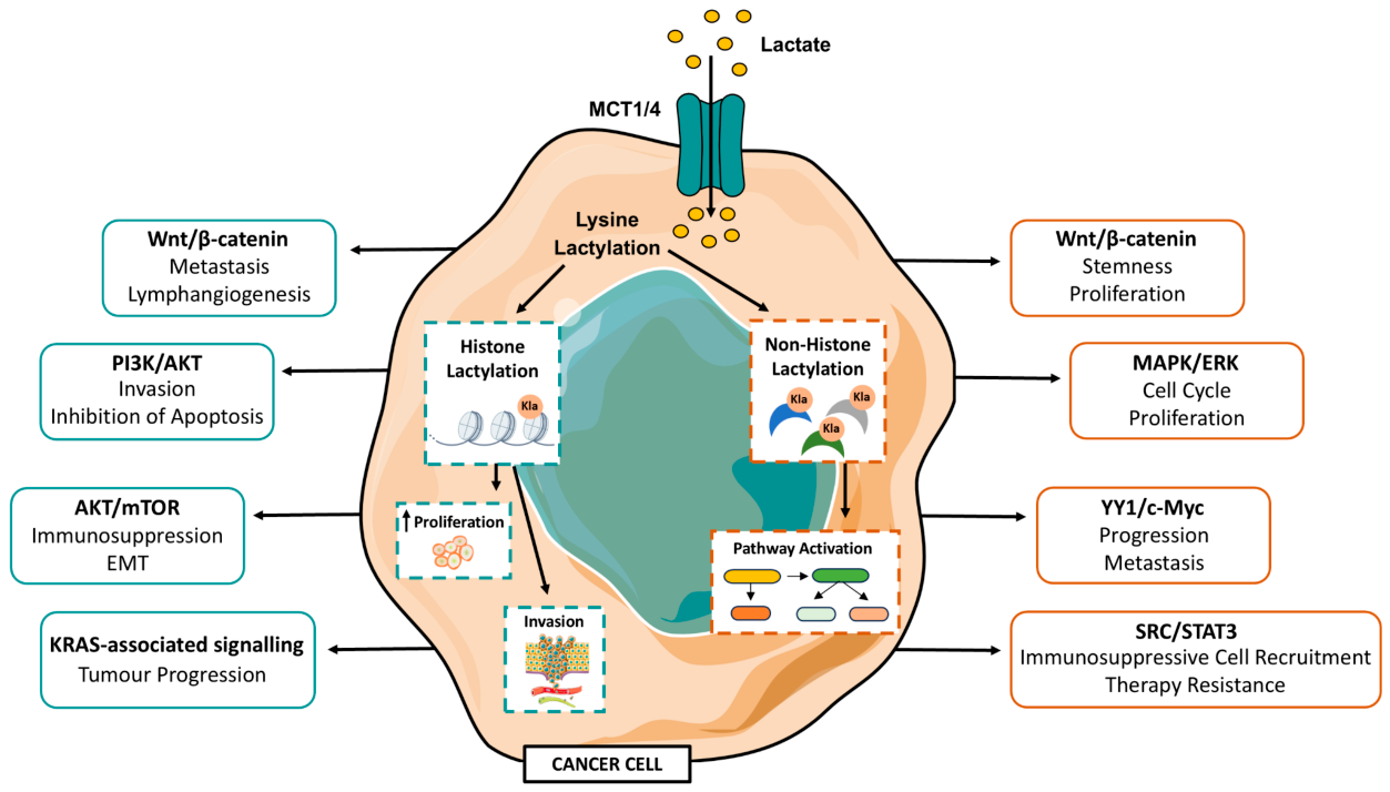
Figure 2.
Lactylation coordinates an integrated exosystem of cancer, stromal and immune-cell reprogramming that reinforces the desmoplastic niche. Lactate generated by elevated glycolysis in tumour cells and CAFs drives lysine Kla of histone and non-histone targets. Box border colours reflect mechanistic origin: blue borders denote histone Kla-driven compartments (mainly H3K18la, H3K56la, H4K8la and H4K12la), which alter chromatin accessibility at target loci; orange borders denote non-histone Kla-driven compartments, in which direct modification of transcription factors and metabolic enzymes alters their activity, stability and localisation; and green borders denote stromal compartments. Solid blue arrows indicate mechanistic connections driven by histone Kla; solid orange arrows indicate connections driven by non-histone Kla. These complementary mechanisms converge on three interconnected biological programmes. First, lactylation sustains cancer stem-cell maintenance by activating self-renewal, metabolic adaptation, stress resistance, ferroptosis suppression and cellular plasticity programmes; an important exception is observed in gastric cancer, where H3K18la activates METTL14 to suppress CSC properties, illustrating the context-dependence of lactylation outcomes (black-bordered box, lower left). Second, lactylation drives CAF activation and stromal remodelling, promoting ECM deposition, matrix stiffening, angiogenesis, immune exclusion and tumour invasion. Third, lactylation mediates immune-cell reprogramming across four populations: TAMs acquire an M2-like immunosuppressive phenotype; CD8+ T cells undergo exhaustion; dendritic cells exhibit impaired maturation and antigen presentation; and TANs adopt a pro-tumoral N2-like phenotype. Critically, these three programmes are not independent: CAF-derived lactate reinforces CSC stemness and CAF-driven ECM remodelling establishes physical barriers that compound Kla-dependent immune exclusion; both feedback interactions are depicted as green dashed arrows to distinguish them from direct Kla-driven mechanisms. ↑ and ↓ indicate upregulation and downregulation, respectively. Together, these coordinated events promote tumour progression, immune exclusion and resistance to multiple therapeutic modalities. Abbreviations: ARG1, arginase 1; CAF, cancer-associated fibroblast; CSC, cancer stem cell; DC, dendritic cell; ECM, extracellular matrix; Kla, lysine lactylation; NF-kB, nuclear factor kappa B; PD-1, programmed cell death protein 1; PD-L1, programmed death-ligand 1; RARγ, retinoic acid receptor gamma; TAN, tumour-associated neutrophil; TME, tumour microenvironment.
Figure 2.
Lactylation coordinates an integrated exosystem of cancer, stromal and immune-cell reprogramming that reinforces the desmoplastic niche. Lactate generated by elevated glycolysis in tumour cells and CAFs drives lysine Kla of histone and non-histone targets. Box border colours reflect mechanistic origin: blue borders denote histone Kla-driven compartments (mainly H3K18la, H3K56la, H4K8la and H4K12la), which alter chromatin accessibility at target loci; orange borders denote non-histone Kla-driven compartments, in which direct modification of transcription factors and metabolic enzymes alters their activity, stability and localisation; and green borders denote stromal compartments. Solid blue arrows indicate mechanistic connections driven by histone Kla; solid orange arrows indicate connections driven by non-histone Kla. These complementary mechanisms converge on three interconnected biological programmes. First, lactylation sustains cancer stem-cell maintenance by activating self-renewal, metabolic adaptation, stress resistance, ferroptosis suppression and cellular plasticity programmes; an important exception is observed in gastric cancer, where H3K18la activates METTL14 to suppress CSC properties, illustrating the context-dependence of lactylation outcomes (black-bordered box, lower left). Second, lactylation drives CAF activation and stromal remodelling, promoting ECM deposition, matrix stiffening, angiogenesis, immune exclusion and tumour invasion. Third, lactylation mediates immune-cell reprogramming across four populations: TAMs acquire an M2-like immunosuppressive phenotype; CD8+ T cells undergo exhaustion; dendritic cells exhibit impaired maturation and antigen presentation; and TANs adopt a pro-tumoral N2-like phenotype. Critically, these three programmes are not independent: CAF-derived lactate reinforces CSC stemness and CAF-driven ECM remodelling establishes physical barriers that compound Kla-dependent immune exclusion; both feedback interactions are depicted as green dashed arrows to distinguish them from direct Kla-driven mechanisms. ↑ and ↓ indicate upregulation and downregulation, respectively. Together, these coordinated events promote tumour progression, immune exclusion and resistance to multiple therapeutic modalities. Abbreviations: ARG1, arginase 1; CAF, cancer-associated fibroblast; CSC, cancer stem cell; DC, dendritic cell; ECM, extracellular matrix; Kla, lysine lactylation; NF-kB, nuclear factor kappa B; PD-1, programmed cell death protein 1; PD-L1, programmed death-ligand 1; RARγ, retinoic acid receptor gamma; TAN, tumour-associated neutrophil; TME, tumour microenvironment.
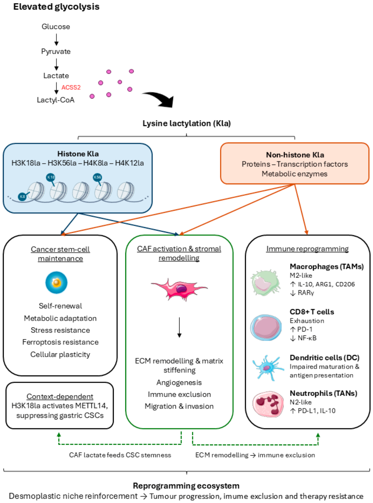

Table 1.
Histone Lactylation in Desmoplastic Cancers.
| Cancer Type | Histone | Lysine (K) | Target Genes | Mechanism of Lactylation Enrichment | Target Gene Effect (Downstream Mechanism) | Relevance for tumour | Supporting source / DOI |
| PDAC | H3 | K9 | ACSL4 | The CD147 /MCT4 axis drives intracellular lactate accumulation, promoting H3K9la at the ACSL4 promoter. | Regulates lipid metabolism and the balance of polyunsaturated fatty acids to maintain membrane integrity under stress. | Inhibits ferroptosis to enhance tumour survival. | 10.1016/j.xcrm.2025.102292 |
| PDAC | H3 | K18 | IGF2BP2 | CTCF recruits the writer EP300 (E1A Binding Protein p300) to the promoter to increase H3K18la. | Acts as an m6A reader that stabilizes the mRNA of oncogenes like MYC and CSF1. | Promotes M2 macrophage polarization and tumour cell proliferation. | 10.1038/s41418-024-01423-1 |
| PDAC | H3 | K18 | ACAT2 | Lactate-driven H3K18la activates ACAT2 transcription. | ACAT2 acetylates the protein MTCH2 (Mitochondrial Carrier Homolog 2), which disrupts oxidative phosphorylation and boosts further lactate production. | Establishes a metabolic feedback loop that drives immunosuppression. | 10.1136/gutjnl-2024-334361 |
| PDAC | H3 | K18 | TTK and BUB1B | High glycolytic flux drives p300-mediated H3K18la at the promoter. | TTK phosphorylates the enzyme LDHA at Y239, significantly increasing its catalytic activity and lactate output. BUB1B serves as a core component of the spindle assembly checkpoint, ensuring accurate chromosome segregation during mitosis. | Accelerates the cell cycle and reinforces glycolytic metabolism. | 10.1186/s12943-024-02008-9 |
| GC | H3 | K18 | VCAM1 | Glycolysis-derived lactate induces H3K18la enrichment at the transcription start site. | Triggers the AKT/mTOR signalling pathway within the tumour cell. | Promotes EMT and recruits immunosuppressive mesenchymal stem cells. | 10.1016/j.bcp.2024.116120 |
| GC | H3 | K18 | H19 (lncRNA - Long Non-Coding RNA) | Loss of the "eraser" SIRT1 leads to aberrant H3K18la accumulation at the H19 promoter. | Upregulates the expression of various glycolytic markers, including LDHA, to shift metabolism toward aerobic glycolysis. | Facilitates a positive feedback loop that enhances cell proliferation and migration. | 10.1038/s41388-024-03243-6 |
| GC | H3 | K18 | POM121 (POM121 Transmembrane Nucleoporin) | LDHA/B-driven lactate production increases H3K18la at the promoter. | Activates the PI3K/AKT survival signalling pathway. | Promotes cell invasion, survival, and inhibits apoptosis. | 10.1016/j.prp.2026.156427 |
| GC | H3 | K9 | TRIM29 | Glycolytic lactate increases H3K9la enrichment at the TRIM29 promoter. | Competitively binds to hnRNPA1 , preventing its degradation and activating Wnt/β-catenin signalling. | Facilitates lymph node metastasis. | 10.1038/s41419-026-08468-9 |
| HCC | H3 | K56 | ESM1 | Acetylation of PDHX disrupts the pyruvate dehydrogenase complex, increasing lactate and H3K56la. | Modulates cellular pathways to facilitate EMT and tumour cell motility. | Facilitates tumour growth and metastasis. | 10.1002/mc.23794 |
| HCC | H4 | K5 | ENO2 | p300 acts as a specific writer to increase H4K5la at the promoter in response to lactate. | Catalyses the conversion of 2-phosphoglycerate to phosphoenolpyruvate, increasing glycolysis | Established a self-sustaining loop that drives malignant progression. | 10.1016/j.jhepr.2026.101786 |
| HCC | H2B | K58 | NDRG1 | Hypoxia-induced LDHA increases H2BK58la at the NDRG1 promoter. | Functions as an anti-senescence factor that allows cells to bypass normal aging and death checkpoints. | Creates senescence-resistant cell lineages that drive aggressive clonal evolution. | 10.1016/j.canlet.2025.217567 |
| HCC | H3 | K18 | CD44 | The chaperone ASF1B recruits p300 to increase H3K18la at the promoter. | Acts as a key cell-surface marker and regulator of cancer stemness and self-renewal. | Increases tumour cell stemness and proliferation. | 10.3390/cells15100952 |
| HCC | H3 | K18 | YBX1 | O-GlcNAcylation of YBX1 increases PKM2 and lactate, driving H3K18la at the YBX1 promoter | YBX1 activates the transcription of p300, which in turn drives the lactylation of histones, particularly H3K18la. | Promotes tumour progression through a YBX1-glycolysis-lactylation loop | 10.1016/j.canlet.2025.217957 |
| CRC | H4 | K12 | LBX2 | Lactate-mediated H4K12la activates LBX2 expression. | Transcriptionally upregulates GFPT2, which increases O-GlcNAcylation of Raptor and activates mTORC1. | Enhances glucose uptake and fuels rapid tumour growth. | 10.1038/s41420-025-02888-w |
| CRC | H4 | K8 | LINC00152 | Bacterial lipopolysaccharide or lactate specifically induces H4K8la at the promoter. | Acts as an oncogenic scaffold that modulates various signalling pathways, including cytokine suppression. | Facilitates tumour cell invasion and migration. | 10.1016/j.cmet.2023.07.005 |
| CRC | H3 | K9 | GRAMD1A | Oncogenic KRAS mutations increase H3K9la by altering chromatin accessibility. | GRAMD1A acts as a cholesterol transporter that redistributes lipids to support membrane synthesis and signalling. | Promotes malignant progression and poor patient survival. | 10.1038/s41418-025-01533-4 |
| CRC | H3 | K18 | AURKB | High lactate levels in CRC drive H3K18la enrichment at the AURKB promoter. | Interacts with and interferes with HNRNPM preventing the degradation of PSAT1 mRNA. | Enhances serine biosynthesis to fuel tumour progression. | 10.1186/s13046-025-03498-1 |
| CRC | H3 | K18 | TROP2 | TROP2-mediated glycolysis promotes H3K18la | H3K18la in turn sustains TROP2 expression in a positive feedback loop | Drives metastatic colonization and progression | 10.1002/ctm2.70562 |
| TNBC | H4 | K12 | SLFN5 | Lactate-induced H4K12la at the promoter suppresses its transcription. | Its suppression prevents programmed cell death. | Leads to apoptosis evasion and TNBC malignant progression. | 10.1016/j.cellsig.2024.111468 |
| TNBC | H3 | K18 | MCM7 | LDHA/B-driven lactate production induces H3K18la at the promoter. | Serves as an essential DNA replication licensing factor required for the initiation of genomic duplication. | Boosts cancer cell proliferation and migration. | 10.1620/tjem.2025.J096 |
Table 2.
Non-Histone Protein Lactylation in Desmoplastic Cancers.
|
Cancer Type |
Protein | Lysine (K) | Mechanism | Relevance for tumour | Supporting source / DOI |
| iCCA | Nucleolin | K477 | Binds to the primary transcript of MADD (MAP kinase-activating death domain protein), preventing alternative splicing and mRNA decay; activation of the MAPK/ERK pathway. | Promotes tumour cell proliferation and invasion. | 10.1016/j.jhep.2024.04.010 |
| iCCA | EGR1 (Early Growth Response 1) | K422 | Required for nuclear translocation and binds to the CDKN1A (p21) promoter. | Induces cellular senescence and creates an immunosuppressive TME. | 10.1016/j.tranon.2026.102750 |
| iCCA | PSMD14 | K100 | Stabilizes ENO1 (Enolase 1) protein by delaying its proteasomal degradation. Reinforces glycolysis. | Confers resistance to ferroptosis and immunotherapy. | 10.1016/j.canlet.2026.218394 |
| PDAC | ENSA | K63 | Inactivates PP2A (protein phosphatase 2A), activating the SRC/STAT3/CCL2 signalling. | Creates an immunosuppressive TME and confers resistance to immunotherapy. | 10.1172/JCI187024 |
| GC | NSUN2 | K508 | Enhances m5C methyltransferase activity under acidic conditions. Stabilizes GCLC (Glutamate-Cysteine Ligase Catalytic Subunit) mRNA; increases GSH (glutathione) synthesis. | Fosters resistance to ferroptosis in the TME. | 10.1016/j.redox.2024.103479 |
| GC | PD-L1 | Not specified | Increases protein stability by preventing lysosomal degradation, increasing PD-L1 surface expression | Suppresses CD8+ T cell function and promotes immune evasion. | 10.1016/j.cpt.2024.09.006 |
| TNBC | ACAA2 | K214 | Increases the catalytic activity of ACAA2 in mitochondrial fatty acid oxidation. Accumulation of free fatty acids; induction of autophagy. | Promotes cell cycle progression, invasion, and metastasis. | 10.1002/advs.202511849 |
| TNBC | ENO1 | K262 | Prevents the lysosomal degradation of the ENO1 protein. Reinforces a positive feedback loop between lactylation and glycolysis. | Sustains tumour survival in hypoxic microenvironments. | 10.1038/s41419-026-08517-3 |
| HCC | ABCF1 | K430 | Induces a structural shift exposing the nuclear localization signal (NLS), leading to its entrance in the nucleus. Acts as a transcription factor, activating the KDM3A/H3K9me2/HIF1A axis. | Creates a self-sustaining lactate feedback loop. Drives metastasis. |
10.1038/s41418-024-01436-w |
| HCC | ASH2L | K312 | Synergistic activation of VEGFA enhancers and promoters. | Stimulates tumour angiogenesis and malignant progression. | 10.1002/advs.202509477 |
| HCC | CENPA | K124 | Forms a transcriptional complex with the factor YY1 (Yin Yang 1). | Drives cell cycle progression and unlimited proliferation. | 10.7150/ijbs.85656 |
| HCC | Cyclin E2 | K347, K348 | Essential for the G1/S transition in the cell cycle. | Accelerates tumour growth and increases cellular aggressiveness. | 10.15252/embr.202256052 |
| HCC | Rab7A | K21 | Inhibits Rab7A GTPase activity. Shifts MVB (multivesicular bodies) fate from degradation toward exosome secretion. | Orchestrates pulmonary pre-metastatic niche formation. | 10.1016/j.canlet.2025.217636 |
| HCC | c-Myc | Not specified | Prevents the degradation of the c-Myc oncoprotein. | Sustains unlimited proliferation and metabolic reprogramming. | 10.1080/13813455.2023.2206982 |
| HCC | YAP | K102 | Promotes nuclear translocation and Hippo pathway inhibition. | Promotes the transcription of proliferation, migration, invasion and apoptotic resistance genes. | 10.1186/s13062-025-00655-6 |
| HCC | TPX2 | K249 | Disrupts the binding of the phosphatase PP1 to AURKA (Aurora Kinase A), maintaining its active phosphorylated state. | Sustains mitotic progression and hepatocellular tumorigenesis. | 10.26508/lsa.202402978 |
| CRC | NSUN2 | K356 | Increases protein affinity for target RNAs like ENO1mRNA. | Reprograms metabolism and promotes CRC metastasis. | 10.1002/advs.202309840 |
| CRC | β-catenin | Not specified | Increases protein stability by preventing degradation. Activates the Wnt signalling pathway. | Promotes cell proliferation and sustains cancer stemness. | 10.1016/j.yexcr.2022.113439 |
| CRC | eEF1A2 | K408 | Enhances translation elongation. Increases global protein synthesis rates. | Facilitates oncogenic adaptation and rapid tumour growth. | 10.1073/pnas.2314128121 |
| CRC | HDAC1 | K412 | Essential for its deacetylase activity. | Confers robust resistance to ferroptosis. | 10.1002/advs.202408845 |
| CRC | PD-L1 | Not specified | Prevents lysosomal degradation of PD-L1, increasing its surface expression. | Impairs CD8+ T cell function and enables immune evasion. | 10.1016/j.cmet.2024.10.019 |
| CRC | PRMT5 | K240 | Represses the transcription of the m6A eraser ALKBH5. Stabilizes SLC7A11 (solute carrier family 7 member 11) mRNA. | Provides defence against lipid peroxidation and ferroptosis. | 10.1038/s41388-025-03457-2 |
| CRC | eEF2 | K439, K594 | Required for maintaining translational efficiency and fidelity. Accentuates global protein synthesis. | Supports the high translational demands of metastatic cells. | 10.1016/j.bbrc.2025.153119 |
| CRC | S100A8 | K23, K36 | Prevents ubiquitination and proteasomal degradation. | Leads to the accumulation of S100A8 and tumour progression. | 10.1016/j.bbrc.2026.153827 |
| CRC | NOL6 | K54 | Inhibits ubiquitination and subsequent degradation of NOL6. Stabilizes the c-Myc transcriptional axis. | Promotes CRC cell proliferation and metastasis. | 10.1016/j.celrep.2025.116774 |
| CRC | YBX1 | K92 | Facilitates interaction with the protein G3BP2 (G3BP Stress Granule Assembly Factor 2). Recognition and stabilization of m5C-modified transcripts. | Promotes oncogenic translation and aggressive tumour phenotypes. | 10.1016/j.ijbiomac.2026.150686 |
Table 3.
Lactylation-activated adaptive programmes driving resistance across therapeutic modalities in desmoplastic tumours.
Table 3.
Lactylation-activated adaptive programmes driving resistance across therapeutic modalities in desmoplastic tumours.
| Lactylation-activated adaptive programmes | Therapy modalities affected | ||
| Chemotherapy | Immunotherapy | Targeted therapy | |
| 1. Stemness maintenance: Histone and non-histone Kla activate OCT4, SOX2 and WNT/Hedgehog programmes; stromal-derived lactate reinforces stemness via ANTXR1 K453 | ✓ | — | — |
| 2. Metabolic rewiring: Kla-driven positive feedback amplifies glycolytic flux (ENO1, AKR1B10) and serine/antioxidant metabolism (PCK2, NRF2), sustaining lactate production under therapeutic pressure | ✓ | — | ✓ |
| 3. Ferroptosis suppression: Kla induces GPX4-independent ferroptosis resistance via glutathione synthesis (GCLC), lipid homeostasis (ZFP64/GCH1) and iron-sulphur cluster assembly (NFS1) | ✓ | ○ | ○ |
| 4. Stress-induced cell death suppression: Kla preserves mitochondrial network integrity (ABHD6 K245) and activates NRF2 via KEAP1 degradation, blocking apoptosis under targeted therapeutic stress | — | — | ✓ |
| 5. Immune evasion: Kla promotes TAM M2 polarisation, DC maturation failure, T-cell exhaustion and tumour-cell PD-L1 upregulation through convergent upstream pathways | ○ | ✓ | — |
| 6. Stromal protection: Tumour-derived lactate induces H3K18la/H4K8la in CAFs, driving FAP expression, collagen deposition and immune exclusion that limit drug and immune-cell access | ✓ | ○ | ○ |
| 7. Stress-adaptive survival programmes: Kla activates cytoprotective autophagy (HNRNPC/TRAF6), ABC-transporter-mediated drug efflux and METTL3-dependent DNA damage repair, enabling survival across treatment modalities | ✓ | — | ○ |
Symbol key: ✓ strong evidence (supported by ≥3 independent studies across ≥2 tumour types) ○ limited evidence (supported by 1 – 2 studies or a single tumour type) — not reported in the reviewed literature.
Table 4.
Original mechanistic and preclinical studies evaluating lactylation-dependent and broader lactate-axis therapeutic targets across desmoplastic and stroma-rich tumour contexts.
Table 4.
Original mechanistic and preclinical studies evaluating lactylation-dependent and broader lactate-axis therapeutic targets across desmoplastic and stroma-rich tumour contexts.
| Cancer Type | Lactylation / lactate axis | Mechanistic link to lactylation | Key evidence / models | Intervention tested | Relevance for desmoplastic tumour | Supporting source / DOI |
| PDAC | H3K18la-TTK/BUB1B-p300-LDHA feedback loop | High glycolysis increases lactate and H3K18la; H3K18la induces TTK/BUB1B; TTK/BUB1B reinforce p300/LDHA activity and sustain glycolysis–lactylation feedback. | Human PDAC tissues; PDAC cell lines; CUT&Tag/ChIP; xenograft validation. | Glycolysis blockade, LDHA knockdown, p300 inhibition, TTK/BUB1B knockdown. | Direct tumour-cell lactylation axis linking glycolytic flux to aggressive PDAC biology and potential biomarker stratification. | 10.1186/s12943-024-02008-9 |
| PDAC | pCAF-derived lactate-H3K18la-L1CAM/SLIT1-perineural invasion | Perineural-invasion-associated CAFs generate a high-lactate niche; lactate uptake by PDAC cells increases H3K18la and activates neural invasion genes including L1CAM and SLIT1. | Single-cell sequencing, metabolomics, patient tissues, PDAC models, ChIP-seq. | Targeting lactate metabolism and H3K18la; glycolytic modulation of pCAFs. | Strong desmoplastic/stromal example: CAF metabolism imposes lactylation-dependent invasive programming on cancer cells. | 10.1158/0008-5472.CAN-24-3173 |
| PDAC | Cancer-derived lactate-CAF metabolic reuse and fibrosis/immunosuppression | Cancer-derived lactate is taken up by CAFs through MCT1 and used as a carbon source; lactate metabolism sustains alpha-SMA-positive CAFs, collagen deposition and immune exclusion. | Mouse PDAC models; CAF metabolic assays; tumour lactate, collagen and immune infiltration readouts. | LDHA inhibition with FX11; MCT1 knockdown/AZD3965 in CAF models. | Not a Kla-specific study, but essential upstream evidence connecting lactate availability to CAF persistence, matrix deposition and CD8 T-cell exclusion. | 10.1172/jci.insight.163022 |
| PDAC | Tumour lactate-epigenomic reprogramming during pancreatic CAF formation | Tumour-derived lactate drives CAF-like differentiation and epigenomic reprogramming, including DNA methylation/TET-dependent inflammatory CAF programmes. | Human pancreatic CAF formation models; metabolomics; DNA methylation and gene-expression analyses. | Manipulation of lactate metabolism and TET-linked epigenetic programmes. | Provides foundational stromal evidence for lactate as a durable epigenetic signal in pancreatic desmoplasia. | 10.7554/eLife.50663 |
| PDAC | Tumour lactate-ENSA-K63la-PP2A-SRC/STAT3-CCL2 | Glycolytic lactate induces ENSA K63 lactylation, inhibits PP2A and sustains SRC/STAT3 activation; CCL2 secretion recruits TAMs and suppresses cytotoxic T-cell activity, promoting resistance to immune checkpoint blockade. | Human PDAC cohorts and tissue microarrays; metabolic imaging; single-cell/proteomic analyses; cell, organoid and immunocompetent mouse models. | ENSA-K63la-blocking peptide; CCL2/CCR2 blockade; combination with immune checkpoint blockade. | Direct metabolic-immune mechanism connecting high glycolysis to macrophage-rich immune exclusion in desmoplastic PDAC; potential response biomarker. | 10.1172/JCI187024 |
| PDAC | UBE2T-p53-glycolysis-CAF H3K18la-ECM deposition | UBE2T-driven p53 degradation rewires tumour glycolysis and increases lactate; tumour-derived lactate induces H3K18la in CAFs, activating stromal programmes and collagen/extracellular-matrix deposition. | Spatial metabolomics/transcriptomics; PDAC organoid-CAF co-cultures; spontaneous PDAC mice; immune-reconstituted patient-derived xenografts. | UBE2T genetic deletion or pentagalloylglucose; glycolysis/lactate inhibition; pentagalloylglucose plus anti-PD-1. | Strong desmoplasia-focused study linking tumour metabolism to CAF lactylation, matrix densification and CD8 T-cell exclusion. | 10.1002/advs.202514933 |
| iCCA | Nucleolin lactylation-MADD alternative splicing | Lactylation of nucleolin contributes to iCCA pathogenesis by regulating RNA splicing of MADD. | ICCA tissues and experimental models; RNA-splicing and functional validation. | Mechanistic targeting of nucleolin lactylation/splicing axis proposed; no established clinical lactylation trial yet. | Important because iCCA is commonly stroma-rich/desmoplastic, and this gives a direct non-histone lactylation mechanism in the selected tumour list. | 10.1016/j.jhep.2024.04.010 |
| iCCA | L-lactate-PSMD14 lactylation-ENO1-ferroptosis resistance | L-lactate lactylates and stabilizes PSMD14; PSMD14 supports ENO1-dependent glycolysis and lactate production, reinforcing a feed-forward programme that limits ferroptotic death. | Human iCCA samples; iCCA cell models; lactylation, protein-stability and ferroptosis assays; in vivo tumour validation. | PSMD14/ENO1 genetic silencing; lactate or glycolysis blockade; ferroptosis-inducing strategies. | Direct non-histone Kla mechanism coupling metabolic feedback to ferroptosis resistance in a highly fibrotic liver tumour. | 10.1016/j.canlet.2026.218394 |
| iCCA | EGR1-K422 lactylation-senescence-immunosuppressive TME | EGR1 K422 lactylation promotes nuclear accumulation and a tumour-cell senescence/interferon-stimulated gene programme that remodels the microenvironment towards immune suppression. | Human iCCA cohorts and transcriptomic/single-cell analyses; iCCA cell models; site-specific lactylation and in vivo validation. | EGR1 knockdown or K422 mutation; inhibition of lactate/lactylation; senescence-associated immune modulation. | Links tumour-cell lactylation to senescence-associated paracrine signalling and immune dysfunction in stroma-rich iCCA. | 10.1016/j.tranon.2026.102750 |
| HCC | H3K14la-NEDD4-PTEN-PI3K/AKT/mTOR multidrug resistance | Glycolysis-driven H3K14la increases NEDD4, promotes PTEN ubiquitination/degradation, activates PI3K/AKT/mTOR signalling and reinforces drug resistance. | Oxaliplatin/5-FU-resistant HCC models; lactylation/PTEN assays; xenografts. | Oxamate/glycolysis inhibition; targeting NEDD4/PTEN axis. | Links lactylation to therapy resistance in a tumour that usually develops in chronically injured/fibrotic liver. | 10.1038/s41419-025-07359-9 |
| HCC | Histone lactylation-ESM1 transcription | Histone lactylation activates ESM1 transcription, promoting HCC proliferation, migration, invasion, EMT and tumour growth/metastasis. | HCC cell lines, xenograft and metastasis models, ChIP validation. | 2-DG-mediated inhibition of histone lactylation; ESM1 knockdown. | Candidate biomarker/effector axis for aggressive HCC; useful for lactylation-driven progression discussion. | 10.1002/mc.23794 |
| HCC | HSC/AKR1B10-H3K18la stromal-tumour interaction | Hepatic stellate cell interaction with tumour cells is associated with activation of histone lactylation; AKR1B10 links stromal crosstalk to Pan-Kla/H3K18la and HCC proliferation. | scRNA-seq, spatial transcriptomics, LX-2/HepG2/HuH7 co-cultures. | AKR1B10 knockdown. | Particularly relevant to fibrotic HCC because hepatic stellate cells are central to liver fibrosis and tumour-stroma signalling. | 10.1016/j.canlet.2024.217243 |
| HCC | IGF2BP3 lactylation-PCK2/NRF2-serine metabolism-m6A-lenvatinib resistance | Glycolysis-derived lactate induces IGF2BP3 lactylation, increasing capture and stability of PCK2 and NRF2 mRNAs; serine metabolism and SAM-dependent m6A establish a feed-forward antioxidant programme. | Lenvatinib-resistant HCC cell lines; patient tissues; multi-omics and lactylome analyses; xenografts. | Liposomal siIGF2BP3; 2-DG; combination with lenvatinib. | Clinically relevant mechanism connecting Kla to targeted-therapy resistance in fibrotic/cirrhotic HCC. | 10.1002/advs.202401399 |
| HCC | Tumour lactate-histone lactylation-NUPR1-TAM immunosuppression | Tumour-derived lactate increases histone lactylation and NUPR1 in infiltrating macrophages; NUPR1 suppresses ERK/JNK activity, promotes an M2-like state and contributes to CD8 T-cell dysfunction. | Single-cell and patient-cohort analyses; macrophage-T-cell co-cultures; subcutaneous and spontaneous HCC mouse models. | NUPR1 inhibitors TFP-2HCl or ZZW-115-2HCl; combination with anti-PD-1. | Adds an immune-cell lactylation axis and a pharmacologically actionable strategy for macrophage-rich HCC. | 10.1002/advs.202413095 |
| TNBC | KCNK1-LDHA-H3K18la feedback | KCNK1 binds and activates LDHA, increasing lactate and H3K18la; H3K18la supports transcriptional programmes linked to proliferation, invasion and metastasis. | Breast cancer cell lines including TNBC-relevant models; ChIP-qPCR; xenografts; clinical datasets. | KCNK1 knockdown; LDHA manipulation. | Connects lactate production to histone lactylation in aggressive breast cancer; direct desmoplasia link remains less developed. | 10.1371/journal.pbio.3002666 |
| TNBC | H4K12la-SLFN5 downregulation and H4K12la biomarker axis | H4K12 lactylation is increased in TNBC and has been linked to malignancy progression, partly through SLFN5 downregulation; lactylproteomics identifies H4K12la as a candidate TNBC biomarker. | TNBC tissues; 4D label-free quantitative proteomics; IHC/immunoblotting; functional assays. | H4K12la/SLFN5 modulation in experimental systems. | Provides biomarker-oriented evidence for lactylation in TNBC and a candidate axis for metabolic-epigenetic stratification. | 10.3389/fendo.2024.1328679; 10.1016/j.cellsig.2024.111468 |
| TNBC | CAF lactate-H3K18la-ZFP64-GCH1/FTH1-ferroptosis and doxorubicin resistance | CAF-derived lactate increases histone/H3K18 lactylation and ZFP64 transcription; ZFP64 induces GCH1 and FTH1, limits lipid peroxidation and iron availability, and suppresses ferroptosis. | CAFs from doxorubicin-sensitive/resistant tumours; TNBC co-cultures and patient tissues; ChIP/reporter assays; orthotopic mouse models. | ZFP64 silencing; lactate/histone-lactylation modulation; ferroptosis sensitization combined with doxorubicin. | Direct CAF-metabolite mechanism linking desmoplastic stroma to ferroptosis escape and anthracycline resistance. | 10.1186/s12967-025-06246-3 |
| TNBC | HDAC2-METTL3 delactylation-m6A-DNA repair-cisplatin resistance | HDAC2 removes lactylation from METTL3, strengthening METTL3-WTAP complex formation and m6A modification of DNA-repair transcripts, enabling survival after cisplatin. | Cisplatin-tolerant TNBC models; patient samples; site-specific lactylation, m6A and DNA-repair assays; in vivo validation. | HDAC2 inhibition with tucidinostat; METTL3/repair-pathway disruption; tucidinostat plus cisplatin. | Demonstrates that delactylation, not only hyperlactylation, can drive treatment resistance and identifies a clinically relevant HDAC inhibitor. | 10.1002/advs.202413121 |
| GC | AARS1-mediated YAP-K90/TEAD1-K108 lactylation | AARS1 senses intracellular lactate, translocates to the nucleus and lactylates YAP/TEAD1, forming a positive feedback loop that activates YAP signalling and tumour cell proliferation. | GC cell lines; MNU-induced mouse GC; human GC tissue arrays. | AARS1 genetic depletion; lactate/ATP-dependent in vitro lactylation assays. | Key non-histone lactyltransferase example in a stroma-rich digestive tumour context. | 10.1172/JCI174587 |
| GC | H3K18la-VCAM1-AKT/mTOR-CXCL1 axis | H3K18la transcriptionally activates VCAM1; VCAM1 activates AKT/mTOR and CXCL1-associated signalling, promoting migration/metastasis and recruitment of GC-derived mesenchymal stromal cells. | GC cells; functional assays; immune/stromal recruitment readouts. | Targeting H3K18la/VCAM1 axis proposed. | Links lactylation to inflammatory stromal recruitment and immune suppression, a useful parallel to desmoplastic TME biology. | 10.1016/j.bcp.2024.116120 |
| GC | GLUT3-LDHA-histone lactylation | GLUT3 increases glycolysis and LDHA-dependent lactate production, raising histone lactylation and supporting GC progression/invasiveness. | scRNA-seq analyses, GC tissues, GC cell lines. | GLUT3/LDHA modulation. | Upstream metabolic source of Kla; supports patient stratification by glycolytic/lactate phenotype. | 10.1186/s12935-023-03162-8 |
| GC | CAF-secreted LOX-histone lactylation-PD-L1 and TGFβ/IGF1-EMT | CAF-derived LOX promotes tumour-cell histone lactylation and PD-L1 expression, while CAF-tumour TGFβ/IGF1 signalling reinforces EMT and invasive behaviour. | Patient gastric tumour/CAF samples; gastric-cancer cell-CAF co-cultures; histone-lactylation, PD-L1 and EMT assays; in vivo validation. | LOX depletion/inhibition; blockade of lactylation, PD-L1 or TGFbeta/IGF1 signalling. | Connects a matrix-remodelling CAF product with lactylation-dependent immune escape and EMT in a stroma-rich cancer. | 10.1016/j.cellsig.2024.111462 |
| GC | CAF lactate-H3K18la-ASPM-NCAPG-SRC/STAT3-PD-L1 | CAF-derived lactate raises H3K18la and induces ASPM; ASPM promotes BUB3-mediated NCAPG deubiquitination, activating SRC/STAT3 and PD-L1 and reducing anti-PD-1 response. | CUT&Tag/transcriptomics; gastric-cancer cell, organoid and animal models; patient anti-PD-1 response samples. | Lactate reduction or ASPM silencing; NCAPG inhibitor daturilin; daturilin plus anti-PD-1. | Strong stromal-immune example directly linking CAF metabolism to checkpoint resistance in desmoplastic-enriched GC. | 10.1186/s12967-025-07013-0 |
| CRC | KAT8-mediated eEF1A2 lactylation | KAT8 catalyses eEF1A2 lactylation, increasing translational elongation/protein synthesis and promoting colorectal carcinogenesis. | CRC tissues and cell lines; lactylome profiling; in vitro/in vivo functional validation. | KAT8 genetic/inhibitory approaches. | Strong non-histone lactylation target; suggests writer/substrate-level therapeutic vulnerability. | 10.1073/pnas.2314128121 |
| CRC | H3K18la-RUBCNL-autophagy and bevacizumab resistance | Tumour-derived lactate induces H3K18la at RUBCNL, promoting autophagy and resistance to bevacizumab treatment. | CRC models of bevacizumab resistance; autophagy assays; mechanistic validation. | Lactate/H3K18la/autophagy modulation. | Directly links lactate-driven histone lactylation to anti-angiogenic therapy resistance. | 10.1080/15548627.2023.2249762 |
| CRC | GPR37-Hippo/YAP-LDHA-H3K18la-CXCL1/CXCL5 | GPR37 activates Hippo/YAP signalling, enhances LDHA and glycolysis, increases H3K18la and drives CXCL1/CXCL5-mediated neutrophil recruitment in colorectal liver metastasis. | Human CRLM specimens; MC38/CT26 liver metastasis models; cytokine and metabolic assays. | GPR37 knockdown; CXCL1/CXCL5 silencing. | Important immune-stromal axis linking lactylation to neutrophil recruitment and metastatic niche formation. | 10.1038/s41388-023-02841-0 |
| CRC | CAF lactate-histone/ANTXR1-K453 lactylation-RhoC/ROCK1/SMAD5-stemness | CAF-derived lactate increases ANTXR1 transcription through histone lactylation and directly stabilizes ANTXR1 by K453 lactylation; this activates RhoC/ROCK1/SMAD5, stemness and oxaliplatin resistance. | CRC patient tissues and CAF co-cultures; lactylation-site/protein-stability assays; organoid/xenograft and oxaliplatin-response models. | CAF/lactate blockade; ANTXR1 knockdown or K453 mutation; inhibition of RhoC/ROCK1/SMAD5. | Highly relevant stromal mechanism linking CAF-derived lactate to cancer stemness and chemotherapy failure. | 10.1016/j.canlet.2025.217917 |
| CRC | Tumour lactate-macrophage histone lactylation-RARgamma-TRAF6/IL-6/STAT3 | Tumour-derived lactate drives macrophage histone lactylation and suppresses RARγ, releasing TRAF6/IL-6/STAT3 signalling and creating a tumour-promoting inflammatory niche. | Human CRC tissues; macrophage-cancer co-cultures; genetic and biochemical pathway studies; mouse CRC models. | RARγ restoration/agonism; inhibition of lactate or histone lactylation; TRAF6/IL-6/STAT3 blockade. | Adds direct immune-cell evidence that tumour lactate reprograms macrophages to support CRC growth. | 10.1016/j.celrep.2024.113688 |
Disclaimer/Publisher’s Note: The statements, opinions and data contained in all publications are solely those of the individual author(s) and contributor(s) and not of MDPI and/or the editor(s). MDPI and/or the editor(s) disclaim responsibility for any injury to people or property resulting from any ideas, methods, instructions or products referred to in the content. |
© 2026 by the authors. Licensee MDPI, Basel, Switzerland. This article is an open access article distributed under the terms and conditions of the Creative Commons Attribution (CC BY) license.
Copyright: This open access article is published under a Creative Commons CC BY 4.0 license, which permit the free download, distribution, and reuse, provided that the author and preprint are cited in any reuse.