

Submitted:
13 November 2026
Posted:
16 November 2026
You are already at the latest version
Abstract
Acetylcholinesterase (AChE) inhibition is an established strategy for the symptomatic treatment of Alzheimer’s disease. Convolvamine is a tropane alkaloid from Convolvulus pluricaulis, a plant traditionally used for neurological indications. However, the potential of isolated convolvamine as a cholinesterase inhibitor remains poorly characterized. We compared convolvamine with donepezil, galantamine and rivastigmine using docking, ADMET prediction, triplicate 200 ns AChE and butyrylcholinesterase (BChE) simulations, and a screen against six proteins with AChE-like binding sites. Convolvamine remained in the AChE gorge while contacting the catalytic-site residues Trp86 and Tyr337 and the peripheral residues Tyr124 and Tyr341. The mean molecular mechanics Poisson-Boltzmann surface area (MM-PBSA) ΔTOTAL of convolvamine was closest to galantamine. No AChE-over-BChE difference was resolved within the sampled simulations. We performed simulations for likely off-targets and identified the α2C adrenergic and glucocorticoid receptors as potential off-target liabilities that should be considered. ADMET models predicted high gastrointestinal absorption and blood-brain-barrier permeation alongside hERG and CYP2D6 liabilities and further alerts consistent with the approved comparators. These calculations support experimental evaluation of convolvamine activity against AChE and BChE and identify potential liabilities for further study.
Keywords:
acetylcholinesterase
; cholinergic signaling
; convolvamine
; molecular dynamics
; ADMET
; receptor selectivity
1. Introduction
Alzheimer’s disease is the most common cause of dementia, and available therapies often fail to halt or reverse the underlying neurodegeneration [1,2]. Acetylcholinesterase (AChE) terminates cholinergic signaling by hydrolyzing acetylcholine, and as cholinergic neurons are progressively lost in Alzheimer’s disease that signaling is impaired. Clinical studies of cholinesterase inhibitors report modest symptomatic improvement in cognition, which remains the basis for their continued use [1,2,3,4]. Disease-modifying anti-amyloid antibodies have since entered clinical use, but they address a different target and have not displaced symptomatic cholinergic therapy [5].
Human AChE has a deep active-site gorge with two functionally distinct regions. The catalytic active site (CAS) lies at the base of the gorge near Trp86, Tyr337 and Phe338. The peripheral anionic site (PAS) lies at its entrance and is formed by Tyr72, Asp74, Tyr124, Trp286 and Tyr341 [6,7]. The human structure 4EY7 was solved with donepezil bound, providing a reference pose against which a docking protocol can be validated.
Three approved inhibitors were included for comparison because they span the recognized binding modes at this site. Donepezil is a reversible piperidine inhibitor that spans the gorge, contacting both the catalytic and peripheral sites, and its co-crystallized pose in 4EY7 also serves as the redocking control [6]. Galantamine is a plant-derived alkaloid that binds competitively at the catalytic site [3]. None of the three is a close structural analog of convolvamine. Tanimoto similarities are 0.23 for donepezil, 0.19 for galantamine and 0.14 for rivastigmine. They were therefore chosen for the binding modes they represent rather than for chemical resemblance. Rivastigmine is a carbamate that inhibits both AChE and BChE by carbamylating the catalytic serine, and so represents a covalent mechanism [8].
Convolvamine is a tropane alkaloid isolated from Convolvulus pluricaulis (Shankhpushpi), a plant traditionally used for cognitive and neurological indications [9], and extracts of the plant inhibit acetylcholinesterase in vivo [10]. Activity of a whole extract does not establish that convolvamine is the constituent responsible, so the present work treats the compound as a hypothesis to be tested rather than as an established inhibitor. The isolated compound has limited pharmacological characterization, with available reports describing plant preparations but not the purified alkaloid [9,11], and its cholinesterase interactions have not been examined against approved inhibitors.
Here we characterize convolvamine alongside these three inhibitors by docking against AChE, triplicate 200 ns molecular dynamics with end-point free-energy analysis, comparative BChE simulations, screening against six structurally related proteins, and ADMET prediction (Figure 1). Ligand identifiers, SMILES and molecular formulas were curated from PubChem [12] and are given in Table S1.
2. Materials and Methods
2.1. Molecular Docking
For the docking studies, the human AChE structure PDB 4EY7 [6,13] was used, a crystal structure of the wild-type enzyme with donepezil bound. The receptor was prepared in UCSF Chimera 1.19 [14] by deleting water and heteroatoms, modeling missing side chains with the Dunbrack backbone-dependent rotamer library [15], adding hydrogens, and assigning Gasteiger charges. Ligands were prepared with Chimera Dock Prep, which adds hydrogens, derives charges with ANTECHAMBER [16] and energy-minimizes each conformer, with ionization set for pH 7.4. The tropane nitrogen of convolvamine and the tertiary amines of the three reference ligands are protonated, giving each ligand a net charge of +1. The exact ligand structure and topology files are in the archive cited under Data availability. Receptor and ligand files were written in PDBQT format [17]. For the simulations, protein hydrogens were rebuilt by gmx pdb2gmx against the amber99sb-ildn port with TIP3P water, discarding any input hydrogens. Histidine protonation was assigned automatically by the hydrogen-bonding optimization in pdb2gmx. The remaining titratable residues were left in their standard states for pH 7, with aspartate and glutamate deprotonated and lysine and arginine protonated.
Ligands were docked with AutoDock Vina 1.2.0 [18,19] into a box centered at (9.671, -56.552, -23.837) Å with dimensions 28 x 32 x 22 Å, using an exhaustiveness of 32, 20 requested modes and a 5 kcal/mol energy range. For convolvamine, galantamine and rivastigmine the top-scoring pose initiated the molecular-dynamics simulation. For donepezil, which is co-crystallized in 4EY7, the crystallographic ligand was used. Structural figures were rendered in UCSF ChimeraX 1.10 [20]. Docking accuracy was validated by redocking donepezil and comparing the resulting modes with the crystallographic pose using symmetry-aware RDKit CalcRMS without superposition [21].
BChE docking used human BChE (PDB 4BDS), chain A, residues Ile4-Val529, prepared metal-free with waters and the co-crystallized ligand removed, in a box centered on the co-crystallized ligand position.
2.2. Molecular Dynamics
Simulations were run in GROMACS 2025.3 [22]. Proteins were described with AMBER99SB-ILDN [23] and ligands with GAFF2 and AM1-BCC charges assigned through ACPYPE and AmberTools [24,25], with hydrogen mass repartitioning to 3.024 amu. Each complex was solvated with TIP3P water in a dodecahedral box, neutralized and brought to 0.15 M NaCl. Systems were energy-minimized by steepest descent to 500 kJ mol-1 nm-1, then equilibrated for 100 ps in the NVT ensemble and 1 ns in the NPT ensemble under position restraints. Production runs used a 4 fs time step, the Verlet cut-off scheme, particle-mesh Ewald electrostatics, 1.2 nm Coulomb and van der Waals cut-offs, and LINCS constraints on all bonds. Temperature was maintained at 310 K with the velocity-rescaling thermostat (coupling constant 1.0 ps) and pressure at 1.0 bar with the Parrinello-Rahman barostat (coupling constant 5.0 ps). AChE complexes were simulated for 200 ns in triplicate and BChE and off-target complexes for 100 ns, with coordinates saved every 500 ps. Replicates were started from the same minimized coordinates with different random velocity seeds.
The membrane-embedded receptors (5DSG and 6KUW) were built with PACKMOL-Memgen [26] in a POPC bilayer of 287 lipids, using AMBER ff14SB for the protein [27] and Lipid21 for the lipids [28], with missing loops modeled in PDBFixer. These systems used semi-isotropic pressure coupling at 1.0 bar with a compressibility of 4.5e-5 bar-1.
End-point energies were estimated with the molecular mechanics Poisson-Boltzmann surface area (MM-PBSA) and generalized Born surface area (MM-GBSA) methods in gmx_MMPBSA [29,30]. Energies were taken over the final 20 ns of the AChE and BChE trajectories and the final 10 ns of the off-target trajectories, in both cases at one frame per 500 ps. Linear Poisson-Boltzmann calculations used ipb = 2 and inp = 1 with an internal dielectric of 1.0, an external dielectric of 80.0, a 1.4 Å probe, radiopt = 1, mbondi3 radii, a grid scale of 2.0 and a fill ratio of 4.0. MM-GBSA used igb = 5 with the same dielectrics. The AChE and BChE calculations used the ff99SBildn and GAFF2 parameter sets at an ionic strength of 0.15 M over a 20 ns window, whereas the off-target panel used ff99SB and GAFF at zero ionic strength over a 10 ns window. The twelve AChE replicates were also recomputed under the off-target convention, and this matched reference is used for the AChE-referenced off-target differences. It shifted the AChE end point by -1.5 to -2.3 kcal/mol across the four ligands and is reported per replicate in Table S12. The complete input files are included in the archive cited under Data availability. Interaction-entropy corrections were computed but not interpreted, as the gmx_MMPBSA reliability criterion was not met for most trajectories [31]. Cα RMSF was computed on fitted coordinates over the same window and referenced to three apo AChE replicates simulated under the same parameters. The scalar pocket RMSF quoted in the text is the unweighted mean of the per-residue Cα RMSF values over the pocket residues, and the pocket ΔRMSF is that scalar for a ligand-bound replicate minus the mean of the three apo replicates.
Ligand persistence was assessed over the complete 200 ns of each AChE trajectory. Trajectories were made whole and centered on the protein with gmx trjconv before analysis. Protein Cα coordinates were superposed on the first frame by Kabsch rotation and the same transform applied to the ligand, so that the reported ligand RMSD is measured in the pocket reference frame. Contact occupancy is the fraction of frames in which a ligand heavy atom lies within 4 Å of a heavy atom of the residue. Peripheral-anionic-site residues were taken as Tyr72, Asp74, Leu76, Tyr124, Trp286 and Tyr341, and catalytic-site residues as Trp86, Glu202, Ser203, Tyr337, Phe338 and His447. All ligands were simulated at pH 7.4 with a net charge of +1 on the protonated amine.
Differences between two estimated means combine the variance of both terms across replicates and use Welch-Satterthwaite degrees of freedom for 95% confidence intervals, uncorrected for multiple comparisons.
2.3. Off-Target Panel
Candidate off-targets were retrieved with PocketScope [32], which uses protein language model embeddings to identify proteins with binding sites similar to a query pocket across the human AlphaFold proteome. Using the AChE site as the query, candidates were retained only where the Protein Data Bank held an experimental human structure spanning the predicted pocket. Eight such targets entered simulation. The six carried into the analysis (Table S5) are angiotensin-converting enzyme (1O8A), adenosine deaminase (3IAR), PPAR-α (3VI8), the glucocorticoid receptor (4P6X), the M4 muscarinic receptor (5DSG) and the α2C adrenergic receptor (6KUW). Receptors were prepared as above and each ligand docked into a box centered on the co-crystallized ligand position. Zinc and magnesium ions were treated as unbonded ions with standard AMBER parameters. The zinc sites of angiotensin-converting enzyme and adenosine deaminase remained coordinated throughout. For the membrane receptors the bilayer was present during simulation but omitted from the MM-PBSA calculation, so their energies are qualitative. Each ligand-target pair was simulated in triplicate, so every difference reported for the panel rests on three independent replicates (Table S10).
2.4. Computational ADMET Prediction
Physicochemical, pharmacokinetic and toxicity properties were predicted with SwissADME [33] and ADMETlab 3.0 [34]. Blood-brain-barrier and gastrointestinal predictions were interpreted in the BOILED-Egg framework [35]. The end points examined were gastrointestinal absorption, blood-brain-barrier permeability, P-glycoprotein substrate status, cytochrome-P450 inhibition, hERG, drug-induced liver injury, AMES mutagenicity, hepatotoxicity, nephrotoxicity, solubility, logP and rule-based drug-likeness alerts (Tables S3 and S4).
2.5. Ethics Statement
This study is entirely computational. It used only previously published, publicly available data: protein structures deposited in the Protein Data Bank and compound records retrieved from PubChem. No human participants, human material, human data or animals were involved at any stage, and no protected or non-public dataset, collection or site was used, so approval by an institutional review board or ethics committee and informed consent were not applicable. The work was nonetheless conducted in accordance with the ethical principles of the Declaration of Helsinki [36].
3. Results
3.1. Molecular Docking
Redocking donepezil against 4EY7 recovered the crystallographic pose to a heavy-atom RMSD of 0.802 Å, with several further modes within about 2 Å, confirming that the docking setup reproduces a known binding geometry. Among the four ligands donepezil scored most favorably (-11.82 kcal/mol), followed by galantamine (-10.30), convolvamine (-8.76) and rivastigmine (-8.14). Docking scores against both cholinesterases are given in Table S2 and the redocked donepezil pose in Figure S6. The docked poses are shown in Figure 2. All four systems were built from the same prepared receptor, so the starting poses can be compared directly (Figure 2). The four compounds lie along the gorge axis but reach different depths. Galantamine is confined to the catalytic side, approaching Trp86 to 3.4 Å while its closest approach to Trp286 is 6.8 Å. The other three contact both anchors, coming within 3.4 to 4.3 Å of Trp86 and 3.4 to 3.7 Å of Trp286, so convolvamine begins in contact with both sites.
3.2. AChE Binding-Pose Persistence and Residue Contacts
Across all twelve ligand-bound replicates we measured, over the full 200 ns, the ligand heavy-atom RMSD after superposing the protein Cα frame and per-residue contact occupancy at a 4 Å heavy-atom cutoff (Figure 3).
In the donepezil simulations, Trp286 contact occupancy was 0.99 ± 0.00 and Trp86 occupancy 0.99 ± 0.02. Galantamine and rivastigmine contacted Trp286 for 0.00 and 0.06 of frames respectively.
Convolvamine contacted at least one peripheral residue in 0.99, 0.99 and 0.83 of sampled frames across the three replicates, and showed persistent catalytic-side contacts at Trp86 (0.99 ± 0.01), Tyr337 (0.94 ± 0.06) and His447 (0.71 ± 0.15). Within the peripheral set, Tyr124 contact occupancy was 0.80 ± 0.15 and Tyr341 0.67 ± 0.39, while Trp286 occupancy varied across replicates at 0.26 ± 0.24 (0.49, 0.28 and 0.01). Convolvamine remained within the AChE gorge across all three simulations, with a ligand RMSD of 5.64 ± 1.69 Å after protein alignment, compared with 2.53 ± 1.23 Å for donepezil, indicating that it samples several orientations within the gorge.
3.3. AChE Pocket Flexibility and Binding-Energy Estimates
Pocket Cα RMSF was compared with three apo replicates simulated under identical parameters. The differences were -0.059 Å for donepezil, -0.040 Å for galantamine, -0.031 Å for rivastigmine and -0.010 Å for convolvamine, and all four 95% confidence intervals included zero (Figure 4A, Table S6). Per-replicate profiles are in Figure S2.
Mean MM-PBSA ΔTOTAL values were -37.59 ± 7.72 kcal/mol for donepezil, -36.68 ± 9.16 for rivastigmine, -29.49 ± 1.90 for convolvamine, and -28.25 ± 10.87 for galantamine. Per-replicate values are in Table S8 for AChE and Table S9 for BChE, and component decompositions are in Figure S3.
3.4. Cholinesterase Selectivity and Off-Target Screening
Across three replicates per ligand, donepezil and rivastigmine gave the largest AChE-favored differences between the two cholinesterases, at +9.54 and +7.52 kcal/mol, and galantamine and convolvamine gave +1.69 and -1.15 kcal/mol. The between-replicate standard deviations on the BChE energies range from 4.3 to 9.6 kcal/mol, and every confidence interval on the difference includes zero (Table 1), so selectivity between the two enzymes could not be established for any compound.
Across the panel, most targets gave less favorable calculated energies for convolvamine than AChE, including angiotensin-converting enzyme (+97.29 kcal/mol), adenosine deaminase (+12.14), the M4 muscarinic receptor (+10.53) and PPAR-α (+3.25) (Figure 5, Table S7). Two combinations gave negative values: the glucocorticoid receptor (-3.86, 95% CI -30.03 to +22.31) and the α2C adrenergic receptor (-1.10, 95% CI -8.03 to +5.82). Both intervals include zero, as does donepezil at the α2C receptor (-7.07, 95% CI -26.96 to +12.82), and no negative difference in the panel reached significance. Convolvamine stayed in the pocket of both complexes, with contact retained in essentially every analyzed frame.
3.5. Predicted ADMET Properties
SwissADME predicted high gastrointestinal absorption and blood-brain-barrier permeability for all four ligands (Figure 6, Table S4). Convolvamine and rivastigmine were predicted not to be P-gp substrates, with no Lipinski violations and no PAINS or Brenk alerts, whereas donepezil and galantamine were predicted P-gp substrates. Convolvamine had a consensus logP of 2.35 and a soluble ESOL classification, against 4.02 and moderate solubility for donepezil.
The toxicity end points carried into Figure 6 are hERG, DILI, AMES, hepatotoxicity and nephrotoxicity, shown alongside the exposure parameters. On these, ADMETlab classified convolvamine as moderate for hERG (62.2%) and hepatotoxicity (32.1%) and, on a second hERG end point measuring blockade at 10 μM, likely positive (72.4%); DILI (5.4%), AMES (26.3%) and nephrotoxicity (14.5%) were classified unlikely. Donepezil was classified likely positive for hERG blockade (93.7%) and hepatotoxicity (76.3%). ADMETlab returns further organ-toxicity models that are not shown in Figure 6, and on four of these convolvamine is classified likely positive: neurotoxicity (94.0%), respiratory toxicity (89.0%), skin sensitization (82.8%) and ototoxicity (80.6%); carcinogenicity is moderate (66.9%). The approved comparators receive the same or higher probabilities on three of the four (donepezil: neurotoxicity 95.7%, respiratory toxicity 97.0%, ototoxicity 75.9%), so these calls describe the compound class rather than convolvamine specifically. The full output for all four ligands is given in Table S3.
4. Discussion
Convolvamine occupies the AChE gorge with persistent contacts at the catalytic-site residues Trp86 and Tyr337 and frequent contacts at Tyr124 and Tyr341, while its Trp286 occupancy varies between replicates. This pattern places it between the two binding modes represented by the comparators. Donepezil spans the gorge and held Trp286 in almost every frame, whereas galantamine is confined to the catalytic side and did not contact Trp286 at all. Convolvamine engages the catalytic anchor as consistently as donepezil but visits the peripheral anchor only intermittently, which is the behavior expected of a compact ligand that occupies the gorge without being long enough to bridge both sites at once. Its ligand RMSD of 5.64 Å after protein alignment exceeds donepezil’s 2.53 Å, indicating that it samples several orientations while remaining within the gorge.
Its mean MM-PBSA ΔTOTAL was numerically closest to galantamine among the three reference compounds. Galantamine is a licensed inhibitor of modest potency, so an end-point energy in that range is consistent with a real but unremarkable interaction rather than with donepezil-like affinity. Between-replicate variability limits how far the comparison can be taken. No ligand-associated difference in pocket Cα RMSF relative to the apo enzyme was resolved, which indicates that convolvamine occupies the gorge without measurably restraining the backbone of the residues that line it.
Selectivity between the two cholinesterases could not be established, and among the proteins screened only the glucocorticoid and α2C adrenergic receptors gave negative differences relative to AChE. Both are signaling receptors rather than hydrolases. The α2C adrenergic receptor is a Gi-coupled G-protein-coupled receptor that modulates central noradrenergic tone, and the glucocorticoid receptor is a ligand-activated transcription factor. Engagement of either would carry consequences distinct from cholinesterase inhibition, which is why they were retained as the priority for follow-up even though neither interval excluded zero. Convolvamine remained in the pocket of both complexes, so these point estimates describe a ligand that stayed engaged with the site rather than one that drifted out of it.
The predicted absorption and distribution profile suits a central nervous system target. SwissADME predicted high gastrointestinal absorption, blood-brain-barrier permeation and no P-glycoprotein efflux for convolvamine, a combination the approved comparators do not all share, since donepezil and galantamine were predicted to be P-gp substrates. Against this, ADMETlab returned a moderate hERG-blocker classification, a likely-positive call on the 10 μM hERG end point and predicted CYP2D6 inhibition. hERG blockade and CYP2D6 inhibition are the two liabilities most likely to complicate development of a centrally acting cholinergic agent, and both are shared with donepezil, which is itself classified likely positive for hERG blockade. The further organ-toxicity calls are largely shared with the comparators and describe the compound class rather than convolvamine specifically.
5. Conclusions
Convolvamine was characterized computationally alongside three FDA-approved cholinesterase inhibitors. Across triplicate 200 ns simulations it remained within the AChE gorge, maintaining contacts with the catalytic-site residues Trp86 and Tyr337, and its mean MM-PBSA ΔTOTAL was numerically closest to that of galantamine. Neither selectivity between the two cholinesterases nor any difference in pocket flexibility was resolved at the sampling used. The receptor screen and ADMET predictions identify the α2C adrenergic and glucocorticoid receptors and the hERG and CYP2D6 end points as the liabilities any experimental program on this scaffold would have to weigh. Collectively, these findings establish convolvamine as a promising starting point for cholinesterase-directed development. The work required to advance it comprises inhibition kinetics at AChE and BChE, hERG channel assays and CYP2D6 phenotyping.
Author Contributions
S.G.: conceptualization, ligand selection, initial docking and ADMET analysis, writing - original draft. Y.B.: data curation, molecular-dynamics and docking analysis, visualization, writing - review and editing, supervision.
Funding
This research received no external grant funding. Computational resources were provided by Bhargava Systems Research Inc.
Data Availability Statement
The curated derived data underlying this study, comprising the docking outputs, ADMET exports, per-replicate RMSF and contact-occupancy tables, and the per-replicate MM-PBSA and MM-GBSA energy tables for AChE, BChE and the off-target panel, are provided as supplementary tables with this article. Molecular-dynamics trajectories are available from the corresponding author on request.
Declaration of Generative AI and AI-Assisted Technologies in the Manuscript Preparation Process
During the preparation of this work, the authors used OpenAI ChatGPT (GPT-5) and Grammarly to assist with language editing and revision of the manuscript text. The authors reviewed, revised, and approved all text and take full responsibility for the content of the article.
Conflicts of Interest
The authors report there are no competing interests to declare.
References
- Terry, A.V., Jr.; Buccafusco, J.J. The cholinergic hypothesis of age and Alzheimer’s disease-related cognitive deficits: recent challenges and their implications for novel drug development. J. Pharmacol. Exp. Ther. 2003, 306, 821–827. [Google Scholar] [CrossRef] [PubMed]
- Hampel, H.; Mesulam, M.M.; Cuello, A.C.; Farlow, M.R.; Giacobini, E.; Grossberg, G.T.; Khachaturian, A.S.; Vergallo, A.; Cavedo, E.; Snyder, P.J.; Khachaturian, Z.S. The cholinergic system in the pathophysiology and treatment of Alzheimer’s disease. Brain 2018, 141, 1917–1933. [Google Scholar] [CrossRef] [PubMed]
- Čolović, M.B.; Krstić, D.Z.; Lazarević-Pašti, T.D.; Bondžić, A.M.; Vasić, V.M. Acetylcholinesterase inhibitors: pharmacology and toxicology. Curr. Neuropharmacol. 2013, 11, 315–335. [Google Scholar] [CrossRef] [PubMed]
- Birks, J.S.; Harvey, R.J. Donepezil for dementia due to Alzheimer’s disease. Cochrane Database Syst. Rev. 2018, 6, CD001190. [Google Scholar] [CrossRef] [PubMed]
- van Dyck, C.H.; Swanson, C.J.; Aisen, P.; Bateman, R.J.; Chen, C.; Gee, M.; et al. Lecanemab in early Alzheimer’s disease. N. Engl. J. Med. 2023, 388, 9–21. [Google Scholar] [CrossRef] [PubMed]
- Cheung, J.; Rudolph, M.J.; Burshteyn, F.; Cassidy, M.S.; Gary, E.N.; Love, J.; Franklin, M.C.; Height, J.J. Structures of human acetylcholinesterase in complex with pharmacologically important ligands. J. Med. Chem. 2012, 55, 10282–10286. [Google Scholar] [CrossRef] [PubMed]
- Johnson, G.; Moore, S.W. The peripheral anionic site of acetylcholinesterase: structure, functions and potential role in rational drug design. Curr. Pharm. Des. 2006, 12, 217–225. [Google Scholar] [CrossRef] [PubMed]
- Bar-On, P.; Millard, C.B.; Harel, M.; Dvir, H.; Enz, A.; Sussman, J.L.; Silman, I. Kinetic and structural studies on the interaction of cholinesterases with the anti-Alzheimer drug rivastigmine. Biochemistry 2002, 41, 3555–3564. [Google Scholar] [CrossRef] [PubMed]
- Sharma, R.; Singla, R.K.; Banerjee, S.; Sinha, B.; Shen, B.; Sharma, R. Role of Shankhpushpi (Convolvulus pluricaulis) in neurological disorders: an umbrella review covering evidence from ethnopharmacology to clinical studies. Neurosci. Biobehav. Rev. 2022, 140, 104795. [Google Scholar] [CrossRef] [PubMed]
- Karunakaran, K.B.; Thiyagaraj, A.; Santhakumar, K. Novel insights on acetylcholinesterase inhibition by Convolvulus pluricaulis, scopolamine and their combination in zebrafish. Nat. Product. Bioprospecting 2022, 12, 6. [Google Scholar] [CrossRef] [PubMed]
- Khan, S.; Kala, C.; Goyal, M.; Yadav, S.K.; Taleuzzaman, M. Evaluation of CNS depressant and anti-anxiety activities of leaves of Convolvulus pluricaulis. Cent. Nerv. Syst. Agents Med. Chem. 2023, 23, 48–56. [Google Scholar] [CrossRef] [PubMed]
- Kim, S.; Chen, J.; Cheng, T.; Gindulyte, A.; He, J.; He, S.; Li, Q.; Shoemaker, B.A.; Thiessen, P.A.; Yu, B.; Zaslavsky, L.; Zhang, J.; Bolton, E.E. PubChem 2023 update. Nucleic Acids Res. 2023, 51, D1373–D1380. [Google Scholar] [CrossRef] [PubMed]
- Berman, H.M.; Westbrook, J.; Feng, Z.; Gilliland, G.; Bhat, T.N.; Weissig, H.; Shindyalov, I.N.; Bourne, P.E. The Protein Data Bank. Nucleic Acids Res. 2000, 28, 235–242. [Google Scholar] [CrossRef] [PubMed]
- Pettersen, E.F.; Goddard, T.D.; Huang, C.C.; Couch, G.S.; Greenblatt, D.M.; Meng, E.C.; Ferrin, T.E. UCSF Chimera: a visualization system for exploratory research and analysis. J. Comput. Chem. 2004, 25, 1605–1612. [Google Scholar] [CrossRef] [PubMed]
- Shapovalov, M.V.; Dunbrack, R.L., Jr. A smoothed backbone-dependent rotamer library for proteins derived from adaptive kernel density estimates and regressions. Structure 2011, 19, 844–858. [Google Scholar] [CrossRef] [PubMed]
- Wang, J.; Wang, W.; Kollman, P.A.; Case, D.A. Automatic atom type and bond type perception in molecular mechanical calculations. J. Mol. Graph. Model. 2006, 25, 247–260. [Google Scholar] [CrossRef] [PubMed]
- Morris, G.M.; Huey, R.; Lindstrom, W.; Sanner, M.F.; Belew, R.K.; Goodsell, D.S.; Olson, A.J. AutoDock4 and AutoDockTools4: automated docking with selective receptor flexibility. J. Comput. Chem. 2009, 30, 2785–2791. [Google Scholar] [CrossRef] [PubMed]
- Trott, O.; Olson, A.J. AutoDock Vina: improving the speed and accuracy of docking with a new scoring function, efficient optimization, and multithreading. J. Comput. Chem. 2010, 31, 455–461. [Google Scholar] [CrossRef] [PubMed]
- Eberhardt, J.; Santos-Martins, D.; Tillack, A.F.; Forli, S. AutoDock Vina 1.2.0: new docking methods, expanded force field, and Python bindings. J. Chem. Inf. Model. 2021, 61, 3891–3898. [Google Scholar] [CrossRef] [PubMed]
- Meng, E.C.; Goddard, T.D.; Pettersen, E.F.; Couch, G.S.; Pearson, Z.J.; Morris, J.H.; Ferrin, T.E. UCSF ChimeraX: tools for structure building and analysis. Protein Sci. 2023, 32, e4792. [Google Scholar] [CrossRef] [PubMed]
- RDKit Developers. RDKit: open-source cheminformatics software, version 2022.09.5. 2022. Available online: https://www.rdkit.org.
- Abraham, M.J.; Murtola, T.; Schulz, R.; Páll, S.; Smith, J.C.; Hess, B.; Lindahl, E. GROMACS: High performance molecular simulations through multi-level parallelism from laptops to supercomputers. SoftwareX 2015, 1-2, 19–25. [Google Scholar] [CrossRef]
- Lindorff-Larsen, K.; Piana, S.; Palmo, K.; Maragakis, P.; Klepeis, J.L.; Dror, R.O.; Shaw, D.E. Improved side-chain torsion potentials for the Amber ff99SB protein force field. Proteins 2010, 78, 1950–1958. [Google Scholar] [CrossRef] [PubMed]
- Wang, J.; Wolf, R.M.; Caldwell, J.W.; Kollman, P.A.; Case, D.A. Development and testing of a general amber force field. J. Comput. Chem. 2004, 25, 1157–1174. [Google Scholar] [CrossRef] [PubMed]
- Case, D.A.; Aktulga, H.M.; Belfon, K.; Cerutti, D.S.; Cisneros, G.A.; Cruzeiro, V.W.D.; Forouzesh, N.; Giese, T.J.; Götz, A.W.; Gohlke, H.; et al. AmberTools. J. Chem. Inf. Model. 2023, 63, 6183–6191. [Google Scholar] [CrossRef] [PubMed]
- Schott-Verdugo, S.; Gohlke, H. PACKMOL-Memgen: a simple-to-use, generalized workflow for membrane-protein-lipid-bilayer system building. J. Chem. Inf. Model. 2019, 59, 2522–2528. [Google Scholar] [CrossRef] [PubMed]
- Maier, J.A.; Martinez, C.; Kasavajhala, K.; Wickstrom, L.; Hauser, K.E.; Simmerling, C. ff14SB: improving the accuracy of protein side chain and backbone parameters from ff99SB. J. Chem. Theory Comput. 2015, 11, 3696–3713. [Google Scholar] [CrossRef] [PubMed]
- Dickson, C.J.; Walker, R.C.; Gould, I.R. Lipid21: complex lipid membrane simulations with AMBER. J. Chem. Theory Comput. 2022, 18, 1726–1736. [Google Scholar] [CrossRef] [PubMed]
- Genheden, S.; Ryde, U. The MM/PBSA and MM/GBSA methods to estimate ligand-binding affinities. Expert Opin. Drug Discov. 2015, 10, 449–461. [Google Scholar] [CrossRef] [PubMed]
- Valdés-Tresanco, M.S.; Valdés-Tresanco, M.E.; Valiente, P.A.; Moreno, E. gmx_MMPBSA: a new tool to perform end-state free energy calculations with GROMACS. J. Chem. Theory Comput. 2021, 17, 6281–6291. [Google Scholar] [CrossRef] [PubMed]
- Duan, L.; Liu, X.; Zhang, J.Z.H. Interaction entropy: a new paradigm for highly efficient and reliable computation of protein-ligand binding free energy. J. Am. Chem. Soc. 2016, 138, 5722–5728. [Google Scholar] [CrossRef] [PubMed]
- Mohan, K.; Bhargava, Y. Retrieval of binding sites across the AlphaFold human proteome using protein language model representations. bioRxiv 2026. [Google Scholar] [CrossRef]
- Daina, A.; Michielin, O.; Zoete, V. SwissADME: a free web tool to evaluate pharmacokinetics, drug-likeness and medicinal chemistry friendliness of small molecules. Sci. Rep. 2017, 7, 42717. [Google Scholar] [CrossRef] [PubMed]
- Fu, L.; Shi, S.; Yi, J.; Wang, N.; He, Y.; Wu, Z.; Peng, J.; Deng, Y.; Wang, W.; Wu, C.; Lyu, A.; Zeng, X.; Zhao, W.; Hou, T.; Cao, D. ADMETlab 3.0: an updated comprehensive online ADMET prediction platform enhanced with broader coverage, improved performance, API functionality and decision support. Nucleic Acids Res. 2024, 52, W422–W431. [Google Scholar] [CrossRef] [PubMed]
- Daina, A.; Zoete, V. A BOILED-Egg to predict gastrointestinal absorption and brain penetration of small molecules. ChemMedChem 2016, 11, 1117–1121. [Google Scholar] [CrossRef] [PubMed]
- World Medical Association. World Medical Association Declaration of Helsinki: ethical principles for medical research involving human subjects. JAMA 2013, 310, 2191–2194. [Google Scholar] [PubMed]
Figure 1.
Study design and compound panel. (A) Structures, PubChem CIDs and molecular formulas of the four compounds. (B) Workflow from receptor preparation through docking and ADMET screening to the AChE, BChE and off-target simulations.
Figure 1.
Study design and compound panel. (A) Structures, PubChem CIDs and molecular formulas of the four compounds. (B) Workflow from receptor preparation through docking and ADMET screening to the AChE, BChE and off-target simulations.
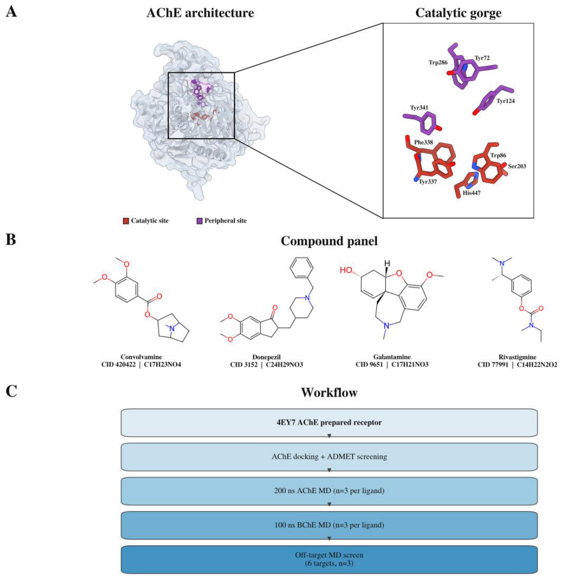
Figure 2.
Starting poses of the four compounds in the AChE gorge. (A) Convolvamine, (B) donepezil, (C) galantamine and (D) rivastigmine, in the same receptor frame and camera, with the catalytic site at the bottom and the peripheral site at the top.
Figure 2.
Starting poses of the four compounds in the AChE gorge. (A) Convolvamine, (B) donepezil, (C) galantamine and (D) rivastigmine, in the same receptor frame and camera, with the catalytic site at the bottom and the peripheral site at the top.
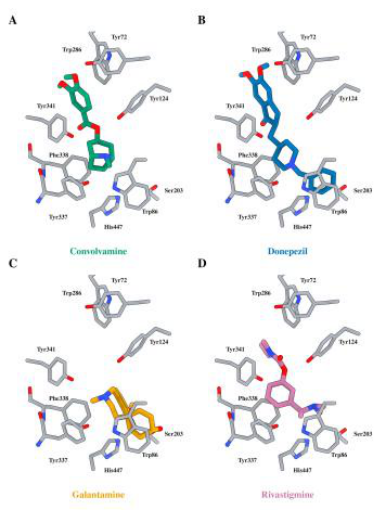
Figure 3.
Persistence of the binding pose over the full 200 ns. (A) Ligand heavy-atom RMSD after superposition of the protein Cα frame on the first frame, averaged within each replicate; points are the three replicates and bars their mean. (B) Contact occupancy at the peripheral anchor Trp286 (red) and the catalytic anchor Trp86 (blue), as the fraction of frames with any ligand heavy atom within 4 Å; points are individual replicates. (C) Per-residue contact occupancy across the peripheral and catalytic residue sets, averaged over three replicates.
Figure 3.
Persistence of the binding pose over the full 200 ns. (A) Ligand heavy-atom RMSD after superposition of the protein Cα frame on the first frame, averaged within each replicate; points are the three replicates and bars their mean. (B) Contact occupancy at the peripheral anchor Trp286 (red) and the catalytic anchor Trp86 (blue), as the fraction of frames with any ligand heavy atom within 4 Å; points are individual replicates. (C) Per-residue contact occupancy across the peripheral and catalytic residue sets, averaged over three replicates.
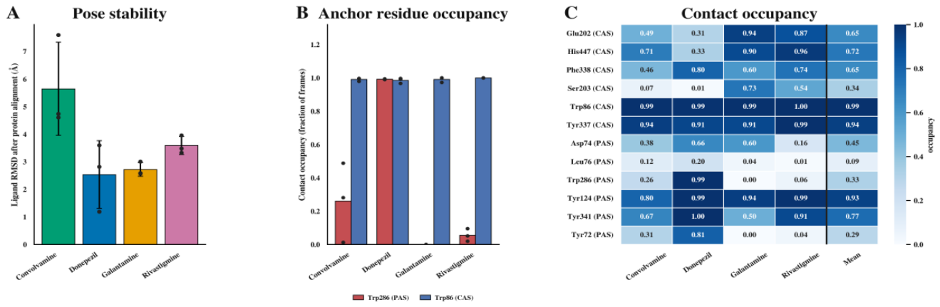
Figure 4.
AChE pocket flexibility, binding energy, and cholinesterase selectivity. (A) Pocket ΔRMSF against three force-field-matched AMBER apo replicates, with 95% confidence intervals; every interval spans zero. (B) AChE MM-PBSA ΔTOTAL, the primary end point, as mean ± SD over three replicates. (C) BChE minus AChE ΔTOTAL, where positive values indicate an AChE-favored direction.
Figure 4.
AChE pocket flexibility, binding energy, and cholinesterase selectivity. (A) Pocket ΔRMSF against three force-field-matched AMBER apo replicates, with 95% confidence intervals; every interval spans zero. (B) AChE MM-PBSA ΔTOTAL, the primary end point, as mean ± SD over three replicates. (C) BChE minus AChE ΔTOTAL, where positive values indicate an AChE-favored direction.
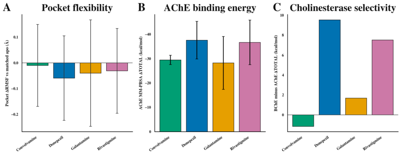
Figure 5.
Receptor and off-target assessment. Heatmap of the AChE-referenced MM-PBSA difference, off-target ΔTOTAL minus AChE ΔTOTAL for BChE and the six off-targets. Positive values indicate less favorable off-target binding than AChE; negative values are outlined in black. The outline marks the sign of the point estimate only, not statistical significance, and every negative cell has a confidence interval spanning zero (Table S11). Every cell is the mean of three independent replicates.
Figure 5.
Receptor and off-target assessment. Heatmap of the AChE-referenced MM-PBSA difference, off-target ΔTOTAL minus AChE ΔTOTAL for BChE and the six off-targets. Positive values indicate less favorable off-target binding than AChE; negative values are outlined in black. The outline marks the sign of the point estimate only, not statistical significance, and every negative cell has a confidence interval spanning zero (Table S11). Every cell is the mean of three independent replicates.
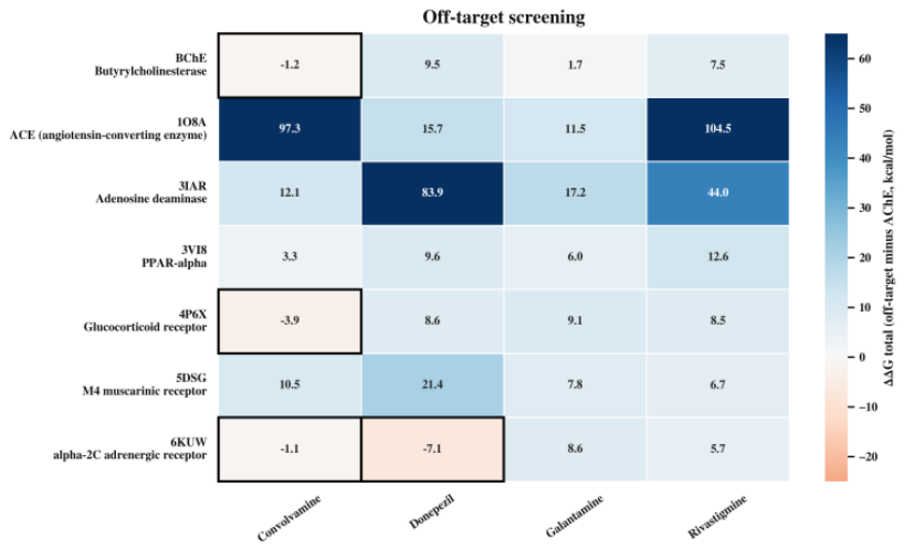
Figure 6.
Predicted ADMET properties. Selected SwissADME and ADMETlab end points for the four compounds, with the predicted value printed in each cell. Cell color codes each end point on a three-level scale (green, amber, orange) applied uniformly across compounds. End points span blood-brain-barrier permeation, gastrointestinal absorption, P-gp substrate status, CYP inhibitor burden, hERG, DILI, AMES, hepatotoxicity, nephrotoxicity, solubility and consensus logP.
Figure 6.
Predicted ADMET properties. Selected SwissADME and ADMETlab end points for the four compounds, with the predicted value printed in each cell. Cell color codes each end point on a three-level scale (green, amber, orange) applied uniformly across compounds. End points span blood-brain-barrier permeation, gastrointestinal absorption, P-gp substrate status, CYP inhibitor burden, hERG, DILI, AMES, hepatotoxicity, nephrotoxicity, solubility and consensus logP.
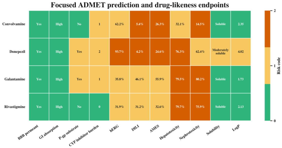

Table 1.
AChE flexibility, binding energy and selectivity. ΔRMSF is given in Å with its 95% confidence interval, and energies in kcal/mol as means ± SD over three replicates.
Table 1.
AChE flexibility, binding energy and selectivity. ΔRMSF is given in Å with its 95% confidence interval, and energies in kcal/mol as means ± SD over three replicates.
| Compound | Pocket ΔRMSF vs matched apo (Å, 95% CI) | AChE PBSA ΔTOTAL (mean ± SD) | BChE minus AChE ΔTOTAL (95% CI) |
| Convolvamine | -0.010 (-0.169 to +0.149) | -29.49 ± 1.90 | -1.15 (-23.84 to +21.54) |
| Donepezil | -0.059 (-0.223 to +0.105) | -37.59 ± 7.72 | +9.54 (-8.87 to +27.95) |
| Galantamine | -0.040 (-0.247 to +0.167) | -28.25 ± 10.87 | +1.69 (-21.71 to +25.09) |
| Rivastigmine | -0.031 (-0.196 to +0.134) | -36.68 ± 9.16 | +7.52 (-11.38 to +26.41) |
Disclaimer/Publisher’s Note: The statements, opinions and data contained in all publications are solely those of the individual author(s) and contributor(s) and not of MDPI and/or the editor(s). MDPI and/or the editor(s) disclaim responsibility for any injury to people or property resulting from any ideas, methods, instructions or products referred to in the content. |
© 2026 by the authors. Licensee MDPI, Basel, Switzerland. This article is an open access article distributed under the terms and conditions of the Creative Commons Attribution (CC BY) license.
Copyright: This open access article is published under a Creative Commons CC BY 4.0 license, which permit the free download, distribution, and reuse, provided that the author and preprint are cited in any reuse.