
Submitted:
11 November 2026
Posted:
12 November 2026
You are already at the latest version
Abstract
Esophageal cancer (EC), comprising esophageal squamous cell carcinoma and esophageal adenocarcinoma, remains a highly aggressive malignancy characterized by substantial intratumoral heterogeneity, cellular plasticity, therapeutic resistance, and poor clinical outcomes. Increasing evidence indicates that EC progression is governed not only by tumor-intrinsic genetic alterations but also by dynamic interactions among signaling networks, metabolic and epigenetic programs, cancer stem-like states, stromal and immune cells, extracellular matrix, vasculature, neural components, and the microbiome. In this review, we synthesize advances in the cellular and molecular biology of EC and discuss their implications for precision therapeutic strategies, with emphasis on oncogenic signaling, pathway rewiring, tumor–microenvironment interactions, cancer stem-cell plasticity, and emerging biomarker platforms, including multi-omics, liquid biopsy, extracellular vesicles, single-cell sequencing, and spatial transcriptomics. We further examine patient-derived organoids, organoid–immune co-culture systems, three-dimensional bioprinted tumor models, and computational approaches as functional platforms for investigating tumor heterogeneity, therapeutic response, and treatment resistance. Beyond tumor control, tissue-engineering and regenerative strategies for repairing treatment-associated mucosal injury, fibrosis, stricture, and tissue loss are discussed. Importantly, regenerative interventions require careful oncologic consideration because pathways involved in tissue repair, stem-cell activation, angiogenesis, and extracellular-matrix remodeling may influence residual malignant cells. Finally, we highlight the convergence of precision oncology, functional tumor modeling, regenerative medicine, and artificial intelligence, while addressing challenges in standardization, validation, scalability, regulation, and clinical translation.
Keywords:
esophageal cancer
; tumor microenvironment
; cellular plasticity
; precision oncology
; patient-derived organoids
; regenerative medicine
Introduction
Esophageal cancer (EC) comprises two major histological subtypes with distinct epidemiological, molecular, and pathological characteristic esophageal squamous cell carcinoma (ESCC) and esophageal adenocarcinoma (EAC) [1]. Although considerable progress has been achieved in endoscopic diagnosis, surgical techniques, chemotherapy, radiotherapy, targeted therapy, and immunotherapy, EC remains one of the most lethal malignancies worldwide because of late diagnosis, aggressive tumor biology, extensive intratumoral heterogeneity, and the rapid development of therapeutic resistance [2]. These challenges underscore the need to move beyond conventional disease models toward a more comprehensive understanding of EC pathogenesis and treatment [3]
Increasing evidence suggests that EC should be regarded as a complex systems disease in which tumor initiation, progression, metastasis, and therapeutic response arise from dynamic interactions among genetic alterations, epigenetic regulation, metabolic reprogramming, immune modulation, stromal remodeling, and environmental influences [4]. Rather than being driven by a single oncogenic event, disease progression results from coordinated changes across multiple cellular and molecular networks. Consequently, therapeutic strategies directed against individual molecular targets frequently provide only transient clinical benefit because compensatory signaling pathways and adaptive cellular responses enable tumor persistence and disease recurrence [5].
Current clinical decision-making relies primarily on histopathological evaluation and TNM staging, which remain indispensable for diagnosis, prognosis, and treatment planning. However, these conventional approaches do not adequately reflect the biological diversity or functional characteristics of individual tumors [6,7] Patients with similar clinicopathological features often exhibit markedly different therapeutic responses and clinical outcomes, emphasizing the importance of integrating molecular, cellular, and functional information into precision oncology [8].
Recent advances in high-throughput molecular profiling, single-cell technologies, spatial biology, patient-derived experimental models, computational biology, and regenerative medicine are reshaping the landscape of EC research [9]. These complementary approaches provide unprecedented opportunities to characterize tumor heterogeneity, identify clinically relevant biomarkers, understand mechanisms of therapeutic resistance, and develop individualized treatment strategies [10]. Collectively, they are accelerating the transition from descriptive pathology toward mechanism-based and patient-centered cancer management.
Recent reviews have separately summarized the molecular pathogenesis and precision treatment of esophageal cancer, the construction and application of esophageal organoids, and tissue-engineering strategies for esophageal repair and reconstruction. However, these fields are often discussed as parallel developments rather than as components of a unified translational pathway. The distinctive contribution of the present review is to connect them within a precision-regenerative oncology framework. Specifically, we examine how subtype-specific tumor evolution, cellular plasticity, and microenvironmental states can inform biomarker selection and functional testing in patient-derived models; how these models may support treatment prioritization; and how treatment-associated mucosal injury, fibrosis, stricture, and tissue loss may subsequently be addressed through indication-specific regenerative strategies. We further position residual-disease assessment and oncologic safety as essential constraints on regenerative intervention. Thus, rather than cataloguing emerging technologies, this review maps a continuum from tumor characterization to functional treatment testing, oncologic control, tissue restoration, and longitudinal surveillance.
In this integrative narrative review, we first examine subtype-specific biological processes that shape esophageal cancer progression and treatment resistance, including clonal evolution, cellular plasticity, signaling-network rewiring, cancer stem-like states, and tumor–microenvironment interactions. We then evaluate molecular and functional platforms (including multi-omics, liquid biopsy, single-cell and spatial profiling, patient-derived organoids, immune co-cultures, bioprinted tumor models, and computational approaches) that may translate biological observations into testable therapeutic hypotheses.
Finally, we examine regenerative strategies for mucosal repair, stricture prevention, and partial- or full-thickness esophageal reconstruction. By connecting these domains, we propose a precision-regenerative framework in which tumor control and functional restoration are planned as interdependent, but temporally and oncologically constrained, components of esophageal cancer management.
Evolutionary Dynamics and Cellular Plasticity
Esophageal tumorigenesis follows Darwinian evolutionary principles, whereby a single transformed cell progressively acquires driver mutations, including TP53, CDKN2A, SMAD4, NOTCH1, and PIK3CA, leading to the expansion of genetically distinct cellular clones [11]. As tumors evolve, additional genetic alterations accumulate within different subpopulations, generating extensive intratumoral heterogeneity (ITH). Phylogenetic analyses have demonstrated a branched evolutionary pattern in both esophageal squamous cell carcinoma (ESCC) and esophageal adenocarcinoma (EAC), in which only specific subclones acquire metastatic competence while others remain relatively indolent [12]. The evolutionary trajectories of the two major EC subtypes also differ. EAC typically develops through the Barrett’s esophagus dysplasia adenocarcinoma sequence, with TP53 mutation representing a critical early event [2]. In contrast, ESCC frequently exhibits field cancerization, in which prolonged exposure to carcinogens generates multiple genetically independent premalignant clones throughout the esophageal epithelium, thereby increasing the likelihood of recurrence even after complete surgical resection [13].
The remarkable evolutionary capacity of EC is further enhanced by cellular plasticity, a hallmark of aggressive disease that enables tumor cells to reversibly transition between differentiated, stem-like, epithelial, and mesenchymal phenotypic states [14,15]. Rather than existing as discrete phenotypes, EC cells occupy a continuum of intermediate epithelial-mesenchymal states that differ in proliferative capacity, invasiveness, metastatic potential, and therapeutic sensitivity [16]. Experimental lineage-tracing studies have shown that ESCC cells can dedifferentiate into fetal-like stem cell states in response to cellular stress, thereby acquiring resistance to chemotherapy [17]. Similarly, EAC cells frequently undergo epithelial-to-mesenchymal transition (EMT), characterized by loss of epithelial polarity and cell–cell adhesion together with enhanced migratory and invasive capabilities.
Therapeutic interventions further accelerate tumor evolution by imposing strong selective pressures on heterogeneous cancer cell populations. Chemotherapy, radiotherapy, and targeted therapies effectively eliminate treatment-sensitive clones, whereas pre-existing resistant subclones survive and subsequently expand [18]. Beyond simple clonal selection, therapy can actively reprogram surviving tumor cells through phenotypic switching, promoting the expression of drug efflux transporters such as ABCB1 and ABCG2, activation of cytoprotective autophagy, and entry into slow-cycling or dormant states [19]. Consistent with these observations, residual tumors following neoadjuvant therapy frequently exhibit enrichment of EMT-associated transcriptional programs together with increased expression of cancer stem cell markers, including CD44 and ALDH1, highlighting therapy-induced plasticity as a major contributor to disease recurrence [20].
In addition to genetic evolution and therapy-induced selection, EC cells continuously adapt to environmental stress through dynamic interactions with their surrounding microenvironment. These adaptive responses include metabolic reprogramming, reciprocal signaling with cancer-associated fibroblasts (CAFs), immune modulation, and extracellular matrix remodeling, all of which promote tumor survival under therapeutic pressure [21]. Importantly, many of these adaptations are reversible and may diminish following drug withdrawal, providing a biological explanation for certain forms of acquired resistance [22]. Consequently, emerging therapeutic strategies increasingly aim to target tumor plasticity itself rather than individual genetic alterations. Pharmacological agents such as histone deacetylase inhibitors are being investigated for their ability to restrict phenotypic switching, stabilize therapy-sensitive cellular states, and enhance the durability of anticancer responses [23].
Signaling Network Rewiring in Esophageal Carcinogenesis
The molecular landscape of EC is shaped by extensive dysregulation of interconnected signaling pathways that collectively regulate cell proliferation, survival, invasion, metastasis, and therapeutic resistance [24]. Among these, aberrant activation of the epidermal growth factor receptor (EGFR) signaling axis is one of the most common molecular alterations, occurring in 40–80% of ESCCs and a subset of EACs [25]. EGFR activation stimulates the PI3K–AKT and RAS–RAF–MAPK cascades, which exhibit substantial functional crosstalk through shared downstream regulators, thereby coordinating cellular proliferation, protein synthesis, metabolism, and survival [26]. Additional signaling pathways further contribute to EC progression. Dysregulation of the Hippo pathway results in nuclear accumulation of the transcriptional co-activators YAP and TAZ, promoting stemness, epithelial plasticity, invasion, and resistance to apoptosis while integrating biomechanical signals from the extracellular matrix [27]. The role of Notch signaling is subtype dependent, functioning predominantly as a tumor suppressor in ESCC through frequent NOTCH1 loss-of-function mutations, whereas persistent pathway activation promotes intestinal metaplasia and tumor progression in EAC. Likewise, aberrant activation of the Wnt/β-catenin pathway enhances cancer stem cell maintenance and self-renewal through induction of genes such as MYC, LGR5, and AXIN2, thereby sustaining tumor initiation and progression [28]. Rather than acting independently, these signaling networks form an integrated regulatory system in which reciprocal pathway activation enables tumor cells to maintain growth and adapt to changing environmental conditions [29].
In parallel with signaling abnormalities, EC undergoes profound metabolic and epigenetic reprogramming that further enhances tumor fitness. Enhanced aerobic glycolysis and glutamine metabolism provide metabolic intermediates required for rapid proliferation, whereas dysregulation of the AMPK–mTOR axis shifts cellular metabolism toward sustained anabolic growth [30]. Epigenetic alterations further reinforce these oncogenic programs through aberrant DNA methylation, histone modifications, and chromatin remodeling. Promoter hypermethylation frequently silences tumor suppressor genes, whereas overexpression of chromatin regulators such as EZH2 promotes transcriptional repression of differentiation-associated genes [31]. Together, these metabolic and epigenetic adaptations cooperate with oncogenic signaling pathways to establish a highly plastic cellular state that facilitates tumor progression, metastatic dissemination, and resistance to therapy [14].
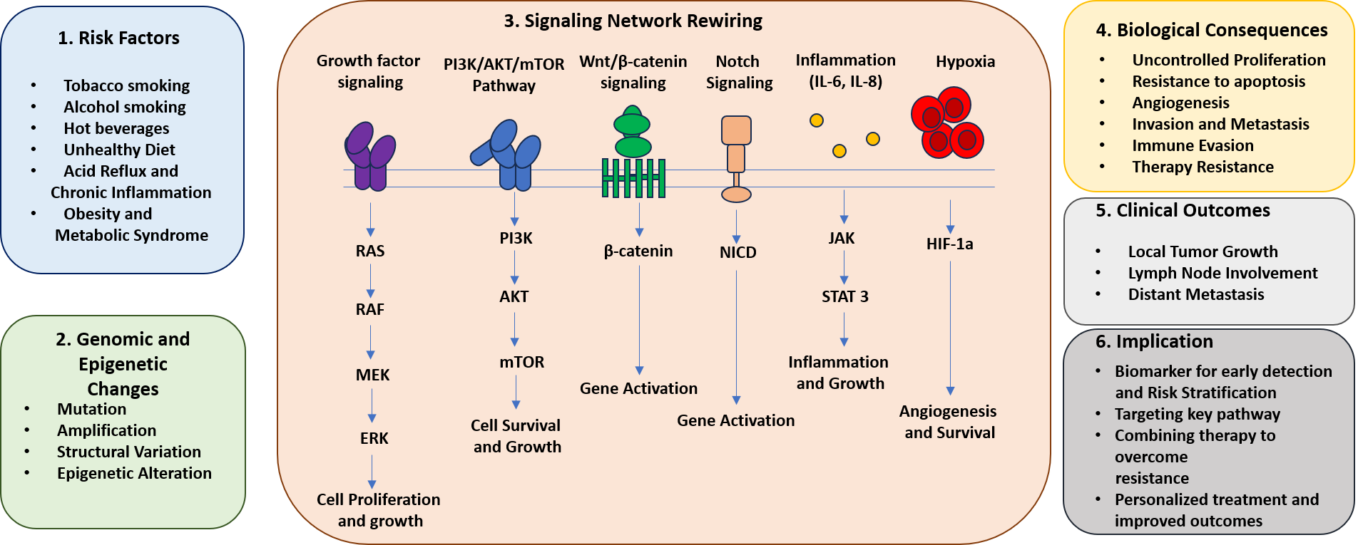
The extensive interconnectivity among these molecular pathways has important therapeutic implications. Clinical experience has demonstrated that inhibition of a single signaling pathway often produces only transient responses because compensatory signaling networks rapidly restore tumor growth [32]. Consequently, current therapeutic strategies increasingly focus on simultaneously targeting multiple oncogenic pathways or disrupting key regulatory hubs. Combination approaches targeting the EGFR/PI3K–AKT–MAPK axis have shown greater antitumor activity than single-agent therapy, although toxicity remains a significant challenge [33]. Novel modalities, including proteolysis-targeting chimeras (PROTACs), are being developed to selectively degrade oncogenic proteins such as EGFR and mutant KRAS while minimizing adaptive resistance. Additional investigational therapies include inhibitors of the YAP–TEAD complex (verteporfin and VT3989), γ-secretase inhibitors targeting aberrant Notch signaling, porcupine inhibitors (WNT974) and Frizzled decoy receptors directed against Wnt signaling, glutaminase inhibitors (CB-839), dual PI3K/mTOR inhibitors such as gedatolisib, and epigenetic agents including histone deacetylase inhibitors (entinostat and vorinostat), the EZH2 inhibitor tazemetostat, and bromodomain inhibitors such as JQ1 and OTX015 [34]. Increasingly, these targeted therapies are being evaluated in rational combination with chemotherapy, radiotherapy, and immunotherapy to overcome compensatory pathway activation and improve treatment durability. Collectively, these advances highlight a shift from targeting isolated molecular alterations toward integrated network-based therapeutic strategies for precision management of esophageal cancer [35] as shown in Figure 1.
The Tumor Ecosystem: Beyond the Cancer Cell
Cancer-Associated Fibroblast Heterogeneity
Cancer-associated fibroblasts (CAFs) represent the predominant stromal cell population within the esophageal tumor microenvironment and exhibit considerable functional heterogeneity [36]. Distinct CAF subtypes include inflammatory CAFs (iCAFs), which secrete cytokines such as IL-6 and CXCL12; myofibroblastic CAFs (myCAFs), characterized by α-smooth muscle actin (α-SMA) expression and extracellular matrix (ECM) remodeling; and antigen-presenting CAFs (apCAFs), which express major histocompatibility complex (MHC) class II molecules [37]. Through the secretion of growth factors including hepatocyte growth factor (HGF), fibroblast growth factor (FGF), and transforming growth factor-β (TGF-β), CAFs promote tumor proliferation, angiogenesis, immune modulation, matrix remodeling, and the establishment of a chemoprotective niche that supports disease progression [38].
Immune-Suppressive Niches and T-Cell Exhaustion
The EC microenvironment is profoundly immunosuppressive, facilitating immune evasion and limiting antitumor responses. This environment is characterized by the accumulation of regulatory T cells (Tregs), myeloid-derived suppressor cells (MDSCs), and alternatively activated M2-polarized tumor-associated macrophages (TAMs), together with increased expression of inhibitory immune checkpoint molecules, including PD-L1, CTLA-4, LAG-3, and TIM-3 [39]. Persistent antigen exposure drives progressive T-cell exhaustion, resulting in impaired cytokine production, reduced cytotoxic function, and diminished immune surveillance, thereby contributing to tumor persistence and resistance to immunotherapy [40].
Endothelial Remodeling and Neovascular Architecture
Tumor-associated angiogenesis in EC generates structurally abnormal vascular networks that are tortuous, poorly organized, and highly permeable [41]. Overexpression of vascular endothelial growth factor-A (VEGF-A) promotes rapid neovascularization; however, the resulting vessels exhibit impaired perfusion, increased vascular leakage, elevated interstitial pressure, and regional hypoxia [42]. These abnormalities restrict drug delivery, promote metabolic adaptation, and create conditions that facilitate tumor invasion and metastatic dissemination.
Neural Components in Tumor Progression
Perineural invasion (PNI) is recognized as an independent predictor of poor prognosis in EC and reflects dynamic bidirectional communication between tumor cells and the nervous system [43,44]. Cancer cells secrete neurotrophic factors, including nerve growth factor (NGF), brain-derived neurotrophic factor (BDNF), and glial cell line-derived neurotrophic factor (GDNF), which stimulate neural infiltration into the tumor microenvironment [45]. In turn, infiltrating nerves release neurotransmitters such as norepinephrine and acetylcholine that enhance tumor cell proliferation, migration, invasion, and survival, highlighting the emerging importance of neuro–tumor interactions in EC progression [46].
Extracellular Matrix Dynamics and Mechanotransduction
The extracellular matrix undergoes extensive remodeling during EC progression, resulting in increased tissue stiffness primarily through collagen crosslinking mediated by lysyl oxidase (LOX) family enzymes [47]. Enhanced matrix rigidity activates mechanotransduction pathways involving YAP/TAZ, β1 integrins, and Rho–ROCK signaling, thereby promoting epithelial-to-mesenchymal transition, cellular proliferation, invasion, and therapeutic resistance [48]. Matrix metalloproteinases (MMPs) further regulate ECM homeostasis, and their dysregulated activity contributes to both matrix degradation and pathological remodeling, emphasizing the complex role of the extracellular matrix in tumor progression [49,50].
Microbiome-Tumor Interactions
Emerging evidence indicates that alterations in the esophageal microbiome contribute to carcinogenesis and disease progression. In EAC, chronic gastroesophageal reflux is associated with a shift from Gram-positive to Gram-negative bacterial communities, including Campylobacter and Fusobacterium, which promote chronic inflammation and genotoxic stress [51]. In ESCC, enrichment of oral pathogens such as Porphyromonas gingivalis and Fusobacterium nucleatum has been associated with advanced disease and poor clinical outcomes [52]. Microbial metabolites, including short-chain fatty acids and secondary bile acids, further influence epithelial barrier integrity, inflammatory signaling, and host immune responses, illustrating the complex interactions between microbial communities and the tumor microenvironment [53].
Collectively, these diverse stromal, immune, vascular, neural, extracellular matrix, and microbial components form an integrated tumor ecosystem that dynamically interacts with malignant epithelial cells throughout disease progression [54] also shown in Figure 2. Increasing evidence suggests that durable therapeutic responses will require simultaneous modulation of both tumor-intrinsic signaling pathways and the surrounding microenvironment [55]. Consequently, combination strategies targeting multiple components of the tumor ecosystem including CAFs, immune checkpoints, tumor vasculature, mechanotransduction pathways, neural signaling, and microbiome-associated mechanisms are emerging as promising directions for improving the efficacy of precision therapies in esophageal cancer [56].
Cancer Stem Cell Niches and Cellular Regeneration
Cancer stem cells (CSCs) are a subpopulation with self-renewal and tumor-initiating capacity. In EC, CSCs have been identified using surface markers: CD44, CD133, ALDH1, and LGR5. CD44+ cells are enriched in post-chemotherapy residual tumors and show high tumorigenicity in limiting dilution assays [57]. LGR5, a Wnt target gene, marks intestinal stem cells and is expressed in EAC CSCs. Notably, CSC marker expression is plastic and context-dependent, complicating therapeutic targeting. Transcriptomic analyses have defined a stemness signature in EC, including upregulation of pluripotency factors (SOX2, OCT4, NANOG), EMT regulators (ZEB1, SNAI1), and developmental pathways (Wnt, Notch, Hedgehog) [58]. SOX2 amplification is common in ESCC and drives stemness and chemoresistance. Targeting core stemness transcription factors is challenging, but downstream effectors (e.g., BRD4, CDK7) are druggable [59]. CSCs can enter a reversible quiescent (dormant) state, characterized by low proliferation, low metabolic activity, and resistance to cell cycle-dependent therapies. Dormant CSCs persist after treatment and can re-emerge years later as recurrent disease [60]. Mechanisms include p38 MAPK activation, autophagy upregulation, and niche-mediated signals. Eliminating dormant CSCs requires a “wake-and-kill” strategy consisting in the induction of cell cycle re-entry (using G-CSF or IL-15) followed by conventional chemotherapy. CSCs reside in specialized niches composed of CAFs, endothelial cells, and ECM [61]. CAFs secrete IL-6, CXCL12, and HGF, activating JAK/STAT and AKT pathways in CSCs. Hypoxic niches stabilize HIF1α/2α, which upregulate CSC markers. Endothelial cells produce Notch ligands that maintain stemness [62]. Targeting niche components (e.g., CXCR4 antagonists, HIF inhibitors) may disrupt CSC maintenance. Many pathways governing normal tissue regeneration are co-opted by CSCs [63]. For instance, the Hippo-YAP axis controls liver and intestinal regeneration; in EC, it drives stemness. WNT-β-catenin mediates colonic crypt regeneration; in EAC, it is constitutively active. Understanding these hijacked regenerative programs offers opportunities for therapeutic intervention that might preferentially target CSCs while sparing normal stem cells if contextual differences exist [64,65].
Biomarker Landscapes for Precision Stratification
Multi-Omics Biomarker Discovery
The TP53 mutation (present in >80% ESCC, >70% EAC) is prognostic but not yet therapeutically actionable [66]. ERBB2 (HER2) amplification (15–30% of EAC) guides trastuzumab use. PIK3CA mutations predict PI3K inhibitor sensitivity. MSI-H (2–5%) predicts pembrolizumab response. Epigenomic biomarkers: DNA methylation panels (e.g., CDKN2A, MGMT) can detect EC from cell-free DNA. Proteomic biomarkers: serum SCC antigen is used clinically for ESCC but has low sensitivity [67].
Circulating Tumor DNA and Liquid Biopsy Platforms
Circulating tumor DNA (ctDNA) offers real-time, minimally invasive monitoring [68]. In EC, ctDNA detection has 80–90% sensitivity in advanced disease. Applications include: early detection (methylation-based assays), molecular residual disease (MRD) detection post-resection, monitoring clonal evolution, and identifying resistance mutations (e.g., ERBB2 loss upon trastuzumab) [69]. Serial ctDNA levels correlate with treatment response and precede radiographic progression by weeks to months [70,71].
Extracellular Vesicle-Based Biomarkers
The emergence of advanced molecular profiling technologies has significantly expanded the biomarker landscape in EC, enabling more precise characterization of tumor heterogeneity and therapeutic vulnerabilities [72]. Among these approaches, extracellular vesicles (EVs), particularly exosomes, have attracted considerable attention as minimally invasive biomarker platforms. Tumor-derived EVs carry a diverse cargo of proteins, messenger RNAs, microRNAs, and DNA fragments that reflect the molecular status of their cells of origin [73]. In EC, EV-associated microRNAs such as miR-21, miR-1246, and miR-223 have been linked to disease stage, progression, and clinical outcomes, while EV surface proteins including EGFR, PD-L1, and CD44 mirror tumor aggressiveness and immune status [74]. Compared with circulating tumor DNA (ctDNA), EV-based biomarkers offer several advantages, including greater abundance, protection from enzymatic degradation, and the ability to simultaneously provide genomic, transcriptomic, and proteomic information [75]. Nevertheless, challenges related to isolation methods, analytical reproducibility, and standardization continue to hinder their routine clinical application [76].
The advent of single-cell RNA sequencing (scRNA-seq) has further revolutionized biomarker discovery by uncovering rare cellular subpopulations that remain undetectable in conventional bulk analyses [77]. This technology has revealed the presence of therapy-resistant persister cells and distinct transcriptional states that contribute to disease progression and treatment failure. Several clinically relevant single-cell signatures have been identified in EC, including a stress-response program characterized by HSPA1A, DNAJB1, and JUN expression in chemoresistant cells [78]. Two important patterns were observed. First, some tumors showed features similar to early fetal cells, with high levels of the proteins SOX2, KLF5, and YAP1. These tumors tend to grow and spread more aggressively. Second, some tumors produced high levels of immune-related proteins (PD-L1, CD47, and HLA-E), which help them avoid being recognized and attacked by the body’s immune system, even when immune cells are present around the tumor. These findings highlight the complexity of intratumoral heterogeneity and provide new opportunities for precision therapeutic targeting [79].
Complementing single-cell analyses, spatial transcriptomics has emerged as a powerful approach for mapping gene expression within its native tissue context. Unlike dissociative sequencing techniques, spatial profiling preserves tissue architecture, enabling the identification of biologically distinct microenvironmental niches [80].
In EC, several spatially resolved transcriptional programs have been described, including a tumor stroma interface signature enriched for transforming growth factor-β (TGF-β), matrix metalloproteinases (MMPs), and cancer-associated fibroblast markers at invasive fronts [81]. A hypoxic core signature characterized by HIF1A, CA9, and VEGFA expression; and an immune-excluded phenotype in which chemokines are retained within the stromal compartment, preventing effective T-cell infiltration into tumor nests. Such spatial biomarkers may serve as valuable predictors of response to immunotherapy and facilitate classification of tumors into inflamed, immune-excluded, or immune-desert phenotypes [82].
Beyond established biomarkers such as HER2 amplification and microsatellite instability (MSI), several emerging predictive biomarkers are shaping the future of precision oncology in EC. High tumor mutational burden (TMB >10 mutations/Mb) has been associated with enhanced responsiveness to immune checkpoint blockade, while loss of ARID1A, observed in a subset of ESCC cases, may confer sensitivity to EZH2-targeted therapies (Mokhtari et al., 2025). Similarly, FBXW7 mutations have been linked to increased susceptibility to WEE1 inhibition, providing additional opportunities for biomarker-driven treatment selection. Programmed death-ligand 1 (PD-L1) expression, measured using the combined positive score (CPS), remains an important clinical biomarker, with pembrolizumab demonstrating efficacy in ESCC patients exhibiting CPS values of 10 or greater [84]. Despite these promising developments, robust validation of emerging biomarkers through large-scale prospective clinical trials remains essential before their widespread implementation in routine clinical practice as shown in Table 1 [85].
Targeted Therapeutic Innovations
Receptor-Directed Therapeutic Strategies
EGFR-directed therapies, including cetuximab and panitumumab, have demonstrated limited efficacy in esophageal cancer. The addition of cetuximab to chemotherapy did not improve outcomes in the REAL-3 trial in esophagogastric adenocarcinoma, while the SCOPE1 trial showed that adding cetuximab to definitive chemoradiotherapy in ESCC increased toxicity and was associated with inferior survival. Consequently, EGFR-targeted therapy is not recommended as standard treatment for unselected patients with esophageal cancer [92]. HER2-directed therapies: trastuzumab with chemotherapy is standard for HER2+ EAC (ToGA trial). Trastuzumab deruxtecan (T-DXd), a HER2-directed antibody–drug conjugate (ADC), has expanded treatment options for previously treated HER2-positive gastric and gastroesophageal-junction adenocarcinoma, whereas its role in primary esophageal adenocarcinoma and HER2-low disease remains under investigation [93]. Other receptors under investigation: FGFR2 (amplified in 5–10% of ESCC; futibatinib, pemigatinib), MET (dysregulated in 15% of EAC; capmatinib, crizotinib). Synthetic lethality exploits genetic vulnerabilities [94]. PARP inhibitors (olaparib, rucaparib) target homologous recombination deficiency (HRD). While EC has low frequency of BRCA mutations, HRD occurs via ATM loss, ARID1A mutations, or promoter methylation. Early trials show modest activity [95]. WEE1 inhibitors (adavosertib) cause synthetic lethality in TP53-mutant tumors (most ECs) by forcing premature mitotic entry. Phase II trials in EC show promising activity but significant toxicity (myelosuppression, gastrointestinal). ATR inhibitors (ceralasertinib) are under investigation [96].
Epigenetic Targeting
HDAC inhibitors (vorinostat, entinostat) induce differentiation, apoptosis, and increase chemo/radiation sensitivity [97]. In EC, entinostat plus chemotherapy showed disease control in phase I/II. EZH2 inhibitors (tazemetostat) are effective in preclinical models of ARID1A-mutant EC. BET inhibitors (OTX015, ZEN-3694) suppress MYC and inflammatory signaling; clinical trials in EC are ongoing. DNA methyltransferase inhibitors (azacitidine, decitabine) have been disappointing as monotherapy but may re-sensitize to chemotherapy [98]. ADCs combine tumor-targeting antibodies with potent cytotoxic payloads. T-DXd (HER2) has transformed HER2-low EC treatment. Enfortumab vedotin (nectin-4) is under evaluation. Sacituzumab govitecan (Trop-2) shows activity in multiple solid tumors; Trop-2 is expressed in 80% of EC. Novel therapeutic targets include CLDN18.2, which is expressed in a subset of esophageal adenocarcinomas (reported in approximately 30–40% of cases).
Zolbetuximab, a CLDN18.2-directed cytolytic monoclonal antibody, is approved in combination with chemotherapy for patients with CLDN18.2-positive, HER2-negative gastric or gastroesophageal junction (GEJ) adenocarcinoma, and its potential role in esophageal adenocarcinoma is under investigation. Tissue factor is another promising target, particularly in ESCC, with agents such as tisotumab vedotin being evaluated in clinical studies. RNA-based therapeutics under investigation include antisense oligonucleotides (ASOs) targeting STAT3 and KRAS, small interfering RNAs (siRNAs) delivered via lipid nanoparticles (LNPs), and microRNA mimics or inhibitors (e.g., miR-34 mimics and anti-miR-21). Despite challenges related to delivery, stability, and off-target effects, the clinical approval of RNA therapeutics such as patisiran (siRNA) and nusinersen (ASO) provides a framework for the future development of these approaches in esophageal cancer [99]. In EC preclinical models, LNP-encapsulated siRNAs targeting MYC or CD44 reduce tumor growth. Nanocarriers (liposomes, polymeric nanoparticles, inorganic nanoparticles) improve drug pharmacokinetics, tumor targeting, and reduce toxicity. Passive targeting exploits enhanced permeability and retention (EPR) effect. Active targeting uses surface ligands (folate, aptamers, antibodies) [100]. Theranostic nanoparticles combine diagnostic imaging and targeted therapy. In esophageal cancer, nanoparticle-based drug delivery systems, including albumin-bound paclitaxel (nab-paclitaxel, Abraxane®), have been evaluated in clinical studies to improve drug delivery and therapeutic efficacy; however, Abraxane is not FDA-approved for the treatment of esophageal cancer. Next-generation nanoparticle platforms incorporate pH-responsive or reactive oxygen species (ROS)-responsive drug release and enable the co-delivery of multiple therapeutic agents to overcome treatment resistance [101].
Established Immunotherapy in Clinical Practice
Immune-checkpoint inhibition is now an established component of esophageal cancer treatment, although regimen selection depends on histological subtype, disease setting, PD-L1 expression, previous treatment, and regional regulatory criteria. In patients with completely resected esophageal or gastroesophageal-junction cancer who have residual pathologic disease after neoadjuvant chemoradiotherapy, adjuvant nivolumab administered for up to one year significantly prolonged median disease-free survival compared with placebo in the CheckMate 577 trial 22.4 versus 11.0 months [102,103]. In previously untreated, locally advanced unresectable or metastatic esophageal cancer, pembrolizumab combined with cisplatin and fluorouracil improved overall and progression-free survival compared with chemotherapy alone in KEYNOTE-590, with a greater magnitude of benefit observed in esophageal squamous cell carcinoma and tumors with a PD-L1 combined positive score (CPS) of 10 or greater [104]. For advanced esophageal squamous cell carcinoma, CheckMate 648 demonstrated that nivolumab plus fluoropyrimidine–platinum chemotherapy and, in selected patients, the chemotherapy-free combination of nivolumab plus ipilimumab improved overall survival compared with chemotherapy, with the greatest benefit observed in tumors with tumor-cell PD-L1 expression of at least 1% [105] . Nivolumab monotherapy also improved overall survival relative to paclitaxel or docetaxel in previously treated advanced esophageal squamous cell carcinoma in ATTRACTION-3, with benefit observed irrespective of tumor PD-L1 expression [104]. In advanced non-HER2-positive gastric, gastroesophageal-junction, and esophageal adenocarcinoma, nivolumab plus fluoropyrimidine–platinum chemotherapy improved survival in CheckMate 649, with the clearest benefit among patients with PD-L1 CPS of 5 or greater [106]Collectively, these phase III trials establish PD-1-based therapy in adjuvant and advanced-disease settings while emphasizing the importance of histology-specific, biomarker-informed patient selection and careful monitoring for immune-related adverse events.
Patient-Derived Organoids as Functional Tumor Avatars
Establishment of Esophageal Tumor Organoids
Patient-derived organoids (PDOs) are three-dimensional cultures derived from tumor biopsies or resected tissue, grown in basement membrane extract with defined growth factors (WNT, R-spondin, Noggin, EGF, FGF10).
Establishment success rates for EC range 60–80% depending on tumor cellularity. PDOs retain genetic and phenotypic features of parental tumors, including mutations, copy number alterations, and histology. Biobanks of EC PDOs (e.g., 100+ lines) have been established, representing both ESCC and EAC subtypes [107].
Modeling Tumor Heterogeneity
PDOs capture intratumoral heterogeneity by deriving multiple organoid clones from different regions of the same tumor. Clonal PDOs show differential drug responses, recapitulating the subclonal architecture. Co-culture of multiple PDO clones recapitulates competitive dynamics observed in patients. Additionally, PDOs can model the transition from primary to metastatic lesions [108].
Drug Screening Applications
PDO-based drug screening identifies active agents and predicts clinical responses. In a landmark study, EC PDOs were screened against >100 FDA-approved drugs. Results showed high sensitivity to conventional agents (5-FU, cisplatin, paclitaxel) but also uncovered non-standard sensitivities (e.g., to PARP inhibitors in HRD-mutant tumors) (Smabers et al., 2025)(Smabers et al., 2025)(Smabers et al., 2025). Importantly, ex vivo PDO drug responses correlated with patient clinical outcomes (positive predictive value ~80%, negative predictive value ~90%). PDO screens can also identify drug combinations that overcome resistance [110,111].
Organoid–Immune Cell Co-Culture Systems
To model immunotherapy responses, PDOs are co-cultured with autologous immune cells (T cells, NK cells, macrophages). This requires establishing patient-matched tumor organoids and peripheral blood immune cells [111]. Co-culture assays measure tumor cell killing, cytokine production, and immune cell activation. Challenges include: maintaining immune cell viability, replicating tumor-immune synapse, and recapitulating T-cell exhaustion over time. Microfluidic organoid-immune platforms are under development [111].
Precision Treatment Prediction Using Organoids
The ultimate goal is “personalized organoid-guided therapy”: generate PDOs from a patient’s biopsy (2–4 weeks), screen a panel of relevant drugs, and select the most active regimen. Early proof-of-concept studies in EC show that PDO-guided therapy improves outcomes compared to physician’s choice [112]. Barriers include: turnaround time, cost, scalability, and lack of standardized drug panels. However, efforts to automate organoid culture and drug screening (robotics, AI-based image analysis) are accelerating clinical translation [113].
Three-Dimensional Bioprinting for Disease Modeling
Three-dimensional bioprinting uses layer-by-layer deposition of bioinks hydrogels containing living cells and biocompatible materials. For esophageal reconstruction, ideal bioinks mimic native esophageal mechanical properties (strain, stiffness, viscoelasticity) and support epithelial and fibroblast growth [114]. Common bioinks: collagen, gelatin methacryloyl (GelMA), alginate, decellularized esophageal ECM (dECM). dECM bioinks retain native biochemical cues (growth factors, ECM proteins) and show superior cell behavior compared to synthetic alternatives [115]. Bioprinting allows spatial patterning of cancer cells, CAFs, endothelial cells, and immune cells to recapitulate the tumor microenvironment (TME). For EC, “tumor-stroma interface” models print concentric rings: tumor core (cancer cells + hypoxia-mimetic), surrounding stroma (CAFs + ECM), and outer vascular layer (endothelial cells) [116]. These constructs enable study of invasion dynamics, CAF-mediated drug resistance, and drug penetration gradients. Vascularization remains a major challenge. Strategies include: co-printing endothelial cells and pericytes in a sacrificial bioink (e.g., Pluronic F127) that is later removed to create perfusable channels; using angiogenic growth factors (VEGF, Ang-1) to induce self-assembly of capillaries post-printing; or pre-vascularizing constructs with microvascular fragments [117].
Vascularized EC bioprinted models show more realistic drug responses (reduced sensitivity due to diffusion barriers) than avascular models. Personalized bioprinting uses a patient’s own cells: cancer cells from biopsy, CAFs and immune cells from adjacent tissue or blood, and endothelial cells from peripheral blood (reprogrammed to induced endothelial cells). Bioprinted personalized models are then used for drug screening. Current limitations: need for large cell numbers (millions of cells), slow printing times, and reproducibility. However, recent advances in high-throughput bioprinting (arrayed 96-well formats) are overcoming these barriers [118]. Organoids and bioprinting are complementary. Organoids provide pre-assembled, functionally relevant units of cancer cells with stem-progenitor hierarchies. Bioprinting spatially organizes organoids within a stromal matrix. “Organoid-on-a-chip” platforms combine bioprinted organoid arrays with microfluidic perfusion, enabling dynamic drug testing and real-time imaging. For EC, bioprinted organoid chips have been used to test sequential drug combinations and to model metastatic dissemination [119].
Regenerative Medicine Approaches Following Esophageal Injury
Tissue-Engineered Esophageal Substitutes
Following esophagectomy (surgical removal of the esophagus) or extensive chemoradiation injury, reconstruction is needed. Current options (gastric pull-up, colon interposition, jejunal grafts) have significant morbidity. Tissue-engineered esophageal substitutes aim to recapitulate native structure: a stratified squamous epithelium, a submucosa with vasculature, and a muscularis propria [120]. Scaffold-based approaches use synthetic (PCL, PLGA) or natural (collagen, decellularized ECM) scaffolds seeded with epithelial cells (autologous oral mucosal epithelial cells) and smooth muscle cells [121].
Biomaterial Scaffolds for Structural Restoration
Scaffolds provide temporary mechanical support while guiding endogenous regeneration. Ideal scaffolds: biocompatible, biodegradable, porous (for cell infiltration), and mechanically matched to native esophagus. Electrospun nanofiber scaffolds (PCL/collagen blends) mimic ECM architecture. Hydrogels (GelMA, alginate) can be injected endoscopically for focal defects. Decellularized esophageal scaffolds from donor or porcine tissue preserve native basement membrane and ECM cues, showing promise in large animal models [122].
Stem Cell-Assisted Tissue Repair
Stem cells enhance regeneration. Mesenchymal stem cells (MSCs) from bone marrow, adipose tissue, or umbilical cord secrete paracrine factors (VEGF, HGF, IL-10) that reduce inflammation, promote angiogenesis, and inhibit fibrosis [123]. In animal models of esophageal injury, locally delivered MSCs improve healing and reduce stricture formation. Epithelial stem cells (e.g., from esophageal biopsies or induced pluripotent stem cells, iPSCs) can regenerate the epithelial lining. iPSC-derived esophageal organoids have been transplanted into injured rat esophagus, forming functional epithelium [124].
Growth Factor-Guided Regeneration
Growth factors stimulate endogenous repair. EGF and FGF promote epithelial proliferation and migration. VEGF induces angiogenesis. TGFβ inhibitors (e.g., fresolimumab) reduce fibrosis and stricture formation [125]. Controlled release from scaffolds (e.g., heparin-binding nanoparticles) prolongs growth factor activity. Combination strategies (EGF + VEGF + FGF) are more effective than single factors [126].
Bioengineered Esophageal Grafts
Several bioengineered grafts have entered preclinical and early clinical testing. One approach: decellularized porcine esophagus recellularized with patient-derived oral epithelial cells and smooth muscle cells [127]. Implanted in a canine model, the graft developed stratified epithelium, vascularized submucosa, and oriented muscle bundles, allowing oral feeding.
First-in-human studies have reported feasibility, but long-term outcomes (stricture formation, functionality) remain suboptimal. Challenges include: achieving innervation (peristalsis), preventing anastomotic leaks, and avoiding chronic inflammation as shown in Table 2.
Oncologic Safety and Residual-Disease Considerations
Regenerative interventions following esophageal cancer treatment require dedicated oncologic safety assessment because several biological programs that promote wound healing (including epithelial proliferation, stem-cell activation, angiogenesis, immune modulation, and extracellular-matrix remodeling) may also support the survival or expansion of residual malignant cells. The effects of mesenchymal stromal cell (MSC)-based products appear to be highly context dependent: co-injection of human umbilical-cord-derived MSCs with esophageal carcinoma cells promoted tumor growth and lymph-node metastasis in mice [133] whereas selected MSC-derived exosomes carrying miR-375 inhibited esophageal squamous cell carcinoma progression in experimental models [133]. These contrasting findings indicate that whole cells, secretomes, extracellular vesicles, and engineered MSC derivatives should be evaluated as distinct therapeutic products rather than regarded as biologically interchangeable. MSC-derived Gremlin-1 has also been shown to promote epithelial–mesenchymal transition in esophageal squamous cell carcinoma [134], while fibroblast growth factor-2–FGFR–ERK signaling supports the maintenance of esophageal cancer stem-like cells [134], raising concern that uncontrolled or prolonged delivery of proliferative and pro-angiogenic factors could theoretically support residual disease. Pluripotent-stem-cell-derived products additionally require sensitive testing for residual undifferentiated or transformed cells and product-specific in vivo tumorigenicity assessment [135]. Preliminary long-term observations after autologous oral mucosal epithelial cell-sheet transplantation are reassuring, but the available evidence derives from small, indication-specific cohorts and cannot be extrapolated to MSC-based products, growth-factor-eluting scaffolds, iPSC-derived tissues, or full-thickness bioengineered grafts [136]. Future studies should therefore prespecify local recurrence, dysplasia, clonal evolution, ectopic tissue formation, cellular biodistribution, and late graft-associated neoplasia as long-term safety outcomes.
Digital Oncology and Artificial Intelligence Integration
Artificial intelligence (AI) is being investigated across the EC care continuum, although the most mature evidence currently concerns computer-assisted analysis of endoscopic images. In a single-center retrospective validation study, a convolutional neural network trained on endoscopic images demonstrated high diagnostic performance for detecting esophageal cancer, including ESCC and EAC [137]. For Barrett’s-associated neoplasia, a multicenter deep-learning computer-aided detection system demonstrated strong image-based performance and outperformed benchmark endoscopists, supporting its potential use in identifying early EAC precursor lesions [138]. Prospective evidence in ESCC is encouraging but remains inconsistent. One randomized controlled study reported that deep-learning assistance improved the detection of esophageal cancer and high-risk precursor lesions during endoscopy [139]. In contrast, a multicenter tandem, double-blind randomized trial found that AI assistance increased lesion detection but did not significantly reduce the prespecified per-lesion or per-patient miss-rate endpoints [140]. Similarly, a separate randomized clinical trial did not demonstrate a significant improvement in ESCC detection with AI assistance compared with conventional endoscopy [141]. These findings indicate that strong diagnostic performance in retrospective image datasets does not necessarily translate into consistent clinical benefit and that implementation should account for lesion prevalence, histological subtype, endoscopist experience, imaging modality, equipment characteristics, and external validation.
Beyond endoscopic detection, computational pathology and radiomics are being evaluated for prognostic stratification and prediction of treatment response. Deep-learning classification of the desmoplastic reaction on routine hematoxylin-and-eosin-stained tissue sections provided objective prognostic information in patients with ESCC [142]. Imaging-based models have also been developed to predict pathological response to neoadjuvant treatment. A two-center study combining computed-tomography deep-learning radiomics with hematological biomarkers evaluated the prediction of pathological complete response after neoadjuvant chemoradiotherapy in ESCC [138]. In addition, a multicenter magnetic-resonance radiomics model demonstrated promising performance for predicting pathological complete response after neoadjuvant chemoradiotherapy, although the study was retrospective and included only patients with ESCC [143]. Despite these encouraging findings, most computational pathology and radiomics models have been developed using relatively small or institution-specific datasets and should therefore be considered investigational until their reproducibility, calibration, generalizability, and clinical utility have been established through independent prospective validation.
Within the precision-regenerative framework proposed in this review, AI may eventually provide an integrative layer connecting endoscopic and radiological imaging, digital pathology, molecular and multi-omics profiles, circulating tumor DNA, and quantitative results from patient-derived organoids and bioprinted models. Automated image analysis could support more standardized measurement of organoid morphology, viability, and treatment response and, in regenerative studies, facilitate the evaluation of epithelial coverage, vascularization, tissue organization, scaffold integration, and structural integrity. Nevertheless, the term digital twin should be used cautiously. A retrospective virtual-twin analysis using random-forest methods has been applied to estimate patient-level heterogeneity in the potential benefit of adjuvant therapy after neoadjuvant treatment for esophageal cancer [144]. However, this approach is not equivalent to a continuously updated, patient-specific digital model that prospectively simulates tumor evolution and guides oncologic or regenerative interventions in real time. Clinical translation of AI in esophageal cancer will require multicenter external validation, prospective evaluation of clinical benefit, assessment of calibration and algorithmic bias, transparent reporting, robust data-governance procedures, and continued clinician oversight.
Convergence of Precision Oncology and Regenerative Medicine
Personalized Therapeutic Ecosystems
The most advanced vision combines precision oncology (targeting molecular drivers) with regenerative medicine (repairing therapy-induced damage). For example: a patient with ESCC receives a personalized drug combination identified through PDO screening, which effectively kills cancer cells but injures normal esophageal mucosa [145]. Concurrently, an injectable hydrogel with growth factors and MSCs is delivered endoscopically to regenerate the mucosa and prevent stricture formation. This “personalized therapeutic ecosystem” addresses both tumor control and quality of life.
Bioengineered Platforms for Drug Development
Pharmaceutical companies are adopting EC bioengineered platforms for drug discovery. High-throughput screening using EC PDO arrays (384-well format) identifies novel compounds. Bioprinted EC tumor-stroma constructs are used for lead optimization and toxicity assessment [146]. Patient-derived xenograft (PDX)-on-a-chip platforms combine humanized immune systems with perfusable tumor constructs, enabling preclinical evaluation of immunotherapies. These platforms reduce reliance on animal models and improve clinical translatability.
Future Clinical Translation Roadmap
A realistic 10-year roadmap: Years 1-3: Standardize PDO generation and drug screening protocols; validate correlative algorithms (PDO response clinical outcome) in multi-center trials. Years 4-6: Implement PDO-guided therapy in select academic centers; obtain regulatory approval for specific bioengineered scaffolds for esophageal reconstruction [147].
Years 7-10: Integrate digital twins for treatment planning; launch registries tracking long-term outcomes of integrated precision-regenerative approaches; commercialize leading platforms [148].
Challenges, Knowledge Gaps, and Future Perspectives
Significant variability exists in PDO culture conditions (growth factor cocktails, matrix composition, passage number), bioprinting protocols (bioink lot-to-lot variability), and drug screening readouts (ATP-based, imaging, viability dyes) [149]. This lack of standardization hampers cross-study comparisons and clinical adoption. International consortia (e.g., OCCAMS for EC) are developing consensus guidelines, but implementation is slow. Regulatory frameworks have not kept pace with technological advances. Are PDOs considered “human subjects research” or “laboratory test”? Do bioprinted tumor models require animal rule approval? How is a digital twin validated as a medical device? The FDA’s Center for Devices and Radiological Health (CDRH) has issued draft guidance on organoids and organs-on-chips, but final regulations are pending [150]. For regenerative products (scaffolds, cell therapies), the regulatory pathway is clearer but costly (premarket approval, biologics license application). PDO generation is labor-intensive and requires specialized expertise. Turnaround time (3–4 weeks) is too long for many EC patients who require immediate treatment.
Conclusions
Esophageal cancer stands at a transformative crossroads. The reductionist era focused on single genes, histologic slides, and one-size-fits-all chemotherapy is giving way to an integrated systems approach. This review has synthesized a new paradigm: understanding EC requires mapping evolutionary and cellular hierarchies within an ecosystem of stroma, immune cells, vessels, nerves, and microbiome. Molecularly, we must target not static mutations but rewired signaling networks and epigenetic circuitry. Clinically, functional avatars, including PDOs, bioprinted models, and digital twins enable personalized prediction of drug responses. Regeneratively, we can now envision repairing the very damage caused by surgery, radiation, and chemotherapy. The convergence of precision oncology and regenerative medicine, powered by AI and multi-omics, promises a future where EC is not a uniformly lethal diagnosis but a condition managed through personalized therapeutic ecosystems. Challenges remain standardization, regulation, scalability but the trajectory is clear. The patient with esophageal cancer of 2035 will benefit from organoid-guided drug selection, bioprinted tumor models to optimize combination regimens, and regenerative scaffolds that restore function after successful treatment. This integrated vision, beyond conventional oncology, is the new frontier.
Funding
This review received no external funding.
Institutional Review Board Statement
Not applicable.
Data Availability Statement
No new data were created or analyzed in this study. Data sharing does not apply to this article.
Conflicts of Interest
The authors declare no conflicts of interest.
References
- Salem, M. E.; et al. , Comparative Molecular Analyses of Esophageal Squamous Cell Carcinoma, Esophageal Adenocarcinoma, and Gastric Adenocarcinoma. Oncologist 2018, vol. 23(no. 11), 1319–1327. [Google Scholar] [CrossRef] [PubMed]
- Zhang, S.; et al. Esophageal cancer: from pathogenesis to precision therapies; 01 Dec 2026. [Google Scholar] [CrossRef] [PubMed]
- Wolde, T.; Belay, M. Unraveling the complexity of gynecological cancers: challenges, resistance, and the road to precision medicine - A Narrative Review. In Elsevier Inc.; 01 Dec 2026. [Google Scholar] [CrossRef]
- Wang, X.; et al. Targeting metabolic-epigenetic-immune axis in cancer: molecular mechanisms and therapeutic implications; 01 Dec 2026. [Google Scholar] [CrossRef] [PubMed]
- Huang, Y.; Hong, W.; Wei, X. The molecular mechanisms and therapeutic strategies of EMT in tumor progression and metastasis. In BioMed Central Ltd; 01 Dec 2022. [Google Scholar] [CrossRef] [PubMed]
- Pulumati, A. Pulumati; Dwarakanath, B. S.; Verma, A.; Papineni, R. V. L. Technological advancements in cancer diagnostics: Improvements and limitations. In John Wiley and Sons Inc; 01 Feb 2023. [Google Scholar] [CrossRef] [PubMed]
- Zubair, M.; Wang, S.; Ali, N. Advanced Approaches to Breast Cancer Classification and Diagnosis. In Frontiers Media S.A.; 26 Feb 2021. [Google Scholar] [CrossRef] [PubMed]
- Rusciano, D. “Molecular Oncodiagnostics in Precision Oncology: Integrating Tumor Transcriptomics, Patient Pharmacogenetics, and Ex Vivo Chemoresistance Testing to Improve Individual Chemotherapy Response,” Apr. 01, 2026. In Multidisciplinary Digital Publishing Institute (MDPI). [CrossRef] [PubMed]
- Liang, et al. Advancements and applications of single-cell multi-omics techniques in cancer research: Unveiling heterogeneity and paving the way for precision therapeutics. In Elsevier B.V.; 01 Mar 2024. [Google Scholar] [CrossRef] [PubMed]
- Zhang, X.; Xie, J.; Yang, Z.; Yu, C. K. W.; Hu, Y.; Qin, J. Tumour heterogeneity and personalized treatment screening based on single-cell transcriptomics. Comput. Struct. Biotechnol. J. 2025, vol. 27, 307–320. [Google Scholar] [CrossRef] [PubMed]
- Testa, U.; Castelli, G.; Pelosi, E. Esophageal Cancer: Genomic and Molecular Characterization, Stem Cell Compartment and Clonal Evolution. Medicines 2017, vol. 4(no. 3), 67. [Google Scholar] [CrossRef] [PubMed]
- Li, R.; Li, P.; Xing, W.; Qiu, H. “Heterogeneous genomic aberrations in esophageal squamous cell carcinoma: a review,” 2020. Available online: www.ajtr.org.
- Knapp, K.; et al. Exploring cell competition for the prevention and therapy of esophageal squamous cell carcinoma. In Elsevier Inc.; 01 Aug 2023. [Google Scholar] [CrossRef] [PubMed]
- Ghorbian, S. Cancer cell plasticity and therapeutic resistance: mechanisms, crosstalk, and translational perspectives. In BioMed Central Ltd; 01 Dec 2025. [Google Scholar] [CrossRef] [PubMed]
- Jayachandran; Dhungel, B.; Steel, J. C. Epithelial-to-mesenchymal plasticity of cancer stem cells: Therapeutic targets in hepatocellular carcinoma. In BioMed Central Ltd.; 30 Aug 2016. [Google Scholar] [CrossRef] [PubMed]
- Sha, Y.; Haensel, D.; Gutierrez, G.; Du, H.; Dai, X.; Nie, Q. Intermediate cell states in epithelial-to-mesenchymal transition. In Institute of Physics Publishing; 18 Jan 2019. [Google Scholar] [CrossRef] [PubMed]
- Kirino, S.; et al. Fetal reversion from diverse lineages sustains the intestinal stem cell pool and confers stress resilience. Commun. Biol. 2026. [Google Scholar] [CrossRef] [PubMed]
- Mubin, N.; Alnukhali, M.; Ahmad, N.; Driscoll, J. J.; Ahmad, A. Multidimensional tumor heterogeneity and its role in therapeutic resistance. 2026. [Google Scholar] [CrossRef] [PubMed]
- Ashique, S.; et al. Multi drug resistance in Colorectal Cancer- approaches to overcome, advancements and future success. Adv. Cancer Biol.-Metastasis 2024, vol. 10. [Google Scholar] [CrossRef]
- Walcher, L.; et al. Cancer Stem Cells—Origins and Biomarkers: Perspectives for Targeted Personalized Therapies. In Frontiers Media S.A.; 07 Aug 2020. [Google Scholar] [CrossRef] [PubMed]
- Zhang, F.; et al. Cancer associated fibroblasts and metabolic reprogramming: unraveling the intricate crosstalk in tumor evolution. In BioMed Central Ltd; 01 Dec 2024. [Google Scholar] [CrossRef] [PubMed]
- Bell, C. C.; Gilan, O. Principles and mechanisms of non-genetic resistance in cancer. In Springer Nature; 18 Feb 2020. [Google Scholar] [CrossRef] [PubMed]
- Al-khreisat, M. J.; et al. Targeting cancer stem cell plasticity and tumor microenvironment crosstalk: a comprehensive review. In Discover Oncology; Dec 2025. [Google Scholar] [CrossRef] [PubMed]
- Bou Antoun, N.; Chioni, A. M. Dysregulated Signalling Pathways Driving Anticancer Drug Resistance. In Multidisciplinary Digital Publishing Institute (MDPI); 01 Aug 2023. [Google Scholar] [CrossRef] [PubMed]
- Abedi-Ardekani, B.; et al. “Epidermal growth factor receptor (EGFR) mutations and expression in squamous cell carcinoma of the esophagus in central Asia,” 2012. Available online: http://www.sanger.ac.uk/.
- Wee, P.; Wang, Z. Epidermal growth factor receptor cell proliferation signaling pathways. In MDPI AG; 17 May 2017. [Google Scholar] [CrossRef] [PubMed]
- Cunningham, R.; Hansen, C. G. The Hippo pathway in cancer: YAP/TAZ and TEAD as therapeutic targets in cancer. In Portland Press Ltd; 01 Feb 2022. [Google Scholar] [CrossRef] [PubMed]
- Song, P.; et al. Wnt/β-catenin signaling pathway in carcinogenesis and cancer therapy. In BioMed Central Ltd; 01 Dec 2024. [Google Scholar] [CrossRef] [PubMed]
- Zhang, X.; et al. “The Tumor Microenvironment: Signal Transduction,” Apr. 01, 2024. In Multidisciplinary Digital Publishing Institute (MDPI). [CrossRef] [PubMed]
- Liu, S.; et al. Metabolic reprogramming and therapeutic resistance in primary and metastatic breast cancer. In BioMed Central Ltd; 01 Dec 2024. [Google Scholar] [CrossRef] [PubMed]
- Marei, H. E. Epigenetic regulators in cancer therapy and progression. In Nature Research; 01 Dec 2025. [Google Scholar] [CrossRef] [PubMed]
- Yip, H. Y. K.; Papa, A. Signaling pathways in cancer: Therapeutic targets, combinatorial treatments, and new developments. 2021, MDPI. [Google Scholar] [CrossRef] [PubMed]
- Fadhal, E. Network centric identification of PI3K/Akt hub proteins as key oncogenic drivers and therapeutic targets. Sci. Rep. 2025, vol. 15(no. 1). [Google Scholar] [CrossRef] [PubMed]
- Nakamura, R.; et al. Verteporfin, an inhibitor of nuclear YAP, improved multi-ciliated cell differentiation in the airway epithelium. J. Transl. Med. 2025. [Google Scholar] [CrossRef] [PubMed]
- Giercuszkiewicz-Haśnik, K.; Morak-Młodawska, B.; Jeleń, M. “Molecularly Targeted Therapies in Oncology: Mechanisms, Resistance, and Combination Strategies,” Apr. 01, 2026. In Multidisciplinary Digital Publishing Institute (MDPI). [CrossRef] [PubMed]
- Li, Y.; et al. Cancer-Associated Fibroblasts: Heterogeneity, Cancer Pathogenesis, and Therapeutic Targets. In John Wiley and Sons Inc; 01 Jul 2025. [Google Scholar] [CrossRef] [PubMed]
- Fan, C.; Zhu, W.; Chen, Y.; Zhu, W.; Ding, J. Cancer-Associated Fibroblasts: Origin, Classification, Tumorigenicity, and Targeting for Cancer Therapy. In John Wiley and Sons Inc; 01 Nov 2025. [Google Scholar] [CrossRef] [PubMed]
- Grugan, K. D.; et al. Fibroblast-secreted hepatocyte growth factor plays a functional role in esophageal squamous cell carcinoma invasion. Proc. Natl. Acad. Sci. U. S. A. 2010, vol. 107(no. 24), 11026–11031. [Google Scholar] [CrossRef] [PubMed]
- Barnestein, R.; Galland, L.; Kalfeist, L.; Ghiringhelli, F.; Ladoire, S.; Limagne, E. Immunosuppressive tumor microenvironment modulation by chemotherapies and targeted therapies to enhance immunotherapy effectiveness. In Taylor and Francis Ltd.; 2022. [Google Scholar] [CrossRef] [PubMed]
- Wu, Y.; Wu, Y.; Gao, Z.; Yu, W.; Zhang, L.; Zhou, F. Revitalizing T cells: breakthroughs and challenges in overcoming T cell exhaustion. In Springer Nature; 01 Dec 2026. [Google Scholar] [CrossRef] [PubMed]
- Lugano, R.; Ramachandran, M.; Dimberg, A. Tumor angiogenesis: causes, consequences, challenges and opportunities. In Springer; 01 May 2020. [Google Scholar] [CrossRef] [PubMed]
- Spilsbury, K.; Garrett, K. L.; Shen, W.-Y.; Constable, I. J.; Rakoczy, P. E. Overexpression of Vascular Endothelial Growth Factor (VEGF) in the Retinal Pigment Epithelium Leads to the Development of Choroidal Neovascularization. [CrossRef] [PubMed]
- Pang, J.-Y.; et al. Perineural invasion in digestive tract tumors: Immune system interactions and therapeutic strategies. World J. Clin. Oncol. 2026, vol. 17(no. 2). [Google Scholar] [CrossRef] [PubMed]
- Liebig, C.; et al. , Perineural invasion is an independent predictor of outcome in colorectal cancer. J. Clin. Oncol. 2009, vol. 27(no. 31), 5131–5137. [Google Scholar] [CrossRef] [PubMed]
- Nguyen, T. M.; Ngoc, D. T. M.; Choi, J. H.; Lee, C. H. Unveiling the Neural Environment in Cancer: Exploring the Role of Neural Circuit Players and Potential Therapeutic Strategies. In Multidisciplinary Digital Publishing Institute (MDPI); 01 Aug 2023. [Google Scholar] [CrossRef] [PubMed]
- Wang, X.; et al. Tumor-infiltrating nerves: unraveling the role of cancer neuroscience in tumorigenesis, disease progression, and emerging therapies. In Springer Science and Business Media B.V.; 01 Dec 2025. [Google Scholar] [CrossRef] [PubMed]
- Zhang, M.; Zhang, B. Extracellular matrix stiffness: mechanisms in tumor progression and therapeutic potential in cancer. In BioMed Central Ltd; 01 Dec 2025. [Google Scholar] [CrossRef] [PubMed]
- Wei, Y.; Hui, V. L. Z.; Chen, Y.; Han, R.; Han, X.; Guo, Y. YAP/TAZ: Molecular pathway and disease therapy. In John Wiley and Sons Inc; 01 Aug 2023. [Google Scholar] [CrossRef] [PubMed]
- Chen, K.; Xu, M.; Lu, F.; He, Y. “Development of Matrix Metalloproteinases-Mediated Extracellular Matrix Remodeling in Regenerative Medicine: A Mini,” Aug. 01, 2023. In Korean Tissue Engineering and Regenerative Medicine Society. [CrossRef] [PubMed]
- Tanase, D. M.; et al. “Matrix Metalloproteinases: Pathophysiologic Implications and Potential Therapeutic Targets in Cardiovascular Disease,” Apr. 01. In Multidisciplinary Digital Publishing Institute (MDPI); 2025. [Google Scholar] [CrossRef] [PubMed]
- Li, Y.; Wei, B.; Xue, X.; Li, H.; Li, J. Microbiome changes in esophageal cancer: implications for pathogenesis and prognosis. Cancer Biol. Med. 2024. [Google Scholar] [CrossRef] [PubMed]
- Kong, J.; Liu, Y.; Qian, M.; Xing, L.; Gao, S. The relationship between Porphyromonas gingivalis and oesophageal squamous cell carcinoma: a literature review. In Cambridge University Press; 03 Apr 2023. [Google Scholar] [CrossRef] [PubMed]
- Duan, H.; Xu, B.; Luo, P.; Chen, T.; Zou, J. Microbial metabolites and their influence on the tumor microenvironment. In Frontiers Media SA; 2025. [Google Scholar] [CrossRef] [PubMed]
- Hu, Q.; Zhu, Y.; Mei, J.; Liu, Y.; Zhou, G. Extracellular matrix dynamics in tumor immunoregulation: from tumor microenvironment to immunotherapy. In BioMed Central Ltd; 01 Dec 2025. [Google Scholar] [CrossRef] [PubMed]
- Klemm, F.; Joyce, J. A. Microenvironmental regulation of therapeutic response in cancer. In Elsevier Ltd; 01 Apr 2015. [Google Scholar] [CrossRef] [PubMed]
- Chiodelli, P.; Papait, A.; Agoni, L.; Quaglia, F.; Silini, A. R.; Parolini, O. The tumor ecosystem: Rewiring an open, systemically integrated network for therapeutic gain. 2026. [Google Scholar] [CrossRef] [PubMed]
- Sadri, M.; et al. “Targeting cancer stem cells as the most aggressive and tumor-initiating cells,” Mar. 01, 2026. In Neoplasia Press, Inc. [CrossRef] [PubMed]
- High, P. C.; Cappellino, M. G.; Blackburn, T. A.; Carmon, K. S. LGR5: from stem cell marker to therapeutic target. In Cell Press; 01 Jun 2026. [Google Scholar] [CrossRef] [PubMed]
- Wang, Z.; et al. SOX2 drives esophageal squamous carcinoma by reprogramming lipid metabolism and histone acetylation landscape. Nat. Commun. 2025, vol. 16(no. 1). [Google Scholar] [CrossRef] [PubMed]
- Luo, M.; et al. , Stem cell quiescence and its clinical relevance. World J. Stem Cells 2020, vol. 12(no. 11), 1307–1326. [Google Scholar] [CrossRef] [PubMed]
- Porcelli, G.; et al. Decoding cancer dormancy: integrative genomic, phenotypic and live-cell imaging analysis to reveal the hidden cancer cell reservoir. In BioMed Central Ltd; 01 Dec 2025. [Google Scholar] [CrossRef] [PubMed]
- Guo, T.; Xu, J. Cancer-associated fibroblasts: a versatile mediator in tumor progression, metastasis, and targeted therapy. In Springer; 01 Sep 2024. [Google Scholar] [CrossRef] [PubMed]
- López-Gil, J. C.; Martin-Hijano, L.; Hermann, P. C.; Sainz, B. The CXCL12 crossroads in cancer stem cells and their niche. In MDPI AG; 2021. [Google Scholar] [CrossRef] [PubMed]
- Seo, Y.; Park, S. Y.; Kim, H. S.; Nam, J. S. The hippo–yap signaling as guardian in the pool of intestinal stem cells. In MDPI AG; 01 Dec 2020. [Google Scholar] [CrossRef] [PubMed]
- Chen, C.; Yun, Q.; Ran, J.; Zhou, Z.; Zhang, P.; Li, R. The Hippo-YAP/β-catenin signaling axis coordinates odontogenic differentiation in dental pulp stem cells: Implications for dentin-pulp regeneration. PLoS ONE 2025, vol. 20. [Google Scholar] [CrossRef] [PubMed]
- Li, V. D.; Li, K. H.; Li, J. T. TP53 mutations as potential prognostic markers for specific cancers: analysis of data from The Cancer Genome Atlas and the International Agency for Research on Cancer TP53 Database. J. Cancer Res. Clin. Oncol. 2019, vol. 145(no. 3), 625–636. [Google Scholar] [CrossRef] [PubMed]
- Battaglin, F.; Naseem, M.; Puccini, A.; Lenz, H. J. Molecular biomarkers in gastro-esophageal cancer: Recent developments, current trends and future directions. Cancer Cell Int. 2018, vol. 18(no. 1). [Google Scholar] [CrossRef] [PubMed]
- Bartolomucci, et al. “Circulating tumor DNA to monitor treatment response in solid tumors and advance precision oncology,” Dec. 01. In Nature Research; 2025. [Google Scholar] [CrossRef] [PubMed]
- Chin, R. I.; et al. Detection of Solid Tumor Molecular Residual Disease (MRD) Using Circulating Tumor DNA (ctDNA). In Adis; 01 Jun 2019. [Google Scholar] [CrossRef] [PubMed]
- Jun, S.; et al. , Analysis of Circulating Tumor DNA Predicts Outcomes of Short-Course Consolidation Immunotherapy in Unresectable Stage III NSCLC. J. Thorac. Oncol. 2024, vol. 19(no. 10), 1427–1437. [Google Scholar] [CrossRef] [PubMed]
- Shohdy, K. S.; et al. Serial ctDNA analysis predicts clinical progression in patients with advanced urothelial carcinoma. Br. J. Cancer 2022. [Google Scholar] [CrossRef] [PubMed]
- Wang, W.-J.; Li, P.; Ling, Z.-Q. Biomarker-targeted therapies for advanced-stage esophageal cancer. Heliyon 2026, vol. 12(no. 11), e45079. [Google Scholar] [CrossRef]
- Primorac, D.; et al. Therapeutic Potential of Extracellular Vesicles: From Biogenesis, Isolation and Molecular Characterization to Addressing Translational Gaps and Regulatory Barriers. In Multidisciplinary Digital Publishing Institute (MDPI); 01 Feb 2026. [Google Scholar] [CrossRef] [PubMed]
- Tian, W.; et al. Extracellular vesicles in ovarian cancer chemoresistance, metastasis, and immune evasion. In Springer Nature; 01 Jan 2022. [Google Scholar] [CrossRef] [PubMed]
- Wu, T.; et al. Circulating Tumour DNA and Extracellular Vesicle-Associated DNA as Biomarkers for Cancer Detection: A Systematic Review and Meta-Analysis. J. Extracell. Biol. 2026, vol. 5(no. 4). [Google Scholar] [CrossRef] [PubMed]
- Witwer, K. W.; et al. Standardization of sample collection, isolation and analysis methods in extracellular vesicle research. J. Extracell. Vesicles 2013, vol. 2(no. 1). [Google Scholar] [CrossRef] [PubMed]
- Nesari, M.; MotieGhader, H.; Ghorbian, S. Advances and challenges in single-cell RNA sequencing data analysis: a comprehensive review. 2026. [Google Scholar] [CrossRef] [PubMed]
- Aissa, F.; et al. , Single-cell transcriptional changes associated with drug tolerance and response to combination therapies in cancer. Nat. Commun. 2021, vol. 12(no. 1). [Google Scholar] [CrossRef] [PubMed]
- Wei, X.; et al. “Ascl2 activation by YAP1/KLF5 ensures the self-renewability of colon cancer progenitor cells,” 2017. Available online: www.impactjournals.com/oncotarget.
- Robles-Remacho; Sanchez-Martin, R. M.; Diaz-Mochon, J. J. Spatial Transcriptomics: Emerging Technologies in Tissue Gene Expression Profiling. In American Chemical Society; 24 Oct 2023. [Google Scholar] [CrossRef] [PubMed]
- Krstic, J.; Santibanez, J. F. Transforming growth factor-beta and matrix metalloproteinases: Functional interactions in tumor stroma-infiltrating myeloid cells; 2014. [Google Scholar] [CrossRef] [PubMed]
- Laitala; Erler, J. T. Hypoxic signalling in tumour stroma. In Frontiers Media S.A.; 29 May 2018. [Google Scholar] [CrossRef] [PubMed]
- Mokhtari, R. B.; et al. “A Comprehensive Oncological Biomarker Framework Guiding Precision Medicine,” Sep. 01. In Multidisciplinary Digital Publishing Institute (MDPI); 2025. [Google Scholar] [CrossRef] [PubMed]
- Shin, M.; et al. , Impact of programmed death-ligand 1 (PD-L1) positivity on clinical and molecular features of patients with metastatic gastric cancer. Cancer Med. 2023, vol. 12(no. 18), 18633–18642. [Google Scholar] [CrossRef] [PubMed]
- Alobaidi, S. “Emerging Biomarkers and Advanced Diagnostics in Chronic Kidney Disease: Early Detection Through Multi-Omics and AI,” May 01, 2025. In Multidisciplinary Digital Publishing Institute (MDPI). [CrossRef] [PubMed]
- Peng, Y.; Mei, W.; Ma, K.; Zeng, C. “Circulating Tumor DNA and Minimal Residual Disease (MRD) in Solid Tumors: Current Horizons and Future Perspectives,” Nov. 18. In Frontiers Media S.A.; 2021. [Google Scholar] [CrossRef] [PubMed]
- Gambella, et al. The clinical impact of precise assessment of predictive biomarkers in gastroesophageal cancer: focus on the PD-L1 combined positive score (CPS) and tumor area positivity (TAP) systems. 2025. [Google Scholar] [CrossRef] [PubMed]
- Koinis, F.; et al. Emerging Role of YAP and the Hippo Pathway in Prostate Cancer. In MDPI; 01 Nov 2022. [Google Scholar] [CrossRef] [PubMed]
- Liu, Y.; et al. Molecular phenotypes and spatial archetypes: A new framework for cancer-associated fibroblasts. In Cell Press; 2026. [Google Scholar] [CrossRef] [PubMed]
- Williams, H. L.; et al. The current landscape of spatial biomarkers for prediction of response to immune checkpoint inhibition. In Nature Research; 01 Dec 2024. [Google Scholar] [CrossRef] [PubMed]
- Ye, et al. Fusobacterium nucleatum in tumors: from tumorigenesis to tumor metastasis and tumor resistance. In Taylor and Francis Ltd.; 2024. [Google Scholar] [CrossRef] [PubMed]
- Kennecke, H.; Chen, L.; Blanke, C. D.; Cheung, W. Y.; Schaff, K.; Speers, C. Panitumumab monotherapy compared with cetuximab and irinotecan combination therapy in patients with previously treated KRAS wild-type metastatic colorectal cancer. Curr. Oncol. 2013, vol. 20(no. 6), 326–332. [Google Scholar] [CrossRef] [PubMed]
- Kotani; Shitara, K. Trastuzumab deruxtecan for the treatment of patients with HER2-positive gastric cancer. In SAGE Publications Inc.; 2021. [Google Scholar] [CrossRef] [PubMed]
- Wu, Q.; et al. , Landscape of Clinical Resistance Mechanisms to FGFR Inhibitors in FGFR2-Altered Cholangiocarcinoma. Clin. Cancer Res. 2024, vol. 30(no. 1), 198–208. [Google Scholar] [CrossRef] [PubMed]
- van Wilpe, S.; et al. “Homologous recombination repair deficiency and implications for tumor immunogenicity,” May 01, 2021. In MDPI. [CrossRef] [PubMed]
- Maldonado, et al. A Phase II Trial of the WEE1 Inhibitor Adavosertib in SETD2-Altered Advanced Solid Tumor Malignancies (NCI 10170). Cancer Res. Commun. 2024, vol. 4(no. 7), 1793–1801. [Google Scholar] [CrossRef] [PubMed]
- Bernhart, et al. Histone deacetylase inhibitors vorinostat and panobinostat induce G1 cell cycle arrest and apoptosis in multidrug resistant sarcoma cell lines. Available online: www.impactjournals.com/oncotarget.
- Julia; Salles, G. EZH2 inhibition by tazemetostat: Mechanisms of action, safety and efficacy in relapsed/refractory follicular lymphoma. Future Oncol. 2021, vol. 17(no. 17), 2127–2140. [Google Scholar] [CrossRef] [PubMed]
- Ertural, B.; Çiçek, B. N.; Kurnaz, I. A. “RNA Therapeutics: Focus on Antisense Oligonucleotides in the Nervous System,” Jul. 01, 2025. In Korean Society of Applied Pharmacology. [CrossRef] [PubMed]
- Venturini, J.; Chakraborty, A.; Baysal, M. A.; Tsimberidou, A. M. Developments in nanotechnology approaches for the treatment of solid tumors. In BioMed Central Ltd; 01 Dec 2025. [Google Scholar] [CrossRef] [PubMed]
- Qiao, J. xin; et al. Research progress of paclitaxel nanodrug delivery system in the treatment of triple-negative breast cancer. In Elsevier B.V.; 01 Dec 2024. [Google Scholar] [CrossRef] [PubMed]
- Lynch, E.; Duffy, A. G.; Kelly, R. J. “Immune Checkpoint Inhibitors: Changing the Treatment Landscape in Esophagogastric Adenocarcinoma,” Jan. 01, 2023. In MDPI. [CrossRef] [PubMed]
- Metges, J. P.; et al. Pembrolizumab plus chemotherapy versus chemotherapy for advanced esophageal cancer: 5-year extended follow-up for the randomized phase III KEYNOTE-590 study. ESMO Open 2025, vol. 10(no. 12). [Google Scholar] [CrossRef] [PubMed]
- Chin, K.; et al. Effectiveness of taxanes following nivolumab in patients with advanced esophageal squamous cell carcinoma: a retrospective chart review of patients in ATTRACTION-3. Esophagus 2023, vol. 20(no. 2), 302–308. [Google Scholar] [CrossRef] [PubMed]
- Doki, Y.; et al. , Nivolumab Combination Therapy in Advanced Esophageal Squamous-Cell Carcinoma. N. Engl. J. Med. 2022, vol. 386(no. 5), 449–462. [Google Scholar] [CrossRef] [PubMed]
- Janjigian, Y. Y.; et al. First-line nivolumab plus chemotherapy versus chemotherapy alone for advanced gastric, gastro-oesophageal junction, and oesophageal adenocarcinoma (CheckMate 649): a randomised, open-label, phase 3 trial. The Lancet 2021, vol. 398(no. 10294), 27–40. [Google Scholar] [CrossRef] [PubMed]
- Wang, E.; Xiang, K.; Zhang, Y.; Wang, X. F. Patient-derived organoids (PDOs) and PDO-derived xenografts (PDOXs): New opportunities in establishing faithful pre-clinical cancer models. In Chinese National Cancer Center; 01 Dec 2022. [Google Scholar] [CrossRef] [PubMed]
- Xue, Z.; Yang, R.; Liu, Y.; Luo, H. Organoid Models: Revolutionizing Disease Modeling and Personalized Therapeutics. In Multidisciplinary Digital Publishing Institute (MDPI); 01 Mar 2026. [Google Scholar] [CrossRef]
- Smabers, L. P.; et al. Patient-Derived Organoids Predict Treatment Response in Metastatic Colorectal Cancer. Clin. Cancer Res. 2025, vol. 31(no. 23), 5015–5026. [Google Scholar] [CrossRef] [PubMed]
- Taverna, J. A.; et al. Ex vivo drug testing of patient-derived lung organoids to predict treatment responses for personalized medicine. In Elsevier Ireland Ltd; 01 Apr 2024. [Google Scholar] [CrossRef] [PubMed]
- Palacios, P. A.; et al. Patient-Derived Organoid Models for NKT Cell-Based Cancer Immunotherapy. In Multidisciplinary Digital Publishing Institute (MDPI); 01 Feb 2025. [Google Scholar] [CrossRef] [PubMed]
- Liu, L.; Yu, L.; Li, Z.; Li, W.; Huang, W. R. Patient-derived organoid (PDO) platforms to facilitate clinical decision making. In BioMed Central Ltd; 01 Dec 2021. [Google Scholar] [CrossRef] [PubMed]
- Zhao, R.; Feng, Q.; Xia, Y.; Liao, L.; Xie, S. “Iteration of Tumor Organoids in Drug Development: Simplification and Integration,” Oct. 01. In Multidisciplinary Digital Publishing Institute (MDPI); 2025. [Google Scholar] [CrossRef] [PubMed]
- Mirsky, N. A.; et al. Three-Dimensional Bioprinting: A Comprehensive Review for Applications in Tissue Engineering and Regenerative Medicine. In Multidisciplinary Digital Publishing Institute (MDPI); 01 Aug 2024. [Google Scholar] [CrossRef] [PubMed]
- Dzobo, K.; Motaung, K. S. C. M.; Adesida, A. Recent trends in decellularized extracellular matrix bioinks for 3D printing: An updated review. In MDPI AG; 02 Sep 2019. [Google Scholar] [CrossRef] [PubMed]
- Du, L.; Yang, H. Spatial omics in 3D culture model systems: decoding cellular positioning mechanisms and microenvironmental dynamics. In BioMed Central Ltd; 01 Dec 2025. [Google Scholar] [CrossRef] [PubMed]
- Son, J.; Li, S.; Jeong, W. Bioprinting Vascularized Constructs for Clinical Relevance: Engineering Hydrogel Systems for Biological Maturity. In Multidisciplinary Digital Publishing Institute (MDPI); 01 Aug 2025. [Google Scholar] [CrossRef] [PubMed]
- Mazzocchi; Soker, S.; Skardal, A. 3D bioprinting for high-throughput screening: Drug screening, disease modeling, and precision medicine applications. In American Institute of Physics Inc.; 01 Mar 2019. [Google Scholar] [CrossRef] [PubMed]
- Mierke, T. “Bioprinting of Cells, Organoids and Organs-on-a-Chip Together with Hydrogels Improves Structural and Mechanical Cues,” Oct. 01. In Multidisciplinary Digital Publishing Institute (MDPI); 2024. [Google Scholar] [CrossRef] [PubMed]
- Hung, P. C.; Chen, H. Y.; Tu, Y. K.; Kao, Y. S. A Comparison of Different Types of Esophageal Reconstructions: A Systematic Review and Network Meta-Analysis. J. Clin. Med. 2022, vol. 11(no. 17). [Google Scholar] [CrossRef] [PubMed]
- Dong, C.; Lv, Y. Application of collagen scaffold in tissue engineering: Recent advances and new perspectives. In MDPI AG; 2016. [Google Scholar] [CrossRef] [PubMed]
- Tomić, S. L.; Babić Radić, M. M.; Vuković, J. S.; Filipović, V. V.; Nikodinovic-Runic, J.; Vukomanović, M. Alginate-Based Hydrogels and Scaffolds for Biomedical Applications. In Multidisciplinary Digital Publishing Institute (MDPI); 01 Mar 2023. [Google Scholar] [CrossRef] [PubMed]
- Pankajakshan, D.; Agrawal, D. K. Mesenchymal Stem Cell Paracrine Factors in Vascular Repair and Regeneration. [CrossRef] [PubMed]
- Kantarcioglu, M.; et al. , The efficacy of mesenchymal stem cell transplantation in caustic esophagus injury: An experimental study. Stem Cells Int. vol. 2014, 2014. [CrossRef] [PubMed]
- Farooq, M.; Khan, A. W.; Kim, M. S.; Choi, S. The role of fibroblast growth factor (FGF) signaling in tissue repair and regeneration. In MDPI; 01 Nov 2021. [Google Scholar] [CrossRef] [PubMed]
- Nurkesh; Jaguparov, A.; Jimi, S.; Saparov, A. Recent Advances in the Controlled Release of Growth Factors and Cytokines for Improving Cutaneous Wound Healing. In Frontiers Media S.A.; 14 Jul 2020. [Google Scholar] [CrossRef] [PubMed]
- Nayakawde, N. B.; Methe, K.; Banerjee, D.; Berg, M.; Premaratne, G. U.; Olausson, M. In Vitro Regeneration of Decellularized Pig Esophagus Using Human Amniotic Stem Cells. Biores. Open Access 2020, vol. 9(no. 1), 22–36. [Google Scholar] [CrossRef] [PubMed]
- Chaitin; Lu, M. L.; Wallace, M. B.; Kang, Y. Development of a decellularized porcine esophageal matrix for potential applications in cancer modeling. Cells 2021, vol. 10(no. 5). [Google Scholar] [CrossRef] [PubMed]
- Arifka, M.; Wilar, G.; Elamin, K. M.; Wathoni, N. Polymeric Hydrogels as Mesenchymal Stem Cell Secretome Delivery System in Biomedical Applications. 2022, MDPI. [Google Scholar] [CrossRef] [PubMed]
- Farzamfar, S.; Chabaud, S.; Fradette, J.; Rioux, Y.; Bolduc, S. Electrospun Polycaprolactone/Collagen Scaffolds Enhance Manipulability and Influence the Composition of Self-Assembled Extracellular Matrix. Bioengineering 2025, vol. 12(no. 10). [Google Scholar] [CrossRef] [PubMed]
- Liang, T.; et al. Application of Pro-angiogenic Biomaterials in Myocardial Infarction. In American Chemical Society; 10 Sep 2024. [Google Scholar] [CrossRef] [PubMed]
- Farhat, W.; et al. Trends in 3D bioprinting for esophageal tissue repair and reconstruction. In Elsevier Ltd; 01 Jan 2021. [Google Scholar] [CrossRef] [PubMed]
- Yang, X.; et al. , Human umbilical cord mesenchymal stem cells promote carcinoma growth and lymph node metastasis when co-injected with esophageal carcinoma cells in nude mice. Cancer Cell Int. 2014, vol. 14(no. 1). [Google Scholar] [CrossRef] [PubMed]
- Hong, D.; et al. , Gremlin1 delivered by mesenchymal stromal cells promoted epithelial-mesenchymal transition in human esophageal squamous cell carcinoma. Cell. Physiol. Biochem. 2018, vol. 47(no. 5), 1785–1799. [Google Scholar] [CrossRef] [PubMed]
- Ito, E.; et al. Tumorigenicity assay essential for facilitating safety studies of hiPSC-derived cardiomyocytes for clinical application. Sci. Rep. 2019, vol. 9(no. 1). [Google Scholar] [CrossRef] [PubMed]
- Maruya, Y.; et al. Long-term prognosis and DNA damage status after oral mucosal epithelial cell sheet transplantation following esophageal endoscopic submucosal dissection for squamous cell carcinoma: A case series. Regen. Ther. 2024, vol. 26, 557–563. [Google Scholar] [CrossRef] [PubMed]
- Bencardino, S.; et al. Artificial intelligence in advanced endoscopic imaging: transforming optical diagnosis in gastroenterology. In Frontiers Media SA; 2026. [Google Scholar] [CrossRef] [PubMed]
- Fockens, K. N.; et al. A deep learning system for detection of early Barrett’s neoplasia: a model development and validation study. Lancet Digit. Health 2023, vol. 5(no. 12), e905–e916. [Google Scholar] [CrossRef] [PubMed]
- Gong, E. J.; et al. Deep-Learning for the Diagnosis of Esophageal Cancers and Precursor Lesions in Endoscopic Images: A Model Establishment and Nationwide Multicenter Performance Verification Study. J. Pers. Med. 2022, vol. 12(no. 7). [Google Scholar] [CrossRef] [PubMed]
- Facanali Junior, M. R.; Sousa Junior, A. H. da S.; Marques, C. F. S.; Safatle-Ribeiro, A. V. Artificial intelligence-assisted colonoscopy for colorectal lesion detection: a case-control study on diagnostic accuracy and histopathological agreement. Arq. Bras. De Cir. Dig. vol. 38, 2025. [CrossRef] [PubMed]
- Quek, S. X. Z.; Ho, K. Y. Artificial Intelligence in Upper Gastrointestinal Diagnosis. Korean J. Helicobacter Up. Gastrointest. Res. 2025, vol. 25(no. 3), 251–260. [Google Scholar] [CrossRef] [PubMed]
- Liu, K.; et al. Artificial intelligence models: transforming early diagnosis and precise treatment of gastrointestinal cancers. In BioMed Central Ltd; 01 Dec 2026. [Google Scholar] [CrossRef] [PubMed]
- Liu, Y.; et al. MR radiomics predicts pathological complete response of esophageal squamous cell carcinoma after neoadjuvant chemoradiotherapy: a multicenter study. Cancer Imaging 2024, vol. 24(no. 1). [Google Scholar] [CrossRef] [PubMed]
- Raja, S.; et al. , Adjuvant Therapy after Neoadjuvant Therapy for Esophageal Cancer: Who Needs It? Ann. Surg. 2023, vol. 278(no. 2), E240–E249. [Google Scholar] [CrossRef] [PubMed]
- Shen, S.; et al. Advancing precision medicine in esophageal squamous cell carcinoma using patient-derived organoids. J. Transl. Med. 2024, vol. 22(no. 1). [Google Scholar] [CrossRef] [PubMed]
- Zheng, L.; et al. The application of experimental models for the drug discovery for digestive tumors. 2026. [Google Scholar] [CrossRef] [PubMed]
- Skowronska, M.; Yildiz, E.; Grosch, J.; Coto-Llerena, M. “Patient-Derived Organoids in Clinical Medicine: Proven Impact and Future Directions,” Jun. 01, 2026. In Multidisciplinary Digital Publishing Institute (MDPI). [CrossRef]
- Silva; Vale, N. Digital Twins in Personalized Medicine: Bridging Innovation and Clinical Reality. In Multidisciplinary Digital Publishing Institute (MDPI); 01 Nov 2025. [Google Scholar] [CrossRef] [PubMed]
- Fashemi, E.; van Biljon, L.; Rodriguez, J.; Graham, O.; Mullen, M.; Khabele, D. Ovarian Cancer Patient-Derived Organoid Models for Pre-Clinical Drug Testing. J. Vis. Exp. 2023, vol. 2023(no. 199). [Google Scholar] [CrossRef] [PubMed]
- Lal; Dang, J.; Nabzdyk, C.; Gajic, O.; Herasevich, V. Regulatory oversight and ethical concerns surrounding software as medical device (SaMD) and digital twin technology in healthcare. Ann. Transl. Med. 2022, vol. 10(no. 18), 950–950. [Google Scholar] [CrossRef] [PubMed]
Figure 1.
Schematic illustration of the principal signaling pathways implicated in esophageal cancer progression. Activation of receptor-mediated signaling, including EGFR, Wnt/β-catenin, PI3K/AKT/mTOR, MAPK/ERK, JAK/STAT, and TGF-β/SMAD pathways, promotes cell proliferation, survival, angiogenesis, invasion, epithelial–mesenchymal transition (EMT), metastasis, and therapeutic resistance through the regulation of downstream transcription factors and oncogenic genes. Abbreviations: AKT, protein kinase B; EGFR, epidermal growth factor receptor; E2F, E2F transcription factor; ERK, extracellular signal-regulated kinase; JAK, Janus kinase; MAPK, mitogen-activated protein kinase; MEK, mitogen-activated protein kinase kinase; mTOR, mechanistic target of rapamycin; MYC, MYC proto-oncogene; NF-κB, nuclear factor kappa B; PI3K, phosphoinositide 3-kinase; PP3, protein phosphatase 3; SMAD, Sma and Mad-related proteins; STAT, signal transducer and activator of transcription; TGF-β, transforming growth factor-beta; Wnt, Wingless/Integrated signaling pathway.
Figure 1.
Schematic illustration of the principal signaling pathways implicated in esophageal cancer progression. Activation of receptor-mediated signaling, including EGFR, Wnt/β-catenin, PI3K/AKT/mTOR, MAPK/ERK, JAK/STAT, and TGF-β/SMAD pathways, promotes cell proliferation, survival, angiogenesis, invasion, epithelial–mesenchymal transition (EMT), metastasis, and therapeutic resistance through the regulation of downstream transcription factors and oncogenic genes. Abbreviations: AKT, protein kinase B; EGFR, epidermal growth factor receptor; E2F, E2F transcription factor; ERK, extracellular signal-regulated kinase; JAK, Janus kinase; MAPK, mitogen-activated protein kinase; MEK, mitogen-activated protein kinase kinase; mTOR, mechanistic target of rapamycin; MYC, MYC proto-oncogene; NF-κB, nuclear factor kappa B; PI3K, phosphoinositide 3-kinase; PP3, protein phosphatase 3; SMAD, Sma and Mad-related proteins; STAT, signal transducer and activator of transcription; TGF-β, transforming growth factor-beta; Wnt, Wingless/Integrated signaling pathway.
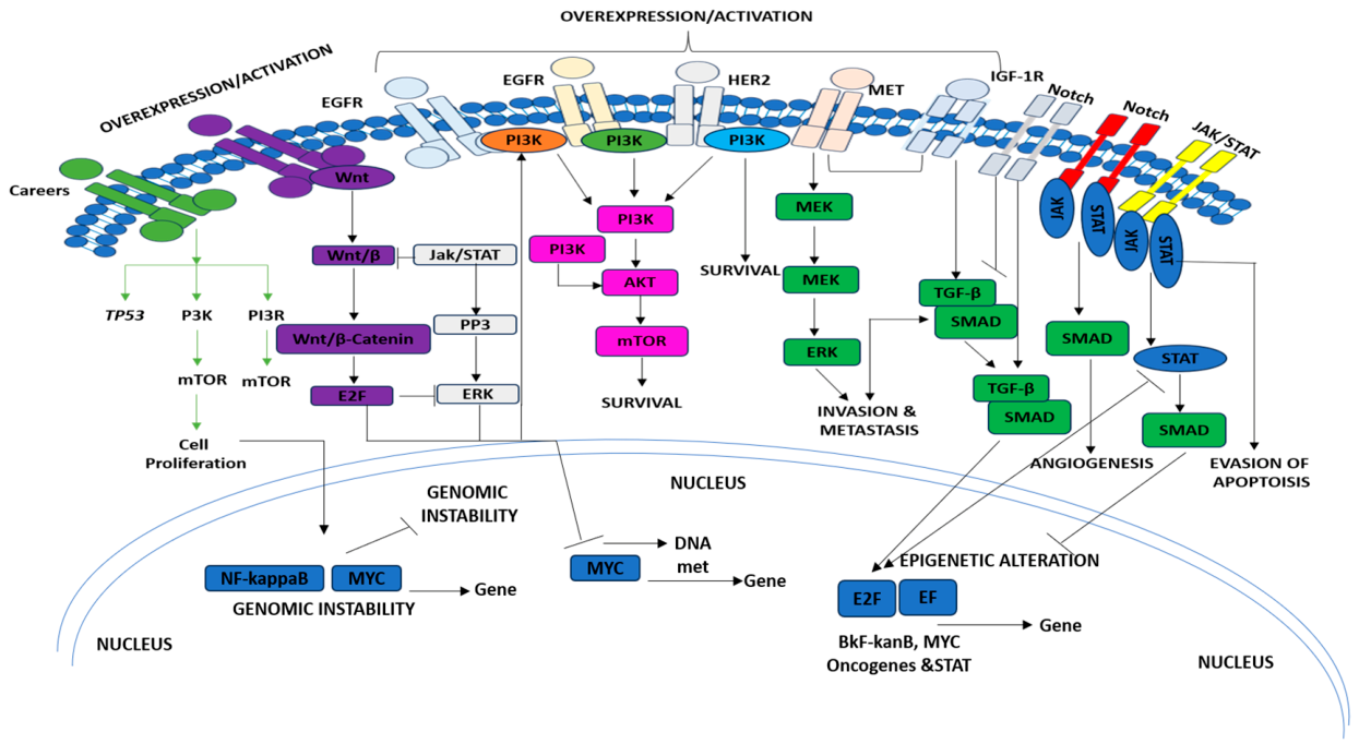
Figure 2.
Schematic representation of the esophageal tumor microenvironment illustrating the interactions between tumor cells, cancer-associated fibroblasts, immune cells, extracellular matrix, blood and lymphatic vessels, and nerve fibers. These dynamic interactions promote tumor growth, angiogenesis, immune evasion, invasion, metastasis, and therapeutic resistance through cytokine- and growth factor-mediated signaling.
Figure 2.
Schematic representation of the esophageal tumor microenvironment illustrating the interactions between tumor cells, cancer-associated fibroblasts, immune cells, extracellular matrix, blood and lymphatic vessels, and nerve fibers. These dynamic interactions promote tumor growth, angiogenesis, immune evasion, invasion, metastasis, and therapeutic resistance through cytokine- and growth factor-mediated signaling.
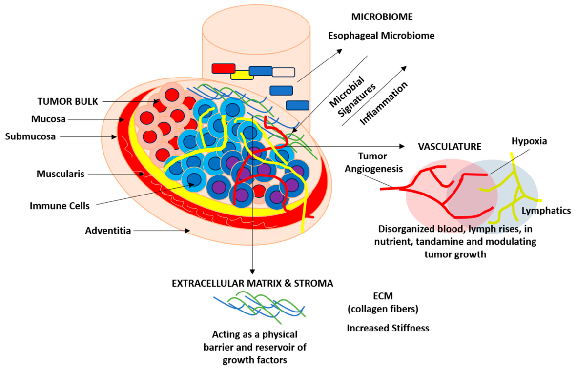

Table 1.
Emerging Biomarkers for Precision Stratification and Therapeutic Guidance in Esophageal Cancer.
Table 1.
Emerging Biomarkers for Precision Stratification and Therapeutic Guidance in Esophageal Cancer.
| Biomarker | Biological Source | Clinical Utility | Validation Status | Future Potential | References |
| ctDNA Methylation Panel | Plasma | Early detection, minimal residual disease (MRD) monitoring, recurrence surveillance | Retrospective cohort studies | Large-scale prospective screening and monitoring trials | [86] |
| PD-L1 Combined Positive Score (CPS) | Tumor tissue | Selection of patients for immune checkpoint inhibitors (e.g., pembrolizumab) |
FDA-approved companion diagnostic for pembrolizumab in eligible ESCC patients. |
Optimization of cutoff values and combination with TMB-based stratification | [87] |
| YAP/TAZ Nuclear Localization | Tumor tissue | Assessment of tumor aggressiveness and Hippo pathway activation | Retrospective clinical studies | Companion diagnostic biomarker for TEAD/YAP-targeted therapies | [88] |
| CAF Signature (FAP, α-SMA, IL6) | Tumor tissue (IHC/RNA profiling) | Prognostic evaluation and characterization of stromal remodeling | Research-stage investigations | Stratification of patients for CAF-targeted therapeutic trials | [89] |
| Spatial Immune Score | Spatially profiled tissue sections | Prediction of immunotherapy responsiveness and immune landscape characterization | Proof-of-concept and translational studies | Clinical implementation in precision immuno-oncology workflows | [90] |
| Fusobacterium nucleatum | Tumour tissue and microbiome samples | Prognostic marker and indicator of metabolic and immune modulation | Correlative clinical studies | Development of microbiome-based therapeutic interventions and risk stratification models | [91] |
Abbreviations: ctDNA, circulating tumor DNA; MRD, minimal residual disease; PD-L1, programmed death-ligand 1; CPS, combined positive score; ESCC, esophageal squamous cell carcinoma; CAF, cancer-associated fibroblast; FAP, fibroblast activation protein; α-SMA, alpha-smooth muscle actin; IL6, interleukin-6; TMB, tumor mutational burden; IHC, immunohistochemistry.
Table 2.
Regenerative Medicine and Bioengineering Strategies for Esophageal Reconstruction and Tissue Regeneration.
Table 2.
Regenerative Medicine and Bioengineering Strategies for Esophageal Reconstruction and Tissue Regeneration.
| Technology | Biological Component | Mechanism of Action | Current Development Stage | Translational Challenges | References |
| Decellularized Esophageal Scaffold | Porcine- or human-derived extracellular matrix (ECM) | Provides a native three-dimensional architecture with preserved basement membrane and bioactive ECM cues to support host cell migration and tissue regeneration | Large-animal studies (canine and porcine models); limited early human case reports | Residual immunogenicity, efficient recellularization, long-term graft integration, sterility assurance | [128] |
| MSC-Seeded Hydrogel Constructs | Mesenchymal stem cells (MSCs) embedded in alginate, GelMA, or hybrid hydrogels | Delivers anti-inflammatory, pro-angiogenic, and anti-fibrotic factors while supporting tissue repair and remodeling | Preclinical studies in rodent and rabbit models | Limited cell retention, uncontrolled differentiation, manufacturing complexity, high production costs | [129] |
| Electrospun PCL/Collagen Nanofiber Scaffolds | Polycaprolactone (PCL) combined with collagen or other natural polymers | Mimics extracellular matrix topography while providing mechanical support for cell attachment, proliferation, and differentiation | Preclinical evaluation in rat and canine models | Optimization of degradation kinetics, biomechanical matching, long-term biocompatibility | [130] |
| Growth Factor-Eluting Scaffolds | EGF, VEGF, FGF, and anti-fibrotic signaling modulators | Promotes epithelialization, angiogenesis, and tissue remodeling while reducing scar formation and fibrosis | Preclinical stage | Controlled release kinetics, growth factor stability, dosage optimization, manufacturing cost | [131] |
| Three-Dimensional Bioprinted Esophageal Grafts | Bioinks containing epithelial cells, smooth muscle cells, and biomaterials | Generates anatomically precise, patient-specific tubular constructs that recapitulate native esophageal architecture | Preclinical studies in small-animal models | Insufficient vascularization, lack of functional innervation, mechanical durability, scale-up for clinical use | [132] |
Abbreviations: ECM, extracellular matrix; MSC, mesenchymal stem cell; GelMA, gelatin methacryloyl; iPSC, induced pluripotent stem cell; PCL, polycaprolactone; EGF, epidermal growth factor; VEGF, vascular endothelial growth factor; FGF, fibroblast growth factor.
Disclaimer/Publisher’s Note: The statements, opinions and data contained in all publications are solely those of the individual author(s) and contributor(s) and not of MDPI and/or the editor(s). MDPI and/or the editor(s) disclaim responsibility for any injury to people or property resulting from any ideas, methods, instructions or products referred to in the content. |
© 2026 by the authors. Licensee MDPI, Basel, Switzerland. This article is an open access article distributed under the terms and conditions of the Creative Commons Attribution (CC BY) license (http://creativecommons.org/licenses/by/4.0/).
Copyright: This open access article is published under a Creative Commons CC BY 4.0 license, which permit the free download, distribution, and reuse, provided that the author and preprint are cited in any reuse.