
Submitted:
11 November 2026
Posted:
12 November 2026
You are already at the latest version
Abstract
Nanoplastics have been detected in food matrices and food-contact materials, yet data on the biological consequences of dietary nano-scale plastic exposure remain limited. We evaluated the effects of oral administration of polystyrene nanoparticles (1 mg/kg bw), silver nanoparticles (1 mg/kg bw), their co-exposure, and polyethylene nanoparticles (0.5 μg/kg bw) isolated from commercial rice packaging in male and female Wistar rats. After 28 days, colonic immune cell populations, antioxidant parameters, gut microbiota composition, short-chain fatty acids, and intestinal metabolomic profiles were analyzed. Exposure to polyethylene nanoparticles derived from food packaging and polystyrene nanoparticles significantly altered lymphocyte distributions, reduced total antioxidant status, modified microbiota structure, and affected metabolic pathways associated with steroid biosynthesis and inflammatory regulation. Several responses exhibited sex-specific patterns. Co-exposure with silver nanoparticles further influenced microbial metabolites. These findings demonstrate that dietary exposure to nano-sized plastic particles from food-contact materials induces measurable biochemical and metabolic alterations in the colon.
Keywords:
nanoplastic
; gut-brain axis
; immune system
; oxidative stress
; microbiome
; metabolic pathways
; nanotoxicity
1. Introduction
In recent years, interest in the influence of synthetic pollutants on the health and functioning of diverse organisms has been steadily growing. Humanity generates substantial amounts of plastic debris, with global production reported to have reached 413.8 million tons in 2023 [1]. Despite increasing awareness of their biological implications, plastic production continues to rise, and it has been estimated that nearly 60% of all plastics produced since the onset of large-scale manufacturing have ultimately become waste [2]. Once released into the environment, plastics undergo physical, chemical, and biological degradation processes leading to the formation of micro- (less than 5 mm) and nanoplastic (less than 100 nm) particles (MNPs). These particles not only contaminate terrestrial and aqueous environments, but may also disperse through the atmosphere, contributing to secondary pollution as plastic fallout [3,4,5].
Even though MNPs vary greatly in shape and chemical composition, their small size makes them prone to internalization by organisms, with nano-sized particles posing a particular threat [6]. One of the most relevant exposure routes in humans and other terrestrial organisms is oral intake, originating primarily from food, beverages, and food packaging materials [7,8,9,10]. Moreover, plastics may act not only as toxic agents themselves, but also as carriers for co-occurring environmental contaminants, including metals and metallic nanoparticles [11,12]. Such plastic–metal interactions may modify particle behavior and biological effects, and in some cases have been suggested to enhance the toxicity of individual agents acting alone [12,13,14].
Among metallic nanoparticles, silver nanoparticles (AgNPs) are of particular importance due to their broad application in consumer, medical, and industrial products, primarily resulting from their antimicrobial properties [15]. Their release into the environment and potential co-occurrence with plastic particles raise concern about combined exposure to nano-sized plastic and metallic particles [11,12]. AgNPs have been shown to affect oxidative balance, immune response, and gut microbiota composition, making them an appropriate model metallic nanoparticle for studying mixed-contaminant scenarios [16]. Therefore, their inclusion in the present study allowed us to assess whether co-exposure with a well-established nanotoxic agent modifies the biological effects of model nanoplastics.
Following oral intake, MNPs, either alone or in combination with other particulate pollutants, can interact with the gastrointestinal tract, which represents the first biological interface between ingested particles and the host. Available evidence suggests that exposure to MNPs may affect intestinal immune function, oxidative balance, and gut microbiota composition [17]. MNPs may also enter the blood circulation and reach distant body compartments, including the nervous system [18]. These gut-associated responses may therefore be relevant to broader systemic communication pathways, including the gut-brain axis (GBA) linking the gastrointestinal tract and the brain, which has been proposed as a potential target of MNP-related effects due to its vulnerability [19].
Given the increasing detection of nano-sized plastic fractions in food and food-contact materials, their influence on biological systems, as well as their potential interactions with co-occurring contaminants, particularly metallic nanoparticles, experimental models reflecting such dietary exposure scenarios are needed. In the present study, we investigated the effects of oral exposure to polystyrene nanoparticles (PSNPs) and AgNPs, used as model plastic and metallic nanoparticles, respectively, including their co-exposure as a standardized mixed-contaminant scenario. While much of the existing research relies on commercially available model nanoparticles, we additionally included high-density polyethene nanoparticles (HDPE) isolated directly from consumer-ready food-packaging material to reflect realistic, day-to-day human exposure. The study is focused on colonic immune responses, gut microbiota composition, microbial metabolites, and intestinal metabolic pathways in male and female rats.
2. Results
2.1. Histopathological Assessment of Colon Tissue Following Nanoparticle Exposure
As part of the evaluation of nanoplastic effects, we examined the histology of the colon in all exposure groups. In male rats, all NP-exposed groups showed a higher frequency of inflammatory and vascular alterations, including lymphocytic infiltrates (often accompanied by eosinophils), dilatation of lymphatic and venous vessels (lymphangiectasia and venous ectasia), and, occasionally, lymphoid hyperplasia (Supplementary materials, Figure S1). The most extensive changes — characterized by multifocal inflammatory infiltrates were observed in the AgNPs (Supplementary materials, Figure S1B) and PSNPs+AgNPs groups (Supplementary materials, Figure S1C). In the female colon, the histological picture was predominantly unaltered across all exposure groups, or limited to small, isolated lymphocytic aggregates and solitary lymphoid follicles. Only animals from the AgNPs group exhibited more pronounced inflammatory changes, including plasma-cell infiltration and marked dilatation of lymphatic vessels (Supplementary materials, Figure S1B).
2.2. Impact of Nanoplastic Particles on the Intestinal Lymphocyte Populations
We investigated lymphocytes from two locations of the intestinal wall – IELs and LPLs – in the colon tissue of rats exposed to experimental treatments and the control group. Results yielded by the flow cytometry analysis provided insight into the percentage distribution of CD3+ cells (T lymphocytes), within which CD4+ (T helper lymphocytes) and CD8+ (T cytotoxic lymphocytes) were differentiated, as well as CD45+ cells (B lymphocytes) and CD161+ cells (NK cells) in each population. Regarding IELs, the only type of cells affected were NK cells in animals treated with HDPE. Females showed a statistically significant increase (Figure 1A), while no such impact was observed in males (Figure 1A). All the other cell types (B lymphocytes, T lymphocytes, Th cells, Tc cells) remained unaffected. The effects of the intervention were more pronounced in the LPLs population. Administration of HDPE resulted in a significant increase in B lymphocytes in the colon of male rats compared to the control group (Figure 1B). On the other hand, an inverse reaction was observed in females, whose B lymphocytes decreased significantly relative to control (Figure 1B).
In the case of T lymphocytes, the effects caused by the intervention were strikingly different between the sexes. In male animals, all experimental groups were characterized by a lower level of T lymphocytes compared to the control group (Figure 1C). Furthermore, rats administered with AgNPs had a lower percentage of T lymphocytes than those fed with PSNPs and the mixture of PSNPs+AgNPs. However, none of these changes were present in females (Figure 1C). With regard to the Th lymphocytes measured in the LPLs population, both sexes were affected, although to different extents. In males, all experimental substances caused a marked increase in the lymphocyte level compared to the control group (Figure 1D). In females, only those receiving the mixture of PSNPs+AgNPs differed significantly from the other groups, having a decreased level of Th lymphocytes relative to the PSNPs, AgNPs, and control (Figure 1D). None of the investigated substances affected either NK cells or Tc lymphocytes in the LPLs population in the colon of exposed animals.
2.3. Antioxidant Potential and Oxidative Stress in the Colon
We found that the exposure to nanoparticles caused oxidative stress in the colon tissue. TAS was significantly lower in males receiving nanoplastic alone (both PSNPs and HDPE) and in females in all groups that contained nanoplastic (PSNPs, HDPE, PSNPs+AgNPs). The activity of antioxidative enzymes was also affected. SOD activity was higher compared to control in females administered with PSNPs and lower in the HDPE group. GR activity was elevated in all groups in males, while in females, its activity increased in the PSNPs and HDPE groups, but decreased in the PSNPs+AgNPs group. GPx activity was significantly lower in all exposure scenarios in both sexes, with the exception of female animals receiving HDPE, where this parameter was increased, as compared with the control group. Moreover, intensified lipid peroxidation, evidenced by TBARS, was observed in both males (AgNPs, PSNPs+AgNPs, HDPE) and females (PSNPs+AgNPs, HDPE) (Supplementary materials, Table 1).
2.4. Impact of Nanoplastic Particles on the Gut Microbiota Composition
The gut microbiota plays a crucial role in maintaining the host’s health, and its composition can be influenced by various factors, including exposure to environmental pollutants. Therefore, we aimed to explore the microbiome profiles of animals of both sexes exposed to various experimental agents (PSNPs, AgNPs, PSNPs+AgNPs, HDPE), and their corresponding control groups. At the genus level, distinct dynamic patterns between treatment groups within sexes were apparent. In males, the PSNPs+AgNPs group showed a microbiota composition similar to the control, while the HDPE group was close to the AgNPs group. The PSNPs group showed the most pronounced divergence in bacterial composition. In females, the AgNPs group was the closest to the control, and the HDPE group was similar to the PSNPs+AgNPs group. The PSNPs group, again, was the most distinct microbial profile. The relative abundance at the genus level is presented in Figure 2A,B.
Moreover, we chose a two-step filtering strategy to identify and exclude low-prevalence bacteria. In the first step, to reduce the impact of sequencing depth on diversity measurements, we performed resampling to equalize the sequence numbers for each sample (to 53100 reads per sample). In the second step, we applied filtering with the following criteria: relative abundance threshold of 0.0001 and a minimum occurrence frequency of 0.1 (10% of samples should have this feature). After filtering out the low-prevalence bacteria, group-specific ASVs emerged. In females, 4 ASVs uniquely characterized the HDPE group (Anaerostipes, Ruminococcus 1, Blautia and Lachnospiraceae NK4A136 group), two groups were specific to the AgNPs exposure (Ruminococcaceae NK4A214 group and an ASV from the Lachnospiraceae family), one was specific to the PSNPs group (Ruminococcaceae UCG-014), one specific to the PSNPs+AgNPs group (an ASV from the Lachnospiraceae family), and one ASV characterized the control (Ruminiclostridium 9). No exposure-specific ASVs were identified in males (Figure 2C,D).
Exposure to various substances can significantly influence gut microbiota diversity. Bacterial composition in terms of alpha-diversity and beta-diversity (exposure-driven) revealed no statistically relevant differences, while beta-diversity was statistically different between males and females (MANOVA, P < 0.001) (Supplementary materials, Figure S2 and Figure 3). When assessing differential abundance, the Linear Discriminant Analysis (LDA) showed statistically significant differences between experimental groups in both sexes. In females, we found several bacteria species specific to each experimental group, while in males, only a few ASVs were identified, specifically in the PSNPs, AgNPs, and HDPE groups. The results are shown in Figure 2E,F. In addition, differentiation between sexes was explored using the Principal Component Analysis (PCA), which identified ASVs contributing to group separation (Supplementary Materials, Figure S4).
We also assessed differences in mean bacterial abundance between exposure and control groups, identifying specific families and genera with statistically significant changes. In particular, in the PSNPs group, we observed an increased mean abundance of Akkermansia, Odoribacter, Parasutterella, and Coriobacteriaceae UCG-002 at the genus level (Figure 3A). Exposure to AgNPs had a statistically significant impact on the Erysipelatoclostridium, Roseburia, Erysipelotrichaceae UCG-004, Odoribacter, and Rothia genera (Figure 3B). The co-exposure to PSNPs+AgNPs affected Erysipelatoclostridium and Odoribacter (also present in the single-substance exposure scenarios) as well as Rikenellaceae RC9 gut group, Tyzzerella 3, and Mucispirillum (not present in either of the single-substance exposure scenarios) (Figure 3C). Meanwhile, the group administered with HDPE showed two genera differentiating it from the control group: Family XIII UCG-001 and Mucispirillum (Figure 3D). Even though more experiments would be needed, it seems likely that particular bacteria respond differently to different types of (plastic) nanoparticles, suggesting substance-specific effects.
2.5. Impact of Nanoplastic Particles on the Fecal SCFA Content
In addition to the microbiota composition, we also examined the fecal content of the microbial metabolites, SCFA. In particular, we looked at six different fatty acids: lactic acid, acetic acid, propionic acid, iso-butyric acid, butyric acid, and valeric acid. Two of them – propionic and butyric acid – showed no significant differences in any exposure scenario, in either sex. While no changes were observed in males, lactic acid levels increased significantly in females treated with AgNPs in comparison to PSNPs and control (Figure 4A). Acetic acid followed a similar pattern, with no changes in males (Figure 5B) and a significant increase observed in females exposed to AgNPs groups, which differed significantly from the PSNPs+AgNPs and control groups (Figure 4B). On the other hand, iso-butyric acid content was elevated only in males and only in the group receiving AgNPs. Its concentration was significantly higher than in all other groups, except the HDPE (Figure 4C). Meanwhile, there was no visible impact on females (Figure 4C). In contrast, valeric acid showed a significant decrease in males exposed to PSNPs or AgNPs. Both groups differed from the mixed PSNPs+AgNPs and control groups (Figure 4D). No such differences were apparent in females (Figure 5D). Overall, AgNPs exposure exerted the strongest effect on SCFA content, followed by PSNPs. Importantly, the observed differences were clearly sex-specific, with different metabolites being affected in males or females only.
2.6. Metabolomic Profile of the Colon Tissue
To quantify changes in the metabolomic profiles of rats exposed to different types of nanoparticles, we conducted a comparative metabolomic data analysis of each treatment group relative to the control. The PCA analysis (Supplementary materials, Figure S5) revealed that the metabolomic profile in the colon tissue samples of rats was significantly affected by different exposure scenarios, showing a separation between groups. Among the affected metabolic pathways, the ones common to all groups were selected for further examination. We identified 9 such pathways: C21-steroid hormone biosynthesis and metabolism, androgen and estrogen biosynthesis and metabolism, prostaglandin formation from arachidonate, aspartate and asparagine metabolism, leukotriene metabolism, tryptophan metabolism, pyrimidine metabolism, fatty acid activation, and xenobiotics metabolism.
Within these shared pathways, we sought significantly altered metabolites present in all experimental groups or in at least three out of four (PSNPs, AgNPs, PSNPs+AgNPs, HDPE). This approach yielded 21 metabolites, from which 5 were selected for subsequent analysis based on their potential relevance for the GBA-related processes. The only metabolite affected across all exposure groups was caprylic acid, from the fatty acid activation pathway. In males, its levels decreased in all groups except the HDPE compared to the control group. Moreover, its level was also significantly lower in PSNPs-exposed rats compared to AgNPs and PSNPs+AgNPs (Figure 5A). In females, the AgNPs group was the only outlier, with lower levels from the PSNPs, PSNPs+AgNPs, and control groups (Figure 5A).
Another metabolite altered significantly in at least three scenarios (PSNPs, AgNPs, HDPE) was anandamide, within prostaglandin formation from the arachidonate pathway. In males, its concentration was markedly increased in PSNPs+AgNPs compared to the control, PSNPs, and AgNPs, with a slight elevation also observed in the HDPE group (Figure 5B). Meanwhile, in females, there was a significant increase in the PSNPs group as compared with AgNPs and control, and similar to males, HDPE exposure also resulted in higher levels than in control animals (Figure 5B). Exposure to nanoparticles also had an impact on androsterone glucuronide, a metabolite identified in the androgen and estrogen biosynthesis and metabolism pathways; however, significant differences were observed only in males. Its levels increased in all exposure groups compared to the control, except HDPE, and were higher in PSNPs-exposed rats than in the mixed PSNPs+AgNPs group (Figure 5C). In females, no significant effects were observed (Figure 5C).
A similar pattern was true for 2-hydroxyestrone, another metabolite, belonging to the same metabolic pathway. It was affected in males of PSNPs- and PSNPs+AgNPs-treated groups; its concentration significantly decreased compared to the control (Figure 5D). In females, it remained unchanged (Figure 5D). We also observed statistically significant differences in the case of 18-hydroxycorticosterone associated with the C21-steroid hormone biosynthesis and metabolism pathway. Even though no significant impact was found in males (Figure 5E), in females, there was an increase in the PSNPs group in comparison with both AgNPs and PSNPs-AgNPs groups (Figure 5E).
3. Discussion
The current state of knowledge shows that plastic particles of various shapes and sizes are ubiquitous in the human environment [20]. Oral exposure may originate not only from contaminated food itself, but also from food containers and even products of particularly strict safety standards, such as infant formulas [21,22]. Moreover, MNPs are increasingly recognized by policy-makers as major pollutants, even though data concerning the health implications of MNPs contamination remain scarce [23]. Nevertheless, a handful of studies involving humans have shown plastic accumulation in various organs and tissues. Leslie et al. found different types of plastic particles, including those falling within the NPs category (<1000 nm), in the blood of healthy volunteers [18], while García et al. showed that plastic exposure may begin as early as the initial stages of fetal development [24]. MNPs have also been detected in human stools, which supports ingestion as one of the main exposure routes [25]. Yan et al. went a step further and investigated the association between the amount of plastic in the feces and IBD status. They found that patients with IBS had more MNPs in their stools, but also showed a positive correlation between fecal plastic concentration and the severity of the IBS [26]. Finally, a recent study by Nilhart et al. provided evidence for the presence of MNPs in human kidneys, liver, and brain, with a marked increase in the degree of plastic pollution observed between 2016 and 2024 [27]. Given the prevalence of the oral exposure route and the aforementioned detection of plastics in both the feces and the brain, particular concern arises regarding the potential influence of MNPs on the GBA and the communication pathways between the gastrointestinal tract and the nervous system, discussed by our as well as other teams [19,28].
As an approximation for human exposure, we investigated the effects of nanoplastics on the first part of the GBA, i.e., the digestive system, in an animal model widely adopted in toxicological studies. To assess the impact of NPs ingestion, we took a closer look at the immune cell profile of the intestine, in particular at the IELs and LPLs populations. IELs form part of the gut-associated lymphoid tissue (GALT) and are located close to the intestinal lumen. They regulate the immune response, modulate the tolerance to dietary antigens, and affect inflammatory processes. LPLs, on the other hand, constitute the primary effector part of the GALT, situated further apart from the lumen and sensitive to different stimuli originating from the microbiota as well as the epithelium. In IELs, we observed a sharp increase in the percentage of NK cells in rats exposed to HDPE, when compared to the control group. Interestingly, this effect seemed to be sex-specific, since it was observed only in females. No significant difference between groups in males was observed.
In contrast, LPLs were affected to a greater degree. In the HDPE group, the B lymphocyte count increased in males but decreased in females. The percentage of T lymphocytes significantly decreased in all experimental groups compared to the control, but only in males. The most pronounced decrease was noted in the positive control (AgNPs) group. Finally, we observed a more complex scenario for the Th cells. In males, their percentage was significantly higher in all groups relative to the control, while in females, it decreased in the co-exposed PSNPs+AgNPs group. In this context, higher susceptibility of males was observed previously by Xiuli et al., who reported increased immune and apoptotic responses in male offspring of mice exposed to PSNPs during pregnancy, as compared with their female counterparts [29]. A similar outcome was described by Fowler et al. in a systematic review and experimental study on mice, where male immunity appeared to be more compromised by MNPs treatment, as compared to females. The authors hypothesized that this recurring trend could be explained by chromosomal differences, with females being more resilient due to the potential biallelic expression of immune-related genes in the X chromosome [30].
To the best of our knowledge, there are no studies investigating the effects of MNPs on the intestinal immune cell profile in rats. However, reports involving other organs and other particle types do exist. In a long-term study on mice (both sexes) treated with 0.125-2 mg of polyethylene microparticles, Park et al. observed no changes in either NK cells, T lymphocytes or B lymphocytes in the spleens of female animals after 90 days of administration [31]. Similarly, Hu et al. found no significant impact of polystyrene microparticles on NK cells in the blood and spleen of pregnant mice; however, they reported a decrease in B lymphocytes, akin to the results obtained in our study [32]. Małaczewska reported a decreased NK cell count in the blood of male mice treated orally with AgNPs for 28 days [33]. In a similar experiment on male mice orally exposed for 28 days to polystyrene microparticles, Zhao et al. observed a decrease in B and Th lymphocytes, but no impact on T lymphocytes [34]. Harusato et al. conducted a study on female mice administered orally with polyethylene microplastic particles for 8 weeks (every 3 days) and reported no changes in the T lymphocyte population, which corresponds to our observation [35]. Overall, the lack of directly comparable data from other experiments concerning intestinal immune cells response to the MNPs treatment makes conclusions in this regard hard to formulate, although our results show a clear difference in effects depending on sex. Nanoplastic impact on the immune system in the colon also highlights its threat to the well-functioning of the GBA, heavily influenced by GALT [36].
A known effect caused by AgNPs and MNPs, closely related to immune system activation, is oxidative stress, as described and shown by our team in previous works [19]. These nanoparticles are capable of inducing numerous detrimental effects related to reactive oxygen species (ROS) generation and inflammation, including neurotoxicity as well as disruption of the intestinal homeostasis, affecting the main parts of the GBA. We found that administration of MNPs, either alone as PNSPs and HDPE, or in combination with AgNPs, resulted in clearly reduced TAS and caused an increase in the activity of major enzymes involved in antioxidant defense processes, such as SOD, GR, and GPx. These effects were evident in animals of either sex. Furthermore, the presence of the nanoparticles in the gut led to lipid peroxidation of the colon tissue, as evidenced by elevated levels of TBARS, primarily in groups receiving AgNPs (alone or with PSNPs) and HDPE. Similar results were also described by Augustyniak et al. who orally exposed young rats to PSNPs in the same dosage as we did (1 mg/kg bw) for 3 weeks [37]. They showed decreased antioxidant capacity in the intestine and oxidative damage to proteins, accompanied by increased expression of proinflammatory cytokines.
The mammalian digestive tract is home to a community of bacteria and other microorganisms crucial to the host’s health and well-being, believed to be an essential part of the GBA, influencing the communication between the intestines and the brain via nerve stimulation and specific metabolites [38]. As we described previously [19], there is evidence suggesting that MNPs can exert an adverse influence on these processes, hence our focus on the impact of nanoplastics on the gut microbiota. At the genus level, exposure-specific differences showed that in both sexes the animals receiving PSNPs-only were consistently the outliers, differing from the control and groups involving AgNPs, known for their antimicrobial properties and impact on the microbiota [16].
In females, we found ASVs specific for each group. In the PSNPs-treated group, it was Ruminococcaceae UCG-014, a bacterium with a controversial health impact, previously linked to inflammatory bowel disease (IBD) in mice [39]. In the AgNPs-treated group, a unique ASVs included Ruminococcaceae NK4A214 and a bacterium from the Lachnospiraceae family. Both can play a mixed role in the host’s health and are involved in SCFA metabolism, contributing to the production of butyrate as well as acetate, propionate (Ruminococcaceae), and lactate (Lachnospiraceae) [40,41]. Thus, their presence corresponded with the observed increase in acetic and lactic acid levels in the feces of female animals in the AgNPs (positive control) group. The latter seems particularly interesting, since it has been suggested that higher lactic acid concentrations in the stool might reflect its impaired metabolism by the microbiota and could be a sign of an undergoing inflammation [42,43].
When examining the microbiota abundance in male rats, the PSNPs group was differentiated by the Akkermansia genus in particular. Akkermansia is a beneficial microorganism and an SCFA producer (acetate, propionate) [44]. It degrades mucin, which under normal physiological conditions is a positive process, promoting mucin production via upregulation of associated gene expression. However, combined with low fiber consumption mucin degradation by Akkermansia could lead to increased inflammation. It would suggest that in this context the impact of nanoplastic exposure could be modulated by diet composition [45]. Meanwhile, ASVs differentially abundant in the AgNPs group were commensal bacteria Roseburia and Coprococcus, which prevent intestinal inflammation and whose presence is inversely correlated with neurological disorders. Both of them produce butyrate, and Coproccocus has been linked to elevated levels of isobutyric acid, which could be reflected in the observed increase in this SCFA content in males administered with AgNPs.
Overall, our findings indicate that all treatments induced changes in gut microbiota, although to a different extent and not in a uniform direction. Some of the affected microorganisms are related to inverse health outcomes, such as Ruminococcaceae, Alloprevotella, Erysipelotrichaceae, or Mucispirillum. On the other hand, there were also notable increases in Bifidobacterium, Lactobacillus, Akkermansia, or Odoribacter. Although research focusing on MNPs-microbiota interactions is scarce, the available studies point towards a negative impact of MNPs exposure on the gut environment, leading to dysbiosis and, consequently, GBA dysregulation [46]. Zhang et al. reported altered microbiota composition in mice treated orally with PSNPs of different sizes for 14 or 28 days. They employed three dosages roughly translated to 0.15, 1.5, and 150 mg/kg bw of PSNPs with sizes of 20, 500, and 5000 nm [47]. Thus, the medium-dosage, low-size group was the one most similar to the setting used in our study (20 nm, 1 mg/kg bw). In this scenario, the authors observed a decreased Firmicutes/Bacteroidetes ratio as well as an increased presence of potentially harmful bacteria, such as Escherichia-Shigella, Enterobacteriaceae, and Erysipelotrichales. We found an increase in the abundance of Erysipelotrichaceae (a family within the same class of Erysipelotrichales), as well as in both the PSNPs group and the positive control AgNPs group. Furthermore, a recent study on mice conducted by Hsu et al. also showed an imbalance in microbiota composition in response to MNPs administration [48]. They treated the animals orally for 12 weeks with 2 × 10¹¹ of 100 nm particles/day, and observed an increase in Akkermansia and Ruminococcaceae UCG-014, similar to that reported in our study in the PSNPs group. Interestingly, we did not observe a significant influence of plastic administration on the SCFA content, except for a decrease in valeric acid in the PSNPs compared to control (in males only). We found, however, a consistent impact of AgNPs on the content of several bacteria-derived SCFA and BCFA, including lactic, acetic, iso-butyric, and valeric acid. This result seems consistent with the established impact of AgNPs on bacterial communities and their antimicrobial properties [49]. In addition, we also found marked differences in microbiota and SCFA content between sexes, with females being seemingly more susceptible to the nanoparticle activity than males. To the best of our knowledge, this is the first study reporting such sex-specific results.
One of the objectives of the present study was to assess the impact of MNPs on the metabolomic profile of the colon tissue. We found that oral exposure to nanoplastics affects several metabolic pathways related to essential processes, such as steroid, fatty acid, and xenobiotic metabolism, or inflammatory response. These changes are in line with the aforementioned alterations in the immune cell populations and antioxidant status of the animals. Although research focusing on the metabolomic effects of MNPs is scarce, a study by Garcia et al. showed similar alterations. The authors reported an impact on pathways such as steroid hormone biosynthesis, xenobiotic metabolism, and fatty acid metabolism in mice after 4 weeks of oral treatment with polystyrene and mixed plastic polymers [50]. In our study, one of the affected metabolites was anandamide, a key ligand of CB1 and CB2 cannabinoid receptors, which has an impact on the anti-inflammatory response of organisms, is linked to cognitive and behavioral processes, and plays a role in the maintenance of intestinal barrier integrity as well as the functioning of the GBA [51,52,53]. We detected a sharp increase in the PSNPs+AgNPs group in males as well as in the PSNPs in females. This observation might suggest a compensation mechanism, where anandamide could be elevated in response to oxidative stress in the colon tissue caused by the presence of MNPs. Moreover, it has been shown to play a crucial role in the immune homeostasis of the gut in mice, further linking its increased level to the changes in the LPLs immune cell populations we described before [54]. Another noteworthy metabolite is 18-hydroxycorticosterone, an aldosterone precursor involved in the regulation of adrenal steroidogenesis [55]. Its increase in females administered with PSNPs suggests hypothalamic-pituitary-adrenal axis (HPA) activation, which forms part of the GBA communication pathways and has an influence on the intestinal barrier [19]. It could be, therefore interpreted as a stress response to the PSNPs, further reflected as shifts in the microbiota composition, susceptible to HPA-modulated cortisol release [56].
By linking environmentally realistic nanoplastic exposure to disruptions in gut-associated immune, microbial, and metabolic systems, our findings provide a mechanistic framework through which nanoplastics present in terrestrial environments may influence organismal fitness and, over time, contribute to broader ecological consequences. These findings highlight the importance of incorporating nanoplastic fractions and mixed nanoparticle scenarios into environmental monitoring, regulatory evaluation, and pollution mitigation strategies addressing emerging contaminants in terrestrial ecosystems.
4. Materials and Methods
4.1. Characterization and Acquisition of Nanoparticles
4.1.1. Silver nanoparticles
Silver nanoparticles (AgNPs) applied in the present study were previously characterized in detail by our research group [57,58,59]. Commercial AgNPs with a nominal particle size of approximately 20 ± 5 nm were acquired from PlasmaChem (Berlin, Germany). Transmission and scanning electron microscopy images of the same AgNP material have been reported elsewhere [57,58]. Comprehensive descriptions of the physicochemical characteristics of AgNPs, as well as the protocols used for preparation of working dispersions, are provided in the cited studies [57,58,60]. Key parameters relevant to the current work are summarized Table 1.
4.1.2. Plastic Nanoparticles
High-density polyethylene nanoparticles (HDPE) were obtained from perforated polymer films supplied by PLASTEXIM (Tychy, Poland), a manufacturer of HDPE foil used for commercially available cooking bags for rice, groats, flakes, and other dry food products in Poland. Detailed isolation procedure was described in our previous study [61]. Monodisperse spherical polystyrene nanoparticles (PSNPs) with a nominal diameter of 20 ± 5 nm were purchased from NANOCS (New York, NY, USA) and employed as a reference plastic nanoparticle material. Particle visualization and size distribution analysis in aqueous suspension for both nanoparticle types were conducted using nanoparticle tracking analysis (NTA). In addition, zeta potential and polydispersity index measurements were performed by dynamic light scattering. Detailed methodological descriptions have been reported in our earlier publications [60,61]. An overview of the key characteristics of plastic nanoparticles investigated in this study, as well as their mixture with AgNPs (PSNPs+AgNPs), is provided in the Table 1.
4.2. Animal Treatment and Experimental Design
The experiment was conducted on 7-week-old outbred Wistar Wu rats (Wistar Wu [Crl:WI(WU)] strain), male (n = 50) and female (n = 50), purchased from Charles River (Charles River Laboratories, Sulzfeld, Germany). The rats were free of specific pathogens (SPF) and had initial body weights of 275.72 ± 8.06 g (males) and 180.96 ± 10.51 g (females). Animals were placed under standard housing conditions (12 h light/12 h dark cycles, 15 times/hour air change ratio, temperature 22 °C, relative humidity 55%) and provided with both water and food ad libitum (Altromin 1324 maintenance diet, providing 65% of energy from carbohydrates, 11% from fat, and 24% from protein with metabolized energy of 3,227 kcal/kg). Environmental enrichment in the rats’ cages was provided by introducing glass balls and wooden round objects.
After a 1-week adaptation period, rats were assigned to one of the five groups, receiving either PSNPs, silver nanoparticles AgNPs (positive control), a mixture of both commercial polystyrene and silver nanoparticles (PSNPs+AgNPs), HDPE isolated from consumer-available rice bags, or water (negative control). Ten animals per sex were allocated to each experimental group; however, two animals per group were transcardially perfused for histological analyses, resulting in n = 8 animals per group being available for biochemical, immunological, microbiome, and metabolomic analyses. Animals were treated with corresponding substances orally by gavage every day for a period of 28 days in accordance with OECD 407 guidelines [62]. All the experimental procedures performed during the study were approved by the Second Local Committee for Ethics in Animal Research at the Warsaw University of Life Sciences (Approval resolution no. WAW2/102/2021, 14 July 2021), and conducted in compliance with the UE Directive (2010/63/UE), the Polish legal regulation, and with respect to the 3R (Replacement, Reduction and Refinement) rules. The study was designed and conducted in accordance with the core principles outlined in the ARRIVE 2.0 guidelines. Throughout the experimental period, no signs of compromised well-being were observed, so the application of humane endpoints was deemed unnecessary. To achieve hormonal and metabolic standardization in females, the estrous cycle was synchronized using a natural method (male odor-induced estrous cycle synchronization). It involved housing females in cages containing bedding from male cages throughout the entire experiment. After 28 days of nanoparticle administration, rats were anesthetized with Isoflurane (Aerrane Isoflurane USP, Baxter, Poland). Eight animals from each group were bled by cardiac puncture, while the other two underwent transcardial perfusion via the left ventricle, first with physiological saline and then with 4% paraformaldehyde solution. An Ismatec pump (ISM831C; Ismatec, Wertheim, Germany) was used for the procedure. Subsequently, the entire large intestine of each rat was collected. Part of the fresh tissue was intended for cytometric analysis. A sample of the colon was fixed in 10% buffered formalin for histopathological examination, while the remaining sections were immediately frozen in liquid nitrogen and stored at −80 °C for later biochemical analysis. The large intestine samples that were preserved using 10% neutral buffered formalin were processed through paraffin embedding, sectioned at a thickness of 5 µm, and subsequently stained with hematoxylin and eosin (H&E) for evaluation by light microscopy, NIKON Eclipse Ti2 (Nikon, Melville, NY, USA).
4.2.1. Dosages and Experimental Groups
Each of the experimental groups comprised 10 animals. The number was determined using the methodology proposed by Dell et al. [63], which includes tools designed for estimating sample size for in vivo experiments. Rats in the PSNPs group and the AgNPs group, which served as a positive control given the widely known toxic effects of nanosilver particles [15], were treated with 1 mg/kg bw of the corresponding substance. As demonstrated by the physicochemical analysis, both types of nanoparticles had comparable sizes of 20 ± 5 nm, ensuring comparability of the results. Since MNPs are known to act as vehicles for other contaminants [13], we decided to include a mixed group, receiving a combination of PSNPs and AgNPs, an established nanotoxic agent. This hybrid PSNPs+AgNPs group received a combined dose of individual amounts of PSNPs and AgNPs (1 mg/kg bw + 1 mg/kg bw). These amounts were chosen based on the higher-end dosages commonly used in research concerning the toxicity of plastic particles and the fact that, in the case of nanosilver, it has been shown to exert toxic effects in mammals [64]. Moreover, applying the rat-human conversion ratio outlined in the guidelines by Nair and Jacob [65], 1 mg/kg bw corresponds to roughly 0.16 mg/kg bw/day or ~11 mg/capita/day (assuming human bodyweight of 70 kg). According to the exposure data by Zhao and You [66] this is more than ten-fold less than the average ingestion of MNPs (153–269 mg/capita/day) in countries considered to be major consumers worldwide (East Asian countries, China, South Korea, and Japan). In the HDPE group, which received nanoplastic harvested from consumer products, we had to take into account the technical limitations of the isolation process and eventually opted for a conservative dosage 2000-fold lower than the one used in the remaining experimental groups, resulting in the final dose of 0.5 μg/kg bw. The last group, which served as a negative control, received water (0.2 ml). The human-animal dosage conversion was performed according to the. The design of the experiment and description of the groups are provided in Figure 6.
4.3. Isolation of Lymphocytes from the Colon Tissue
Lymphocytes from the colon tissue were isolated using a previously described method [67]. Briefly, during dissection, 1–2 cm fragments of the colon tissue were collected and placed in centrifuge tubes with Ca2+- and Mg2+-free Hank’s balanced salt solution (HBSS). Under stable laboratory conditions, the samples were rinsed with pre-warmed HBSS and incubated for 30 min with 2 mM dithiothreitol (DTT) in HBSS at 37 °C. To release intraepithelial lymphocytes (IELs) into the solution, continuous stirring (200 rpm) was applied. Subsequently, each cell-containing suspension was filtered through a 3-layer sterilized gauze to remove connective colon tissue, and each sample was then maintained on ice before further IELs staining. The remaining tissue fragments were transferred into 50 mL of 5 mM EDTA in HBSS. Vigorous shaking was employed to facilitate the isolation of lamina propria lymphocytes (LPLs). Once EDTA-HBSS was removed, tissue fragments were incubated in RPMI 1640 medium supplemented with 5.96 g/L of HEPES, antibiotics (100 mg/L of streptomycin sulfate, 7 mg/L of penicillin G potassium), 5% fetal bovine serum, and 2 mg of collagenase type I (125 units/mg) for 1 h at 37 °C. To release leukocytes from the lamina propria in the intestinal mucosa, continuous stirring (200 rpm) was applied. Afterwards, samples were transferred into 15 mL of cold complete RPMI medium. Each set of suspensions, the gauze-filtered in DTT-HBSS (for IELs) as well as cells treated with collagenase (for LPLs), was centrifuged separately at 400 g for 5 min at 4 °C. The pellets were then resuspended in 10 mL of phosphate-buffered saline (PBS) and centrifuged again under the same conditions. Eventually, both fractions of immune cells were resuspended in PBS and aliquoted before the following immunofluorescent staining and flow cytometry analysis.
4.4. Phenotypic Analysis of Isolated Lymphocytes
After isolation from the colon tissue, fractions of immune cells (IELs and LPLs) were stained with antibodies specific to surface markers characteristic of the assessed lymphocyte population. Staining was performed using commercially available sets of antibodies: Rat T Lymphocyte Cocktail (BD PharmingenTM, USA, cat. no: 558493) and Rat T/B/NK Cell Cocktail (BD PharmingenTM, USA, cat no: 558495). Cells were centrifuged at 400 x g for 5 min at 4 °C, and then resuspended in 100 μL of PBS before the addition of 10 µL of corresponding antibodies. Samples were incubated at room temperature for 30 min in the dark. Afterwards, cells were washed with 1 mL of PBS, centrifuged again (400 x g, 5 min,4 °C) and resuspended in 0.5 mL of PBS. Flow cytometry was performed using a BD FACSAria™ II flow cytometer (BD Biosciences, USA), acquiring approximately 20,000 events per sample. Data were analyzed with FlowJo software (TreeStar Inc., Ashland, USA).
During analysis, lymphocyte populations were first differentiated based on their morphological characteristics, measurements of the forward scatter (FSC) and side scatter (SSC) parameters (P1 Gate). Single cells within the P1 gate were then analyzed for positive staining with corresponding antibodies (CD3, CD4, CD8, CD45RA, CD161alfa). The results were expressed as percentages of cells within the gated area (P1 gate). CD3+T cells were further analyzed to determine the Th (CD4+) and Tc (CD8+) cell subsets, expressed as percentages of total CD3+ cells. The compensation procedure was performed using a commercially available rat compensation set by BD Pharmingen. Gating strategy used throughout the cytometry analysis is illustrated in the Supplementary Materials (Supplementary materials, Figure S6).
4.5. Antioxidant Assays
Activities of glutathione peroxidase (GPx), glutathione reductase (GR), and superoxide dismutase (SOD), together with total antioxidant status (TAS), were quantified in colon homogenates. Tissue was homogenized in PBS (pH 7.4; Sigma-Aldrich, St. Louis, MO, USA) at a 1:5 (w/v) tissue-to-buffer ratio using a Bio-Gen PRO 200 homogenizer (PRO Scientific, Oxford, MS, USA). The homogenates were centrifuged at 5,000 × g for 5 min at 4 °C (Multifuge 3L-R, Kendro, Hanau, Germany), and supernatants were aliquoted into 200 μL tubes and stored at −80 °C until use. Enzyme activities were determined by spectrophotometry with Randox kits: RANSel (GPx), GR (glutathione reductase), and RANSOD (SOD), following the manufacturer’s manuals, and read at 505 nm on a Biochrom Anthos Zenyth 200 spectrophotometer (Cambridge, UK). Results are reported as Ug−1 tissue. TAS was measured with the Randox Total Antioxidant Status kit at 600 nm and expressed as μmol·g−1 tissue.
4.6. Lipid Peroxidation
Lipid peroxidation was evaluated as thiobarbituric acid–reactive substances (TBARS), including malondialdehyde (MDA). Colon homogenate supernatants were combined with 0.1 M sulfuric acid and 1% (w/v) thiobarbituric acid (TBA; Sigma-Aldrich, St. Louis, MO, USA), incubated at 90 °C for 1 h, cooled on ice, and extracted with n-butanol. After phase separation on ice, the upper organic layer was collected, clarified by centrifugation (2,000 × g, 5 min), and absorbance was recorded at 534 nm using the Biochrom Anthos Zenyth 200 instrument (Cambridge, UK). TBARS concentrations were calculated from a standard curve prepared with 1,1,3,3-tetramethoxypropane (TMP), which hydrolyzes to MDA under assay conditions, and expressed as nmol MDA per mg tissue.
4.7. DNA Extraction for Microbiota Analysis
Feces were collected one day before the end of the experimental period, and genomic DNA was immediately extracted using the QIAamp PowerFecal ProDNA isolation kit (Qiagen, Hilden, Germany). Samples of approximately 200 mg were placed into PowerBead Pro tubes with the addition of 800 μL of CD1 solution. After vortexing, the material was homogenized using an Omni® Bead Ruptor 12 Homogenizer (OMNI International, NW Kennesaw, GA, USA), under the following conditions: an initial step at a speed of 3.1 m/s (30 s), two subsequent steps at a speed of 1 m/s (30 s) with a 5 min interval between them. Immediately afterwards, the PowerBead Pro tubes were centrifuged at 15,000 g for 60 s. The obtained supernatant was then placed in new, sterile microcentrifuge tubes, and the DNA isolation process was continued according to the manufacturer’s instructions. The concentration of the extracted DNA and its purity (A260/A280 ratio) were measured using the NanoDrop™ 2000 spectrophotometer (Thermo Fisher Scientific, Waltham, MA, USA). The quality of the samples was determined by agarose gel electrophoresis. High-molecular-weight DNA, without RNA contamination, was successfully achieved.
4.8. 16S rDNA Microbiome Sequencing
The genetic material isolated from feces samples was used for library preparation and sequencing. Sequencing of the 16S rDNA amplicon (V3–V4 region) was performed on the Illumina MiSeq platform. To synthesize a single amplicon (approximately ~460 bp long) of the hypervariable regions (V3–V4) of 16S rRNAs, forward (TCGTCGGCAGCGTCAGATGTGTATAAGAGACAGCCTACGGGNGGCWGCAG) and reverse (GTCTCGTGGGCTCGGAGATGTGTATAAGAGACAGGACTACHVGGGTATCTAATCC) PCR primers were used. The preparation of the library was performed following the Illumina protocol “16S Metagenomic Sequencing Library Preparation. Preparing 16S Ribosomal RNA Gene Amplicons for the Illumina MiSeq System” with an increased number of cycles in the amplicon synthesis PCR reaction (28 instead of 25). The obtained libraries were pooled and sequenced with a 10% phiX spike-in on an Illumina MiSeq sequencer using the paired-end 2 × 300-bp v3 reagent kit as outlined in the instructions supplied by the manufacturer.
4.9. Analysis of the Fecal Content of Short-Chain Fatty Acids
The quantification of short-chain fatty acids (SCFAs) and branched-chain fatty acids (BCFAs) in fecal samples was carried out according to the following procedure. Samples were initially diluted at a 1:10 ratio in 25 mM sulfuric acid (H2SO4) and then homogenized using an Omni® Bead Ruptor 12 Homogenizer (OMNI International, Kennesaw, GA, USA) in two 30-second cycles at a speed of 3.1 m/s, with a 5-minute interval between the cycles. Following homogenization, the samples were centrifuged at 12,000 rpm for 20 minutes at 4 °C, and the supernatant was filtered using a 0.22 μm syringe filter.
SCFA and BCFA concentrations were determined using a Shimadzu high-performance liquid chromatography (HPLC) system comprising an LC-20CE pump, CBM-20A system controller, CTD-20AC column oven, SIL-20AC autosampler, and an SPD-M30A photodiode array detector (Kyoto, Japan). The samples were fractionated on an Aminex HPX-87H column (300 × 7.8 mm; Bio-Rad, Warsaw, Poland) under isocratic conditions using 10 mM H2SO4 as the mobile phase at a flow rate of 1 mL/min and a column temperature of 40 °C. Detection was performed at 210 nm. The compounds were identified based on retention times and compared to known standards. External calibration curves were constructed over a concentration range of 0.20 to 100 μg per injection for each compound. All external standards (purity ≥99%) were sourced from Sigma-Aldrich (Poznan, Poland).
4.10. Metabolomic Analysis
Metabolomic profiling of colon tissue samples was conducted using a Symbiosis Pico UHPLC system equipped for high-pressure liquid chromatography. Detection was carried out with a SCIEX TripleTOF 5600+ system with a DuoSpray Source (TurboIonSpray and APCI). Data processing and interpretation were performed using SCIEX MarkerView™ (version 1.3.1), the online version of XCMSplus, and the MetaboAnalyst 5.0 platform to enable comprehensive extraction of metabolomic information. Colon tissue samples were homogenized in PBS, then mixed with a 1:1 solution of acetonitrile and methanol. Following this, the samples were vortexed at 2000 rpm for 15 min and subsequently centrifuged at 20,000 × g for 15 minutes. The resulting supernatants were transferred into glass autosampler vials and stored at 4 °C before analysis. Samples were then injected directly into the Spark Holland Symbiosis™ Pico system.
Chromatographic separation was achieved using a Hypersil BDS C18 column (150 × 4.6 mm, 5 μm), accompanied by a Hypersil C18 guard column (10 × 2.1 mm, 5 μm). The mobile phases consisted of (A) methanol with formic acid (99:1, v/v) and (B) water with formic acid (99:1, v/v), delivered at a constant flow rate of 500 µL/min. The gradient elution program was as follows: 100% phase A from 1.1 to 40 minutes; a linear gradient transition to 100% phase B from 40.1 to 50 minutes; followed by a return to 100% phase A between 50.1 and 60 minutes. Mass spectrometry (MS/MS) detection was performed under optimized conditions, including curtain gas (N2) at 25 psi, nebulizer gas (N2) at 22 psi, and heater gas (N2) at 50 psi. The ion source voltage was set to 5500 V (floating), and the source temperature was maintained at 400 °C. Analyses were carried out in positive ionization mode using a heated electrospray 3 ionization (H-ESI+) probe.
4.11. Data analysis and Visualization
The microbiome analyses were performed according to the protocol described in our previous work [68] and using MicrobiomeStat tool. Metabolite profiling was performed within the mass range of 100–1100 Da at a sensitivity threshold of 5 cps, using SCIEX MarkerView™ software (version 1.3.1). Group comparisons were carried out through Student’s t-tests and principal component analysis (PCA). The initial data sets generated by the SCIEX Analyst® mass spectrometer (version 1.7.3) were saved in a proprietary *.raw format. These raw files were then converted into the open-source *.mzXML format using MSConvert (version 1.5.2). The converted files were subsequently processed through the XCMSplus platform. PCA was used to derive comparative metabolomic profiles across groups, which were further evaluated using SCIEX MarkerView™ software. Identified metabolites were classified into specific metabolic pathways using both the XCMSplus online platform and MetaboAnalyst 5.0, applying a significance threshold of p < 0.0001. Metabolite identification was performed through the ChemSpider database via SCIEX PeakView™ (version 2.2), with only those structures showing a confirmation probability exceeding 80% being cross-referenced with metabolite data from MetaboAnalyst 5.0. Significantly altered metabolites were identified as follows: (1) performing Functional Analysis in MetaboAnalyst 5.0 for each experimental group against the control, (2) filtering altered metabolic pathways common for all of the groups, (3) within each common pathway selecting the metabolites statistically significant for the majority (at least 3) of the groups.
Data obtained from cytometry analysis, SCFA analysis, and concentrations of the individual metabolites determined during metabolite profiling were analyzed using Statistica software (version 13.1 PL; StatSoft, Kraków, Poland). The normality of distribution and equality of variance were determined for all datasets. One-way analysis of variance (ANOVA) was performed separately for each sex. Groups receiving the same dosages of experimental substances (PSNPs, AgNPs, PSNPs+AgNPs) were compared both between themselves and the control group, while the HDPE group (different dosage) was compared to the control only. A significance level of p < 0.05 was considered a statistically significant difference. All data were expressed as mean ± standard error of the mean (mean ± SE). The significance of differences between the groups was determined by the Fischer post hoc test.
5. Conclusions
In summary, oral exposure to different types of nanoplastics resulted in significant biological alterations in the colon of exposed animals, affecting immune responses, oxidative balance, microbial composition, and metabolic pathways. A relatively short exposure period (28 days) was sufficient to induce changes in intraepithelial and lamina propria lymphocyte populations, accompanied by oxidative stress in colonic tissue. Alterations in gut microbiota composition and selected short-chain and branched-chain fatty acids further indicate that nano-sized plastic particles can influence microbial community structure and metabolism following oral exposure.
Metabolomic analyses revealed perturbations in pathways associated with steroid biosynthesis, fatty acid metabolism, xenobiotic processing, and inflammatory regulation, suggesting coordinated responses across immune, microbial, and metabolic systems. Several responses exhibited sex-specific patterns, highlighting the importance of considering biological variability when evaluating the effects of nano-scale contaminants.
Importantly, the inclusion of polyethylene nanoparticles isolated from commercial food-packaging materials demonstrates that particles derived from food-contact materials can induce measurable biological changes even at low-dose exposure. Together with the observed effects of polystyrene nanoplastics and their co-exposure with silver nanoparticles, these findings provide experimental evidence that nano-sized plastic contaminants entering the food chain may influence gut-associated biochemical and microbial processes.
Supplementary Materials
The following supporting information can be downloaded at the website of this paper posted on Preprints.org, Figure S1: Representative examples of samples collected from colon of animals from groups: PSNPs (A), AgNPs (B), PSNPs+AgNPs (C), HDPE (D), and (E) control, magnification 10x.; Table S1: Oxidative stress and antioxidative defense parameters of the colon tissue.; Figure S2: Beta diversity assessed using Bray-Curtis visualized with Principal Coordinates Analysis (PCoA) plot. Sex-driven differences, the first two principal coordinate axes explaining 31.80% of the variation.; Figure S3: Beta diversity was assessed using Bray-Curtis matrices visualized with Principal Coordinates Analysis (PCoA) plot. Exposure-driven differences, the first two principal coordinate axes explaining 31.80% of the variation.; Figure S4: Sex-differentiating bacteria visualized with Principal Component Analysis (PCA), the first two principal coordinate axes explaining 56.90% of the variation. ASV components of the highest significance: Bifidobacterium, Lactobacillus, Romboutsia.; Figure S5: Principal components analysis (PCA) of colon tissue metabolome of rats exposed to commercial polystyrene nanoparticles (PSNPs, n = 8), silver nanoparticles (AgNPs, n = 8), both commercial polystyrene and silver nanoparticles (PSNPs+AgNPs, n = 8), plastic nanoparticles isolated from rice bags (HDPE, n = 8) and the control group (n = 8).; Figure S6: Gating strategy used for flow cytometry analysis of isolated lymphocytes. IELs - intraperitoneal lymphocytes.
Author Contributions
Conceptualization: [Wojciech Grodzicki, Katarzyna Dziendzikowska, Joanna Gromadzka-Ostrowska, Marcin Kruszewski]; Funding acquisition: [Marcin Kruszewski]; Investigation: [Wojciech Grodzicki, Katarzyna Dziendzikowska, Jacek Wilczak, Małgorzata Gajewska, Kinga Majchrzak-Kuligowska, Magdalena Guzowska, Katarzyna Sikorska, Marcin Kruk, Joanna Gromadzka-Ostrowska]; Methodology: [Wojciech Grodzicki, Katarzyna Dziendzikowska, Jacek Wilczak, Małgorzata Gajewska, Kinga Majchrzak-Kuligowska, Magdalena Guzowska, Marcin Kruk]; Project administration: [Katarzyna Dziendzikowska, Marcin Kruszewski]; Visualization: [Wojciech Grodzicki, Jacek Wilczak, Magdalena Guzowska]; Resources: [Wojciech Grodzicki, Katarzyna Dziendzikowska]; Supervision: [Katarzyna Dziendzikowska, Marcin Kruszewski, Joanna Gromadzka-Ostrowska]; Writing – original draft: [Wojciech Grodzicki]; Writing – review & editing: [Katarzyna Dziendzikowska, Małgorzata Gajewska, Joanna Gromadzka-Ostrowska, Marcin Kruszewski]. All authors read and approved the final manuscript.
Funding
This research was funded by the National Science Centre, Poland, through grant number 2019/35/B/NZ7/04133.
Institutional Review Board Statement
All procedures were approved by The Second Local Committee for Ethics in Animal Research at the Warsaw University of Life Sciences (approval number WAW2/102/2021, 14 July 2021) and were conducted in compliance with the UE Directive (2010/63/UE), the Polish law regulation, ARRIVE guidelines 2.0, and with respect to the 3R (Replacement, Reduction and Refinement) rules.
Data Availability Statement
The original contributions presented in this study are included in the article/supplementary material. Further inquiries can be directed to the corresponding author.
Acknowledgments
The authors wish to thank Prof. dr hab. Rafał Sapierzyński from WULS for assistance in histology analysis.
Conflicts of Interest
The authors declare no conflicts of interest.
Abbreviations
The following abbreviations are used in this manuscript:
| MNPs | micro- and nanoplastic particles |
| AgNPs | silver nanoparticles |
| GBA | gut-brain axis |
| PSNPs | polystyrene nanoparticles |
| HDPE | high-density polyethene nanoparticles |
| PCA | principal component analysis |
| GALT | gut-associated lymphoid tissue |
| ROS | reactive oxygen species |
| IBD | inflammatory bowel disease |
| HPA | hypothalamic-pituitary-adrenal axis |
| NTA | nanoparticle tracking analysis |
| SPF | nanoparticle tracking analysis |
| H&E | hematoxylin and eosin |
| HBSS | Hank’s balanced salt solution |
| DTT | dithiothreitol |
| IELs | intraepithelial lymphocytes |
| LPLs | lamina propria lymphocytes |
| PBS | phosphate-buffered saline |
| FSC | forward scatter |
| SSC | side scatter |
| GPx | glutathione peroxidase |
| GR | glutathione reductase |
| SOD | superoxide dismutase |
| TAS | total antioxidant status |
| TBARS | thiobarbituric acid–reactive substances |
| MDA | malondialdehyde |
| TBA | thiobarbituric acid |
| TMP | 1,1,3,3-tetramethoxypropane |
| SCFA | short-chain fatty acids |
| BCFA | branched-chain fatty acids |
| HPLC | high-performance liquid chromatography |
| ANOVA | analysis of variance |
References
- Plastics – the Fast Facts 2024 • Plastics Europe. Available online: https://plasticseurope.org/knowledge-hub/plastics-the-fast-facts-2024/ (accessed on 8 March 2025).
- Nayanathara Thathsarani Pilapitiya, P.G.C.; Ratnayake, A.S. The World of Plastic Waste: A Review. Clean. Mater. 2024, 11, 100220. [Google Scholar] [CrossRef]
- Alimba, C.G.; Faggio, C. Microplastics in the Marine Environment: Current Trends in Environmental Pollution and Mechanisms of Toxicological Profile. Environ. Toxicol. Pharmacol. 2019, 68, 61–74. [Google Scholar] [CrossRef] [PubMed]
- Sangkham, S.; Faikhaw, O.; Munkong, N.; Sakunkoo, P.; Arunlertaree, C.; Chavali, M.; Mousazadeh, M.; Tiwari, A. A Review on Microplastics and Nanoplastics in the Environment: Their Occurrence, Exposure Routes, Toxic Studies, and Potential Effects on Human Health. Mar. Pollut. Bull. 2022, 181, 113832. [Google Scholar] [CrossRef] [PubMed]
- Bhat, M.A.; Gedik, K.; Gaga, E.O. Atmospheric Micro (Nano) Plastics: Future Growing Concerns for Human Health. Air Qual. Atmos. Health 2022, 16, 233. [Google Scholar] [CrossRef] [PubMed]
- Tang, K.H.D. Toxic Effects of Nanoplastics on Animals: Comparative Insights into Microplastic Toxicity. Environ.-MDPI 2025, 12. [Google Scholar] [CrossRef]
- Domenech, J.; Marcos, R. Pathways of Human Exposure to Microplastics, and Estimation of the Total Burden. Curr. Opin. Food Sci. 2021, 39, 144–151. [Google Scholar] [CrossRef]
- Ramsperger, A.F.R.M.; Bergamaschi, E.; Panizzolo, M.; Fenoglio, I.; Barbero, F.; Peters, R.; Undas, A.; Purker, S.; Giese, B.; Lalyer, C.R.; et al. Nano- and Microplastics: A Comprehensive Review on Their Exposure Routes, Translocation, and Fate in Humans. NanoImpact 2023, 29, 100441. [Google Scholar] [CrossRef] [PubMed]
- Can-Tuncelli, I.; Erkan, N.; Sarıçam, M.; Harbeck, S. Assessment of Potential Health Risk from Microplastic Contamination in Packaged Frozen Seafood. Food Chem. 2026, 506, 148166. [Google Scholar] [CrossRef] [PubMed]
- Fard, N.J.H.; Jahedi, F.; Turner, A. Microplastics and Nanoplastics in Tea: Sources, Characteristics and Potential Impacts. Food Chem. 2025, 466, 142111. [Google Scholar] [CrossRef] [PubMed]
- Domenech, J.; Cortés, C.; Vela, L.; Marcos, R.; Hernández, A. Polystyrene Nanoplastics as Carriers of Metals. Interactions of Polystyrene Nanoparticles with Silver Nanoparticles and Silver Nitrate, and Their Effects on Human Intestinal Caco-2 Cells. Biomol. 2021, 11, 859. [Google Scholar] [CrossRef] [PubMed]
- Santos, A.; Oliveira, M.; Venâncio, C. Concomitant Presence of Nanosized Plastics and Metal(Loid)s: Is There Cause for Alarm? State-of-the-Art and Recommendations for Future Studies. TrAC Trends Anal. Chem. 2023, 164, 117110. [Google Scholar] [CrossRef]
- Sun, N.; Shi, H.; Li, X.; Gao, C.; Liu, R. Combined Toxicity of Micro/Nanoplastics Loaded with Environmental Pollutants to Organisms and Cells: Role, Effects, and Mechanism. Environ. Int. 2023, 171, 107711. [Google Scholar] [CrossRef] [PubMed]
- Domenech, J.; Cortés, C.; Vela, L.; Marcos, R.; Hernández, A. Polystyrene Nanoplastics as Carriers of Metals. Interactions of Polystyrene Nanoparticles with Silver Nanoparticles and Silver Nitrate, and Their Effects on Human Intestinal Caco-2 Cells. Biomolecules 2021, 11, 859. [Google Scholar] [CrossRef] [PubMed]
- Kruszewski, M.; Brzoska, K.; Brunborg, G.; Asare, N.; Dobrzyńska, M.; Dušinská, M.; Fjellsbø, L.M.; Georgantzopoulou, A.; Gromadzka-Ostrowska, J.; Gutleb, A.C.; et al. Toxicity of Silver Nanomaterials in Higher Eukaryotes. Adv. Mol. Toxicol. 2011, 5, 179–218. [Google Scholar] [CrossRef]
- Wang, X.L.; Yu, N.; Wang, C.; Zhou, H.R.; Wu, C.; Yang, L.; Wei, S.; Miao, A.J. Changes in Gut Microbiota Structure: A Potential Pathway for Silver Nanoparticles to Affect the Host Metabolism. ACS Nano 2022, 16, 19002–19012. [Google Scholar] [CrossRef]
- Covello, C.; Di Vincenzo, F.; Cammarota, G.; Pizzoferrato, M. Micro(Nano)Plastics and Their Potential Impact on Human Gut Health: A Narrative Review. Curr. Issues Mol. Biol. 2024, 46, 2658. [Google Scholar] [CrossRef] [PubMed]
- Leslie, H.A.; van Velzen, M.J.M.; Brandsma, S.H.; Vethaak, A.D.; Garcia-Vallejo, J.J.; Lamoree, M.H. Discovery and Quantification of Plastic Particle Pollution in Human Blood. Environ. Int. 2022, 163, 107199. [Google Scholar] [CrossRef] [PubMed]
- Grodzicki, W.; Dziendzikowska, K.; Gromadzka-Ostrowska, J.; Kruszewski, M. Nanoplastic Impact on the Gut-Brain Axis: Current Knowledge and Future Directions. Int. J. Mol. Sci. 2021, 22. [Google Scholar] [CrossRef] [PubMed]
- OECD Global Plastics Outlook: Economic Drivers, Environmental Impacts and Policy Options. OECD Publ. 2022. [CrossRef]
- Caponigro, V.; Di Fiore, C.; Carriera, F.; Iannone, A.; Malinconico, A.; Campiglia, P.; Crescenzi, C.; Avino, P. Evaluating Microplastic Emission from Takeaway Containers: A Micro-Raman Approach across Diverse Exposure Scenarios. Food Chem. 2025, 464, 141716. [Google Scholar] [CrossRef] [PubMed]
- Kadac-Czapska, K.; Jutrzenka Trzebiatowska, P.; Mazurkiewicz, M.; Kowalczyk, P.; Knez, E.; Behrendt, M.; Mahlik, S.; Zaleska-Medynska, A.; Grembecka, M. Isolation and Identification of Microplastics in Infant Formulas – A Potential Health Risk for Children. Food Chem. 2024, 440, 138246. [Google Scholar] [CrossRef] [PubMed]
- Impacts of Microplastics on Health (Signal) | European Zero Pollution Dashboards. Available online: https://www.eea.europa.eu/en/european-zero-pollution-dashboards/indicators/impacts-of-microplastics-on-health-signal (accessed on 6 April 2025).
- Garcia, M.A.; Liu, R.; Nihart, A.; El Hayek, E.; Castillo, E.; Barrozo, E.R.; Suter, M.A.; Bleske, B.; Scott, J.; Forsythe, K.; et al. Quantitation and Identification of Microplastics Accumulation in Human Placental Specimens Using Pyrolysis Gas Chromatography Mass Spectrometry. Toxicol. Sci. 2024, 199, 81–88. [Google Scholar] [CrossRef] [PubMed]
- Hartmann, C.; Lomako, I.; Schachner, C.; El Said, E.; Abert, J.; Satrapa, V.; Kaiser, A.M.; Walch, H.; Köppel, S. Assessment of Microplastics in Human Stool: A Pilot Study Investigating the Potential Impact of Diet-Associated Scenarios on Oral Microplastics Exposure. Sci. Total Environ. 2024, 951, 175825. [Google Scholar] [CrossRef] [PubMed]
- Yan, Z.; Liu, Y.; Zhang, T.; Zhang, F.; Ren, H.; Zhang, Y. Analysis of Microplastics in Human Feces Reveals a Correlation between Fecal Microplastics and Inflammatory Bowel Disease Status. Environ. Sci. Technol. 2022, 56, 414–421. [Google Scholar] [CrossRef]
- Nihart, A.J.; Garcia, M.A.; El Hayek, E.; Liu, R.; Olewine, M.; Kingston, J.D.; Castillo, E.F.; Gullapalli, R.R.; Howard, T.; Bleske, B.; et al. Bioaccumulation of Microplastics in Decedent Human Brains. Nat. Med. 2025, 1–6. [Google Scholar] [CrossRef] [PubMed]
- Shi, L.; Feng, Y.; Wang, J.; Xiao, R.; Wang, L.; Tian, P.; Jin, X.; Zhao, J.; Wang, G. Innovative Mechanisms of Micro- and Nanoplastic-Induced Brain Injury: Emphasis on the Microbiota-Gut-Brain Axis. Life Sci. 2024, 357, 123107. [Google Scholar] [CrossRef] [PubMed]
- Chen, X.; Wang, L.; Liu, K.; Wang, Q.; Li, R.; Niu, L.; Wu, H. Maternal Exposure to Polystyrene Nanoplastics Induces Sex-Specific Kidney Injury in Offspring. Ecotoxicol. Environ. Saf. 2025, 293, 118006. [Google Scholar] [CrossRef] [PubMed]
- Fowler, L.F.; Burry, T.N.; Maekawa, A.S.; Cahill, L.S. A Systematic Review and Experimental Study of Micro/Nanoplastic-Induced Endocrine Disruption in Rodents: Potential Links to Autism Spectrum Disorder. Horm. Behav. 2025, 175, 105818. [Google Scholar] [CrossRef] [PubMed]
- Park, E.J.; Han, J.S.; Park, E.J.; Seong, E.; Lee, G.H.; Kim, D.W.; Son, H.Y.; Han, H.Y.; Lee, B.S. Repeated-Oral Dose Toxicity of Polyethylene Microplastics and the Possible Implications on Reproduction and Development of the next Generation. Toxicol. Lett. 2020, 324, 75–85. [Google Scholar] [CrossRef] [PubMed]
- Hu, J.; Qin, X.; Zhang, J.; Zhu, Y.; Zeng, W.; Lin, Y.; Liu, X. Polystyrene Microplastics Disturb Maternal-Fetal Immune Balance and Cause Reproductive Toxicity in Pregnant Mice. Reprod. Toxicol. 2021, 106, 42–50. [Google Scholar] [CrossRef] [PubMed]
- Małaczewska, J. Effect of 28-Day Oral Administration of Silver Nanocolloid on the Peripheral Blood Leukocytes in Mice. Pol. J. Vet. Sci. 2014, 17, 263–273. [Google Scholar] [CrossRef] [PubMed]
- Zhao, L.; Shi, W.; Hu, F.; Song, X.; Cheng, Z.; Zhou, J. Prolonged Oral Ingestion of Microplastics Induced Inflammation in the Liver Tissues of C57BL/6J Mice through Polarization of Macrophages and Increased Infiltration of Natural Killer Cells. Ecotoxicol. Environ. Saf. 2021, 227, 112882. [Google Scholar] [CrossRef] [PubMed]
- Harusato, A.; Seo, W.; Abo, H.; Nakanishi, Y.; Nishikawa, H.; Itoh, Y. Impact of Particulate Microplastics Generated from Polyethylene Terephthalate on Gut Pathology and Immune Microenvironments. iScience 2023, 26, 106474. [Google Scholar] [CrossRef] [PubMed]
- Abo-Shaban, T.; Sharna, S.S.; Hosie, S.; Lee, C.Y.Q.; Balasuriya, G.K.; McKeown, S.J.; Franks, A.E.; Hill-Yardin, E.L. Issues for Patchy Tissues: Defining Roles for Gut-Associated Lymphoid Tissue in Neurodevelopment and Disease. J. Neural Transm. 2022 1303 2022, 130, 269–280. [Google Scholar] [CrossRef] [PubMed]
- Augustyniak, J.; Toczylowska, B.; Dąbrowska-Bouta, B.; Adamiak, K.; Sulkowski, G.; Zieminska, E.; Struzynska, L. Nanoplastic Disrupts Intestinal Homeostasis in Immature Rats by Altering the Metabolite Profile and Gene Expression. Int. J. Mol. Sci. 2025, 26, 7207. [Google Scholar] [CrossRef]
- Cryan, J.F.; O’riordan, K.J.; Cowan, C.S.M.; Sandhu, K. V.; Bastiaanssen, T.F.S.; Boehme, M.; Codagnone, M.G.; Cussotto, S.; Fulling, C.; Golubeva, A. V.; et al. The Microbiota-Gut-Brain Axis. Physiol. Rev. 2019, 99, 1877–2013. [Google Scholar] [CrossRef] [PubMed]
- Hu, L.; Jin, L.; Xia, D.; Zhang, Q.; Ma, L.; Zheng, H.; Xu, T.; Chang, S.; Li, X.; Xun, Z.; et al. Nitrate Ameliorates Dextran Sodium Sulfate-Induced Colitis by Regulating the Homeostasis of the Intestinal Microbiota. Free Radic. Biol. Med. 2020, 152, 609–621. [Google Scholar] [CrossRef] [PubMed]
- Vacca, M.; Celano, G.; Calabrese, F.M.; Portincasa, P.; Gobbetti, M.; De Angelis, M. The Controversial Role of Human Gut Lachnospiraceae. Microorganisms 2020, 8, 573. [Google Scholar] [CrossRef] [PubMed]
- Kim, Y.J.; Jung, D.H.; Park, C.S. Important Roles of Ruminococcaceae in the Human Intestine for Resistant Starch Utilization. Food Sci. Biotechnol. 2024, 33, 2009–2019. [Google Scholar] [CrossRef]
- Kaczmarczyk, O.; Dąbek-Drobny, A.; Woźniakiewicz, M.; Paśko, P.; Dobrowolska-Iwanek, J.; Woźniakiewicz, A.; Targosz, A.; Ptak-Belowska, A.; Piątek-Guziewicz, A.; Wcisło, K.; et al. Association between Fecal Levels of Short-Chain Fatty Acids and Serum Pro- and Anti-Inflammatory Cytokines in Patients with Inflammatory Bowel Disease. Folia Med. Cracov. 2022, 62, 43–54. [Google Scholar] [CrossRef] [PubMed]
- Louis, P.; Duncan, S.H.; Sheridan, P.O.; Walker, A.W.; Flint, H.J. Microbial Lactate Utilisation and the Stability of the Gut Microbiome. Gut Microbiome 2022, 3, e3. [Google Scholar] [CrossRef] [PubMed]
- Ghotaslou, R.; Nabizadeh, E.; Memar, M.Y.; Law, W.M.H.; Ozma, M.A.; Abdi, M.; Yekani, M.; Kadkhoda, H.; hosseinpour, R.; Bafadam, S.; et al. The Metabolic, Protective, and Immune Functions of Akkermansia Muciniphila. Microbiol. Res. 2023, 266, 127245. [Google Scholar] [CrossRef] [PubMed]
- Panzetta, M.E.; Valdivia, R.H. Akkermansia in the Gastrointestinal Tract as a Modifier of Human Health. Gut Microbes 2024, 16. [Google Scholar] [CrossRef]
- Kandpal, M.; Indari, O.; Baral, B.; Jakhmola, S.; Tiwari, D.; Bhandari, V.; Pandey, R.K.; Bala, K.; Sonawane, A.; Jha, H.C. Dysbiosis of Gut Microbiota from the Perspective of the Gut–Brain Axis: Role in the Provocation of Neurological Disorders. Metabolites 2022, 12, 1064. [Google Scholar] [CrossRef] [PubMed]
- Zhang, Z.; Xu, M.; Wang, L.; Gu, W.; Li, X.; Han, Z.; Fu, X.; Wang, X.; Li, X.; Su, Z. Continuous Oral Exposure to Micro- and Nanoplastics Induced Gut Microbiota Dysbiosis, Intestinal Barrier and Immune Dysfunction in Adult Mice. Environ. Int. 2023, 182. [Google Scholar] [CrossRef] [PubMed]
- Hsu, W.H.; Chen, Y.Z.; Chiang, Y.T.; Chang, Y.T.; Wang, Y.W.; Hsu, K.T.; Hsu, Y.Y.; Wu, P.T.; Lee, B.H. Polystyrene Nanoplastics Disrupt the Intestinal Microenvironment by Altering Bacteria-Host Interactions through Extracellular Vesicle-Delivered MicroRNAs. Nat. Commun. 2025 161 2025, 16, 1–13. [Google Scholar] [CrossRef] [PubMed]
- Ma, Y.; Zhang, J.; Yu, N.; Shi, J.; Zhang, Y.; Chen, Z.; Jia, G. Effect of Nanomaterials on Gut Microbiota. Toxics 2023, 11, 384. [Google Scholar] [CrossRef] [PubMed]
- Garcia, M.M.; Romero, A.S.; Merkley, S.D.; Meyer-Hagen, J.L.; Forbes, C.; El Hayek, E.; Sciezka, D.P.; Templeton, R.; Gonzalez-Estrella, J.; Jin, Y.; et al. In Vivo Tissue Distribution of Polystyrene or Mixed Polymer Microspheres and Metabolomic Analysis after Oral Exposure in Mice. Environ. Health Perspect. 2024, 132. [Google Scholar] [CrossRef]
- Simankowicz, P.; Stępniewska, J. The Role of Endocannabinoids in Physiological Processes and Disease Pathology: A Comprehensive Review. J. Clin. Med. 2025, 14, 2851. [Google Scholar] [CrossRef]
- Cuddihey, H.; MacNaughton, W.K.; Sharkey, K.A. Role of the Endocannabinoid System in the Regulation of Intestinal Homeostasis. Cell. Mol. Gastroenterol. Hepatol. 2022, 14, 947. [Google Scholar] [CrossRef] [PubMed]
- Sharkey, K.A.; Wiley, J.W. The Role of the Endocannabinoid System in the Brain-Gut Axis. Gastroenterology 2016, 151, 252. [Google Scholar] [CrossRef] [PubMed]
- Acharya, N.; Penukonda, S.; Shcheglova, T.; Hagymasi, A.T.; Basu, S.; Srivastava, P.K. Endocannabinoid System Acts as a Regulator of Immune Homeostasis in the Gut. Proc. Natl. Acad. Sci. U. S. A. 2017, 114, 5005–5010. [Google Scholar] [CrossRef]
- Schumacher, C.D.; Steele, R.E.; Brunner, H.R. Aldosterone Synthase Inhibition for the Treatment of Hypertension and the Derived Mechanistic Requirements for a New Therapeutic Strategy. J. Hypertens. 2013, 31, 2085–2093. [Google Scholar] [CrossRef] [PubMed]
- Rusch, J.A.; Layden, B.T.; Dugas, L.R. Signalling Cognition: The Gut Microbiota and Hypothalamic-Pituitary-Adrenal Axis. Front. Endocrinol. . 2023, 14. [Google Scholar] [CrossRef] [PubMed]
- Dziendzikowska, K.; Gromadzka-Ostrowska, J.; Lankoff, A.; Oczkowski, M.; Krawczyńska, A.; Chwastowska, J.; Sadowska-Bratek, M.; Chajduk, E.; Wojewódzka, M.; Dušinská, M.; et al. Time-Dependent Biodistribution and Excretion of Silver Nanoparticles in Male Wistar Rats. J. Appl. Toxicol. 2012, 32, 920–928. [Google Scholar] [CrossRef] [PubMed]
- Lankoff, A.; Sandberg, W.J.; Wegierek-Ciuk, A.; Lisowska, H.; Refsnes, M.; Zena Sartowska, B.˙.; Schwarze, E.; Meczynska-Wielgosz, S.; Wojewodzka, M.; Kruszewski, M. The Effect of Agglomeration State of Silver and Titanium Dioxide Nanoparticles on Cellular Response of HepG2, A549 and THP-1 Cells. Toxicol. Lett. 2011, 208, 197–213. [Google Scholar] [CrossRef] [PubMed]
- Meczyńska-Wielgosz, S.; Wojewódzka, M.; Matysiak-Kucharek, M.; Czajka, M.; Jodlowska-Jedrych, B.; Kruszewski, M.; Kapka-Skrzypczak, L. Susceptibility of HepG2 Cells to Silver Nanoparticles in Combination with Other Metal/Metal Oxide Nanoparticles. Mater. 2020, 13, 2221. [Google Scholar] [CrossRef] [PubMed]
- Dziendzikowska, K.; Czerwińska, M.; Grodzicki, W.; Oczkowski, M.; Królikowski, T.; Gromadzka-Ostrowska, J.; Męczyńska-wielgosz, S.; Sikorska, K.; Kamola, D.; Sapierzyński, R.; et al. Comparative Impact of Polystyrene, Rice Bag-Derived High-Density Polyethylene Nanoparticles, and Polystyrene – Silver Nanoparticle Interactions in a 28-Day in Vivo Study in Male and Female Wistar Rats. Sci. Rep. 2026, 1–16. [Google Scholar] [CrossRef] [PubMed]
- Sikorska, K.; Czerwi, M.; Grzelak, A.; Sylwia, M. Toxicity of High-Density Polyethylene Nanoparticles in Combination with Silver Nanoparticles to Caco-2 and HT29MTX Cells Growing in 2D or 3D Culture. 2026, 1–28. [Google Scholar] [PubMed]
- OECD Guidelines for the Testing of Chemicals, Section 4: Health Effects | OECD Guidelines for the Testing of Chemicals | OECD ILibrary. Available online: https://www.oecd-ilibrary.org/environment/oecd-guidelines-for-the-testing-of-chemicals-section-4-health-effects_20745788 (accessed on 7 December 2024).
- Dell, R.B.; Holleran, S.; Ramakrishnan, R. Sample Size Determination. ILAR J. 2002, 43, 207–212. [Google Scholar] [CrossRef] [PubMed]
- Prüst, M.; Meijer, J.; Westerink, R.H.S. The Plastic Brain: Neurotoxicity of Micro- And Nanoplastics. Part. Fibre Toxicol. 2020, 17, 1–16. [Google Scholar] [CrossRef] [PubMed]
- Nair, A.; Jacob, S. A Simple Practice Guide for Dose Conversion between Animals and Human. J. Basic Clin. Pharm. 2016, 7, 27. [Google Scholar] [CrossRef] [PubMed]
- Zhao, X.; You, F. Microplastic Human Dietary Uptake from 1990 to 2018 Grew across 109 Major Developing and Industrialized Countries but Can Be Halved by Plastic Debris Removal. Environ. Sci. Technol. 2024, 58, 8709–8723. [Google Scholar] [CrossRef] [PubMed]
- Wilczak, J.; Błaszczyk, K.; Kamola, D.; Gajewska, M.; Harasym, J.P.; Jałosińska, M.; Gudej, S.; Suchecka, D.; Oczkowski, M.; Gromadzka-Ostrowska, J. The Effect of Low or High Molecular Weight Oat Beta-Glucans on the Inflammatory and Oxidative Stress Status in the Colon of Rats with LPS-Induced Enteritis. Food Funct. 2015, 6, 590–603. [Google Scholar] [CrossRef] [PubMed]
- Guzowska, M.; Dziendzikowska, K.; Kopiasz, Ł.; Gajewska, M.; Wilczak, J.; Harasym, J.; Czerwińska, M.; Gromadzka-Ostrowska, J. Oat Beta-Glucans Modulate the Gut Microbiome, Barrier Function, and Immune Responses in an In Vivo Model of Early-Stage Colorectal Cancer. Int. J. Mol. Sci. 2024, 25, 13586. [Google Scholar] [CrossRef]
Figure 1.
Percentage of immune cells in the corresponding population. (A): NK cells within the intraepithelial lymphocytes (IELs); (B): B lymphocytes within the lamina propria lymphocytes (LPLs); (C): T lymphocytes within the LPLs; (D): Th lymphocytes within the LPLs. Data are expressed as mean ± SEM. *, **, *** significantly different from the respective control group (* P < 0.05, ** P < 0.01, *** P < 0.001) (Fischer post-hoc test). ●, ●● significant difference between exposure groups in the same sex according to Fischer post-hoc test (● p < 0.05, ●● p < 0.01). PSNPs – rats exposed to commercial polystyrene nanoparticles (n = 8); AgNPs – rats exposed to silver nanoparticles (n = 8); PSNPs+AgNPs – rats exposed to both commercial polystyrene and silver nanoparticles (n = 8); HDPE – rats exposed to plastic nanoparticles isolated from rice bags (n = 8); Control group – rats administered with water (n = 8).
Figure 1.
Percentage of immune cells in the corresponding population. (A): NK cells within the intraepithelial lymphocytes (IELs); (B): B lymphocytes within the lamina propria lymphocytes (LPLs); (C): T lymphocytes within the LPLs; (D): Th lymphocytes within the LPLs. Data are expressed as mean ± SEM. *, **, *** significantly different from the respective control group (* P < 0.05, ** P < 0.01, *** P < 0.001) (Fischer post-hoc test). ●, ●● significant difference between exposure groups in the same sex according to Fischer post-hoc test (● p < 0.05, ●● p < 0.01). PSNPs – rats exposed to commercial polystyrene nanoparticles (n = 8); AgNPs – rats exposed to silver nanoparticles (n = 8); PSNPs+AgNPs – rats exposed to both commercial polystyrene and silver nanoparticles (n = 8); HDPE – rats exposed to plastic nanoparticles isolated from rice bags (n = 8); Control group – rats administered with water (n = 8).
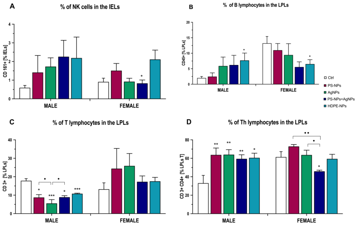
Figure 2.
Microbiota composition at the taxonomic level of genus in males (A) and females (B). Bars represent microbial abundance in each experimental group for each sex. PSNPs – rats exposed to commercial polystyrene nanoparticles (n = 10); AgNPs – rats exposed to silver nanoparticles (n = 10); PSNPs+AgNPs – rats exposed to both commercial polystyrene and silver nanoparticles (n = 10); HDPE – rats exposed to plastic nanoparticles isolated from rice bags (n = 10); Control group – rats administered with water (n = 10). Venn diagrams of bacterial ASVs depending on the experimental group in males (C) and females (D). Differential abundance with group-specific bacteria in males (E) and females (F). Values of the Linear Discriminant Analysis (LDA) above the threshold of 2 are considered significant (P < 0.05, without correction).
Figure 2.
Microbiota composition at the taxonomic level of genus in males (A) and females (B). Bars represent microbial abundance in each experimental group for each sex. PSNPs – rats exposed to commercial polystyrene nanoparticles (n = 10); AgNPs – rats exposed to silver nanoparticles (n = 10); PSNPs+AgNPs – rats exposed to both commercial polystyrene and silver nanoparticles (n = 10); HDPE – rats exposed to plastic nanoparticles isolated from rice bags (n = 10); Control group – rats administered with water (n = 10). Venn diagrams of bacterial ASVs depending on the experimental group in males (C) and females (D). Differential abundance with group-specific bacteria in males (E) and females (F). Values of the Linear Discriminant Analysis (LDA) above the threshold of 2 are considered significant (P < 0.05, without correction).
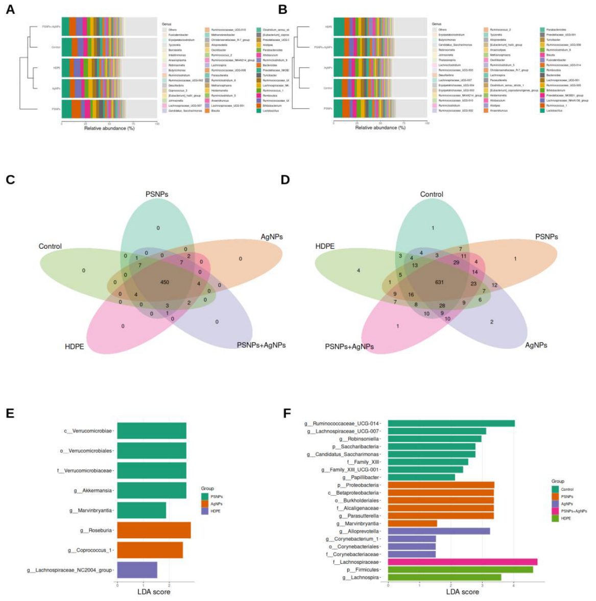
Figure 3.
Bacteria differentiating the experimental groups (A – PSNPs, B – AgNPs, C – PSNPs+AgNPs, D – HDPE) from the control group at the genus level. The X axis encapsulates the scope of the difference and the Y axis reflects the statistical significance, with named families above the threshold of 1 being statistically different. The left part of the plot shows bacteria specific to the control, and the right part the ones specific to the corresponding experimental group. The transparency of the dots corresponds to the prevalence of the individual family or genus within the experimental group, while its size corresponds to the mean abundance.
Figure 3.
Bacteria differentiating the experimental groups (A – PSNPs, B – AgNPs, C – PSNPs+AgNPs, D – HDPE) from the control group at the genus level. The X axis encapsulates the scope of the difference and the Y axis reflects the statistical significance, with named families above the threshold of 1 being statistically different. The left part of the plot shows bacteria specific to the control, and the right part the ones specific to the corresponding experimental group. The transparency of the dots corresponds to the prevalence of the individual family or genus within the experimental group, while its size corresponds to the mean abundance.
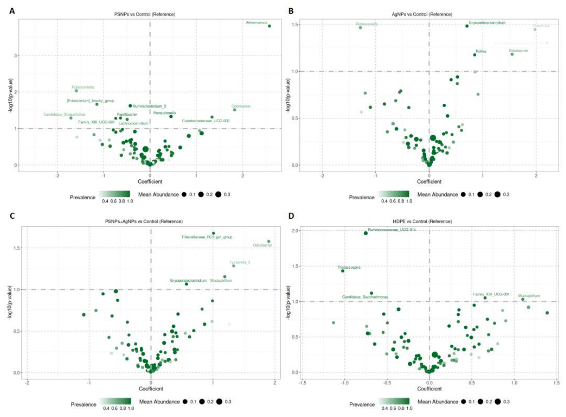
Figure 4.
Fecal content of short-chain fatty acids (SCFA). (A): lactic acid, (B): acetic acid, (C): iso-butyric acid, and (D): valeric acid. Data are expressed as mean ± SEM. **, *** significantly different from the respective group (** P < 0.01, *** P < 0.001) (Fischer post-hoc test). ●, ●● significant difference between exposure groups in the same sex according to Fischer post-hoc test (● p < 0.05, ●● p < 0.01). PSNPs – rats exposed to commercial polystyrene nanoparticles (n = 10); AgNPs – rats exposed to silver nanoparticles (n = 10); PSNPs+AgNPs – rats exposed to both commercial polystyrene and silver nanoparticles (n = 10); HDPE – rats exposed to plastic nanoparticles isolated from rice bags (n = 10); Control group – rats administered with water (n = 10).
Figure 4.
Fecal content of short-chain fatty acids (SCFA). (A): lactic acid, (B): acetic acid, (C): iso-butyric acid, and (D): valeric acid. Data are expressed as mean ± SEM. **, *** significantly different from the respective group (** P < 0.01, *** P < 0.001) (Fischer post-hoc test). ●, ●● significant difference between exposure groups in the same sex according to Fischer post-hoc test (● p < 0.05, ●● p < 0.01). PSNPs – rats exposed to commercial polystyrene nanoparticles (n = 10); AgNPs – rats exposed to silver nanoparticles (n = 10); PSNPs+AgNPs – rats exposed to both commercial polystyrene and silver nanoparticles (n = 10); HDPE – rats exposed to plastic nanoparticles isolated from rice bags (n = 10); Control group – rats administered with water (n = 10).
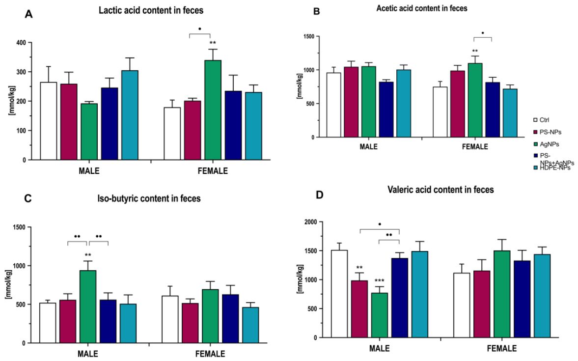
Figure 5.
Sum of m/z metabolite counts of selected metabolites in the colon tissue of rats: (A): caprylic acid, (B): anandamide, (C): androsterone glucuronide, (D): 2-hydroxyestrone, and (E): 18-hydroxycorticosterone. Data are expressed as mean ± SEM. *, **, *** significantly different from the respective control group (* P < 0.05, ** P < 0.01, *** P < 0.001) (Fischer post-hoc test). ●, ●●,●●● significant difference between exposure groups in the same sex according to Fischer post-hoc test (● p < 0.05, ●● p < 0.01, ●●● p < 0.001). PSNPs – rats exposed to commercial polystyrene nanoparticles (n = 8); AgNPs – rats exposed to silver nanoparticles (n = 8); PSNPs+AgNPs – rats exposed to both commercial polystyrene and silver nanoparticles (n = 8); HDPE – rats exposed to plastic nanoparticles isolated from rice bags (n = 8); Control group – rats administered with water (n = 8).
Figure 5.
Sum of m/z metabolite counts of selected metabolites in the colon tissue of rats: (A): caprylic acid, (B): anandamide, (C): androsterone glucuronide, (D): 2-hydroxyestrone, and (E): 18-hydroxycorticosterone. Data are expressed as mean ± SEM. *, **, *** significantly different from the respective control group (* P < 0.05, ** P < 0.01, *** P < 0.001) (Fischer post-hoc test). ●, ●●,●●● significant difference between exposure groups in the same sex according to Fischer post-hoc test (● p < 0.05, ●● p < 0.01, ●●● p < 0.001). PSNPs – rats exposed to commercial polystyrene nanoparticles (n = 8); AgNPs – rats exposed to silver nanoparticles (n = 8); PSNPs+AgNPs – rats exposed to both commercial polystyrene and silver nanoparticles (n = 8); HDPE – rats exposed to plastic nanoparticles isolated from rice bags (n = 8); Control group – rats administered with water (n = 8).
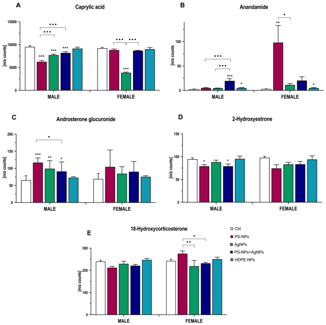
Figure 6.
Scheme of the experimental design. Designed using elements by ©irasutoya via Canva.com.
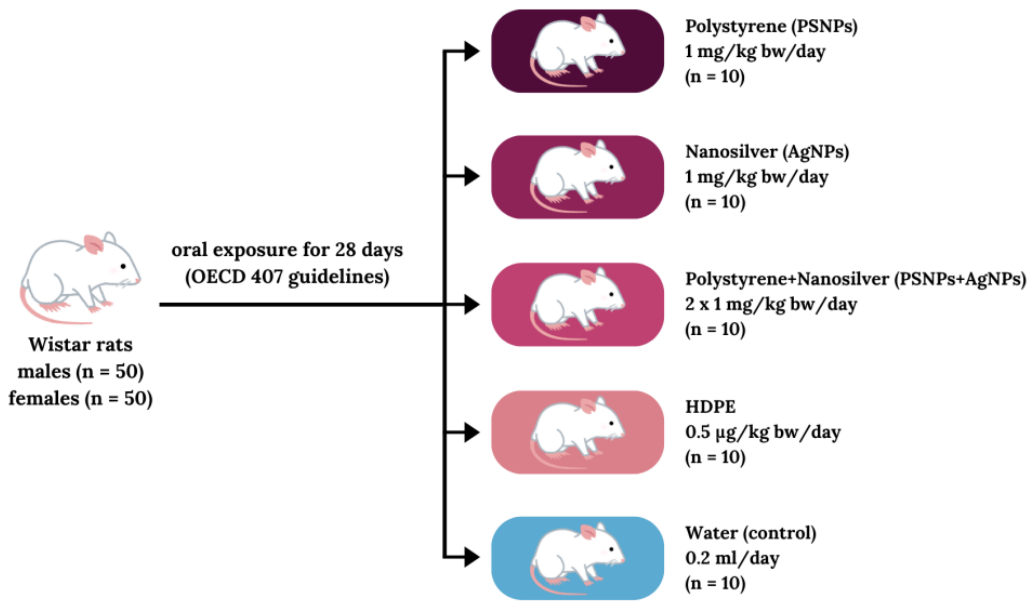

Table 1.
Characteristics of nanoparticles (measured in deionized water). AgNPs, PSNPs and HDPE characteristics adapted from [57,58,60].
| PSNPs | AgNPs | PSNPs+AgNPs | HDPE | |
| Nominal size of particles [nm] | 20 ± 5 | 20 ± 5 | - | - |
| Hydrodynamic Diameter [nm] | 62.4 ± 3.1 | 197.4 ± 2.7 | 290.1 ± 34.1 | 217.0 ± 24.1 |
| Polydispersity index | 0.231 ± 0.24 | 0.295 | 0.701 ± 0.63 | 0.580 ± 0.44 |
| Zeta potential [mV] | -40.02 ± 2.41 | -33.6 | -31.23 ± 1.09 | -33.92 ± 1.02 |
| BET surface area [m2/g] | ND | 2.2419 | ND | ND |
| Micropore volume [cm3/g] | ND | 0.0076 | ND | ND |
| Adsorption average pore width (nm) | ND | 13.6698 | ND | ND |
| Desorption average pore width (nm) | ND | 23.8934 | ND | ND |
Data expressed as mean ± SD (n = 3). ND – not determined, due to the limited amount of material.
Disclaimer/Publisher’s Note: The statements, opinions and data contained in all publications are solely those of the individual author(s) and contributor(s) and not of MDPI and/or the editor(s). MDPI and/or the editor(s) disclaim responsibility for any injury to people or property resulting from any ideas, methods, instructions or products referred to in the content. |
© 2026 by the authors. Licensee MDPI, Basel, Switzerland. This article is an open access article distributed under the terms and conditions of the Creative Commons Attribution (CC BY) license (http://creativecommons.org/licenses/by/4.0/).
Copyright: This open access article is published under a Creative Commons CC BY 4.0 license, which permit the free download, distribution, and reuse, provided that the author and preprint are cited in any reuse.