

Submitted:
09 November 2026
Posted:
10 November 2026
You are already at the latest version
Abstract
The intersection of radiotherapy and immunology has emerged as a highly promising frontier: localized radiotherapy can act as an in-situ vaccine, triggering prompt systemic anti-tumor responses. Carbon ion radiotherapy (CIRT) outperforms conventional photon radiotherapy (XRT) by delivering a highly concentrated dose to the target volume while minimizing collateral damage to adjacent healthy structures and preserving peripheral immune tissues. Emerging preclinical and clinical data indicate that CIRT possesses ex-traordinary immunomodulatory potential. High-LET carbon ion irradiation strongly drives immunogenic cell death (ICD), promotes mature dendritic cell antigen presentation, and strongly upregulates the cGAS–STING signaling cascade, shifting the TME from a "cold" immunosuppressive state to a "hot" immunostimulatory environment. Recent find-ings also show that carbon ions potently trigger non-apoptotic cell death modalities, fer-roptosis, necroptosis, and autophagy, which amplify ICD. When combined with IT, this triggers a potent synergistic effect that targets both local tumor recurrence and distant me-tastases. However, clinical translation faces critical issues, including patient selection and optimal dosing and scheduling. This review synthesizes key preclinical breakthroughs and emerging clinical data on CIRT combined with IT, exploring the molecular mecha-nisms underlying the synergy of this combination. We also discuss perspectives and fu-ture directions needed to optimize this promising therapeutic strategy.
Keywords:
carbon ion radiotherapy
; immunotherapy
; synergy
1. Introduction
The clinical landscape of oncology has changed profoundly with the success of immunotherapy (IT), which has provided durable survival benefits in advanced malignancies. However, some patients fail to achieve objective responses because of the immunosuppressive tumor microenvironment (TME). Therefore, identifying effective combinatorial strategies that can remodel the TME and sensitize tumors to systemic IT is a priority in modern oncology. The success of immunotherapy, which has provided important survival benefits in advanced malignancies, has changed the clinical landscape of oncology. In many patients, however, objective responses are difficult to achieve because the tumor microenvironment (TME) is immunosuppressive [1,2]. Therefore, identifying effective combinatorial strategies to remodel the TME and sensitize tumors to systemic immunotherapy is a leading priority in modern oncology. The intersection of radiation oncology and immunology has emerged as a highly promising frontier: localized radiotherapy (RT) can act as an in-situ vaccine, triggering prompt systemic anti-tumor responses, best illustrated by the abscopal effect [3]. Carbon ion radiotherapy (CIRT), characterized by its unique Bragg peak, provides a superior therapeutic alternative to conventional XRT by delivering a highly concentrated radiation dose immunotherapy to the target volume while minimizing collateral damage to adjacent healthy structures and preserving peripheral immune tissues. Its high linear energy transfer (LET) generates dense, complex, clustered deoxyribonucleic acid (DNA) double-strand breaks, making it highly effective against traditionally radioresistant, hypoxic, or deeply situated tumors [4,5,6,7].
Emerging preclinical and clinical data indicate that CIRT possesses extraordinary immunomodulatory potential. High-LET carbon ion irradiation strongly drives immunogenic cell death (ICD), promotes mature dendritic cell (DC) antigen presentation, and strongly upregulates the cyclic GMP-AMP synthase-stimulator of interferon genes (cGAS–STING) signaling cascade, shifting the TME from a “cold,” immunosuppressive state to a “hot,” immunostimulatory environment [8,9,10]. When combined with immunotherapy, this triggers a potent synergistic effect that targets both local tumor recurrence and distant micrometastases.
Recent preclinical data also show that high-LET carbon ions potently trigger non-apoptotic regulated cell death modalities, ferroptosis, necroptosis, and autophagy, which amplify ICD [11,12,13].
However, the clinical translation of this combined therapy faces critical issues regarding optimal dosing and scheduling, patient selection, and potential toxicities across different cancer types. This comprehensive review synthesizes the latest preclinical breakthroughs and emerging clinical data on CIRT combined with immunotherapy.
Here, we explore the synergistic potential of combining CIRT with immunotherapy, detail the underlying molecular mechanisms, and summarize key preclinical breakthroughs and active clinical trials. We also discuss perspectives and future directions needed to optimize this promising therapeutic strategy.
2. Comparative Profile: Carbon Ion Radiotherapy (CIRT) vs. Photon Radiotherapy (XRT)
For decades, photon radiotherapy (XRT) has been the cornerstone of oncology. However, it has intrinsic physical and biological limitations that often hinder its effectiveness as an in-situ tumor vaccine. Because photons follow an exponential attenuation path, XRT inevitably deposits significant radiation doses into surrounding healthy tissues, draining lymph nodes and circulating blood, triggering radiation-induced lymphopenia and destroying the systemic immune apparatus at the very moment it needs to be primed. Furthermore, XRT’s reliance on oxygen-mediated free radical generation renders it poorly effective against the severely hypoxic cores of bulky, advanced tumors.
Carbon ion radiotherapy (CIRT) represents a paradigm shift in both radiation ballistics and tumor immunogenicity. Carbon ions have a unique depth-dose profile characterized by the Bragg Peak, enabling sub-millimeter, almost surgical precision. CIRT travels through healthy tissue with minimal energy deposition, releases a massive, lethal energy burst precisely within the tumor volume, and drops to virtually zero immediately afterward. Beyond this physical superiority, CIRT delivers high linear energy transfer (high-LET) radiation, causing dense, complex, clustered double-strand DNA breaks that bypass cellular repair mechanisms and overcome hypoxic radioresistance.
To illustrate these differences, Table 1 contrasts the biophysical and immunological profiles of XRT and CIRT.
3. Molecular and Cellular Mechanisms of CIRT and IT Combination
3.1. Immunogenic Cell Death (ICD)
While conventional photon-based radiotherapy relies on inducing apoptosis, a process often immunologically silent or tolerogenic, CIRT fundamentally alters the tumor microenvironment by triggering robust immunogenic cell death [14]. Because of its high linear energy transfer and distinctive Bragg peak, CIRT induces dense, complex, clustered DNA double-strand breaks (DSBs) that overwhelm the cell’s DNA damage repair mechanisms and force cells to bypass canonical apoptotic pathways, promoting inflammatory, highly immunogenic, non-apoptotic forms of programmed cell death. This catastrophic cellular demise drives widespread spatial exposure and massive release of damage-associated molecular patterns (DAMPs)—such as surface calreticulin (CRT), extracellular adenosine triphosphate (ATP), and high-mobility group box 1 (HMGB1)—at levels significantly higher than those achieved by low-LET photon modalities. These danger signals act as potent “eat me signals” (calreticulin) and adjuvants that orchestrate DC maturation, enhance tumor antigen cross-presentation, and prime the TME for synergistic combinations with immune checkpoint inhibitors (ICIs) [13,15].
3.1.1. The cGAS-STING Pathway: The Bridge from Clustered DNA Damage to Innate Immunity
Central to CIRT’s immunomodulatory efficacy is activation of the cyclic GMP-AMP synthase (cGAS)-stimulator of interferon genes (STING) pathway. High-LET carbon ions induce chromosomal aberrations and micronuclei formation, leading to leakage of genomic double-stranded DNA (dsDNA) into the cytoplasm during aberrant mitoses. Cytosolic dsDNA is rapidly recognized by the innate immune sensor cGAS, catalyzing the synthesis of the secondary messenger 2’,3’-cyclic GMP-AMP (cGAMP) [16,17].
Upon binding cGAMP, STING recruits and activates the TANK-binding kinase 1 (TBK1) cascade, which phosphorylates interferon regulatory factor 3 (IRF3) and nuclear factor-kappa B (NF-κB), driving their nuclear translocation [18]. The resulting transcriptional program strongly upregulates type I interferons (primarily IFN-α and IFN-β) and pro-inflammatory chemokines (e.g., C-X-C motif chemokine ligand CXCL9, CXCL10). Crucially, this type I interferon signature links local CIRT-induced damage to systemic immunity by promoting the recruitment, activation, and clonal expansion of cytotoxic CD8+ T lymphocytes and natural killer (NK) cells. By converting a cold tumor architecture into an immune-receptive niche, cGAS-STING activation addresses a primary mechanism of resistance to anti-PD-1/anti-PD-L1 therapies [14,17,19,20].
However, cell-intrinsic upregulation of Three-Prime Repair Exonuclease 1 (TREX1) can actively undermine the immunogenic potential of CIRT. TREX1 serves as the primary mammalian cytosolic 3’-to-5’ exonuclease whose homeostatic function is to clear accessory DNA fragments to prevent aberrant autoimmunity. In cancer therapy, malignant cells exploit TREX1 to execute an immune-evasion program by rapidly degrading radiation-induced cytosolic dsDNA before cGAS can sense it, extinguishing downstream type I IFN signaling and subsequent T-cell-dependent antitumor immunity [21,22,23].
3.1.2. Non-Apoptotic Mechanisms of CIRT-Induced Cell Death
To maximize the therapeutic potential of combined immunotherapy, CIRT actively orchestrates three interconnected, non-apoptotic, cell death pathways that amplify the inflammatory context of the TME.
- Ferroptosis: Unlocking Iron-Dependent Cell Death for Enhanced Efficacy
CIRT delivers an intense localized burst of reactive oxygen species (ROS), severely disrupting cellular redox homeostasis and triggering ferroptosis—an iron-dependent form of regulated necrosis driven by lethal lipid peroxidation. The synergy between CIRT and IT depends on specific molecular pathways and biomarkers that link high-LET radiation damage to immune-mediated tumor clearance. These mechanisms center on iron accumulation, lipid peroxidation and the impairment of cellular antioxidant defense mechanisms. Three primary metabolic and signaling cascades drive the cross-talk between CIRT and immunotherapy:
The System / GSH / GPX4 Axis. This primary cellular defense pathway against ferroptosis imports cystine into the cell to synthesize glutathione (GSH), which the enzyme glutathione peroxidase 4 GPX4) uses as a cofactor to neutralize toxic lipid peroxides. Immunotherapy-activated CD8+ T cells release IFN-gamma (IFN-γ), which downregulates expression of the System subunit. Concurrently, CIRT suppresses DHODH (dihydroorotate dehydrogenase) [24] and downregulates GPX4. Together, they collapse the tumor’s antioxidant shield, leaving it completely vulnerable to lipid peroxidation [25,26,27].
Lipid and Amino Acid Metabolism Pathways. For a cell to undergo ferroptosis, it must incorporate polyunsaturated fatty acids (PUFAs) into the cell membrane. This process depends heavily on ACSL4 (acyl-CoA synthetase long-chain family member 4), which esterifies PUFAs [28,29]. CIRT significantly upregulates ACSL4 expression, enriching lipid metabolism pathways and providing the “fuel” for lipid peroxidation. IFN-γ from immunotherapy further primes these lipid metabolism genes, including 2,4-Dienoyl-CoA Reductase 2 (DECR2) and ACSL4, creating an explosive accumulation of membrane-destroying reactive oxygen species (ROS) [30,31].
The cGAS-STING & Ferritinophagy Pathway. Activated cGAS-STING (through dsDNA leaked into the cytoplasm) induces ferritinophagy - the autophagic degradation of ferritin via the Nuclear Receptor Coactivator 4 (NCOA4) - Ferritin Heavy Chain 1 (FTH1) axis) [32,33,34] - which releases a flood of labile intracellular iron. The free iron drives the Fenton reaction, amplifying the oxidative stress generated by radiation and transforming the local tumor environment into an immunogenic zone [35,36].
- Necroptosis: Evading Apoptosis Resistance
Conventional radiation relies mainly on apoptosis to eliminate tumor cells, a process that intrinsic tumor mutations such as protein p53 mutations or B-cell lymphoma 2 (Bcl-2) overexpression can easily block. CIRT bypasses these resistance mechanisms through specific molecular changes termed “the epigenetic switch.” The complex, clustered DNA double-strand breaks induced by carbon ions trigger a global super-enhancer remodeling network [12]12 that suppresses pro-survival regulators such as caspase-8. Without caspase-8 activity to cleave and deactivate necroptotic machinery, the cell is forcefully directed into necroptosis [37].
Once the apoptotic checkpoints are bypassed, CIRT triggers a molecular cascade for necrosome assembly: receptor-interacting serine/threonine kinase 1 (RIPK1) recruits and phosphorylates RIPK3, which subsequently phosphorylates mixed lineage kinase domain-like pseudokinase (pMLKL), which translocates to the plasma membrane, forming pore complexes that cause the cell to swell and burst [38,39]. Preclinical evidence demonstrates that CIRT induces a massive upregulation of pMLKL (up to a 28-fold increase in pMLKL levels in resistant cancer cell lines) compared to XRT [40]. Necrotic cell death is a major source of high concentrations of DAMPs, including HMGB1, heat shock proteins (HSPs), and unmethylated cytosine-phosphate-guanine DNA (CpG DNA), which act as powerful immunological signals to recruit myeloid cells and break peripheral immune tolerance within the TME.
- Autophagy: A Double-Edged Sword in Tumor Fate
High-LET carbon ion beams induce profound cellular stress, heavily stimulating autophagic pathways. Under the intensive stress of high-LET carbon ions, hyper-activated autophagy can cross the threshold from survival to autophagic cell death, directly contributing to tumor elimination. Depending on the tumor microenvironment and therapeutic timing, autophagy can either amplify the immunogenicity of dying cancer cells or act as a survival shield that promotes immune evasion [41,42].
1. The pro-immunogenic role: When behaving as an ally to IT, CIRT-induced autophagy enhances ICD via the active secretion of extracellular ATP who binds specific receptors (P2RX7) on dendritic cells, serving as a powerful chemoattractant, and via the released tumor-associated antigens, encapsulated in the autophagosomes, who are highly efficiently cross-presented on Major Histocompatibility Complex Class I (MHC-I) molecules to CD8+ cytotoxic T cells and boost the efficacy of anti-PD-1/anti-PD-L1 check-point inhibitors [43,44].
2. The pro-tumorigenic role: elevated autophagy in stressed tumor cells degrades critical cytotoxic components (such as granzyme B delivered by T cells) and can stabilize immune checkpoints like Programmed Death-Ligand 1 (PD-L1), allowing sub-lethally irradiated tumor cells to survive both CIRT and subsequent T-cell attacks. Autophagy also supports the metabolic survival and stability of regulatory T cells (Tregs) in the hypoxic tumor core; therefore, high autophagic activity in the TME can favor Treg-mediated immune tolerance [41,45]. Combined with IT, targeted inhibition of the cytoprotective phase of autophagy can accelerate cell transit into lytic necrosis and ferroptosis, enabling a finely adjustable mechanism that balances maximal tumor cell death with optimized release of immunogenic material [12,46].
These above-mentioned cooperating mechanisms are presented in Figure 1.
3.2. Modulation of the TME.
CIRT generates a unique tumor immune microenvironment through radiation-induced coordinated changes. Gameiro et al. investigated radiation-induced alterations in the tumor immune microenvironment. They found increased infiltration of cytotoxic T lymphocytes (CD8+) and helper T cells (CD4+), enhanced natural killer (NK) cell activity, DC maturation, decreased regulatory T cell (Treg) populations and suppressed myeloid-derived suppressor cell (MDSC) function. They also showed that high-LET radiation upregulates a broad spectrum of immunologically relevant molecules at the cellular level, including immune recognition molecules - Human Leukocyte Antigen (HLA), Intercellular Adhesion Molecule 1 (ICAM-1), Carcinoembryonic Antigen CEA, Mucin-1 (MUC-1), surface-exposed Calreticulin (CRT), damage-associated molecular patterns (HMGB1, ATP), and the immune checkpoint protein PD-L1 [47]. The most important mechanisms are:
- Overcoming tumor hypoxia. Hypoxic zones in tumors resist conventional photon radiation and strongly suppress the immune system. Scientific and clinical evidence clearly shows that increased particle LET affects cell killing and tumor response in hypoxia [6]. CIRT destroys cells through direct ionization rather than relying on oxygen-derived free radicals [50]. By eliminating hypoxic tumor cores, CIRT disrupts the hypoxia-driven accumulation of immunosuppressive cells (Tregs and MDSCs), opening the door for immunotherapies to function effectively.
- Downregulation of myeloid-derived suppressor cells (MDSCs) [15] and regulatory T-cells (Tregs), while shifting tumor-associated macrophages from the pro-tumor M2 phenotype to the anti-tumor M1 phenotype [51]. The abundance of myeloid-derived suppressor cells (MDSC) in the tumor microenvironment is associated with therapeutic efficacy and disease outcome. We established murine melanoma B16, MelanA, and S91 tumor models in syngeneic immunocompetent mice and irradiated them with carbon ion beams. Carbon ion beam irradiation (5 Gy) in melanoma-bearing mice reduced MDSC populations in the bone marrow, peripheral blood and spleen via a Janus kinase 2/Signal Transducer and Activator of Transcription 3 (JAK2/STAT3) -dependent mechanism. After radiation, the percentages of CD3+, CD4+, CD8+ T cells, macrophages, and natural killer cells increased, resulting in reduced tumor growth and prolonged overall survival [52].
- Improved Chimeric Antigen Receptor T (CAR-T) cell recognition of tumor cells. CIRT induces double-stranded DNA breaks more frequently and with greater complexity, making them harder to repair. This unique damage pattern is more likely to trigger ICD and promote DAMP release [53]. This mechanism not only enhances antitumor immunity but also improves CAR-T cell recognition of tumor cells. At the spatial level, radiation therapy improves CAR-T cell homing and infiltration. At the molecular level, it overcomes antigen heterogeneity, enhances T cell function, and extends the antitumor immune response over time [54].
3.3. Lymphocyte Sparing
Radiation-induced lymphopenia (RIL) is a known side effect of radiotherapy [55]. When XRT with large radiation fields encompasses tumor-draining lymph nodes and hematopoietic and lymphoid organs such as the bone marrow, spleen, and thymus, it can compromise both lymphatic circulation and hematopoiesis, reducing lymphocyte numbers. Lymphocytes are essential for mounting effective anti-tumor immune responses. In contrast, proton and heavy-ion beams exhibit a sharp dose fall-off at the field edges, causing less damage to surrounding lymphoid tissues and preserving more functional lymphocytes. A recent statistical analysis further showed that patients treated with XRT have consistently lower relative lymphocyte counts at treatment completion than those receiving ion therapy. This finding solidifies the aforementioned perspective [56].
3.4. Up-Regulation of Immune Checkpoints
Massive DNA damage activates downstream STAT1 and IFN signaling pathways. This causes tumor cells to highly upregulate PD-L1 expression. While this upregulation is originally a tumor defense mechanism, it creates the perfect target for anti-PD-1/anti-PD-L1 drugs (a synergistic trap). Administering checkpoint inhibitors precisely when CIRT forces PD-L1 expression removes the tumor’s brakes and creates a highly lethal synergy.
In a study conducted by Zhou et al [14], they demonstrated that carbon ion irradiation (5Gy) of human osteosarcoma U2OS cells and mouse melanoma B16/S91 cells and also of B16 and S91 melanoma-bearing mice induced ICD characterized by the exposure of calreticulin on the plasma membrane surface, the phosphorylation of eukaryotic initiation factor 2, alpha subunit (eIF2a), the release of ATP into the extracellular space, the exodus of HMGB1 from the nucleus, and the induction of the type-1 interferon response in vitro and in vivo. 5 Gy of CIRT induced tumor immunogenicity more effectively than 5 Gy of XRT. Combining irradiation with anti-PD-1 therapy further improved CIRT’s effect and enhanced immune response markers in blood, spleen, and tumors in vivo; increased PD-L1 expression; and significantly prolonged survival in treated animals [14]. Carbon ion irradiation caused more DNA damage and activated the cGAS–STING pathway than X-ray irradiation and it slowed melanoma growth in a syngeneic model. Although X-ray irradiation is not effective for melanoma treatment, carbon ion irradiation showed a significant anti-tumor effect. Tumor infiltrating lymphocytes (TIL) analysis revealed that CIRT boosted infiltration of NK, CD4+ and CD8+ T cells, while increasing the levels of immune checkpoints (programmed death-1, PD-1; lymphocyte activation gene 3, LAG-3; and T-cell immunoglobulin and mucin domain-containing protein 3, TIM-3). Moreover, CIRT increased PD-L1 exposure on the cell surface compared with X-ray group. Furthermore, CIRT combined with PD-L1 inhibitor therapy increased the number of T cells and NK cells in melanoma and slowed melanoma growth compared with other therapies. CIRT increased DNA damage in tumor cells and improved the immune response in melanoma, suggesting that CIRT could be a potential synergistic treatment with radiotherapy and radioimmunotherapy in melanoma patients [19].
The immunogenic potential of CIRT, which is highly efficient at generating dense, clustered double-strand breaks and a significantly higher yield of cytosolic double stranded DNA (dsDNA) fragments per unit dose than conventional photon therapy, can be actively undermined by cell-intrinsic upregulation of Three-Prime Repair Exonuclease 1 (TREX1) [57]. This enzyme clears accessory DNA fragments to prevent aberrant autoimmunity, acting as a critical gatekeeper of the cGAS-STING axis in CIRT-IT synergy. In cancer therapy, malignant cells exploit TREX1 to execute an immune-evasion program by rapidly degrading radiation-induced cytosolic dsDNA before cGAS can sense it, thereby extinguishing downstream type I IFN signaling and subsequent T-cell-dependent antitumor immunity [21]. In managing CIRT and IT combinations, radiation dose and treatment scheduling rely critically on TREX1 expression and kinetics: delivering high-dose carbon-ion fractions consecutively can trigger a cell-intrinsic threshold that upregulates TREX1, which rapidly degrades cytosolic dsDNA before immune sensing occurs [58]. Consequently, identifying the exact therapeutic window of CIRT that avoids TREX1 activation, or combining CIRT with emerging small-molecule TREX1 inhibitors, represents a vital strategy to maximize the efficacy of next-generation radio-immunotherapy combinations. By sparing adjacent lymphoid structures and circulating lymphocytes while simultaneously triggering a robust cascade of immunogenic danger signals, CIRT creates an ideal microenvironmental window. When paired with immunotherapy, this profound remodeling transforms the tumor site from an immunosuppressive sanctuary into a powerful inductive hub for systemic antitumor immunity.
4. Preclinical Evidence and Emerging Clinical Trials Supporting the Combination of CIRT and ICIs
4.1. Preclinical Foundations
Growing evidence supports the idea that, when paired with ICIs, CIRT outperforms conventional XRT. Main arguments (mechanisms), described in detail above (section 5), are: amplified ICD, due to complex, clustered DNA double-strand breaks who drastically upregulate the cGAS-STING-IFN1 pathway; reshaping of the TME and converting a “cold” (immunosuppressive) tumor into a “hot” (immunostimulatory) one; overcoming the hypoxia-driven resistance by eliminating a major source of immunosuppressive signaling (such as hypoxia-inducible factors) and creating a fertile environment for ICIs to function; the abscopal effect and development of durable, tumor-specific immune memory.
In vitro studies on cell lines evaluated the combination’s cytotoxicity and the immune activation induced by CIRT [14,19,59,60], while in vivo studies in animal models evaluated the efficacy, toxicity, and safety profile of CIRT and various checkpoint inhibitor combinations [14,19,58,60,61,62,63,64,65]. Table 2 presents the most relevant preclinical studies supporting the enhanced efficacy of CIRT combined with ICIs.
4.2. Emerging Clinical Trials.
Based on available preclinical data, a growing number of clinical trials [66,67,68,69,70] explore the safety and tolerability of combination treatments, efficacy outcomes in radioresistant and advanced malignancies, and predictive and response biomarkers. Table 3 describes some relevant clinical trials.
5. Future Perspectives and Challenges
The integration of CIRT and IT represents one of the most promising frontiers in precision oncology. CIRT’s unique physical and biological properties make it a potent partner for transforming “cold” tumors into immunologically “hot” ones.
5.1. Focusing on Key Mechanisms of This Combined Therapeutic Axis Drives Future Synergy, Optimizing Clinical Design and Overcoming Existing Operational Barriers:
- Maximizing the abscopal effect: complex, clustered double-strand DNA damage triggers ICD and releases tumor-associated antigens and damage-associated molecular patterns, acting as an in-situ vaccine that, combined with immune checkpoint inhibitors (ICIs), uses local CIRT to eradicate distant, un-irradiated metastatic lesions [14,71].
- Modulation of TME: activating the cGAS-STING pathway upregulates Type I IFN, driving DC maturation and heavy infiltration of cytotoxic CD8+ T cells and NK cells into previously “cold”, immunosuppressive tumors, transforming them into “hot”, inflamed, and therapy-responsive ones [48].
- Overcoming hypoxia-induced resistance: with a very low Oxygen Enhancement Ratio (OER), CIRT destroys hypoxic tumor cores and disrupts the hypoxia-driven accumulation of immunosuppressive cells, opening the door for immunotherapies to function effectively [6].
- Clinically integrate new data on regulated cell death pathways, specifically ferroptosis, necroptosis, and autophagy. To translate these biological insights from bench to bedside, future research must prioritize developing new biomarkers and identifying synergistic therapeutic combinations, such as pairing CIRT with specific ferroptosis inducers or necroptosis sensitizers. Implementing high-throughput screening platforms and patient-derived organoid models will be essential to map individual tumor susceptibilities to these cell death mechanisms.
5.2. Optimizing Clinical Design (Dose, Timing and Sequencing). Determining Whether Immunotherapy Should Be Administered Concurrently, Sequentially or as Neoadjuvant Priming before CIRT is of Major Concern.
- In conventional XRT, the threshold dose that triggers a massive, immunity-blunting upregulation of TREX1 is 12 to 18 Gy per fraction. Carbon ions possess high LET and are far more potent than photons, so single high doses (such as 8 to 10 Gy or higher can dose-dependently upregulate TREX1 expression. Single-fraction doses should ideally be optimized to stay below the 8–10 Gy (roughly 2-5Gy) threshold to prevent up-regulation of TREX1, when systemic immune priming is the primary goal. This optimization window maximizes cytoplasmic dsDNA without triggering the TREX1 clearance mechanism [22,58,72].
- Determining whether immunotherapy should be administered concurrently, sequentially, or as neoadjuvant priming before CIRT remains a major issue. Because immunotherapy agents remodel the TME via specific kinetic timelines, the choice between concurrent vs sequential delivery depends on the class of IT used. Anti-Cytotoxic T-Lymphocyte-Associated Protein 4 (CTLA4) agents act primarily in the lymph nodes to deplete Tregs and expand naïve T cell clones before they travel to the tumor, therefore giving them 1-2 w. before CIRT ensure a robust pool of responsive T-cells ready to receive the wave of antigens released by carbon ions [73]. For anti-PD-1 –anti-PD-L1 agents, because the antigen peak window is 2-7 days post-irradiation and maximum upregulation of PD-L1 on surviving tumor cells is at 24- 72 h after the last radiation fraction, the sequential delivery starting 1 to 7 days after the final CIRT fraction perfectly intercepts this window [58]. Concurrent delivery is reserved only for costimulatory agonists (anti-OX40), as they require immediate, concurrent interaction with the cytokine storm triggered during delivery of CIRT [74].
5.3. Multi-OMICS and AI for Patient selection.
The future relies on integrating artificial intelligence, radiomics signatures, liquid biopsies, and tumor RNA sequencing to identify genetic signatures beyond PD-L1 expression (such as tumor mutational burden or TME signatures and distinct mesenchymal resistance markers) to precisely select candidates who will benefit and maximize this synergistic loop [75,76]. Integrating synergistic therapeutic combinations using ferroptosis inducers or necroptosis sensitizers also holds immense potential to overcome radioresistance in hypoxic and immunologically cold tumors. Ultimately, AI-driven predictive modeling can orchestrate these multi-pathway dynamics to enable highly personalized CIRT regimens, maximizing tumor ablation while minimizing toxicity to surrounding healthy tissues.
5.4. Overcoming Technological and Access Barriers.
CIRT centers are limited, with fewer than twenty operational centers globally [77]. The immense initial capital investment, structural and engineering complexities, and high establishment costs have limited the number of clinical centers with long-term CIRT experience and reduced large-scale clinical trials. Concurrently, traditional treatment planning systems remain limited by physical and localized RBE algorithms and cannot simulate dynamic immune kinetics. Overcoming this software barrier requires developing immunologically guided adaptive planning platforms. Driven by machine learning, these advanced algorithms could integrate real-time, spatially resolved lymphocyte-sparing trajectories, model tumor microenvironment hypoxia, and optimize LET maps to avoid TREX1-inducing hot spots.
6. Conclusions
Integrating carbon ion radiotherapy and immunotherapy represents a paradigm shift in precision oncology, offering a powerful strategy to overcome immune tolerance and trigger systemic tumor regression. By using precision carbon ions to preserve circulating immune cells, keeping doses below the TREX1 threshold to protect STING signaling, and carefully timing the checkpoint blockades, we can turn a localized treatment into a powerful systemic cure.
However, realizing the full potential of this combined therapy requires overcoming significant and logistical and biological hurdles. Logistically, the field must move beyond geographic centralization and high capital costs. Developing compact, superconducting gantry designs and immunologically guided treatment-planning software will be vital to increase patient access. Biologically, future strategies must account for the TREX1 homeostatic threshold to prevent immune silencing through optimized hypofractionation schedules, incorporate discoveries about regulated cell death pathways, and validate robust multi-omic biomarkers to identify patients who could benefit most from this combined treatment. By bridging advanced physics and dynamic immune kinetics, the oncology community could eventually translate this modality into a global pillar of systemic cancer care.
Author Contributions
Conceptualization, I.B., M.P.S., P.V. and D.M.; methodology, I.B., M.P.S., P.V., C.B., E.C.G., I.L. and D.M.; software, I.B., M.P.S. and P.V.; validation, I.B., M.P.S., P.V., C.B., E.C.G., I.L. and D.M.; formal analysis, I.B., M.P.S., P.V., C.B., E.C.G., I.L. and D.M.; investigation, I.B., M.P.S., P.V., C.B., E.C.G., I.L. and D.M.; resources, I.B., M.P.S., P.V., C.B., E.C.G., I.L. and D.M.; data curation, I.B., M.P.S., P.V., C.B., E.C.G., I.L. and D.M.; writing—original draft preparation, I.B., and M.P.S.; writing—review and editing, I.B., M.P.S. and P.V.; visualization, I.B., M.P.S. and P.V.; supervision, I.L. and D.M.; project administration, I.B., M.P.S., P.V., I.L. and D.M.; funding acquisition, I.B., M.P.S., P.V., C.B., E.C.G., I.L. and D.M. All authors have read and agreed to the published version of the manuscript.
Funding
The biological part of this work was supported by a grant of the Romanian Government and the Health Program, within the project: “Medical applications of high-power lasers – Dr. LASER”, SMIS Code 326475.
Data Availability Statement
No new data were created or analyzed in this study. Data sharing is not applicable to this article.
Acknowledgments
During the preparation of this review manuscript, the authors used ChatGPT (OpenAI) to assist with text generation, literature searching, and the analysis and synthesis of information retrieved from scientific sources. The authors critically reviewed and verified the AI-assisted content, checked the relevant scientific information against the original sources, and take full responsibility for the accuracy, integrity, and content of the published article.
Conflicts of Interest
The authors declare no conflicts of interest.
Abbreviations
The following abbreviations are used in this manuscript:
| ACSL4 | Acyl-CoA Synthetase Long Chain Family Member 4 |
| ATM | Ataxia- teleangiectazia- mutated |
| ATP | Adenosine triphosphate |
| ATR | Ataxia telangiectasia and Rad3-related protein |
| Bcl-2 | B-cell lymphoma 2 |
| BRCA | Breast cancer gene |
| CAR-T | Chimeric Antigen Receptor T-cell |
| CD | Cluster of differentiation |
| CEA | Carcinoembryonic antigen |
| cGAMP | Cyclic guanosine monophosphate–adenosine monophosphate |
| cGAS | Cyclic GMP-AMP synthase |
| CIRT | Carbon ion radiotherapy |
| CpG-DNA | Cytosine phosphate guanine- deoxyribonucleic acid |
| CR | Complete response |
| CRT | Calreticulin |
| CTLA-4 | Cytotoxic T-Lymphocyte-Associated Protein 4 |
| CXCL | C-X-C motif chemokine ligand |
| DAMPs | Damage-associated molecular patterns |
| DC | Dendritic cells |
| DECR2 | 2,4-Dienoyl-CoA Reductase 2 |
| DHODH | Dihydroorotate dehydrogenase |
| DNA | Deoxyribonucleic acid |
| dsDNA | Double stranded DNA |
| DSB | Double strand breaks |
| eIF2a | Eukaryotic Initiation Factor 2, alpha subunit |
| GPX4 | Glutathione peroxidase 4 |
| GSH | Glutathione |
| HLA | Human Leukocyte Antigen |
| HMGB1 | High Mobility Group Box 1 |
| HSP | Heat Shock Protein |
| ICAM1 | Intercellular Adhesion Molecule 1 |
| ICD | Immunogenic cell death |
| ICIs | Immune checkpoint inhibitor |
| IFN | Interferon |
| IRF3 | Interferon Regulatory Factor 3 |
| IT | Immunotherapy |
| JAK2 | Janus kinase 2 |
| LAG3 | Lymphocyte-activation gene 3 |
| LET | Linear energy transfer |
| MDSCs | Myeloid-derived suppressor cells |
| MHC1 | Major Histocompatibility Complex class I |
| Muc1 | Mucin-1 |
| NCOA4- FTH1 | Nuclear receptor coactivator 4- Ferritin Heavy Chain 1 |
| NF-kB | Nuclear factor kappa-light-chain-enhancer of activated B cells |
| NK | Natural killer |
| OER | Oxygen enhancement ratio |
| OS | Overall survival |
| P2RX7 | Gene encoding P2X7 receptor- ATP-gated ligand-gated ion channel on the surface of immune and glial cells |
| PD-1 | Programmed cell death protein 1 |
| PD-L1 | Programmed Death-Ligand 1 |
| pMLK1 | Phosphorylated mixed-lineage domain-like pseudokinase |
| PR | Partial response |
| PUFAs | Polyunsaturated fatty acid |
| RBE | Relative Biological Effectiveness |
| RIL | Radiation induced lymphopenia |
| RIPK1 | Receptor-Interacting Serine/Threonine-Protein Kinase 1 |
| RNA | Ribonucleic acid |
| ROS | Reactive oxygen species |
| RT | Radiotherapy |
| STAT | Signal Transducer and Activator of Transcription |
| STING | Stimulator of Interferon Genes |
| SV | Survival |
| TAMS | Tumor-associated macrophages |
| TBK1 | TANK-Binding Kinase 1 |
| TIL | Tumor infiltrating lymphocytes |
| TIM3 | T-cell immunoglobulin and mucin domain-containing protein 3 |
| TME | Tumor microenvironment |
| Tregs | Regulatory T cells |
| TREX1 | Three Prime Repair Exonuclease 1 |
| XRT | Photon radiotherapy |
References
- Sharma P, Hu-Lieskovan S, Wargo JA, Ribas A. Primary, Adaptive, and Acquired Resistance to Cancer Immunotherapy. Cell. 2017 Feb 9;168(4):707-723. PMID: 28187290; PMCID: PMC5391692. [CrossRef]
- Liu M, Zhu Y, Liang H, Li Y, Liu H and Li J (2026) Immunosuppressive cells as barriers to cancer therapy: mechanisms and emerging solutions. Front. Immunol. 17:1896156. [CrossRef]
- Formenti S, Demaria S. Radiation Therapy to Convert the Tumor Into an In Situ Vaccine. International Journal of Radiation Oncology, Biology, Physics, 84, 879-880.
- Jung-in Kim, Jong Min Park, Hong-Gyun Wu: Carbon Ion Therapy: A Review of an Advanced Technology. Progress in Medical Physics 31(3), 2020 . [CrossRef]
- Helm, A., Fournier, C. High-LET charged particles: radiobiology and application for new approaches in radiotherapy. Strahlenther Onkol 199, 1225–1241 (2023). [CrossRef]
- Sokol O, Durante M. Carbon Ions for Hypoxic Tumors: Are We Making the Most of Them? Cancers (Basel). 2023 Sep 9;15(18):4494. PMID: 37760464; PMCID: PMC10526811. [CrossRef]
- Tinganelli W, Durante M. Carbon Ion Radiobiology. Cancers (Basel). 2020 Oct 17;12(10):3022. PMID: 33080914; PMCID: PMC7603235. [CrossRef]
- Guo, S., Yao, Y., Tang, Y. et al. Radiation-induced tumor immune microenvironments and potential targets for combination therapy. Sig Transduct Target Ther 8, 205 (2023). [CrossRef]
- Zhu S, Wang Y, Tang J, Cao M. Radiotherapy induced immunogenic cell death by remodeling tumor immune microenvironment. Front Immunol. 2022 Dec 1;13:1074477. PMID: 36532071; PMCID: PMC9753984. [CrossRef]
- Liu, S., He, X., Liang, S. et al. Carbon ion irradiation mobilizes antitumor immunity: from concept to the clinic. Radiat Oncol 20, 85 (2025). [CrossRef]
- Wang Y, Cai H. Carbon-ion radiotherapy induces ferroptosis and M1 macrophage polarization to inhibit the development of gastric cancer by downregulating DHODH. Front Med (Lausanne). 2025 Aug 22;12:1592116. PMID: 40917841; PMCID: PMC12411497. [CrossRef]
- Li C, Liu X, Zhu L, Liu M, Konishi T, Lou X, Chen L, Liao J, Hong Y, Deng Y, Xiao M, Shao C, Kong L, Guo X, Su F. Carbon ion irradiation at a clinically used dose rewires super-enhancers to drive necroptosis and systemic immunity. Sci Adv. 2026 Aug 28;12(35):eaec7590. PMID: 42647624; PMCID: PMC13510677. Epub 2026 Aug 26. [CrossRef]
- Seneviratne, D.; Biswal, P.; Krishnan, S. Novel Physical and Biological Applications of Carbon Ion Radiotherapy. Cancers 2026, 18, 113. [CrossRef]
- Zhou H, Tu C, Yang P, Li J, Kepp O, Li H, Zhang L, Zhang L, Zhao Y, Zhang T, Sheng C, Wang J. Carbon ion radiotherapy triggers immunogenic cell death and sensitizes melanoma to anti-PD-1 therapy in mice. Oncoimmunology. 2022 Mar 25;11(1):2057892. PMID: 35355680; PMCID: PMC8959514. [CrossRef]
- Sudo, M.; Tsutsui, H.; Fujimoto, J. Carbon Ion Irradiation Activates Anti-Cancer Immunity. Int. J. Mol. Sci. 2024, 25, 2830. [CrossRef]
- Qingyu Jiang, Zhiheng Chen, Jin Jiang, Qianping Chen, Huiyin Lan, Ji Zhu, Wei Mao, The role of cGAS-STING in remodeling the tumor immune microenvironment induced by radiotherapy, Critical Reviews in Oncology /Hematology, Volume 209, 2025, 104658, ISSN 1040-8428. [CrossRef]
- Yang, C., Liang, Y., Liu, N. et al. Role of the cGAS-STING pathway in radiotherapy for non-small cell lung cancer. Radiat Oncol 18, 145 (2023). [CrossRef]
- Jiawen Zhang, Sihui Yu, Qiao Peng, Ping Wang, Lan Fang: Emerging mechanisms and implications of cGAS-STING signaling in cancer immunotherapy strategies. Cancer Biol Med 2024. [CrossRef]
- Guo, Y., Shen, R., Wang, F. et al. Carbon ion irradiation induces DNA damage in melanoma and optimizes the tumor microenvironment based on the cGAS–STING pathway. J Cancer Res Clin Oncol 149, 6315–6328 (2023). [CrossRef]
- Alabi F, Imodoye SO, Adedokun KA, Eltokhy MA, Curcic M, Okoyeocha E, Thapa I, Tiwari S, Ogundele AV, Ayo TE, Awe OB, Abd-Rouf MSK. The cGAS-STING pathway in cancer immunotherapy: prognostic value and therapeutic potential. Front Immunol. 2026 Jun 2; 17:1760947. PMID: 42311657; PMCID: PMC13269242. [CrossRef]
- Vanpouille-Box, C.; Alard, A.; Aryankalayil, M.J.; Babb, J.S.; Myles, D.R.; Formenti, S.C.; Demaria, S. DNA exonuclease TREX1 dictates the immune fate of irradiated cancer cells. Nat. Commun. 2017, 8, 15618 doi.org/10.3389/fimmu.2021.660184.
- Hawillo, K.; Kemiha, S.; Técher, H. Cancer Immunotherapy: Targeting TREX1 Has the Potential to Unleash the Host Immunity against Cancer Cells. Onco 2024, 4, 322-334. [CrossRef]
- Totis, C.; Tommasino, F.; Friedrich, T.; Jakob, B.; Scholz, M.; Durante, M.; Helm, A. Induction of Cytoplasmic dsDNA and cGAS-STING Immune Signaling After Exposure of Breast Cancer Cells to X-ray or High-Energetic Carbon Ions. Adv. Radiat. Oncol. 2025, 10, 100703. [CrossRef]
- Wang Y and Cai H (2025) Carbon-ion radiotherapy induces ferroptosis and M1 macrophage polarization to inhibit the development of gastric cancer by downregulating DHODH. Front. Med. 12:1592116. [CrossRef]
- Dixon, S.J.; Lemberg, K.M.; Lamprecht, M.R.; Skouta, R.; Zaitsev, E.M.; Gleason, C.E.; Patel, D.N.; Bauer, A.J.; Cantley, A.M.; Yang, W.S.; et al. Ferroptosis: An Iron-Dependent Form of Nonapoptotic Cell Death. Cell 2012, 149, 1060–1072.
- Wang, W.; Green, M.; Choi, J.E.; Gijón, M.; Kennedy, P.D.; Johnson, J.K.; Liao, P.; Lang, X.; Kryczek, I.; Sell, A.; et al. CD8+ T Cells Regulate Tumour Ferroptosis during Cancer Immunotherapy. Nature 2019, 569, 270–274.
- Mao, C.; Liu, X.; Zhang, Y.; Lei, G.; Yan, Y.; Lee, H.; Koppula, P.; Wu, S.; Zhuang, L.; Fang, B.; et al. DHODH-Mediated Ferroptosis Defence Is a Targetable Vulnerability in Cancer. Nature 2021, 593, 586–590.
- Doll, S.; Proneth, B.; Tyurina, Y.Y.; Panzilius, E.; Kobayashi, S.; Ingold, I.; Irmler, M.; Beckers, J.; Aichler, M.; Walch, A.; et al. ACSL4 Dictates Ferroptosis Sensitivity by Shaping Cellular Lipid Composition. Nat. Chem. Biol. 2017, 13, 91–98.
- Di, W.; Chen, Y.; Zhang, C.; Lu, T.; Tan, S.; Zhang, Q.; Gu, J.; Zhang, X. ACSL4-Mediated Ferroptosis and Its Potential Role in Central Nervous System Diseases or Injuries. Int. J. Mol. Sci. 2023, 24, 10021.
- Liao, P.; Wang, W.; Wang, W.; Kryczek, I.; Li, X.; Bian, Y.; Sell, A.; Wei, S.; Grove, S.; Johnson, J.K.; et al. CD8+ T Cells and Interferon-γ Enhance Ferroptosis In Vivo. Cancer Res. 2022, 82, 124–135.
- Sun, L.; Green, M.D.; Choi, J.E.; Gijón, M.A.; Johnson, J.K.; Kennedy, P.D.; Liao, P.; Al-Khami, A.A.; Vasan, R.; Maj, T.; et al. Tumor Cell–Intrinsic Decr2 Regulates Ferroptosis and Immunotherapy Efficacy. Cancer Immunol. Res. 2024, 12, 1560–1574.
- Li, Y.; Zhang, M.; Wang, X.; Liu, J.; Chen, H.; Zhou, L.; Wang, G.; Zhang, J.; Liu, Y.; Wang, R.; et al. Carbon Ion Combined Photon Radiotherapy Induces Ferroptosis via NCOA4-Mediated Ferritinophagy in Glioblastoma. Redox Biol. 2025, 86, 103321.
- Mancias, J.D.; Wang, X.; Gygi, S.P.; Harper, J.W.; Kimmelman, A.C. Quantitative Proteomics Identifies NCOA4 as the Cargo Receptor Mediating Ferritinophagy. Nature 2014, 509, 105–109.
- Song, X.; Cao, J.; Zhao, Y.; Liang, X.; Kang, R.; Tang, D.; Zhang, L. Delphinidin Inhibits Retinal Photochemical Damage by Suppressing the cGAS–STING–NCOA4 Axis. J. Photochem. Photobiol. B 2026, 262, 113350.
- Zhao, L.; Jin, Y.; Shen, H.; Wang, Y.; Wu, Q.; Chen, J.; Zhang, L.; Zhou, M.; Wang, X. NCOA4-Mediated Ferroptosis Drives cGAS–STING-Dependent Inflammation in Radiation-Induced Tissue Injury. Free Radic. Biol. Med. 2026, 213, 45–58.
- Li C, Zhao W, Geng D, Jin Y, Guan W. Targeting the interplay of cGAS-STING and ferroptosis by nanomedicine in the treatment of cancer. J Exp Clin Cancer Res. 2025 Aug 22;44(1):249. PMID: 40846966; PMCID: PMC12372235. [CrossRef]
- Zhang, Z.; Zhang, F.; Xie, W.; Niu, Y.; Wang, H.; Li, G.; Zhao, L.; Wang, X.; Xie, W. Induced Necroptosis and Its Role in Cancer Immunotherapy. Int. J. Mol. Sci. 2024, 25, 10760. ine in the Treatment of Cancer. J. Nanobiotechnology 2025, 23, 112. [CrossRef]
- Saha S, Saso L, Buttari B. Targeting MLKL-Driven Necroptosis: A Therapeutic Target in Inflammation and Host Defense. Biomolecules. 2026 Feb 28;16(3):360. PMID: 41897296; PMCID: PMC13024110. [CrossRef]
- Martinez-Osorio V, Abdelwahab Y, Ros U. The Many Faces of MLKL, the Executor of Necroptosis. Int J Mol Sci. 2023 Jun 14;24(12):10108. PMID: 37373257; PMCID: PMC10298923. [CrossRef]
- Youwei Ai,1 Yutong Meng,2 Bo Yan,3 Qinyi Zhou,2 and Xiaodong Wang: The biochemical pathways of apoptotic, necroptotic, pyroptotic, and ferroptotic cell death. Molecular Cell, 84, 170-179. [CrossRef]
- Jalouli, M.; Harrath, A.H.; Al-Zharani, M.; Rahman, M.A. Autophagy Modulation in Cancer Immunotherapy, Emerging Molecular Targets and Drug Selection Strategies. Int. J. Mol. Sci. 2026, 27, 2183. [CrossRef]
- Liao Y, Tan X, Ren S, Duan C, Hu J. Autophagy-Driven Cancer Immunotherapy via Tumor Immune Ecosystem Remodeling: Autophagy in cancer immunotherapy. Cancer Biome Target Ther. [Internet]. 2026 Jan. 24 [cited 2026 Oct. 1];1(1):219-52. Available from: https://cancerbiometherapy.com/index.php/cbtt/article/view/10.
- Matsushita M, Moriwaki M. Autophagy Modulates Immunogenic Cell Death in Cancer. Cancers (Basel). 2026 Jan 8;18(2):205. PMID: 41595126; PMCID: PMC12838838. [CrossRef]
- Gupta G, Borglum K, Chen H. Immunogenic Cell Death: A Step Ahead of Autophagy in Cancer Therapy. J Cancer Immunol. 2021; 3(1): 47-59. [CrossRef]
- Lei Y, Zhang E, Bai L, Li Y. Autophagy in Cancer Immunotherapy. Cells. 2022 Sep 26;11(19):2996. PMID: 36230955; PMCID: PMC9564118. [CrossRef]
- Makoto Sudo, Hiroko Tsutsui, Shuhei Hayashi, Koubun Yasuda, Keiko Mitani, Nana Iwami, Makoto Anzai, Toshiro Tsubouchi, Mitsuaki Ishida, Sohei Satoi, Tatsuaki Kanai, Seiko Hirono, Etsuro Hatano, Jiro Fujimoto: Autophagy Inhibition Increased Sensitivity of Pancreatic Cancer Cells to Carbon Ion Radiotherapy. Cell Physiol Biochem 2023;57: 212-225. [CrossRef]
- Gameiro SR, Malamas AS, Bernstein MB, Tsang KY, Vassantachart A, Sahoo N, et al. Tumor cells surviving exposure to proton or photon radiation share a common immunogenic modulation signature, rendering them more sensitive to T cell-mediated killing. Int J Radiat Oncol Biol Phys. (2016) 95:120–30. [CrossRef]
- Storozynsky, Q.; Hitt, M.M. The Impact of Radiation-Induced DNA Damage on cGAS-STING-Mediated Immune Responses to Cancer. Int. J. Mol. Sci. 2020, 21, 8877. [CrossRef]
- Totis C, Averbeck N, Jakob B, Schork M, Volpi G, Hintze Dannis F, Durante M, Fournier C, Halm A: Induction of Cytoplasmic dsDNA and cGAS-STING Immune Signaling After Exposure of Breast Cancer Cells to X-ray or High-Energetic Carbon Ions. Advances in Radiation Oncology, 2025; 10; [CrossRef]
- Liu M, Ma N, Ren C, Song S, Wu K, Sun Y, Mao J, Cheng J, Hypoxia predicts favorable response to carbon ion radiotherapy in non-small cell lung cancer (NSCLC) defined by 18F-FMISO positron emission tomography/ computed tomography (PET/CT) imaging. Quantitative Imaging in Medicine and Surgery, 2024, 14(5). https://qims.amegroups.org/article/view/123983.
- Chiang, C.-H.; Chan, H.-W.; Chuang, H.-Y. The Double-Edged Sword: How Radiotherapy Shapes the Tumor Immune Microenvironment to Modulate Responses to Checkpoint Inhibitors. Int. J. Mol. Sci. 2026, 27, 4525. [CrossRef]
- Zhou, H., Yang, P., Li, H. et al. Carbon ion radiotherapy boosts anti-tumour immune responses by inhibiting myeloid-derived suppressor cells in melanoma-bearing mice. Cell Death Discov. 7, 332 (2021). [CrossRef]
- Laurent PA, Morel D, Meziani L, Depil S, Deutsch E. Radiotherapy as a means to increase the efficacy of T-cell therapy in solid tumors. Oncoimmunology. 2023; 12(1):2158013. [CrossRef]
- Qiu L, Zhou X, Xue J, Zhang X, Qin S. Combining radiotherapy with CAR-T cell therapy for solid tumors: Challenges, mechanisms, and future directions. Prec Radiat Oncol. 2026; 10: 192–204. [CrossRef]
- Durante M. Kaplan lecture 2023: lymphopenia in particle therapy. Int J Radiat Biol. (2024) 100:669–77. [CrossRef]
- Sandul V, Al-Hamami SS, Kubeš J, Durante M, Friedrich T. Radiation-induced lymphopenia: A data compilation to unveil relevant factors and mitigation strategies. Clin Transl Radiat Oncol. 2025 Nov 10;56:101071. PMID: 41321701; PMCID: PMC12662089. [CrossRef]
- Jiawen Zhang, Sihui Yu, Qiao Peng, Ping Wang, Lan Fang: Emerging mechanisms and implications of cGAS-STING signaling in cancer immunotherapy strategies. Cancer Biol Med 2024. [CrossRef]
- Liu R, Geng Y, Luo H, Zhang Q, Yang Z, Li S, Sun S, Liu Z, Dong M, Du T, Che T, Wang X. Carbon ion irradiation combined with PD-1 inhibitor trigger abscopal effect in Lewis lung cancer via a threshold dose. J Cancer. 2024 Feb 25;15(8):2245-2259. PMID: 38495488; PMCID: PMC10937268. [CrossRef]
- Permata TBM, Sato H, Gu W, Kakoti S, Uchihara Y, Yoshimatsu Y, et al. High linear energy transfer carbon-ion irradiation upregulates PD-L1 expression more significantly than X-rays in human osteosarcoma U2OS cells. J Radiat Res. 2021 Sep 13;62(5): 773-781. [CrossRef]
- Takahashi Y, Yasui T, Minami K, et al. Carbon ion irradiation enhances the antitumor efficacy of dual immune checkpoint blockade therapy both for local and distant sites in murine osteosarcoma. Oncotarget. 2019 Jan 18;10(6):633-646. [CrossRef]
- Hartmann L, Osen W, Eichmüller OL, Kordaß T, Furkel J, Dickes E, Reid C, Debus J, Brons S, Abdollahi A, Moustafa M, Rieken S, Eichmüller SB. Carbon ion irradiation plus CTLA4 blockade elicits therapeutic immune responses in a murine tumor model. Cancer Lett. 2022 Dec 1; 550:215928. [CrossRef]
- Huang Q, Hu J, Chen L, Lin W, Yang J, Hu W, Gao J, Zhang H, Lu JJ, Kong L. Carbon ion radiotherapy combined with immunotherapy: synergistic anti-tumor efficacy and preliminary investigation of ferroptosis. Cancer Immunol Immunother. 2023 Dec;72(12):4077-4088. [CrossRef]
- Helm A, Tinganelli W, Simoniello P, Kurosawa F, Fournier C, Shimokawa T, Durante M. Reduction of Lung Metastases in a Mouse Osteosarcoma Model Treated with Carbon Ions and Immune Checkpoint Inhibitors. Int J Radiat Oncol Biol Phys. 2021, 109(2):594-602. [CrossRef]
- Spina CS, Tsuruoka C, Mao W, Sunaoshi MM, Chaimowitz M, Shang Y, Welch D, Wang YF, Venturini N, Kakinuma S, Drake CG. Differential Immune Modulation with Carbon-Ion Versus Photon Therapy. Int J Radiat Oncol Biol Phys. 2021 Mar 1;109(3):813-818. [CrossRef]
- Takeshima T, Hirayama R, Hasegawa S. Experimental evidence that carbon-ion radiotherapy utilizes cytotoxic T lymphocyte-mediated anti-tumor immunity for shrinking tumors compared to X-ray therapy. Biochem Biophys Res Commun. 2024 Jul 23;718:150058. [CrossRef]
- Cavalieri S, Vitolo V, Barcellini A, Ronchi S, Facoetti A, Campo C, Klersy C, Molinelli S, Agustoni F, Ferretti VV, Silvestri A, Platania M, Del Vecchio M, Durante M, Helm A, Fournier C, Braud F, Pedrazzoli P, Orlandi E, Licitra L. Immune checkpoint inhibitors and Carbon iON radiotherapy In solid Cancers with stable disease (ICONIC). Future Oncol. 2023 Jan;19(3):193-203. Epub 2023 Mar 28. PMID: 36974574. [CrossRef]
- Ogasawara S, Koroki K, Makishima H, Wakatsuki M, Takahashi A, Yumita S, Nakagawa M, Ishino T, Ogawa K, Fujiwara K, Iwanaga T, Sakuma T, Fujita N, Kojima R, Kanzaki H, Kobayashi K, Kiyono S, Nakamura M, Kanogawa N, Saito T, Kondo T, Nakagawa R, Nakamoto S, Muroyama R, Chiba T, Ozawa Y, Kawasaki Y, Kurokawa T, Hanaoka H, Tsuji H, Kato N. Durvalumab with or without tremelimumab combined with particle therapy for advanced hepatocellular carcinoma with macrovascular invasion: protocol for the DEPARTURE phase Ib trial. BMJ Open. 2022 Apr 8;12(4):e059779. PMID: 35396315; PMCID: PMC8995959. [CrossRef]
- Koroki K, Wakatsuki M, Ogasawara S, Makishima H, Watabe T, Anzai H, Fujiya M, Sawada M, Tsuchiya T, Katayama K, Yoshino R, Izai R, Akatsuka T, Miwa C, Yonemoto T, Yumita S, Inoue M, Nakamura M, Kanogawa N, Nakamoto S, Takahashi K, Ozawa Y, Inaba Y, Kurokawa T, Isozaki T, Takahashi A, Kato J, Ohtsuka M, Ohno T, Ueno Y, Furuse J, Kudo M, Hanaoka H, Ishikawa H. Phase Ib/II trial to evaluate the safety and efficacy of atezolizumab and bevacizumab as adjuvant therapy following carbon-ion radiotherapy in hepatocellular carcinoma (VANGUARD): study protocol. BMJ Open. 2026 Jun 2;16(6):e115731. PMID: 42229957; PMCID: PMC13239675. [CrossRef]
- Okonogi N, Murata K, Yamada S, Habu Y, Hori M, Kurokawa T, et al. Phase Ib Study of Durvalumab (MEDI4736) in Combination with Carbon-Ion Radiotherapy and Weekly Cisplatin for Patients with Locally Advanced Cervical Cancer (DECISION Study): The Early Safety and Efficacy Results. Int J Mol Sci. 2023 Jun 23;24(13):10565. [CrossRef]
- Hu J, Huang Q, Hu W, Gao J, Yang J, Zhang H, Lu JJ, Kong L. A protocol for a randomized trial evaluating the role of carbon-ion radiation therapy plus camrelizumab for patients with locoregionally recurrent nasopharyngeal carcinoma. Cancer Med. 2024 Feb;13(3):e6742. [CrossRef]
- Anghel, Rodica Maricela, Anca Florina Zgură, and Liviu Bîlteanu. 2026. ‘Immuno-Radiation in Breast Cancer: Mechanistic Insights and Combinatorial Strategies’. Oncology. IntechOpen. [CrossRef]
- Tani T, Mathsyaraja H, Campisi M, Li ZH, Haratani K, Fahey CG, Ota K, Mahadevan NR, Shi Y, Saito S, Mizuno K, Thai TC, Sasaki N, Homme M, Yusuf CFB, Kashishian A, Panchal J, Wang M, Wolf BJ, Barbie TU, Paweletz CP, Gokhale PC, Liu D, Uppaluri R, Kitajima S, Cain J, Barbie DA. TREX1 Inactivation Unleashes Cancer Cell STING-Interferon Signaling and Promotes Antitumor Immunity. Cancer Discov. 2024 May 1;14(5):752-765. PMID: 38227896; PMCID: PMC11062818. [CrossRef]
- Ma L, Li Y, Sakamoto Y, Xie L, Suzuki S, Yoshida Y, Sui L, Guo G, Wen J, Ren W, Kakimi K, Osada K, Takahashi A, Shimokawa T. Optimal radiation dose to induce an abscopal effect by combining carbon-ion radiotherapy and anti-CTLA4 antibody. Neoplasia. 2025 Feb;60:101099. Epub 2024 Dec 13. PMID: 39674115; PMCID: PMC11699741. [CrossRef]
- Linch SN, McNamara MJ and Redmond WL (2015) OX40 Agonists and Combination Immunotherapy: Putting the Pedal to the Metal. Front. Oncol. 5:34. [CrossRef]
- Mei T, Wang T, Zhou Q. Multi-omics and artificial intelligence predict clinical outcomes of immunotherapy in non-small cell lung cancer patients. Clin Exp Med. 2024 Mar 30;24(1):60. PMID: 38554212; PMCID: PMC10981593. [CrossRef]
- Nativel, A.; Dandou, S.; Favier, F.; Lesage, C.; Mangé, A.; Aligon, J.; Coopman, P.J.; Larive, R.M. Integration of Multi-Omics in AI-Based Models for Predictive Oncology. Genes 2026, 17, 1231. [CrossRef]
- Adam L. Holtzman, Huan Giap, Hitoshi Ishikawa, Tatsuya Ohno, Guoliang Jiang, Qiang Li, Keng-Li Lan, Woong Sub Koom, Sebastian Adeberg, Ester Orlandi, Piero Fossati, Eugen Hug, Jacques Balosso, Semi Harrabi, Bradford S. Hoppe: Global Landscape of Clinical Studies Open to Enrollment in Carbon Ion Radiotherapy: A PTCOG Review. International Journal of Particle Therapy, 2026, 22. [CrossRef]
Figure 1.
Carbon-ion radiotherapy (CIRT) - induced activation of cGAS–STING pathway and the consecutive antitumor immune responses. CIRT induces DNA damage and micronuclei formation in tumor cells, activating the cyclic GMP–AMP synthase–stimulator of interferon genes (cGAS–STING) pathway. Downstream signaling promotes type I interferon (IFN) production and the release of damage-associated molecular patterns (DAMPs), including calreticulin, HMGB1, and ATP, thereby stimulating innate immune activation, natural killer (NK) cell responses, and CD8⁺ T-cell infiltration of the tumor. These processes may promote immunological memory and systemic antitumor effects, including the abscopal response. Autophagy and TREX1-mediated DNA degradation can attenuate cGAS–STING signaling, whereas deficiencies in ATM, ATR, or BRCA-associated DNA repair pathways may increase PD-L1 expression and facilitate immune evasion. Anti-PD-1/PD-L1 immune checkpoint blockade may help restore antitumor immune activity. Created in BioRender. Perde-Schrepler, M. (2026) https://.BioRender.com/fue0rsg.
Figure 1.
Carbon-ion radiotherapy (CIRT) - induced activation of cGAS–STING pathway and the consecutive antitumor immune responses. CIRT induces DNA damage and micronuclei formation in tumor cells, activating the cyclic GMP–AMP synthase–stimulator of interferon genes (cGAS–STING) pathway. Downstream signaling promotes type I interferon (IFN) production and the release of damage-associated molecular patterns (DAMPs), including calreticulin, HMGB1, and ATP, thereby stimulating innate immune activation, natural killer (NK) cell responses, and CD8⁺ T-cell infiltration of the tumor. These processes may promote immunological memory and systemic antitumor effects, including the abscopal response. Autophagy and TREX1-mediated DNA degradation can attenuate cGAS–STING signaling, whereas deficiencies in ATM, ATR, or BRCA-associated DNA repair pathways may increase PD-L1 expression and facilitate immune evasion. Anti-PD-1/PD-L1 immune checkpoint blockade may help restore antitumor immune activity. Created in BioRender. Perde-Schrepler, M. (2026) https://.BioRender.com/fue0rsg.
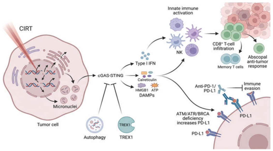

Table 1.
Key differences between conventional photon radiotherapy and carbon ion radiotherapy.
| Parameter | Conventional photon radiotherapy (XRT) |
Carbon ion radiotherapy (CIRT) |
| Dose profile | Exponential attenuation; high entry and exit doses that induce damage to adjacent healthy tissues. | Bragg Peak distribution, exhibiting a low surface dose, a distinct peak at the target depth and zero exit dose. |
| Linear energy transfer (LET) | Low (0.2–2 keV/µm); sparse ionization along the radiation path | High (80-100keV/µm); dense ionization track within the target |
| Relative biological effectiveness (RBE) | 1.0 (baseline reference radiation) | 2.0-5.0 (highly variable and elevated) |
| Mechanism and type of DNA damage | Predominantly indirect; mostly easily repairable single-strand breaks | Direct; complex, clustered and unrepairable double-strand breaks |
| Oxygen dependence | High: requires oxygen to permanently fix DNA damage | Low: overcomes hypoxia via direct, dense DNA damage |
| Immune Effects | Radiation-induced lymphopenia | Immunogenic cell death, driving systemic abscopal efects |
Table 2.
Relevant preclinical studies exploring the activity and mechanisms of CIRT combined with IT.
Table 2.
Relevant preclinical studies exploring the activity and mechanisms of CIRT combined with IT.
| Aim | Samples | Treatments | Endpoints |
Results |
Reference | |
| 1. | To examine the impact of CIRT vs XRT on the expression of PD-L1 | U2OS human osteosarcoma cells | CIRT and XRT 30 min after treatment with an ATR or ATM inhi[14,19,58,60–65bitor | PD-L1 expression on cell surface |
CIRT induced higher PD-L1 expression than the same doses of XRT, depending predominantly on the ATR kinase activity | [59] |
| 2. | To investigate whether CIRT combined with dual ICI (anti-PD-L1 and anti-CTLA-4) is efficient for both local and distant tumor sites. |
In vitro: LM8-mouse osteosarcoma cell line In vivo: LM8 cells inoculated into hind legs of C3H mice |
4 treatment groups: no treatment; ICI; CIRT; combination groups |
In vitro: protein and gene expression of PD-L1, HMGB-1 and CD80 after CIRT In vivo: clinical response (tumor volume, type of response and survival); immune activation (TILs - NK, CTLs, Tregs) |
In vitro: CIRT enhanced PD-L1 and CD80 expression on LM8 cells In combination groups (in vivo): tumor growth delay in both irradiated and unirradiated tumors; CR of unirradiated tumors in 64% of mice; OS significantly prolonged; significant activation of immune cells; |
[60] |
| 3. | To find out if there is a rationale to combine CIRT with CTLA4 blockade for durable anti-tumor immunity | Bilateral tumor model on mice: EO771 breast cancer cells inoculated in one leg and the contralateral flank | In-field CIRT 3x3.08 Gy or XRT 3x5 Gy to the leg tumors Anti-CTLA4 /anti-PD-L1, 3 x 100μg, 3 days apart, starting with the first fraction of RT |
Growth control of locally (irradiated) and distant (unirradiated) tumors Composition of TILS following CIRT+ICIs Gene expression in the intra-tumoral myeloid compartment. |
CIRT with anti-PD-L1 reduced the tumor growth in directly irradiated tumors, but not in the out-of-field tumors. CIRT plus anti-CTLA4: PR (50%) remission of the out-of-field tumors and CR of in-field tumors; CR and improved SV in5/9 mice The combination with CTLA4-blocking antibody was superior to the combination with PD-L1 blockade CIRT + anti-CTLA4: potentiated activation of TAMs and gene expression patterns; activated CD8+ T cells in both in-field and out-of-field tumors inducing protective long-term immunity against EO771 tumors |
[61] |
| 4. |
To evaluate the synergistic effects of ICIs with CIRT and the involvement of ferroptosis | B16-OVA melanoma cell line inoculated into both hind legs of C57BL/6 mice | 4 groups: control, CIRT (4Gy one leg), ICIs (anti-PD-L1 and anti-CTLA4), CIRT+ICIs |
Local and abscopal effects following CIRT+ICIs combination Gene expression, cytokine secretion, immune cell infiltration in irradiated tumors Involvement of ferroptosis |
- All treatment groups: reduced tumor growth, reversed by anti-CD8+ antibodies -In the combination group: longest SV and inhibition of distant tumor growth, enriched immune-related pathways, the most prominent CD45+ and CD8+ cell infiltration, elevated inflammatory cytokine and IFN responses, enrichment of lipid and amino acid metabolism pathways; ferroptosis promoting genes upregulated whilst suppressor genes downregulated |
[62] |
| 5. | To compare the effectiveness of XRT and CIRT in combination with ICI | LM8 sarcoma cells injected in both hind limbs of C3H/He mice 7 days before exposure to CIRT or XRT | 2 ICIs (anti–PD-1 and anti–CTLA-4) injected 1 day before, concomitantly to and after exposure to CIRT/XRT |
The reduction of the non-irradiated, distant tumors (lung metastasis) Evaluation of TILs |
Reduced growth and number of abscopal tumors (lung metastasis), most pronounced after the combined CIRT and ICI administered sequentially. Increase in CD8+ cells in the abscopal tumors in the combination group |
[63] |
| 6. | To examine the immune effects and the mechanism of action of CIRT vs XRT combination with PD-1 inhibitors. | A C57BL/6 mouse model bearing Lewis cells tumors growing on the right hind leg and on the left rear | CIRT or XRT (10Gy) either alone or in combination with PD-1 inhibitor, administered only on the left back tumor. |
Tumor growth The expression of molecules linked to immunogenicity, IFN-β and CD8+ T cells |
Bilateral tumors after CIRT/XRT + PD-1, particularly the non-irradiated tumor Increased CD8+ T cells and IFN-β expression after CIRT in the irradiated tumor. Elevated levels of PD-L1 and TREX1 expression. TREX1 silencing considerably increased IFN-β levels. |
[58] |
| 7. | To elucidate the mechanisms underlying the antitumor immune responses elicited by XRT and CIRT |
Cultured B16, S91 melanoma cells (in vitro model) or inoculated into wild-type C57BL/6 mice (in vivo model) | CIRT or XRT followed by anti-PD-1 i.p. injections on days 2, 4 and 6 after irradiation |
Biomarkers of immunogenic cell death in vitro. Tumor growth and the presence TILs |
In vitro: CIRT+ anti-PD-1 triggered the release of ATP, HMGB1 In vivo: CIRT+ anti-PD-1 significantly decreased tumor growth and prolonged SV, -increased infiltration of CD4+ and CD8+ lymphocytes into the tumor bed and induced type-1 IFN responses |
[14] |
| 8. | To explore the effect of cGAS/STING pathway on TME after CIRT |
In vitro: B16F10 (melanoma) cells In vivo: C57BL/6 mouse tumor models |
In vitro: XRT 6Gy, CIRT 2Gy In vivo: same doses of XRT and CIRT 3 days after PD-L1 inhibitor |
DNA damage and activation of cGAS-STING pathway The expression of molecules linked to immunogenicity |
CIRT caused more DNA damage and cGAS–STING activation, slowed the melanoma growth, boosted the infiltration of NK cells, CD4+, and CD8+ T cells, increased the number of immune checkpoint receptors (PD-1, LAG-3 and TIM-3) and increased PD-L1 exposure on cell surface. CIRT with PD-L1 inhibitor increased the number of T and NK cells and slowed tumor growth |
[19] |
| 9. | To compare the immune regulatory effects of CIRT and XRT | In situ 4T1 breast cancer mouse model | CIRT or biologically equivalent doses of XRT | The immune characteristics of the TME | Low dose CIRT spared lymphocytes, higher dose induced the secretion of pro-inflammatory cytokines. XRT was lympho-toxic even at low doses |
[64] |
| 10. | To assess the immunological contribution to the therapeutic effectiveness of XRT and CIRT | Mouse models of intradermal tumors (B16F10-OVA melanoma and 3LL-OVA lung cancer cells) | CIRT or XRT in the presence or absence of CTLs. | The importance of CTLs-mediated immunity in shrinking tumors irradiated with CIRT or XRT | CIRT utilizes CTLs-mediated immunity to a greater extent compared to XRT | [65] |
Table 3.
Ongoing clinical trials exploring combinations of CIRT and IT.
| Nr and acronim | Phase and size | Aim | Tumor site | IT agents | CIRT | Endpoints | Results-Efficacy (survival) | Ref |
| NCT05229614 ICONIC |
IIb 27p. |
Feasibility and clinical activity of CIRT+ICI in p. with SD and Pembro as standard of care |
Solid cancers with SD: NSCLC, H&N, MM, URO |
Pembro |
CIRT to one measurable lesion (untreated previously) 24Gy/3fr |
Primary: ORR Secondary: Safety, PFS, OS *Analysis of cytokines (ILs, IFN) soluble markers: HMGB1, PD-L1, tumor cfDNA and immune cell composition in blood, before and after irradiation |
None reported yet |
[66] |
| jRCT2031210046 DEPARTURE |
Ib 15p. |
Safety and preliminary efficacy of the CIRT + ICI | Advanced HCC with MVI |
Durva (3p.) or Durva + Treme (12p.) | CIRT 60Gy/4fr. to the primary tumor |
Primary: DLT, AE Secondary: PFS and OS |
No DLT Gr 3-4 AE: 53.3% PFS 4.7 mo, OS 10.4 mo |
[67] |
| jRCT2031240284 VANGUARD |
Ph Ib (6)/II (42p) | Safety and efficacy of Atezo and Beva following CIRT | HCC | Atezo followed by Beva every 21 days | CIRT 60Gy/4 fr to the primary tumor | Phase Ib: DLT Phase II: 1year RFS, OS, 6-month OS rate, extended RFS tracking over time |
None reported yet | [68] |
| jRCT2031210083 DECISION |
Ph Ib 3p | The safety of Durva + CIRT and weekly CDDP |
Cervical cancer | Durva at weeks 2 and 6 | CIRT 74.4 Gy in 20 fr and concurrent weekly CDDP | Primary: AE and DLT Secondary: OS, PFS, DMR, OR and CR rates Collection of tumor tissue and blood |
1 serious AE; No AE associated with DLT All p. had CR within the CIRT region |
[69] |
| NCT04143984A | Ph 2, 146p | The Efficacy of CIRT (arm1) vs CIRT+Camre (arm2) | Locally recurrent NPC | Camre started with CIRT and given for maximum 1 year. | CIRT 63 Gy/21 fr |
Primary: PFS Secondary: OS, PFS, MFS; AE *Potential biomarkers for treatment outcome based on omics. |
No results published yet |
[70] |
| NCT06805864 BROTHER |
Ph 2 | The superiority of this regimen vs XRT+CDDP | Locally advanced cervical carcinoma | CIRT+CDDP+Pembro | CIRT 16Gy/4fr followed by 3 cycles of IGBT |
Primary: PFS Secondary: OS, LC, CR, AE |
Not yet recruiting |
Abbr.: Pembro - Pembrolizumab; Durva - Durvalumab; Treme - Tremelimumab; Atezo - Atezolizumab; Beva - Bevacizumab; Camre - Camrelizumab; CDDP – Cisplatin; CIRT - carbon ion radiotherapy; IGBT – image-guided brachytherapy; ICI - immune checkpoint inhibitor; NSCLC - non-small cell lung cancer; H&N - head and neck cancer; MM - malignant melanoma; URO - urothelial cancer; AE – adverse effects; HCC – hepatocellular carcinoma; MVI – macrovascular invasion; OS – overall survival; PFS – progression free survival; ORR – overall response rate; CR – complete response, LC – local controlDLT - dose- limiting toxicity; RFS - recurrence-free survival; DMR - distant metastasis rate; NPC – nasopharyngeal carcinoma; MFS – metastasis-free survival; SD - stable disease.
Disclaimer/Publisher’s Note: The statements, opinions and data contained in all publications are solely those of the individual author(s) and contributor(s) and not of MDPI and/or the editor(s). MDPI and/or the editor(s) disclaim responsibility for any injury to people or property resulting from any ideas, methods, instructions or products referred to in the content. |
© 2026 by the authors. Licensee MDPI, Basel, Switzerland. This article is an open access article distributed under the terms and conditions of the Creative Commons Attribution (CC BY) license (http://creativecommons.org/licenses/by/4.0/).
Copyright: This open access article is published under a Creative Commons CC BY 4.0 license, which permit the free download, distribution, and reuse, provided that the author and preprint are cited in any reuse.