

Submitted:
08 November 2026
Posted:
09 November 2026
You are already at the latest version
Abstract
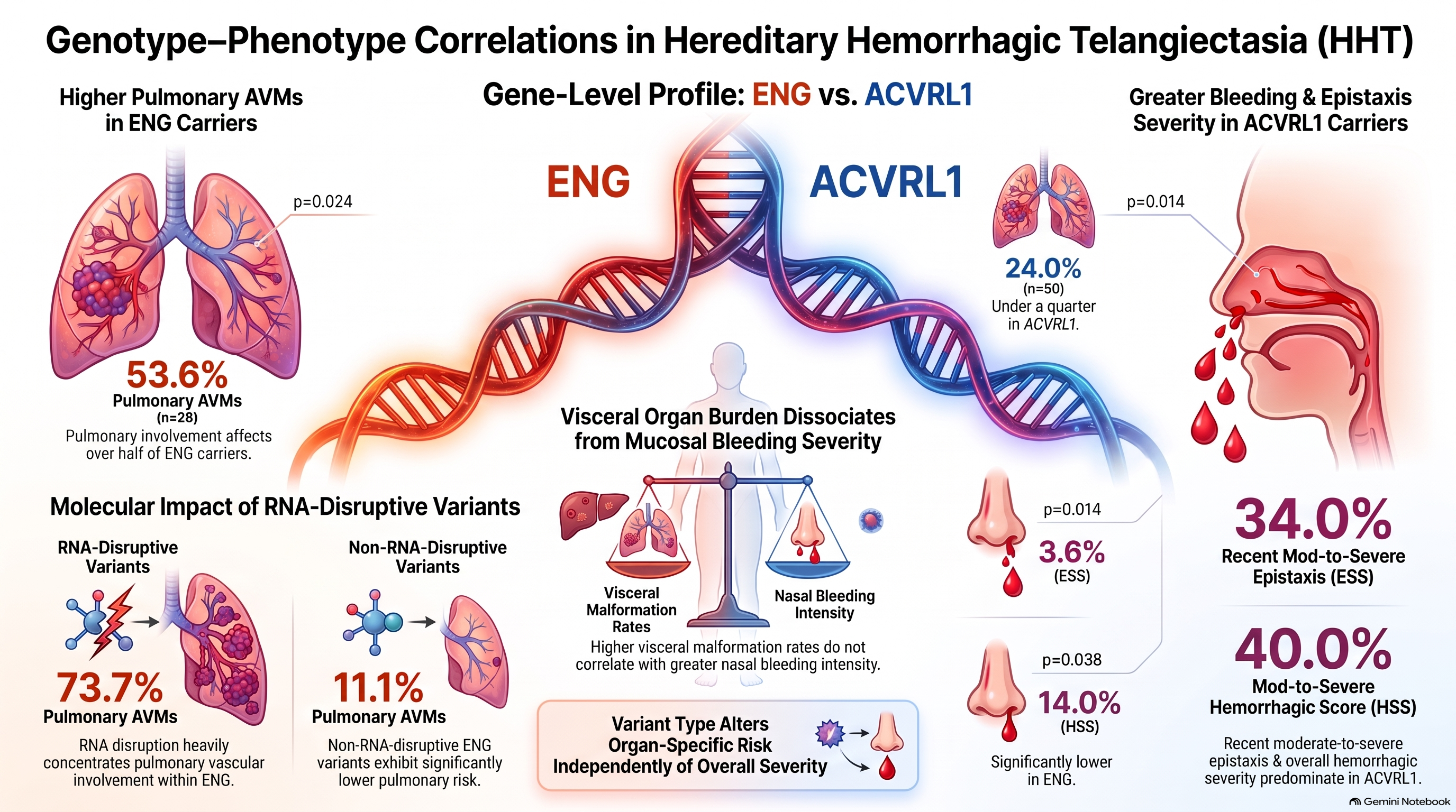
Background: Hereditary hemorrhagic telangiectasia (HHT) shows substantial phenotypic variability. We investigated genotype-phenotype associations, focusing on variant type and predicted effects on RNA processing. Methods: We conducted a retrospective observational study of genetically confirmed HHT patients carrying ENG or ACVRL1 variants. Clinical manifestations, epistaxis severity, treatment and genetic characteristics were assessed. Variants were classified according to molecular type and predicted RNA-disrupting effect. Results: Seventy-eight patients were included, 28 carrying ENG and 50 ACVRL1 variants. Pulmonary arteriovenous malformations (PAVMs) were more frequent among ENG than ACVRL1 carriers (53.6% vs. 24.0%, p = 0.024), whereas hepatic AVMs showed a nonsignificant predominance in ACVRL1 (44.0% vs. 21.4%, p = 0.072). Moderate-to-severe recent epistaxis was more frequent among ACVRL1 carriers (34.0% vs. 3.6%, p = 0.014). Overall, PAVMs were more frequent in patients carrying RNA-disruptive variants than in those with non-RNA-disruptive variants (60.9% vs. 23.6%, p = 0.004). Importantly, this association persisted within ENG, with PAVMs occurring in 73.7% of patients with RNA-disruptive variants compared with 11.1% of those with non-RNA-disruptive variants (p = 0.004). Among patients with non-RNA-disruptive variants, cerebral vascular malformations were more frequent in ENG than in ACVRL1 carriers (44.4% vs. 8.7%, p = 0.013). Conclusions: Beyond established ENG-ACVRL1 phenotypic differences, variant-level molecular characteristics may contribute to HHT phenotypic heterogeneity.
Keywords:
hereditary haemorrhagic telangiectasia
; ENG
; ACVRL1
; genotype-phenotype
; RNA-disrupting variants
1. Introduction
Hereditary haemorrhagic telangiectasia (HHT), also known as Rendu–Osler–Weber disease, is an autosomal dominant vascular disorder characterized by recurrent epistaxis, mucocutaneous telangiectasias, and visceral arteriovenous malformations (AVMs)[1]. HHT is the second-most-common inherited bleeding disorder worldwide, the most morbid inherited bleeding disorder of women, and results in major morbidity, increased mortality, and substantially worse health-related quality of life [2,3,4]. The disease usually affects multiple organs, including the nasal cavity, lungs, liver, brain, and/or gastrointestinal tract, resulting in substantial clinical heterogeneity among affected individuals [5,6].
HHT is genetically heterogeneous, with ENG and ACVRL1 representing the major disease-associated genes and defining HHT type 1 (HHT1) and HHT type 2 (HHT2), respectively. Both genes are predominantly expressed in vascular endothelial cells and participate in signalling pathways involved in vascular homeostasis. Reduced levels of functional endoglin or ALK1 are considered central to HHT pathogenesis [7]. However, the marked inter and intrafamilial variability observed among patients suggests that the affected gene alone does not fully determine disease expression [8,9].
Several genotype–phenotype associations have been suggested. ENG variants may be associated with earlier mucosal telangiectasias and more pulmonary and cerebral AVMs, whereas hepatic vascular malformations appear more common with ACVRL1 [5,10]. However, substantial phenotypic variability persists both between and within HHT1 and HHT2, indicating that additional molecular determinants may influence clinical expression.
Limited evidence further suggests that variant class may contribute to phenotypic differences. Truncating ENG variants have been associated with greater organ involvement and haemorrhagic severity than missense variants [10]. Experimental and patient-derived studies indicate that individual variants may differ in their effects on endoglin/ALK1 function and their clinical consequences [11,12].
These observations raise the possibility that a more refined characterization incorporating the affected gene, variant type, and its molecular consequences could improve our understanding of phenotypic variability in HHT. However, the clinical relationship between the reported or predicted effects of HHT-associated variants on RNA and specific vascular phenotypes remains insufficiently characterized.
Therefore, the aim of the present study was to investigate genotype–phenotype associations in a geographically defined cohort of genetically confirmed patients with HHT. We first compared the clinical phenotype of patients carrying ENG and ACVRL1 variants and subsequently evaluated phenotypic differences according to variant type and the reported or predicted effect of each variant on RNA. Finally, RNA-related associations were examined within each gene to explore whether molecular characteristics at the variant level may contribute to the clinical heterogeneity of HHT.
2. Materials and Methods
2.1. Study Design and Population
This retrospective observational study included patients with genetically confirmed HHT from Cantabria, Spain. Patients were eligible regardless of age, and only cases with genetically confirmed ENG and ACVRL1 variants were included.
Demographic characteristics, HHT-related manifestations, epistaxis and bleeding severity scores, and treatment were collected from the available medical records.
2.2. Clinical Manifestations and Severity
HHT-related manifestations included mucocutaneous telangiectasias and pulmonary, hepatic, cerebral, and gastrointestinal vascular malformations. Telangiectasias were recorded according to their presence and anatomical distribution (lips, tongue, ears, and fingertips). Visceral AVMs were diagnosed by lung CT scan, brain angio-CT or angio-MRI, and Doppler ultrasound and/or CT of the liver. PAVMs were additionally classified as unilateral or bilateral, and previous embolization was recorded.
Epistaxis severity was assessed using the Epistaxis Severity Score (ESS)[13], the Frequency, Intensity and Duration score (FID score) [14] and Sadick classification [15]. A composite Hemorrhagic severity score (HSS in this study) was estimated according to the bleeding severity classification recently proposed by the International Consensus Report on Standardization of Terminology, Definitions, and Outcome Criteria in HHT [16]. When available, both recent severity and the worst recorded clinical status were considered, with recent severity defined by assessments performed within the preceding 3 months for epistaxis scores and 12 months for the HSS.
HHT-related treatments, including iron supplementation, antifibrinolytic and antiangiogenic therapies, local nasal treatments, PAVM embolization, and liver transplantation, were also recorded.
2.3. Genetic Study and Data Analysis
DNA was extracted from the peripheral blood and analysed by Sanger sequencing of NGS, using standard laboratory procedures. Variants were classified according to the American College of Medical Genetics and Genomics (ACMG) criteria.
All patients had genetically confirmed HHT. Variants were classified according to the affected gene (ENG or ACVRL1) and molecular type (missense, splice-site, deletion, nonsense, or frameshift).
Variants were additionally categorized according to their reported or predicted effect on RNA, as RNA-altering variants (i.e., those causing frame-shift, exon deletions, splicing-alterations, or premature stop codons) or non-RNA-altering variants (missense).
Genotype–phenotype associations were initially evaluated by comparing ENG and ACVRL1 carriers and subsequently according to molecular variant type.
The phenotype was also compared according to the reported/predicted effect on RNA. Because RNA-effect status was unequally distributed between ENG and ACVRL1, analyses were additionally stratified by gene.
2.4. Statistical Analyses
Categorical variables were expressed as n (%) and continuous variables as median and interquartile range (IQR). In the presence of missing data, percentages were calculated using evaluable patients as the denominator.
Categorical variables were compared using Fisher’s exact test and continuous variables using the Mann–Whitney U test. All tests were two-sided, and p < 0.05 was considered statistically significant. All statistical analyses were performed using SPSS version 27.0 (IBM Corp., Armonk, NY, USA).
2.5. Ethics
The study was conducted in accordance with the Declaration of Helsinki, and approved by the Institutional Review Board (Comité de Ética de Investigación con Medicamentos de Cantabria, acta 18/2025, dated 21 November 2025).
3. Results
3.1. Clinical Manifestations According to the Mutated Gene
The study population included 28 patients carrying ENG variants and 50 carrying ACVRL1 variants (Table 1). The manifestations of both groups are shown in Table 2. No significant differences were observed in sex or age. Mucocutaneous telangiectasias were highly prevalent in both groups, without significant differences between them. Pulmonary arteriovenous malformations (PAVMs) were significantly more frequent among ENG carriers (53.6% vs. 24.0%, p = 0.024). Conversely, hepatic AVMs were numerically more frequent in ACVRL1 carriers (44.0% vs. 21.4%, p = 0.072), whereas cerebral AVMs showed the opposite pattern (25% in ENG vs. 8.0% in ACVRL1; p = 0.082) (Figure 1).
Epistaxis, as assessed by the FID, ESS, or Sadick score, tended to be more severe in carriers of ACVRL1 variants. The difference between groups was statistically significant for recent ESS, which was moderate-to-severe in 34% of ACVRL1 carriers and only in 3.6% of ENG carriers (p=0.014). Likewise, the previous year combined HSS was moderate-to-severe in 40% of ACVRL1 carriers and only 14% of ENG variant carriers (p=0.038). Consequently, more frequent systemic drug therapy was given in the ACVRL1 group (antifibrinolytic use 32% vs 18%, p=0.053; antiangiogenic drugs, 12% vs 3.6%, p=0.22).
3.2. Genotype–Phenotype Associations According to Variant Type
Twenty-three patients had RNA-disruptive variants and 55 had non-RNA-disruptive variants. PAVMs were more frequent in patients with RNA-disruptive variants (60.9% vs. 26.4%, p=0.009), with higher proportions of bilateral involvement (57.1% vs. 21.4%, p=0.120) and previous embolization (84.6% vs. 57.1%, p=0.209). Conversely, hepatic AVMs were numerically less frequent in the RNA-disruptive group (27.3% vs. 47.5%, p=0.123), whereas cerebral vascular lesions and anemia showed similar distributions. No relevant differences in epistaxis severity were observed between both groups. Given the unequal distribution of the RNA-disruptive variants, gene-stratified analyses were subsequently performed.
Among ENG variant carriers, 19 patients had RNA-disruptive variants and 9 had non-RNA-disruptive variants (Table 3). PAVMs were significantly more frequent in patients with RNA-disruptive variants (73.7% vs. 11.1%, p = 0.004). Bilateral PAVMs (42.1% vs. 0%) and embolization (57.9% vs. 11.1%) were also numerically more frequent in the RNA-disruptive group, although these differences were not statistically significant. We did not observe differences in extrapulmonary vascular malformations or bleeding severity.
Among ACVRL1 variant carriers, only four patients had RNA-disruptive variants compared with 46 with non-RNA-disruptive variants. No significant differences were observed in vascular involvement or epistaxis severity (Table 4). However, this finding should be interpreted cautiously given the very small number of patients with RNA-disruptive variants (Figure 2).
3.3. Genotype-phenotype Comparison in Patients with non-RNA-disruptive variants
The proportion of RNA-disrupting variants differed substantially between the ENG and ACVRL1 variant groups. Therefore, to better characterize differences in the clinical profiles of patients with ENG and ACVRL1 variants, we compared the two groups among those carrying non–RNA-disrupting variants (Table 5). Consistent with the overall analysis, cerebral vascular lesions were significantly more frequent among ENG variant carriers than among ACVRL1 variant carriers (44.4% vs. 8.7%; p = 0.013). Conversely, mucocutaneous telangiectasias and visceral vascular lesions tended to be more frequent among ACVRL1 variant carriers. Similarly, epistaxis scores and HSS tended to be higher in the ACVRL1 group, although these differences did not reach statistical significance.
4. Discussion
We aimed to investigate genotype-phenotype associations in a geographically defined cohort of patients with genetically confirmed HHT, focusing not only on the affected gene but also on variant type and the reported molecular consequences of individual variants on RNA. HHT is characterized by substantial inter and intrafamilial variability, suggesting that the clinical phenotype cannot be completely explained by the causal gene alone [17].
Our findings confirm several of the classical phenotypic differences between ENG and ACVRL1 variants, particularly regarding visceral vascular involvement. More importantly, our results suggest that molecular characteristics beyond the affected gene may contribute to phenotypic heterogeneity, with the most prominent signal being the association between variants with a RNA-disruptive effect on RNA and PAVMs among ENG carriers [18]. Given that a signal was recently identified for differential response to pomalidomide by underlying genotype [19], these findings additionally raise the question of whether variant type could also contribute to therapeutic response [20]. Given that therapeutic development in HHT is rapidly expanding and novel agents designed for HHT are being evaluated in formal clinical trials [21,22,23,24], recognizing a potential impact of genotype or variant on therapeutic response is important.
The distribution of visceral AVMs in our cohort was largely consistent with the established HHT1 and HHT2 phenotypes. PAVMs were significantly more frequent among ENG carriers than among ACVRL1 variant carriers (53.6% vs. 24.0%). This finding closely mirrors previous genotype–phenotype studies. Bayrak-Toydemir et al. reported intrapulmonary shunting in 59.0% of patients with HHT1 compared with 28.9% of those with HHT2 [10], while previous studies also supported preferential pulmonary involvement in HHT1 [18,25]. More recent evidence summarized in a recent review estimates PAVM prevalence at approximately 50–75% among carriers of ENG variants compared with 20–30% among patients carrying ACVRL1 variants [1]. Thus, the frequencies observed in our cohort are remarkably similar to those reported in larger HHT populations and reinforce the strong association between ENG variants and pulmonary vascular involvement across different populations.
Hepatic AVMs were more frequent in ACVRL1 variant carriers (44.0% vs. 21.4%), whereas cerebral AVMs were more common in carriers of ENG variants (25.0% vs. 8.0%), although neither difference reached statistical significance. These patterns are consistent with previous reports and support the role of the affected gene in determining the anatomical distribution of visceral vascular lesions [10,26].
Mucocutaneous telangiectasias were highly prevalent in both genotypes, with no significant difference in overall prevalence; however, genotype-related differences may exist in their anatomical distribution and age of onset, with earlier nasal and oral involvement in HHT1 and more frequent, earlier dermal lesions in HHT2 [10,26,27,28,29,30]. Another relevant finding was the apparent dissociation between visceral vascular involvement and bleeding and epistaxis severity scores. Despite the greater pulmonary vascular burden observed in ENG variant carriers, moderate-to-severe bleeding according to HSS classification during the previous year was more frequent among ACVRL1 carriers (40.0% vs. 14.2%). Similarly, moderate-to-severe recent ESS was observed more frequently in carriers of ACVRL1 variants (34.0% vs. 3.6%). Nevertheless, when ESS was analyzed as a continuous variable, median recent scores did not differ significantly between genotypes. These observations are not necessarily inconsistent with previous reports. These findings should be interpreted in the context of previous studies reporting heterogeneous genotype-epistaxis associations. Bayrak-Toydemir et al. also reported an earlier onset of epistaxis in HHT1 (ENG) than in HHT2 (ACVRL) but found that epistaxis severity became comparable between genotypes by middle age [10,18]. In contrast, other studies have reported greater epistaxis severity, with higher ESS values, among ACVRL1 carriers [31]. Our findings further suggest that the determinants of visceral AVM burden and nasal bleeding severity may differ and that a more severe visceral vascular phenotype does not necessarily translate into more severe epistaxis.
Beyond the classical comparison between HHT1 and HHT2, we aimed to determine whether variant type could further contribute to phenotypic variability. This analysis revealed a marked gradient in pulmonary involvement. PAVMs were observed in 85.7% of patients carrying deletions, 53.8% of those with splice-site variants, and 23.6% of those carrying missense variants. This pattern became even more pronounced when the analysis was restricted to ENG: PAVMs occurred in 85.7% of patients with deletions and 63.6% of those with splice-site variants, compared with only 11.1% of those carrying missense variants.
These findings are conceptually consistent with the genotype–phenotype analysis by Bayrak-Toydemir et al., who found that truncating ENG variants were associated with a greater number of affected organs and more severe hemorrhagic manifestations than missense variants [10]. Our results potentially extend this observation by suggesting that pulmonary involvement may be particularly concentrated among specific molecular subgroups, especially deletions and splice-site variants. However, this finding should be interpreted cautiously because the deletion subgroup included only seven patients.
Further evidence suggests that variant class may influence clinically relevant manifestations independently of the affected HHT gene. In a recent cross-sectional study of 85 patients with HHT, Viteri-Noël et al. found that truncating variants were independently associated with anemia after adjustment for potential confounders, including age, sex, and causal gene. Notably, the affected gene (ENG vs. ACVRL1) was not independently associated with anemia. Conversely, truncating variants were not significantly associated with epistaxis severity, suggesting that the phenotypic relevance of variant class may differ according to the clinical manifestation considered [18].
Evidence regarding visceral AVMs is less consistent. In a recent cohort of 142 patients with HHT, Sturiale et al. found no significant association between truncating versus non-truncating variants and pulmonary, cerebral, or hepatic AVMs [32]. Moreover, Palermo et al. reported lower odds of brain AVMs among carriers of truncating variants compared with those carrying non-truncating variants [33]. These findings contrast with the marked pulmonary gradient observed across variant classes in our cohort and suggest that the relationship between variant type and visceral vascular involvement may be organ-specific rather than reflecting a uniform effect on overall disease severity. Differences in gene distribution, variant classification, age, screening strategies, and cohort composition may also contribute to the discrepancies between studies.
This interpretation is also supported by the pattern of epistaxis observed in our cohort. Despite having the highest prevalence of PAVMs, patients carrying deletions had a lower median worst HHT-ESS than those carrying splice-site or missense variants, whereas splice-site variants combined relatively frequent pulmonary involvement with greater historical epistaxis severity. Together with the observations of Viteri-Noël et al. [18], these findings suggest that visceral vascular malformations, mucosal bleeding, and hematological consequences may represent partially independent phenotypic dimensions rather than manifestations of a single molecular severity gradient.
The most distinctive aspect of our study was the evaluation of phenotype according to the predicted and reported molecular effect of individual variants on RNA. In the overall cohort, PAVMs were more frequent among patients carrying RNA-disruptive variants than among those without such an effect (60.9% vs. 26.4%). Bilateral pulmonary involvement and previous embolization were also more frequent in the RNA-disruptive group. However, these overall findings require careful interpretation because the variants’ effects on RNA were not equally distributed between genes: the RNA-disruptive variants group was strongly enriched among ENG carriers, whereas variants without a predicted RNA-disrupting effect predominantly occurred in the ACVRL1 group. Consequently, the overall association could at least partly reflect the already established relationship between ENG variants and PAVMs rather than an independent RNA-related effect.
For this reason, the within-gene analysis is particularly relevant. Among ENG carriers, PAVMs were present in 73.7% of patients carrying variants with an RNA-disruptive effect compared with only 11.1% of those with a non-disruptive effect (p = 0.004). Bilateral pulmonary involvement and previous embolization were also numerically more frequent in the first subgroup. In contrast, no significant differences were observed in extrapulmonary AVMs or epistaxis severity. This within-ENG association reduces, although does not eliminate, confounding by the causal gene and suggests that variant-level molecular characteristics may contribute to pulmonary phenotypic variability. Nevertheless, the observational design and limited subgroup size preclude establishing an independent or causal effect.
The biological plausibility of a variant-level effect is supported by the findings of Bernabéu-Herrero et al., who showed that variants generating premature termination codons (PTCs) were associated with differences in hemorrhagic phenotype and could account for part of the clinical variability observed among patients with HHT [34]. Importantly, their functional analyses demonstrated variable persistence of PTC-containing transcripts despite nonsense-mediated decay, providing evidence that pathogenic variants affecting the same HHT gene may differ in their downstream molecular consequences [34].
Nevertheless, our classification should not be considered equivalent to the PTC/non-PTC classification used by Bernabéu-Herrero et al. In the present study, RNA effects were derived from reported annotations available in genetic databases rather than from direct experimental analysis of patient-derived RNA. Accordingly, our results demonstrate an association with variant-level RNA annotations, rather than a direct association between experimentally demonstrated RNA alterations.
A further exploratory observation supports the possibility that gene and variant effects may contribute differently according to the vascular territory involved. When the analysis was restricted to patients without an RNA-disruptive variant effect, cerebral VMs remained significantly more frequent among ENG than ACVRL1 carriers (44.4% vs. 8.7%; p = 0.013), whereas differences in pulmonary involvement were no longer significant. The preferential association between ENG and cerebral vascular malformations is well established in the literature [1,35]. However, given that this restricted analysis included only nine carriers of ENG variants, the magnitude of this difference should be interpreted cautiously.
Taken together, our findings support a multilevel interpretation of genotype–phenotype variability in HHT. The affected gene remains a major determinant of phenotype, particularly with respect to the distribution of pulmonary, hepatic, and cerebral vascular malformations [17]. However, the substantial variability observed within the same gene suggests that the traditional HHT1/HHT2 classification alone is unlikely to explain the full clinical heterogeneity of the disease. Variant type and its downstream molecular consequences may provide an additional level of phenotypic information, particularly for pulmonary involvement in ENG. This concept is consistent with emerging molecular evidence demonstrating that pathogenic variants affecting the same HHT gene are not necessarily biologically equivalent [34,36].
Several limitations should be acknowledged. First, the retrospective design may have introduced selection and information bias, and some phenotypic variables were not available for all patients. Second, the relatively small sample size substantially limits subgroup analyses, particularly for deletions, nonsense and frameshift variants and for ACVRL1 variants with a reported or predicted RNA effect. Third, multiple phenotypic comparisons were performed; consequently, some statistically significant associations, particularly those arising from small subgroups, should be considered exploratory and require external validation. Fourth, RNA consequences were inferred from genetic database annotations and were not directly assessed in patient-derived samples, precluding mechanistic or causal conclusions. Finally, several HHT manifestations are age-dependent, and residual confounding by age and other clinical or genetic modifiers cannot be excluded.
Despite these limitations, our study has several strengths. All included patients had genetically confirmed HHT and belonged to a geographically defined population, limiting diagnostic heterogeneity. Furthermore, our study extends beyond the conventional comparison between ENG and ACVRL by integrating gene, variant type, and RNA consequences with detailed clinical phenotyping. The reproducibility of the classical ENG–pulmonary and ACVRL1 –hepatic patterns within our cohort provides internal consistency, while the marked association between RNA-disruptive variants and PAVMs within ENG identifies a potentially relevant molecular subgroup that warrants confirmation in larger independent cohorts and, ideally, direct functional characterization.
5. Conclusions
Our findings confirm established genotype–phenotype associations in HHT while suggesting that molecular characterization beyond the affected gene may contribute to explaining interindividual phenotypic variability. ENG variants were associated with greater pulmonary involvement, whereas ACVRL1 variants showed greater hepatic involvement and higher epistaxis severity according to selected measures. Within ENG variants, greater PAVM burden was observed in deletions and splice-site variants and, consistently, among variants classified as having an RNA-disruptive effect. These findings should be regarded as exploratory but support a more refined model of genotype–phenotype correlation in HHT incorporating the affected gene, variant class, and downstream molecular consequences. Further studies integrating larger clinical cohorts with direct RNA and protein characterization are needed to determine whether these molecular features may ultimately contribute to individualized risk stratification in HHT.
Author Contributions
Conceptualization, E.U. H.A. and J.A.R.; methodology: E.U., N.P. and J.A.R.; formal analysis, E.U. and J.A.R.; investigation, E.U., V.D., C.M., F.J.G., J.M.C., A.F., P.S., N.P.; data curation, E.U., P.S. and J.A.R.; writing—original draft preparation, E.U. and J.A.R.; writing—review and editing, All; supervision, H.A., J.A.R. All authors have read and agreed to the published version of the manuscript.
Funding
This research did not receive any specific grant from funding agencies in the public, commercial, or not-for-profit sectors.
Institutional Review Board Statement
The study was conducted in accordance with the Declaration of Helsinki, and approved by the Institutional Review Board (Comité de Ética de Investigación con Medicamentos de Cantabria, acta 18/2025, dated 21 November 2025).
Informed Consent Statement
Given the retrospective nature of the study, informed consent was waived.
Data Availability Statement
The original data that support the findings of this study are available from the corresponding author upon reasonable request. The individual sequence data are not publicly available due to privacy and ethical restrictions related to patient confidentiality and consent.
Acknowledgments
During the preparation of this work, ChatGPT was used for language editing. After using this tool, the authors reviewed and extensively edited the manuscript and take full responsibility for the content of the published article. The authors developed and verified all scientific content, interpretation, and conclusions.
Conflicts of Interest
H.A. declares the following universal disclosures: Consultancy (Agios, Amgen, Alnylam, Alpine, Atavistik, Diagonal, Novartis, Pharmacosmos, Sobi, Sanofi, Takeda, Terremoto, Tectonic, Vaderis) and Research Funding to Institution (Agios, Amgen, Alnylam, Novartis, Sobi, Atavistik, Vaderis). All other authors declare that they have no known competing financial interests or personal relationships that could have appeared to influence the work reported in this paper.
Abbreviations
The following abbreviations are used in this manuscript:
| HHT | Hereditary Hemorrhagic Telangiectasia |
| PAVM / PAVMs | Pulmonary Arteriovenous Malformation(s) |
| AVM / AVMs | Arteriovenous Malformation(s) |
| HHT1 | Hereditary Hemorrhagic Telangiectasia type 1 |
| HHT2 | Hereditary Hemorrhagic Telangiectasia type 2 |
| ALK1 | Activin receptor-like kinase 1 |
| CT | Computed Tomography |
| MRI | Magnetic Resonance Imaging |
| ESS | Epistaxis Severity Score |
| FID | Frequency, Intensity and Duration score |
| HSS | Hemorrhagic Severity Score |
| DNA | Deoxyribonucleic Acid |
| NGS | Next-Generation Sequencing |
| ACMG | American College of Medical Genetics and Genomics |
| RNA | Ribonucleic Acid |
| IQR | Interquartile Range |
| SPSS | Statistical Package for the Social Sciences |
| IBM | International Business Machines Corporation |
| PTC / PTCs | Premature Termination Codon(s) |
| VM / VMs | Vascular Malformation(s) |
References
- Hermann, R.; Shovlin, C.L.; Kasthuri, R.S.; et al. Hereditary haemorrhagic telangiectasia. Nat. Rev. Dis. Primers 2025, 11, 1. [Google Scholar] [CrossRef] [PubMed]
- Al-Samkari, H.; Mayne, T.J.; Troutt, M.; et al. Characterizing the healthcare utilization and costs of hereditary hemorrhagic telangiectasia. Am. J. Hematol. 2025, 100, 1722–1735. [Google Scholar] [CrossRef] [PubMed]
- Al-Samkari, H.; Kasthuri, R.S.; Iyer, V.N.; et al. Validation and clinical application of the hereditary hemorrhagic telangiectasia-specific quality of life scale. J. Thromb. Haemost. 2026, 24, 108–118. [Google Scholar] [CrossRef] [PubMed]
- Zhang, E.; Virk, Z.M.; Rodriguez-Lopez, J.; et al. Hereditary hemorrhagic telangiectasia may be the most morbid inherited bleeding disorder in women. Blood Adv. 2024, 8, 3166–3172. [Google Scholar] [CrossRef] [PubMed]
- Al-Samkari, H.; Friday, C.; Kasthuri, R.S.; et al. Clinical spectrum of hereditary hemorrhagic telangiectasia: Data from the Comprehensive HHT Outcomes Registry of the US (CHORUS). Blood 2026, 148, 417–432. [Google Scholar] [CrossRef] [PubMed]
- Hayama, M.; Maeda, Y.; Obata, S.; et al. Understanding hereditary hemorrhagic telangiectasia: From genetic anomalies to systemic manifestations, quality of life, and epistaxis management—Exploring the otolaryngologist’s integral role. Auris Nasus Larynx 2024, 51, 305–312. [Google Scholar] [CrossRef] [PubMed]
- Al Tabosh, T.; Al Tarrass, M.; Tourvieilhe, L.; et al. Hereditary hemorrhagic telangiectasia: From signaling insights to therapeutic advances. J. Clin. Invest. 2024, 134, e176379. [Google Scholar] [CrossRef] [PubMed]
- Giorgio, V.; Di Foggia, C.; Quatrale, G.; et al. Family phenotypic profile in hereditary hemorrhagic telangiectasia: Genotype-phenotype correlation in a pediatric Italian population. Ital. J. Pediatr. 2025, 51, 268. [Google Scholar] [CrossRef] [PubMed]
- Kritharis, A.; Al-Samkari, H.; Kuter, D.J. Hereditary hemorrhagic telangiectasia: Diagnosis and management from the hematologist’s perspective. Haematologica 2018, 103, 1433–1443. [Google Scholar] [CrossRef] [PubMed]
- Bayrak-Toydemir, P.; McDonald, J.; Markewitz, B.; et al. Genotype-phenotype correlation in hereditary hemorrhagic telangiectasia: Mutations and manifestations. Am. J. Med. Genet. A 2006, 140A, 463–470. [Google Scholar] [CrossRef] [PubMed]
- Gariballa, N.; Badawi, S.; Ali, B.R. Endoglin mutants retained in the endoplasmic reticulum exacerbate loss of function in hereditary hemorrhagic telangiectasia type 1 (HHT1) by exerting dominant negative effects on the wild type allele. Traffic 2024, 25, e12928. [Google Scholar] [CrossRef] [PubMed]
- Mallet, C.; Lamribet, K.; Giraud, S.; et al. Functional analysis of endoglin mutations from hereditary hemorrhagic telangiectasia type 1 patients reveals different mechanisms for endoglin loss of function. Hum. Mol. Genet. 2015, 24, 1142–1154. [Google Scholar] [CrossRef] [PubMed]
- Hoag, J.B.; Terry, P.; Mitchell, S.; et al. An epistaxis severity score for hereditary hemorrhagic telangiectasia. Laryngoscope 2010, 120, 838–843. [Google Scholar] [CrossRef] [PubMed]
- Pagella, F.; Maiorano, E.; Matti, E.; et al. FID score: An effective tool in hereditary haemorrhagic telangiectasia-related epistaxis. Rhinology 2020, 58, 516–521. [Google Scholar] [CrossRef] [PubMed]
- Bergler, W.; Sadick, H.; Gotte, K.; Riedel, F.; Hörmann, K. Topical estrogens combined with argon plasma coagulation in the management of epistaxis in hereditary hemorrhagic telangiectasia. Ann. Otol. Rhinol. Laryngol. 2002, 111, 222–228. [Google Scholar] [CrossRef] [PubMed]
- Al-Samkari, H.; Kasthuri, R.S.; Mager, H.J.; et al. Standardization of terminology, definitions, and outcome criteria for bleeding in hereditary hemorrhagic telangiectasia: International consensus report. Am. J. Hematol. 2025, 100, 1813–1827. [Google Scholar] [CrossRef] [PubMed]
- Bossler, A.D.; Richards, J.; George, C.; et al. Novel mutations in ENG and ACVRL1 identified in a series of 200 individuals undergoing clinical genetic testing for hereditary hemorrhagic telangiectasia (HHT): Correlation of genotype with phenotype. Hum. Mutat. 2006, 27, 667–675. [Google Scholar] [CrossRef] [PubMed]
- Viteri-Noël, A.; Patier, J.L.; Bara-Ledesma, N.; et al. Genotype–phenotype relationship in hereditary hemorrhagic telangiectasia: Quality of life and cardiovascular risk evaluation. J. Clin. Med. 2025, 14, 4409. [Google Scholar] [CrossRef] [PubMed]
- Al-Samkari, H.; Thomas, S.M.; Marsh, D.; et al. Characteristics associated with clinical response to pomalidomide in hereditary hemorrhagic telangiectasia. Blood Adv. 2026, 10, 2967–2976. [Google Scholar] [CrossRef] [PubMed]
- Al-Samkari, H. How I treat bleeding in hereditary hemorrhagic telangiectasia. Blood 2024, 144, 940–954. [Google Scholar] [CrossRef] [PubMed]
- Al-Samkari, H.; Kasthuri, R.S.; Parambil, J.G.; et al. An international, multicenter study of intravenous bevacizumab for bleeding in hereditary hemorrhagic telangiectasia: The InHIBIT-Bleed study. Haematologica 2021, 106, 2161–2169. [Google Scholar] [CrossRef] [PubMed]
- Parambil, J.G.; Gossage, J.R.; McCrae, K.R.; et al. Pazopanib for severe bleeding and transfusion-dependent anemia in hereditary hemorrhagic telangiectasia. Angiogenesis 2022, 25, 87–97. [Google Scholar] [CrossRef] [PubMed]
- Al-Samkari, H.; Kasthuri, R.S.; Iyer, V.N.; et al. Pomalidomide for epistaxis in hereditary hemorrhagic telangiectasia. N. Engl. J. Med. 2024, 391, 1015–1027. [Google Scholar] [CrossRef] [PubMed]
- Al-Samkari, H.; Hessels, J.; Riera-Mestre, A.; et al. Engasertib versus placebo for bleeding in hereditary hemorrhagic telangiectasia. N. Engl. J. Med. 2025, 393, 2131–2141. [Google Scholar] [CrossRef] [PubMed]
- Berg, J.; Porteous, M.; Reinhardt, D.; et al. Hereditary haemorrhagic telangiectasia: A questionnaire based study to delineate the different phenotypes caused by endoglin and ALK1 mutations. J. Med. Genet. 2003, 40, 585–590. [Google Scholar] [CrossRef] [PubMed]
- McDonald, J.E.; Miller, F.J.; Hallam, S.E.; et al. Clinical manifestations in a large hereditary hemorrhagic telangiectasia (HHT) type 2 kindred. Am. J. Med. Genet. 2000, 93, 320–327. [Google Scholar] [CrossRef] [PubMed]
- Letteboer, T.G.W.; Mager, H.J.; Snijder, R.J.; et al. Genotype-phenotype relationship for localization and age distribution of telangiectases in hereditary hemorrhagic telangiectasia. Am. J. Med. Genet. A 2008, 146A, 2733–2739. [Google Scholar] [CrossRef] [PubMed]
- Kilian, A.; Latino, G.A.; White, A.J.; et al. Genotype–phenotype correlations in children with HHT. J. Clin. Med. 2020, 9, 2714. [Google Scholar] [CrossRef] [PubMed]
- Gaetani, E.; Peppucci, E.; Agostini, F.; et al. Distribution of cerebrovascular phenotypes according to variants of the ENG and ACVRL1 genes in subjects with hereditary hemorrhagic telangiectasia. J. Clin. Med. 2022, 11, 2685. [Google Scholar] [CrossRef] [PubMed]
- Plauchu, H.; De Chadarévian, J.-P.; Bideau, A.; Robert, J.-M. Age-related clinical profile of hereditary hemorrhagic telangiectasia in an epidemiologically recruited population. Am. J. Med. Genet. 1989, 32, 291–297. [Google Scholar] [CrossRef] [PubMed]
- Beckman, J.D.; Li, Q.; Hester, S.T.; et al. Integration of clinical parameters, genotype and epistaxis severity score to guide treatment for hereditary hemorrhagic telangiectasia associated bleeding. Orphanet J. Rare Dis. 2020, 15, 185. [Google Scholar] [CrossRef] [PubMed]
- Sturiale, C.L.; Cocilovo, F.; Trevisi, G.; et al. Genotype–phenotype correlations and protein domain-level predictors of cerebrovascular malformations in hereditary hemorrhagic telangiectasia. J. Neurol. 2026, 273, 237. [Google Scholar] [CrossRef] [PubMed]
- Palermo, M.; Cocilovo, F.; Trevisi, G.; et al. Genotype-driven cerebrovascular risk across age and sex in hereditary hemorrhagic telangiectasia. Neurol. Sci. 2026, 47, 428. [Google Scholar] [CrossRef] [PubMed]
- Bernabéu-Herrero, M.E.; Patel, D.; Bielowka, A.; et al. Mutations causing premature termination codons discriminate and generate cellular and clinical variability in HHT. Blood 2024, 143, 2314–2331. [Google Scholar] [CrossRef] [PubMed]
- Letteboer, T.G.W.; Mager, J.J.; Snijder, R.J.; et al. Genotype-phenotype relationship in hereditary haemorrhagic telangiectasia. J. Med. Genet. 2006, 43, 371–377. [Google Scholar] [CrossRef] [PubMed]
- Govani, F.S.; Giess, A.; Mollet, I.G.; et al. Directional next-generation RNA sequencing and examination of premature termination codon mutations in endoglin/hereditary haemorrhagic telangiectasia. Mol. Syndromol. 2013, 4, 184–196. [Google Scholar] [CrossRef] [PubMed]
Figure 1.
Distribution of organ manifestations according to the affected gene (ENG and ACVRL1).
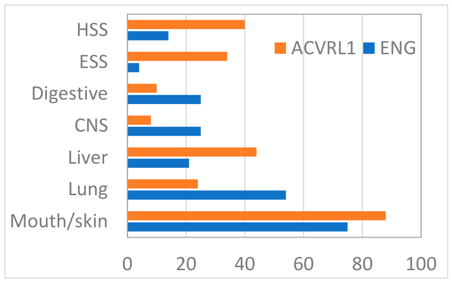
Figure 2.
Organ involvement according to affected gene and RNA-disrupting variants.
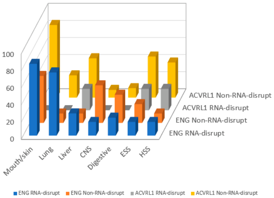

Table 1.
Sex, age, and variant type of study subjects.
|
ENG (n=28) |
ACVRL1 (n=50) |
p-value | |
|
Sex Male Female |
16 (57.1%) 12 (42.9%) |
19 (38%) 31 (62%) |
0.154 |
| Age, yr, median (IQR) | 51 (34.25-75) | 45 (27.50-63.50) | 0.225 |
| Variant type | |||
| Missense | 9 | 46 | <0.001 |
| Nonsense | 0 | 2 | 0.534 |
| Frame-shift | 1 | 0 | 0.359 |
| Splicing-altering | 11 | 2 | <0.001 |
| Exon deletion | 7 | 0 | <0.001 |
Table 2.
Manifestations according to the mutated gene.
| ENG | ACVRL1 | p-value | |
| Vascular lesions | |||
| Mucocutaneous telangiectasias | 21 (75%) | 43 (86%) | 0.237 |
| Pulmonary AVMs | 15 (53.6%) | 12 (24%) | 0.024 |
| Bilateral PAVMs | 8 (28.6%) | 2 (4%) | 0.107 |
| Embolized PAVMs | 12 (42.8%) | 7 (14%) | 0.190 |
| Hepatic VMs | 6 (21.4%) | 22 (44%) | 0.072 |
| Cerebral VMs | 7 (25%) | 4 (8%) | 0.082 |
| Gastrointestinal VMs | 7 (25%) | 5 (10%) | 0.505 |
| Worst recorded epistaxis severity | |||
| Moderate-to-severe FID | 8 (26.8%) | 25 (50%) | 0.511 |
| Moderate-to-severe ESS | 5 (17.9%) | 23 (46%) | 0.074 |
| ESS, median (IQR) | 3.08 (1.19-4.78) | 4.24 (1.52-5.47) | 0.353 |
| Sadick II–III, frequency | 11(39.3%) | 28 (56%) | 0.287 |
| Sadick II–III, amount | 10 (35.7) | 25 (50%) | 0.586 |
| Recent epistaxis/bleeding severity (*) | |||
| Moderate-to-severe FID | 7 (46.4%) | 23 (46%) | 0.294 |
| Moderate-to-severe ESS | 1 (3.6%) | 17 (34%) | 0.014 |
| ESS, median (IQR) | 0.51 (0-2.43) | 1.41 (0-4) | 0.131 |
| Sadick II–III, frequency | 5 (17.9%) | 18 (36%) | 0.402 |
| Sadick II–III, amount | 3 (10.7%) | 19 (38%) | 0.086 |
| Moderate-to-severe HSS | 4 (14.8%) | 20 (40%) | 0.038 |
(*) Past 3 months for FID, ESS and Sadick scores;12 months for the Hemorrhagic severity score Data are presented as n (%) unless otherwise indicated. Continuous variables are presented as median (interquartile range [IQR]). Categorical variables were compared using Fisher’s exact test, whereas continuous variables were compared using the Mann–Whitney U test. AVM, arteriovenous malformation; VM, vascular malformation; ESS, Epistaxis Severity Score; HHT, hereditary hemorrhagic telangiectasia. HSS, hemorrhagic severity score.
Table 3.
Manifestations of patients with ENG variants according to the variant type.
|
RNA-disruptive Variants (n=19) |
Non-RNA-disruptive variants (n=9) |
p-value | |
| Vascular lesions | |||
| Mucocutaneous telangiectasias | 16 (84.2%) | 5 (55.5%) | 0.165 |
| Pulmonary AVMs | 14 (73.7%) | 1 (11.1%) | 0.004 |
| Hepatic VMs | 5 (26.3%) | 1 (11.1%) | 1 |
| Cerebral VMs | 3 (15.8%) | 4 (44.4%) | 0.134 |
| Gastrointestinal VMs | 4 (21.1%) | 3 (33.3%) | 0.290 |
| Worst recorded epistaxis score | |||
| Moderate-to-severe FID | 5 (26.3%) | 3 (33.3%) | 0.580 |
| Moderate-to-severe ESS | 3 (15.8%) | 2 (22.2%) | 1 |
| ESS, median (IQR) | 3.73 (0.91-4.57) | 2.43 (1.21-7.52) | 0.583 |
| Sadick II–III, frequency | 8 (42.1%) | 3 (33.3%) | 1 |
| Sadick II–III, amount | 7 (35.8) | 3 (33.3%) | 1 |
| Recent Epistaxis/Bleeding severity | |||
| Moderate-to-severe FID | 5 (26.3%) | 2 (22.2%) | 1 |
| Moderate-to-severe ESS | 1 (5.3%) | 4 (44.4%) | 1 |
| ESS, median (IQR) | 0.71 (0-2.53) | 0 (0-1.72) | 0.444 |
| Sadick II–III, frequency | 4 (21.1%) | 1 (11.1%) | 1 |
| Sadick II–III, amount | 3 (15.8%) | 0 | 0.521 |
| Moderate-to-severe HSS | 3 (15.8%) | 1 (11.1%) | 1 |
(*) Past 3 months for FID, ESS and Sadick scores;12 months for the Hemorrhagic severity score Data are presented as n (%) unless otherwise indicated. Continuous variables are presented as median (interquartile range [IQR]). Categorical variables were compared using Fisher’s exact test, whereas continuous variables were compared using the Mann–Whitney U test. AVM, arteriovenous malformation; VM, vascular malformation; ESS, Epistaxis Severity Score; HHT, hereditary hemorrhagic telangiectasia. HSS, hemorrhagic severity score.
Table 4.
Manifestations of patients with ACVRL1 variants according to the variant type.
|
RNA-disruptive variants (n=4) |
Non-RNA-disruptive variants (n=46) |
p-value | |
| Vascular lesions | |||
| Mucocutaneous telangiectasias | 0 | 39 (84.8%) | 1 |
| Pulmonary AVMs | 0 | 12 (26.1%) | 0.560 |
| Hepatic VMs | 1 (25%) | 21 (45.7%) | 0.607 |
| Cerebral VMs | 0 | 4 (8.7%) | 1 |
| Gastrointestinal VMs | 0 | 5 (10.9%) | 1 |
| Worst recorded epistaxis score | |||
| Moderate-to-severe FID | 1 (25%) | 24 (52.2%) | 1 |
| Moderate-to-severe ESS | 1 (25%) | 22 (47.8%) | 1 |
| ESS, median (IQR) | 3.53 (2.83-3.53) | 4.24 (1.26-5.74) | 0.826 |
| Sadick II–III, frequency | 0 | 28 (60.9%) | 0.106 |
| Sadick II–III, amount | 1 (25%) | 24 (52.2%) | 1 |
| Recent Epistaxis/Bleeding severity | |||
| Moderate-to-severe FID | 1 (25%) | 22 (47.8%) | 1 |
| Moderate-to-severe ESS | 1 (25%) | 16 (34.8%) | 1 |
| ESS, median (IQR) | 0.705 (0-3.53) | 1.41 (0-4) | 0.463 |
| Sadick II–III, frequency | 0 | 18 (39.1%) | 0.283 |
| Sadick II–III, amount | 1 (25%) | 18 (39.1%) | 1 |
| Moderate-to-severe HSS | 1 (25%) | 19 (41.3%) | 0.636 |
(*) Past 3 months for FID, ESS and Sadick scores;12 months for the Hemorrhagic severity score Data are presented as n (%) unless otherwise indicated. Continuous variables are presented as median (interquartile range [IQR]). Categorical variables were compared using Fisher’s exact test, whereas continuous variables were compared using the Mann–Whitney U test. AVM, arteriovenous malformation; PAVM, pulmonary arteriovenous malformation; ESS, Epistaxis Severity Score; HHT, hereditary hemorrhagic telangiectasia. HSS, hemorrhagic severity score.
Table 5.
Clinical characteristics of patients with non-RNA-disrupting variants according to the gene mutated.
Table 5.
Clinical characteristics of patients with non-RNA-disrupting variants according to the gene mutated.
|
ENG (n=9) |
ACVLR1 (n=46) |
p-value | |
| Vascular lesions | |||
| Mucocutaneous telangiectasias | 5 (55.5%) | 39 (84.8%) | 0.067 |
| Pulmonary AVMs | 1 (11.1%) | 12 (26.1%) | 0.420 |
| Hepatic VMs | 1 (11.1%) | 21 (45.7%) | 0.346 |
| Cerebral VMs | 4 (44.4%) | 4 (8.7%) | 0.013 |
| Gastrointestinal VMs | 3 (33.3%) | 5 (10.9%) | 0.281 |
| Worst recorded epistaxis score | |||
| Moderate-to-severe FID | 3 (33.3%) | 24 (52.2%) | 1 |
| Moderate-to-severe ESS | 2 (22.2%) | 22 (47.8%) | 0.633 |
| ESS, median (IQR) | 2.4 (1.2-7.5) | 4.2 (1.3-5.7) | 0.984 |
| Sadick II–III, frequency | 3 (33.3%) | 28 (60.9%) | 0.639 |
| Sadick II–III, amount | 3 (33.3%) | 24 (52.2%) | 1 |
| Recent Epistaxis/bleeding severity | |||
| Moderate-to-severe FID | 2 (22.2%) | 22 (47.8%) | 0.420 |
| Moderate-to-severe ESS | 4 (44.4%) | 16 (34.8%) | 0.158 |
| ESS, median (IQR) | 0 (0-1.72) | 1.41 (0-4) | 0.118 |
| Sadick II–III, frequency | 1 (11.1%) | 18 (39.1%) | 0.384 |
| Sadick II–III, amount | 0 | 18 (39.1%) | 0.075 |
| Moderate-to-severe HSS | 1 (11.1%) | 19 (41.3%) | 0.228 |
(*) Past 3 months for FID, ESS and Sadick scores;12 months for the Hemorrhagic severity score Data are presented as n (%) unless otherwise indicated. Continuous variables are presented as median (interquartile range [IQR]). Categorical variables were compared using Fisher’s exact test, whereas continuous variables were compared using the Mann–Whitney U test. AVM, arteriovenous malformation; PAVM, pulmonary arteriovenous malformation; ESS, Epistaxis Severity Score; HHT, hereditary hemorrhagic telangiectasia. HSS, hemorrhagic severity score.
Disclaimer/Publisher’s Note: The statements, opinions and data contained in all publications are solely those of the individual author(s) and contributor(s) and not of MDPI and/or the editor(s). MDPI and/or the editor(s) disclaim responsibility for any injury to people or property resulting from any ideas, methods, instructions or products referred to in the content. |
© 2026 by the authors. Licensee MDPI, Basel, Switzerland. This article is an open access article distributed under the terms and conditions of the Creative Commons Attribution (CC BY) license (http://creativecommons.org/licenses/by/4.0/).
Copyright: This open access article is published under a Creative Commons CC BY 4.0 license, which permit the free download, distribution, and reuse, provided that the author and preprint are cited in any reuse.