

Submitted:
06 November 2026
Posted:
09 November 2026
You are already at the latest version
Abstract
Protein abundance helps identify candidate cancer targets. Their therapeutic relevance depends on the protein’s molecular state, its role in the tumor, and how the treatment acts on it. This review synthesizes evidence from quantitative proteomics, protein interaction mapping, structural analysis, genetic perturbation, and pharmacological studies. Preclinical examples involving epidermal growth factor receptor trafficking, hexokinase 2 scaffolding, chromatin complex assembly, and protein turnover illustrate how localization, binding partners, and post-translational modifications shape protein function. Studies of targeted degraders and molecular glues show why target engagement, ubiquitin ligase recruitment, and protein loss require separate verification. Clinical responses to revumenib in genetically defined acute leukemias support targeting the menin–KMT2A interaction, while acquired MEN1 mutations reveal a mechanism of resistance. Trastuzumab deruxtecan in HER2-low breast cancer illustrates a distinct use of protein expression to guide patient selection when the target serves as a delivery antigen. We propose a framework that evaluates protein expression, physical interaction, functional dependence, drug mechanism, and clinical benefit as distinct forms of evidence. The framework matches validation experiments and patient-selection biomarkers to the therapeutic modality. Confidence in a target is greatest when independent evidence connects the proposed mechanism to a cancer-relevant effect at achievable and tolerable drug exposures.
Keywords:
cancer
; protein expression
; protein interactions
; target validation
; proteomics
; molecular glues
; targeted protein degradation
1. Introduction
Cancer target discovery increasingly depends on understanding the functional consequences of molecular alterations rather than simply compiling proteins that are highly expressed in tumors (Elmas et al., 2021; Gonçalves et al., 2022). Large proteomic datasets reveal candidate vulnerabilities that are not apparent from transcript abundance alone, supporting the inclusion of protein measurements in precision oncology (Guo et al., 2019; Roumeliotis et al., 2017). However, protein concentration represents a biological outcome as well as a potential cause, because interacting partners can determine whether a newly synthesized protein becomes incorporated into a stable complex or is removed (Senger & Schaefer, 2021; Sousa et al., 2019). An expression difference therefore becomes therapeutically informative only when the relevant cellular process, disease context and proposed intervention are specified (Coleman et al., 2009; Vdovin et al., 2022).
The relationship between messenger RNA and protein is particularly important when expression is used to nominate targets from sequencing data (Srivastava et al., 2022). Analyses across cancer proteogenomic datasets show that some proteins are poorly predicted by their own transcripts and that information from other genes can improve prediction (Srivastava et al., 2022). Such patterns are compatible with regulation through interacting partners, but predictive performance alone cannot establish the identity or direction of the underlying causal interaction (Ryan et al., 2017; Sousa et al., 2019). Proteomic associations should consequently be treated as hypotheses about regulatory organization until perturbation experiments distinguish direct mechanisms from shared upstream regulation (Roumeliotis et al., 2017; Vizeacoumar et al., 2013).
Protein interaction studies provide one route from expression to mechanism by identifying partners that connect a candidate to signaling, chromatin regulation, metabolism or protein quality control (Li et al., 2017; Rodina et al., 2023). The term interaction nevertheless encompasses experimentally different observations, including direct molecular contact, membership of the same isolated complex, spatial proximity and genetic dependence (Breitkopf et al., 2016; Havugimana et al., 2022; Vizeacoumar et al., 2013). These observations answer different questions and should remain distinct in a therapeutic argument (Huttlin et al., 2021; Porta-Pardo et al., 2015). A proximity-labeling hit can identify a useful cellular neighborhood without demonstrating a druggable binding surface, whereas a purified binding experiment can establish contact without showing that the contact matters in a tumor (Branon et al., 2018; Swayampakula et al., 2017).
The functional unit may also differ from the protein conventionally named as the target (Blaha et al., 2022; Morozov et al., 2023). Hexokinase 2 (HK2) can support metastatic behavior through scaffolding functions, while changes in chromatin composition can reorganize androgen receptor complexes without requiring a simple increase in receptor abundance (Blaha et al., 2022; Morozov et al., 2023). Similarly, altered epidermal growth factor receptor (EGFR) trafficking can preserve signaling-competent receptor pools, making receptor location and partner recruitment relevant to the interpretation of an expression measurement (Chung et al., 2009; Yu et al., 2020).
Pharmacological development adds a further requirement because inhibition, protein degradation and antibody-mediated payload delivery exploit different properties of a target (Gadd et al., 2017; Lv et al., 2020; Modi et al., 2022). A catalytic inhibitor may leave scaffolding functions intact, whereas degradation removes several protein functions but requires productive engagement of cellular degradation machinery (Alabi et al., 2021; Blaha et al., 2022). An antibody–drug conjugate can use a surface protein as an entry point even when the malignant cell is not dependent on that protein's intracellular signaling function (Modi et al., 2022). Thus, a universal rule that every useful target must be overexpressed, directly oncogenic and essential would misclassify several valid therapeutic strategies (Lin et al., 2024; Modi et al., 2022).
This review examines the evidence needed to connect protein expression to an intervention that changes a cancer-relevant phenotype, with emphasis on experimentally defined interactions and recent chemical approaches (Lv et al., 2020; Yoshinari et al., 2025; Zhai et al., 2023). Its organizing question is whether each additional experiment changes the strength or meaning of the therapeutic claim, rather than merely adding another measurement (Lin et al., 2019; Meyers et al., 2017). Figure 1 distinguishes the principal evidence categories, and Table 1 compares the inferences supported by commonly used assays (Branon et al., 2018; Gadd et al., 2017; Uhlen et al., 2016). The framework developed here is an interpretive proposal for evaluating studies, rather than a validated clinical scoring system.
2. Literature Search and Evidence Selection
This critical narrative review began with a supplied index of 100 publications concerning cancer protein expression, interaction networks, functional dependencies and interaction-directed therapeutics. Ninety-seven indexed full texts were available through PubMed Central or Europe PMC at the time of preparation; DOI-linked publisher and bibliographic records supplemented these texts. Targeted searches added methodological and clinical examples concerning antibody validation, proximity labeling, CRISPR artifacts, drug mechanism validation, menin inhibition, antibody–drug conjugates and structural prediction. Source checking was completed on 1 November 2026, and the sampled literature spans foundational and recent studies rather than a prespecified exhaustive publication interval.
Targeted lookup used article titles, DOI strings and PubMed or PMC identifiers. Concepts covering protein abundance, protein complexes, interaction interfaces, cancer dependency, rescue, molecular glue and targeted degradation guided the synthesis. Selection prioritized primary studies that connected at least two evidence categories or exposed a limitation in the inference between them. Expression-only observations were retained when they illustrated biomarker limitations, while structural studies were included when an identified interface informed function or chemical intervention.
For each cited study, the assessment recorded the experimental model, interaction assay, perturbation strategy, phenotype and strength of therapeutic inference. Bibliographic checking compared source identifiers and article metadata with DOI registration or publisher records; differences between online publication and final issue years were resolved using the version cited. The separate reference audit provides source links and claim locations. No prospective screening protocol, systematic-review flow diagram or meta-analysis was undertaken, so the synthesis should not be interpreted as an exhaustive estimate of target-validation success or treatment efficacy.
3. Scientific Background
3.1. Expression Is Constrained by Complex Assembly
Protein abundance emerges from synthesis, compartmental distribution and removal, with complex formation providing an additional layer of regulation (Ori et al., 2016; Sousa et al., 2019). When unassembled subunits are less stable than assembled proteins, changes in one component can alter the abundance of its partners without corresponding transcriptional changes (Ryan et al., 2017; Senger & Schaefer, 2021). Breast cancer proteomics identified collateral losses of complex members after alteration of another subunit, including effects associated with E-cadherin loss (Ryan et al., 2017). Such observations explain why a protein decrease may report the disruption of an entire assembly rather than independent suppression of each component (Roumeliotis et al., 2017; Ryan et al., 2017).
Stoichiometry also changes the meaning of apparently comparable expression levels (Morales-Betanzos et al., 2017; Ori et al., 2016). The same concentration of a signaling component can produce different outputs when binding partners compete for it or when an inhibitory partner becomes limiting (Coleman et al., 2009). Combined assessment of eIF4E and its binding proteins provided prognostic information beyond eIF4E alone, illustrating how a pathway-level estimate can be more informative than a single abundance measurement (Coleman et al., 2009). This remains a prognostic observation unless treatment-specific analyses establish that the combined marker identifies differential benefit from an intervention (Coleman et al., 2009; Modi et al., 2022).
Measurements from bulk tumors add a spatial complication because tumor, stromal and immune compartments contribute differently to the recovered protein pool (Morales-Betanzos et al., 2017). Quantitative melanoma measurements of PD-1, PD-L1 and PD-L2 showed that ligand abundance and relative stoichiometry cannot be inferred reliably from the concentration of a single component (Morales-Betanzos et al., 2017). In this setting, total abundance does not directly establish receptor occupancy or the strength of signaling across the tumor–immune interface (Morales-Betanzos et al., 2017). Spatially resolved measurements and an explicit identification of the expressing cell population are therefore logical complements to bulk proteomics when an interaction crosses cell boundaries (Morales-Betanzos et al., 2017; Swayampakula et al., 2017).
3.2. Physical and Functional Interactions
A direct physical interaction requires evidence that two molecular species contact one another under defined conditions, while co-complex association can be mediated by additional components (Breitkopf et al., 2016; Wang et al., 2024). Reciprocal isolation, orthogonal assays and structural data can strengthen an association, but their interpretation depends on expression level, biochemical conditions and preservation of the relevant assembly (Havugimana et al., 2022; Huttlin et al., 2021). Proximity labeling captures nearby proteins in living cells and can recover spatial relationships that would be lost during biochemical purification (Branon et al., 2018; Swayampakula et al., 2017). Its principal limitation for this review is that proximity does not identify which pair of proteins directly binds (Branon et al., 2018).
Genetic interactions describe a different relationship because the phenotype of combined perturbations differs from that expected from the individual perturbations (van Pel et al., 2013; Vizeacoumar et al., 2013). Two genes can therefore exhibit synthetic lethality while their products never contact one another physically (Vizeacoumar et al., 2013; Zhao et al., 2023). Conversely, two proteins can bind reproducibly while the interaction is redundant for the phenotype being measured (Guarnaccia et al., 2021; Lambert et al., 2019). Separating these concepts prevents a network diagram from implying that every connected pair represents both a physical interface and a therapeutic dependency (Li et al., 2017; Vizeacoumar et al., 2013).
3.3. Dependence and Intervention
Functional dependence is conditional on genotype, lineage, cell state and the experimental endpoint (Gonçalves et al., 2022; Lin et al., 2024). A proliferation assay can miss a role in invasion or metastasis, as illustrated by tumor-intrinsic ICAM-1–EGFR signaling in breast cancer models (Kang et al., 2024). An apparent dependency can also arise from technical effects, including the response to CRISPR-induced DNA breaks in amplified genomic regions (Meyers et al., 2017). Correction of such artifacts and confirmation with independent perturbations are necessary before interpreting a screening score as evidence for a drug target (Lin et al., 2019; Meyers et al., 2017).
Target engagement is the demonstration that an intervention reaches and acts on its proposed molecular partner, whereas therapeutic efficacy requires a favorable disease outcome at tolerable exposure (Issa et al., 2023; Ketcham et al., 2022). These requirements remain separate because a compound can bind a protein without producing the necessary functional change, or kill cells through another molecular activity (Beyett et al., 2022; Lin et al., 2019). The appropriate validation experiment therefore follows the proposed modality, including interface disruption for interaction inhibitors and ligase-dependent protein loss for degraders (Gadd et al., 2017; Lv et al., 2020).

Table 1.
What common assays establish.
| Evidence | Supported inference | Additional test |
|---|---|---|
| Expression (Gonçalves et al., 2022; Uhlen et al., 2016) | Candidate abundance association | Validated assay and cell identity |
| Co-isolation (Havugimana et al., 2022; Huttlin et al., 2021) | Shared biochemical assembly | Direct binding and abundance normalization |
| Proximity labeling (Branon et al., 2018) | Cellular neighborhood | Interface-specific validation |
| Structural binding (Gadd et al., 2017; Wang et al., 2024) | Defined molecular contact | Functional perturbation |
| Genetic perturbation (Meyers et al., 2017; Vizeacoumar et al., 2013) | Contextual requirement | Independent guides and rescue |
| Drug response (Lin et al., 2019; Perner et al., 2023) | Intervention-associated phenotype | Engagement and mechanism controls |
4. Mechanisms Linking Expression to Functional Interaction
4.1. Receptor Trafficking and Signaling Competence
EGFR provides a useful example because receptor abundance, activation state and intracellular routing can change together while contributing differently to a phenotype (Chung et al., 2009; Li et al., 2013). In lung cancer models, TRIB3 associated with EGFR and recruited machinery that favored receptor recycling and stability, including PKCα-associated phosphorylation and WWP1-associated ubiquitination (Yu et al., 2020). Disruption of the TRIB3–EGFR interaction with a stapled peptide accelerated receptor degradation and suppressed malignant phenotypes in the studied models (Yu et al., 2020). The causal argument is therefore stronger than an expression correlation because the intervention altered a proposed interaction and changed receptor fate (Yu et al., 2020).
The interpretation remains conditional on the particular receptor state and cellular background, because mutant EGFR trafficking through recycling compartments can promote interaction with Src (Chung et al., 2009). Likewise, a mutant-KRAS colorectal cancer model exhibited extensive remodeling of the EGFR network, showing that a downstream oncogenic alteration can change the context in which receptor interactions operate (Kennedy et al., 2020). Network remodeling can reflect altered protein concentration as well as altered phosphorylation, so increased recovery of a partner should not automatically be attributed to stronger binding (Grossmann et al., 2015; Kennedy et al., 2020). Experiments that normalize interaction measurements to available bait and partner abundance help distinguish these possibilities (Havugimana et al., 2022; Huttlin et al., 2021).
FBXW7 supplies a complementary mechanism in which an E3 ubiquitin ligase contributes to EGFR turnover through receptor-associated phosphodegron-like motifs (Boretto et al., 2024). Reduced degradation after alteration of those motifs supports a mechanistic connection between recognition and receptor stability (Boretto et al., 2024). The associated treatment outcomes in colorectal cancer patients are clinically relevant, but an observational association cannot establish that FBXW7 status alone predicts benefit from every EGFR-directed modality (Boretto et al., 2024). A prospective treatment comparison would be needed to separate predictive value from broader disease aggressiveness (Boretto et al., 2024; Modi et al., 2022).
ICAM-1 further demonstrates why the measured endpoint must match the biological claim (Kang et al., 2024). In the reported triple-negative breast cancer models, the ICAM-1–EGFR interaction favored JAK1/STAT3 signaling and metastatic progression without a corresponding increase in primary tumor growth (Kang et al., 2024). A screen restricted to short-term viability could therefore underestimate this interaction's importance, while an expression-based survival association would not reveal the specific mechanism (Kang et al., 2024). Figure 2A compares two independently reported routes controlling receptor fate in the studied cancer models (Boretto et al., 2024; Yu et al., 2020).
4.2. Scaffolds Separate Abundance from Catalytic Activity
The activity of a cancer-associated protein can include physical organization of a signaling complex independently of its canonical catalytic reaction (Blaha et al., 2022; Romano et al., 2015). Hexokinase 2 formed a complex involving PRKAR1a and GSK3β, facilitating inhibitory regulation of GSK3β and stabilization of proteins including SNAIL (Blaha et al., 2022). The same study also connected glucose kinase activity to SNAIL glycosylation, indicating that catalytic and scaffolding routes can converge on the same downstream phenotype (Blaha et al., 2022). Consequently, a decrease in glucose phosphorylation cannot be assumed to quantify complete inhibition of HK2-dependent metastatic behavior (Blaha et al., 2022).
FKBP51 contributed both scaffolding and isomerase functions to NF-κB activation in melanoma, while FKBP4 organized chaperone-linked complexes affecting IKK and RelA in lung adenocarcinoma (Romano et al., 2015; Zong et al., 2021). The prohibitin–CRAF interaction in pancreatic cancer models similarly linked membrane organization to RAS–ERK signaling (Luan et al., 2014). Together, these studies support measuring the assembly and downstream output of a complex when evaluating proteins whose activity depends on positioning other molecules (Blaha et al., 2022; Luan et al., 2014).
Chaperone dependence also complicates the attribution of selectivity because disrupting one organizing factor can alter several client proteins (Ha et al., 2022; Wang et al., 2019). GRP78 perturbation reduced oncogenic KRAS protein expression in several cancer models, while disruption of the Hsp90–Cdc37 association affected kinase clients (Ha et al., 2022; Wang et al., 2019). These findings support therapeutic exploration but leave open how much of an observed phenotype results from the named client rather than broader proteostasis disruption (Rodina et al., 2023; Wang et al., 2019). Rescue with appropriately expressed client proteins and comparison with interaction-selective perturbations would help distinguish these mechanisms (Blaha et al., 2022; Lin et al., 2019).
4.3. Chromatin Assembly Determines Transcriptional Output
Transcription factors operate within assemblies whose formation depends on chromatin state and cofactor availability (Morozov et al., 2023; Seo et al., 2013). CCAR1 stabilized the association between androgen receptor and GATA2 and supported recruitment of transcriptional machinery to androgen-responsive regulatory regions (Seo et al., 2013). HIRA-dependent loading of histone H3.3 influenced H3.3 phosphorylation, histone acetylation, BRD4 recruitment and androgen receptor organization at enhancers (Morozov et al., 2023). These observations make receptor concentration an incomplete description of the transcriptionally active state (Morozov et al., 2023; Seo et al., 2013).
Drug exposure can also reorganize rather than simply eliminate a chromatin-associated interactome (Guarnaccia et al., 2021; Lambert et al., 2019). BET bromodomain inhibition affected distinct classes of interactions, including associations mediated through domains outside the inhibitor-binding pocket (Lambert et al., 2019). WDR5 WIN-site inhibition likewise changed a broader interaction network, showing that chemical occupancy of one pocket can have consequences beyond the initially nominated partner (Guarnaccia et al., 2021). The key analytical question is which altered association mediates the phenotype, because a global interaction change can contain both causal effects and secondary responses (Guarnaccia et al., 2021; Lambert et al., 2019).
HDAC6 inactivation provides a further example of the distinction between removing an enzyme activity and changing regulatory protein abundance (Zamperla et al., 2024). The reported interventions altered P300 ubiquitination and stabilization alongside changes in chromatin accessibility and transcription (Zamperla et al., 2024). These results suggest a role for interaction-dependent P300 regulation, but they do not establish that every anticancer effect of every HDAC6 inhibitor proceeds through the same pathway (Lin et al., 2019; Zamperla et al., 2024). Figure 2C summarizes chromatin assembly as a context-dependent mechanism rather than a universal linear cascade across cancers (Morozov et al., 2023; Zamperla et al., 2024).
4.4. Post-Translational Modification and Protein Turnover
Post-translational modifications can change binding preferences without appreciably changing total protein abundance (Grossmann et al., 2015; Zhao et al., 2025). At Ku80 lysine 568, the reported transition from crotonylation to SUMOylation promoted DNA-PK complex assembly and DNA repair after radiation exposure (Zhao et al., 2025). Perturbation of this modification site altered radiosensitivity in tumor models, connecting a defined molecular state with a treatment-relevant phenotype (Zhao et al., 2025). The relevant target hypothesis therefore concerns modification-dependent assembly rather than simple Ku80 overexpression (Zhao et al., 2025).
Autophagy machinery provides another example in which partner binding and stability reinforce one another (Wible et al., 2019). ATG16L1 binding protected free ATG5 from degradation and favored ATG12 conjugation, while cancer-associated changes affecting the binding region impaired productive complex assembly (Wible et al., 2019). This mechanism shows why the concentration of a component and the fraction incorporated into an active complex can diverge (Senger & Schaefer, 2021; Wible et al., 2019). Functional assays should therefore accompany total-protein measurements when an assembly step controls pathway flux (Wible et al., 2019).
Interaction-dependent protection from degradation can itself become a therapeutic vulnerability (Vdovin et al., 2022; Zhai et al., 2023). N1DARP disrupted the association between USP10 and the Notch1 intracellular domain, increasing ubiquitination and degradation in pancreatic cancer models (Zhai et al., 2023). In multiple myeloma, OTUD1 protected PRDX4 and supported immunoglobulin synthesis, linking protein production and endoplasmic reticulum stress to proteasome-inhibitor sensitivity (Vdovin et al., 2022). These mechanisms point in different therapeutic directions, so a general assumption that lowering an abundant protein will always improve response would be inappropriate (Vdovin et al., 2022; Zhai et al., 2023).
5. Recent Experimental Advances
5.1. Proteomics and Context-Specific Interaction Maps
Large cancer cell-line proteomes allow expression, drug response and genetic essentiality to be analyzed within a shared experimental resource (Gonçalves et al., 2022). The 949-cell-line proteomic map identified protein-level associations with vulnerabilities that were not consistently detected at the transcript level, but overall drug-response prediction was similar for proteomic and transcriptomic inputs (Gonçalves et al., 2022). This combination of results supports complementarity rather than a universal claim that protein measurements outperform RNA for every therapeutic question (Gonçalves et al., 2022; Srivastava et al., 2022). Biomarkers should therefore be evaluated for added value in a specified prediction problem instead of ranked solely by molecular layer (Elmas et al., 2021; Guo et al., 2019).
Cancer-focused interaction resources add mechanistic hypotheses to these association maps (Li et al., 2013; Li et al., 2017). OncoPPi identified previously unreported cancer-associated interactions, while perturbation of a mutant-EGFR interaction network located proteins contributing to viability and resistance (Li et al., 2013; Li et al., 2017). These resources become particularly informative when the same interaction is examined across genotypes or under therapeutic exposure (Kennedy et al., 2020; Lambert et al., 2019). However, physical network coverage remains uneven, and the absence of an edge can reflect detection limits or experimental context rather than biological absence (Havugimana et al., 2022; Huttlin et al., 2021).
Modern proximity labeling improves access to dynamic or compartment-restricted protein neighborhoods in living cells (Branon et al., 2018). Co-fractionation mass spectrometry offers a complementary strategy for inferring assemblies from coordinated biochemical behavior, reducing reliance on a separate affinity reagent for every candidate (Havugimana et al., 2022). Neither approach eliminates the need for orthogonal validation when a proposed binding interface is central to the therapeutic mechanism (Breitkopf et al., 2016; Havugimana et al., 2022). Table 2 compares representative studies according to their mechanism, model and remaining inferential gap (Blaha et al., 2022; Yoshinari et al., 2025; Yu et al., 2020; Zhai et al., 2023).
5.2. Interaction Inhibitors and Mechanistic Precision
Interaction inhibitors can interfere with selected interfaces while preserving other functions of the same protein, provided that selectivity is demonstrated experimentally (Filippakopoulos et al., 2012; Ketcham et al., 2022). MRTX0902 was developed as a SOS1 binder that disrupts the SOS1–KRAS interaction, with combination activity alongside a KRAS inhibitor in preclinical models (Ketcham et al., 2022). This strategy illustrates an indirect way to influence the nucleotide state of KRAS, but the combination phenotype alone cannot identify which pharmacological properties determine efficacy in patients (Ketcham et al., 2022). Exposure, target engagement and downstream pathway measurements are needed to connect biochemical activity with an achievable treatment effect (Issa et al., 2023; Ketcham et al., 2022).
Structural analysis can test specific explanations for drug cooperation rather than inferring mechanism from a combination score (Beyett et al., 2022). In EGFR, analysis of ATP-site and allosteric inhibitor combinations identified P-loop contacts contributing to cooperative binding, and mutation of a relevant residue reduced cooperation (Beyett et al., 2022). This is stronger evidence for a defined mechanism than a demonstration that two compounds reduce viability more effectively together (Beyett et al., 2022; Lin et al., 2019). Nevertheless, the tested receptor variants and compounds define the scope of that conclusion (Beyett et al., 2022).
The CPAP–tubulin study illustrates an interaction-directed intervention with a distinct phenotypic endpoint (Mariappan et al., 2019). Perturbation of the interface increased microtubule nucleation from extra centrosomes, impaired centrosome clustering and promoted lethal multipolar division in the examined cancer models (Mariappan et al., 2019). Its therapeutic rationale therefore depends on a cellular structural state, centrosome amplification, as well as the binding interaction (Mariappan et al., 2019). This example broadens target evaluation beyond expression thresholds to measurable organizational features of malignant cells (Mariappan et al., 2019; Ori et al., 2016).
5.3. Degraders Require Productive Induced Interactions
Targeted degraders, including proteolysis-targeting chimeras (PROTACs), convert a binding event into a sequence of recruitment, ubiquitination and protein removal, making induced molecular proximity part of the mechanism (Gadd et al., 2017; Lv et al., 2020). Structural work on the BRD4 degrader MZ1 showed how protein contacts within a ternary complex influence cooperative recognition and selectivity (Gadd et al., 2017). A target-binding affinity measurement alone therefore cannot establish degradation efficiency or specificity (Alabi et al., 2021; Gadd et al., 2017). Figure 3 contrasts interface blockade with induced recruitment and includes an attributed experimental structure of the MZ1-containing complex (Gadd et al., 2017; Ketcham et al., 2022).
BRAF-targeting PROTACs demonstrated that degradation selectivity can depend on protein conformation rather than simply the affinity of the target-binding component (Alabi et al., 2021). In the reported experiments, wild-type BRAF was relatively spared because its inactive conformation supported weaker ternary-complex formation, while activation sensitized it to degradation (Alabi et al., 2021). It also suggests that conformational state should be included when testing biomarkers of degrader response (Alabi et al., 2021; Vetma et al., 2025).
Recent KRAS degraders provide further examples of this requirement (Vetma et al., 2025; Yoshinari et al., 2025). ASP3082 was characterized through a KRAS(G12D)–degrader–VHL structure, cellular protein depletion and activity in xenograft models (Yoshinari et al., 2025). A separate study of ACBI4 examined cooperative ternary-complex formation involving active-state KRAS and demonstrated degradation of selected KRAS variants in cancer cells (Vetma et al., 2025). These findings establish valuable preclinical mechanisms, but they do not justify transferring efficacy claims between alleles, compounds or clinical settings (Vetma et al., 2025; Yoshinari et al., 2025).
5.4. Molecular Glues Reveal Unexpected Causal Intermediates
Molecular glues emphasize that the protein contacted by a compound need not be the protein ultimately removed (Houles et al., 2023; Lv et al., 2020). HQ461 promoted association of CDK12 with DDB1 and the associated ubiquitin-ligase machinery, leading to degradation of cyclin K (Lv et al., 2020). SR-4835 was also shown to promote cyclin K degradation in melanoma, complicating a description based exclusively on catalytic CDK12 inhibition (Houles et al., 2023). Mechanistic evaluation must therefore trace the recruited complex and degraded species separately rather than assigning all observed effects to a conventional inhibitor label (Houles et al., 2023; Lv et al., 2020).
Two structural studies connected neomorphic KBTBD4 mutations with recruitment of HDAC-containing complexes and compared this process with UM171-induced molecular gluing (Chen et al., 2025; Xie et al., 2025). Their convergence strengthens the interpretation that an altered interface can reprogram substrate recruitment, while the different assemblies examined should remain distinguishable (Chen et al., 2025; Xie et al., 2025). This is also a reminder that induced degradation can reproduce a disease-associated neomorphic interaction, so the biological consequence depends on which protein complex is destabilized (Chen et al., 2025; Xie et al., 2025). Interface stabilization is therefore a mechanism to evaluate, not an inherently beneficial therapeutic outcome (Wang et al., 2024; Xie et al., 2025).
The 2026 HuR-degrader study connected a CRBN-associated ternary complex with HuR loss, altered BRAF splicing and suppression of BRAF-mutant colorectal cancer models (Lu et al., 2026). Its mechanism places an RNA-binding protein upstream of an oncogenic protein output, showing how expression changes can be downstream indicators of an induced interaction (Lu et al., 2026). A separate allosteric molecular glue stabilized KEAP1–CUL3 organization to increase NRF2 degradation in the reported cancer models (Roy et al., 2026). These advances broaden the available intervention types, while preserving the need for genetic specificity controls and normal-tissue assessment (Lin et al., 2019; Lu et al., 2026; Roy et al., 2026).
6. Therapeutic Applications and Clinical Implications
6.1. Clinical Testing of an Interaction-Defined Dependency
Menin inhibition provides an example in which a protein interaction hypothesis progressed to patient treatment rather than remaining a biochemical observation (Issa et al., 2023). In the first-in-human revumenib study, inhibition of the menin–KMT2A interaction was evaluated in relapsed or refractory acute leukemias with KMT2A rearrangements or NPM1 mutations (Issa et al., 2023). Complete remission or complete remission with partial hematologic recovery occurred in 30% of the efficacy analysis population, establishing clinical activity within the defined study setting (Issa et al., 2023). The result supports the therapeutic relevance of the interaction but does not imply equivalent benefit across all leukemias expressing menin (Issa et al., 2023; Rasouli et al., 2023).
The accompanying identification of acquired MEN1 mutations at the inhibitor-binding interface added evidence that drug engagement contributed to selective pressure in patients (Perner et al., 2023). Functional characterization showed how these alterations could preserve leukemogenic programs while reducing sensitivity to menin inhibitors (Perner et al., 2023). Response and resistance therefore provide complementary evidence, with the latter helping connect clinical outcome to a specific molecular interface (Issa et al., 2023; Perner et al., 2023). Longitudinal resistance sampling should be treated as a mechanistic opportunity as well as a clinical challenge (Perner et al., 2023).
NUP98-rearranged acute myeloid leukemia (AML) models also responded to menin-directed perturbation, extending the biological hypothesis beyond the original genomic categories (Rasouli et al., 2023). However, ex vivo responses, xenograft effects and a clinical response rate answer different questions and should not be combined into a single efficacy estimate (Issa et al., 2023; Rasouli et al., 2023). This separation is especially important when a compelling mechanistic similarity motivates expansion into an additional patient population (Perner et al., 2023; Rasouli et al., 2023).
6.2. Matching the Biomarker to the Therapeutic Modality
The clinical purpose of an expression assay depends on how a treatment uses the protein (Modi et al., 2022). In the randomized DESTINY-Breast04 trial, trastuzumab deruxtecan improved outcomes compared with physician-selected chemotherapy in previously treated HER2-low advanced breast cancer (Modi et al., 2022). HER2 expression in this setting served a treatment-selection role for an antibody–drug conjugate, demonstrating that expression can remain actionable without proving a classical high-level oncogenic signaling dependency (Modi et al., 2022). This example prevents the evidence framework from incorrectly excluding proteins that function as delivery addresses (Modi et al., 2022).
For degraders, the corresponding biomarker may need to include a competent recruitment and degradation system rather than target expression alone (Kohsaka et al., 2024; Pogacar et al., 2022). Indisulam response was modified by factors identified through genetic and compound screens, while RBM39-directed molecular glues showed activity associated with homologous recombination repair defects in preclinical models (Kohsaka et al., 2024; Pogacar et al., 2022). These observations justify testing composite biomarkers involving target state, ligase machinery and cellular repair context (Kohsaka et al., 2024; Singh et al., 2021). They do not yet establish that a single expression cutoff can identify all clinically responsive patients (Kohsaka et al., 2024; Pogacar et al., 2022).
6.3. Dependencies Created by Treatment and Genotype
Functional target hypotheses can emerge after the loss of another cancer regulator rather than from overexpression of the candidate target (Vizeacoumar et al., 2013; Zhao et al., 2023). Protein arginine methyltransferase 5 (PRMT5) was identified as a vulnerability in estrogen receptor (ER)-positive, retinoblastoma protein (RB)-deficient breast cancer models resistant to CDK4/6 inhibition (Lin et al., 2024). Proteomic and mechanistic analysis linked PRMT5 perturbation to altered FUS–RNA polymerase II association, RNA processing and reduced expression of proteins involved in DNA synthesis (Lin et al., 2024). Combined ER and PRMT5 targeting inhibited growth in cell-derived and patient-derived xenografts, supporting a genotype- and treatment-context hypothesis that requires clinical evaluation (Lin et al., 2024).
Similarly, loss of MLL3/4-COMPASS function generated a metabolic vulnerability associated with nucleotide synthesis in experimental systems (Zhao et al., 2023). These results illustrate how a functionally important target can occupy a compensatory pathway rather than the initiating oncogenic lesion (Vizeacoumar et al., 2013; Zhao et al., 2023). Expression and interaction measurements are most useful here when they explain why the altered genotype changes dependence and identify an intervention that can exploit that difference (Bei et al., 2024; Lin et al., 2024). Table 3 summarizes unresolved questions that arise when these mechanisms are translated into treatment-selection strategies (Lin et al., 2024; Modi et al., 2022; Pogacar et al., 2022).
7. Critical Analysis and Controversies
7.1. An Expression Association Can Support Competing Explanations
Elevated protein abundance can result from increased gene dosage, altered translation, reduced degradation or stabilization by an interaction partner (Sousa et al., 2019; Srivastava et al., 2022). Conversely, a decrease in one component can produce collateral loss of associated proteins, generating multiple correlated expression changes from a single initiating event (Ryan et al., 2017). Treating every correlated protein as an independent driver would therefore inflate the number of apparent therapeutic targets (Roumeliotis et al., 2017; Senger & Schaefer, 2021). A useful analysis asks whether perturbing the candidate changes the phenotype after accounting for the assembly or pathway responsible for its expression pattern (Roumeliotis et al., 2017; Vizeacoumar et al., 2013).
The OTUD1–PRDX4 mechanism shows that an expression-associated process can also predict sensitivity through cellular stress rather than direct addiction to the measured protein (Vdovin et al., 2022). Immunoglobulin production increased proteostatic load and influenced proteasome-inhibitor sensitivity in the examined myeloma systems (Vdovin et al., 2022). Lowering the production machinery could consequently reduce a tumor-associated feature while also weakening sensitivity to a relevant therapy (Vdovin et al., 2022). Direction of expression change alone is therefore insufficient to select the intended direction of intervention (Vdovin et al., 2022; Zhai et al., 2023).
7.2. Physical Evidence Can Be Strong but Functionally Incomplete
A reproducible co-complex association or high-resolution structure substantially strengthens a physical mechanism without establishing that the interaction is necessary for a malignant phenotype (Breitkopf et al., 2016; Wang et al., 2024). Interface mutations can improve functional resolution, but a mutation that destabilizes a protein may affect several associations simultaneously (Engin et al., 2016; Wible et al., 2019). The most informative mutant retains appropriate expression, folding and localization while selectively changing the interaction being tested (Beyett et al., 2022; Gadd et al., 2017). When this selectivity cannot be established, conclusions should remain at the level of protein or complex function rather than a uniquely required interface (Wang et al., 2024; Wible et al., 2019).
Rescue also requires careful interpretation because overexpression can restore a phenotype through an alternative route or create nonphysiological assemblies (Huttlin et al., 2021; Ori et al., 2016). Comparing wild-type rescue with an interaction-deficient variant at similar protein levels provides stronger discrimination than showing that either construct is simply detectable (Beyett et al., 2022; Blaha et al., 2022). These standards improve causal inference without implying that a single rescue experiment removes every source of uncertainty (Lin et al., 2019; Meyers et al., 2017).
7.3. Drug Labels Can Obscure Mechanism
Cancer compounds can produce cytotoxicity even after removal of their proposed target, directly challenging the attribution of their effects to the named protein (Lin et al., 2019). This observation is particularly relevant when an expression association, RNA interference phenotype and inhibitor response are treated as three independent validations despite shared specificity limitations (Lin et al., 2019). Chemically unrelated probes, genetic perturbation and resistance-conferring substitutions provide more independent tests of the mechanism (Beyett et al., 2022; Perner et al., 2023).
Degradation adds a related ambiguity because the directly recruited protein and the protein lost from the cell can differ (Lv et al., 2020). Cyclin K degradation following CDK12 recruitment to DDB1 illustrates why a drug-class label cannot replace a molecular mechanism (Houles et al., 2023; Lv et al., 2020). Likewise, a phenotype associated with degradation may reflect loss of catalytic and scaffolding functions together, making comparison with an active-site inhibitor informative but not mechanistically equivalent (Alabi et al., 2021; Blaha et al., 2022). The intervention should therefore be described in terms of the molecular event actually demonstrated (Gadd et al., 2017; Lv et al., 2020).
7.4. Generalization Across Cancers Remains Conditional
Protein interactions vary between cellular contexts, and oncogenic alterations can remodel an existing signaling network (Huttlin et al., 2021; Kennedy et al., 2020). An interface essential in one lineage can be buffered by alternative partners or pathways in another, even when the target protein is similarly expressed (Lin et al., 2024; Ori et al., 2016). Differences between findings should first be examined in relation to genotype, assembly state, exposure and endpoint rather than assumed to represent irreproducibility (Alabi et al., 2021; Kang et al., 2024). Evidence of a metastasis-specific effect, for example, need not contradict a weak short-term proliferation phenotype (Blaha et al., 2022; Kang et al., 2024).
8. Current Methodological and Translational Challenges
Reliable measurement remains a prerequisite because an incorrect abundance estimate contaminates every downstream interpretation (Uhlen et al., 2016). Antibody validation should match the intended application, with genetic or orthogonal confirmation where feasible rather than reliance on a reagent's name alone (Uhlen et al., 2016). Mass spectrometry provides complementary specificity but still requires attention to protein inference, quantification depth and the distinction between measured abundance and the active molecular species (Gonçalves et al., 2022; Morales-Betanzos et al., 2017). A low-abundance interactor missing from a dataset cannot automatically be classified as absent from the cell (Branon et al., 2018; Havugimana et al., 2022).
Temporal resolution is another limitation because assembly changes can precede total-protein loss or emerge secondarily after cell stress (Lambert et al., 2019; Lv et al., 2020). A late endpoint may therefore combine target engagement, adaptation and nonspecific consequences of reduced viability (Lin et al., 2019; Lv et al., 2020). Early molecular measurements followed by later functional assays can help establish order, although temporal precedence alone does not prove causality (Yu et al., 2020; Zhao et al., 2025). Matched measurements of target amount, interaction state and downstream output offer a more interpretable basis for connecting these stages (Beyett et al., 2022; Lin et al., 2024).
Model choice also limits translation because purified proteins, established cell lines, organoids and xenografts preserve different features of the human disease (Yoshinari et al., 2025; Zhai et al., 2023). Immunodeficient xenografts can support a tumor-cell mechanism while leaving immune-mediated consequences unresolved (Kang et al., 2024; Zhai et al., 2023). Preclinical selectivity should consequently be evaluated in relevant normal-cell systems and across distinct cancer contexts before it is treated as a therapeutic window (Alabi et al., 2021; Kohsaka et al., 2024). Pharmacokinetic exposure and sustained target modulation remain necessary even when the cellular mechanism is persuasive (Ketcham et al., 2022; Yoshinari et al., 2025).
Clinical implementation creates an additional measurement problem when an informative research assay cannot be reproduced on routine samples (Modi et al., 2022; Morales-Betanzos et al., 2017). A composite marker requiring fresh tissue, interaction-preserving processing or spatial measurements may be biologically richer but harder to standardize than conventional staining (Branon et al., 2018; Morales-Betanzos et al., 2017). Assay feasibility should therefore be evaluated alongside the mechanism, so that a target hypothesis does not advance with an unusable selection strategy (Modi et al., 2022; Uhlen et al., 2016).
9. Future Perspectives and an Evidence Framework
9.1. A Modality-Specific Sequence of Questions
The proposed framework begins by specifying the intended intervention and the phenotype it should change, because these choices determine which evidence is relevant (Mariappan et al., 2019; Modi et al., 2022). Expression then identifies candidate patients or model systems, with cell type, localization and modification state recorded whenever they affect interpretation (Morales-Betanzos et al., 2017; Zhao et al., 2025). Physical assays test the nominated assembly or interface, while functional perturbations determine whether that relationship contributes to the chosen phenotype (Blaha et al., 2022; Breitkopf et al., 2016). Pharmacological experiments subsequently establish that the intervention acts through the proposed event at an exposure compatible with biological selectivity (Ketcham et al., 2022; Lin et al., 2019).
These categories should be retained as separate judgments rather than collapsed into an unvalidated numerical score (Lin et al., 2019; Meyers et al., 2017). A clinically supported delivery antigen can have strong treatment evidence while lacking evidence of intracellular dependency, whereas a convincing genetic dependency can remain chemically inaccessible (Lin et al., 2024; Modi et al., 2022). Figure 4 presents a branching evaluation workflow that accommodates these differences and identifies experiments suited to the remaining uncertainty (Kohsaka et al., 2024; Modi et al., 2022; Perner et al., 2023). The workflow is proposed here as a way to organize reasoning, not as a validated predictor of clinical success.
9.2. Experimental Priorities
The most useful next experiments isolate the difference between plausible mechanisms rather than repeat an already established association (Beyett et al., 2022; Lin et al., 2019). For expression-defined candidates, this means testing whether altered abundance precedes the phenotype and whether partner availability explains the effect (Ryan et al., 2017; Sousa et al., 2019). For interface-defined targets, it means comparing interaction-selective mutants with wild-type rescue while preserving expression and localization (Beyett et al., 2022; Blaha et al., 2022). For degraders, it means distinguishing binding, recruitment, ubiquitination and loss of the relevant protein, with appropriate dependence on the recruited degradation machinery (Gadd et al., 2017; Lv et al., 2020).
Disease relevance should be built into these experiments through paired models that differ in a nominated genotype, assembly state or resistance mechanism (Lin et al., 2024; Vizeacoumar et al., 2013). Such comparisons can identify conditional vulnerabilities more directly than a broad collection of unrelated cell lines selected only for high target expression (Bei et al., 2024; Lin et al., 2024). Complementary models remain useful for testing the boundaries of the hypothesis after its mechanism has been established (Gonçalves et al., 2022; Kohsaka et al., 2024). Prospective assessment of the proposed biomarker should then determine whether it improves treatment selection beyond available clinical and molecular variables (Issa et al., 2023; Modi et al., 2022).
Resistance analysis deserves a place early in target evaluation because it can expose both the mechanism and the limits of a therapeutic strategy (Perner et al., 2023; Pogacar et al., 2022). Interface substitutions that reduce drug binding support a particular engagement model, while changes in ligase components or compensatory pathways suggest different routes of escape (Alabi et al., 2021; Perner et al., 2023; Pogacar et al., 2022). Repeated sampling can determine whether the original dependency persists after resistance or whether the tumor has changed its reliance on the targeted process (Li et al., 2013; Perner et al., 2023). These alternatives should guide combination development and subsequent inhibitor design rather than be grouped under a single resistance label (Beyett et al., 2022; Ketcham et al., 2022).
9.3. Computational Prediction and Transparent Evidence Reporting
Structure prediction can prioritize plausible molecular contacts and guide interface experiments, especially when the candidate interaction has no available experimental structure (Abramson et al., 2024). However, a predicted complex does not establish that the proteins are coexpressed, colocalized or functionally coupled in the relevant cancer state (Abramson et al., 2024; Huttlin et al., 2021). Predictions should therefore be reported separately from experimentally measured contacts, with uncertainty preserved in diagrams and target-ranking arguments (Abramson et al., 2024; Porta-Pardo et al., 2015). Structural plausibility becomes most useful when it generates a discriminating mutation or chemical hypothesis that can be tested (Beyett et al., 2022; Gadd et al., 2017).
Future target reports would also benefit from a compact statement of the strongest demonstrated inference and the principal unresolved one (Lin et al., 2019; Uhlen et al., 2016). Reporting a candidate as expression-associated, interaction-supported, functionally dependent or clinically evaluated can prevent stronger language from outrunning the experiments (Issa et al., 2023; Vizeacoumar et al., 2013). Shared assay details and source-linked evidence tables would make it easier to compare apparently conflicting studies and to identify which experiment would most change the conclusion (Havugimana et al., 2022; Meyers et al., 2017). Table 3 links recurring uncertainties to such experiments rather than assigning certainty from the number of supporting publications (Lin et al., 2019; Modi et al., 2022; Pogacar et al., 2022).
10. Conclusion
Protein expression is a valuable entry point for cancer target discovery, but its interpretation depends on the processes that produce and stabilize the measured protein (Sousa et al., 2019; Srivastava et al., 2022). Interactions add mechanistic information by connecting abundance to receptor routing, scaffolding, chromatin organization and selective protein turnover (Blaha et al., 2022; Morozov et al., 2023; Yu et al., 2020; Zhai et al., 2023). Functional perturbation determines whether these relationships matter for a defined malignant phenotype, while pharmacological validation establishes whether an intervention exploits the proposed mechanism (Lin et al., 2019; Lin et al., 2024; Mariappan et al., 2019). Clinical menin inhibition and resistance-associated interface mutations illustrate the value of convergence across these evidence categories (Issa et al., 2023; Perner et al., 2023).
The appropriate standard remains specific to the therapeutic modality, including delivery antigens, interface inhibitors and induced degraders (Gadd et al., 2017; Lv et al., 2020; Modi et al., 2022). Confidence is greatest when independent experiments connect a molecular event to cancer-selective benefit at achievable exposure and when the remaining uncertainty is explicitly stated (Alabi et al., 2021; Ketcham et al., 2022; Lin et al., 2019). An evidence framework that preserves these distinctions can make target prioritization more informative, reduce overinterpretation of expression associations and direct experimental effort toward the weakest link in the therapeutic argument (Gonçalves et al., 2022; Meyers et al., 2017; Vizeacoumar et al., 2013).
Author Contributions
Majid Ayaz: Conceptualization, literature review, analysis and interpretation, manuscript preparation, critical revision, and approval of the final manuscript.
Funding
This research received no external funding.
Acknowledgments
The author acknowledges the contributions of researchers whose published work provided the scientific foundation for this review.
Conflicts of Interest
The author declares that there are no known financial or non-financial competing interests that could have influenced the preparation, interpretation, or presentation of this manuscript.
Ethics Approval and Consent to Participate
This manuscript is a review based on previously published scientific literature and does not involve new studies with human participants or animals.
References
- Abramson, J., Adler, J., Dunger, J., Evans, R., Green, T., Pritzel, A., Ronneberger, O., Willmore, L., Ballard, A. J., Bambrick, J., Bodenstein, S. W., Evans, D. A., Hung, C.-C., O’Neill, M., Reiman, D., Tunyasuvunakool, K., Wu, Z., Žemgulytė, A., Arvaniti, E., … Jumper, J. M. (2024). Accurate structure prediction of biomolecular interactions with AlphaFold 3. Nature, 630(8016), 493–500. [CrossRef]
- Alabi, S., Jaime-Figueroa, S., Yao, Z., Gao, Y., Hines, J., Samarasinghe, K. T. G., Vogt, L., Rosen, N., & Crews, C. M. (2021). Mutant-selective degradation by BRAF-targeting PROTACs. Nature Communications, 12(1), Article 920. [CrossRef]
- Bei, Y., Bramé, L., Kirchner, M., Fritsche-Guenther, R., Kunz, S., Bhattacharya, A., Rusu, M.-C., Gürgen, D., Dubios, F. P., Köppke, J. K., Proba, J., Wittstruck, N., Sidorova, O. A., Chamorro González, R., Dorado Garcia, H., Brückner, L., Xu, R., Giurgiu, M., Rodriguez-Fos, E., … Henssen, A. G. (2024). Passenger gene coamplifications create collateral therapeutic vulnerabilities in cancer. Cancer Discovery, 14(3), 492–507. [CrossRef]
- Beyett, T. S., To, C., Heppner, D. E., Rana, J. K., Schmoker, A. M., Jang, J., De Clercq, D. J. H., Gomez, G., Scott, D. A., Gray, N. S., Jänne, P. A., & Eck, M. J. (2022). Molecular basis for cooperative binding and synergy of ATP-site and allosteric EGFR inhibitors. Nature Communications, 13(1), Article 2530. [CrossRef]
- Blaha, C. S., Ramakrishnan, G., Jeon, S.-M., Nogueira, V., Rho, H., Kang, S., Bhaskar, P., Terry, A. R., Aissa, A. F., Frolov, M. V., Patra, K. C., Brooks Robey, R., & Hay, N. (2022). A non-catalytic scaffolding activity of hexokinase 2 contributes to EMT and metastasis. Nature Communications, 13(1), Article 899. [CrossRef]
- Boretto, M., Geurts, M. H., Gandhi, S., Ma, Z., Staliarova, N., Celotti, M., Lim, S., He, G.-W., Millen, R., Driehuis, E., Begthel, H., Smabers, L., Roodhart, J., van Es, J., Wu, W., & Clevers, H. (2024). Epidermal growth factor receptor (EGFR) is a target of the tumor-suppressor E3 ligase FBXW7. Proceedings of the National Academy of Sciences of the United States of America, 121(12), Article e2309902121. [CrossRef]
- Branon, T. C., Bosch, J. A., Sanchez, A. D., Udeshi, N. D., Svinkina, T., Carr, S. A., Feldman, J. L., Perrimon, N., & Ting, A. Y. (2018). Efficient proximity labeling in living cells and organisms with TurboID. Nature Biotechnology, 36(9), 880–887. [CrossRef]
- Breitkopf, S. B., Yang, X., Begley, M. J., Kulkarni, M., Chiu, Y.-H., Turke, A. B., Lauriol, J., Yuan, M., Qi, J., Engelman, J. A., Hong, P., Kontaridis, M. I., Cantley, L. C., Perrimon, N., & Asara, J. M. (2016). A cross-species study of PI3K protein-protein interactions reveals the direct interaction of P85 and SHP2. Scientific Reports, 6(1), Article 20471. [CrossRef]
- Chen, Z., Chi, G., Balo, T., Chen, X., Montes, B. R., Clifford, S. C., D’Angiolella, V., Szabo, T., Kiss, A., Novak, T., Herner, A., Kotschy, A., & Bullock, A. N. (2025). Structural mimicry of UM171 and neomorphic cancer mutants co-opts E3 ligase KBTBD4 for HDAC1/2 recruitment. Nature Communications, 16(1), Article 3144. [CrossRef]
- Chung, B. M., Raja, S. M., Clubb, R. J., Tu, C., George, M., Band, V., & Band, H. (2009). Aberrant trafficking of NSCLC-associated EGFR mutants through the endocytic recycling pathway promotes interaction with Src. BMC Cell Biology, 10(1), Article 84. [CrossRef]
- Coleman, L. J., Peter, M. B., Teall, T. J., Brannan, R. A., Hanby, A. M., Honarpisheh, H., Shaaban, A. M., Smith, L., Speirs, V., Verghese, E. T., McElwaine, J. N., & Hughes, T. A. (2009). Combined analysis of eIF4E and 4E-binding protein expression predicts breast cancer survival and estimates eIF4E activity. British Journal of Cancer, 100(9), 1393–1399. [CrossRef]
- Elmas, A., Tharakan, S., Jaladanki, S., Galsky, M. D., Liu, T., & Huang, K.-L. (2021). Pan-cancer proteogenomic investigations identify post-transcriptional kinase targets. Communications Biology, 4(1), Article 1112. [CrossRef]
- Engin, H. B., Kreisberg, J. F., & Carter, H. (2016). Structure-based analysis reveals cancer missense mutations target protein interaction interfaces. PLoS ONE, 11(4), Article e0152929. [CrossRef]
- Filippakopoulos, P., Picaud, S., Fedorov, O., Keller, M., Wrobel, M., Morgenstern, O., Bracher, F., & Knapp, S. (2012). Benzodiazepines and benzotriazepines as protein interaction inhibitors targeting bromodomains of the BET family. Bioorganic & Medicinal Chemistry, 20(6), 1878–1886. [CrossRef]
- Gadd, M. S., Testa, A., Lucas, X., Chan, K.-H., Chen, W., Lamont, D. J., Zengerle, M., & Ciulli, A. (2017). Structural basis of PROTAC cooperative recognition for selective protein degradation. Nature Chemical Biology, 13(5), 514–521. [CrossRef]
- Gonçalves, E., Poulos, R. C., Cai, Z., Barthorpe, S., Manda, S. S., Lucas, N., Beck, A., Bucio-Noble, D., Dausmann, M., Hall, C., Hecker, M., Koh, J., Lightfoot, H., Mahboob, S., Mali, I., Morris, J., Richardson, L., Seneviratne, A. J., Shepherd, R., … Reddel, R. R. (2022). Pan-cancer proteomic map of 949 human cell lines. Cancer Cell, 40(8), 835–849.e8. [CrossRef]
- Grossmann, A., Benlasfer, N., Birth, P., Hegele, A., Wachsmuth, F., Apelt, L., & Stelzl, U. (2015). Phospho-tyrosine dependent protein–protein interaction network. Molecular Systems Biology, 11(3), 0794. [CrossRef]
- Guarnaccia, A. D., Rose, K. L., Wang, J., Zhao, B., Popay, T. M., Wang, C. E., Guerrazzi, K., Hill, S., Woodley, C. M., Hansen, T. J., Lorey, S. L., Shaw, J. G., Payne, W. G., Weissmiller, A. M., Olejniczak, E. T., Fesik, S. W., Liu, Q., & Tansey, W. P. (2021). Impact of WIN site inhibitor on the WDR5 interactome. Cell reports, 34(3), 108636. [CrossRef]
- Guo, T., Luna, A., Rajapakse, V. N., Koh, C. C., Wu, Z., Liu, W., Sun, Y., Gao, H., Menden, M. P., Xu, C., Calzone, L., Martignetti, L., Auwerx, C., Buljan, M., Banaei-Esfahani, A., Ori, A., Iskar, M., Gillet, L., Bi, R., … Aebersold, R. (2019). Quantitative proteome landscape of the NCI-60 cancer cell lines. iScience, 21, 664–680. [CrossRef]
- Ha, D. P., Huang, B., Wang, H., Rangel, D. F., Van Krieken, R., Liu, Z., Samanta, S., Neamati, N., & Lee, A. S. (2022). Targeting GRP78 suppresses oncogenic KRAS protein expression and reduces viability of cancer cells bearing various KRAS mutations. Neoplasia (New York, N.Y.), 33, 100837. [CrossRef]
- Havugimana, P. C., Goel, R. K., Phanse, S., Youssef, A., Padhorny, D., Kotelnikov, S., Kozakov, D., & Emili, A. (2022). Scalable multiplex co-fractionation/mass spectrometry platform for accelerated protein interactome discovery. Nature Communications, 13(1), Article 4043. [CrossRef]
- Houles, T., Boucher, J., Lavoie, G., MacLeod, G., Lin, S., Angers, S., & Roux, P. P. (2023). The CDK12 inhibitor SR-4835 functions as a molecular glue that promotes cyclin K degradation in melanoma. Cell Death Discovery, 9(1), Article 459. [CrossRef]
- Huttlin, E. L., Bruckner, R. J., Navarrete-Perea, J., Cannon, J. R., Baltier, K., Gebreab, F., Gygi, M. P., Thornock, A., Zarraga, G., Tam, S., Szpyt, J., Gassaway, B. M., Panov, A., Parzen, H., Fu, S., Golbazi, A., Maenpaa, E., Stricker, K., Guha Thakurta, S., … Gygi, S. P. (2021). Dual proteome-scale networks reveal cell-specific remodeling of the human interactome. Cell, 184(11), 3022–3040.e28. [CrossRef]
- Issa, G. C., Aldoss, I., DiPersio, J., Cuglievan, B., Stone, R., Arellano, M., Thirman, M. J., Patel, M. R., Dickens, D. S., Shenoy, S., Shukla, N., Kantarjian, H., Armstrong, S. A., Perner, F., Perry, J. A., Rosen, G., Bagley, R. G., Meyers, M. L., Ordentlich, P., … Stein, E. M. (2023). The menin inhibitor revumenib in KMT2A-rearranged or NPM1-mutant leukaemia. Nature, 615(7954), 920–924. [CrossRef]
- Kang, J.-H., Uddin, N., Kim, S., Zhao, Y., Yoo, K.-C., Kim, M.-J., Hong, S.-A., Bae, S., Lee, J.-Y., Shin, I., Jin, Y. W., O’Hagan, H. M., Yi, J. M., & Lee, S.-J. (2024). Tumor-intrinsic role of ICAM-1 in driving metastatic progression of triple-negative breast cancer through direct interaction with EGFR. Molecular Cancer, 23(1), Article 230. [CrossRef]
- Kennedy, S. A., Jarboui, M.-A., Srihari, S., Raso, C., Bryan, K., Dernayka, L., Charitou, T., Bernal-Llinares, M., Herrera-Montavez, C., Krstic, A., Matallanas, D., Kotlyar, M., Jurisica, I., Curak, J., Wong, V., Stagljar, I., LeBihan, T., Imrie, L., Pillai, P., … Kolch, W. (2020). Extensive rewiring of the EGFR network in colorectal cancer cells expressing transforming levels of KRASG13D. Nature Communications, 11(1), Article 499. [CrossRef]
- Ketcham, J. M., Haling, J., Khare, S., Bowcut, V., Briere, D. M., Burns, A. C., Gunn, R. J., Ivetac, A., Kuehler, J., Kulyk, S., Laguer, J., Lawson, J. D., Moya, K., Nguyen, N., Rahbaek, L., Saechao, B., Smith, C. R., Sudhakar, N., Thomas, N. C., … Marx, M. A. (2022). Design and discovery of MRTX0902, a potent, selective, brain-penetrant, and orally bioavailable inhibitor of the SOS1:KRAS protein–protein interaction. Journal of Medicinal Chemistry, 65(14), 9678–9690. [CrossRef]
- Kohsaka, S., Yagishita, S., Shirai, Y., Matsuno, Y., Ueno, T., Kojima, S., Ikeuchi, H., Ikegami, M., Kitada, R., Yoshioka, K.-I., Toshimitsu, K., Tabata, K., Yokoi, A., Doi, T., Yamamoto, N., Owa, T., Hamada, A., & Mano, H. (2024). A molecular glue RBM39-degrader induces synthetic lethality in cancer cells with homologous recombination repair deficiency. NPJ Precision Oncology, 8(1), 117. [CrossRef]
- Lambert, J.-P., Picaud, S., Fujisawa, T., Hou, H., Savitsky, P., Uusküla-Reimand, L., Gupta, G. D., Abdouni, H., Lin, Z.-Y., Tucholska, M., Knight, J. D., Gonzalez-Badillo, B., St-Denis, N., Newman, J. A., Stucki, M., Pelletier, L., Bandeira, N., Wilson, M. D., Filippakopoulos, P., & Gingras, A.-C. (2019). Interactome rewiring following pharmacological targeting of BET bromodomains. Molecular Cell, 73(3), 621–638.e17. [CrossRef]
- Li, J., Bennett, K., Stukalov, A., Fang, B., Zhang, G., Yoshida, T., Okamoto, I., Kim, J., Song, L., Bai, Y., Qian, X., Rawal, B., Schell, M., Grebien, F., Winter, G., Rix, U., Eschrich, S., Colinge, J., Koomen, J., … Haura, E. B. (2013). Perturbation of the mutated EGFR interactome identifies vulnerabilities and resistance mechanisms. Molecular Systems Biology, 9(1), 705. [CrossRef]
- Li, Z., Ivanov, A. A., Su, R., Gonzalez-Pecchi, V., Qi, Q., Liu, S., Webber, P., McMillan, E., Rusnak, L., Pham, C., Chen, X., Mo, X., Revennaugh, B., Zhou, W., Marcus, A., Harati, S., Chen, X., Johns, M. A., White, M. A., … Fu, H. (2017). The OncoPPi network of cancer-focused protein–protein interactions to inform biological insights and therapeutic strategies. Nature Communications, 8(1), Article 14356. [CrossRef]
- Lin, A., Giuliano, C. J., Palladino, A., John, K. M., Abramowicz, C., Yuan, M. L., Sausville, E. L., Lukow, D. A., Liu, L., Chait, A. R., Galluzzo, Z. C., Tucker, C., & Sheltzer, J. M. (2019). Off-target toxicity is a common mechanism of action of cancer drugs undergoing clinical trials. Science Translational Medicine, 11(509), Article eaaw8412. [CrossRef]
- Lin, C.-C., Chang, T.-C., Wang, Y., Guo, L., Gao, Y., Bikorimana, E., Lemoff, A., Fang, Y. V., Zhang, H., Zhang, Y., Ye, D., Soria-Bretones, I., Servetto, A., Lee, K.-M., Luo, X., Otto, J. J., Akamatsu, H., Napolitano, F., Mani, R., … Arteaga, C. L. (2024). PRMT5 is an actionable therapeutic target in CDK4/6 inhibitor-resistant ER+/RB-deficient breast cancer. Nature Communications, 15(1), Article 2287. [CrossRef]
- Lu, X., Wang, X., Yang, Z., Wang, X., Wang, L., Xu, C., Lo, I.-C., Geng, C., Wang, L., Pu, Y., Zhang, K., Zhu, Z., Ye, L., Huang, J., Wei, X., Bai, F., Zhu, Y., Qian, X., Dou, H., … Cang, Y. (2026). Molecular glue degraders of HuR suppress BRAF-mutant colorectal cancer. Nature, 655(8123), 780–789. [CrossRef]
- Luan, Z., He, Y., Alattar, M., Chen, Z., & He, F. (2014). Targeting the prohibitin scaffold-CRAF kinase interaction in RAS-ERK-driven pancreatic ductal adenocarcinoma. Molecular Cancer, 13(1), Article 38. [CrossRef]
- Lv, L., Chen, P., Cao, L., Li, Y., Zeng, Z., Cui, Y., Wu, Q., Li, J., Wang, J.-H., Dong, M.-Q., Qi, X., & Han, T. (2020). Discovery of a molecular glue promoting CDK12-DDB1 interaction to trigger cyclin K degradation. eLife, 9, Article e59994. [CrossRef]
- Mariappan, A., Soni, K., Schorpp, K., Zhao, F., Minakar, A., Zheng, X., Mandad, S., Macheleidt, I., Ramani, A., Kubelka, T., Dawidowski, M., Golfmann, K., Wason, A., Yang, C., Simons, J., Schmalz, H., Hyman, A. A., Aneja, R., Ullrich, R., … Gopalakrishnan, J. (2019). Inhibition of CPAP–tubulin interaction prevents proliferation of centrosome-amplified cancer cells. The EMBO Journal, 38(2), EMBJ201899876. [CrossRef]
- Meyers, R. M., Bryan, J. G., McFarland, J. M., Weir, B. A., Sizemore, A. E., Xu, H., Dharia, N. V., Montgomery, P. G., Cowley, G. S., Pantel, S., Goodale, A., Lee, Y., Ali, L. D., Jiang, G., Lubonja, R., Harrington, W. F., Strickland, M., Wu, T., Hawes, D. C., … Tsherniak, A. (2017). Computational correction of copy number effect improves specificity of CRISPR-Cas9 essentiality screens in cancer cells. Nature Genetics, 49(12), 1779–1784. [CrossRef]
- Modi, S., Jacot, W., Yamashita, T., Sohn, J., Vidal, M., Tokunaga, E., Tsurutani, J., Ueno, N. T., Prat, A., Chae, Y. S., Lee, K. S., Niikura, N., Park, Y. H., Xu, B., Wang, X., Gil-Gil, M., Li, W., Pierga, J.-Y., Im, S.-A., … Cameron, D. A. (2022). Trastuzumab deruxtecan in previously treated HER2-low advanced breast cancer. New England Journal of Medicine, 387(1), 9–20. [CrossRef]
- Morales-Betanzos, C. A., Lee, H., Gonzalez Ericsson, P. I., Balko, J. M., Johnson, D. B., Zimmerman, L. J., & Liebler, D. C. (2017). Quantitative mass spectrometry analysis of PD-L1 protein expression, N-glycosylation and expression stoichiometry with PD-1 and PD-L2 in human melanoma. Molecular & Cellular Proteomics : MCP, 16(10), 1705–1717. [CrossRef]
- Morozov, V. M., Riva, A., Sarwar, S., Kim, W.-J., Li, J., Zhou, L., Licht, J. D., Daaka, Y., & Ishov, A. M. (2023). HIRA-mediated loading of histone variant H3.3 controls androgen-induced transcription by regulation of AR/BRD4 complex assembly at enhancers. Nucleic Acids Research, 51(19), 10194–10217. [CrossRef]
- Ori, A., Iskar, M., Buczak, K., Kastritis, P., Parca, L., Andrés-Pons, A., Singer, S., Bork, P., & Beck, M. (2016). Spatiotemporal variation of mammalian protein complex stoichiometries. Genome Biology, 17(1), 47. [CrossRef]
- Perner, F., Stein, E. M., Wenge, D. V., Singh, S., Kim, J., Apazidis, A., Rahnamoun, H., Anand, D., Marinaccio, C., Hatton, C., Wen, Y., Stone, R. M., Schaller, D., Mowla, S., Xiao, W., Gamlen, H. A., Stonestrom, A. J., Persaud, S., Ener, E., … Cai, S. F. (2023). MEN1 mutations mediate clinical resistance to menin inhibition. Nature, 615(7954), 913–919. [CrossRef]
- Pogacar, Z., Groot, K., Jochems, F., Dos Santos Dias, M., Mulero-Sánchez, A., Morris, B., Roosen, M., Wardak, L., De Conti, G., Velds, A., Lieftink, C., Thijssen, B., Beijersbergen, R. L., Bernards, R., & Leite de Oliveira, R. (2022). Genetic and compound screens uncover factors modulating cancer cell response to indisulam. Life Science Alliance, 5(9), Article e202101348. [CrossRef]
- Porta-Pardo, E., Garcia-Alonso, L., Hrabe, T., Dopazo, J., & Godzik, A. (2015). A pan-cancer catalogue of cancer driver protein interaction interfaces. PLoS Computational Biology, 11(10), Article e1004518. [CrossRef]
- Rasouli, M., Blair, H., Troester, S., Szoltysek, K., Cameron, R., Ashtiani, M., Krippner-Heidenreich, A., Grebien, F., McGeehan, G., Zwaan, C. M., & Heidenreich, O. (2023). The MLL–menin interaction is a therapeutic vulnerability in NUP98-rearranged AML. HemaSphere, 7(8), Article e935. [CrossRef]
- Rodina, A., Xu, C., Digwal, C. S., Joshi, S., Patel, Y., Santhaseela, A. R., Bay, S., Merugu, S., Alam, A., Yan, P., Yang, C., Roychowdhury, T., Panchal, P., Shrestha, L., Kang, Y., Sharma, S., Almodovar, J., Corben, A., Alpaugh, M. L., … Chiosis, G. (2023). Systems-level analyses of protein-protein interaction network dysfunctions via epichaperomics identify cancer-specific mechanisms of stress adaptation. Nature Communications, 14(1), Article 3742. [CrossRef]
- Romano, S., Xiao, Y., Nakaya, M., D'Angelillo, A., Chang, M., Jin, J., Hausch, F., Masullo, M., Feng, X., Romano, M. F., & Sun, S.-C. (2015). FKBP51 employs both scaffold and isomerase functions to promote NF-κB activation in melanoma. Nucleic Acids Research, 43(14), 6983–6993. [CrossRef]
- Roumeliotis, T. I., Williams, S. P., Gonçalves, E., Alsinet, C., Del Castillo Velasco-Herrera, M., Aben, N., Ghavidel, F. Z., Michaut, M., Schubert, M., Price, S., Wright, J. C., Yu, L., Yang, M., Dienstmann, R., Guinney, J., Beltrao, P., Brazma, A., Pardo, M., Stegle, O., … Choudhary, J. S. (2017). Genomic determinants of protein abundance variation in colorectal cancer cells. Cell Reports, 20(9), 2201–2214. [CrossRef]
- Roy, N., Wyseure, T., Lo, I.-C., Lu, J., Eissler, C. L., Bernard, S. M., Bok, I., Snead, A. N., Parker, A., Lo, U.-G., Green, J. C., Inloes, J., Jacinto, S. R., Kuenzi, B., Pariollaud, M., Negri, K., Le, K., Horning, B. D., Ibrahim, N., … Patricelli, M. P. (2026). A covalent allosteric molecular glue suppresses NRF2-dependent cancer growth. Cancer Discovery, 16(5), 953–975. [CrossRef]
- Ryan, C. J., Kennedy, S., Bajrami, I., Matallanas, D., & Lord, C. J. (2017). A compendium of co-regulated protein complexes in breast cancer reveals collateral loss events. Cell Systems, 5(4), 399–409.e5. [CrossRef]
- Senger, G., & Schaefer, M. H. (2021). Protein complex organization imposes constraints on proteome dysregulation in cancer. Frontiers in Bioinformatics, 1, Article 723482. [CrossRef]
- Seo, W.-Y., Jeong, B. C., Yu, E. J., Kim, H. J., Kim, S.-H., Lim, J. E., Kwon, G. Y., Lee, H. M., & Kim, J. H. (2013). CCAR1 promotes chromatin loading of androgen receptor (AR) transcription complex by stabilizing the association between AR and GATA2. Nucleic Acids Research, 41(18), 8526–8536. [CrossRef]
- Shi, A., Murai, M. J., He, S., Lund, G., Hartley, T., Purohit, T., Reddy, G., Chruszcz, M., Grembecka, J., & Cierpicki, T. (2012). Structural insights into inhibition of the bivalent menin-MLL interaction by small molecules in leukemia. Blood, 120(23), 4461–4469. [CrossRef]
- Singh, S., Quarni, W., Goralski, M., Wan, S., Jin, H., Van de Velde, L.-A., Fang, J., Wu, Q., Abu-Zaid, A., Wang, T., Singh, R., Craft, D., Fan, Y., Confer, T., Johnson, M., Akers, W. J., Wang, R., Murray, P. J., Thomas, P. G., … Yang, J. (2021). Targeting the spliceosome through RBM39 degradation results in exceptional responses in high-risk neuroblastoma models. Science Advances, 7(47), Article eabj5405. [CrossRef]
- Sousa, A., Gonçalves, E., Mirauta, B., Ochoa, D., Stegle, O., & Beltrao, P. (2019). Multi-omics characterization of interaction-mediated control of human protein abundance levels. Molecular & Cellular Proteomics : MCP, 18(8), S114-S125. [CrossRef]
- Srivastava, H., Lippincott, M. J., Currie, J., Canfield, R., Lam, M. P. Y., & Lau, E. (2022). Protein prediction models support widespread post-transcriptional regulation of protein abundance by interacting partners. PLOS Computational Biology, 18(11), Article e1010702. [CrossRef]
- Swayampakula, M., McDonald, P. C., Vallejo, M., Coyaud, E., Chafe, S. C., Westerback, A., Venkateswaran, G., Shankar, J., Gao, G., Laurent, E. M. N., Lou, Y., Bennewith, K. L., Supuran, C. T., Nabi, I. R., Raught, B., & Dedhar, S. (2017). The interactome of metabolic enzyme carbonic anhydrase IX reveals novel roles in tumor cell migration and invadopodia/MMP14-mediated invasion. Oncogene, 36(45), 6244–6261. [CrossRef]
- Uhlen, M., Bandrowski, A., Carr, S., Edwards, A., Ellenberg, J., Lundberg, E., Rimm, D. L., Rodriguez, H., Hiltke, T., Snyder, M., & Yamamoto, T. (2016). A proposal for validation of antibodies. Nature Methods, 13(10), 823–827. [CrossRef]
- van Pel, D. M., Barrett, I. J., Shimizu, Y., Sajesh, B. V., Guppy, B. J., Pfeifer, T., McManus, K. J., & Hieter, P. (2013). An evolutionarily conserved synthetic lethal interaction network identifies FEN1 as a broad-spectrum target for anticancer therapeutic development. PLoS Genetics, 9(1), Article e1003254. [CrossRef]
- Vdovin, A., Jelinek, T., Zihala, D., Sevcikova, T., Durech, M., Sahinbegovic, H., Snaurova, R., Radhakrishnan, D., Turi, M., Chyra, Z., Popkova, T., Venglar, O., Hrdinka, M., Hajek, R., & Simicek, M. (2022). The deubiquitinase OTUD1 regulates immunoglobulin production and proteasome inhibitor sensitivity in multiple myeloma. Nature Communications, 13(1), Article 6820. [CrossRef]
- Vetma, V., Puoti, I., Karolak, N. K., Chakraborti, S., Diers, E., Girardi, E., Khan, S., Kidd, G., Kropatsch, K. G., Mclennan, R., O’Connor, S., Samwer, M., Trainor, N., Whitworth, C., Wijaya, A. J., Wong, J. Y. F., Zollman, D., Farnaby, W., Popow, J., … McAulay, K. (2025). Identification of a highly cooperative PROTAC degrader targeting GTP-loaded KRAS(On) alleles. Journal of the American Chemical Society, 147(45), 41367–41378. [CrossRef]
- Vizeacoumar, F. J., Arnold, R., Vizeacoumar, F. S., Chandrashekhar, M., Buzina, A., Young, J. T. F., Kwan, J. H. M., Sayad, A., Mero, P., Lawo, S., Tanaka, H., Brown, K. R., Baryshnikova, A., Mak, A. B., Fedyshyn, Y., Wang, Y., Brito, G. C., Kasimer, D., Makhnevych, T., … Moffat, J. (2013). A negative genetic interaction map in isogenic cancer cell lines reveals cancer cell vulnerabilities. Molecular Systems Biology, 9(1), 696. [CrossRef]
- Wang, L., Zhang, L., Li, L., Jiang, J., Zheng, Z., Shang, J., Wang, C., Chen, W., Bao, Q., Xu, X., Jiang, Z., Zhang, J., & You, Q. (2019). Small-molecule inhibitor targeting the Hsp90-Cdc37 protein-protein interaction in colorectal cancer. Science Advances, 5(9), Article eaax2277. [CrossRef]
- Wang, J. C. K., Baddock, H. T., Mafi, A., Foe, I. T., Bratkowski, M., Lin, T.-Y., Jensvold, Z. D., Preciado López, M., Stokoe, D., Eaton, D., Hao, Q., & Nile, A. H. (2024). Structure of the p53 degradation complex from HPV16. Nature Communications, 15(1), Article 1842. [CrossRef]
- Wible, D. J., Chao, H.-P., Tang, D. G., & Bratton, S. B. (2019). ATG5 cancer mutations and alternative mRNA splicing reveal a conjugation switch that regulates ATG12–ATG5-ATG16L1 complex assembly and autophagy. Cell Discovery, 5(1), Article 42. [CrossRef]
- Xie, X., Zhang, O., Yeo, M. J. R., Lee, C., Tao, R., Harry, S. A., Payne, N. C., Nam, E., Paul, L., Li, Y., Kwok, H. S., Jiang, H., Mao, H., Hadley, J. L., Lin, H., Batts, M., Gosavi, P. M., D’Angiolella, V., Cole, P. A., … Liau, B. B. (2025). Converging mechanism of UM171 and KBTBD4 neomorphic cancer mutations. Nature, 639(8053), 241–249. [CrossRef]
- Yoshinari, T., Nagashima, T., Ishioka, H., Inamura, K., Nishizono, Y., Tasaki, M., Iguchi, K., Suzuki, A., Sato, C., Nakayama, A., Amano, Y., Tateishi, Y., Yamanaka, Y., Osaki, F., Yoshino, M., Kuramoto, K., Imaizumi, T., & Hayakawa, M. (2025). Discovery of KRAS(G12D) selective degrader ASP3082. Communications Chemistry, 8(1), Article 254. [CrossRef]
- Yu, J.-J., Zhou, D.-D., Yang, X.-X., Cui, B., Tan, F.-W., Wang, J., Li, K., Shang, S., Zhang, C., Lv, X.-X., Zhang, X.-W., Liu, S.-S., Yu, J.-M., Wang, F., Huang, B., Hua, F., & Hu, Z.-W. (2020). TRIB3-EGFR interaction promotes lung cancer progression and defines a therapeutic target. Nature Communications, 11(1), Article 3660. [CrossRef]
- Zamperla, M. G., Illi, B., Barbi, V., Cencioni, C., Santoni, D., Gagliardi, S., Garofalo, M., Zingale, G. A., Pandino, I., Sbardella, D., Cipolla, L., Sabbioneda, S., Farsetti, A., Ripamonti, C., Fossati, G., Steinkühler, C., Gaetano, C., & Atlante, S. (2024). HDAC6 inhibition disrupts HDAC6-P300 interaction reshaping the cancer chromatin landscape. Clinical Epigenetics, 16(1), 109. [CrossRef]
- Zhai, S., Lin, J., Ji, Y., Zhang, R., Zhang, Z., Cao, Y., Liu, Y., Tang, X., Liu, J., Liu, P., Lin, J., Li, F., Li, H., Shi, Y., Fu, D., Deng, X., & Shen, B. (2023). A microprotein N1DARP encoded by LINC00261 promotes Notch1 intracellular domain (N1ICD) degradation via disrupting USP10-N1ICD interaction to inhibit chemoresistance in Notch1-hyperactivated pancreatic cancer. Cell Discovery, 9(1), 95. [CrossRef]
- Zhao, Z., Cao, K., Watanabe, J., Philips, C. N., Zeidner, J. M., Ishi, Y., Wang, Q., Gold, S. R., Junkins, K., Bartom, E. T., Yue, F., Chandel, N. S., Hashizume, R., Ben-Sahra, I., & Shilatifard, A. (2023). Therapeutic targeting of metabolic vulnerabilities in cancers with MLL3/4-COMPASS epigenetic regulator mutations. The Journal of Clinical Investigation, 133(13), Article e169993. [CrossRef]
- Zhao, H., Gao, S., Han, Y., Xie, D., Xuan, L., Huang, X., Luo, J., Ran, Q., Li, G., Guo, H., Hu, W., Jia, J., Liu, X., Liu, Y., Tan, J., Bai, C., Gu, Y., Ma, T., Li, Z., … Zhou, P.-K. (2025). Conversion of Ku80 K568 crotonylation to SUMOylation facilitates DNA non-homologous end joining and cancer radioresistance. Signal Transduction and Targeted Therapy, 10(1), Article 127. [CrossRef]
- Zong, S., Jiao, Y., Liu, X., Mu, W., Yuan, X., Qu, Y., Xia, Y., Liu, S., Sun, H., Wang, L., Cui, B., Liu, X., Li, P., & Zhao, Y. (2021). FKBP4 integrates FKBP4/Hsp90/IKK with FKBP4/Hsp70/RelA complex to promote lung adenocarcinoma progression via IKK/NF-κB signaling. Cell Death & Disease, 12(6), 602. [CrossRef]
Figure 1.
Evidence connecting expression to therapeutic action. Separate evidence domains and their principal inferential gaps. The proposed framework accommodates different therapeutic modalities and does not assign a validated numerical score (Gonçalves et al., 2022; Lin et al., 2019; Modi et al., 2022; Vizeacoumar et al., 2013). WT, wild type; E3, ubiquitin ligase; Ub, ubiquitin.
Figure 1.
Evidence connecting expression to therapeutic action. Separate evidence domains and their principal inferential gaps. The proposed framework accommodates different therapeutic modalities and does not assign a validated numerical score (Gonçalves et al., 2022; Lin et al., 2019; Modi et al., 2022; Vizeacoumar et al., 2013). WT, wild type; E3, ubiquitin ligase; Ub, ubiquitin.
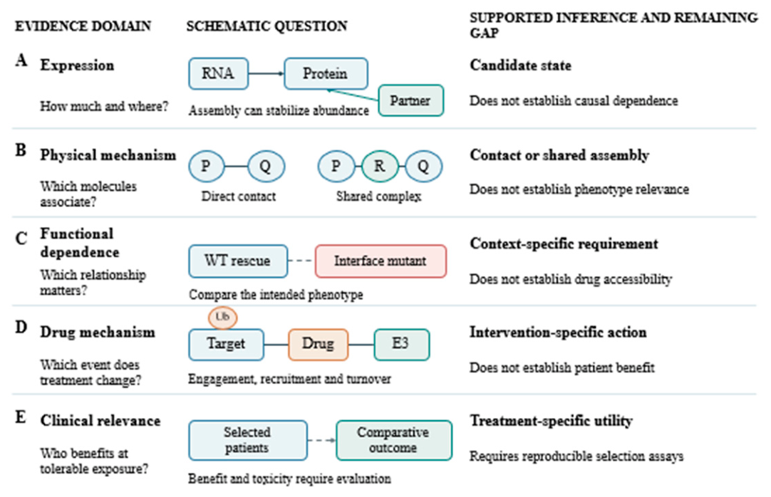
Figure 2.
Context-dependent interaction mechanisms in cancer. Independent cancer model systems illustrate EGFR routing, HK2 scaffolding, chromatin assembly and protection from degradation. Arrows summarize experimentally reported mechanisms without implying that all processes occur together in one malignant cell (Blaha et al., 2022; Boretto et al., 2024; Morozov et al., 2023; Yu et al., 2020; Zhai et al., 2023). EMT, epithelial–mesenchymal transition; G6P, glucose 6-phosphate.
Figure 2.
Context-dependent interaction mechanisms in cancer. Independent cancer model systems illustrate EGFR routing, HK2 scaffolding, chromatin assembly and protection from degradation. Arrows summarize experimentally reported mechanisms without implying that all processes occur together in one malignant cell (Blaha et al., 2022; Boretto et al., 2024; Morozov et al., 2023; Yu et al., 2020; Zhai et al., 2023). EMT, epithelial–mesenchymal transition; G6P, glucose 6-phosphate.
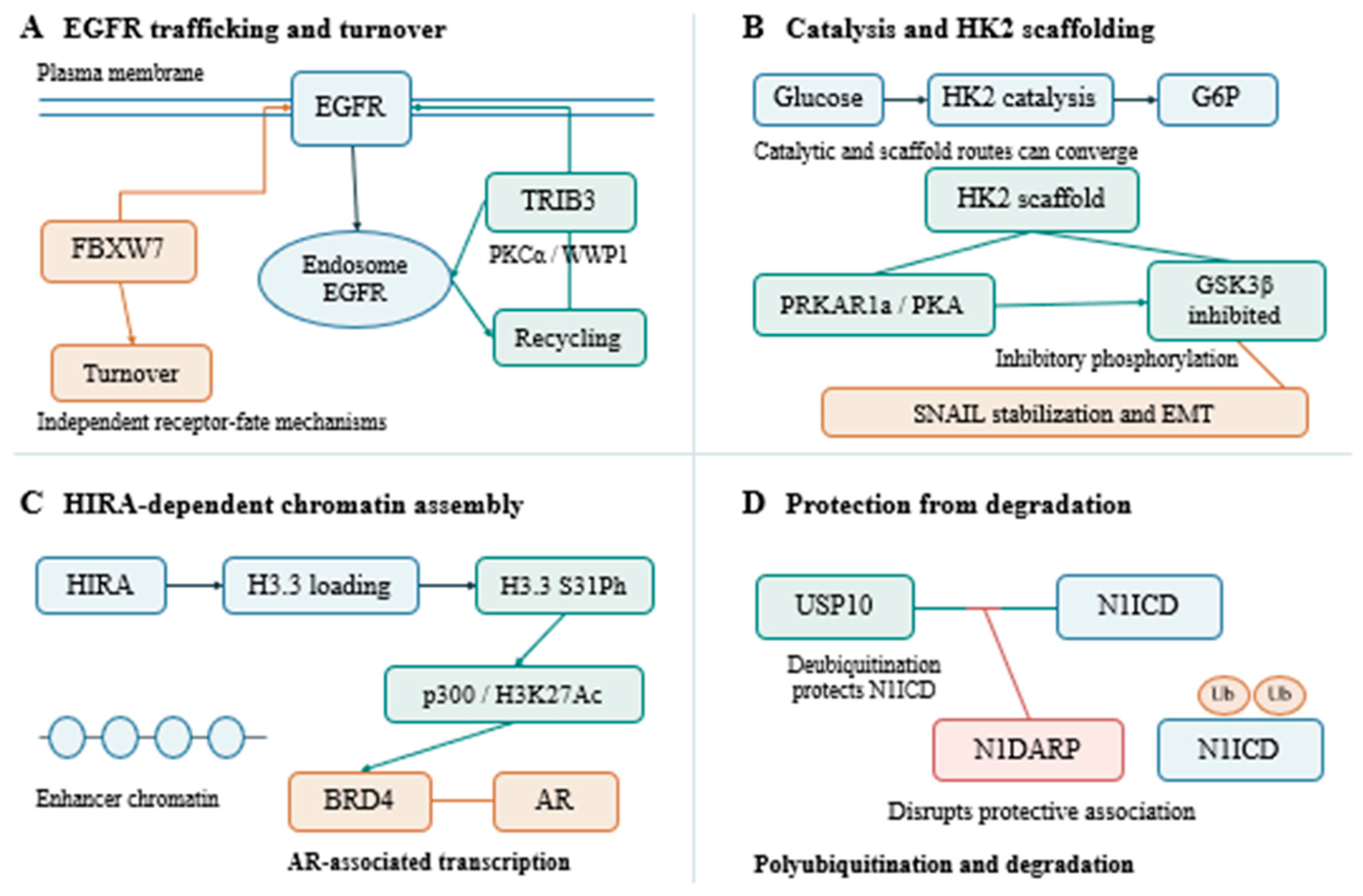
Figure 3.
Blocking and inducing molecular interactions. Interface inhibitors, PROTACs and molecular glues alter different molecular events. RCSB PDB images show menin–MI-2, 4GQ3, and the MZ1-containing BRD4–VHL complex, 5T35 (Gadd et al., 2017; Lv et al., 2020; Shi et al., 2012). Schematic colors do not map to structure colors.
Figure 3.
Blocking and inducing molecular interactions. Interface inhibitors, PROTACs and molecular glues alter different molecular events. RCSB PDB images show menin–MI-2, 4GQ3, and the MZ1-containing BRD4–VHL complex, 5T35 (Gadd et al., 2017; Lv et al., 2020; Shi et al., 2012). Schematic colors do not map to structure colors.
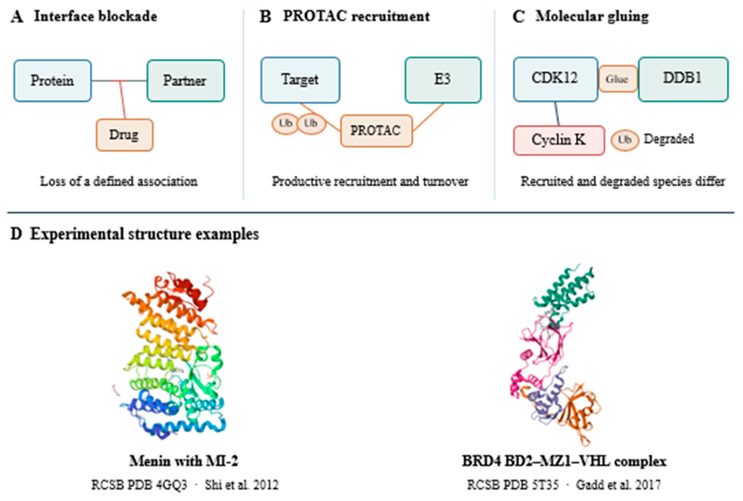
Figure 4.
A proposed workflow for evaluating therapeutic targets. Modality-specific questions guide experiments and interpretation. Dashed connectors indicate proposed evaluation paths rather than measured biological interactions. Clinical utility requires validation beyond the preclinical evidence supporting a candidate (Lin et al., 2019; Modi et al., 2022; Perner et al., 2023; Pogacar et al., 2022).
Figure 4.
A proposed workflow for evaluating therapeutic targets. Modality-specific questions guide experiments and interpretation. Dashed connectors indicate proposed evaluation paths rather than measured biological interactions. Clinical utility requires validation beyond the preclinical evidence supporting a candidate (Lin et al., 2019; Modi et al., 2022; Perner et al., 2023; Pogacar et al., 2022).
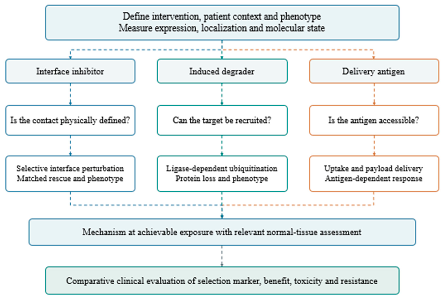
Table 2.
Representative mechanisms and evidence limits.
| Study | System | Demonstrated mechanism | Remaining question |
|---|---|---|---|
| (Yu et al., 2020) | Lung cancer models | TRIB3 supports EGFR recycling | Patient selection |
| (Blaha et al., 2022) | Breast cancer models | HK2 scaffolds GSK3 regulation | Interface-selective therapy |
| (Zhai et al., 2023) | Pancreatic cancer models | N1DARP disrupts USP10 protection | Peptide delivery |
| (Yoshinari et al., 2025) | KRAS(G12D) models | VHL recruitment drives degradation | Clinical benefit |
| (Lin et al., 2024) | RB-deficient breast models | PRMT5 regulates FUS-associated transcription | Clinical predictive biomarker |
| (Issa et al., 2023; Perner et al., 2023) | Acute leukemia patients | Menin inhibition and interface resistance | Durable response |
Table 3.
Evidence gaps and discriminating experiments.
| Uncertainty | Next experiment | Decision informed |
|---|---|---|
| Expression or dependence (Ryan et al., 2017; Vizeacoumar et al., 2013) | Perturbation with matched rescue | Target causality |
| Binding or proximity (Branon et al., 2018; Breitkopf et al., 2016) | Purified binding and interface mutation | Physical mechanism |
| Recruitment or degradation (Gadd et al., 2017; Lv et al., 2020) | Ligase dependence and turnover kinetics | Degrader mechanism |
| Response or specificity (Lin et al., 2019; Perner et al., 2023) | Resistance allele and engagement | Drug mechanism |
| Marker or treatment predictor (Modi et al., 2022) | Prospective comparative validation | Patient selection |
Disclaimer/Publisher’s Note: The statements, opinions and data contained in all publications are solely those of the individual author(s) and contributor(s) and not of MDPI and/or the editor(s). MDPI and/or the editor(s) disclaim responsibility for any injury to people or property resulting from any ideas, methods, instructions or products referred to in the content. |
© 2026 by the authors. Licensee MDPI, Basel, Switzerland. This article is an open access article distributed under the terms and conditions of the Creative Commons Attribution (CC BY) license (http://creativecommons.org/licenses/by/4.0/).
Copyright: This open access article is published under a Creative Commons CC BY 4.0 license, which permit the free download, distribution, and reuse, provided that the author and preprint are cited in any reuse.