

Submitted:
31 October 2026
Posted:
04 November 2026
You are already at the latest version
Abstract
Chronic inflammation and subfertility converge on a number of ligand-dependent transcription factors induced by chemically heterogeneous environmental exposures. Foremost among them is the aryl hydrocarbon receptor (AhR), of the basic helix-loop-helix (bHLH)–PER-ARNT-SIM (PAS) superfamily. Identified first as the receptor for dioxins and long regarded as a mediator of xenobiotic toxicity, AhR is now established as a regulator of physiological homeostasis, responding to microbial, dietary, phytochemical substances and endogenously generated metabolites. In this review, we examine AhR ligands and the transcriptional programmes they elicit, organising the evidence around one distinction: whether a ligand is a substrate for the cytochrome P450 enzymes it induces, and therefore self-limiting, or whether it persists and converts a single exposure into a chronic stimulus. We follow the receptor through the gastrointestinal tract, skin and adipose tissue; through the eye, where biallelic loss-of-function genetic variants cause foveal hypoplasia and retinitis pigmentosa and through the reproductive axis, where AhR sets ovarian aromatase expression, follicular recruitment, spermatogenesis and trophoblast invasion. Spanning the good, the bad, the ugly and the blind, we argue that therapeutic progress depends not on deciding whether to activate or inhibit AhR, but on confining modulation to a defined tissue, ligand residence time and transcriptional output.
Keywords:
AhR
; transcription factor
; environmental toxins
; physiology
; inflammation
; gut
; skin
; adipose tissue
; retina
; retinal degeneration
; folliculogenesis
; spermatogenesis
1. Introduction
Pathological inflammatory processes are largely involved in the broad range of health issues that account for the majority of global morbidity and mortality today, rather than just a few specific conditions [1,2,3]. More than half of all deaths worldwide are now attributed to conditions in which chronic inflammation is a constitutive component – ischaemic heart disease, stroke, diabetes mellitus, chronic kidney disease, non-alcoholic fatty liver disease, autoimmune disease and neurodegeneration – so that low-grade inflammation is best treated not as a feature of a few diagnoses but as a shared pathogenic axis running through the commonest causes of death [4].
It was revealed that certain environmental factors may contribute to systemic chronic inflammation, which results in a number of disorders that cause disability and may lead to death. The majority of the discussed substances are industrial and environmental pollutants that have been classified as endocrine disruptors due to their capacity to interfere with systemic regulatory mechanisms [5]. Dioxins and polychlorinated biphenyls are among the substances that inhibit the process of normal adipogenesis. Despite being prohibited in the 1970s, a class of industrial chemicals known as polychlorinated biphenyls, or PCBs, continue to exist in the environment because of their persistent nature. It has been demonstrated that 3,3'4,4'5-pentachlorobiphenyl (PCB126) exposure affects adipocyte metabolism and impedes adipogenesis [6]. Dioxins, dioxin-like PCBs and most other persistent pollutants filed under “endocrine disruptors” are not united by a shared hormone receptor. With few exceptions, their biological effects vanish in the absence of a single intracellular sensor, which converts a chemical exposure into a defined transcriptional programme [7]. Identifying that sensor therefore spotlights the point at which an environmental exposure transforms into gene expression.
A member of the periodic circadian protein (Per)—AhR nuclear translocator (ARNT)—single-minded protein (Sim) (Per-ARNT-Sim, PAS) domain and helix-loop-helix (bHLH) transcription factor superfamilies, the aryl hydrocarbon receptor (AhR) is distinguished among its relatives as the only member that is directly activated by small-molecule ligands rather than by protein–protein interaction or oxygen tension. The bHLH-PAS superfamily in vertebrates comprises at least nine groups, each consisting of two or three closely related genes (paralogs) that have arisen from the duplication of a precursor gene in vertebrates [8,9,10]. The first gene coding for AhR was characterized in mice [11]. There has been a second form of AhR identified in fish, where it has been referred to as AHR2 [12]. AHR2, a more divergent homologous gene, has been found in both teleosts and elasmobranchii [13]. However, in mammalian genomes there is no evidence for the existence of AhR2 [10,14].
Initially, AhR was considered to primarily act as a mediator between the body and environmental contaminants. Numerous recently conducted studies have demonstrated the significance of AhR in barrier tissues, including the placenta, skin, lungs, gastrointestinal system, and liver, in addition to related immune cells [15,16,17,18]. In these tissues, its function is essential for sustaining the physiological processes and relaying signals from the environment, food, and microbes.
The interactions of AhR ligands in the gut, skin, adipose tissue, eye and gonads are the main topic of our review. The gastrointestinal system is not only the place of entry for dietary molecules, but the home for human microbiota, billions of microorganisms able to produce diverse molecules. Whereas the main endogenous ligand is produced in skin, adipose tissue is one of the tissues that is most regulated by AhR. The eye is treated last among the barrier organs and at length, because it is the one organ in which human loss-of-function mutations in AHR have now been shown to cause disease, which converts AhR from a receptor that modifies pathology into a gene that causes it; the gonads follow, because they are the compartment in which the receptor is at once hormonally regulated and hormonally regulating. In particular, we outline the properties of AhR ligands and the physiological roles currently understood for them, and we return throughout to a single organising distinction: whether a given ligand is metabolised by the enzymes it induces, or whether it is not.
2. AhR Structure and Functions
The AhR is a ligand-activated transcription factor, belonging to a superfamily of transcription factors that includes bHLH domain-containing factors together with PAS domain-containing factors. Homology within the PAS domain amino acid sequence was first determined by aligning the sequences of the period circadian protein (PER) and single-minded homolog (SIM) proteins in Drosophila melanogaster, and the sequence of the human ARNT (nuclear translocator of aromatic hydrocarbon receptor) gene product was found to be similar as well [19]. In order to acknowledge the homology between these proteins an acronym for PAS was introduced.
The members of basic helix-loop-helix and PAS superfamily of proteins have highly conserved domain structure at the N-terminal part of the sequence (Figure 1). The bHLH region, which comprises about 60 amino acid residues, is divided into two functionally distinct parts: the main region responsible for DNA binding (about 15 amino acids) and the adjacent C-terminal HLH region involved in protein dimerization [19]. The PAS domain, which is located in the central part of the protein, comprises about 300 amino acid residues. It is divided into two structurally conserved regions, PAS-A and PAS-B, which are often linked by a PAS-associated C-terminal (PAC) motif [20]. The PAS-A and PAS-B regions are separated by a slightly conserved linker. The PAS-A domain stabilizes heterodimerization with ARNT, while the PAS-B domain controls the ligand binding pocket [21]. The PAS domain is one of the important signaling modules that controls changes in light, redox potential, oxygen content and ligands (polyaromatic hydrocarbons and environmental toxins) [22]. It binds small molecules, participates in protein-protein interactions and transduces input signals to mediate an adaptive physiological response [19]. In contrast to the conserved domains, the C-termini of bHLH-PAS family proteins exhibit considerable variability and contain transcriptional activation/repression domains (TAD/RPD). Accordingly, the C-termini are responsible for the specific modulation of the function of bHLH-PAS proteins and the recognition of partner proteins necessary for their unique action [20].
AhR was first discovered in mouse hepatocytes in 1976 as a receptor that mediates the effects of 2,3,7,8-tetrachlorodibenzo-p-dioxin (TCDD) [23]. Since then, the research was focused on the role of AhR in mediating the toxicity of polycyclic aromatic hydrocarbons (PAHs) and the halogenated dioxins dibenzofurans and biphenyls. Early studies of PAHs metabolism and the toxic effects of dioxins led to the discovery of their receptor and confirmed that this receptor mediates numerous biological effects of these harmful substances [24,25].
The ligand-binding domain (LBD) of AhR, contained within PAS-B, is what permits activation by so chemically diverse a set of compounds [26]. Specific amino acid residues in LBD determine the specificity and effectiveness of ligand binding, contributing to significant interspecific differences in AhR functionality [27]. A striking example is a single alanine-to-valine substitution at the position equivalent to mouse Ala375 (valine in human AhR), which reduces TCDD-binding affinity by approximately one order of magnitude. This helps explain why the high-affinity murine Ahr allele responds to dioxin at body burdens that leave human AhR comparatively unaffected, and why potency values measured in rodent cytosol cannot be transferred directly to human risk assessment [28]. These structural determinants have been identified using homology modeling and directed mutagenesis, providing additional information on the molecular mechanisms of AhR activation and potential directions for the development of therapeutic drugs [29]. Thus, the presence of a hidden cavity within the structural core of LBD was previously revealed. Experiments on directed mutagenesis have confirmed a set of residues, side chains of which play a major role in ligand binding through steric effects on the cavity of a ligand binding pocket: for example, common to all ligands are T283, F289, F318, I319, F345, L347 and Q377 [30].
However, additional residues and interactions specific to different ligand groups have also been identified. Thus, TCDD, 2,3,7,8-tetrachlorodibenzofuran (TCDF) and benzo(a)pyrene bind at the bottom of the cavity (region Bß, Ca, Da) and are stabilized mainly by hydrophobic interactions. Ligands such as 3-methylcholanthrene, PCB126 bind closer to the Fa/Gß site. β-naphthoflavone and 6-Formylindolo(3,2-b)carbazole (FICZ) differ in that they establish hydrogen bonds and polar interactions [29]. Thus, binding regions for artificial ligands were discovered.
AhR was later found to be highly expressed in tissues of barrier organs such as the placenta, skin, lung, gastrointestinal tract and liver, as well as in associated immune cells, where it transduces signals from the environment, food, and microorganisms [16,31,32]. AhR modulates metabolic processes that are important for tissue homeostasis, such as proliferation, differentiation, cell cycle, adhesion, migration, invasion and pluripotency [33]. It also plays a key role in mediating physiological functions, including maintaining the homeostasis of the cardiovascular [34], immune [35], reproductive [36], nervous and intestinal systems [37]. It is also involved in the regulation of insulin-glucose metabolism [38], the management of oxidative stress and adaptation to hypoxia. In addition, AhR influences the development of pathological conditions such as inflammation and cancer [39].
Understanding that AhR is an important regulator of homeostatic processes in barrier organs has led to an intensive search for natural ligands [40]. Multiple research has shown that the gastrointestinal tract is a rich source of such compounds, which may be derived from food and microbial metabolites [37,41,42,43]. There is also evidence that cellular function and thus nuclear localization of AhR in mammals is regulated by endogenous systems acting through low affinity ligands. These ligands include some prostaglandins, 2-(1′H-indole-3′-carbonyl)-thiazole-4-carboxylic acid methyl ester (ITE) [44], heme metabolites, and specific endogenous activators [45]. The action of small molecule ligands enables the activation of AhR-dependent functions at the cellular level. The multifaceted nature of ligands and diversity of their origin thus determines the complex role of AhR in maintenance of a constant internal stability despite environmental changes.
2.1. AhR Signalling
In the absence of a ligand, AhR resides in cytosol as a part of inactive multicomponent protein complex that includes the AhR-interacting protein (AIP, also known as immunophilin homolog ARA9 or HBV-associated X-activated protein XAP2), the heat shock protein 90 dimer (Hsp90), the co-chaperone p23 and possibly ARA3 (also known as NS1BP) [46] (Figure 2). One of the Hsp90 proteins binds the PAS B domain of the AhR and the other interacts with the bHLH and the PAS regions responsible for DNA and ligand binding, respectively [47]. Hsp90 not only keeps the AhR in an inactive form by binding to its PAS B domain, but also enhances activation of the receptor upon ligand binding. AIP stabilizes this complex by directly interacting with HSP90 and AhR. AIP in complex with co-chaperone p23 are also involved in the folding and stabilization of AhR, which leads to its cytoplasmic localization by inhibiting interaction of the AhR with transporting protein importin-β [48]. The co-chaperone p23 also protects AhR from ubiquitination-mediated degradation [49].
In addition, the association of the protein tyrosine kinase c-Src with the cytosolic AhR multiprotein complex has been observed in different cell types [50]. It is thought that carcinogenic effects of AhR agonists are possibly related to c-Src kinase signaling, as it was demonstrated activation of c-Src induced by TCDD. Phosphorylation of c-Src after TCDD treatment was detected in HepG2 and MCF-10A cells [51]. The AhR-mediated induction of c-Src leads to activation of focal adhesion kinase, which promotes integrin clustering, contributes to cell motility and metastatic programing in response to xenobiotic exposure [52]. It has also been shown that the events initiating the inflammatory actions of TCDD occur through the non-genomic action of AhR, which does not require ARNT, but involves c-Src [53].
Ligand engagement results in activation of AhR and its translocation to the nucleus. When ligand is bound the AhR alters its conformation within the Hsp90/AhR complex in a way that releases the nuclear localization signal (NLS) of AhR, thus promoting interaction with importin-β and translocation of the complex to the nucleus [25]. In the nucleus, AhR forms a heterodimer with ARNT and becomes capable of controlling the expression of target genes harboring specific DNA elements known as xenobiotic response elements (XRE) or dioxin responsive elements (DRE) in their regulatory regions. It was shown that localization of AhR is not dependent on interactions with ARNT [54].
Binding of the AhR-ARNT heterodimer to XREs is considered as canonical AhR signaling to induce the transcription of target genes, which encode for enzymes of the cytochrome P450 family (CYP1A1, CYP1A2, CYP1B1), glutathione S transferase alpha 1 and the repressor of AhR (AhRR at Figure 2) [8,24,49]. For instance, eight XREs located less than 1.4kb upstream from the CYP1A1 transcription start site were identified, and the sequences of five of them are identical in humans, mice and rats. The positions of these elements were also found: -9738, -1379, -1203, -1058, -981, -892, -821, -488 [55]. The consensus sequence of XRE has the following form: 5'-(T/G)NGCGTG(A/C)(G/C)A-3'. In this case, 5'-GCGTG-3' is considered as the core part. Sequences flanking the consensus also contribute to transcriptional activation of CYP1A1, and the evidence for this is that an isolated XRE cloned upstream of a reporter is not by itself sufficient to confer ligand-inducible expression: the core 5'-GCGTG-3' motif specifies heterodimer binding, but not activation. These flanking sequences are themselves conserved between species [56].
The disposal of PAHs proceeds in three phases – functionalisation (phase I), conjugation (phase II) and export (phase III) – and the enzymes of the first two phases are themselves AhR target genes, so that the receptor induces the very machinery that metabolises its own ligands. AhR target genes comprise phase I metabolic enzymes, including proteins of the cytochrome P450 superfamily: CYP1A1, CYP1A2 and CYP1B1 that introduce reactive and polar groups into xenobiotic molecules [45]. These enzymes, therefore, promote the detoxification of PAHs. However, in addition to detoxification, previously mentioned enzymes are also involved in the bioactivation of PAHs, which leads to the formation of reactive intermediates. These intermediates in turn promote the formation of DNA adducts that have genotoxic effects [57,58]. The apparent paradox – that one set of enzymes both detoxifies and form genotoxic adducts – dissolves once the chemistry is made explicit. CYP1A1 and CYP1B1 do not destroy polyaromatic compounds; they insert an oxygen atom to generate an arene oxide, which is itself an electrophile. In the case of benzo(a)pyrene, successive monooxygenation and hydration by epoxide hydrolase yield benzo(a)pyrene-7,8-dihydrodiol-9,10-epoxide, which alkylates the exocyclic N2 of guanine. Whether such an intermediate is quenched by phase II conjugation or escapes to react with DNA depends on the stoichiometry between phase I and phase II capacity in the exposed cell. Sustained AhR activation that induces CYP1A1 out of proportion to the conjugating enzymes therefore converts a detoxification pathway into a mutagenic one, which is the mechanistic sense in which one receptor is at once protective and carcinogenic.
During the metabolism of ligands, such as dioxins, by cytochrome P450 enzymes, significant amounts of reactive oxygen species (ROS) are generated, specifically superoxide (Figure 2). The source of that superoxide is the catalytic cycle itself: when a bound substrate is turned over slowly, or when induced CYP1A1 is present in excess of the substrate available to it, the oxyferrous intermediate decays before oxygen is transferred and releases superoxide instead – the uncoupling of the P450 cycle. Dismutation then yields H2O2 which, unlike superoxide, diffuses freely between cellular compartments. This is the step at which an oxidative signal becomes an inflammatory one: H2O2 oxidises reactive cysteine thiols in redox-sensitive signalling proteins, relieving the constraint on IKK and licensing NF-κB-dependent transcription of TNF, IL1B and IL6, while oxidised mitochondrial products supply a second signal for NLRP3 inflammasome assembly and the maturation of IL-1β [59]. Regarding DNA, ROS oxidise nucleobases and the deoxyribose backbone – most diagnostically to 8-oxo-7,8-dihydroguanine, which mispairs with adenine and fixes G:C→T:A transversions at the next round of replication – causing mutations, which lead to carcinogenesis, apoptosis, necrosis, and genetic diseases [60].
2.2. AhR Ligands
With the growing interest in AhR and its ligands, additional potential high-affinity exogenous and endogenous AhR ligands have been discovered, whose chemical structure and binding affinities can vary considerably. Thus, the highest relative affinity of binding to AhR is observed for FICZ (IC50 is 0.07 nM) and 2,3,7,8-tetrachlorodibenzo-p-dioxin (0.48 nM) [61], whereas for benzo(a)pyrene this indicator is one of the lowest (617 nM). These are competitive binding constants determined in cytosolic preparations rather than cellular potencies, and because the affinity of AhR for any given ligand is itself species-dependent, it is the rank order rather than the absolute values that can safely be carried across species. Another AhR ligands include 3-methylcholanthrene (1.6 nM), 3,3'4,4'5-pentachlorobiphenyl (5.9 nM), β-naphthoflavone (7.2 nM) [29]. The main source of exogenous AhR ligands are environmental pollutants such as 2,3,7,8-tetrachlorodibenzo-p-dioxin (TCDD) and other PAHs as well as dietary metabolites [23].
Endogenous AhR ligands include intermediate and microbiological tryptophan metabolites such as kynurenine and its derivatives [62], tetrapyrroles such as biliverdin and bilirubin [63] and eicosanoids such as prostaglandin G2 [64]. These endogenous ligands play an important role in physiological processes regulated by the AhR, such as the immune response, inflammation and the maintenance of tissue barrier function [49,65].
2.2.1. Environmental Toxins
2,3,7,8-TCDD is the AhR agonist described as one of the most potent inducers of CYP1A1 [66]. The toxicity of TCDD and other dioxins is directly linked to their ability to disrupt the function of AhR-controlled gene products. TCDD, similarly to other ligands, binds and activates intracellular AhR, therefore inducing the transcription of xenobiotic-metabolizing cytochromes P450. In general, induction of CYP1A1 leads to increased biodegradation of exogenous and endogenous chemicals, and is coupled with excessive generation of ROS [67]. The decisive variable is whether the inducing ligand is itself a substrate for the enzyme it induces. Endogenous agonists such as FICZ are rapidly hydroxylated by the CYP1A1 they induce, so that induction extinguishes the signal that generated it and receptor activation is self-limiting, lasting minutes to hours. By contrast, dioxin-like polyhalogenated aromatic hydrocarbons are chemically stable and poor P450 substrates; they therefore degrade slowly, causing constant AhR activation and sustained production of ROS. With an elimination half-life in human organism measured in years, a single exposure to TCDD constitutes a chronic transcriptional stimulus. This is why receptor-level potency and organism-level toxicity are not equivalent quantities. Such TCDD-induced oxidative processes [67] can be inhibited both by AhR and CYP1A1 silencing [67]. In contrast, silencing of CYP1B1 did not affect TCDD-induced ROS generation, which proves that AhR-mediated CYP1A1 induction is responsible for oxidative stress caused by dioxins [67]. This ROS generation contributes to immune, liver, heart, and skin toxicity, as well as alters cell growth, proliferation, and differentiation, which can possibly promote tumor growth [62,68].
Molecular dynamics simulations show that binding of TCDD to the AhR–ARNT dimer reorganises the interdomain contact network, loosening particular interfaces – notably between AhR PAS-B and the PAS-A domains of AhR and ARNT – while markedly reducing the mobility of PAS-B itself. The net effect is stabilising rather than destabilising: the ligand-bound dimer samples a narrower conformational ensemble than the apo dimer, so that the local loss of contact is more than offset by rigidification of the ligand-binding domain. This effect also extends to the interactions between the AhR-ARNT complex and DNA, allowing for ligand-mediated transcriptional regulation [69].
Classical ligands with a planar structure and properties similar to TCDD are the most well-studied high-affinity ligands for AhR [26,70,71,72]. This group includes numerous PAHs and similar substances (e.g. benzo(a)pyrene, anthracene and 3-methylcholanthrene) as well as toxic halogenated aromatic hydrocarbons (HAHs) such as polychlorinated and polybrominated dibenzo-p-dioxins, dibenzofurans and biphenyls, which have a higher affinity for AhR compared to PAHs [26,73,74] (Figure 3). Such ligands play a key role in triggering AhR-associated toxic responses and were the first AhR agonists identified as they are components of environmental pollutants [47,75]. It is important to note that these ligands are metabolically stable and accumulate in adipose tissue, leading to chronic toxicity over time [76,77,78].
2.2.2. Endogenous Chemicals
The involvement of AhR in regulation of various physiological processes raised the question of the origin of the dioxin receptor, and the existence of an endogenous AhR ligand that regulates normal physiological functions [49].
Quite early it was reported that some naturally occurring compounds, such as products of tryptophan photooxidation [61], indole derivatives as condensation products of indole-3-carbinol [79] and flavones [80], exert the activating effects on the AhR. It is now known that the endogenous ligands of the AhR include other tryptophan metabolites such as kynurenic, xanthurenic and cinnamic acids, which can bind and activate the AhR [49].
There are three metabolic pathways by which tryptophan is utilized in the gastrointestinal system: through indole produced by the gut microbiota, kynurenine in liver, immune cells and mucous membranes, and by the serotonin system in enterochromaffin cells. Both studies in vivo and in vitro have demonstrated that the endogenous regulation of AhR by these compounds significantly influence the regulation of protective immune processes in the gut [81]. Through interactions in various immune cells, including dendritic and innate lymphoid cells, this transcription factor appears to play an important role in immune modulation at barrier sites such as gut and skin [82].
The homology that AhR shares with canonical circadian clock genes (periodic circadian protein Per), and its potent ligands are formed from tryptophan under ultraviolet radiation, suggests a role for AhR in circadian rhythm. Since 1987, when it was demonstrated that certain photo-oxidized derivatives of tryptophan bind to the AhR with very high affinity, the structure of 6-Formylindole[3,2-b]carbazole (FICZ) was identified. FICZ is formed in the skin upon exposure to the light, and has one of the highest affinities for the AhR [83]. FICZ, being a potent agonist of AhR, is able to induce the expression of CYP1A1, and at the same time is metabolized by cytochrome P450 [84]. Thus, it was proven that a feedback mechanism for controlling the constitutive activity of AhR-ARNT transcription complexes is regulated by the catalytic activity of the CYP1A1 enzyme. Endogenous CYP1A1 substrates can be AhR activators in the absence of exogenous ligands [83]. It should be noted that most tryptophan degradation products are low AhR agonists [10], but this group plays an important role in the body's self-regulation through interaction with the AhR [85,86].
Kynurenine, an intermediate product of the enzymatic degradation of tryptophan, spontaneously condenses in intestinal cells to form trace extended aromatic condensation products (TEACOPs), dimeric and trimeric adducts that react with AhR at far higher affinity than kynurenine itself [41,42]. The distinction matters for reading the kynurenine literature: a substantial part of the AhR activity attributed to kynurenine in cell culture is contributed by such condensation products formed in the medium, which is why the kynurenine concentrations required in vitro exceed those that are physiologically relevant [41]. In a physiologically normal condition, indole metabolites are assumed to have more potency and activity than kynurenine [43]. Bacterial metabolites, which may be tryptophan derivatives, are an important source of AhR ligands. Intestinal indole is mainly produced by gut microbiota such as Escherichia coli through hydrolysis of tryptophan by the microbial tryptophanase enzyme [87] Additionally, multiple Bifidobacterium and Lactobacillus species are able to convert tryptophan in the gut into the indole-3-lactic acid using specific aromatic lactate dehydrogenase enzyme [88]. Indole itself behaves as a partial agonist, and its net effect is therefore concentration-dependent: at the low micromolar concentrations prevailing in the healthy colonic lumen it activates AhR, whereas at higher concentrations it occupies the binding pocket while recruiting coactivators inefficiently and so competes with more efficacious agonists such as FICZ, behaving functionally as an antagonist. Agonism and antagonism are in this sense not fixed properties of a molecule but properties of the ratio between its occupancy and its intrinsic efficacy relative to the other ligands present [41,77]. Indole is then absorbed and metabolized in the liver to indoxyl-3-sulfate and other derivatives, which are also AhR agonists [78].
Indole derivatives are also produced during the metabolism of tryptophan by the intestinal microbiota from dietary products. Glucobrassicin, for example, is contained in large quantities in cruciferous vegetables. When consumed, it is enzymatically cleaved by myrosinase to form indole-3-acetonitrile (I3ACN) and indole-3-carbinol (I3C). In the stomach, I3C condenses to by-products such as 3,3'-diindolylmethane (DIM), 2-(indol-3-ylmethyl)-3'-3-diindolylmethane (Ltr-1) and indole 3,2-b carbazole (ICZ), which are affinity ligands for AhR [84]. In animal models of atopic dermatitis, I3C, which is similarly generated by Bifidobacterium longum during the metabolism of tryptophan, reduces inflammation by activating the immunological signaling pathway mediated by AhR. This is due to the fact that disturbances in the intestinal microbiome also affect the development of allergic diseases [84].
Indirubin and indigo are isomeric condensed oxindoles, derivatives of indican (indoxyl-P-D-glucoside), found primarily in plant sources, such as Indigofera [89]. Indigo and its structural isomer indirubin are also produced from indoxyl sulfate during the microbial degradation of tryptophan. Multiple times they were found in human urine, and apparently their presence indicates a corresponding bacterial metabolism [90].
The representatives of the genus Lactobacillus, which colonize the gut, urinary tract and skin of humans and other mammals, can use tryptophan instead of glucose as an energy source and produce AhR ligands such as 3-methylindole (skatole), indole-3-acetic acid and tryptamine [22].
AhR agonists can also be produced by the human skin microbiota. For example, skin Malassezia furfur produces the indole alkaloid pityriazepine, malassezine, pityriacitrin, indirubin, tryptanthrin, ICZ and FICZ [91,92]. Dysbiosis of the gut microbiota has been shown to disrupt the integrity of the blood-brain barrier (BBB) by activating AhR signaling, thereby exacerbating the development of Alzheimer's disease, for example [42]. Indoxyl-3-sulfate crosses the BBB and causes CNS inflammation through mechanisms controlled by AhR in astrocytes and microglia [22].
However, due to the interaction with AhR, endogenous ligands also cause many positive effects related to immunity. The ability of indole metabolites and kynurenine to interact with AhR enables them to facilitate the differentiation of FoxP3+ T regulatory cells [88]. This interaction also influences the differentiation pathways of Th17 and Th1 cells and contributes to the production of anti-inflammatory mediators [93]. Activation of FoxP3+ T regulatory cells occurs via several mechanisms, including direct transactivation and induction of epigenetic modifications that affect Foxp3 transcription [94,95]. Quintana et al also suggested that the FOXP3 promoter of T-regulatory cells also contains elements of the dioxin response, so ligand binding to AhR activates these cells and promotes their differentiation [96]. At the same time, AhR-dependent induction of FoxP3+ T regulatory cells suppresses Th17 differentiation and inflammation in mice [95]. AhR may also act in conjunction with с-Maf, a transcription factor regulating IL-10 expression, promoting the development of Tr1 cells [97].
A metabolic pathway through which microbiota tryptophan metabolites regulate mucosal reactivity in mice has also been described. Intestinal lactobacilli produce indole-3-aldehyde, which activates AhR-dependent transcription of IL-22, which, in turn, protects the mucosa from Candida albicans and inflammation [65,98]. This provided insight into why dietary tryptophan deficiency weakens intestinal immunity in mice and alters the gut microbial community [65,81].
2.2.3. Phytochemicals
Beyond natural ligands of endogenous origin, several plant-derived substances have agonistic effects on the AhR, as has been shown in mouse and in human cell lines. Such alkaloids are norisoboldine (Radix linderae) [99], berberine (Hydrastis canadensis) [100], rutaecarpine (Euodia rutaecarpa) [101]. Anti-inflammatory features of these substances are inevitably linked to their agonistic effects, as was confirmed for norisoboldine in a mouse model of collagen-induced arthritis. Oral administration of norisoboldine in mice promoted Treg cell differentiation and function in the gut, ensuring a substantial anti-arthritic effect through enhancing AhR activation, which was inhibited by resveratrol (plant-derived antagonist of AhR) [102].
The plant-derived AhR ligands quercetin and I3C have been shown to reduce clinical symptoms in mice with experimental dextran sulfate sodium-induced colitis. The treatment restored epithelial integrity by increasing the synthesis of tight junction proteins and significantly reduced intestinal inflammation [81,91]. Moreover, patients with ulcerative colitis also showed positive changes when receiving I3C [103].
In addition, pelargonidin, a water-soluble anthocyanidin, can activate AhR and reduce intestinal inflammation by decreasing the induction of angiotensin-converting enzyme (ACE2) in the inflamed colon [104]. Baicalein, extracted from the root of Scutellaria baicalensis, directly activates the AhR in the innate lymphoid cells 3 (ILC3), increases the production of IL-22 by these cells and improves the intestinal barrier [105].
Indigo plant extract proved to be a powerful inducer of IL-10 and IL-22, protecting against insulin resistance. The underlying sequence runs as follows. A high-fat diet produces dysbiosis, which drives intestinal inflammation and loosens the epithelial barrier [106]; lipopolysaccharide then translocates into the portal circulation, reaches the liver and sustains metabolic endotoxaemia, local inflammation and, consequently, insensitivity to insulin [107]. AhR helps maintain the number of active regulatory T cells and resident anti-inflammatory polarized macrophages that produce IL-10 and help maintain insulin sensitivity. In particular, indigo increased the number of Lactobacillus bacteria and caused the production of IL-22 in the intestine, which improved the permeability of the intestinal barrier and reduced endotoxemia [108]. It has been shown that AhR ligands extracted from Indigofera exert beneficial effects. For instance, indirubin and its derivative 6′-bromoindirubin-3′-acetoxime have demonstrated anti-tumor activity both in vitro and in vivo [109,110].
Based on the results of these studies, non-toxic AhR agonists of plant origin may be considered as promising therapeutics for the treatment of different chronic inflammatory diseases, specifically Crohn's disease and ulcerative colitis [93].
2.2.4. AhR Antagonists and Selective Modulators
Putative natural ligands have been identified that can inhibit the action of AhR in vitro. Functionally, the actions of AhR ligands include full agonists, partial agonists and competitive antagonists. Some phytochemicals and dietary components can modulate physiological processes by antagonizing AhR signaling [111].
Many bioactive plant metabolites of polyphenolic nature give us an opportunity to unveil the molecular mechanisms of inflammatory diseases. In particular, isoprenoids and phenylpropanoids [112], as quercetin and kaempferol [113], curcuminoids and coumarins [114], have been shown to antagonize aryl hydrocarbon receptors. These ligands interact directly with the binding site of the AhR ligand, blocking transcription induced by the agonist [115]. A specific group of monoterpenoids, the carvones, found in the essential oils of dill, caraway and peppermint, have been reported to be non-competitive antagonists of the human AhR, as carvones exerted similar antagonizing effects for high and low concentrations of tested agonists. They act by binding allosterically to the region of the AhR involved in heterodimerization with ARNT, preventing the formation of functional AhR-ARNT heterodimers [116]. In addition, vitamin B12 and folic acid have also shown the ability to bind to the AhR and block its activation [117]. Research has also shown that other phytochemicals, such as brevifoline carboxylic acid from Polygonum capitatum and extracts from green tea (Camellia sinensis) and Phellinus linteus, can inhibit AhR activation or contribute to lowering the expression level of CYP1A1, however it has not been shown by what mechanisms they interact with AhR [118].
Therapeutic targeting of the AhR has long been neglected, mainly due to the misconception that the AhR is a receptor that exclusively mediates the toxicity of xenobiotics. With increasing knowledge of the physiological and pathophysiological role of the AhR, attempts have been made to target the AhR, including therapies for cancer, inflammatory bowel disease and atopic dermatitis [119,120]. Several AhR antagonists have been developed, such as BAY-2416964 [121], StemRegenin 1 (SR-1) [122], KYN-101 [123] and IK-175 [124], which have shown high efficacy in inhibiting AhR activation in mice and humans. These compounds have potential for further preclinical and clinical studies for the treatment of diseases associated with abnormal AhR activation.
SR-1 promotes the expansion of CD34+ cells by directly binding to and inhibiting the AhR signaling pathway [125]. In addition, a phase 1/2 study was conducted that led to an increase in CD34+ cells, resulting in rapid engraftment in the patients studied [126]. Furthermore, SR1 has been shown to improve the expansion of human primary hematopoietic stem cells as an inhibitor of the AhR signaling pathway [122,127,128]. SR1 has the potential to stimulate the development of human plasmacytoid and myeloid dendritic cells from hematopoietic progenitor cells [128]. It has also been reported that SR1 can promote terminal differentiation of human erythroblasts and attenuate endothelial progenitor cell senescence by inhibiting AhR signaling [129,130]. These results provide a basis for the further treatment of vascular diseases.
The natural AhR ligand I3C has been shown to regulate microglial homeostasis and protect the retina from light-induced degeneration [131]. Recently, a novel indole-containing synthetic AhR ligand 2, 2′-aminophenylindole (2AI), was identified to protect the retinal pigment epithelium (RPE) and retina from environmental stress, highlighting the potential role of AhR in retinal neuroprotection [132]. I3C and 2AI modulate key functional parameters of microglial activation, significantly reducing migration and phagocytic ability and eliminating the typical amoeboid phenotype of BV-2 microglia [131]. Interestingly, new antagonists have also been developed - indole and stilbene derivatives that have shown high efficacy in vitro [133], for example PX-A24590 and GNF351 [120].
3. Physiological Roles of Aryl Hydrocarbon Receptor
3.1. AhR in the Gastrointestinal Tract
AhR is expressed by epithelial cells and immune cells of lamina propria and is activated in malignant and inflammatory bowel diseases [137]. AhR controls the self-renewal of intestinal epithelial cells, preventing the increased growth of their precursors and the transformation of malignant epithelial cells, thereby maintaining the integrity of the barrier. The mechanism is the ligand-dependent induction of E3 ubiquitin ligase components that direct nuclear β-catenin to the proteasome: AhR does not switch Wnt signalling off but sets a ceiling on its output, so that crypt-base stem cells proliferate within a bounded range rather than continuously [138]. Consistent with this, intestinal epithelium lacking AhR becomes hyperproliferative and, on a tumour-prone background, progresses to adenoma (Figure 4).
It is believed that AhR also stimulates the expression of IL-10 receptors in epithelial cells, which enhances the barrier function of the epithelium during inflammation [139]. Nevertheless, there is evidence that AhR activation can also restrain IL-10 production: in animals exposed to dietary and environmental oxazoles, receptor engagement suppressed rather than enhanced IL-10 [140]. Whether the opposing outcomes are set by the ligand, by the cell type in which the receptor is engaged, or by the duration of engagement has not been resolved, and this is one of several places in which “AhR activation” is used in the literature to denote experimentally non-equivalent conditions.
Also, there is a decrease in the expression of TNFa, IL1-β and IL-8 and the secretion of IL-8 in vitro, since AhR prevents the inflammatory reaction of intestinal epithelial cells during chemical damage [141]. As already mentioned, when AhR is activated, the expression of tight junction proteins in the intestinal epithelium increases. This occurs in response to IL-22 produced by dendritic and CD4+ T cells [142]. The group of intraepithelial lymphocytes (IELs) includes separate populations of γδ T cells in the skin and intestine, as well as TCRαβCD8αα+ cells in the intestinal lamina propria. The lack of AhR activation reduces their survival rate, which violates the control of microbial load and composition [143] (Figure 4).
The cells of the ILC3 group are involved in the innate response to bacterial and fungal infection. They play an important role in ensuring the homeostasis of intestinal bacteria and in regulating the work of T-helper cells in the Th17 population. The development and functioning of ILC3 depend on the nuclear receptor RORγt, which is not induced upon pathogen detection but is the lineage-defining transcription factor of these cells, expressed constitutively and required for their specification [144]. AhR can synergize with RORγt by inducing IL-22 transcription and controlling the homeostasis of ILC3 [94]. In addition, AhR is shown to be able to stimulate the development of intestinal ILC3 through the induction of Notch transcriptional factor expression [32]. Finally, it was found that AhR expression in ILC3 decreases with inflammation of intestinal tissues [145].
Interestingly, a connection between Notch and IL-22 has been demonstrated, since Notch signaling controls the production of IL-22 by CD4+ cells through the induction of AhR ligands, and IL-22 production in vivo is regulated by Notch signaling [146].
In general, these data suggest that AhR activity in intestinal epithelial and immune cells plays an important role in tissue homeostasis at the structural and functional level. Disruption of AhR signaling leads to an imbalance of the microflora and increases susceptibility to autoimmune, tumor and pathogenic diseases.
3.2. AhR in the Skin
Skin cells of various types were found to express AhR: keratinocytes, sebocytes, fibroblasts, melanocytes, endothelial cells, Langerhans cells, lymphocytes and mast cells [147]. Activation of AhR accelerates the terminal differentiation of keratinocytes and enhances the expression of proteins involved in the creation of a structural barrier (for example, filaggrin, involucrin and loricrin) [148,149]. It is believed that skin homeostasis, as well as pathological processes such as atopic dermatitis, psoriasis and chloracne, can be modulated through specific ligand-dependent activation of AhR [35,150].
Indeed, AhR expression in keratinocytes increases with inflammation [151,152]. The skin microbiome in mice with AhR deficiency is more variable than that in mice with normal AhR, reflecting its role in controlling microflora stability [153]. Also in mice with AhR deletion, as well as in mice with constitutive AhR expression in keratinocytes, the skin acquires disorders characteristic of the epidermis with atopic dermatitis [154,155].
As for the immune cells of the skin, mice contain several populations of γδ T cells in the dermis, where IL-17 is secreted, as well as in the epidermis (dendritic epidermal γδ T cells). All of the subtypes of γδ T cells express AhR [156]. AhR expression is necessary for the maintenance of dendritic epidermal γδ T cells in the skin of mice and also contributes to the preservation of resident memory T cells (TRM) [157,158]. TRM cells provide an early immune response, playing a crucial role in skin protection [157]. However, the increased activity of TRM cells secreting IL-17A and IL-22 contributes to the chronic state of inflammatory skin diseases [159]. AhR is an important regulator of the activation and function of Langerhans cells, the antigen-presenting dendritic cells resident in the epidermis. In mice, deletion of AhR causes their decrease in number and activity [160].
The role of AhR and its ligands in the functioning of TRM in psoriasis or atopic dermatitis has not been fully evaluated (Figure 5).
AhR is crucial for the production of IL-22 by Th17 cells: this conclusion was made when comparing Ahr −/− mice with the psoriasis phenotype and wild-type mice. Th17 cells play an important role in the immune response of the skin, participating in the development of inflammatory diseases such as psoriasis. IL-22 has a pro- or anti-inflammatory effect, depending on the site of inflammation. It can play a detrimental role in skin inflammation [161]. A naturally derived small molecule produced by bacterial symbionts of entomopathogenic nematodes, tapinarof (GSK2894512) [162], was approved in 2022 for the treatment of plaque psoriasis in adults and subsequently for atopic dermatitis, making it the first AhR agonist licensed as a therapeutic [163]. This molecule is an AhR agonist that effectively reduces inflammation in skin, similarly to treatment with FICZ. It was shown that tapinarof acts through suppression of Th17 cells and IL-17+ γδ T cells secretory activity. The mechanism is therefore twofold rather than circular: receptor activation in keratinocytes restores barrier gene expression and NRF2-dependent antioxidant defence, while receptor activation in skin-resident lymphocytes limits IL-17A and IL-22 output – the same ligand acting on two cell types towards one phenotypic end [162,164]
The most recent study showed that in keratinocytes, under the action of ligands such as TCDD and FICZ, AhR is almost completely degraded. In addition, after treatment with I3C the level of AhR substantially decreased. The proteasomal degradation of the AhR, perhaps, is one of the major mechanisms of its regulation [165]. It has been shown that such degradation of AhR is a mechanism for limiting its transcriptional activity [166]. This process involves the formation of cullin–RING ligase 4B complex (CUL4BAHR), which directly catalyzes the ubiquitination of AhR interacting via its N-terminal extension. In addition, for proteasome degradation of AhR, after ligand activation and ubiquitination, it must be exported from the nucleus to the cytoplasm, which is facilitated by TCDD-inducible poly(ADP-ribose) polymerase (TiPARP), which ADP-ribosylates the AhR site and targets it for proteasome degradation [166].
Experimental data also revealed that AhR decay could be inhibited by ligand-selective AhR antagonist CH223191. Moreover, the degradation of AhR was blocked after knockdown of its main dimerization partner ARNT in the N/TERT cell line, suggesting that ARNT is required for AhR proteolysis [165].
A rather interesting fact is that inhibition of HDACs with Trichostatin A led to enhanced expression of AhR in both untreated and AhR ligand-treated cells [165]. The most likely mechanism of activation of some genes, both the AhR gene and AhR target genes, when exposed to HDAC inhibitors is to facilitate the binding of transcription factors (for example, AhR) to sequences in the promoter region. DNA in these places becomes more accessible due to the binding of acetylated histones nearby [167].
These experimental data indicate that in epidermal keratinocytes, AhR is regulated posttranslationally through degradation mediated by proteasomes or at the transcription stage by epigenetic modifications. We suggest that artificial control of AhR levels can potentially be carried out with the help of small molecules regulating the cytosolic stability of its multimeric complex. In one of the studies, chimeric degrader molecules containing the AhR ligand were developed and they can activate the AhR E3 Ligase complex, thus ubiquitinating the target protein [168]. Perhaps, due to the ubiquitination system, the concentration of AhR can also be regulated.
AhR can also promote wound healing by stimulating the production of TGF-β by fibroblasts, which in turn promotes keratinocyte migration and accelerates healing [156].
The evidence on mast cells is indirect and comes from another organ. Mast cells of the lung parenchyma constitutively express AhR, and its activation by the high affinity FICZ ligand leads to accelerated degranulation. Mast cells exposed to FICZ produce IL-6 and IL-17, which play an important role in chronic inflammation and allergic reaction [169]. Considering that mast cells are also an important type of skin immune cells and play an active role in wound healing, it can be assumed that AhR may have a similar function here [169]. On the other hand, in 2016 it was shown that AhR may not play a role in the homeostasis of skin mast cells [170]. The authors noted that AhR can still regulate mast cell function, but its role in changing the number of mast cells in the population has not been noticed. Discrepancies with the results of previous studies were explained by possible differences in the housing of experimental animals.
3.3. AhR in Adipose Tissue
Activation of AhR is closely related to the process of adipogenesis and regulation of energy balance. AhR is expressed in adipocytes [171] and, in particular, in the 3T3-L1 model cell line. Specifically, it has been demonstrated that the level of AhR decreases during differentiation in 3T3-L1 cells [172,173]. The level of AhR not only decreases during adipogenic differentiation, but also its affinity towards the response elements and the response to TCDD declines.
Various fatty products people consume with food, in particular animal fats, can contain dioxin-like AhR ligands. Particularly, persistent organic pollutants (POPs) are lipophilic, so they accumulate in adipose tissue [77]. Despite the activation of xenobiotic metabolism-related enzymes these molecules decay insufficiently, and their excretion and degradation occurs slowly [171]. It has also been shown that AhR interaction with POPs is associated with insulin resistance [174,175]. In humans, exposure to TCDD and other dioxins can cause anorexia and weight loss, disrupting the process of adipogenesis [176,177,178].
It was shown that activation of AhR led to suppression of lipogenesis, as AhR deficient mouse embryonic fibroblasts displayed enhanced synthesis of triacylglycerols [177]. Activation of AhR by TCDD inhibited adipogenesis through peroxisome proliferator–activated receptor gamma (PPARγ) activity suppression. Ligand-dependent AhR activation could thus impair adipogenesis of 3T3-L1 cells in vitro. Consistent with the effect being receptor-mediated rather than a property of the compound, ligand-treated mouse embryonic fibroblasts isolated from AhR-/- mice retained normal adipogenic potential [177].
Activation of AhR by dioxin is associated with the inhibition of PPARγ activity, which is a key transcriptional activator of adipogenesis [179]. The interaction of PPARy with AhR occurs mainly through its DNA-binding domain (DBD, or C) and part of the flexible hinge region (D). AhR acts as a substrate receptor in the ubiquitin ligase complex CUL4B-RING E3 (as already mentioned, AhR is part of this complex), aimed at the ubiquitination of PPARγ. AhR binds two lysine residues 268 and 293 in the D-region and recruits PPARy onto CRL4BAHR E3 complex, which ubiquitinates PPARy and subsequently causes degradation of this transcription factor [166,180]. Most likely, activation of AhR by dioxin leads to inhibition of PPARγ and suppression of adipogenesis also through exposure to the ERK signaling pathway [181,182,183]. Recent in vitro experiments have also demonstrated that adipocyte differentiation is suppressed by overexpression of AhR due to a decrease in the stability of PPARγ and, conversely, siRNA-mediated silence of the AhR gene leads to differentiation of 3T3-L1 cells [180].
On the other hand, it was found that systemic deficiency of AhR prevents obesity in mice receiving a high-fat diet [172,173]. In wild-type mice, the number of tryptophan metabolites that activate AhR and promote obesity phenotype increases [184]. Thus, kynurenine activates the AhR/Stat3/IL-6 pathway in adipocytes, mediating the development of obesity and insulin resistance [185]. Adipocyte-restricted deletion of AhR produces the opposite phenotype [186], and that apparent contradiction is taken up below.
Various dioxin-like substances, such as PCB126, cause a proinflammatory reaction associated with adipogenesis inhibition and metabolism changes in mouse adipocytes and human stem cells derived from fat [187,188,189]. At the same time, the number of differentiated adipocytes decreases due to PPARγ inhibition simultaneously with the activation of AhR [6].
The congener PCB77 complicates this picture: it likewise elicits a proinflammatory response, yet increases rather than decreases the number of differentiated adipocytes [190]. Since PCB126 and PCB77 differ in AhR affinity by roughly an order of magnitude while sharing a coplanar configuration, the divergence suggests that the direction of the adipogenic effect is set by the degree and duration of receptor occupancy rather than by the presence of agonism as such – a proposition that remains to be tested directly.
In addition to PPARγ-related effects AhR modulates the expression of other genes that regulate metabolism and energy homeostasis [191,192]. A direct relationship has been demonstrated between AhR deficiency and loss of function in the liver of the peroxisome proliferator-activated receptor alpha (PPARα), which interferes with the metabolism of glucose and fatty acids [193].
AhR was shown to regulate the action of fibroblast growth factor (FGF21), which affects the metabolism of carbohydrates and fats, regulates thermogenesis and insulin sensitivity [194,195]. FGF21 is produced by the liver, and is also released from the white adipose tissue, and primarily affects preadipocytes and adipocytes [196,197,198]. By enhancing the expression of uncoupling protein 1 (UCP1) and induction of PPARγ FGF21 promotes differentiation into brown adipocytes [199]. However, it is not clear whether AhR increases or decreases FGF21 expression. On the one hand, it was shown that due to the binding of the AhR-ARNT heterodimer to XRE regions in the FGF21 promoter, FGF21 expression was suppressed in mice [200]. However, other studies show that TCDD AhR activation promotes FGF21 expression in the liver [201]. Most likely, the differences in effects may be related to the duration of AhR activation [202].
AhR plays an important role in the regulation of ILC. One group of innate lymphoid cells ILC2 has previously been identified in human white adipose tissue, and their number was also shown to decrease with obesity [203]. Previous studies have shown that AhR is expressed in ILC2s at the high level [202]. Pharmacological activation of AhR could inhibit the function of this lymphoid cell population [204]. Earlier we proposed that AhR regulates the beigeing of adipose through tissue resident immune cells [205]. The supporting evidence is still indirect: it is established that AhR regulates ILC number and function, and separately that ILC2s promote beigeing, but no experiment has yet tested whether AhR acts on beigeing through ILC2s. It was demonstrated that ILC2 promote the beigeing process in WAT through the mechanism related to the interleukin 33 (IL-33) [203]. IL-33, critical for the maintenance of ILC2s in WAT, was found to correlate with the recruitment of uncoupling protein 1 (UCP1)+ beige adipocytes in WAT. IL-33-induced beigeing was connected to the production of methionine-enkephalin peptides by ILC2s, which directly promotes Ucp1 upregulation in adipocytes and beigeing in vivo [203] ILC2s residing in WAT are able to produce BMP7 and, thus, induce the white-to-beige transdifferentiation of adipose [206]. According to the suggestion that ILC2s regulate basic adipose functions and metabolic homeostasis, it is highly likely that AhR depletion in these cells may protect from obesity.
Figure 6.
The role of AhR in the adipose tissue. AhR regulates the function of ILCs through reducing their activity, thus promoting the proliferation of adipocytes in WAT. The major cellular mechanism of white-to-beige adipocytes transdifferentiation is production of IL-33 by macrophages, and BMP7 by ILC2s.
Figure 6.
The role of AhR in the adipose tissue. AhR regulates the function of ILCs through reducing their activity, thus promoting the proliferation of adipocytes in WAT. The major cellular mechanism of white-to-beige adipocytes transdifferentiation is production of IL-33 by macrophages, and BMP7 by ILC2s.
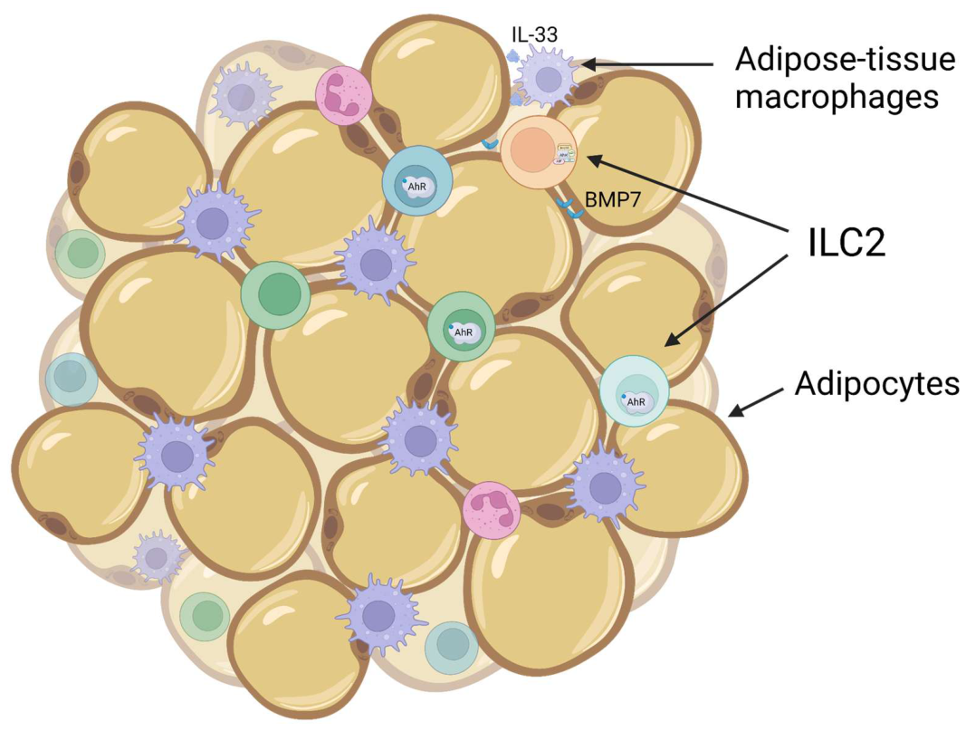
The discrepancy between whole-body and adipocyte-specific AhR deletion is the most instructive conflict in this literature, and it ceases to be a contradiction once the compared genotypes are specified. Protection of whole-body AhR-null mice from diet-induced obesity tracks with energy expenditure rather than with intake: expression of the thermogenic gene UCP1 in brown adipose tissue, and of the genes of mitochondrial β-oxidation in muscle, is significantly higher in AhR(-/-) and AhR(+/-) mice than in wild-type littermates, while insulin sensitivity in adipose tissue and muscle is unaltered. Deleting AhR only in mature adipocytes, by contrast, removes the receptor from the single compartment in which it restrains adipose expansion while leaving it intact in liver, muscle and immune cells, and the outcome is reversed: increased fat mass, adipose tissue inflammation and decreased glucose tolerance on a high-fat diet [186]. The phenotype of a whole-body knockout is therefore the sum of opposing cell-autonomous contributions, and no single-tissue experiment can be used to predict it [207].
Another study recently revealed sex-dependent phenotype of AhR depletion specific for mature adipose tissue (CadKO) cells [208]. CadKO females had a lean phenotype and healthy adipose–hypothalamic crosstalk. Interestingly, HFD-induced leptin rise was reduced in CadKO females, while the leptin receptor was increased in the energy regulatory regions of the hypothalamus, suggesting an increase of leptin sensitivity. It was estrogen receptor α (ERα) presence increased in CadKO female adipose tissue and the hypothalamus [205]. This phenomenon underlines the existing crosstalk between ERα and AhR-induced gene expression, as 17β-estradiol (E2) influences on AhR-dependent transcription of CYP1 family through the interaction between estrogen receptor and AhR. [209]. It is possible that the increase in ERα compensated for the physical absence of AhR.
CadKO males displayed a delayed progression of obesity and insulin resistance. In males, beneficial effects of adipose-specific AhR depletion were mediated through the maintenance of healthy crosstalk between adipocytes and immune cells: proinflammatory adipocytokines (such as TNFα, IL1β, IL6) secretion was improved and inflammatory macrophage infiltration into adipose was reduced. Overall, adipose-specific knockout of AhR gene improved weight control and systemic glucose homeostasis in high fat diet-induced conditions with more pronounced effects in females [205].
Taken together, these data place AhR among the transcriptional regulators that set the balance between adipocyte expansion and thermogenic remodelling – with an effect whose sign depends on the cell type in which the receptor is engaged, and on the sex of the animal, rather than on ligand dose alone
3.4. AhR in Ocular Homeostasis and Disease
Over the past few years, the scientific paradigm surrounding the AhR has shifted from viewing it merely as a sensor of environmental toxicants to recognizing it as a nodal regulator of tissue homeostasis in the eye, particularly within the retina, retinal pigment epithelium (RPE), choroid, cornea, and orbit [210,211,212,213]. This evolution in comprehension is highly relevant to ophthalmology, as ocular tissues constantly operate under high metabolic loads, light exposure, and oxidative stress, while the eye's immune privilege requires exquisite tuning of both innate and adaptive immunity [214,215,216].
Currently, AhR is firmly linked to ocular pathologies, and AhR ligands appear highly promising for ocular tissue preservation. However, this remains largely a preclinical domain with robust data supporting its role in inflammation, RPE and retinal protection, and vascular remodeling, rather than direct clinical application in humans. AhR consistently acts as a regulator of immune responses, oxidative stress, barrier function, and the survival of retinal and RPE cells; in AhR deficiency, these systems typically shift toward inflammation, degeneration, and pathological angiogenesis [210,211,212,217,218].
The most compelling ophthalmological data converge on three main points. First, the loss or attenuation of AhR signaling intrinsically leads to age-related macular degeneration (AMD)-like, inflammatory, or vascular ocular phenotypes [217,218,219]. Second, AhR activation by various endogenous, dietary, microbial, and synthetic ligands reduces inflammation, cell death, and tissue damage in models of uveitis, glaucoma, light- and ischemia-induced damage, diabetic retinopathy, and thyroid eye disease [211,220,221,222,223]. Third, not every AhR activation is beneficial; persistent toxic ligands like TCDD, alongside certain infection or neurotrauma contexts, highlight the "double-edged" nature of this pathway [224,225,226,227]. Consequently, the effect of AhR is fundamentally dependent on the ligand, tissue, dose, signal duration, and disease context, precluding broad statements about "general beneficial AhR activation" [211,213,226,228,229].
3.4.1. Ocular Phenotypes of AhR Deficiency and Activation
In the posterior segment of the eye, the protective role of basal AhR signaling is best substantiated. Aged Ahr−/− mice develop signs of dry AMD, including deteriorated visual function, sub-RPE lipid deposits, increased autofluorescence, RPE atrophy, and choroidal thinning [217]. Other AhR-deficient models similarly exhibit subretinal microglial accumulation, RPE atrophy, immune activation, and heightened sensitivity to light stress [212,217].
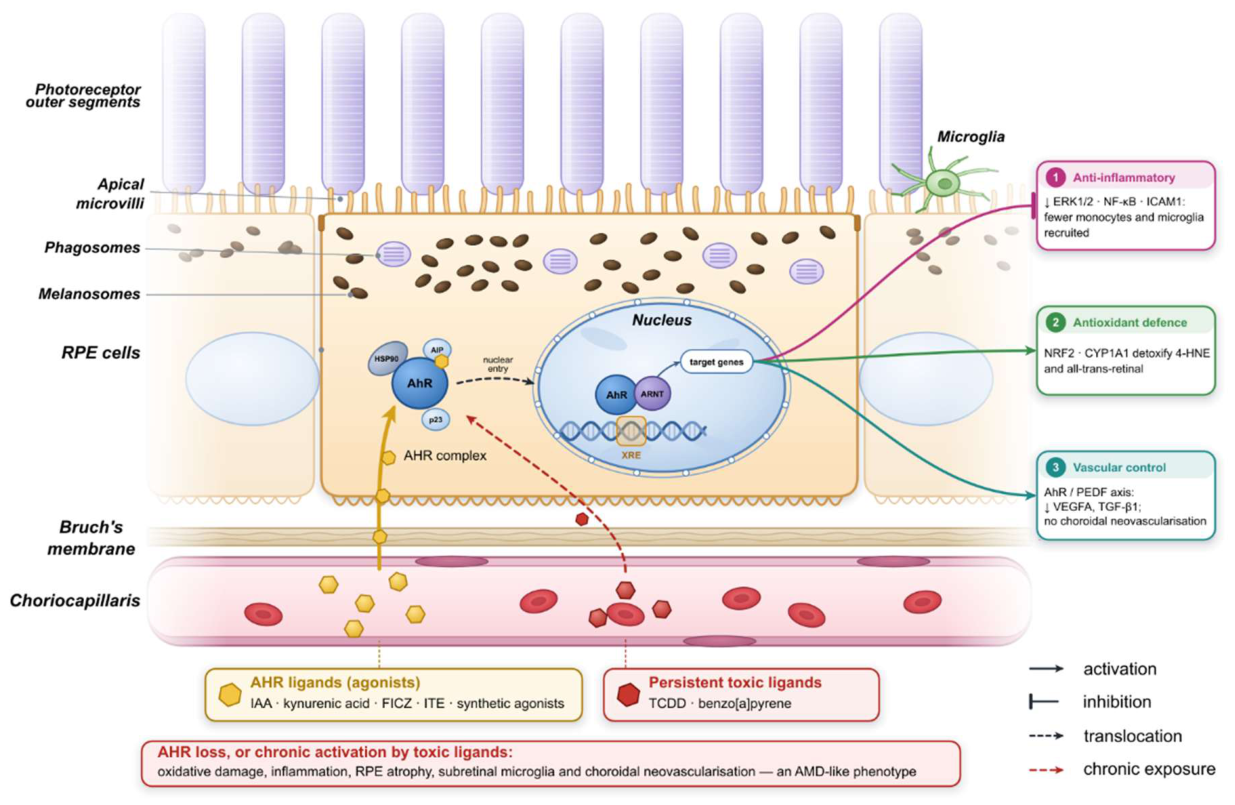
Conversely, AhR activation by multiple ligands protects the RPE and neuroretina. For instance, the synthetic AhR ligand 2AI protected the RPE cells in vitro from lipid peroxidation cytotoxicity mediated by 4-hydroxynonenal (4-HNE) and the retina in vivo from light-induced damage [230]. Independent screening in ARPE-19 cells demonstrated that AhR agonists — DF 203, FICZ, and kynurenic acid — suppressed RPE death induced by all-trans-retinal and 4-HNE, with FICZ being the most potent in both AhR activation and 4-HNE protection [231]. These effects are linked to the AhR-NRF2 antioxidant circuit and the maintenance of RPE cell survival [131,164,231]
In glaucoma and retinal ischemic injury, AhR appears to restrict microglial inflammation. Glaucoma patients exhibit altered tryptophan metabolism and reduced AhR expression in the retina, interpreted as insufficient activation by endogenous ligands [220]. In a retinal ischemia/reperfusion model, the tryptophan metabolite 2-(1′H-indole-3′-carbonyl)-thiazole-4-carboxylic acid methyl ester (ITE) reduced local inflammation, protected retinal ganglion cells (RGC) from apoptosis, and suppressed ERK- and NFκB-dependent microglial activation [220]. Recent work also links the gut-eye axis to the microbial metabolite indoleacetic acid (IAA); glaucoma is associated with reduced serum IAA, and administering IAA or B. fragilis restored AhR activation, reduced RGC loss, and preserved visual function via the AhR/RAGE/NF-κB axis [232].
For retinal vascular pathology, the signal is generally protective but context-dependent. In diabetic retinopathy, the AhR agonist VAF347 reduced leukostasis, oxidative stress, inflammation, and capillary degeneration in streptozotocin-induced diabetic C57BL/6 mice [221]. AhR deficiency exacerbated vascular permeability, blood-retinal barrier breakdown, capillary degeneration, and neovascularization, whereas agonists restored AhR/PEDF axis activity [219]. In AMD models, leflunomide and flutamide reduced choroidal neovascularization and fibrosis, an effect that vanished in AhR-knockout animals [211,217].
3.4.2. Molecular Mechanisms of AhR in the Eye
AhR influences ocular tissue health through three primary mechanistic categories: canonical transcriptional regulation with co-regulators, suppression of pro-inflammatory signaling cascades, and non-canonical interactions with other receptors.
As it was described in detail earlier, AhR resides in the cytosol in a complex with HSP90, the co-chaperone p23, the AIP, and the kinase SRC [211]. Upon ligand binding, chaperones dissociate, and AhR translocates to the nucleus, forming a heterodimer with ARNT. This complex binds to DRE/XRE DNA elements, activating the transcription of genes including CYP1A1, CYP1B1, and AHRR [211,233,234]. In retinal pigment epithelial cells specifically, this output requires recruited co-activators: the chromatin remodeller BRG1 binds the C-terminal activation domain of AhR and opens chromatin at the CYP1A1 enhancer [235], while the histone methyltransferase MLL1 and the co-activator FLII act together to recruit BRG1 and RNA polymerase II, FLII binding AhR in a ligand-dependent manner and MLL1 independently of ligand [236].
Suppression of inflammatory cascades by AhR covers several directions. Loss of AhR in RPE cells enhances αvβ3-integrin, ERK1/2, and NF-κB p65 subunit activity, inducing ICAM1 and monocyte adhesion; pharmacological inhibition of each node prevents this effect [218]. Prolonged exposure to the carcinogen benzo[a]pyrene disrupts the SOCS3–STAT3 axis via AhR dysfunction, leading to STAT3 hypo-ubiquitination, weakened negative feedback via SOCS3, and the development of AMD-like retinal damage [237]. In uveitis models, AhR activation by TCDD reduces pro-inflammatory cytokines and inhibits NF-κB and STAT pathways, shifting macrophage/microglial polarization from M1 to M2 [238].
Beyond canonical pathways, non-canonical interactions of AhR further expand its regulatory network through several key axes. In ARPE-19 cells, for instance, AhR physically associates with the glucocorticoid receptor (GR), forming a complex where GR suppresses AhR nuclear translocation and DNA recruitment, while AhR conversely enhances GR-mediated transcription [239]. In the context of thyroid eye disease, AhR activation by FICZ and tapinarof blocks IGF1R signaling, reducing GSK3β phosphorylation and inhibiting the β-catenin/Wnt pathway, thereby suppressing the proliferation, migration, and hyaluronan production of orbital fibroblasts [217,222]. Furthermore, the AhR pathway critically regulates angiogenic and fibrotic mediators; AhR knockdown in choroidal endothelial cells and the RPE increases the expression of VEGFA, CCL2, SPP1, and TGFβ1, ultimately exacerbating angiogenesis, fibrosis, and inflammation in wet AMD models [240]. Finally, a functional link between AhR and HIF-1α is particularly relevant in smoking-related ocular diseases, where both transcription factors may compete for the common dimerization partner ARNT, thereby modulating the hypoxic response in the retina and cornea [50].
The collective data best support a model where physiological or selectively enhanced AhR signaling protects ocular tissues from chronic inflammation, oxidative damage, pathological extracellular matrix remodeling, and cell death, particularly in the RPE, microglia, endothelium, and orbital fibroblasts [132,217,219,231,241]. This is supported by genetic loss-of-function models, independent agonists, and recurring mechanisms such as NF-κB/ERK suppression, antioxidant responses, and the attenuation of Th17/IL-17 signaling [211,219,220,221,241,242].
Figure 7.
AhR signalling in the retinal pigment epithelium. Agonists reaching the RPE from the choriocapillaris and from the gut–eye axis – indoleacetic acid, kynurenic acid, FICZ, ITE and synthetic ligands such as 2AI – release AhR from its cytosolic complex with HSP90, AIP and p23; the receptor enters the nucleus, heterodimerises with ARNT and binds xenobiotic response elements (XRE) in the regulatory regions of its target genes. Three output branches are established in ocular tissue: (1) suppression of ERK1/2 and NF-κB signalling, which limits ICAM1 induction and the recruitment of monocytes and microglia; (2) an AhR–NRF2 antioxidant circuit that protects the RPE against the lipid peroxidation product 4-hydroxynonenal (4-HNE) and against all-trans-retinal; and (3) restraint of angiogenic and fibrotic mediators through the AhR/PEDF axis, holding down VEGFA and TGF-β1 and preventing choroidal neovascularisation.
Figure 7.
AhR signalling in the retinal pigment epithelium. Agonists reaching the RPE from the choriocapillaris and from the gut–eye axis – indoleacetic acid, kynurenic acid, FICZ, ITE and synthetic ligands such as 2AI – release AhR from its cytosolic complex with HSP90, AIP and p23; the receptor enters the nucleus, heterodimerises with ARNT and binds xenobiotic response elements (XRE) in the regulatory regions of its target genes. Three output branches are established in ocular tissue: (1) suppression of ERK1/2 and NF-κB signalling, which limits ICAM1 induction and the recruitment of monocytes and microglia; (2) an AhR–NRF2 antioxidant circuit that protects the RPE against the lipid peroxidation product 4-hydroxynonenal (4-HNE) and against all-trans-retinal; and (3) restraint of angiogenic and fibrotic mediators through the AhR/PEDF axis, holding down VEGFA and TGF-β1 and preventing choroidal neovascularisation.
3.4.3. Genetic Evidence in Human Ocular Disease
Recently there were two ophthalmologic inherited disorders described where AhR directly acts as a causal gene.
A homozygous nonsense mutation c.1861C>T (p.Gln621*) in a family with foveal hypoplasia and infantile nystagmus completely co-segregated with the disease, identifying AHR as a novel gene for this recessive pathology [243]. The p.Gln621* AhR mutation truncates the protein, losing a significant portion of the C-terminus, a region critical for ligand-induced nuclear accumulation and receptor activation [244]. Specifically, it eliminates the transactivation domain while leaving the bHLH and PAS domains intact; thus, any mutant protein escaping nonsense-mediated decay would be predicted to dimerize with ARNT and bind XREs without recruiting coactivators. Because the pedigree shows strict recessive inheritance, one wild-type allele appears sufficient, indicating that the variant acts as a loss of function rather than as a dominant negative [243]. An independent case report of a novel biallelic AHR splice-site variant c.899_908+15del in isolated foveal hypoplasia without nystagmus expanded the phenotypic spectrum of AhR deficiency [245,246].
Rare homozygous autosomal-recessive splice variant c.1160+1G>A resulting in exon 9 skipping of the AHR gene causing retinitis pigmentosa (RP) was identified in an Indian subcontinent family. This variant is very rare in several exome databases. Described mutant AhR variant loses TCDD-induced XRE activity and fails to activate expression of the downstream target genes CYP1A1 and CYP1A2. Mouse knockout model exhibited reduced electroretinogram responses at 9 months of age, comparing with the wild-type mouse. Histology of model retinas showed the degeneration of photoreceptors with a thinner outer nuclear layer, demonstrating typical RP phenotype [247].
As a genetic model of AHR-caused diseases, retina-specific Ahr knockout causes late-onset retinal degeneration, reduced ERG responses, and a thinner outer nuclear layer, proving that direct AhR deficiency in the neuronal retina is sufficient for a degenerative phenotype compatible with RP [247]. Independent Ahr−/− models show broader tissue maintenance defects, including subretinal microglial accumulation, autofluorescent deposits, and focal RPE atrophy, linking AhR loss to increased vulnerability to light/oxidative stress and AMD-like pathology [217,248].
While the direct mutation of the AHR gene causes severe retinal dystrophy, a critical, yet underexplored [249], dimension of AhR pathogenesis in the retina involves its cytosolic chaperone complex. In the canonical pathway, AhR is maintained in the cytosol via a complex involving HSP90, p23, and the AIP [211]. AIP is a ubiquitous co-chaperone essential for AhR stability and regulation. Notably, AIP shares significant structural homology with AIPL1 (Aryl Hydrocarbon Receptor Interacting Protein-Like 1), particularly within the tetratricopeptide repeat domains responsible for chaperone interactions. While AIP is ubiquitously expressed, AIPL1 is specific for the retina and is a crucial chaperone for photoreceptor-specific proteins (such as PDE6 and guanylate cyclase). Mutations in the AIPL1 gene are a well-established cause of severe early-onset retinal dystrophies, including Leber Congenital Amaurosis and RP [250].
Despite the ubiquitous expression of AhR across tissues, the biallelic mutations reported to date segregate almost exclusively with ocular disease. Two interpretations of this observation are possible, and they are not mutually exclusive. The first is deflationary and concerns ascertainment: the families were identified through ophthalmic genetics services, the number of published familial cases remains in single figures, and systematic phenotyping outside the eye has not been reported. Thus, an apparently eye-restricted phenotype may be no more than an eye-focused bias. The substantive interpretation begins with the fact that AhR loss of function produces a retinal dystrophy at all, and asks what is distinctive about the retina. It prompts a plausible, yet underexplored, hypothesis: given that AhR loss-of-function leads to profound retinal dystrophy, the retina-enriched protein AIPL1 may intersect with, compensate for, or be regulated by the AhR pathway specifically within the eye. Two mechanisms can be distinguished, and they apply to different situations rather than competing to explain the same one. The first is loss of function. If AIPL1 acts as a retina-restricted surrogate for AIP, contributing to AhR stability and cytosolic retention under local oxidative and light stress, then biallelic loss of AhR would deprive the photoreceptor of a component of that system; this is the mechanism consistent with the inherited pedigrees, which are strictly recessive and in which one wild-type allele is evidently sufficient. The second is sequestration. Because AIPL1 is an essential, retina-specific chaperone for photoreceptor proteins, an AhR species that bound it inappropriately would deplete the functional AIPL1 pool, disrupt photoreceptor proteostasis and precipitate degeneration. This mechanism, however, cannot be attributed to the truncated receptor: a mutant protein that actively sequestered AIPL1 would be predicted to cause disease in heterozygous carriers, and no such dominant phenotype has been reported. Sequestration is therefore a hypothesis about the wild-type receptor under sustained activation – the condition produced by persistent environmental ligands rather than by mutation – and it predicts a different population at risk: chronically exposed individuals with an intact AHR genotype, rather than biallelic carriers.
The proposed AhR–AIPL1 axis therefore describes a balance between these two regimes: physiological AhR signalling might recruit AIPL1 for localised stress adaptation in the photoreceptor, whereas sustained activation could draw it away from its clients. It is worth noting how little of this has been tested. AIPL1 was named for its homology to the AhR-interacting protein and not for any demonstrated association with the receptor itself, and that interaction has, to our knowledge, never been examined directly in retinal tissue. We are testing the physical premise common to both regimes – whether AhR and AIPL1 associate at all – by co-immunoprecipitation of the two proteins and by an XRE-driven reporter of AhR transcriptional output; failure to detect an interaction would dispose of the model, which is the point of framing it this way. Should it survive, the therapeutic corollary is unusually direct, because AIPL1 augmentation is already deliverable: subretinal rAAV8.hRKp.AIPL1 has produced sustained gains in visual function in children with AIPL1-associated Leber congenital amaurosis [272], and human retinal organoid models of the disease are available for preclinical testing [244,273].
3.4.4. Research Gaps and Clinical Translation
Despite strong mechanistic preclinical data, clinical readiness remains low. Almost all ocular data derive from mice, cell lines, organoid-like systems, or ex vivo tissues; direct ophthalmological clinical trials in humans are absent from this corpus [220,221,228,231,251]. Furthermore, the broader AhR literature warns that safety and efficacy depend on pharmacokinetics, ligand persistence, feedback via CYP1/AHRR, and tissue context; thus, toxic or persistent agonists are unsuitable as therapeutic templates [213,226,229,252].
Extraocular literature confirms AhR as a general regulator of barrier and immune defense in the lungs, skin, and gut [253,254,255,256]. However, adjacent neurological and infectious literature sometimes favors AhR antagonism or shows opposing ligand effects [42,225,257,258,259,260].
Therefore, the most realistic strategy is not simply "activating AhR", but rather developing selective, short-lived, tissue-specific modulators with appropriate ocular delivery [211,229,261]. The main gap remains the translation from preclinical mechanisms to clinical application, specifically determining which ligands, doses, and ocular cell targets will yield sustainable benefits without toxicity or paradoxical activation of undesirable pathways [226,228,262]. Ultimately, AhR is a central regulator of ocular inflammation, oxidative stress, and cell survival, so making selective, non-toxic AhR modulators a highly promising, albeit currently preclinical, tool for preserving ocular tissues [210,231,232,251].
3.5. AhR in Reproduction: Gonads, Gametes and the Maternal–Fetal Interface
AhR has a dual role at the intersection of development and reproduction: it is both a mediator of environmental teratogenesis and an endogenous regulator of developmental and reproductive physiology. The reproductive evidence has for most of its history been treated as a toxicological appendix – AhR was the receptor through which dioxins impaired fertility, and the interest lay in the exposure rather than in the receptor. Two developments make that framing obsolete. The first is that Ahr-null animals are subfertile in the absence of any exogenous ligand, which establishes a constitutive requirement. The second is that conditional, cell-type-restricted deletions have begun to appear and, exactly as in adipose tissue, they do not phenocopy the whole-body null. The reproductive axis therefore presents the same interpretative problem that has occupied the rest of this review, and admits the same resolution. Disruption at any point along this axis – whether by exogenous ligands, genetic variation or null mutations – produces cascading effects on gametogenesis, pregnancy outcome and offspring development.
3.5.1. Ovarian Folliculogenesis and Steroidogenesis
AhR is not merely upstream of ovarian steroid output; it is also downstream of the gonadotropins that drive it. In granulosa cells of immature mice, Ahr is upregulated in a gonadotropin-dependent manner requiring both FSH and LH activity: FSH alone raises Ahr transcript without raising protein, implying an LH-dependent post-transcriptional step, and the protein increase is specific to large antral follicles [263]. The receptor is thus positioned as an effector of the hypothalamic-pituitary-gonadal (HPG) axis rather than as an independent sensor sitting beside it, which is the structural reason why an environmental ligand entering this compartment does not simply add a signal but perturbs an existing hormonal circuit.
What AhR does once expressed is, in substantial part, to set aromatase. AhR binds and transactivates the ovarian Cyp19 (aromatase) promoter in granulosa cells, cooperating with the orphan nuclear receptor Ad4BP/SF-1, as shown by reporter assay and by chromatin immunoprecipitation in vivo; Ahr-null females are subfertile, with defective late folliculogenesis and reduced intraovarian oestrogen at preovulatory stages, and administration of the AhR ligand DMBA induces ovarian Cyp19 irrespective of the phase of the oestrous cycle [264]. That last observation is the mechanistic hinge of the reproductive argument and deserves to be stated as such: an exogenous agonist does not merely raise aromatase, it decouples aromatase expression from the cycle that normally gates it. A hormone whose physiological signal lies in its timing is thereby converted into a constitutive one, and the consequence is a disordered cycle rather than a simple deficiency.
Follicular dynamics are affected across most stages of maturation. Ahr-null mice have fewer preantral and antral follicles than wild-type during postnatal life, and the deficit arises from slowed follicular growth rather than from increased atresia [265]. By roughly three months of age antral follicle numbers converge on wild-type values, yet Ahr-null females still ovulate fewer oocytes in response to equine chorionic gonadotropin, which localises part of the defect to ovulation itself rather than to the preceding growth phase; and reintroducing Ahr into cultured pubertal antral follicles rescues growth only partially [266]. The incomplete rescue is informative, because it indicates that part of the phenotype is developmental – laid down before the experimental window – rather than a continuing requirement for the receptor.
The cell-type question has now been addressed directly, and the answer parallels the adipose case closely enough to be worth setting side by side with it. Oocyte-specific deletion of Ahr (Ahr fl/fl × Gdf9-Cre) produces subfertility and disrupted oestrous cyclicity, but the follicular phenotype is not that of the whole-body null: the number of primary follicles falls while secondary follicles and corpora lutea increase, indicating accelerated rather than retarded follicular recruitment, with granulosa cell apoptosis, reduced oocyte expression of Gdf9 and Bmp15, impaired bidirectional oocyte-granulosa communication, and reduced serum oestradiol and FSH [267]. Deleting the receptor from the oocyte therefore depletes the primordial pool by releasing the brake on recruitment, whereas deleting it from the whole animal slows the growth of follicles already recruited. These are not contradictory findings but measurements of two functions of one protein in two cells of the same follicle, and they carry the same methodological lesson as the adipocyte-specific knockout discussed in Section 3.3: a systemic deletion reports the sum of opposing cell-autonomous contributions and cannot be used to predict any one of them.
Exogenous ligands act on this system in the direction opposite to the endogenous requirement. TCDD suppresses promoter-specific CYP19A1 expression in granulosa cells [268], so that chronic agonism lowers the same aromatase output that physiological AhR signalling sustains – a pattern consistent with the general principle developed in Section 2.2.1, in which a persistent agonist produces an outcome unlike that of the transient endogenous ligand it displaces.
3.5.2. Spermatogenesis and Male Gamete Quality
The male evidence is thinner and should be presented as such. Ahr-null males show reduced fertility at a young age, accompanied by dysregulated Notch and ERα signalling and – in contrast to females – without a detectable change in testicular aromatase or oestrogen levels, indicating that the male phenotype is not simply the male counterpart of the ovarian aromatase defect [269]. AhR is expressed in Sertoli, Leydig and germ cells, and Ahr deletion has been reported to disturb seminiferous tubule architecture and sperm development [270]. Perinatal TCDD exposure reduces daily sperm production in rats at doses below those producing overt maternal toxicity, and this remains the most frequently replicated reproductive effect of dioxin [271].
Two qualifications should be stated rather than left implicit. First, several of the classical dioxin effects on the male tract were obtained at doses and in strains carrying high-affinity AhR alleles, so the species difference in ligand affinity noted in Section 2 applies here with full force. Second, no human AHR loss-of-function pedigree has had gonadal or endocrine phenotyping reported – the published biallelic variants were ascertained ophthalmologically – so the question of whether human AhR deficiency impairs gametogenesis is at present unanswered rather than answered negatively. It could be settled by endocrine and semen phenotyping of the existing families, and we suggest this as a concrete next step.
3.5.3. The Maternal–Fetal Interface
Beyond the gonads, AhR is expressed throughout the human placenta, where it acts both as a developmental regulator and as a sensor of the maternal chemical environment [272]. The ligands reaching this compartment are largely those already encountered in the gut. Indole-3-lactic acid, generated from dietary tryptophan by Bifidobacterium and Lactobacillus species through aromatic lactate dehydrogenase (Section 2.2.2), promotes trophoblast migration and invasion by activating AhR [273] – which places a maternal microbial metabolite directly upstream of placentation, and gives the gut–eye axis described in Section 3.4.1 a gut–placenta counterpart. The IDO1–kynurenine–AhR axis has in parallel been proposed to contribute to maternal–fetal immune tolerance, since placental indoleamine 2,3-dioxygenase both depletes tryptophan, restraining effector T cells, and generates kynurenine, supplying an AhR agonist that favours regulatory differentiation [274]. Whether this constitutes a dedicated tolerogenic mechanism or a consequence of local tryptophan economy is unresolved, and the human evidence remains associative.
The placental data also illustrate the residence-time distinction in its starkest form, because the consequences of exposure are not confined to the exposed individual. A ligand that is cleared within hours acts on the trophoblast during a defined window; one with a tissue half-life measured in years is present throughout gestation and, being lipophilic, is mobilised from maternal adipose stores precisely when fat is being catabolised. Developmental and reproductive toxicity of persistent AhR ligands is therefore not simply reproductive toxicity delivered to a second individual, and the distinction between an effect on the parent and an effect on the offspring deserves to be drawn explicitly wherever it is available [275].
3.5.4. Human Evidence: Receptor Variants, Exposure and Reproductive Disease
Translating any of this into human terms requires endpoints that clinical reproductive medicine actually measures: ovarian reserve, response to controlled ovarian stimulation, and oocyte and embryo competence, rather than fertility in the abstract. The cumulus-oocyte complex is the natural point of contact between the two literatures, since it is the compartment in which gonadotropin signalling, steroidogenesis and xenobiotic metabolism converge on a single gamete, and it is accessible in the course of routine assisted reproduction. In women with poor ovarian response, bisphenol A exposure has been associated with altered expression and DNA methylation of ICAM1 and HLA-G in cumulus cells, and with impaired cumulus cell development and a reduced yield of mature oocytes [6,16], and transcriptomic analysis of poor-responder cohorts has begun to identify the pathways involved [12]. Bisphenol A is a weak direct AhR ligand whose principal route is the oestrogen receptor, so these associations are not by themselves evidence of AhR-mediated action; the two pathways are nonetheless not mutually exclusive, since AhR-dependent effects of bisphenol A have been demonstrated against AhR-null controls [276]. The relevance of the cumulus cell here is that it is the compartment in which an AhR-directed hypothesis could be tested in humans, using endpoints already collected clinically.
Direct human genetic evidence for AhR in reproduction is at present confined to common variants rather than to the biallelic lesions described for the eye. In a case–control study of 176 men with idiopathic infertility and 352 fertile controls, two of seven AHR single-nucleotide polymorphisms were associated with infertility, the rs2066853 (Arg554Lys) AA genotype carrying an approximately one-third lower risk [277]; cohort studies of women with chronic exposure to industrial AhR agonists report corresponding effects on reproductive outcome [278]. No human AHR loss-of-function pedigree has had gonadal or endocrine phenotyping reported, so the severe end of the genotypic spectrum remains uncharacterised in this respect.
Polycystic ovary syndrome supplies the most informative human disease example, and the three strongest lines of evidence do not point in the same direction. Serum L-kynurenine is elevated in affected women, and in letrozole-induced PCOS-like mice hepatic IDO1 induction activates an AHR–PCSK9 pathway that generates dyslipidaemia alongside ovarian dysfunction [279]. Bisphenol A, which is enriched in the serum of affected women, suppresses GLUT4 expression in granulosa cells in an AhR-dependent manner, an effect absent in AhR-null mice and supported by chromatin immunoprecipitation [276]. Both implicate excessive AhR signalling. The third line points the other way: the gut fungus Aspergillus tubingensis is enriched in women with PCOS across three geographically separate cohorts, its colonisation reproduces a PCOS-like phenotype in mice, and the responsible secondary metabolite AT-C1 behaves as an endogenous AhR antagonist, reducing IL-22 output by intestinal ILC3 through precisely the circuit described in Section 3.1 [280]. A polymorphism in the AhR repressor has separately been associated with the syndrome [281]. The literature therefore contains both gain- and loss-of-signalling models of one disorder. On the framework developed here these are not alternatives: hepatic AhR activation and intestinal AhR suppression are effects in different compartments and could coexist in the same patient, which is a testable claim rather than a reconciliation of convenience.
Endometriosis is frequently listed as an AhR-linked disease, and the attribution should be treated with more caution than it usually receives. AhR and its target genes are expressed in human endometrium, with differences reported between affected and unaffected women [282], and in vitro and animal studies have supported a role for dioxins in the disease [275]. A systematic reassessment of that literature nevertheless concluded that the supportive findings are strongly context-dependent, that negative results are rarely published, and that the epidemiological association remains unproven [283]; a later systematic review of organochlorine exposure reached similarly guarded conclusions [284]. We therefore regard endometriosis as a plausible but unestablished example of AhR-mediated reproductive pathology rather than as a demonstrated one.
3.5.5. Outlook
Therapeutic targeting of AhR in reproduction remains undeveloped, and the obstacle is not a shortage of candidate molecules but the absence of a defined objective. The preceding subsections indicate what that objective would have to be. Agonism and antagonism are the wrong axes on which to choose: in the ovary the receptor is required, and in the oocyte and the granulosa cell it is required for opposite things, so a systemically delivered ligand of either sign will produce the sum of two effects and probably neither of the intended ones. What the reproductive axis needs from AhR pharmacology is what the retina and the gut also need – ligands cleared by the enzymes they induce, delivered to a defined compartment, with a residence time short enough not to decouple aromatase from the cycle that gates it. A further consideration concerns bisphenol A and related environmental oestrogens, which are frequently discussed alongside AhR ligands although their principal route is the oestrogen receptor. An AhR-directed strategy is defensible for such compounds only where the AhR-dependent component of the endpoint has been established rather than assumed – as it has, for instance, for the suppression of GLUT4 in granulosa cells, which is lost in AhR-null animals [276] – or where the route runs through AhR–ERα crosstalk. That crosstalk is direct and bidirectional: ligand-activated AhR associates with ERα and can act as the substrate-recognition component of a ubiquitin ligase that targets the oestrogen receptor for degradation, while also recruiting ERα to AhR target promoters [285], and 17β-oestradiol reciprocally modulates AhR-dependent transcription of the CYP1 family [209] [286]. The sex-dependent effects observed in adipose tissue underline, on the same grounds, the need to evaluate such agents separately in males and females rather than to assume that they behave alike [208].
The path forward is complex, requiring careful consideration of the dual nature of AhR in different tissues, cell types and disease contexts. However, with the convergence of structural biology, medicinal chemistry, and a deeper understanding of its physiological roles, the AhR is poised to become a cornerstone target for a new generation of therapies for inflammatory, metabolic, oncological, and reproductive disorders.
4. Discussion
The apparent contradictions in the AhR literature are not disagreements about what the receptor does. They arise because three variables are rarely specified together: how long a ligand occupies the receptor, which cell it occupies it in, and which transcriptional output follows. Table 1 sets the principal ligand classes against the first of these, and the pattern is systematic. The endogenous, microbial and dietary agonists that maintain barrier function are substrates for the cytochrome P450 enzymes they induce, so that induction terminates the signal which produced it; the halogenated compounds responsible for AhR-associated toxicity are poor substrates, and convert a single exposure into a stimulus lasting years [49,226,287]. Affinity and toxicity are therefore uncoupled. FICZ binds AhR with the highest affinity yet measured – roughly sevenfold tighter than TCDD – and is innocuous, because it is cleared within minutes [61]; benzo[a]pyrene binds three orders of magnitude more weakly and is carcinogenic, because what it induces bioactivates it [57,58]. Potency at the receptor is thus a poor predictor of consequence in the organism, and this single observation accounts for much of what otherwise appears paradoxical in the preceding sections.
The second variable, cellular address, is what makes whole-animal genetics hard to read in this field, and three independent instances in this review follow one pattern. Whole-body deletion of Ahr protects mice from diet-induced obesity through increased energy expenditure, whereas deletion restricted to mature adipocytes increases fat mass, adipose inflammation and glucose intolerance (Section 3.3). Whole-body deletion slows ovarian follicular growth, whereas deletion restricted to the oocyte accelerates follicular recruitment and depletes the primordial pool (Section 3.5.1). In the eye, it is the loss of the receptor rather than its activation that produces inherited disease (Section 3.4.3). A systemic knockout reports the sum of opposing cell-autonomous contributions and cannot be used to predict any one of them; this is a constraint on the literature as a whole, not a peculiarity of individual experiments, and it is the reason that “AhR activation” and “AhR deficiency” are not single experimental conditions.
The third variable is the transcriptional output, and it is the one that pharmacology can address. The aim of selective AhR modulation is to discriminate between the receptor's roles rather than to switch it on or off, and structural work on the ligand-binding domain and its interface with ARNT has made this tractable by revealing cavities and conformations distinct from the orthosteric pocket [69,234,288]. Allosteric modulation offers functional selectivity that competition for the ligand-binding site cannot: the monoterpenoid carvones, for example, act as non-competitive antagonists by binding the region of AhR involved in heterodimerisation and preventing formation of a functional AhR–ARNT complex, so that they suppress signalling without displacing an agonist [116]. Selective AhR modulators such as 6-MCDF and the dietary metabolite DIM exhibit tissue-dependent partial agonism [134,135,136], and chimeric degrader molecules that recruit AhR itself to an E3 ligase complex offer a further mode of control, silencing the receptor by destroying it rather than by blocking its pocket and so mimicking the proteasomal regulation described in Section 3.2 [168].
Clinically the field is no longer theoretical, but its successes are confined to the condition the framework predicts would be easiest. Tapinarof, an agonist derived from bacterial symbionts of entomopathogenic nematodes, is approved for plaque psoriasis and for atopic dermatitis [162,163]; it is applied topically, which confines both the tissue and the systemic exposure, and it acts on an organ in which the receptor's requirement is consistent across the relevant cell types. Antagonists have advanced furthest where the target is a defined immunosuppressive axis rather than a tissue: BAY-2416964 and IK-175 are in immuno-oncology trials directed at kynurenine-driven AhR signalling [121,124,227], and StemRegenin 1 expands human haematopoietic stem cells ex vivo, where exposure is bounded by the culture period [125,126]. Agonists intended to restore epithelial integrity in inflammatory bowel disease remain preclinical or early clinical, and share the same design logic of transient, locally restricted engagement [289]. What is absent is as informative as what exists: no ophthalmological trial of an AhR modulator has been reported despite the strength of the ocular preclinical data [220,221,231,251], and no reproductive application has been attempted at all.
Two limitations of this framework should be stated plainly. First, residence time is inferred for most ligands rather than measured: systematic comparisons of receptor occupancy half-life across the classes in Table 1 do not exist, and the inference rests on metabolic stability together with the elimination kinetics of the compounds in tissue. Second, the cell-type argument depends almost entirely on mouse conditional genetics. The corresponding human evidence is limited to common polymorphisms [277,281], to exposure cohorts [278], and to the small number of biallelic AHR pedigrees, none of which has been phenotyped outside the eye. The framework is therefore a way of organising the existing evidence and of specifying which experiments would discriminate between interpretations; it is not itself an established result.
5. Conclusions
One receptor, engaged by different ligands in different cells, produces outcomes that are not merely different but opposite, and each organ system reviewed here shows this in its own way: in the gut and the skin, intermittent activation by microbial and photochemical ligands sustains the epithelial barrier and restrains IL-17-driven inflammation; in adipose tissue, chronic activation by persistent pollutants suppresses adipogenesis while promoting inflammation, and whole-body and adipocyte-restricted deletion of the same gene give opposite phenotypes; in the ovary and the testis the receptor is required for cyclical aromatase expression and for normal gametogenesis, so that a persistent agonist uncouples a hormonal signal from the cycle that gates it; at the maternal–fetal interface microbial indoles promote trophoblast invasion; and in the retina it is the loss of AhR, not its activation, that causes inherited disease. What unifies these observations is not a direction of effect but the three variables that determine it – how long the ligand occupies the receptor, which cell it occupies it in, and which transcriptional programme follows. Framed this way, the therapeutic question ceases to be whether AhR should be activated or inhibited and becomes whether modulation can be confined: to a defined tissue, to a residence time short enough to preserve the physiological rhythm of the pathway, and to a transcriptional output narrower than pan-activation. The approval of tapinarof for plaque psoriasis and atopic dermatitis shows that a topically confined agonist can be both effective and tolerable; whether comparable control can be achieved systemically, and in organs where the receptor's requirement differs between neighbouring cell types, is the question on which the clinical prospects of this target now rest.
Author Contributions
Conceptualization ADE. AD, TS and ADE searched for scientific data and wrote the manuscript. TS and ADE generated figures. All authors contributed to editorial changes in the manuscript. ADE was responsible for funding acquisition and project supervision. All authors read and approved the final manuscript.
Funding
This research was initially supported by the Russian Science Foundation and Kuban Science Foundation (project No. 22-14-20046). Presented work was funded by the grant of the state program of the «Sirius» Federal Territory «Scientific and technological development of the «Sirius» Federal Territory» (Agreement No. 20-03 date 1 June 2026).
Institutional Review Board Statement
Not applicable.
Informed Consent Statement
Not applicable.
Data Availability Statement
We encourage all authors of articles published in MDPI journals to share their research data. In this section, please provide details regarding where data supporting reported results can be found, including links to publicly archived datasets analyzed or generated during the study. Where no new data were created, or where data is unavailable due to privacy or ethical restrictions, a statement is still required. Suggested Data Availability Statements are available in section “MDPI Research Data Policies” at https://www.mdpi.com/ethics.
Acknowledgments
We gratefully acknowledge the support of funding authorities, the assistance and extensive endorsement from our colleagues Stanislav Boychenko and Vera Egorova.
Conflicts of Interest
The authors declare no conflicts of interest.
References
- Furman, D.; Chang, J.; Lartigue, L.; Bolen, C.R.; Haddad, F.; Gaudilliere, B.; Ganio, E.A.; Fragiadakis, G.K.; Spitzer, M.H.; Douchet, I.; et al. Expression of Specific Inflammasome Gene Modules Stratifies Older Individuals into Two Extreme Clinical and Immunological States. Nat. Med. 2017, 23, 174–184. [Google Scholar] [CrossRef] [PubMed]
- Netea, M.G.; Balkwill, F.; Chonchol, M.; Cominelli, F.; Donath, M.Y.; Giamarellos-Bourboulis, E.J.; Golenbock, D.; Gresnigt, M.S.; Heneka, M.T.; Hoffman, H.M.; et al. A Guiding Map for Inflammation. Nat. Immunol. 2017, 18, 826–831. [Google Scholar] [CrossRef] [PubMed]
- Bennett, J.M.; Reeves, G.; Billman, G.E.; Sturmberg, J.P. Inflammation–Nature’s Way to Efficiently Respond to All Types of Challenges: Implications for Understanding and Managing “the Epidemic” of Chronic Diseases. Front. Med. 2018, 5, 316. [Google Scholar] [CrossRef] [PubMed]
- Furman, D.; Campisi, J.; Verdin, E.; Carrera-Bastos, P.; Targ, S.; Franceschi, C.; Ferrucci, L.; Gilroy, D.W.; Fasano, A.; Miller, G.W.; et al. Chronic Inflammation in the Etiology of Disease across the Life Span. Nat. Med. 2019, 25, 1822–1832. [Google Scholar] [CrossRef] [PubMed]
- Petrakis, D.; Vassilopoulou, L.; Mamoulakis, C.; Psycharakis, C.; Anifantaki, A.; Sifakis, S.; Docea, A.; Tsiaoussis, J.; Makrigiannakis, A.; Tsatsakis, A. Endocrine Disruptors Leading to Obesity and Related Diseases. IJERPH 2017, 14, 1282. [Google Scholar] [CrossRef] [PubMed]
- Aftabsavad, S.; Noormohammadi, Z.; Moini, A.; Karimipoor, M. Effect of Bisphenol A on Alterations of ICAM-1 and HLA-G Genes Expression and DNA Methylation Profiles in Cumulus Cells of Infertile Women with Poor Response to Ovarian Stimulation. Sci. Rep. 2021, 11, 9595. [Google Scholar] [CrossRef] [PubMed]
- Boychenko, S.; Egorova, V.S.; Brovin, A.; Egorov, A.D. White-to-Beige and Back: Adipocyte Conversion and Transcriptional Reprogramming. Pharmaceuticals 2024, 17, 790. [Google Scholar] [CrossRef] [PubMed]
- Karchner, S.I.; Franks, D.G.; Powell, W.H.; Hahn, M.E. Regulatory Interactions among Three Members of the Vertebrate Aryl Hydrocarbon Receptor Family: AHR Repressor, AHR1, and AHR2. J. Biol. Chem. 2002, 277, 6949–6959. [Google Scholar] [CrossRef] [PubMed]
- Kewley, R.J.; Whitelaw, M.L.; Chapman-Smith, A. The mammalian basic helix-loop-helix/PAS family of transcriptional regulators. Int. J. Biochem Cell Biol. 2004, 36, 189–204. [Google Scholar] [CrossRef] [PubMed]
- Hahn, M.E.; Karchner, S.I.; Shapiro, M.A.; Perera, S.A. Molecular Evolution of Two Vertebrate Aryl Hydrocarbon (Dioxin) Receptors (AHR1 and AHR2) and the PAS Family. Proc. Natl. Acad. Sci. U S A 1997, 94, 13743–13748. [Google Scholar] [CrossRef] [PubMed]
- Burbach, K.M.; Poland, A.; Bradfield, C.A. Cloning of the Ah-Receptor cDNA Reveals a Distinctive Ligand-Activated Transcription Factor. Proc. Natl. Acad. Sci. U.S.A. 1992, 89, 8185–8189. [Google Scholar] [CrossRef] [PubMed]
- Bahrami, N.; Nazari, A.; Afshari, Z.; Aftabsavad, S.; Moini, A.; Noormohammadi, Z. Gene Expression and Demographic Analyses in Women with the Poor Ovarian Response: A Computational Approach. J. Assist Reprod. Genet 2023, 40, 2627–2638. [Google Scholar] [CrossRef] [PubMed]
- Bak, S.-M.; Iida, M.; Soshilov, A.A.; Denison, M.S.; Iwata, H.; Kim, E.-Y. Auto-Induction Mechanism of Aryl Hydrocarbon Receptor 2 (AHR2) Gene by TCDD-Activated AHR1 and AHR2 in the Red Seabream (Pagrus Major). Arch. Toxicol. 2017, 91, 301–312. [Google Scholar] [CrossRef] [PubMed]
- Yasui, T.; Kim, E.-Y.; Iwata, H.; Franks, D.G.; Karchner, S.I.; Hahn, M.E.; Tanabe, S. Functional Characterization and Evolutionary History of Two Aryl Hydrocarbon Receptor Isoforms (AhR1 and AhR2) from Avian Species. Toxicol. Sci. 2007, 99, 101–117. [Google Scholar] [CrossRef] [PubMed]
- Murray, I.A.; Perdew, G.H. How Ah Receptor Ligand Specificity Became Important in Understanding Its Physiological Function. IJMS 2020, 21, 9614. [Google Scholar] [CrossRef] [PubMed]
- Aftabsavad, S.; Noormohammadi, Z.; Moini, A.; Karimipoor, M. Interference of Bisphenol A on Cumulus Cells Development and Number of Retrieved Mature Oocytes in Unexpected Poor Ovarian Response Women: A Prospective Cohort Study. Cell J. 2024, 26. [Google Scholar] [CrossRef] [PubMed]
- Furue, M.; Takahara, M.; Nakahara, T.; Uchi, H. Role of AhR/ARNT System in Skin Homeostasis. Arch. Dermatol. Res. 2014, 306, 769–779. [Google Scholar] [CrossRef] [PubMed]
- Shivanna, B.; Chu, C.; Moorthy, B. The Aryl Hydrocarbon Receptor (AHR): A Novel Therapeutic Target for Pulmonary Diseases? IJMS 2022, 23, 1516. [Google Scholar] [CrossRef] [PubMed]
- Kolonko, M.; Greb-Markiewicz, B. bHLH–PAS Proteins: Their Structure and Intrinsic Disorder. IJMS 2019, 20, 3653. [Google Scholar] [CrossRef] [PubMed]
- Wu, D.; Rastinejad, F. Structural Characterization of Mammalian bHLH-PAS Transcription Factors. Curr. Opin. Struct. Biol. 2017, 43, 1–9. [Google Scholar] [CrossRef] [PubMed]
- Partch, C.L.; Gardner, K.H. Coactivator Recruitment: A New Role for PAS Domains in Transcriptional Regulation by the bHLH-PAS Family. J. Cell. Physiol. 2010, 223, 553–557. [Google Scholar] [CrossRef] [PubMed]
- Lin, L.; Dai, Y.; Xia, Y. An Overview of Aryl Hydrocarbon Receptor Ligands in the Last Two Decades (2002–2022): A Medicinal Chemistry Perspective. Eur. J. Med. Chem. 2022, 244, 114845. [Google Scholar] [CrossRef] [PubMed]
- Poland, A.; Glover, E.; Kende, A.S. Stereospecific, High Affinity Binding of 2,3,7,8-Tetrachlorodibenzo-p-Dioxin by Hepatic Cytosol. Evidence That the Binding Species Is Receptor for Induction of Aryl Hydrocarbon Hydroxylase. J. Biol. Chem. 1976, 251, 4936–4946. [Google Scholar] [CrossRef]
- Avilla, M.N.; Malecki, K.M.C.; Hahn, M.E.; Wilson, R.H.; Bradfield, C.A. The Ah Receptor: Adaptive Metabolism, Ligand Diversity, and the Xenokine Model. Chem. Res. Toxicol. 2020, 33, 860–879. [Google Scholar] [CrossRef] [PubMed]
- Vazquez-Rivera, E.; Rojas, B.L.; Parrott, J.C.; Shen, A.L.; Xing, Y.; Carney, P.R.; Bradfield, C.A. The Aryl Hydrocarbon Receptor as a Model PAS Sensor. Toxicol. Rep. 2022, 9, 1–11. [Google Scholar] [CrossRef] [PubMed]
- Denison, M.S.; Pandini, A.; Nagy, S.R.; Baldwin, E.P.; Bonati, L. Ligand Binding and Activation of the Ah Receptor. Chem.-Biol. Interact. 2002, 141, 3–24. [Google Scholar] [CrossRef] [PubMed]
- Xing, Y.; Nukaya, M.; Satyshur, K.A.; Jiang, L.; Stanevich, V.; Korkmaz, E.N.; Burdette, L.; Kennedy, G.D.; Cui, Q.; Bradfield, C.A. Identification of the Ah-Receptor Structural Determinants for Ligand Preferences. Toxicol. Sci. 2012, 129, 86–97. [Google Scholar] [CrossRef] [PubMed]
- Poland, A.; Palen, D.; Glover, E. Analysis of the Four Alleles of the Murine Aryl Hydrocarbon Receptor. Mol. Pharmacol. 1994, 46, 915–921. [Google Scholar] [CrossRef]
- Giani Tagliabue, S.; Faber, S.C.; Motta, S.; Denison, M.S.; Bonati, L. Modeling the Binding of Diverse Ligands within the Ah Receptor Ligand Binding Domain. Sci. Rep. 2019, 9, 10693. [Google Scholar] [CrossRef] [PubMed]
- Pandini, A.; Soshilov, A.A.; Song, Y.; Zhao, J.; Bonati, L.; Denison, M.S. Detection of the TCDD Binding-Fingerprint within the Ah Receptor Ligand Binding Domain by Structurally Driven Mutagenesis and Functional Analysis. Biochemistry 2009, 48, 5972–5983. [Google Scholar] [CrossRef] [PubMed]
- Esser, C.; Rannug, A. The Aryl Hydrocarbon Receptor in Barrier Organ Physiology, Immunology, and Toxicology. Pharmacol. Rev. 2015, 67, 259–279. [Google Scholar] [CrossRef] [PubMed]
- Lee, J.S.; Cella, M.; McDonald, K.G.; Garlanda, C.; Kennedy, G.D.; Nukaya, M.; Mantovani, A.; Kopan, R.; Bradfield, C.A.; Newberry, R.D.; et al. AHR Drives the Development of Gut ILC22 Cells and Postnatal Lymphoid Tissues via Pathways Dependent on and Independent of Notch. Nat. Immunol. 2012, 13, 144–151. [Google Scholar] [CrossRef] [PubMed]
- Singh, K.P.; Bennett, J.A.; Casado, F.L.; Walrath, J.L.; Welle, S.L.; Gasiewicz, T.A. Loss of Aryl Hydrocarbon Receptor Promotes Gene Changes Associated with Premature Hematopoietic Stem Cell Exhaustion and Development of a Myeloproliferative Disorder in Aging Mice. Stem Cells Dev. 2014, 23, 95–106. [Google Scholar] [CrossRef] [PubMed]
- Sauzeau, V.; Carvajal-González, J.M.; Riolobos, A.S.; Sevilla, M.A.; Menacho-Márquez, M.; Román, Á.C.; Abad, A.; Montero, M.J.; Fernández-Salguero, P.; Bustelo, X.R. Transcriptional Factor Aryl Hydrocarbon Receptor (Ahr) Controls Cardiovascular and Respiratory Functions by Regulating the Expression of the Vav3 Proto-Oncogene. J. Biol. Chem. 2011, 286, 2896–2909. [Google Scholar] [CrossRef] [PubMed]
- Neavin, D.R.; Liu, D.; Ray, B.; Weinshilboum, R.M. The Role of the Aryl Hydrocarbon Receptor (AHR) in Immune and Inflammatory Diseases. IJMS 2018, 19, 3851. [Google Scholar] [CrossRef] [PubMed]
- Hernandez-Ochoa, I.; Barnett-Ringgold, K.R.; Dehlinger, S.L.; Gupta, R.K.; Leslie, T.C.; Roby, K.F.; Flaws, J.A. The Ability of the Aryl Hydrocarbon Receptor to Regulate Ovarian Follicle Growth and Estradiol Biosynthesis in Mice Depends on Stage of Sexual Maturity1. Biol. Reprod. 2010, 83, 698–706. [Google Scholar] [CrossRef] [PubMed]
- Gasaly, N.; De Vos, P.; Hermoso, M.A. Impact of Bacterial Metabolites on Gut Barrier Function and Host Immunity: A Focus on Bacterial Metabolism and Its Relevance for Intestinal Inflammation. Front. Immunol. 2021, 12, 658354. [Google Scholar] [CrossRef] [PubMed]
- Roman, Á.C.; Carvajal-Gonzalez, J.M.; Merino, J.M.; Mulero-Navarro, S.; Fernández-Salguero, P.M. The Aryl Hydrocarbon Receptor in the Crossroad of Signalling Networks with Therapeutic Value. Pharmacol. Ther. 2018, 185, 50–63. [Google Scholar] [CrossRef] [PubMed]
- Zhao, H.; Chen, L.; Yang, T.; Feng, Y.-L.; Vaziri, N.D.; Liu, B.-L.; Liu, Q.-Q.; Guo, Y.; Zhao, Y.-Y. Aryl Hydrocarbon Receptor Activation Mediates Kidney Disease and Renal Cell Carcinoma. J. Transl. Med. 2019, 17, 302. [Google Scholar] [CrossRef] [PubMed]
- Hubbard, T.D.; Murray, I.A.; Perdew, G.H. Indole and Tryptophan Metabolism: Endogenous and Dietary Routes to Ah Receptor Activation. Drug Metab. Dispos. 2015, 43, 1522–1535. [Google Scholar] [CrossRef] [PubMed]
- Seok, S.-H.; Ma, Z.-X.; Feltenberger, J.B.; Chen, H.; Chen, H.; Scarlett, C.; Lin, Z.; Satyshur, K.A.; Cortopassi, M.; Jefcoate, C.R.; et al. Trace Derivatives of Kynurenine Potently Activate the Aryl Hydrocarbon Receptor (AHR). J. Biol. Chem. 2018, 293, 1994–2005. [Google Scholar] [CrossRef] [PubMed]
- Salminen, A. Activation of Aryl Hydrocarbon Receptor (AhR) in Alzheimer’s Disease: Role of Tryptophan Metabolites Generated by Gut Host-Microbiota. J. Mol. Med. 2023, 101, 201–222. [Google Scholar] [CrossRef] [PubMed]
- Hubbard, T.D.; Murray, I.A.; Bisson, W.H.; Lahoti, T.S.; Gowda, K.; Amin, S.G.; Patterson, A.D.; Perdew, G.H. Adaptation of the Human Aryl Hydrocarbon Receptor to Sense Microbiota-Derived Indoles. Sci. Rep. 2015, 5, 12689. [Google Scholar] [CrossRef] [PubMed]
- Zhao, L.; Shu, Q.; Sun, H.; Ma, Y.; Kang, D.; Zhao, Y.; Lu, J.; Gong, P.; Yang, F.; Wan, F. [Retracted] 1′H-Indole-3′-Carbonyl-Thiazole-4-Carboxylic Acid Methyl Ester Blocked Human Glioma Cell Invasion via Aryl Hydrocarbon Receptor’s Regulation of Cytoskeletal Contraction. BioMed Res. Int. 2020, 2020, 2616930. [Google Scholar] [CrossRef] [PubMed]
- Ye, W.; Chen, R.; Chen, X.; Huang, B.; Lin, R.; Xie, X.; Chen, J.; Jiang, J.; Deng, Y.; Wen, J. AhR Regulates the Expression of Human Cytochrome P450 1A1 (CYP1A1) by Recruiting Sp1. FEBS J. 2019, 286, 4215–4231. [Google Scholar] [CrossRef] [PubMed]
- Nebert, D.W. Aryl Hydrocarbon Receptor (AHR): “Pioneer Member” of the Basic-Helix/Loop/Helix per - Arnt - Sim (bHLH/PAS) Family of “Sensors” of Foreign and Endogenous Signals. Prog. Lipid Res. 2017, 67, 38–57. [Google Scholar] [CrossRef] [PubMed]
- Tsuji, N.; Fukuda, K.; Nagata, Y.; Okada, H.; Haga, A.; Hatakeyama, S.; Yoshida, S.; Okamoto, T.; Hosaka, M.; Sekine, K.; et al. The Activation Mechanism of the Aryl Hydrocarbon Receptor (AhR) by Molecular Chaperone HSP90. FEBS Open Bio 2014, 4, 796–803. [Google Scholar] [CrossRef] [PubMed]
- Petrulis, J.R.; Kusnadi, A.; Ramadoss, P.; Hollingshead, B.; Perdew, G.H. The Hsp90 Co-Chaperone XAP2 Alters Importin β Recognition of the Bipartite Nuclear Localization Signal of the Ah Receptor and Represses Transcriptional Activity. J. Biol. Chem. 2003, 278, 2677–2685. [Google Scholar] [CrossRef] [PubMed]
- Rothhammer, V.; Quintana, F.J. The Aryl Hydrocarbon Receptor: An Environmental Sensor Integrating Immune Responses in Health and Disease. Nat. Rev. Immunol. 2019, 19, 184–197. [Google Scholar] [CrossRef] [PubMed]
- Sondermann, N.C.; Faßbender, S.; Hartung, F.; Hätälä, A.M.; Rolfes, K.M.; Vogel, C.F.A.; Haarmann-Stemmann, T. Functions of the Aryl Hydrocarbon Receptor (AHR) beyond the Canonical AHR/ARNT Signaling Pathway. Biochem. Pharmacol. 2023, 208, 115371. [Google Scholar] [CrossRef] [PubMed]
- Dong, B.; Cheng, W.; Li, W.; Zheng, J.; Wu, D.; Matsumura, F.; Vogel, C.F.A. FRET Analysis of Protein Tyrosine Kinase C-Src Activation Mediated via Aryl Hydrocarbon Receptor. Biochim. Et. Biophys. Acta (BBA) -General. Subj. 2011, 1810, 427–431. [Google Scholar] [CrossRef] [PubMed]
- Tomkiewicz, C.; Herry, L.; Bui, L.-C.; Métayer, C.; Bourdeloux, M.; Barouki, R.; Coumoul, X. The Aryl Hydrocarbon Receptor Regulates Focal Adhesion Sites through a Non-Genomic FAK/Src Pathway. Oncogene 2013, 32, 1811–1820. [Google Scholar] [CrossRef] [PubMed]
- Matsumura, F. The Significance of the Nongenomic Pathway in Mediating Inflammatory Signaling of the Dioxin-Activated Ah Receptor to Cause Toxic Effects. Biochem. Pharmacol. 2009, 77, 608–626. [Google Scholar] [CrossRef] [PubMed]
- Haidar, R.; Henkler, F.; Kugler, J.; Rosin, A.; Genkinger, D.; Laux, P.; Luch, A. The Role of DNA-Binding and ARNT Dimerization on the Nucleo-Cytoplasmic Translocation of the Aryl Hydrocarbon Receptor. Sci. Rep. 2021, 11, 18194. [Google Scholar] [CrossRef] [PubMed]
- Nukaya, M.; Bradfield, C.A. Conserved Genomic Structure of the Cyp1a1 and Cyp1a2 Loci and Their Dioxin Responsive Elements Cluster. Biochem. Pharmacol. 2009, 77, 654–659. [Google Scholar] [CrossRef] [PubMed]
- Li, S.; Pei, X.; Zhang, W.; Xie, H.Q.; Zhao, B. Functional Analysis of the Dioxin Response Elements (DREs) of the Murine CYP1A1 Gene Promoter: Beyond the Core DRE Sequence. Int. J. Mol. Sci. 2014, 15, 6475. [Google Scholar] [CrossRef] [PubMed]
- Rao, P.S.S.; Kumar, S. Polycyclic Aromatic Hydrocarbons and Cytochrome P450 in HIV Pathogenesis. Front. Microbiol. 2015, 6. [Google Scholar] [CrossRef] [PubMed]
- Moorthy, B.; Gastelum, G.; Veith, A.; Wang, L.; Zhou, G.; Jiang, W. Mechanistic Role of Cytochrome P450 (CYP) 1 Enzymes in Polycyclic Aromatic Hydrocarbon (PAH)-Mediated Carcinogenesis. FASEB J. 2019, 33. [Google Scholar] [CrossRef]
- Moloudizargari, M.; Moradkhani, F.; Asghari, N.; Fallah, M.; Asghari, M.H.; Moghadamnia, A.A.; Abdollahi, M. NLRP Inflammasome as a Key Role Player in the Pathogenesis of Environmental Toxicants. Life Sci. 2019, 231, 116585. [Google Scholar] [CrossRef] [PubMed]
- Juan, C.A.; Pérez De La Lastra, J.M.; Plou, F.J.; Pérez-Lebeña, E. The Chemistry of Reactive Oxygen Species (ROS) Revisited: Outlining Their Role in Biological Macromolecules (DNA, Lipids and Proteins) and Induced Pathologies. IJMS 2021, 22, 4642. [Google Scholar] [CrossRef] [PubMed]
- Rannug, A.; Rannug, U.; Rosenkranz, H.S.; Winqvist, L.; Westerholm, R.; Agurell, E.; Grafström, A.K. Certain Photooxidized Derivatives of Tryptophan Bind with Very High Affinity to the Ah Receptor and Are Likely to Be Endogenous Signal Substances. J. Biol. Chem. 1987, 262, 15422–15427. [Google Scholar] [CrossRef]
- Denison, M.S.; Soshilov, A.A.; He, G.; DeGroot, D.E.; Zhao, B. Exactly the Same but Different: Promiscuity and Diversity in the Molecular Mechanisms of Action of the Aryl Hydrocarbon (Dioxin) Receptor. Toxicol. Sci. 2011, 124, 1–22. [Google Scholar] [CrossRef] [PubMed]
- Phelan, D.; Winter, G.M.; Rogers, W.J.; Lam, J.C.; Denison, M.S. Activation of the Ah Receptor Signal Transduction Pathway by Bilirubin and Biliverdin. Arch. Biochem. Biophys. 1998, 357, 155–163. [Google Scholar] [CrossRef] [PubMed]
- Opitz, C.A.; Holfelder, P.; Prentzell, M.T.; Trump, S. The Complex Biology of Aryl Hydrocarbon Receptor Activation in Cancer and Beyond. Biochem. Pharmacol. 2023, 216, 115798. [Google Scholar] [CrossRef] [PubMed]
- Zelante, T.; Iannitti, R.G.; Cunha, C.; De Luca, A.; Giovannini, G.; Pieraccini, G.; Zecchi, R.; D’Angelo, C.; Massi-Benedetti, C.; Fallarino, F.; et al. Tryptophan Catabolites from Microbiota Engage Aryl Hydrocarbon Receptor and Balance Mucosal Reactivity via Interleukin-22. Immunity 2013, 39, 372–385. [Google Scholar] [CrossRef] [PubMed]
- Mimura, J.; Fujii-Kuriyama, Y. Functional Role of AhR in the Expression of Toxic Effects by TCDD. Biochim. Et. Biophys. Acta (BBA) -General. Subj. 2003, 1619, 263–268. [Google Scholar] [CrossRef] [PubMed]
- Kopf, P.G.; Walker, M.K. 2,3,7,8-Tetrachlorodibenzo-p-Dioxin Increases Reactive Oxygen Species Production in Human Endothelial Cells via Induction of Cytochrome P4501A1. Toxicol. Appl. Pharmacol. 2010, 245, 91–99. [Google Scholar] [CrossRef] [PubMed]
- Okey, A.B. An Aryl Hydrocarbon Receptor Odyssey to the Shores of Toxicology: The Deichmann Lecture, International Congress of Toxicology-XI. Toxicol. Sci. 2007, 98, 5–38. [Google Scholar] [CrossRef] [PubMed]
- Motta, S.; Bonati, L. TCDD-Induced Allosteric Perturbation of the AhR:ARNT Binding to DNA. IJMS 2023, 24, 9339. [Google Scholar] [CrossRef] [PubMed]
- Nagy, S.R.; Liu, G.; Lam, K.S.; Denison, M.S. Identification of Novel Ah Receptor Agonists Using a High-Throughput Green Fluorescent Protein-Based Recombinant Cell Bioassay. Biochemistry 2002, 41, 861–868. [Google Scholar] [CrossRef] [PubMed]
- Seidel, S.D. Ah Receptor-Based Chemical Screening Bioassays: Application and Limitations for the Detection of Ah Receptor Agonists. Toxicol. Sci. 2000, 55, 107–115. [Google Scholar] [CrossRef] [PubMed]
- Harrigan, J.A. DNA Adduct Formation in Precision-Cut Rat Liver and Lung Slices Exposed to Benzo[a]Pyrene. Toxicol. Sci. 2004, 77, 307–314. [Google Scholar] [CrossRef] [PubMed]
- Safe, S. Polychlorinated Biphenyls (PCBs), Dibenzo-p-Dioxins (PCDDs), Dibenzofurans (PCDFs), and Related Compounds: Environmental and Mechanistic Considerations Which Support the Development of Toxic Equivalency Factors (TEFs). Crit. Rev. Toxicol. 1990, 21, 51–88. [Google Scholar] [CrossRef] [PubMed]
- Bohonowych, J.E.; Denison, M.S. Persistent Binding of Ligands to the Aryl Hydrocarbon Receptor. Toxicol. Sci. 2007, 98, 99–109. [Google Scholar] [CrossRef] [PubMed]
- DeGroot, D.; He, G.; Fraccalvieri, D.; Bonati, L.; Pandini, A.; Denison, M.S. AHR Ligands Promiscuity Bind. Divers. Response 2011, 63–79. [CrossRef]
- Lind, P.M.; Lind, L. Are Persistent Organic Pollutants Linked to Lipid Abnormalities, Atherosclerosis and Cardiovascular Disease? A Rev. J. Lipid Atheroscler. 2020, 9, 334–348. [Google Scholar] [CrossRef] [PubMed]
- McMillan, B.J.; Bradfield, C.A. The Aryl Hydrocarbon Receptor Is Activated by Modified Low-Density Lipoprotein. Proc. Natl. Acad. Sci. U S A 2007, 104, 1412–1417. [Google Scholar] [CrossRef] [PubMed]
- Joffin, N.; Noirez, P.; Antignac, J.-P.; Kim, M.; Marchand, P.; Falabregue, M.; Le Bizec, B.; Forest, C.; Emond, C.; Barouki, R. Release and Toxicity of Adipose Tissue-Stored TCDD: Direct Evidence from a Xenografted Fat Model. Environ. Int. 2018, 121, 1113–1120. [Google Scholar] [CrossRef] [PubMed]
- Bjeldanes, L.F.; Kim, J.Y.; Grose, K.R.; Bartholomew, J.C.; Bradfield, C.A. Aromatic Hydrocarbon Responsiveness-Receptor Agonists Generated from Indole-3-Carbinol in Vitro and in Vivo: Comparisons with 2,3,7,8-Tetrachlorodibenzo-p-Dioxin. Proc. Natl. Acad. Sci. U S A 1991, 88, 9543–9547. [Google Scholar] [CrossRef] [PubMed]
- Yf, L.; M, S.; Bd, C.; Md, T.; S, S. Identification of 3’-Methoxy-4’-Nitroflavone as a Pure Aryl Hydrocarbon (Ah) Receptor Antagonist and Evidence for More than One Form of the Nuclear Ah Receptor in MCF-7 Human Breast Cancer Cells. Arch. Biochem. Biophys. 1995, 316. [Google Scholar] [CrossRef] [PubMed]
- Ala, M. Tryptophan Metabolites Modulate Inflammatory Bowel Disease and Colorectal Cancer by Affecting Immune System. Int. Rev. Immunol. 2022, 41, 326–345. [Google Scholar] [CrossRef] [PubMed]
- Agus, A.; Planchais, J.; Sokol, H. Gut Microbiota Regulation of Tryptophan Metabolism in Health and Disease. Cell Host Microbe 2018, 23, 716–724. [Google Scholar] [CrossRef] [PubMed]
- Wincent, E.; Amini, N.; Luecke, S.; Glatt, H.; Bergman, J.; Crescenzi, C.; Rannug, A.; Rannug, U. The Suggested Physiologic Aryl Hydrocarbon Receptor Activator and Cytochrome P4501 Substrate 6-Formylindolo[3,2-b]Carbazole Is Present in Humans *. J. Biol. Chem. 2009, 284, 2690–2696. [Google Scholar] [CrossRef] [PubMed]
- Smirnova, A.; Wincent, E.; Vikström Bergander, L.; Alsberg, T.; Bergman, J.; Rannug, A.; Rannug, U. Evidence for New Light-Independent Pathways for Generation of the Endogenous Aryl Hydrocarbon Receptor Agonist FICZ. Chem. Res. Toxicol. 2016, 29, 75–86. [Google Scholar] [CrossRef] [PubMed]
- Sun, J.; Zhang, Y.; Kong, Y.; Ye, T.; Yu, Q.; Kumaran Satyanarayanan, S.; Su, K.-P.; Liu, J. Microbiota-Derived Metabolite Indoles Induced Aryl Hydrocarbon Receptor Activation and Inhibited Neuroinflammation in APP/PS1 Mice. Brain Behav. Immun. 2022, 106, 76–88. [Google Scholar] [CrossRef] [PubMed]
- Powell, D.N.; Swimm, A.; Sonowal, R.; Bretin, A.; Gewirtz, A.T.; Jones, R.M.; Kalman, D. Indoles from the Commensal Microbiota Act via the AHR and IL-10 to Tune the Cellular Composition of the Colonic Epithelium during Aging. Proc. Natl. Acad. Sci. U S A 2020, 117, 21519–21526. [Google Scholar] [CrossRef] [PubMed]
- Sasaki-Imamura, T.; Yoshida, Y.; Suwabe, K.; Yoshimura, F.; Kato, H. Molecular Basis of Indole Production Catalyzed by Tryptophanase in the Genus Prevotella. FEMS Microbiol. Lett. 2011, 322, 51–59. [Google Scholar] [CrossRef] [PubMed]
- Laursen, M.F.; Sakanaka, M.; Burg, N.; Mörbe, U.; Andersen, D.; Moll, J.M.; Pekmez, C.T.; Rivollier, A.; Michaelsen, K.F.; Mølgaard, C. Bifidobacterium Species Associated with Breastfeeding Produce Aromatic Lactic Acids in the Infant Gut. Nat. Microbiol. 2021, 6, 1367–1382. [Google Scholar] [CrossRef] [PubMed]
- Fabara, A.N.; Fraaije, M.W. An Overview of Microbial Indigo-Forming Enzymes. Appl. Microbiol. Biotechnol. 2020, 104, 925–933. [Google Scholar] [CrossRef] [PubMed]
- Plaçais, L.; Denier, C. Purple Urine after Catheterization. N Engl. J. Med. 2019, 381, 33. [Google Scholar] [CrossRef] [PubMed]
- Mexia, N.; Gaitanis, G.; Velegraki, A.; Soshilov, A.; Denison, M.S.; Magiatis, P. Pityriazepin and Other Potent AhR Ligands Isolated from Malassezia Furfur Yeast. Arch. Biochem. Biophys. 2015, 571, 16–20. [Google Scholar] [CrossRef] [PubMed]
- Gaitanis, G.; Magiatis, P.; Stathopoulou, K.; Bassukas, I.D.; Alexopoulos, E.C.; Velegraki, A.; Skaltsounis, A.-L.A.L. A.L. Malassezin, and Indolo[3,2-b]Carbazole Are Selectively Produced by Malassezia Furfur Strains Isolated from Seborrheic Dermatitis. J. Invest Dermatol. 2008, 128, 1620–1625. [Google Scholar] [CrossRef] [PubMed]
- Riemschneider, S.; Hoffmann, M.; Slanina, U.; Weber, K.; Hauschildt, S.; Lehmann, J. Indol-3-Carbinol and Quercetin Ameliorate Chronic DSS-Induced Colitis in C57BL/6 Mice by AhR-Mediated Anti-Inflammatory Mechanisms. Int. J. Environ. Res. Public Health 2021, 18, 2262. [Google Scholar] [CrossRef] [PubMed]
- Gutiérrez-Vázquez, C.; Quintana, F.J. Regulation of the Immune Response by the Aryl Hydrocarbon Receptor. Immunity 2018, 48, 19–33. [Google Scholar] [CrossRef] [PubMed]
- Singh, N.P.; Singh, U.P.; Singh, B.; Price, R.L.; Nagarkatti, M.; Nagarkatti, P.S. Activation of Aryl Hydrocarbon Receptor (AhR) Leads to Reciprocal Epigenetic Regulation of FoxP3 and IL-17 Expression and Amelioration of Experimental Colitis. PLoS ONE 2011, 6, 23522. [Google Scholar] [CrossRef] [PubMed]
- Quintana, F.J.; Basso, A.S.; Iglesias, A.H.; Korn, T.; Farez, M.F.; Bettelli, E.; Caccamo, M.; Oukka, M.; Weiner, H.L. Control of T(Reg) and T(H)17 Cell Differentiation by the Aryl Hydrocarbon Receptor. Nature 2008, 453, 65–71. [Google Scholar] [CrossRef] [PubMed]
- Apetoh, L.; Quintana, F.J.; Pot, C.; Joller, N.; Xiao, S.; Kumar, D.; Burns, E.J.; Sherr, D.H.; Weiner, H.L.; Kuchroo, V.K. The Aryl Hydrocarbon Receptor Interacts with C-Maf to Promote the Differentiation of Type 1 Regulatory T Cells Induced by IL-27. Nat. Immunol. 2010, 11, 854–861. [Google Scholar] [CrossRef] [PubMed]
- Fang, Z.; Pan, T.; Li, L.; Wang, H.; Zhu, J.; Zhang, H.; Zhao, J.; Chen, W.; Lu, W. Bifidobacterium Longum Mediated Tryptophan Metabolism to Improve Atopic Dermatitis via the Gut-Skin Axis. Gut Microbes 14 2044723. [CrossRef] [PubMed]
- Wei, Z.; Lv, Q.; Xia, Y.; Yue, M.; Shi, C.; Xia, Y.; Chou, G.; Wang, Z.; Dai, Y. Norisoboldine, an Anti-Arthritis Alkaloid Isolated from Radix Linderae, Attenuates Osteoclast Differentiation and Inflammatory Bone Erosion in an Aryl Hydrocarbon Receptor-Dependent Manner. Int. J. Biol. Sci. 2015, 11, 1113–1126. [Google Scholar] [CrossRef] [PubMed]
- Vrzal, R.; Zdařilová, A.; Ulrichová, J.; Bláha, L.; Giesy, J.P.; Dvořák, Z. Activation of the Aryl Hydrocarbon Receptor by Berberine in HepG2 and H4IIE Cells: Biphasic Effect on CYP1A1. Biochem. Pharmacol. 2005, 70, 925–936. [Google Scholar] [CrossRef] [PubMed]
- Liu, Y.; Chen, Y.; Zhu, R.; Xu, L.; Xie, H.Q.; Zhao, B. Rutaecarpine Inhibits U87 Glioblastoma Cell Migration by Activating the Aryl Hydrocarbon Receptor Signaling Pathway. Front Mol. Neurosci. 2021, 14, 765712. [Google Scholar] [CrossRef] [PubMed]
- Tong, B.; Yuan, X.; Dou, Y.; Wu, X.; Chou, G.; Wang, Z.; Xia, Y.; Dai, Y.N.; Alkaloid, I. Acts as an Aryl Hydrocarbon Receptor Ligand to Induce Intestinal Treg Cells and Thereby Attenuate Arthritis. Int. J. Biochem Cell Biol. 2016, 75, 63–73. [Google Scholar] [CrossRef] [PubMed]
- Kabel, A.M.; Omar, M.S.; Alotaibi, S.N.; Baali, M.H. Effect of Indole-3-Carbinol and/or Metformin on Female Patients with Ulcerative Colitis (Premalignant Condition): Role of Oxidative Stress, Apoptosis and Proinflammatory Cytokines. J. Cancer Res. Treat. 2017, 5, 1–8. [Google Scholar] [CrossRef]
- Biagioli, M.; Marchianò, S.; Roselli, R.; Giorgio, C.; Bellini, R.; Bordoni, M.; Gidari, A.; Sabbatini, S.; Francisci, D.; Fiorillo, B. Discovery of a AHR Pelargonidin Agonist That Counter-Regulates Ace2 Expression and Attenuates ACE2-SARS-CoV-2 Interaction. Biochem. Pharmacol. 2021, 188, 114564. [Google Scholar] [CrossRef] [PubMed]
- Li, Y.; Wang, X.; Su, Y.; Wang, Q.; Huang, S.; Pan, Z.; Chen, Y.; Liang, J.; Zhang, M.; Xie, X. Baicalein Ameliorates Ulcerative Colitis by Improving Intestinal Epithelial Barrier via AhR/IL-22 Pathway in ILC3s. Acta Pharmacol. Sin. 2022, 43, 1495–1507. [Google Scholar] [CrossRef] [PubMed]
- Malesza, I.J.; Malesza, M.; Walkowiak, J.; Mussin, N.; Walkowiak, D.; Aringazina, R.; Bartkowiak-Wieczorek, J.; Mądry, E.H.-F. Western-Style Diet, Systemic Inflammation, and Gut Microbiota: A Narrative Review. Cells 2021, 10, 3164. [Google Scholar] [CrossRef] [PubMed]
- Creely, S.J.; McTernan, P.G.; Kusminski, C.M.; Fisher, ff M.; Silva, N.F.; Khanolkar, M.; Evans, M.; Harte, A.L.; Kumar, S. Lipopolysaccharide Activates an Innate Immune System Response in Human Adipose Tissue in Obesity and Type 2 Diabetes. Am. J. Physiol. Endocrinol. Metab. 2007, 292, 740–747. [Google Scholar] [CrossRef] [PubMed]
- Lin, Y.-H.; Luck, H.; Khan, S.; Schneeberger, P.H.H.; Tsai, S.; Clemente-Casares, X.; Lei, H.; Leu, Y.-L.; Chan, Y.T.; Chen, H.-Y. Aryl Hydrocarbon Receptor Agonist Indigo Protects against Obesity-Related Insulin Resistance through Modulation of Intestinal and Metabolic Tissue Immunity. Int. J. Obes. 2019, 43, 2407–2421. [Google Scholar] [CrossRef] [PubMed]
- Williams, S.P.; Nowicki, M.O.; Liu, F.; Press, R.; Godlewski, J.; Abdel-Rasoul, M.; Kaur, B.; Fernandez, S.A.; Chiocca, E.A.; Lawler, S.E. Indirubins Decrease Glioma Invasion by Blocking Migratory Phenotypes in Both the Tumor and Stromal Endothelial Cell Compartments. Cancer Res. 2011, 71, 5374–5380. [Google Scholar] [CrossRef] [PubMed]
- Zdioruk, M.; Jimenez-Macias, J.-L.; Nowicki, M.O.; Manz, K.E.; Pennell, K.D.; Koch, M.S.; Finkelberg, T.; Wu, B.; Boucher, P.; Takeda, Y. PPRX-1701, a Nanoparticle Formulation of 6’-Bromoindirubin Acetoxime, Improves Delivery and Shows Efficacy in Preclinical GBM Models. Cell Rep. Med. 2023, 4, 101019. [Google Scholar] [CrossRef] [PubMed]
- Kanayama, T.; Kobayashi, N.; Mamiya, S.; Nakanishi, T.; Nishikawa, J. Organotin Compounds Promote Adipocyte Differentiation as Agonists of the Peroxisome Proliferator-Activated Receptor Gamma/Retinoid X Receptor Pathway. Mol. Pharmacol. 2005, 67, 766–774. [Google Scholar] [CrossRef] [PubMed]
- Kim, S.; Rabhi, N.; Blum, B.C.; Hekman, R.; Wynne, K.; Emili, A.; Farmer, S.; Schlezinger, J.J. Triphenyl Phosphate Is a Selective PPARγ Modulator That Does Not Induce Brite Adipogenesis in Vitro and in Vivo. Arch. Toxicol. 2020, 94, 3087–3103. [Google Scholar] [CrossRef] [PubMed]
- Kim, S.; Li, A.; Monti, S.; Schlezinger, J.J. Tributyltin Induces a Transcriptional Response without a Brite Adipocyte Signature in Adipocyte Models. Arch. Toxicol. 2018, 92, 2859–2874. [Google Scholar] [CrossRef] [PubMed]
- Merlo, E.; Zimerman, J.; Dos Santos, F.C.F.; Zanol, J.F.; Costa, C.S.; Carneiro, P.H.; Miranda-Alves, L.; Warner, G.R.; Graceli, J.B. Subacute and Low Dose of Tributyltin Exposure Leads to Brown Adipose Abnormalities in Male Rats. Toxicol. Lett. 2023, 376, 26–38. [Google Scholar] [CrossRef] [PubMed]
- Puppala, D.; Gairola, C.G.; Swanson, H.I. Identification of Kaempferol as an Inhibitor of Cigarette Smoke-Induced Activation of the Aryl Hydrocarbon Receptor and Cell Transformation. Carcinogenesis 2007, 28, 639–647. [Google Scholar] [CrossRef] [PubMed]
- Ondrová, K.; Zůvalová, I.; Vyhlídalová, B.; Krasulová, K.; Miková, E.; Vrzal, R.; Nádvorník, P.; Nepal, B.; Kortagere, S.; Kopečná, M. Monoterpenoid Aryl Hydrocarbon Receptor Allosteric Antagonists Protect against Ultraviolet Skin Damage in Female Mice. Nat. Commun. 2023, 14, 2728. [Google Scholar] [CrossRef] [PubMed]
- Walter Bock, K. Aryl Hydrocarbon Receptor (AHR): Towards Understanding Intestinal Microbial Ligands Including Vitamin B12 and Folic Acid as Natural Antagonists. Biochem Pharmacol. 2023, 214, 115658. [Google Scholar] [CrossRef] [PubMed]
- Han, S.G.; Han, S.-S.; Toborek, M.; Hennig, B. EGCG Protects Endothelial Cells against PCB 126-Induced Inflammation through Inhibition of AhR and Induction of Nrf2-Regulated Genes. Toxicol. Appl. Pharmacol. 2012, 261, 181–188. [Google Scholar] [CrossRef] [PubMed]
- Zhao, B.; Degroot, D.E.; Hayashi, A.; He, G.; Denison, M.S. CH223191 Is a Ligand-Selective Antagonist of the Ah (Dioxin) Receptor. Toxicol. Sci. 2010, 117, 393–403. [Google Scholar] [CrossRef] [PubMed]
- Sun, L. Recent Advances in the Development of AHR Antagonists in Immuno-Oncology. RSC Med. Chem. 12 902–914. [CrossRef] [PubMed]
- Kober, C.; Roewe, J.; Schmees, N.; Roese, L.; Roehn, U.; Bader, B.; Stoeckigt, D.; Prinz, F.; Gorjánácz, M.; Roider, H.G. Targeting the Aryl Hydrocarbon Receptor (AhR) with BAY 2416964: A Selective Small Molecule Inhibitor for Cancer Immunotherapy. J. Immunother. Cancer 2023, 11, 007495. [Google Scholar] [CrossRef] [PubMed]
- Zhu, X.; Sun, Q.; Tan, W.; Cai, H. Reducing TGF-Β1 Cooperated with StemRegenin 1 Promoted the Expansion Ex Vivo of Cord Blood CD34+ Cells by Inhibiting AhR Signalling. Cell Prolif. 2021, 54, 12999. [Google Scholar] [CrossRef] [PubMed]
- Campesato, L.F.; Budhu, S.; Tchaicha, J.; Weng, C.-H.; Gigoux, M.; Cohen, I.J.; Redmond, D.; Mangarin, L.; Pourpe, S.; Liu, C. Blockade of the AHR Restricts a Treg-Macrophage Suppressive Axis Induced by L-Kynurenine. Nat. Commun. 2020, 11, 4011. [Google Scholar] [CrossRef] [PubMed]
- McGovern, K.; Castro, A.C.; Cavanaugh, J.; Coma, S.; Walsh, M.; Tchaicha, J.; Syed, S.; Natarajan, P.; Manfredi, M.; Zhang, X.M. Discovery and Characterization of a Novel Aryl Hydrocarbon Receptor Inhibitor, IK-175, and Its Inhibitory Activity on Tumor Immune Suppression. Mol. Cancer Ther. 2022, 21, 1261–1272. [Google Scholar] [CrossRef] [PubMed]
- Boitano, A.E.; Wang, J.; Romeo, R.; Bouchez, L.C.; Parker, A.E.; Sutton, S.E.; Walker, J.R.; Flaveny, C.A.; Perdew, G.H.; Denison, M.S. Aryl Hydrocarbon Receptor Antagonists Promote the Expansion of Human Hematopoietic Stem Cells. Science 2010, 329, 1345–1348. [Google Scholar] [CrossRef] [PubMed]
- Wagner, J.E.; Brunstein, C.G.; Boitano, A.E.; DeFor, T.E.; McKenna, D.; Sumstad, D.; Blazar, B.R.; Tolar, J.; Le, C.; Jones, J. Phase I/II Trial of StemRegenin-1 Expanded Umbilical Cord Blood Hematopoietic Stem Cells Supports Testing as a Stand Alone Graft. Cell Stem Cell 2016, 18, 144–155. [Google Scholar] [CrossRef] [PubMed]
- Jackson, C.S.; Durandt, C.; Rensburg, I.; Praloran, V.; Grange, P.; Pepper, M.S. Targeting the Aryl Hydrocarbon Receptor Nuclear Translocator Complex with DMOG and Stemregenin 1 Improves Primitive Hematopoietic Stem Cell Expansion. Stem Cell Res. 2017, 21, 124–131. [Google Scholar] [CrossRef] [PubMed]
- Singh, J.; Chen, E.L.Y.; Xing, Y.; Stefanski, H.E.; Blazar, B.R.; Zúñiga-Pflücker, J.C. Generation and Function of Progenitor T Cells from StemRegenin-1-Expanded CD34+ Human Hematopoietic Progenitor Cells. Blood Adv. 2019, 3, 2934–2948. [Google Scholar] [CrossRef] [PubMed]
- Chen, Y.; Dong, Y.; Lu, X.; Li, W.; Zhang, Y.; Mao, B.; Pan, X.; Li, X.; Zhou, Y.; An, Q. Inhibition of Aryl Hydrocarbon Receptor Signaling Promotes the Terminal Differentiation of Human Erythroblasts. J. Mol. Cell Biol. 2022, 14, 001. [Google Scholar] [CrossRef] [PubMed]
- Zhou, S.; Li, J.; Ying, T.; Wang, Y.; Wang, Q.; Li, X.; Zhao, F. StemRegenin 1 Attenuates the RANKL-Induced Osteoclastogenesis via Inhibiting AhR-c-Src-NF-κB/p-ERK MAPK-NFATc1 Signaling Pathway. 2024, 27, 109682. [Google Scholar] [CrossRef] [PubMed]
- Khan, A.S.; Langmann, T. Indole-3-Carbinol Regulates Microglia Homeostasis and Protects the Retina from Degeneration. J. Neuroinflammation 2020, 17, 327. [Google Scholar] [CrossRef] [PubMed]
- Khan, A.S.; Wolf, A.; Langmann, T. The AhR Ligand 2, 2′-Aminophenyl Indole (2AI) Regulates Microglia Homeostasis and Reduces pro-Inflammatory Signaling. Biochem. Biophys. Res. Commun. 2021, 579, 15–21. [Google Scholar] [CrossRef] [PubMed]
- Medina, P.; Casper, R.; Savouret, J.-F.; Poirot, M. Synthesis and Biological Properties of New Stilbene Derivatives of Resveratrol as New Selective Aryl Hydrocarbon Modulators. J. Med. Chem. 2005, 48, 287–291. [Google Scholar] [CrossRef] [PubMed]
- Safe, S.; Jin, U.; Park, H.; Chapkin, R.S.; Jayaraman, A. Aryl Hydrocarbon Receptor (AHR) Ligands as Selective AHR Modulators (SAhRMs. Int. J. Mol. Sci. 2020, 21, 6654. [Google Scholar] [CrossRef] [PubMed]
- Fritz, W.A.; Lin, T.-M.; Safe, S.; Moore, R.W.; Peterson, R.E. The Selective Aryl Hydrocarbon Receptor Modulator 6-Methyl-1,3,8-Trichlorodibenzofuran Inhibits Prostate Tumor Metastasis in TRAMP Mice. Biochem. Pharmacol. 2009, 77, 1151–1160. [Google Scholar] [CrossRef] [PubMed]
- Zhu, P.; Yu, H.; Zhou, K.; Bai, Y.; Qi, R.; Zhang, S. 3,3’-Diindolylmethane Modulates Aryl Hydrocarbon Receptor of Esophageal Squamous Cell Carcinoma to Reverse Epithelial-Mesenchymal Transition through Repressing RhoA/ROCK1-Mediated COX2/PGE2 Pathway. J. Exp. Clin. Cancer Res. 2020, 39, 113. [Google Scholar] [CrossRef] [PubMed]
- Ikuta, T.; Kurosumi, M.; Yatsuoka, T.; Nishimura, Y. Tissue Distribution of Aryl Hydrocarbon Receptor in the Intestine: Implication of Putative Roles in Tumor Suppression. Exp. Cell Res. 2016, 343, 126–134. [Google Scholar] [CrossRef] [PubMed]
- Metidji, A.; Omenetti, S.; Crotta, S.; Li, Y.; Nye, E.; Ross, E.; Li, V.; Maradana, M.R.; Schiering, C.; Stockinger, B. The Environmental Sensor AHR Protects from Inflammatory Damage by Maintaining Intestinal Stem Cell Homeostasis and Barrier Integrity. Immunity 2018, 49, 353–362 5. [Google Scholar] [CrossRef] [PubMed]
- Lanis, J.M.; Alexeev, E.E.; Curtis, V.F.; Kitzenberg, D.A.; Kao, D.J.; Battista, K.D.; Gerich, M.E.; Glover, L.E.; Kominsky, D.J.; Colgan, S.P. Tryptophan Metabolite Activation of the Aryl Hydrocarbon Receptor Regulates IL10 Receptor Expression on Intestinal Epithelia. Mucosal Immunol. 2017, 10, 1133–1144. [Google Scholar] [CrossRef] [PubMed]
- Iyer, S.S.; Gensollen, T.; Gandhi, A.; Oh, S.F.; Neves, J.F.; Collin, F.; Lavin, R.; Serra, C.; Glickman, J.; Silva, P.S.A. Dietary and Microbial Oxazoles Induce Intestinal Inflammation by Modulating Aryl Hydrocarbon Receptor Responses. Cell 2018, 173, 1123–1134 11. [Google Scholar] [CrossRef] [PubMed]
- Postal, B.G.; Ghezzal, S.; Aguanno, D.; André, S.; Garbin, K.; Genser, L.; Brot-Laroche, E.; Poitou, C.; Soula, H.; Leturque, A. AhR Activation Defends Gut Barrier Integrity against Damage Occurring in Obesity. Mol. Metab. 2020, 39, 101007. [Google Scholar] [CrossRef] [PubMed]
- Fang, L.; Pang, Z.; Shu, W.; Wu, W.; Sun, M.; Cong, Y.; Liu, Z. Anti-TNF Therapy Induces CD4+ T-Cell Production of IL-22 and Promotes Epithelial Repairs in Patients With Crohn’s Disease. Inflamm. Bowel Dis. 2018, 24, 1733–1744. [Google Scholar] [CrossRef] [PubMed]
- Li, Y.; Innocentin, S.; Withers, D.R.; Roberts, N.A.; Gallagher, A.R.; Grigorieva, E.F.; Wilhelm, C.; Veldhoen, M. Exogenous Stimuli Maintain Intraepithelial Lymphocytes via Aryl Hydrocarbon Receptor Activation. Cell 2011, 147, 629–640. [Google Scholar] [CrossRef] [PubMed]
- Buonocore, S.; Ahern, P.P.; Uhlig, H.H.; Ivanov, I.I.; Littman, D.R.; Maloy, K.J.; Powrie, F. Innate Lymphoid Cells Drive IL-23 Dependent Innate Intestinal Pathology. Nature 2010, 464, 1371–1375. [Google Scholar] [CrossRef] [PubMed]
- Li, J.; Doty, A.; Glover, S.C. Aryl Hydrocarbon Receptor Signaling Involves in the Human Intestinal ILC3/ILC1 Conversion in the Inflamed Terminal Ileum of Crohn’s Disease Patients. Inflamm. Cell Signal 2016, 3, 1404. [Google Scholar] [CrossRef] [PubMed]
- Weidenbusch, M.; Rodler, S.; Song, S.; Romoli, S.; Marschner, J.A.; Kraft, F.; Holderied, A.; Kumar, S.; Mulay, S.R.; Honarpisheh, M. Gene Expression Profiling of the Notch-AhR-IL22 Axis at Homeostasis and in Response to Tissue Injury. Biosci. Rep. 2017, 37, 20170099. [Google Scholar] [CrossRef] [PubMed]
- Esser, C.; Bargen, I.; Weighardt, H.; Haarmann-Stemmann, T.; Krutmann, J. Functions of the Aryl Hydrocarbon Receptor in the Skin. Semin Immunopathol. 2013, 35, 677–691. [Google Scholar] [CrossRef] [PubMed]
- Sutter, C.H.; Bodreddigari, S.; Campion, C.; Wible, R.S.; Sutter, T.R. 2,3,7,8-Tetrachlorodibenzo-p-Dioxin Increases the Expression of Genes in the Human Epidermal Differentiation Complex and Accelerates Epidermal Barrier Formation. Toxicol. Sci. 2011, 124, 128–137. [Google Scholar] [CrossRef] [PubMed]
- Sutter, C.H.; Yin, H.; Li, Y.; Mammen, J.S.; Bodreddigari, S.; Stevens, G.; Cole, J.A.; Sutter, T.R. EGF Receptor Signaling Blocks Aryl Hydrocarbon Receptor-Mediated Transcription and Cell Differentiation in Human Epidermal Keratinocytes. Proc. Natl. Acad. Sci. U S A 2009, 106, 4266–4271. [Google Scholar] [CrossRef] [PubMed]
- Fernández-Gallego, N.; Sánchez-Madrid, F.; Cibrian, D. Role of AHR Ligands in Skin Homeostasis and Cutaneous Inflammation. Cells 2021, 10, 3176. [Google Scholar] [CrossRef] [PubMed]
- Kim, J.E.; Kim, H.R.; Kang, S.Y.; Jung, M.J.; Heo, N.H.; Lee, H.J.; Ryu, A.; Kim, H.O.; Park, C.W.; Chung, B.Y. Aryl Hydrocarbon Receptor and Autophagy-Related Protein Microtubule-Associated Protein Light Chain 3 Expression in Psoriasis. Ann. Dermatol. 2021, 33, 138–146. [Google Scholar] [CrossRef] [PubMed]
- Kim, H.O.; Kim, J.H.; Chung, B.Y.; Choi, M.G.; Park, C.W. Increased Expression of the Aryl Hydrocarbon Receptor in Patients with Chronic Inflammatory Skin Diseases. Exp. Dermatol. 2014, 23, 278–281. [Google Scholar] [CrossRef] [PubMed]
- Haas, K.; Weighardt, H.; Deenen, R.; Köhrer, K.; Clausen, B.; Zahner, S.; Boukamp, P.; Bloch, W.; Krutmann, J.; Esser, C. Aryl Hydrocarbon Receptor in Keratinocytes Is Essential for Murine Skin Barrier Integrity. J. Invest Dermatol. 2016, 136, 2260–2269. [Google Scholar] [CrossRef] [PubMed]
- Fernandez-Salguero, P.M.; Ward, J.M.; Sundberg, J.P.; Gonzalez, F.J. Lesions of Aryl-Hydrocarbon Receptor-Deficient Mice. Vet. Pathol. 1997, 34, 605–614. [Google Scholar] [CrossRef] [PubMed]
- Tauchi, M.; Hida, A.; Negishi, T.; Katsuoka, F.; Noda, S.; Mimura, J.; Hosoya, T.; Yanaka, A.; Aburatani, H.; Fujii-Kuriyama, Y. Constitutive Expression of Aryl Hydrocarbon Receptor in Keratinocytes Causes Inflammatory Skin Lesions. Mol. Cell Biol. 2005, 25, 9360–9368. [Google Scholar] [CrossRef] [PubMed]
- Stockinger, B.; Meglio, P.D.; Gialitakis, M.; Duarte, J.H. The Aryl Hydrocarbon Receptor: Multitasking in the Immune System. Annu. Rev. Immunol. 2014, 32, 403–432. [Google Scholar] [CrossRef] [PubMed]
- Zaid, A.; Mackay, L.K.; Rahimpour, A.; Braun, A.; Veldhoen, M.; Carbone, F.R.; Manton, J.H.; Heath, W.R.; Mueller, S.N. Persistence of Skin-Resident Memory T Cells within an Epidermal Niche. Proc. Natl. Acad. Sci. U S A 2014, 111, 5307–5312. [Google Scholar] [CrossRef] [PubMed]
- Kadow, S.; Jux, B.; Zahner, S.P.; Wingerath, B.; Chmill, S.; Clausen, B.E.; Hengstler, J.; Esser, C. Aryl Hydrocarbon Receptor Is Critical for Homeostasis of Invariant Gammadelta T Cells in the Murine Epidermis. J. Immunol. 2011, 187, 3104–3110. [Google Scholar] [CrossRef] [PubMed]
- Shin, H.; Iwasaki, A. Tissue-Resident Memory T Cells. Immunol. Rev. 2013, 255, 165–181. [Google Scholar] [CrossRef] [PubMed]
- Hong, C.-H.; Lin, S.-H.; Clausen, B.E.; Lee, C.-H. Selective AhR Knockout in Langerin-Expressing Cells Abates Langerhans Cells and Polarizes Th2/Tr1 in Epicutaneous Protein Sensitization. Proc. Natl. Acad. Sci. U S A 2020, 117, 12980–12990. [Google Scholar] [CrossRef] [PubMed]
- Cochez, P.M.; Michiels, C.; Hendrickx, E.; Belle, A.B.; Lemaire, M.M.; Dauguet, N.; Warnier, G.; Heusch, M.; Togbe, D.; Ryffel, B. AhR Modulates the IL-22-Producing Cell Proliferation/Recruitment in Imiquimod-Induced Psoriasis Mouse Model. Eur. J. Immunol. 2016, 46, 1449–1459. [Google Scholar] [CrossRef] [PubMed]
- Smith, S.H.; Jayawickreme, C.; Rickard, D.J.; Nicodeme, E.; Bui, T.; Simmons, C.; Coquery, C.M.; Neil, J.; Pryor, W.M.; Mayhew, D. Tapinarof Is a Natural AhR Agonist That Resolves Skin Inflammation in Mice and Humans. J. Invest Dermatol. 2017, 137, 2110–2119. [Google Scholar] [CrossRef] [PubMed]
- Silverberg, J.I.; Eichenfield, L.F.; Hebert, A.A.; Simpson, E.L.; Stein Gold, L.; Bissonnette, R.; Papp, K.A.; Browning, J.; Kwong, P.; Korman, N.J. Tapinarof Cream 1% Once Daily: Significant Efficacy in the Treatment of Moderate to Severe Atopic Dermatitis in Adults and Children down to 2 Years of Age in the Pivotal Phase 3 ADORING Trials. J. Am. Acad. Dermatol. 2024, 91, 457–465. [Google Scholar] [CrossRef] [PubMed]
- Edamitsu, T.; Taguchi, K.; Okuyama, R.; Yamamoto, M. AHR and NRF2 in Skin Homeostasis and Atopic Dermatitis. Antioxidants 2022, 11, 227. [Google Scholar] [CrossRef] [PubMed]
- Rudyak, S.G.; Usakin, L.A.; Tverye, E.A.; Robertson, E.D.; Panteleyev, A.A. Aryl Hydrocarbon Receptor Is Regulated via Multiple Mechanisms in Human Keratinocytes. Toxicol. Lett. 2023, 382, 58–65. [Google Scholar] [CrossRef] [PubMed]
- Rijo, M.P.; Diani-Moore, S.; Yang, C.; Zhou, P.; Rifkind, A.B. Roles of the Ubiquitin Ligase CUL4B and ADP-Ribosyltransferase TiPARP in TCDD-Induced Nuclear Export and Proteasomal Degradation of the Transcription Factor AHR. J. Biol. Chem. 2021, 297, 100886. [Google Scholar] [CrossRef] [PubMed]
- You, D.; Wen, X.; Gorczyca, L.; Morris, A.; Richardson, J.R.; Aleksunes, L.M. Increased MDR1 Transporter Expression in Human Brain Endothelial Cells Through Enhanced Histone Acetylation and Activation of Aryl Hydrocarbon Receptor Signaling. Mol. Neurobiol. 2019, 56, 6986–7002. [Google Scholar] [CrossRef] [PubMed]
- Ohoka, N.; Tsuji, G.; Shoda, T.; Fujisato, T.; Kurihara, M.; Demizu, Y.; Naito, M. Development of Small Molecule Chimeras That Recruit AhR E3 Ligase to Target Proteins. ACS Chem. Biol. 2019, 14, 2822–2832. [Google Scholar] [CrossRef] [PubMed]
- Ud-Din, S.; Wilgus, T.A.; Bayat, A. Mast Cells in Skin Scarring. A Rev. Anim. Hum. Res. Front Immunol. 2020, 11, 552205. [Google Scholar] [CrossRef] [PubMed]
- Pilz, C.; Feyerabend, T.; Sonner, J.; Redaelli, C.; Peter, K.; Kunze, A.; Haas, K.; Esser, C.; Schäkel, K.; Wick, W. Normal Mast Cell Numbers in the Tissues of AhR-Deficient Mice. Exp. Dermatol. 2016, 25, 62–63. [Google Scholar] [CrossRef] [PubMed]
- Shimba, S.; Todoroki, K.; Aoyagi, T.; Tezuka, M. Depletion of Arylhydrocarbon Receptor during Adipose Differentiation in 3T3-L1 Cells. Biochem Biophys. Res. Commun. 1998, 249, 131–137. [Google Scholar] [CrossRef] [PubMed]
- Kerley-Hamilton, J.S.; Trask, H.W.; Ridley, C.J.A.; DuFour, E.; Ringelberg, C.S.; Nurinova, N.; Wong, D.; Moodie, K.L.; Shipman, S.L.; Moore, J.H. Obesity Is Mediated by Differential Aryl Hydrocarbon Receptor Signaling in Mice Fed a Western Diet. Environ. Health Perspect. 2012, 120, 1252–1259. [Google Scholar] [CrossRef] [PubMed]
- Xu, C.-X.; Wang, C.; Zhang, Z.-M.; Jaeger, C.D.; Krager, S.L.; Bottum, K.M.; Liu, J.; Liao, D.-F.; Tischkau, S.A. ARYL HYDROCARBON RECEPTOR DEFICIENCY PROTECTS MICE FROM DIET-INDUCED ADIPOSITY AND METABOLIC DISORDERS THROUGH INCREASED ENERGY EXPENDITURE. Int J Obes (Lond.) 2015, Vol. 39, 1300–1309. [Google Scholar] [CrossRef] [PubMed]
- Fierens, S.; Mairesse, H.; Heilier, J.; De Burbure, C.; Focant, J.; Eppe, G.; De Pauw, E.; Bernard, A. Dioxin/Polychlorinated Biphenyl Body Burden, Diabetes and Endometriosis: Findings in a Population-Based Study in Belgium. Biomarkers 2003, 8, 529–534. [Google Scholar] [CrossRef] [PubMed]
- Wang, S.-L.; Tsai, P.-C.; Yang, C.-Y.; Leon Guo, Y. Increased Risk of Diabetes and Polychlorinated Biphenyls and Dioxins. Diabetes Care 2008, 31, 1574–1579. [Google Scholar] [CrossRef] [PubMed]
- Poland, A.P.; Smith, D.; Metter, G.; Possick, P. A Health Survey of Workers in a 2,4-D and 2,4,5-T Plan with Special Attention to Chloracne, Porphyria Cutanea Tarda, and Psychologic Parameters. Arch. Environ. Health 1971, 22, 316–327. [Google Scholar] [CrossRef] [PubMed]
- Alexander, D.L.; Ganem, L.G.; Fernandez-Salguero, P.; Gonzalez, F.; Jefcoate, C.R. Aryl-Hydrocarbon Receptor Is an Inhibitory Regulator of Lipid Synthesis and of Commitment to Adipogenesis. J. Cell Sci. 1998, 111, 3311–3322. [Google Scholar] [CrossRef] [PubMed]
- Olsen, H.; Enan, E.; Matsumura, F. 2,3,7,8-Tetrachlorodibenzo-p-Dioxin Mechanism of Action to Reduce Lipoprotein Lipase Activity in the 3T3-L1 Preadipocyte Cell Line. J. Biochem Mol. Toxicol. 1998, 12, 29–39. [Google Scholar] [CrossRef]
- Chawla, A.; Schwarz, E.J.; Dimaculangan, D.D.; Lazar, M.A. Peroxisome Proliferator-Activated Receptor (PPAR) Gamma: Adipose-Predominant Expression and Induction Early in Adipocyte Differentiation. Endocrinology 1994, 135, 798–800. [Google Scholar] [CrossRef] [PubMed]
- Dou, H.; Duan, Y.; Zhang, X.; Yu, Q.; Di, Q.; Song, Y.; Li, P.; Gong, Y. Aryl Hydrocarbon Receptor (AhR) Regulates Adipocyte Differentiation by Assembling CRL4B Ubiquitin Ligase to Target PPARγ for Proteasomal Degradation. J. Biol. Chem. 2019, 294, 18504–18515. [Google Scholar] [CrossRef] [PubMed]
- Hanlon, P.R.; Ganem, L.G.; Cho, Y.C.; Yamamoto, M.; Jefcoate, C.R. AhR- and ERK-Dependent Pathways Function Synergistically to Mediate 2,3,7,8-Tetrachlorodibenzo-p-Dioxin Suppression of Peroxisome Proliferator-Activated Receptor-Gamma1 Expression and Subsequent Adipocyte Differentiation. Toxicol. Appl. Pharmacol. 2003, 189, 11–27. [Google Scholar] [CrossRef] [PubMed]
- Cimafranca, M.A.; Hanlon, P.R.; Jefcoate, C.R. TCDD Administration after the Pro-Adipogenic Differentiation Stimulus Inhibits PPARgamma through a MEK-Dependent Process but Less Effectively Suppresses Adipogenesis. Toxicol. Appl. Pharmacol. 2004, 196, 156–168. [Google Scholar] [CrossRef] [PubMed]
- Hanlon, P.R.; Zheng, W.; Ko, A.Y.; Jefcoate, C.R. Identification of Novel TCDD-Regulated Genes by Microarray Analysis. Toxicol. Appl. Pharmacol. 2005, 202, 215–228. [Google Scholar] [CrossRef] [PubMed]
- Mezrich, J.D.; Fechner, J.H.; Zhang, X.; Johnson, B.P.; Burlingham, W.J.; Bradfield, C.A. AN INTERACTION BETWEEN KYNURENINE AND THE ARYL HYDROCARBON RECEPTOR CAN GENERATE REGULATORY T CELLS. J. Immunol. 2010, 185, 3190–3198. [Google Scholar] [CrossRef] [PubMed]
- Huang, T.; Song, J.; Gao, J.; Cheng, J.; Xie, H.; Zhang, L.; Wang, Y.-H.; Gao, Z.; Wang, Y.; Wang, X. Adipocyte-Derived Kynurenine Promotes Obesity and Insulin Resistance by Activating the AhR/STAT3/IL-6 Signaling. Nat. Commun. 2022, 13, 3489. [Google Scholar] [CrossRef] [PubMed]
- Baker, N.A.; Shoemaker, R.; English, V.; Larian, N.; Sunkara, M.; Morris, A.J.; Walker, M.; Yiannikouris, F.; Cassis, L.A. Effects of Adipocyte Aryl Hydrocarbon Receptor Deficiency on PCB-Induced Disruption of Glucose Homeostasis in Lean and Obese Mice. Environ. Health Perspect. 2015, 123, 944–950. [Google Scholar] [CrossRef] [PubMed]
- Kern, P.A.; Dicker-Brown, A.; Said, S.T.; Kennedy, R.; Fonseca, V.A. The Stimulation of Tumor Necrosis Factor and Inhibition of Glucose Transport and Lipoprotein Lipase in Adipose Cells by 2,3,7,8-Tetrachlorodibenzo-p-Dioxin. Metabolism 2002, 51, 65–68. [Google Scholar] [CrossRef] [PubMed]
- Li, W.; Matsumura, F. Significance of the Nongenomic, Inflammatory Pathway in Mediating the Toxic Action of TCDD to Induce Rapid and Long-Term Cellular Responses in 3T3-L1 Adipocytes. Biochemistry 2008, 47, 13997–14008. [Google Scholar] [CrossRef] [PubMed]
- Kim, M.J.; Pelloux, V.; Guyot, E.; Tordjman, J.; Bui, L.-C.; Chevallier, A.; Forest, C.; Benelli, C.; Clément, K.; Barouki, R. Inflammatory Pathway Genes Belong to Major Targets of Persistent Organic Pollutants in Adipose Cells. Environ. Health Perspect. 2012, 120, 508–514. [Google Scholar] [CrossRef] [PubMed]
- Arsenescu, V.; Arsenescu, R.I.; King, V.; Swanson, H.; Cassis, L.A. Polychlorinated Biphenyl-77 Induces Adipocyte Differentiation and Proinflammatory Adipokines and Promotes Obesity and Atherosclerosis. Environ. Health Perspect. 2008, 116, 761–768. [Google Scholar] [CrossRef] [PubMed]
- Sato, S.; Shirakawa, H.; Tomita, S.; Ohsaki, Y.; Haketa, K.; Tooi, O.; Santo, N.; Tohkin, M.; Furukawa, Y.; Gonzalez, F.J. Low-Dose Dioxins Alter Gene Expression Related to Cholesterol Biosynthesis, Lipogenesis, and Glucose Metabolism through the Aryl Hydrocarbon Receptor-Mediated Pathway in Mouse Liver. Toxicol. Appl. Pharmacol. 2008, 229, 10–19. [Google Scholar] [CrossRef] [PubMed]
- Kurachi, M.; Hashimoto, S.; Obata, A.; Nagai, S.; Nagahata, T.; Inadera, H.; Sone, H.; Tohyama, C.; Kaneko, S.; Kobayashi, K. Identification of 2,3,7,8-Tetrachlorodibenzo-p-Dioxin-Responsive Genes in Mouse Liver by Serial Analysis of Gene Expression. Biochem Biophys. Res. Commun. 2002, 292, 368–377. [Google Scholar] [CrossRef] [PubMed]
- Wang, C.; Xu, C.-X.; Krager, S.L.; Bottum, K.M.; Liao, D.-F.; Tischkau, S.A. Aryl Hydrocarbon Receptor Deficiency Enhances Insulin Sensitivity and Reduces PPAR-α Pathway Activity in Mice. Environ. Health Perspect. 2011, 119, 1739–1744. [Google Scholar] [CrossRef] [PubMed]
- Kharitonenkov, A.; Shiyanova, T.L.; Koester, A.; Ford, A.M.; Micanovic, R.; Galbreath, E.J.; Sandusky, G.E.; Hammond, L.J.; Moyers, J.S.; Owens, R.A. FGF-21 as a Novel Metabolic Regulator. J. Clin. Invest 2005, 115, 1627–1635. [Google Scholar] [CrossRef] [PubMed]
- Fisher, ffolliott M.; Kleiner, S.; Douris, N.; Fox, E.C.; Mepani, R.J.; Verdeguer, F.; Wu, J.; Kharitonenkov, A.; Flier, J.S.; Maratos-Flier, E. FGF21 Regulates PGC-1α and Browning of White Adipose Tissues in Adaptive Thermogenesis. Genes Dev. 2012, 26, 271–281. [Google Scholar] [CrossRef] [PubMed]
- Nishimura, T.; Nakatake, Y.; Konishi, M.; Itoh, N.I. of a N.F.G.F.; F.G.F.-21 Preferentially Expressed in the Liver. Biochim Biophys. Acta 2000, 1492, 203–206. [Google Scholar] [CrossRef] [PubMed]
- Lin, Z.; Tian, H.; Lam, K.S.L.; Lin, S.; Hoo, R.C.L.; Konishi, M.; Itoh, N.; Wang, Y.; Bornstein, S.R.; Xu, A. Adiponectin Mediates the Metabolic Effects of FGF21 on Glucose Homeostasis and Insulin Sensitivity in Mice. Cell Metab. 2013, 17, 779–789. [Google Scholar] [CrossRef] [PubMed]
- Emanuelli, B.; Vienberg, S.G.; Smyth, G.; Cheng, C.; Stanford, K.I.; Arumugam, M.; Michael, M.D.; Adams, A.C.; Kharitonenkov, A.; Kahn, C.R. Interplay between FGF21 and Insulin Action in the Liver Regulates Metabolism. J. Clin. Invest 2014, 124, 515–527. [Google Scholar] [CrossRef] [PubMed]
- Cuevas-Ramos, D.; Mehta, R.; Aguilar-Salinas, C.A. Fibroblast Growth Factor 21 and Browning of White Adipose Tissue. Front Physiol. 2019, 10, 37. [Google Scholar] [CrossRef] [PubMed]
- Girer, N.G.; Murray, I.A.; Omiecinski, C.J.; Perdew, G.H. Hepatic Aryl Hydrocarbon Receptor Attenuates Fibroblast Growth Factor 21 Expression. J. Biol. Chem. 2016, 291, 15378–15387. [Google Scholar] [CrossRef] [PubMed]
- Cheng, X.; Vispute, S.G.; Liu, J.; Cheng, C.; Kharitonenkov, A.; Klaassen, C.D. Fibroblast Growth Factor (Fgf) 21 Is a Novel Target Gene of the Aryl Hydrocarbon Receptor (AhR. Toxicol. Appl. Pharmacol. 2014, 278, 65–71. [Google Scholar] [CrossRef] [PubMed]
- Girer, N.G.; Carter, D.; Bhattarai, N.; Mustafa, M.; Denner, L.; Porter, C.; Elferink, C.J. Inducible Loss of the Aryl Hydrocarbon Receptor Activates Perigonadal White Fat Respiration and Brown Fat Thermogenesis via Fibroblast Growth Factor 21. Int. J. Mol. Sci. 2019, 20, 950. [Google Scholar] [CrossRef] [PubMed]
- Brestoff, J.R.; Kim, B.S.; Saenz, S.A.; Stine, R.R.; Monticelli, L.A.; Sonnenberg, G.F.; Thome, J.J.; Farber, D.L.; Lutfy, K.; Seale, P. Group 2 Innate Lymphoid Cells Promote Beiging of White Adipose Tissue and Limit Obesity. Nature 2015, 519, 242–246. [Google Scholar] [CrossRef] [PubMed]
- Li, S.; Bostick, J.W.; Ye, J.; Qiu, J.; Zhang, B.; Urban, J.F.; Avram, D.; Zhou, L. Aryl Hydrocarbon Receptor Signaling Cell Intrinsically Inhibits Intestinal Group 2 Innate Lymphoid Cell Function. Immunity 2018, 49, 915–928 5. [Google Scholar] [CrossRef] [PubMed]
- Boychenko, S.; Egorova, V.S.; Brovin, A.; Egorov, A.D. White-to-Beige and Back: Adipocyte Conversion and Transcriptional Reprogramming. Pharmaceuticals 2024, 17, 790. [Google Scholar] [CrossRef] [PubMed]
- Miyajima, Y.; Ealey, K.N.; Motomura, Y.; Mochizuki, M.; Takeno, N.; Yanagita, M.; Economides, A.N.; Nakayama, M.; Koseki, H.; Moro, K. Effects of BMP7 Produced by Group 2 Innate Lymphoid Cells on Adipogenesis. Int. Immunol. 2020, 32, 407–419. [Google Scholar] [CrossRef] [PubMed]
- Hamm, J.K.; Jack, A.K.; Pilch, P.F.; Farmer, S.R. Role of PPAR Gamma in Regulating Adipocyte Differentiation and Insulin-Responsive Glucose Uptake. Ann. N Y Acad. Sci. 1999, 892, 134–145. [Google Scholar] [CrossRef] [PubMed]
- Haque, N.; Ojo, E.S.; Krager, S.L.; Tischkau, S.A. Deficiency of Adipose Aryl Hydrocarbon Receptor Protects against Diet-Induced Metabolic Dysfunction through Sexually Dimorphic Mechanisms. Cells 2023, 12, 1748. [Google Scholar] [CrossRef] [PubMed]
- Kretzschmar, G.; Papke, A.; Zierau, O.; Möller, F.J.; Medjakovic, S.; Jungbauer, A.; Vollmer, G. Estradiol Regulates Aryl Hydrocarbon Receptor Expression in the Rat Uterus. Mol. Cell Endocrinol. 2010, 321, 253–257. [Google Scholar] [CrossRef] [PubMed]
- Ji, J.; Xiong, C.; Yang, H.; Jiang, Z.; Zhang, Y.; Wang, X.; Yu, T.; Li, Q.; Zhu, S.; Zhou, Y. The Aryl Hydrocarbon Receptor: A Crucial Mediator in Ocular Disease Pathogenesis and Therapeutic Target. Exp. Eye Res. 2024, 249, 110144. [Google Scholar] [CrossRef] [PubMed]
- Hammond, C.L.; Roztocil, E.; Gupta, V.; Feldon, S.E.; Woeller, C.F. More than Meets the Eye: The Aryl Hydrocarbon Receptor Is an Environmental Sensor, Physiological Regulator and a Therapeutic Target in Ocular Disease. Front. Toxicol. 2022, 4. [Google Scholar] [CrossRef] [PubMed]
- Aryl Hydrocarbon Receptor in Oxidative Stress as a Double Agent and Its Biological and Therapeutic Significance. Available online: https://www.mdpi.com/1422-0067/23/12/6719 (accessed on 15 October 2026).
- Stockinger, B.; Diaz, O.E.; Wincent, E. The Influence of AHR on Immune and Tissue Biology. EMBO Mol. Med. 2024, 16, 2290–2298. [Google Scholar] [CrossRef] [PubMed]
- Kaplan, B.L.F.; Baglole, C.J.; Sulentic, C.E.W. Editorial: Role of the Aryl Hydrocarbon Receptor in Immune Modulation. Front. Toxicol. 2022, 4. [Google Scholar] [CrossRef] [PubMed]
- Ocular Immune Privilege and Retinal Pigment Epithelial Cells | Journal of Leukocyte Biology | Oxford Academic. Available online: https://academic.oup.com/jleukbio/article/113/3/288/7022123 (accessed on 3 December 2025).
- Retinal Pigment Epithelium Under Oxidative Stress: Chaperoning Autophagy and Beyond. Available online: https://www.mdpi.com/1422-0067/26/3/1193 (accessed on 15 October 2026).
- Choudhary, M.; Malek, G. The Aryl Hydrocarbon Receptor: A Mediator and Potential Therapeutic Target for Ocular and Non-Ocular Neurodegenerative Diseases. Int. J. Mol. Sci. 2020, 21, 6777. [Google Scholar] [CrossRef] [PubMed]
- Yang, T.-M.; Fang, T.-C.; Lee, Y.-C.; Lee, C.-C.; Chan, Y.-J.; Fitriana, I.; Cheng, Y.-W.; Li, C.-H. Aryl Hydrocarbon Receptor Deficiency Upregulates Intercellular Adhesion Molecule 1 in Retinal Pigment Epithelial Cells and Contributes to Retinal Inflammation. Lab. Investig. 2025, 105. [Google Scholar] [CrossRef] [PubMed]
- Lee, W.-J.; Lin, K.-H.; Wang, J.-S.; Sheu, W.H.-H.; Shen, C.-C.; Yang, C.-N.; Wu, S.-M.; Shen, L.-W.; Lee, S.-H.; Lai, D.-W.; et al. Aryl Hydrocarbon Receptor Deficiency Augments Dysregulated Microangiogenesis and Diabetic Retinopathy. Biomed. Pharmacother. 2022, 155, 113725. [Google Scholar] [CrossRef] [PubMed]
- Yang, Y.; Wang, N.; Xu, L.; Liu, Y.; Huang, L.; Gu, M.; Wu, Y.; Guo, W.; Sun, H. Aryl Hydrocarbon Receptor Dependent Anti-Inflammation and Neuroprotective Effects of Tryptophan Metabolites on Retinal Ischemia/Reperfusion Injury. Cell Death Dis. 2023, 14, 92. [Google Scholar] [CrossRef] [PubMed]
- Zapadka, T.E.; Lindstrom, S.I.; Batoki, J.C.; Lee, C.A.; Taylor, B.E.; Howell, S.J.; Taylor, P.R. Aryl Hydrocarbon Receptor Agonist VAF347 Impedes Retinal Pathogenesis in Diabetic Mice. Int. J. Mol. Sci. 2021, 22, 4335. [Google Scholar] [CrossRef] [PubMed]
- Roztocil, E.; Husain, F.; Patrick, C.C.; Feldon, S.E.; Woeller, C.F. Targeting the Aryl Hydrocarbon Receptor to Attenuate IGF1R Signaling in Thyroid Eye Disease. Thyroid® 2025, 35, 527–542. [Google Scholar] [CrossRef] [PubMed]
- Therapeutic Effect of Baicalin-Loaded Thermosensitive Liposomal Hydrogel in Autoimmune Uveitis | ACS Omega | ACS Publications. Available online: https://pubs.acs.org/acsodf/article/11/7/11616/5078333/Therapeutic-Effect-of-Baicalin-Loaded (accessed on 15 October 2026).
- Yue, M.S.; Martin, S.E.; Martin, N.R.; Taylor, M.R.; Plavicki, J.S. 2,3,7,8-Tetrachlorodibenzo-p-Dioxin Exposure Disrupts Development of the Visceral and Ocular Vasculature. Aquat. Toxicol. 2021, 234, 105786. [Google Scholar] [CrossRef] [PubMed]
- When AHR Signaling Pathways Meet Viral Infections | Cell Communication and Signaling | Springer Nature Link. Available online: https://link.springer.com/article/10.1186/s12964-023-01058-8 (accessed on 15 October 2026).
- Haarmann-Stemmann, T.; Reichert, D.; Coumoul, X.; Lawrence, B.P.; Perdew, G.H.; Sherr, D.H.; Weighardt, H.; Rolfes, K.M.; Esser, C. The Janus-Facedness of the Aryl Hydrocarbon Receptor Pathway Report of the 6th International AHR Meeting: Research, Prevention, Therapy. Biochem. Pharmacol. 2025, 234. [Google Scholar] [CrossRef] [PubMed]
- Leclerc, D.; Staats Pires, A.C.; Guillemin, G.J.; Gilot, D. Detrimental Activation of AhR Pathway in Cancer: An Overview of Therapeutic Strategies. Curr. Opin. Immunol. 2021, 70, 15–26. [Google Scholar] [CrossRef] [PubMed]
- Aryl Hydrocarbon Receptor in Health and Disease - Li - 2025 - MedComm - Wiley Online Library. Available online: https://onlinelibrary.wiley.com/doi/10.1002/mco2.70426 (accessed on 15 October 2026).
- Bobrovs, R.; Olafsen, N.E.; Åhrling, S.S.; Matthews, J. Aryl Hydrocarbon Receptor Pharmacology—Mechanisms, Ligands, and Therapeutic Potential. Pharmacol. Rev. 2026, 78. [Google Scholar] [CrossRef] [PubMed]
- Gutierrez, M.A.; Davis, S.S.; Rosko, A.; Nguyen, S.M.; Mitchell, K.P.; Mateen, S.; Neves, J.; Garcia, T.Y.; Mooney, S.; Perdew, G.H.; et al. A Novel AhR Ligand, 2AI, Protects the Retina from Environmental Stress. Sci. Rep. 2016, 6, 29025. [Google Scholar] [CrossRef] [PubMed]
- Schustak, J.; Han, H.; Bond, K.; Huang, Q.; Saint-Geniez, M.; Bao, Y. Phenotypic High-Throughput Screening Identifies Aryl Hydrocarbon Receptor Agonism as Common Inhibitor of Toxin-Induced Retinal Pigment Epithelium Cell Death. PLoS ONE 2024, 19, e0301239. [Google Scholar] [CrossRef] [PubMed]
- Wang, N.; Sun, C.; Yang, Y.; Zhang, D.; Huang, L.; Xu, C.; Wang, M.; Xu, M.; Yan, T.; Wu, Y.; et al. Gut Microbiota-Derived Indoleacetic Acid Attenuates Neuroinflammation and Neurodegeneration in Glaucoma through Ahr/Rage Pathway. J. Neuroinflammation 2025, 22, 179. [Google Scholar] [CrossRef] [PubMed]
- Karmoker, J.R.; Bounds, S.E.; Cai, J. Aryl Hydrocarbon Receptor (AhR)-Mediated Immune Responses to Degeneration of the Retinal Pigment Epithelium. Biochim. Et. Biophys. Acta (BBA) -Mol. Basis Dis. 2024, 1870, 167351. [Google Scholar] [CrossRef] [PubMed]
- Diao, X.; Shang, Q.; Guo, M.; Huang, Y.; Zhang, M.; Chen, X.; Liang, Y.; Sun, X.; Zhou, F.; Zhuang, J.; et al. Structural Basis for the Ligand-Dependent Activation of Heterodimeric AHR-ARNT Complex. Nat. Commun. 2025, 16, 1282. [Google Scholar] [CrossRef] [PubMed]
- Jin, H.L.; Jeong, K.W. Regulation of Aryl Hydrocarbon Receptor-Mediated Transcription in Human Retinal Pigmented Epithelial Cells. Biochem. Biophys. Res. Commun. 2016, 472, 366–372. [Google Scholar] [CrossRef] [PubMed]
- Jeong, K.W. FLII and MLL1 Cooperatively Regulate Aryl Hydrocarbon Receptor-Mediated Transcription in ARPE-19 Cells. Curr. Issues Mol. Biol. 2021, 43, 1623–1631. [Google Scholar] [CrossRef] [PubMed]
- Tsai, C.-H.; Lee, Y.; Li, C.-H.; Cheng, Y.-W.; Kang, J.-J. Down-Regulation of Aryl Hydrocarbon Receptor Intensifies Carcinogen-Induced Retinal Lesion via SOCS3-STAT3 Signaling. Cell Biol. Toxicol. 2020, 36, 223–242. [Google Scholar] [CrossRef] [PubMed]
- Huang, Y.; He, J.; Liang, H.; Hu, K.; Jiang, S.; Yang, L.; Mei, S.; Zhu, X.; Yu, J.; Kijlstra, A.; et al. Aryl Hydrocarbon Receptor Regulates Apoptosis and Inflammation in a Murine Model of Experimental Autoimmune Uveitis. Front. Immunol. 2018, 9. [Google Scholar] [CrossRef] [PubMed]
- Crosstalk between Aryl Hydrocarbon Receptor and Glucocorticoid Receptor in Human Retinal Pigment Epithelial Cells. International Journal of Endocrinology - Wiley Online Library 2017. [CrossRef] [PubMed]
- Aryl Hydrocarbon Receptor Knock-out Exacerbates Choroidal Neovascularization via Multiple Pathogenic Pathways - Choudhary. The Journal of Pathology 2015. [CrossRef] [PubMed]
- Roztocil, E.; Hammond, C.L.; Gonzalez, M.O.; Feldon, S.E.; Woeller, C.F. The Aryl Hydrocarbon Receptor Pathway Controls Matrix Metalloproteinase-1 and Collagen Levels in Human Orbital Fibroblasts. Sci. Rep. 2020, 10, 8477. [Google Scholar] [CrossRef] [PubMed]
- Ding, Y.; Chen, L.; Xu, J.; Liu, Q. NR2E3 Inhibits the Inflammation and Apoptosis in Diabetic Retinopathy by Regulating the AHR/IL-17A Signaling Pathway. Naunyn-Schmiedeberg’s Arch. Pharmacol. 2024, 397, 9081–9094. [Google Scholar] [CrossRef] [PubMed]
- Homozygous Stop Mutation in AHR Causes Autosomal Recessive Foveal Hypoplasia and Infantile Nystagmus | Brain | Oxford Academic. Available online: https://academic.oup.com/brain/article/142/6/1528/5476095 (accessed on 16 October 2026).
- Borovok, N.; Weiss, C.; Sharkia, R.; Reichenstein, M.; Wissinger, B.; Azem, A.; Mahajnah, M. Gene and Protein Expression in Subjects With a Nystagmus-Associated AHR Mutation. Front. Genet. 2020, 11. [Google Scholar] [CrossRef] [PubMed]
- AlMoallem, B.; Alharthi, E. Novel Biallelic AHR Splice Site Mutation Cause Isolated Foveal Hypoplasia in Saudi Patient: A Case Report. Ophthalmic Genet. 2022, 43, 425–429. [Google Scholar] [CrossRef] [PubMed]
- AlMoallem, B.; Alharthi, E. Novel Biallelic AHR Splice Site Mutation Cause Isolated Foveal Hypoplasia in Saudi Patient. Wiley Online Library, Acta Ophthalmologica 2022. [Google Scholar] [CrossRef]
- A Splicing Mutation in Aryl Hydrocarbon Receptor Associated with Retinitis Pigmentosa. Available online: https://academic.oup.com/hmg/article/27/14/2587/5042212 (accessed on 8 September 2026).
- Kim, S.-Y.; Yang, H.-J.; Chang, Y.-S.; Kim, J.-W.; Brooks, M.; Chew, E.Y.; Wong, W.T.; Fariss, R.N.; Rachel, R.A.; Cogliati, T.; et al. Deletion of Aryl Hydrocarbon Receptor AHR in Mice Leads to Subretinal Accumulation of Microglia and RPE Atrophy. Invest. Ophthalmol. Vis. Sci. 2014, 55, 6031–6040. [Google Scholar] [CrossRef] [PubMed]
- Chevallier, A. Etude du rôle du récepteur aux hydrocarbures aromatiques ou AhR dans le développement et l’homéostasie du système nerveux de la souris C57BL/6J. Dissertation, Université René Descartes, Paris, 2012. [Google Scholar]
- Restoring Sight: The Journey of AIPL1 from Discovery to Therapy. Available online: https://www.mdpi.com/1422-0067/26/24/12066 (accessed on 15 October 2026).
- Tapinarof, an Aryl Hydrocarbon Receptor Ligand, Mitigates Fibroblast Activation in Thyroid Eye Disease: Implications for Novel Therapy | IOVS | ARVO Journals. Available online: https://iovs.arvojournals.org/article.aspx?articleid=2802260 (accessed on 16 October 2026).
- The Aryl Hydrocarbon Receptor: Structure, Signaling, Physiology and Pathology | Signal Transduction and Targeted Therapy. Available online: https://www.nature.com/articles/s41392-025-02500-8 (accessed on 16 October 2026).
- Endothelial AHR Activity Prevents Lung Barrier Disruption in Viral Infection | Nature. Available online: https://www.nature.com/articles/s41586-023-06287-y (accessed on 16 October 2026).
- Commensal Microbiota Regulates Skin Barrier Function And Repair Via Signaling Through The Aryl Hydrocarbon Receptor | bioRxiv. Available online: https://www.biorxiv.org/content/10.1101/2020.12.05.413096v2 (accessed on 16 October 2026).
- AHR in the Intestinal Microenvironment: Safeguarding Barrier Function | Nature Reviews Gastroenterology & Hepatology. Available online: https://www.nature.com/articles/s41575-021-00430-8 (accessed on 16 October 2026).
- Commensal-Derived Tryptophan Metabolites Fortify the Skin Barrier: Insights from a 50-Species Gnotobiotic Model of Human Skin Microbiome: Cell Chemical Biology. Available online: https://www.cell.com/cell-chemical-biology/fulltext/S2451-9456(24)00517-8?_returnURL=https%3A%2F%2Flinkinghub.elsevier.com%2Fretrieve%2Fpii%2FS2451945624005178%3Fshowall%3Dtrue (accessed on 16 October 2026).
- AhR Inhibition Promotes Axon Regeneration via a Stress–Growth Switch | Nature. Available online: https://www.nature.com/articles/s41586-026-10295-z (accessed on 16 October 2026).
- The Absence or the Pharmacological Blockade of the Aryl Hydrocarbon Receptor Promotes Neuroprotection in the Hippocampus after an Ischemic Insult | PLOS One. Available online: https://journals.plos.org/plosone/article?id=10.1371/journal.pone.0338936 (accessed on 16 October 2026).
- Involvement of the Microglial Aryl Hydrocarbon Receptor in Neuroinflammation and Vasogenic Edema after Ischemic Stroke. Available online: https://www.mdpi.com/2073-4409/10/4/718 (accessed on 16 October 2026).
- Cortés Malagón, E.M.; López Ornelas, A.; Olvera Gómez, I.; Bonilla Delgado, J. The Kynurenine Pathway, Aryl Hydrocarbon Receptor, and Alzheimer’s Disease. Brain Sci. 2024, 14, 950. [Google Scholar] [CrossRef] [PubMed]
- Aryl Hydrocarbon Receptor (AHR) Ligands as Selective AHR Modulators (SAhRMs). Available online: https://www.mdpi.com/1422-0067/21/18/6654 (accessed on 16 October 2026).
- The Aryl Hydrocarbon Receptor (AHR): A Novel Therapeutic Target for Pulmonary Diseases? Available online: https://www.mdpi.com/1422-0067/23/3/1516 (accessed on 16 October 2026).
- Matvere, A.; Teino, I.; Varik, I.; Kuuse, S.; Tiido, T.; Kristjuhan, A.; Maimets, T. FSH/LH-Dependent Upregulation of Ahr in Murine Granulosa Cells Is Controlled by PKA Signaling and Involves Epigenetic Regulation. [CrossRef] [PubMed]
- Baba, T.; Mimura, J.; Nakamura, N.; Harada, N.; Yamamoto, M.; Morohashi, K.; Fujii-Kuriyama, Y. Intrinsic Function of the Aryl Hydrocarbon (Dioxin) Receptor as a Key Factor in Female Reproduction. Mol. Cell Biol. 2005, 25, 10040–10051. [Google Scholar] [CrossRef] [PubMed]
- Benedict, J.C.; Miller, K.P.; Lin, T.M.; Greenfeld, C.; Babus, J.K.; Peterson, R.E.; Flaws, J.A. Aryl Hydrocarbon Receptor Regulates Growth, but Not Atresia, of Mouse Preantral and Antral Follicles. Biol. Reprod. 2002, 68, 1511–1517. [Google Scholar] [CrossRef] [PubMed]
- Dean, A.E.; Flaws, J.A. The Role of the Aryl Hydrocarbon Receptor in Ovarian Development, Function, Xenobiotic Metabolism, and Disease†. Biol. Reprod. 2026, 114, 12–28. [Google Scholar] [CrossRef] [PubMed]
- Chen, P.; Ruan, J.; Xue, F.; Dai, X.; Tang, C.; Chen, M.; Xiao, N.; Cai, Z.; Yang, C.; He, C. Oocyte-Specific Ahr Deletion Disrupts Folliculogenesis and Female Fertility in Mice. Mol. Cell Endocrinol. 2025, 609, 112656. [Google Scholar] [CrossRef] [PubMed]
- Sharma, D.; Rani, P.; Onteru, S.K.; Singh, D. Relevance of Aryl Hydrocarbon Receptor Signalling in the Inhibitory Effect of Environmental Pollutant TCDD on Promoter-Specific Aromatase (CYP19A1) Expression in Granulosa Cells. Reprod. Toxicol. 2025, 138, 109085. [Google Scholar] [CrossRef] [PubMed]
- Huang, B.; Butler, R.; Miao, Y.; Dai, Y.; Wu, W.; Su, W.; Fujii-Kuriyama, Y.; Warner, M.; Gustafsson, J.Å. Dysregulation of Notch and ERα Signaling in AhR-/- Male Mice. Proc. Natl. Acad. Sci. U S A 2016, 113, 11883–11888. [Google Scholar] [CrossRef] [PubMed]
- Hansen, D.A.; Esakky, P.; Drury, A.; Lamb, L.; Moley, K.H. The Aryl Hydrocarbon Receptor Is Important for Proper Seminiferous Tubule Architecture and Sperm Development in Mice. Biol. Reprod. 2014, 90, 8. [Google Scholar] [CrossRef] [PubMed]
- Mably, T.A.; Bjerke, D.L.; Moore, R.W.; Gendron-Fitzpatrick, A.; Peterson, R.E. In Utero and Lactational Exposure of Male Rats to 2,3,7,8-Tetrachlorodibenzo-p-Dioxin. 3. Effects on Spermatogenesis and Reproductive Capability. Toxicol. Appl. Pharmacol. 1992, 114, 268. [Google Scholar] [CrossRef] [PubMed]
- Person, R.; Deval, G.; Ferecatu, I.; Coumoul, X. The Aryl Hydrocarbon Receptor Spectrum in the Human Placenta: From Development to Environmental Sensing and Disease. Environ. Res. 2026, 305, 124959. [Google Scholar] [CrossRef] [PubMed]
- Wei, Y.; Wei, M.; Zhang, L.; Jia, L.; Huang, X.; Duan, T.; He, Q.; Wang, K. Indole-3-Lactic Acid Derived from Tryptophan Metabolism Promotes Trophoblast Migration and Invasion by Activating the AhR/VCAN Pathway. Placenta 2025, 165, 4–15. [Google Scholar] [CrossRef] [PubMed]
- Hao, K.; Zhou, Q.; Chen, W.; Jia, W.; Zheng, J.; Kang, J.; Wang, K.; Duan, T. Possible Role of the “IDO-AhR Axis” in Maternal-Foetal Tolerance. Cell Biol. Int. 2013, 37, 105–108. [Google Scholar] [CrossRef] [PubMed]
- Aldeli, N.; Murphy, D.; Hanano, A. Impact of Dioxins on Reproductive Health in Female Mammals. Front Toxicol. 2024, 6, 1392257. [Google Scholar] [CrossRef] [PubMed]
- Shi, J.; Hu, K.L.; Li, X.X.; Ge, Y.M.; Yu, X.J.; Zhao, J. Bisphenol a Downregulates GLUT4 Expression by Activating Aryl Hydrocarbon Receptor to Exacerbate Polycystic Ovary Syndrome. Cell Commun. Signal 2024, 22, 28. [Google Scholar] [CrossRef] [PubMed]
- Safarinejad, M.R.; Shafiei, N.; Safarinejad, S. Polymorphisms in Aryl Hydrocarbon Receptor Gene Are Associated with Idiopathic Male Factor Infertility. Reprod. Sci. 2013, 20, 1423–1432. [Google Scholar] [CrossRef] [PubMed]
- Cavallini, A.; Lippolis, C.; Vacca, M.; Nardelli, C.; Castegna, A.; Arnesano, F.; Carella, N.; Depalo, R. The Effects of Chronic Lifelong Activation of the AHR Pathway by Industrial Chemical Pollutants on Female Human Reproduction. PLoS ONE 2016, 11, 0152181. [Google Scholar] [CrossRef] [PubMed]
- Wang, Y.; Wu, Y.; Jiang, H.; Li, S.; Li, J.; Wang, C.; Yang, L.; Zhou, X.; Yu, J.; Zhai, J. L-Kynurenine Activates the AHR-PCSK9 Pathway to Mediate the Lipid Metabolic and Ovarian Dysfunction in Polycystic Ovary Syndrome. Metabolism 2025, 168, 156238. [Google Scholar] [CrossRef] [PubMed]
- Wu, J.; Wang, K.; Qi, X.; Zhou, S.; Zhao, S.; Lu, M.; Nie, Q.; Li, M.; Han, M.; Luo, X. The Intestinal Fungus Aspergillus Tubingensis Promotes Polycystic Ovary Syndrome through a Secondary Metabolite. Cell Host Microbe 2025, 33, 119–136 11. [Google Scholar] [CrossRef] [PubMed]
- Chung, Y.K.; Kim, J.J.; Hong, M.A.; Hwang, K.R.; Chae, S.J.; Yoon, S.H.; Choi, Y.M. Association Between Polycystic Ovary Syndrome and the Polymorphisms of Aryl Hydrocarbon Receptor Repressor, Glutathione-S-Transferase T1, and Glutathione-S-Transferase M1 Genes. Gynecol. Endocrinol. 2020, 37, 558–561. [Google Scholar] [CrossRef] [PubMed]
- Igarashi, T.; Osuga, U.; Tsutsumi, O.; Momoeda, M.; Ando, K.; Matsumi, H.; Takai, Y.; Okagaki, R.; Hiroi, H.; Fujiwara, O. Expression of Ah Receptor and Dioxin-Related Genes in Human Uterine Endometrium in Women with or without Endometriosis. Endocr. J. 1999, 46, 765–772. [Google Scholar] [CrossRef] [PubMed]
- Guo, S.W.; Simsa, P.; Kyama, C.M.; Mihályi, A.; Fülöp, V.; Othman, E.E.; D’Hooghe, T.M. Reassessing the Evidence for the Link between Dioxin and Endometriosis: From Molecular Biology to Clinical Epidemiology. Mol. Hum. Reprod. 2009, 15, 609–624. [Google Scholar] [CrossRef] [PubMed]
- Matta, K.; Koual, M.; Ploteau, S.; Coumoul, X.; Audouze, K.; Le Bizec, B.; Antignac, J.P.; Cano-Sancho, G. Associations between Exposure to Organochlorine Chemicals and Endometriosis: A. Syst. Rev. Exp. Stud. Integr. Epidemiol. Evid. Environ. Health Perspect. 2021, 129, 76003. [Google Scholar] [CrossRef] [PubMed]
- Ohtake, F.; Takeyama, K.; Matsumoto, T.; Kitagawa, H.; Yamamoto, Y.; Nohara, K.; Tohyama, C.; Krust, A.; Mimura, J.; Chambon, P. Modulation of Oestrogen Receptor Signalling by Association with the Activated Dioxin Receptor. Nature 2003, 423, 545–550. [Google Scholar] [CrossRef] [PubMed]
- Machtinger, R.; Orvieto, R. Bisphenol A, Oocyte Maturation, Implantation, and IVF Outcome: Review of Animal and Human Data. Reprod. BioMed Online 2014, 29, 404–410. [Google Scholar] [CrossRef] [PubMed]
- Coumoul, X.; Barouki, R.; Esser, C.; Haarmann-Stemmann, T.; Lawrence, B.P.; Lehmann, J.; Moura-Alves, P.; Murray, I.A.; Opitz, C.A.; Perdew, G.H.; et al. The Aryl Hydrocarbon Receptor: Structure, Signaling, Physiology and Pathology. Signal Transduct. Target Ther. 2026, 11, 20. [Google Scholar] [CrossRef] [PubMed]
- Shang, Q.; Khorasanizadeh, S.; Diao, X.; Rastinejad, F.; Wu, D. Advances in Understanding Aryl Hydrocarbon Receptor Structure, Function, and Modulation. Trends Biochem Sci. 2026, 51, 507–523. [Google Scholar] [CrossRef] [PubMed]
- Polonio, C.M.; McHale, K.A.; Sherr, D.H.; Rubenstein, D.; Quintana, F.J. The Aryl Hydrocarbon Receptor: A Rehabilitated Target for Therapeutic Immune Modulation. Nat. Rev. Drug Discov. 2025, 24, 610–630. [Google Scholar] [CrossRef] [PubMed]
Figure 1.
Schematic representation of the structure of an AHR (turquoise) ARNT (blue) heterodimeric complex containing PAS A domain and bHLH bound to its target DNA (orange-yellow).
Figure 1.
Schematic representation of the structure of an AHR (turquoise) ARNT (blue) heterodimeric complex containing PAS A domain and bHLH bound to its target DNA (orange-yellow).
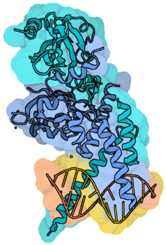
Figure 2.
Schematic representation of the canonical AhR pathway. In the cytoplasm, inactive AhR is bound to Hsp90, AIP, p23, and c-Src, forming a complex that keeps AhR inactive and prevents its degradation. When an agonist binds, AhR changes its conformation, releasing c-Src and AIP, and reveals its NLS, enabling AhR to move between the nucleus and cytoplasm.
Figure 2.
Schematic representation of the canonical AhR pathway. In the cytoplasm, inactive AhR is bound to Hsp90, AIP, p23, and c-Src, forming a complex that keeps AhR inactive and prevents its degradation. When an agonist binds, AhR changes its conformation, releasing c-Src and AIP, and reveals its NLS, enabling AhR to move between the nucleus and cytoplasm.
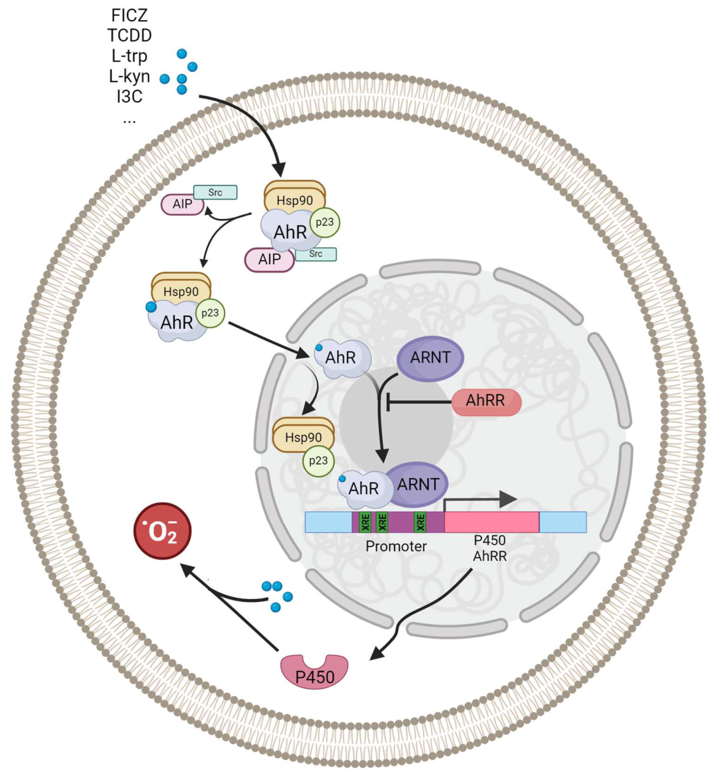
Figure 3.
The structure of AhR classical ligands: benzo(a)pyrene, anthracene, 3-methylcholanthrene (3MC), polychlorinated dibenzo-p-dioxins (PCDDs), polychlorinated dibenzofurans (PCDFs), polychlorinated biphenyls (PCBs).
Figure 3.
The structure of AhR classical ligands: benzo(a)pyrene, anthracene, 3-methylcholanthrene (3MC), polychlorinated dibenzo-p-dioxins (PCDDs), polychlorinated dibenzofurans (PCDFs), polychlorinated biphenyls (PCBs).
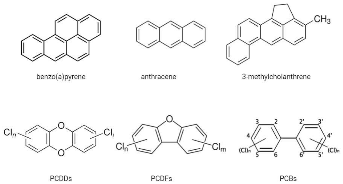
Figure 4.
The role of AhR in the gut. AhR ligands from food are crucial for maintaining IELs like TCRγδ and TCRαβ+ CD8αα+ cells and ILCs, especially ILC3. AhR also controls the regeneration of IECs and crypt stem cell proliferation. AhR deficiency results in the loss of ILC3, IELs, IL-22 depletion, impaired crypt stem cell proliferation, and gut microbiota imbalance.
Figure 4.
The role of AhR in the gut. AhR ligands from food are crucial for maintaining IELs like TCRγδ and TCRαβ+ CD8αα+ cells and ILCs, especially ILC3. AhR also controls the regeneration of IECs and crypt stem cell proliferation. AhR deficiency results in the loss of ILC3, IELs, IL-22 depletion, impaired crypt stem cell proliferation, and gut microbiota imbalance.
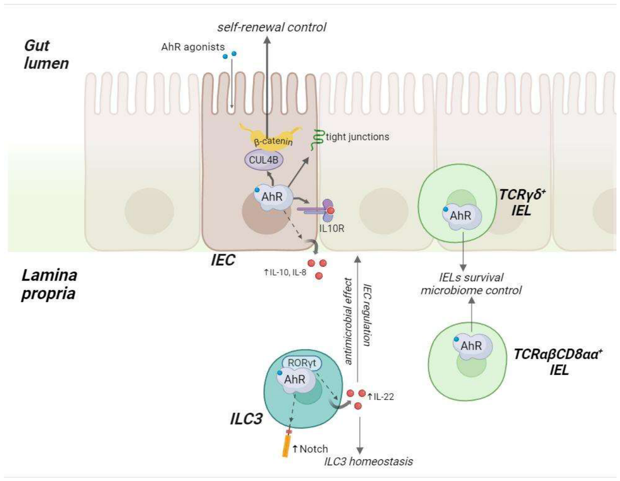
Figure 5.
The role of AhR in the skin. Activation of AhR stimulates keratinocyte differentiation by regulating genes for involucrin, loricrin, and filaggrin. AhR also promotes TGF-β production by fibroblasts, aiding keratinocyte migration and wound healing. Additionally, AhR maintains immune surveillance in the skin, supporting populations of various dendritic cells and T cells, and regulates inflammation through Th17 cells. It may also influence mast cell activity.
Figure 5.
The role of AhR in the skin. Activation of AhR stimulates keratinocyte differentiation by regulating genes for involucrin, loricrin, and filaggrin. AhR also promotes TGF-β production by fibroblasts, aiding keratinocyte migration and wound healing. Additionally, AhR maintains immune surveillance in the skin, supporting populations of various dendritic cells and T cells, and regulates inflammation through Th17 cells. It may also influence mast cell activity.
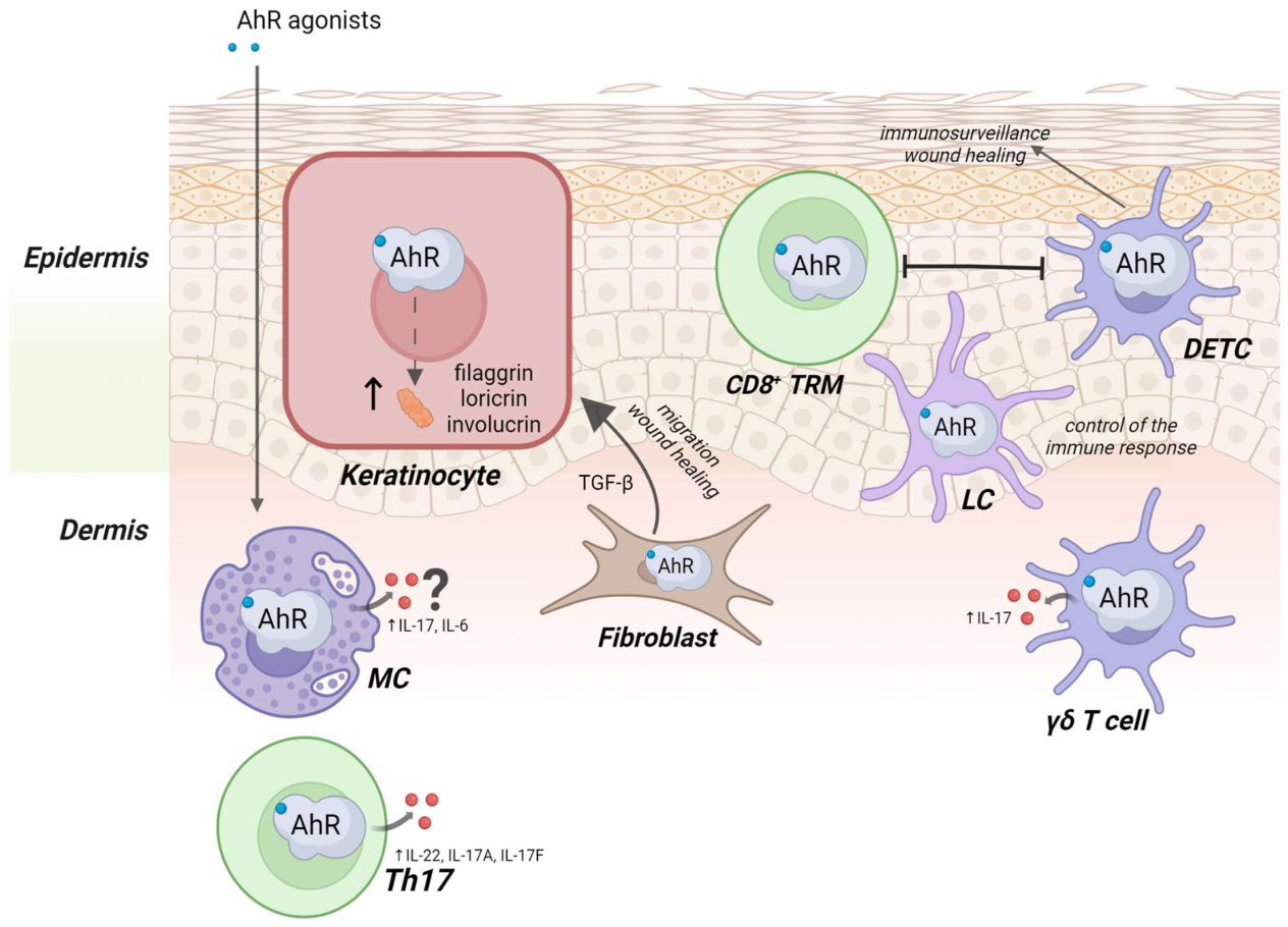
Disclaimer/Publisher’s Note: The statements, opinions and data contained in all publications are solely those of the individual author(s) and contributor(s) and not of MDPI and/or the editor(s). MDPI and/or the editor(s) disclaim responsibility for any injury to people or property resulting from any ideas, methods, instructions or products referred to in the content. |
© 2026 by the authors. Licensee MDPI, Basel, Switzerland. This article is an open access article distributed under the terms and conditions of the Creative Commons Attribution (CC BY) license (http://creativecommons.org/licenses/by/4.0/).
Copyright: This open access article is published under a Creative Commons CC BY 4.0 license, which permit the free download, distribution, and reuse, provided that the author and preprint are cited in any reuse.