
Submitted:
03 November 2026
Posted:
04 November 2026
You are already at the latest version
Abstract
Many Pseudomonas species produce pyoverdines, fluorescent siderophores that comprise a conserved dihydroxyquinoline chromophore, a strain-specific peptide chain, and a small acyl side chain. Their exceptional affinity for Fe(III), and the more-recent discovery that they also bind some other metals, has prompted diverse biotechnological applications. These applications span iron biofertilisation and biocontrol, bioremediation and metal scavenging, infection imaging, Trojan horse antibiotic delivery, direct antibacterial activity, and tumour targeting. To support these and other applications, we establish a robust catalogue of structurally distinct pyoverdines, consolidating published lists with manual literature searches and resolving duplicate and conflicting structural assignments. We identify 83 pyoverdines with discrete peptide chains, ranging from 6 to 14 residues (median 8), together with five chromophore variants and six acyl side chains. Each peptide contains two oxidatively modified iron-binding residues, most often ornithine-derived hydroxamates, although 26.3% are threo-hydroxylated amino acids. Peptide composition is strongly biased towards small and polar residues, with just a few amino acid families accounting for an overwhelming majority of residues, and with L-stereochemistry slightly favoured (60.8%). We conclude that pyoverdines are tightly constrained in chelation chemistry yet highly plastic in recognition, and hope that our catalogue will provide a useful parts list for engineering tailored products.
Keywords:
pyoverdine
; non-ribosomal peptide
; biotechnology
; siderophore
; Pseudomonas
; iron chelation
; NRPS
; bioremediation
; metal scavenging
; Trojan horse antibiotic
1. Introduction
The pyoverdines are an exceptionally diverse family of fluorescent, yellow-green siderophores that are produced by Pseudomonas species, most notably P. aeruginosa, P. fluorescens, and P. putida. Each pyoverdine molecule is comprised of three parts: a conserved dihydroxyquinoline chromophore that gives the molecule its characteristic fluorescence, a strain-specific peptide chain of 6 to 14 amino acids, and a small acyl side chain (Figure 1). Production of these molecules demands a substantial genetic investment. For example, in P. aeruginosa PAO1, at least 14 genes are required for pyoverdine biosynthesis [1]. Four of these, encoding multimodular non-ribosomal peptide synthetases (NRPSs) that govern synthesis of the chromophore and peptide chain, alone span over 40 kb of chromosomal DNA [2]. The biosynthetic genes are in turn accompanied by numerous other genes that govern pyoverdine secretion, uptake and regulation [3]. Across the genus, these genes are typically dispersed among multiple genomic loci [4].
The pyoverdine chromophore provides a bidentate catecholate group, and the peptide two further bidentate groups, most commonly hydroxamates derived from modified ornithine residues (although β-hydroxy acid or α-hydroxycarboxylate groups occur in some pyoverdines), that together bind Fe³⁺ with exceptionally high affinity [3]. This enables pyoverdine-producing pseudomonads to effectively scavenge iron, an essential nutrient that is scarce in most environments and in host tissues because it exists largely as insoluble Fe(III) hydroxides or is tightly bound by host proteins [5]. Once iron-loaded, pyoverdine is recognized by outer-membrane receptors, e.g. P. aeruginosa FpvA or FpvB, and taken back into the cell [6,7].
Although evolved for iron transport, the powerful and versatile chelating properties of pyoverdines offer a wide range of potential applications in biotechnology, sometimes extending beyond binding of iron. Here, we explore these applications and more rigorously define the chemical diversity of known pyoverdines to support future investigations into pyoverdine biotechnology.
2. Uses of Pyoverdines in Biotechnology
2.1. Pyoverdines and Agriculture
The powerful iron-chelating capacity of pyoverdines has been directly useful in several environmental biotechnology applications. One of the most intuitive has been evaluation of pyoverdines as environmentally friendly alternatives to synthetic iron chelates, to provide a bioavailable source of iron to crops grown in alkaline soils. Under such conditions most soil Fe(III) forms insoluble oxides, oxyhydroxides, or hydroxides [8]. Although synthetic chelates such as Fe ethylendiaminetetraacetic acid (EDTA) or Fe ethylenediamine-N,N′-bis(2-hydroxyphenylacetic) acid (EDDHA) are widely used to provide a soluble source of iron, these are poorly retained by soil and can also exert detrimental effects on soil chemistry [9]. In contrast, pyoverdine from Pseudomonas monsensis RMC4 has been shown to mobilize iron effectively to plants with fewer adverse effects [10,11], offering promise as a more effective iron fertilizer if it can be produced economically at scale.
Pyoverdines have also been directly implicated in the biocontrol activities of a range of fluorescent pseudomonads, including suppression of Pythium ultimum fungal root rot by P. rhodesiae HAI-0804 [12], P. chlororaphis YL-1 mediated biocontrol of rice bacterial leaf blight caused by the Gram-negative bacterium Xanthomonas oryzae pv. oryzae [13], and P. fluorescens 2112 mediated suppression of red pepper blight caused by the oomycete Phytophthora capsica [14]. The mechanistic basis for pyoverdine-mediated biocontrol is presumed to be depriving the competing organism of an essential iron supply, but the efficacy of this can vary greatly from strain to strain [15], and it is additionally clear that pyoverdine production is not a universally dominant biocontrol mechanism. For example, a pyoverdine-null mutant of Pseudomonas fluorescens PICF7 retained the ability to suppress Verticillium wilt resulting from infection of olive tree roots by the fungus Verticillium dahlia [16], while a similarly pyoverdine-deficient mutant of Pseudomonas orientalis F9 was still able to prevent fire blight in apple flowers exposed to the Gram-negative pathogen Erwinia amylovora [17].
2.2. Bioremediation Applications of Pyoverdines
The high affinity of pyoverdines for iron has been explored for bioremediation of asbestos waste. It is well known that asbestos can induce mesothelioma and other lung disorders, but less common knowledge that the toxicity of asbestos correlates with its iron content, owing to generation of reactive oxygen species via Fenton chemistry [18]. In seeking to prevent this, pyoverdines from a wide range of fluorescent pseudomonads were found to be significantly more effective at extracting iron from asbestos than the synthetic chelator EDTA [18,19]. Building on this, a strain of P. aeruginosa engineered for constitutive pyoverdine production was shown to be highly effective in both weathering and detoxifying asbestos [20]. The authors concluded that if the effect can be recapitulated in a non-pathogenic species such as P. putida, then this could pave the way to a viable bioremediation process.
Recently, Semenec et al [21] demonstrated that pyoverdine can also act via indirect mechanisms to promote biodegradation of recalcitrant pollutants. They showed that polyurethane degradation by Pseudomonas protegens Pf-5 was substantially enhanced in a mutant where the sensor kinase gene gacS had been disrupted by transposon insertion. Transcriptomics analysis suggested that this was due to a dysregulation of iron and sulphur homeostasis, and this was linked to a marked upregulation of pyoverdine production. The authors propose that the consequent increase in intracellular iron levels generated markedly higher levels of reactive oxygen species via Fenton chemistry, which in turn promoted the oxidative degradation of polyurethane [21].
Of great interest, pyoverdines have also been applied to recover metals other than iron, both for the removal of pollutants from contaminated sites, and for scavenging of valuable minerals. A recent review considers in detail the different complexes that a pyoverdine from P. aeruginosa can form with Ga(III), Mn(III), Cu(II), Ni(II) and Cd(II) (Andrejević et al [22]), and relevance to phytoremediation has been demonstrated for each of the divalent ions in that list. In a hydroponic system for remediation of cadmium and nickel from smectite (a mixture of clay minerals extracted from bentonite mine tailings), metal mobilisation was enhanced both by direct addition of pyoverdine and by inoculation of P. aeruginosa to enable biofilm formation around the roots of hydroponically grown tomato plants [23]. In a similar fashion, sunflowers bioaugmented with P. putida in copper overloaded vineyards were capable of substantially greater Cu(II) removal than non-augmented sunflowers [24].
Pyoverdines have also been observed to mobilise some more exotic metals. In a study involving actinides associated with nuclear waste, pyoverdine produced by P. fluorescens CCUG 32456 (recovered from the Äspö Hard Rock Laboratory at a depth of 70 m underground) was found to bind U(VI), Cm(III) and Np(V)) with stability constants stronger than for EDTA [25]. A more recent study has shown that pyoverdines can also mobilise Pu(IV) [26]. This offers prospects for recovery of such metals, but poses concerns for the containment of nuclear waste buried underground. Perhaps more usefully, pyoverdine production by P. fluorescens (ATCC 17400) was found to promote phytoextraction of cesium from illite clay by red clover, offering prospects for improved cleanup of sites contaminated with radioactive 137Cs after nuclear plant accidents [27,28]. In those studies, the impact of pyoverdine was attributed primarily to enhanced weathering of the illite via iron extraction, rather than direct transport of cesium [27]. Finally, in a recent example, pyoverdine purified from a marine Pseudomonas sp. ASA235, was found to promote leaching of the rare earth metal lanthanum (and to a lesser extent, neodymium and praseodymium) from the anodes of spent nickel–metal hydride batteries [29].
2.3. Biomedical Applications of Pyoverdines
The innate fluorescence of pyoverdine has enabled direct detection of P. aeruginosa in a burn wound model, as a step toward point-of-care assays that convey more immediate information on infection than overnight culture-based methods [30]. However, such approaches are yet to achieve the sensitivity of earlier electrochemical methods for pyoverdine detection [31,32]. An innovative alternative strategy that demonstrated high-level sensitivity and specificity, albeit requiring significant infrastructure, employed 68Ga-associated pyoverdine [33]. That pyoverdine can tightly bind gallium was already well-established, with gallium having previously been employed as a less redox active mimetic of iron to solve the structures of several pyoverdines in solution [34,35,36]. Here, when P. aeruginosa pyoverdine was radiolabelled with 68Ga and injected retro-orbitally into rats that had undergone intramuscular injection with P. aeruginosa or other bacteria, positron emission tomography revealed rapid clearance of signal from the infection site in all cases other than the P. aeruginosa infected wounds [33]. More recently, the approach was advanced by creating a mixed “cocktail” of pyoverdines, each loaded with 68Ga, to detect a broader range of P. aeruginosa strains [37].
Gallium additionally has direct antibacterial properties. Based on the near-identical ionic radius shared by Ga(III) and Fe(III) and because Ga(III) cannot be reduced, it has been employed in “Trojan horse” strategies to disrupt bacterial iron metabolism. In P. aeruginosa, gallium was observed to downregulate pvdS, the transcriptional regulator governing pyoverdine synthesis, and the consequent repression of pyoverdine production was considered to make a substantial contribution to gallium’s antibacterial effect [38]. Additional antibacterial strategies targeting pyoverdine synthesis have since been explored; these were recently reviewed in detail by Dell’Anno et al [39] and are not considered further here.
In an interesting twist, pyoverdine has itself been employed as the equine vehicle for Trojan horse antibacterial strategies. Early success was enjoyed by Kinzel et al [40], who reported that conjugation of ampicillin with each of the pyoverdines produced by two different P. aeruginosa strains resulted in strain-specific antibacterial activity, whereas ampicillin administered on its own was ineffective. Subsequent attempts utilising a variety of conjugated antibiotics proved less effective, which has been attributed to conjugate stability, uptake, and/or cellular localisation being sub-optimal [39]. Indeed, it has recently been confirmed that the site of antibiotic attachment to pyoverdine can impact the ability of the conjugate to be taken up by FpvB [41]. However, a route to modulating this has been offered by another elegant recent study. Puja et al [42] successfully engineered the first A domain of the pyoverdine NRPS pvdD from P. aeruginosa PAO1 to enable incorporation of an azido-L-homoalanine into pyoverdine, in place of the penultimate L-threonine residue. They further showed that the azide moiety enables functionalised antibiotics to be attached to their modified pyoverdine via click chemistry, and subsequently internalised by P. aeruginosa PAO1. Although the functionalised oxazolidinone antibiotic eperezolid used to exemplify their approach did not exhibit an antibacterial effect, their ‘clickable’ pyoverdine represents an exciting and versatile tool that could enable an extensive study of Trojan horse antibiotic strategies to combat P. aeruginosa, e.g., modifying not only pyoverdine’s antibiotic partner but also the linker that joins them [42].
A further twist is that various pyoverdines have themselves demonstrated antibiotic activity against ESKAPE pathogens, both when administered as sole agents in Galleria mellonella infection models [43] and in combination with the cephalosporin ceftazidime in culture-based studies [44]. In the latter case, pyoverdine was found to suppress E. coli ceftazidime resistance phenotypes mediated by the β-lactamase gene blaTEM-1, although it did not prevent new ceftazidime resistance phenotypes from arising via spontaneous mutations [44].
In one final biomedical application, pyoverdine from a strain of P. fluorescens was evaluated for use in an innovative anti-cancer therapy that employs superparamagnetic iron oxide nanoparticles. It was already known that these can be chelated by pyoverdine and the complex subsequently detected via magnetic resonance imaging (MRI) [45]. Nosrati et al [46] demonstrated that an anti-MUC1 aptamer, which targets mucin that is aberrantly displayed on the surface of cells in many types of cancer, could be conjugated to the exposed carboxyl group on the pyoverdine chromophore. They further showed that the aptamer could be loaded with doxorubicin, as a payload that was then preferentially delivered to tumours implanted in BALB/c mice, as confirmed by both MRI and fluorescence imaging [46]. However, one possible caution around advancing this potential therapy is that pyoverdines have also been shown to promote the progression of many cancers by lowering their susceptibility to ferroptosis, a cell death pathway that cancer cells exhibiting dysregulated iron metabolism are particularly susceptible to [47].
3. The Diversity of Pyoverdines
3.1. Curating a List of Unique Pyoverdines
Given the wealth of potential applications for pyoverdines in biotechnology, we considered it important to compile an accurate catalogue of the diversity of structures that have been identified to date (Supplementary data). We reason that this can serve not only as a foundation for selecting the most appropriate natural molecules to explore a desired application, but also as a source of raw materials to rationally engineer novel pyoverdines with expanded functionalities, via substitution of one or more A domains in a target pyoverdine NRPS assembly line as demonstrated by Calcott et al [48] and Puja et al [42].
In the process of compiling this list, 83 pyoverdines were identified that possess a discrete peptide chain relative to one another, based on combining existing lists [49,50,51] and manual searches. Duplicates were removed, with the earliest most complete structure kept as a representative pyoverdine. This included instances in which some publications describe more stereochemistry than others that have ambiguous or incomplete stereochemistry determinations. An example of this is P. fluorescens CHAO that has fewer stereocentres determined than Pseudomonas protegens Pf-5 [52,53] and therefore only the latter pyoverdine was retained.
In our list of discrete pyoverdines, there were two examples where the peptide chains were identical except for differences in their solved stereochemistry. The first instance are the pyoverdines from Pseudomonas aureofaciens and Pseudomonas sp. SXM-1, for which two Thr residues were ascribed as being D-Thr-L-Thr and L-Thr-D-Thr, respectively. For P. aureofaciens, stereochemistry was originally confirmed by partial hydrolysis followed by dansylation and complete hydrolysis before analysis using TLC on chiral plates [54]. For Pseudomonas sp. SXM-1, stereochemistry was determined by comparison to the biosynthetic gene cluster [55]. The second instance is P. fluorescens SBW25 and P. fluorescens A506, which differ in the stereochemistry of the final N⁵-formyl-N⁵-hydroxy-ornithine (Fo-OH-Orn) residue [52]. This difference has been confirmed by examination of the biosynthetic genes and the observation of differences in pyoverdine cross-feeding experiments [52]. In each case, there is compelling evidence to suggest these examples are genuinely different pyoverdines, so they were recorded in our list as four unique pyoverdines.
The pyoverdines from Azotobacter vinelandii strain D [56,57] and A. vinelandii ATCC 12837 [58] have azotobactin chromophores, which are identical to the pyoverdine chromophore except that they contain an extra urea ring instead of the variable acyl chain. While these azotobactins are often considered separate from pyoverdines, the distinction is not entirely clear, as azotobactin chromophores have been detected as side-products from the pyoverdine producing strains Pseudomonas sp. taiwanensis VLB120 [59], P. fluorescens CCM 2798 [60] and P. fluorescens G173 [61]. Due to this, they have each been included in our list of 83 discrete pyoverdines for completeness.
3.2. Chemical Diversity of Pyoverdines
In terms of the acyl side chain, the diversity in pyoverdines is relatively low. This small acyl unit can be succinamide, succinic acid, malic acid, malamide, L-glutamic acid, or alpha-keto-glutaric acid. Succinamide and succinic acid are the most commonly described. However, this likely misrepresents occurrence due to a trend to only describe the succinyl variants [50,62]. In some studies, up to four pyoverdines differing in the acyl chain have been described from a single strain [63,64] with some evidence that there is no difference in biological function between these isoforms [52].
Five chromophore variants have been described (Figure 2). The chromophores are typically described as pyoverdine with rarer instances of isopyoverdines,[65,66,67,68] azotobactins [58,59,60,61,68], succinopyoverdines [61,69] or dihydropyoverdines [69,70]. The non-pyoverdine chromophores are often described as a mixture of products among which a pyoverdine containing the standard pyoverdine chromophore is also present. However, isopyoverdines are always described as the only chromophore when present [65,66,67,68], dihydropyoverdine has been described as the sole pyoverdine once [70], and there are two descriptions of strains producing solely azotobactins [56,58]. The pyoverdine from A. vinelandii ATCC 12837 was originally called azoverdin and later found to belong to the class of isopyoverdines [68]. Isopyoverdines contain a different location for the linkage of the peptide chain due to cyclic 2,4-diaminobutyric acid (Dab) rotating about the peptide backbone prior to cyclisation during chromophore formation.
For the 83 unique pyoverdine peptide chains in our list, the chain length ranges from 6 to 14 amino acids, with a median length of 8 amino acids (IQR 7-10), indicating a skewed distribution towards shorter peptide chains. Every peptide chain contains two oxidatively modified amino acids that are involved in iron-binding. These modifications can be classified into two groups, either hydroxylation at the N5 residue of ornithine (Orn) or β-threo hydroxylation of Asp, His or Asn (Supplementary Figure S1). The N5-hydroxylation of Orn is always combined with further addition of an N5-carbonyl containing moiety or cyclisation (allowing formation of a hydroxamate with the C-terminal carbonyl). These Orn-derivatives are the most common iron binding amino acids, such that pyoverdines are generally described as containing two hydroxamate groups. However, there are four pyoverdines that contain no hydroxamate groups with about a quarter (26.3%) of the iron-binding residues across all pyoverdines being threo hydroxylated amino acids (Figure 3). While one pyoverdine from P. taiwanensis VLB120 has been tentatively identified as bearing a peptide that contains three potential chelating groups, namely, OH-Asn, Ac-OH-Orn and cyclic OH-Orn, the authors concluded that further structural elucidation was required to confirm the presence of OH-Asn [59]. Given the prevalence of hexadentate chelators that form a stable octahedral geometry with iron [71], it seems possible that further characterisation may revise the initial structure.
In terms of the overall composition of amino acids in the peptide chain, there is a strong preference for small or polar residues and Orn-derivatives. Across all peptide chains, there are 693 amino acids and the percentage of each is shown in Figure 4a (abbreviations and structures defined in Supplementary Figure S1 to S3). Ignoring chirality, Ser, Thr and the rarer Ser analogues Fo-Ser, and Hse are the most common and make up 33.0% of the total amino acids. Orn and its N5-modified derivatives Fo-OH-Orn, Ac-OH-Orn, Bu-OH-Orn, and Cit account for 20.3% of the total. The next most common, small aliphatic amino acids Gly, Ala and, to a lesser extent, Val (only 2 occurrences), make up 18.3% of the total amino acids. The other relatively abundant amino acids are Lys and εLys (amide bond formed via the Lys sidechain ε-amine) at 9.8% as well as Asp and OH-Asp at 9.7%. Altogether, these amino acids make up 91.1% of the total amino acids in pyoverdine peptide chains. There are also occurrences of the nonproteinogenic Dab plus proteinogenic amino acids that have charged or polar uncharged sidechains, including some hydroxylation of the sidechain β-carbon: Gln, Arg, Glu, Asn, OH-His and OH-Asn. With the exception of Pro being detected in a minor side product from P. taiwanensis VLB120 [59], when considering proteinogenic amino acids, there is a noticeable absence of larger aliphatic (Cys, Ile, Leu, Pro and Met) as well as the uncharged aromatic (Phe, Trp and Tyr) amino acids. The trend for small and polar residues has been proposed to be important to increase solubility in water and ensure interaction with the pyoverdine receptors [51].
In addition to variations in the abundance of each amino acid sidechain, many amino acids within the peptide chain also differ in their stereochemistry (Figure 4b). Notably, as Thr contains two stereocentres, the D-form is always present as D-allothreonine due to the downstream epimerisation domain acting only on the α-position (Supplementary Figure S4). Excluding Gly due to being achiral, there is a modest preference towards L-amino acids (234 out of 385; 60.8%), representing a statistically significant deviation from a 1:1 ratio of L- to D- amino acids (two-sided binomial test, p = 0.000027). Relative to the overall distribution, Ser, OH-Asp and Asp showed significant deviations with Ser and OH-Asp being D-enriched and Asp being L-enriched (Figure 4b).
3.3. Pyoverdine Classification
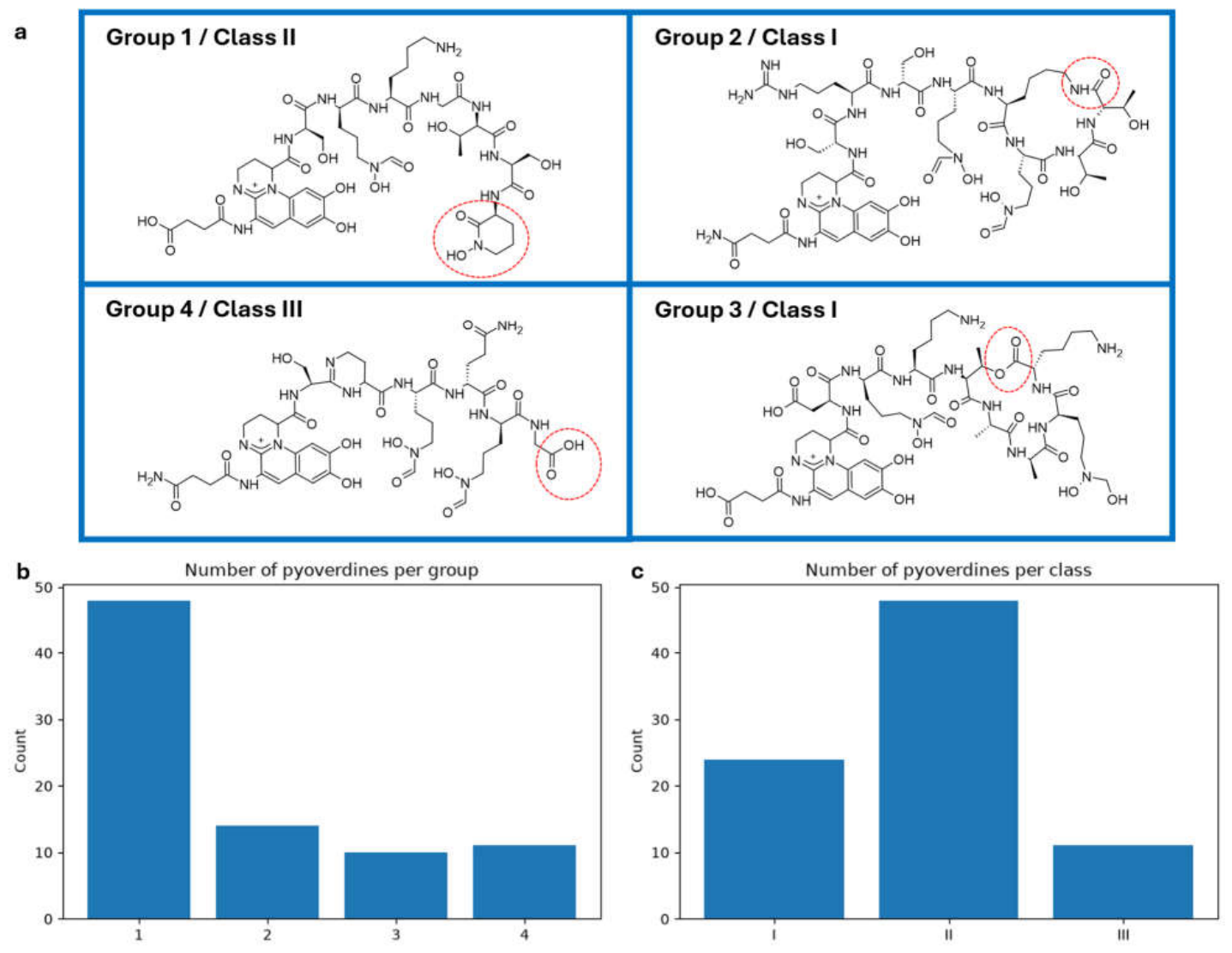
There are two main ways in which pyoverdines have been characterised by the peptide sidechain (Figure 5a). These depend on chemical features of peptide release during biosynthesis without further rationale being provided for groupings [51,72]. The most recent classification, adapted here to include more recently discovered pyoverdines that possess a terminal Hse or cyclisation of the C-terminus with a side-chain of Orn, splits pyoverdines into four groups [72]. These are, group 1 for linear pyoverdines ending in a terminal cyclic Orn or cyclic Hse residue, group 2 for cyclic pyoverdines having an amide bond between the C-terminus and the side-chain of an in-chain amino acid (Lys or Orn), group 3 for cyclic pyoverdines having an ester bond between the C-terminus and the side-chain of an in-chain amino acid (Ser or Thr), and group 4 for linear pyoverdines ending with a free carboxyl group (however, some or all of this last group may be the hydrolysis products of the labile ester bond of group 3) [72]. Group 1 is the most prevalent, representing over half of all pyoverdines (48 out of 83), and there is a relatively even split between groups 2 to 4 (Figure 5b). The second characterisation splits pyoverdines into classes. In this system, class I combines the aforementioned groups 2 and 3, class II is equivalent to group 1 and class III is equivalent to group 4 (Figure 5c) [51]. In this classification, class II remains the most abundant type of pyoverdine.
4. Conclusions
Pyoverdines are highly diverse with at least 83 distinct pyoverdines known. The diversity is almost entirely confined to the peptide chain and acyl side chain, whereas the chromophore is near-invariant, always presenting a catecholate group that contributes two iron-binding residues. These features are consistent with a molecule that is tightly constrained in its chelation chemistry yet highly plastic in the features that govern receptor recognition. The high variation likely represents selection for strain-selective uptake to avoid competition between strains, with a strong bias towards small polar amino acids promoting solubility. The combination of a highly conserved metal-binding core displayed on a variable and intrinsically fluorescent scaffold also offers a breadth of biotechnological potential that extends well beyond iron. Demonstrated applications now span iron biofertilisation and biocontrol in agriculture; bioremediation of asbestos, plastics, and toxic, radioactive or commercially valuable metals; and biomedical uses as diagnostic imaging agents, ‘Trojan horse’ vectors for antibiotics, antibacterials in their own right, and tumour-targeting ligands. However, more widespread applications have been tempered by practical constraints, e.g. economical production at scale, transfer of high-level production from opportunistic pathogens into safe chassis species such as P. putida, and, for conjugate strategies, recognition limitations imposed by Fpv-mediated uptake. Encouragingly, the demonstration that pyoverdine NRPS A domains can be rationally substituted [41] or engineered to accept non-canonical substrates [35] means the 83 known pyoverdines we described here can be viewed as a parts list to support the design of tailored molecules.
Supplementary Materials
The following supporting information can be downloaded at the website of this paper posted on Preprints.org, Supplementary Figure S1–S4. Supplementary Data S1.
Author Contributions
Conceptualization, M.C. and D.A.; data curation, M.C.; writing—original draft preparation, M.C. and D.A.; writing—review and editing, M.C. and D.A.; visualization, M.C.;. All authors have read and agreed to the published version of the manuscript.
Funding
This research was funded by Health Research Council of New Zealand, grant number 23/188/A.
Data Availability Statement
Data analysis is available from: https://github.com/MarkCalcott/pyoverdine_review.
Conflicts of Interest
The authors declare no conflicts of interest.
Abbreviations
The following abbreviations are used in this manuscript:
| Ac-OH-Orn | N⁵-acetyl-N⁵-hydroxy-ornithine |
| Bu-OH-Orn | N⁵-hydroxybutyryl-N⁵-hydroxy-ornithine |
| Cit | citrulline |
| Dab | 2,4-diaminobutyric acid |
| Fo-OH-Orn | N⁵-formyl-N⁵-hydroxy-ornithine |
| Fo-Ser | O-formylserine |
| Hse | homoserine |
| MDPI | Multidisciplinary Digital Publishing Institute |
| OH-Asn | threo-β-hydroxyasparagine |
| OH-Asp | threo-β-hydroxyaspartic acid |
| OH-His | threo-β-hydroxyhitidine |
| Orn | ornithine |
| εLys | Lys in which the amide bond forms with the ε-NH2 |
References
- Lamont, I.L.; Martin, L.W. Identification and characterization of novel pyoverdine synthesis genes in Pseudomonas aeruginosa; Microbiology (Reading), 2003; Volume 149, Pt 4, pp. 833–842. [Google Scholar]
- Stover, C.K.; et al. Complet. Genome Seq. Pseudomonas Aeruginosa PAO1, An. Opportunistic Pathog. Nat. 2000, 406(6799), 959–64.
- Visca, P.; Imperi, F.; Lamont, I.L. Pyoverdine siderophores: from biogenesis to biosignificance Trends Microbiol 2007, 15(1), 22–30.
- Gu, S. Feature sequence-based genome mining uncovers the hidden diversity of bacterial siderophore pathways eLife 2024, 13, RP96719.
- Hood, M.I.; Skaar, E.P. Nutr. Immun. Transit. Met. At. Pathog.-Host Interface Nat. Rev. Microbiol. 2012, 10(8), 525–37.
- Meyer, J.-M.; Stintzi, A.; Poole, K. The ferripyoverdine receptor FpvA of Pseudomonas aeruginosa PAO1 recognizes the ferripyoverdines of P. aeruginosa PAO1 and P. fluorescens ATCC 13525 FEMS Microbiology Letters 1999, 170(1), 145–150.
- Ghysels, B.; et al. FpvB, an alternative type I ferripyoverdine receptor of Pseudomonas aeruginosa; Microbiology (Reading), 2004; Volume 150, Pt 6, pp. 1671–1680. [Google Scholar]
- Lucena, J.J. Synthetic Iron Chelates to Correct Iron Deficiency in Plants, in Iron Nutrition in Plants and Rhizospheric Microorganisms; Barton, L.L., Abadia, J., Eds.; Springer Netherlands: Dordrecht, 2006; pp. 103–128. [Google Scholar]
- Nowack, B. Environ. Chem. Aminopolycarboxylate Chelating Agents Environ. Sci. Technol. 2002, 36(19), 4009–4016.
- Lozano-González, J.M.; Lucena, J.J.; López-Rayo, S. A pyoverdine-based iron biochelate from bacterial secretions as an effective fertilizer under alkaline conditions Front Plant Sci 2025, 16, 1675837.
- Lozano-González, J.M. Evaluation of Siderophores Generated by Pseudomonas Bacteria and Their Possible Application as Fe Biofertilizers Plants 2023, 12(23).
- Takeuchi, K. Pseudomonas rhodesiae HAI-0804 suppresses Pythium damping off and root rot in cucumber by its efficient root colonization promoted by amendment with glutamate; Frontiers in Microbiology, 2024; pp. 15–2024. [Google Scholar]
- Liu, Y.; et al. Pyoverdines Are Essential for the Antibacterial Activity of Pseudomonas chlororaphis YL-1 under Low-Iron Conditions Appl Environ Microbiol 2021, 87(7).
- Eun-Tag, L.; et al. Pyoverdin2112 of Pseudomonas fluorescens 2112 Inhibits Phytophthora capsici, a Red-Pepper Blight-Causing Fungus Journal of Microbiology and Biotechnology 2003, 13(3), 415–421.
- Höfte, M.; Altier, N. Fluorescent pseudomonads as biocontrol agents for sustainable agricultural systems Research in Microbiology 2010, 161(6), 464–471.
- Maldonado-González, M.M. Endophytic Colon. Biocontrol Perform. Pseudomonas Fluoresc. PICF7 Olive (Olea europaea L.) Are Determ. Neither By Pyoverdine Prod. Nor. Swim. Motil. Environ. Microbiol. 2015, 17(9), 3139–53. [CrossRef]
- Santos Kron, A.; et al. Pseudomonas orientalis F9 Pyoverdine, Safracin, and Phenazine Mutants Remain Effective Antagonists against Erwinia amylovora in Apple Flowers Appl Environ Microbiol 2020, 86(8).
- David, S.R.; et al. Iron removal from raw asbestos by siderophores-producing Pseudomonas J Hazard Mater 2020, 385, 121563.
- David, S.R.; et al. Efficiency of pyoverdines in iron removal from flocking asbestos waste: An innovative bacterial bioremediation strategy J Hazard Mater 2020, 394, 122532.
- Lemare, M.; et al. Eng. Siderophore Prod. Pseudomonas To Improv. Asbestos Weather. Microb. Biotechnol. 2022, 15(9), 2351–2363.
- Semenec, L. Molecular insights into polyurethane biodegradation in Pseudomonas protegens mBio 2026, 17(2), e0354425.
- Andrejević, T.P. Metal complexes with valuable biomolecules produced by Pseudomonas aeruginosa: a review of the coordination properties of pyocyanin, pyochelin and pyoverdines Dalton Trans 2023, 52(14), 4276–4289.
- Ferret, C.; et al. Effect of pyoverdine supply on cadmium and nickel complexation and phytoavailability in hydroponics Environmental Science and Pollution Research 2015, 22(3), 2106–2116.
- Emmeline, D.; et al. Effect of Pseudomonas putida-producing pyoverdine on copper uptake by Helianthus annuus cultivated on vineyard soils Sci Total Environ 2022, 152113.
- Moll, H.; et al. The mobilization of actinides by microbial ligands taking into consideration the final storage of nuclear waste: interactions of selected actinides U (VI), Cm (III), and Np (V) with pyoverdins secreted by Pseudomonas fluorescens and related model compounds 2009. [CrossRef]
- Ma, T. Influence of pyoverdine on plutonium-239 speciation evolution in contaminated soils J Environ Radioact 2026, 108083.
- Péron, O.; et al. Effect of the bacterial pyoverdine siderophore on the phytoextraction of cesium from illite Environmental Chemistry Letters 2019, 17(1), 521–526.
- Hazotte, A.; et al. Effect of Pseudomonas fluorescens and pyoverdine on the phytoextraction of cesium by red clover in soil pots and hydroponics Environ Sci Pollut Res Int 2018, 25(21), 20680–20690.
- Hegazy, A.S. Bioleaching of lanthanum from nickel metal hydride dry battery using siderophores produced by Pseudomonas sp World J Microbiol Biotechnol 2025, 41(2), 39.
- Cueva, A.R.; et al. Pyoverdine Assay for Rapid and Early Detection of Pseudomonas aeruginosa in Burn Wounds ACS Appl Bio Mater 2020, 3(8), 5350–5356.
- Ciui, B.; et al. Finger-Based Printed Sensors Integrated on a Glove for On-Site Screening Of Pseudomonas aeruginosa Virulence Factors Analytical Chemistry 2018, 90(12), 7761–7768.
- Gandouzi, I.; et al. Sensitive detection of pyoverdine with an electrochemical sensor based on electrochemically generated graphene functionalized with gold nanoparticles Bioelectrochemistry 2018, 120, 94–103.
- Petrik, M.; et al. Imaging of Pseudomonas aeruginosa infection with Ga-68 labelled pyoverdine for positron emission tomography Sci Rep 2018, 8(1), 15698.
- Mohn, G.; et al. Solut. Struct. Pyoverdin GM-II Biochem. 1994, 33(10), 2843–2851.
- Atkinson, R.A. Bacterial iron transport: 1H NMR determination of the three-dimensional structure of the gallium complex of pyoverdin G4R, the peptidic siderophore of Pseudomonas putida G4R Biochemistry 1998, 37(45), 15965–73.
- Tzou, D.L.; et al. A low-temperature heteronuclear NMR study of two exchanging conformations of metal-bound pyoverdin PaA from Pseudomonas aeruginosa Biopolymers 2005, 79(3), 139–49.
- Krasulova, K.; et al. Targeted PET Imaging of Pseudomonas aeruginosa Using a [ (68) Ga]Ga-Pyoverdines Cocktail J. Nucl. Med. 2026. [CrossRef]
- Kaneko, Y.; et al. The transition metal gallium disrupts Pseudomonas aeruginosa iron metabolism and has antimicrobial and antibiofilm activity J Clin Invest 2007, 117(4), 877–88.
- Dell’Anno, F.; et al. Novel Insights on Pyoverdine: From Biosynthesis to Biotechnological Application International Journal of Molecular Sciences 2022, 23(19), 11507.
- Kinzel, O.; et al. The synthesis and antibacterial activity of two pyoverdin-ampicillin conjugates, entering Pseudomonas aeruginosa via the pyoverdin-mediated iron uptake pathway J Antibiot 1998, 51(5), 499–507.
- Revol-Tissot, J. Transport specificity of FpvA and FpvB for pyoverdine-antibiotic conjugates in Pseudomonas aeruginosa Bioorg Med Chem 2026, 118734.
- Puja, H.; et al. Biosynthesis of a clickable pyoverdine via in vivo enzyme engineering of an adenylation domain Microb Cell Fact 2024, 23(1), 207.
- Vollenweider, V. Antimicrobial activity of iron-depriving pyoverdines against human opportunistic pathogens Elife 2024. [CrossRef]
- Vollenweider, V.; Roncoroni, F.; Kümmerli, R. Pyoverdine-antibiotic combination treatment: its efficacy and effects on resistance evolution in Escherichia coli Microlife 2024, 5, uqae021.
- Górska, A.; Sloderbach, A.; Marszałł, M.P. Siderophore-drug complexes: potential medicinal applications of the 'Trojan horse' strategy Trends Pharmacol Sci 2014, 35(9), 442–9.
- Nosrati, R.; et al. Targeted SPION siderophore conjugate loaded with doxorubicin as a theranostic agent for imaging and treatment of colon carcinoma Sci Rep 2021, 11(1), 13065.
- Yeung, Y.W.S.; et al. Bacterial Iron Siderophore Drives Tumor Survival and Ferroptosis Resistance in a Biofilm-Tumor Spheroid Coculture Model Adv Sci (Weinh) 2024, 11(39), e2404467.
- Calcott, M.J.; Owen, J.G.; Ackerley, D.F. Efficient rational modification of non-ribosomal peptides by adenylation domain substitution Nature Communications 2020, 11(1).
- Gu, S. From sequence to molecules: Feature sequence-based genome mining uncovers the hidden diversity of bacterial siderophore pathways eLife 2024, 13, RP96719.
- Meyer, J.-M.; et al. Siderotyping of fluorescent Pseudomonas: molecular mass determination by mass spectrometry as a powerful pyoverdine siderotyping method BioMetals 2008, 21(3), 259–271.
- Cézard, C.; Farvacques, N.; Sonnet, P. Chem. Biol. Pyoverdines Pseudomonas Prim. Siderophores Curr. Med. Chem. 2015, 22(2), 165–86.
- Hartney, S.L.; et al. Ferric-pyoverdine recognition by Fpv outer membrane proteins of Pseudomonas protegens Pf-5 J Bacteriol 2013, 195(4), 765–76.
- Wong-Lun-Sang, S. Bacterial siderophores: Structure elucidation, 2D 1H and 13C NMR assignments of pyoverdins produced by Pseudomonas fluorescens CHAO Tetrahedron Letters 1996, 37(19), 3329–3332. [CrossRef]
- Beiderbeck, H.; et al. A New Pyoverdin from Pseudomonas aureofaciens Zeitschrift für Naturforschung C 1999, 54(1-2), 1–5. [CrossRef]
- Guo, W. Complete genome sequence of a marine-derived bacterium Pseudomonas sp. SXM-1 and characterization of its siderophore through antiSMASH analysis and with mass spectroscopic method Marine Genomics 2021, 55, 100802.
- Demange, P.; et al. Structure of azotobactin D, a siderophore of Azotobacter vinelandii strain D (CCM 289) Biochem. 1988, 27(8), 2745–2752.
- Page, W.J.; et al. Azotobacter vinelandii strains of disparate origin produce azotobactin siderophores with identical structures Biology of Metals 1991, 4(4), 217–222. [CrossRef]
- Schaffner, E.M.; et al. Structure Elucidation of Azotobactin 87, Isolated from Azotobacter vinelandii ATCC 12837*, ** Zeitschrift für Naturforschung C 1996, 51(3-4), 139–150.
- Scholz, K.; et al. Mass spectrometric characterization of siderophores produced by Pseudomonas taiwanensis VLB120 assisted by stable isotope labeling of nitrogen source BioMetals 2018, 31(5), 785–795.
- Demange, P. Bacterial siderophores: structures of pyoverdins Pt, siderophores of Pseudomonas tolaasii NCPPB 2192, and pyoverdins Pf, siderophores of Pseudomonas fluorescens CCM 2798. Identification of an unusual natural amino acid Biochemistry 1990, 29(50), 11041–11051.
- Fernández, D.U.; et al. The Pyoverdin of Pseudomonas fluorescens G173, a Novel Structural Type Accompanied by Unexpected Natural Derivatives of the Corresponding Ferribactin Zeitschrift für Naturforschung C 2003, 58(1-2), 1–10.
- Rehm, K. Rapid identification of pyoverdines of fluorescent Pseudomonas spp. by UHPLC-IM-MS Biometals 2023, 36(1), 19–34.
- Geisen, K.; Taraz, K.; Budzikiewicz, H. Neue Siderophore des Pyoverdin-Typs ausPseudomonas fluorescens Monatshefte für Chemie / Chemical Monthly 1992, 123(1), 151–178.
- Seinsche, D.; et al. Neue Pyoverdin-Siderophore aus Pseudomonas putida C Journal für Praktische Chemie/Chemiker-Zeitung 1993, 335(2), 157–168.
- Jacques, P.; et al. Structure and Characterization of Isopyoverdin from Pseudomonas putida BTP1 and Its Relation to the Biogenetic Pathway Leading to Pyoverdins Zeitschrift für Naturforschung C 1995, 50(9-10), 622–629.
- Sultana, R.; et al. An isopyoverdin from Pseudomonas putida CFML 90-44 Z Naturforsch C J Biosci 2001, 56(3-4), 303–7. [CrossRef]
- Sultana, R.; et al. An isopyoverdin from Pseudomonas putida CFML 90-331 Tetrahedron 2001, 57(6), 1019–1023.
- Wasielewski, E.; et al. The Three-Dimensional Structure of the Gallium Complex of Azoverdin, a Siderophore of Azomonas macrocytogenes ATCC 12334, Determined by NMR Using Residual Dipolar Coupling Constants Biochemistry 2002, 41(41), 12488–12497. [CrossRef]
- Amann, C.; et al. The Siderophores of Pseudomonas fluorescens 18.1 and the Importance of Cyclopeptidic Substructures for the Recognition at the Cell Surface Zeitschrift für Naturforschung C 2000, 55(9-10), 671–680.
- Ye, L.; et al. A combinatorial approach to the structure elucidation of a pyoverdine siderophore produced by a Pseudomonas putida isolate and the use of pyoverdine as a taxonomic marker for typing P. putida subspecies BioMetals 2013, 26(4), 561–575.
- Kontoghiorghes, G.J.; Kontoghiorghe, C.N. Iron and Chelation in Biochemistry and Medicine: New Approaches to Controlling Iron Metabolism and Treating Related Diseases Cells 2020, 9(6).
- Fuchs, R.; et al. Siderotyping--a powerful tool for the characterization of pyoverdines Curr Top Med Chem 2001, 1(1), 31–57.
Figure 1.
Structure of pyoverdine from P. aeruginosa PAO1 highlighting the sidechain, chromophore and peptide chain. The natural variation in alternative Pseudomonas strains is indicated.
Figure 1.
Structure of pyoverdine from P. aeruginosa PAO1 highlighting the sidechain, chromophore and peptide chain. The natural variation in alternative Pseudomonas strains is indicated.
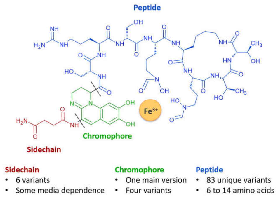
Figure 2.
Chromophore diversity among known pyoverdines. R1 represents the peptide chain attachment point and R2 represents the acyl chain attachment point.
Figure 2.
Chromophore diversity among known pyoverdines. R1 represents the peptide chain attachment point and R2 represents the acyl chain attachment point.
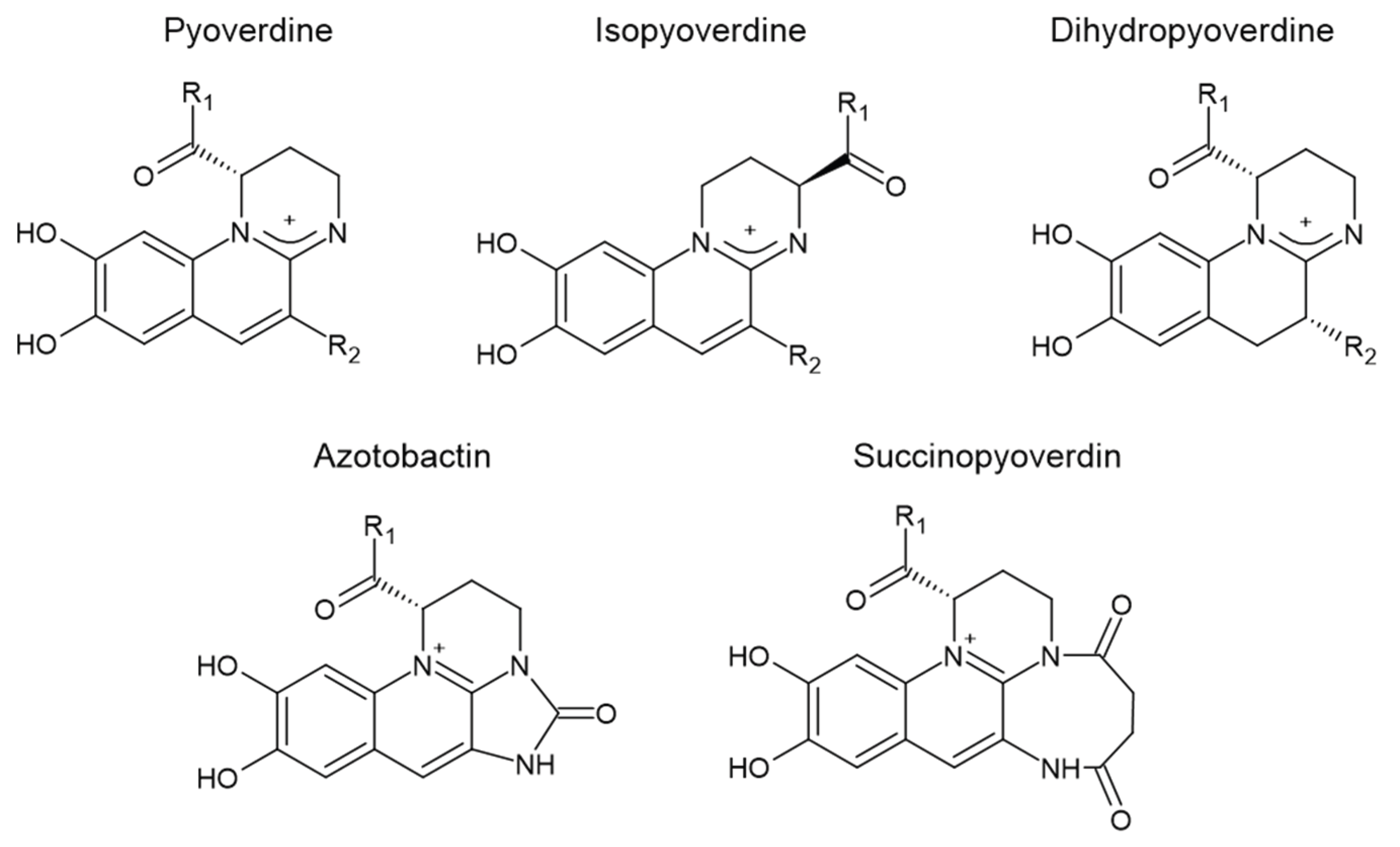
Figure 3.
Percentage of each iron binding residue found within pyoverdines. Abbreviations and structures are given in Supplementary Figure S1.
Figure 3.
Percentage of each iron binding residue found within pyoverdines. Abbreviations and structures are given in Supplementary Figure S1.
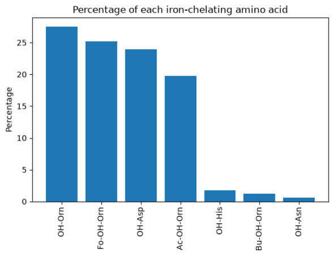
Figure 4.
a) Percentages of amino acids in pyoverdine peptide chains. b) Percentages of amino acids with D- and L-stereochemistry determined. For analysis of stereochemistry, rare amino acids were grouped together when both the L- and D-forms had less than 5 occurrences. A Fisher's exact test was performed to identify whether specific amino acids were enriched for L- or D-stereochemistry relative to the overall L:D distribution. P-values were adjusted for multiple hypothesis testing using the Benjamini-Hochberg procedure to control the false discovery rate (FDR). The following amino acids differed significantly from the expected L:D proportion: Ser (D-enriched, adjusted p = 0.024); OH-Asp (D-enriched, adjusted p = 0.024); Asp (L-enriched, adjusted p = 0.024).
Figure 4.
a) Percentages of amino acids in pyoverdine peptide chains. b) Percentages of amino acids with D- and L-stereochemistry determined. For analysis of stereochemistry, rare amino acids were grouped together when both the L- and D-forms had less than 5 occurrences. A Fisher's exact test was performed to identify whether specific amino acids were enriched for L- or D-stereochemistry relative to the overall L:D distribution. P-values were adjusted for multiple hypothesis testing using the Benjamini-Hochberg procedure to control the false discovery rate (FDR). The following amino acids differed significantly from the expected L:D proportion: Ser (D-enriched, adjusted p = 0.024); OH-Asp (D-enriched, adjusted p = 0.024); Asp (L-enriched, adjusted p = 0.024).
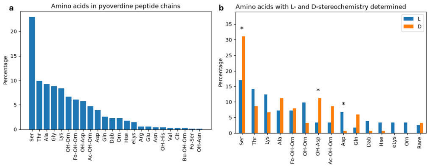
Figure 5.
Group and class nomenclature of pyoverdines. a. Representative structures for each category with the pyoverdine from P. aeruginosa ATCC 27853, P. aeruginosa PAO1, P. protegens Pf-5 and P. aeruginosa R' for groups 1 to 4 respectively. Distinguishing features for each group are indicated by a dashed circle. b. Total counts of all pyoverdines belonging to each group. c. Total counts of all pyoverdines belonging to each class.
Figure 5.
Group and class nomenclature of pyoverdines. a. Representative structures for each category with the pyoverdine from P. aeruginosa ATCC 27853, P. aeruginosa PAO1, P. protegens Pf-5 and P. aeruginosa R' for groups 1 to 4 respectively. Distinguishing features for each group are indicated by a dashed circle. b. Total counts of all pyoverdines belonging to each group. c. Total counts of all pyoverdines belonging to each class.
Disclaimer/Publisher’s Note: The statements, opinions and data contained in all publications are solely those of the individual author(s) and contributor(s) and not of MDPI and/or the editor(s). MDPI and/or the editor(s) disclaim responsibility for any injury to people or property resulting from any ideas, methods, instructions or products referred to in the content. |
© 2026 by the authors. Licensee MDPI, Basel, Switzerland. This article is an open access article distributed under the terms and conditions of the Creative Commons Attribution (CC BY) license (http://creativecommons.org/licenses/by/4.0/).
Copyright: This open access article is published under a Creative Commons CC BY 4.0 license, which permit the free download, distribution, and reuse, provided that the author and preprint are cited in any reuse.