

Submitted:
02 November 2026
Posted:
04 November 2026
You are already at the latest version
Abstract
Survivin (BIRC5), a core component of the chromosomal passenger complex (CPC), is essential for faithful mitosis and frequently overexpressed in human cancers. Despite extensive investigation of its mitotic and cell survival functions, its relationship with cellular senescence remains poorly understood. We investigated Survivin in therapy- and stress-induced senescence models and examined the consequences of transient Survivin depletion by RNA interference. Camptothecin treatment of HCT116 wt and A549 cells and oxidative stress in WI-38 fibroblasts induced characteristic morphological changes, increased senescence-associated β-galactosidase activity and reduced DNA synthesis. In HCT116 wt and A549 cells, senescence induction was accompanied by changes in cell-cycle regulatory proteins and altered immunofluorescence patterns of cell-cycle and chromatin-associated proteins. Survivin-targeting siRNA promoted pronounced senescence-associated morphological changes and increased β-galactosidase activity in HCT116 wt cells, whereas A549 cells exhibited a less pronounced response. These phenotypic differences were accompanied by changes in p53, p21 and CDK1 protein levels during the observation period. However, transient Survivin depletion does not establish whether the observed changes represent stable or irreversible senescence. Together, our findings support a cell-context-dependent association between Survivin perturbation and senescence-associated phenotypes, extending its biological significance beyond mitosis and highlighting cellular senescence as a potential outcome relevant to Survivin-targeted anticancer strategies.
Keywords:
BIRC5
; chromosomal passenger complex
; therapy-induced senescence
; DNA damage response
; RNA interference
1. Introduction
Survivin, also known as baculoviral inhibitor of apoptosis protein repeat-containing protein 5 (BIRC5), is a multifunctional protein involved in essential cellular processes, including mitosis, cell survival, and the maintenance of proliferative capacity. [1] Originally identified as the smallest member of the inhibitor of apoptosis protein (IAP) family through its N-terminal baculoviral IAP repeat (BIR) domain, Survivin is now primarily recognized as a core component of the chromosomal passenger complex (CPC). Together with Borealin, INCENP, and Aurora B kinase, the CPC coordinates chromosome alignment, spindle assembly checkpoint signaling, chromosome segregation, and cytokinesis, thereby ensuring faithful cell division. [1,2] Within this complex, Survivin contributes to the recruitment and localization of Aurora B at centromeres and to the dynamic redistribution of the CPC throughout mitosis. Unlike Aurora B, Survivin lacks intrinsic enzymatic activity and instead exerts its functions through interactions with multiple binding partners. Although indispensable during embryonic development, Survivin expression is generally low or undetectable in many terminally differentiated adult tissues. In contrast, Survivin is frequently overexpressed in human malignancies, where elevated expression has been associated with aggressive disease, resistance to anticancer therapies, tumor recurrence, and poor clinical outcomes. [1,3] Consequently, Survivin has emerged as a promising therapeutic target in cancer. Despite considerable efforts to inhibit Survivin therapeutically, a more comprehensive understanding of its biological functions remains important for interpreting the cellular consequences of Survivin-targeted strategies. [4,5]
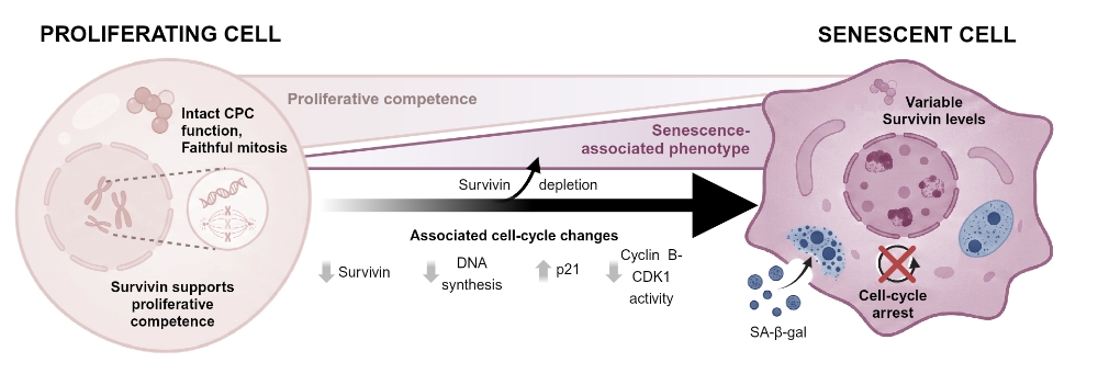
Beyond its canonical mitotic functions, accumulating evidence suggests that Survivin participates in several non-mitotic processes, including DNA damage signaling, cellular metabolism, stem cell maintenance, and adaptation to cellular stress. [1,6] Nuclear Survivin has been implicated in DNA double-strand break repair through functional associations with DNA-PKcs and other components of the repair machinery, [7,8] while our previous work identified an additional role for Survivin in DNA damage tolerance through the regulation of translesion DNA synthesis. [6] These findings suggest that Survivin may contribute to the maintenance of proliferative competence not only during mitosis but also in cells challenged by DNA damage and replication-blocking lesions. Survivin has additionally been linked to aging and cellular senescence. [9] Reduced Survivin expression has been associated with replicative and stress-induced senescence, whereas sustained Survivin expression has been linked to continued proliferative capacity and resistance to senescence-associated growth arrest. [9,10,11] These observations form the basis of a conceptual framework relating Survivin expression to distinct cellular states during development, aging, and senescence (Figure S1). However, whether Survivin actively contributes to the regulation of senescence or whether changes in its expression primarily reflect altered proliferative activity remains unresolved.
Cellular senescence is characterized by a persistent cell-cycle arrest that can be triggered by a variety of intrinsic and extrinsic stresses, including telomere shortening, oncogene activation, oxidative stress, mitochondrial dysfunction, and DNA damage. [12] Initially described as a mechanism limiting the replicative lifespan of primary human fibroblasts, senescence is now recognized as a biological program involved in embryonic development, tissue remodeling, wound healing, and tumor suppression. [13,14] Senescent cells generally remain metabolically active while exhibiting characteristic morphological and molecular features, including cellular enlargement, increased senescence-associated β-galactosidase activity, alterations in chromatin organization, and, in many contexts, the development of a senescence-associated secretory phenotype (SASP). [12,15] Although senescence can contribute to tissue homeostasis and the suppression of damaged-cell proliferation, the persistent accumulation of senescent cells has also been implicated in chronic inflammation, tissue dysfunction, organismal aging, and age-related diseases. [12,14]
The transition from proliferation to senescence is closely associated with changes in cell-cycle regulation and DNA damage signaling (Figure S2). In response to persistent cellular stress, activation of checkpoint pathways involving ATM, ATR, p53, and the cyclin-dependent kinase inhibitor p21 can suppress cyclin–CDK activity and promote cell-cycle arrest. The p16-RB pathway provides an additional mechanism contributing to the maintenance of senescence-associated growth arrest. [12,15] Survivin expression and activity are closely linked to cell-cycle progression, including its mitotic functions within the CPC and its phosphorylation at Thr34 by Cyclin B-CDK1. [1] Alterations in Survivin abundance or regulation may therefore accompany changes in proliferative competence and cellular stress responses. However, the extent to which Survivin itself influences the establishment or maintenance of senescence remains unclear. This distinction is particularly relevant in the context of Survivin-targeted therapies, where perturbation of Survivin function may be associated with different cellular outcomes depending on the biological context.
To investigate the relationship between Survivin and cellular senescence, we employed complementary models of therapy- and stress-induced senescence and examined the consequences of transient Survivin depletion by RNA interference. We assessed senescence-associated phenotypes, cell-cycle regulatory proteins, and immunofluorescence patterns of Survivin and selected nuclear proteins. Our findings reveal a cell-context-dependent association between Survivin perturbation and the acquisition of senescence-associated characteristics, extending the biological context of Survivin beyond its established mitotic functions and providing a basis for further investigation of its potential relevance to senescence-associated responses during anticancer therapy.
2. Results
2.1. Senescence-Inducing Treatments Establish Characteristic Senescence-Associated Phenotypes
To establish experimental models of cellular senescence, HCT116 wt and A549 cells were treated with camptothecin (CPT; 25 nM, 48 h), whereas WI-38 fibroblasts were exposed to oxidative stress induced by hydrogen peroxide (H2O2; 150 µM, 24 h) (Figure 1a). Following treatment, HCT116 wt cells exhibited pronounced morphological alterations, including cellular enlargement and flattening, together with increased senescence-associated β-galactosidase (SA-β-gal) activity (Figure 1b). Similar senescence-associated morphological changes and increased SA-β-gal activity were observed in CPT-treated A549 cells and H2O2-treated WI-38 fibroblasts (Figure 1b). To further characterize the proliferative state of these cells, DNA synthesis was assessed by incorporation of 5-ethynyl-2′-deoxyuridine (EdU). Compared with untreated controls, senescence-inducing treatments were associated with reduced EdU incorporation in HCT116 wt, A549, and WI-38 cells, consistent with diminished DNA synthesis (Figure 1c). Immunofluorescence analysis of Survivin was additionally performed to examine its distribution under the respective treatment conditions (Figure 1c). However, these observations did not reveal a consistent decrease in Survivin staining across the three cellular models. Together, the morphological changes, increased SA-β-gal activity and reduced EdU incorporation supported the establishment of senescence-associated phenotypes in the three experimental models. These models provided the basis for subsequent analyses of cell-cycle regulatory proteins and Survivin-associated responses during senescence induction.
2.2. Senescence Induction Is Accompanied by Changes in Cell-Cycle Regulatory Proteins and Survivin Staining
To further characterize molecular changes accompanying senescence induction, the expression and subcellular distribution of Survivin and selected cell-cycle regulatory proteins were examined in HCT116 wt and A549 cells following CPT treatment (25 nM, 48 h). Immunofluorescence analyses revealed reduced CDK1 staining following treatment, particularly in HCT116 wt cells, together with alterations in Survivin staining patterns (Figure 2a). Immunofluorescence analysis of p21 showed increased nuclear staining in CPT-treated cells, accompanied by changes in Survivin staining (Figure 2b). To examine the phosphorylation-associated distribution of Survivin, immunofluorescence analyses were performed using an antibody recognizing Survivin phosphorylated at threonine 34 (SurvT34ph). Distinct punctate signals were detected in both cell lines, with treatment-associated differences in their distribution (Figure 2c). These observations were considered in the context of the established role of CDK1 in Survivin phosphorylation and mitotic regulation (Figure 2d). Immunoblot analyses confirmed pronounced changes in cell-cycle regulatory proteins following CPT treatment (Figure 2e). CDK1 protein levels decreased in both HCT116 wt and A549 cells, while p21 and phosphorylated ATM (pATM) levels increased. These changes were particularly pronounced for p21 in HCT116 wt cells, whereas the relative p21 increase was more modest in A549 cells. Together, these findings demonstrate that CPT-induced senescence-associated phenotypes are accompanied by changes in cell-cycle regulatory proteins and are consistent with activation of DNA damage signaling. Although Survivin staining patterns were altered following treatment, the immunofluorescence analyses did not establish a uniform reduction in Survivin abundance across the two cell lines.
2.3. Survivin Depletion Promotes Senescence-Associated Phenotypes
To investigate whether reduced Survivin expression contributes to the acquisition of senescence-associated characteristics, HCT116 wt and A549 cells were transfected with non-targeting control siRNA (siNT) or Survivin-targeting siRNA (siSurv). Morphological changes and senescence-associated β-galactosidase (SA-β-gal) activity were assessed 72 h after transfection (Figure 3a). Survivin depletion was associated with distinct phenotypic responses in the two cell lines. In HCT116 wt cells, siSurv transfection resulted in cellular enlargement and flattening compared with siNT-transfected controls. These morphological alterations were accompanied by increased SA-β-gal activity (Figure 3a). In contrast, A549 cells displayed less pronounced morphological changes and a weaker SA-β-gal response under the corresponding conditions (Figure 3a), indicating differences in the cellular response to Survivin depletion. To examine accompanying molecular alterations, cells were analyzed over the indicated observation periods following siRNA transfection, with or without subsequent camptothecin (CPT) treatment (Figure 3b). Immunoblot analyses confirmed reduced Survivin protein levels following siSurv transfection and revealed accompanying changes in cell-cycle regulatory proteins in both cellular models (Figure 3c-e). The corresponding densitometric analyses are presented in Figure S4 and Figure S5. Together, these findings suggest that Survivin depletion can promote senescence-associated phenotypic changes, particularly in HCT116 wt cells, while revealing differences between the two cellular backgrounds. However, the observation period and assays used do not establish whether these changes represent stable or irreversible senescence.
2.4. Survivin Depletion Is Accompanied by Changes in Cell-Cycle Regulatory Proteins
To further characterize the molecular changes associated with Survivin depletion, proteins involved in cell-cycle regulation and DNA damage signaling were analyzed by immunoblotting in HCT116 wt and A549 cells at the indicated time points following siRNA transfection, with or without subsequent CPT treatment. In HCT116 wt cells, Survivin-targeting siRNA was associated with reduced CDK1 protein levels and increased p21 and p53 levels at selected time points (Figure 3c-e; Figure S4). A549 cells also exhibited changes in these regulatory proteins, although their temporal patterns and magnitudes differed from those observed in HCT116 wt cells (Figure 3c-e; Figure S5). In HCT116 wt cells, ATM phosphorylation was primarily associated with CPT treatment rather than Survivin depletion alone, suggesting that the observed DNA damage signaling response was predominantly driven by the genotoxic treatment under the conditions examined (Figure 3c-e; Figure S4). The extent of Survivin depletion varied over the observation period, with partial recovery of detectable Survivin protein levels at later time points under some experimental conditions despite repeated siRNA transfections (Figure 3c-e; Figure S4 and Figure S5). Accordingly, changes in cell-cycle regulatory proteins at these later time points cannot be attributed unequivocally to sustained Survivin depletion. Nevertheless, the observed alterations in CDK1, p21, and p53 were consistent with changes in cell-cycle regulation accompanying the senescence-associated phenotypes described above. As a complementary approach, we examined the expression of ectopically introduced Survivin-GFP during senescence induction in A549 cells. Following transient transfection, cells were either left untreated or exposed to CPT. Fluorescence microscopy revealed a progressive decline in detectable Survivin-GFP fluorescence over the observation period under both conditions (Figure S6). Because sustained Survivin-GFP expression could not be demonstrated, these experiments did not allow an assessment of whether ectopic Survivin expression could prevent or attenuate the senescence-associated phenotype.
2.5. Survivin and Aurora B Exhibit Distinct Nuclear Staining Patterns During Senescence Induction
Because cellular senescence is accompanied by changes in nuclear and chromatin organization, we investigated the subcellular distribution of Survivin and selected chromosomal passenger complex (CPC)-associated proteins in CPT-treated HCT116 wt cells. Immunofluorescence analyses examined Survivin and Aurora B in relation to histone H3 lysine 9 di- and trimethylation (H3K9me2/3), as well as CRM1 and INCENP in relation to HP1α. Survivin and Aurora B exhibited discrete nuclear staining signals showing limited spatial overlap with H3K9me2/3-positive regions (Figure 4a). Additional immunofluorescence analyses did not reveal convincing spatial colocalization of CRM1 or INCENP with HP1α-positive nuclear regions (Figure 4b). These observations did not establish a direct association of the examined proteins with heterochromatin. The experimental observations were considered in the context of established models of heterochromatin organization and senescence-associated heterochromatin foci (SAHF) formation (Figure 4c,d). However, the staining patterns alone did not provide conclusive evidence for the formation of mature SAHF under the conditions examined. Together, these findings indicate that Survivin and Aurora B exhibit distinct subnuclear staining patterns in CPT-treated HCT116 wt cells, with limited spatial overlap with H3K9me2/3-positive regions. Nevertheless, the data do not establish recruitment of CPC-associated proteins to senescence-associated heterochromatin. An overview of senescence-associated phenotypic and molecular changes across the experimental models is provided in Table S3.
3. Discussion
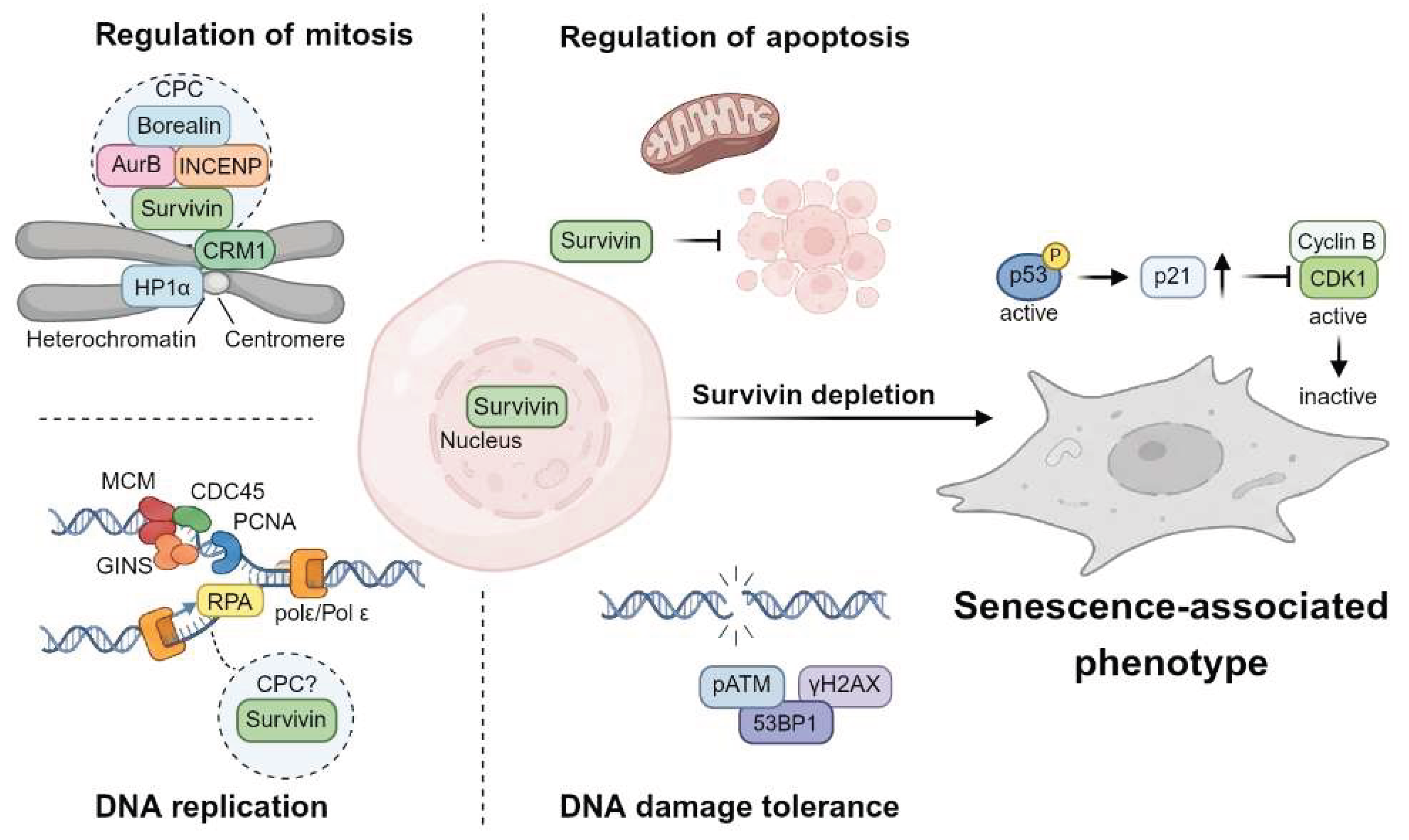
Cellular senescence and proliferation represent distinct cellular states, yet the mechanisms underlying this transition remain incompletely understood. [12,17] Survivin is widely recognized as a multifunctional regulator of mitosis and apoptosis, [1,2] but its relationship to cellular senescence remains less well defined. In the present study, we combined senescence induction models with RNA interference-mediated Survivin depletion to investigate this relationship. Senescence-inducing treatments established characteristic morphological and biochemical phenotypes in HCT116 wt, A549 and WI-38 cells, while targeted Survivin depletion promoted senescence-associated changes, most prominently in HCT116 wt cells. The weaker response observed in A549 cells further indicates that the consequences of Survivin perturbation are influenced by cellular context. Collectively, these findings support a relationship between Survivin availability, cell-cycle regulation and the acquisition of senescence-associated characteristics, although Survivin depletion did not produce equivalent responses across the cellular models examined (Figure 5).
Our observations are consistent with previous studies linking Survivin to cellular immortalization and senescence. Kan and colleagues reported increased Survivin expression during immortalization of human myofibroblasts, accompanied by repression of p16 and increased resistance to oxidative stress. [10] Conversely, Sumi et al. demonstrated that depletion of Survivin induces senescence in KRAS-mutant lung adenocarcinoma cells. [11] Although these studies examined distinct biological settings, they suggest that Survivin may contribute to the maintenance of proliferative competence, whereas its depletion can promote senescence-associated responses. Our findings extend these observations by showing that Survivin depletion is accompanied by morphological changes, increased SA-β-gal activity and alterations in cell-cycle regulatory proteins in HCT116 wt cells. At the same time, the different responses of HCT116 and A549 cells emphasize that reduced Survivin alone does not necessarily produce an equivalent phenotype across different cellular backgrounds. Importantly, the present data do not establish a consistent reduction in endogenous Survivin abundance following senescence induction across all three experimental models. The consequences of experimentally reducing Survivin expression must therefore be distinguished from changes in Survivin expression occurring during senescence induction.
Molecular changes following Survivin depletion offer further insight. In HCT116 wt cells, reduced CDK1 protein levels together with increased p21 and p53 levels were observed at selected time points following Survivin-targeting siRNA transfection and accompanied the development of senescence-associated phenotypic changes. This expression pattern is consistent with altered cell-cycle regulation and is of particular interest given the established role of Survivin in chromosome segregation and mitotic progression. [2] Impaired mitotic progression or cytokinesis represents one possible mechanism through which Survivin depletion could contribute to proliferative arrest, although this was not directly investigated. Persistent mitotic abnormalities have been linked to checkpoint activation and senescence-associated outcomes. [18,19] In addition, Survivin has been linked to the DNA damage response and DNA double-strand break repair through interactions with DNA-PKcs and other components of the repair machinery, suggesting that loss of Survivin may affect proliferative competence through mechanisms extending beyond mitotic disruption. [7,8] The concomitant changes in p21 and CDK1 expression observed following Survivin depletion are compatible with altered cell-cycle regulation and are of particular interest in light of evidence implicating the p21-CDK1/2 axis in therapy-induced senescence following genotoxic stress. [19,20,21] Moreover, ATM phosphorylation in HCT116 wt cells was primarily associated with CPT treatment rather than Survivin depletion alone, suggesting that the observed ATM activation was mainly associated with genotoxic treatment under the experimental conditions examined. However, these experiments cannot determine whether the observed cell-cycle changes result directly from Survivin depletion or secondarily from mitotic defects or other proliferative stress. Thus, the findings support an association between Survivin perturbation and altered cell-cycle regulation without defining the causal sequence connecting these events.
Immunofluorescence analyses of CPT-treated HCT116 wt cells revealed distinct subnuclear staining patterns of Survivin and Aurora B, including discrete signals showing limited spatial overlap with H3K9me2/3-positive regions. Convincing colocalization of CRM1 or INCENP with HP1α-positive nuclear regions was not evident. These observations are of interest because extensive chromatin reorganization accompanies cellular senescence, but they should be interpreted cautiously. Mature senescence-associated heterochromatin foci were not conclusively demonstrated under our experimental conditions, and the localization patterns did not reveal a uniform redistribution of CPC components to heterochromatin. Moreover, spatial overlap observed by immunofluorescence does not establish direct molecular association with heterochromatin. The present data therefore do not establish a direct role for Survivin or the CPC in senescence-associated chromatin organization. Rather, they reveal distinct subnuclear staining patterns of selected CPC components in CPT-treated cells, whose functional significance remains unclear.
Survivin has long been considered an attractive therapeutic target because of its central role in mitosis and its frequent overexpression in human malignancies. [1,5] Accordingly, several conceptually distinct Survivin-targeting strategies have been investigated, [22] including small-molecule approaches such as the Survivin suppressant sepantronium bromide (YM155), antisense oligonucleotides targeting BIRC5, including LY2181308, [23,24] and Survivin-directed immunotherapeutic approaches. [4,25] A Survivin/BIRC5-derived peptide disrupting Survivin dimerization was recently shown to impair cell division and exert anticancer effects. [26] Several of these strategies have progressed to clinical evaluation, but their translation into established therapeutic approaches has remained challenging. [22] One possible contributing factor is the heterogeneity of cellular responses to Survivin perturbation. Indeed, Survivin inhibition does not necessarily result exclusively in apoptosis; alternative cellular outcomes, including prolonged growth arrest and senescence-associated phenotypes, have also been reported depending on cellular context. [11,27] Our observations are consistent with this concept and suggest that senescence-associated responses should be considered alongside cell death when evaluating the biological consequences of Survivin targeting. This distinction may be therapeutically relevant because therapy-induced senescence can suppress tumor cell proliferation, [28] while senescent cells may remain metabolically active and influence their surroundings through the senescence-associated secretory phenotype. [29] Depending on cellular context, these effects may contribute to immune-mediated clearance of senescent tumor cells, but persistent senescent populations have also been associated with tumor-promoting inflammation, cellular plasticity and disease recurrence. Thus, the therapeutic consequences of Survivin targeting may depend not only on the extent and duration of target suppression, but also on the subsequent cellular response. [24] This consideration may be particularly relevant for therapeutic approaches aimed at reducing Survivin expression, as incomplete or temporally variable target suppression could conceivably contribute to heterogeneous cellular outcomes, although this possibility was not directly tested in the present study. If Survivin-targeted interventions were shown to generate persistent senescent tumor-cell populations, sequential strategies aimed at eliminating or modulating these cells, including senolytic or senomorphic approaches, might warrant consideration. [28,30,31] Such combinations remain hypothetical in the context of Survivin targeting and require direct experimental validation. Understanding the factors that determine whether Survivin-targeted interventions result in cell death, prolonged growth arrest or acquisition of a senescent state may therefore be important for the future development and rational combination of Survivin-directed therapeutic strategies.
Several limitations warrant consideration. Despite repeated siRNA transfections, sustained Survivin depletion was not consistently demonstrated, precluding conclusions about its ability to induce stable or irreversible senescence. The weaker A549 response underscores the context dependence of these effects. The inability to maintain detectable Survivin-GFP expression precluded a functional rescue assessment, while immunofluorescence analyses could not establish direct protein-chromatin interactions. Moreover, the findings are limited to cultured cells. Future studies employing stable genetic models, functional rescue experiments and longitudinal analyses are needed to clarify the mechanisms, persistence and biological relevance of Survivin-associated senescence responses.
4. Materials and Methods
4.1. Cell Culture
Eukaryotic cells were cultured in a humidified incubator at 37 °C with 5 % CO2. Human lung carcinoma A549 cells (ATCC CCL-185) were maintained in Ham’s F-12K (Kaighn’s) medium supplemented with 10 % (v/v) fetal bovine serum (FBS) and 1 x Antibiotic-Antimycotic. Human lung fibroblasts WI-38 (ATCC CCL-75) were maintained in Minimum Essential Medium (MEM) supplemented with 10 % (v/v) FBS, 2 mM L-glutamine, 1 x non-essential amino acids, 1.5 g/L sodium bicarbonate, 1 mM sodium pyruvate, and 1 x Antibiotic-Antimycotic. Human colorectal carcinoma HCT116 wild-type (wt) cells (ATCC CCL-247) were cultured in McCoy’s 5A medium supplemented with 10 % (v/v) FBS and 1 x Antibiotic-Antimycotic. All media and supplements were purchased from Thermo Fisher Scientific (Dreieich, Germany).
4.2. Plasmids
The eukaryotic expression plasmids pc3-Survivin-GFP and pc3-GFP, both based on the pcDNA3.1 vector, have been described previously. [16]
4.3. Transfection
Transient plasmid transfection was performed using Lipofectamine™ 2000 for HCT116 wt cells or Lipofectamine LTX with PLUS Reagent for A549 cells (Thermo Fisher Scientific, Dreieich, Germany) according to the manufacturer’s instructions. Cells were seeded 24 h before transfection and transfected with the indicated plasmids. For HCT116 wt cells, 150 ng plasmid DNA and 0.8 µL Lipofectamine™ 2000 were used per well of an 8-well µ-Slide. For A549 cells, 250 ng plasmid DNA, 1 µL Lipofectamine LTX and 0.25 µL PLUS Reagent were used per well. Where indicated, cells were exposed to camptothecin (CPT) 24 h after transfection and analyzed at the indicated time points.
4.4. RNA Interference
Transient knockdown of Survivin (BIRC5) was achieved using a Stealth siRNA targeting the 3′-untranslated region (3′-UTR) of human BIRC5 (siSurvivin B02; 5′-AAGAAGAGCACAGUUGAAACAUCA-3′; Thermo Fisher Scientific, Dreieich, Germany) and HiPerFect Transfection Reagent (Qiagen, Hilden, Germany) according to the manufacturer’s instructions for fast-forward transfection of adherent cells. A non-targeting siRNA with no mammalian homology (siNT; 5′-UUCUCCGAACGUGUCACGUdTdT-3′; Microsynth AG, Balgach, Switzerland) served as a negative control. Cells were transfected with a final siRNA concentration of 20 nM using 12 µL HiPerFect in 300 µL Opti-MEM per 60-mm culture dish. For prolonged depletion experiments, transfections were repeated at 48, 120 and 192 h after the initial transfection. Cells were analyzed at the indicated time points after the initial transfection.
4.5. Senescence Induction
Stress-induced senescence in WI-38 cells was induced by treatment with 150 μM hydrogen peroxide (H2O2; Sigma-Aldrich/Merck KGaA, Darmstadt, Germany) for 24 h. Therapy-induced senescence in HCT116 wt and A549 cells was induced by treatment with 25 nM camptothecin (CPT; Santa Cruz Biotechnology, Heidelberg, Germany) for 48 h. Following treatment, cells were washed twice with phosphate-buffered saline (PBS) and cultured in fresh growth medium for the respective observation periods. For the indicated experiments, immunofluorescence and EdU incorporation analyses were initiated three days after completion of treatment, whereas senescence-associated β-galactosidase activity was assessed at the indicated time points during the subsequent observation period.
4.6. Senescence-Associated β-Galactosidase Staining
Senescence-associated β-galactosidase (SA-β-gal) activity was detected using the Senescence β-Galactosidase Staining Kit (New England Biolabs/Cell Signaling Technology) according to the manufacturer’s instructions. Briefly, cells were washed once with PBS, fixed in the supplied fixation solution for 15 min at room temperature, washed twice with PBS, and incubated overnight at 37 °C in a dry incubator without CO2 in β-galactosidase staining solution (pH 6.0). Development of the blue reaction product was assessed after 12-16 h by bright-field microscopy using an Olympus CKX41 inverted microscope.
4.7. Immunofluorescence
Cells were seeded either in 8-well μ-Slides (ibidi®, Gräfelfing, Germany) or on glass-bottom dishes (MatTek Corporation, Ashland, MA, USA) and allowed to adhere for 24 h before the respective treatment or transfection. Cells were fixed with 4 % Roti®-Histofix (Carl Roth, Karlsruhe, Germany) for 15 min at room temperature, permeabilized and blocked with 5 % normal goat serum (NGS) in 0.3 % Triton X-100/PBS for 1 h. Primary antibodies were diluted in 1 % bovine serum albumin (BSA)/0.3 % Triton X-100/PBS and incubated overnight at 4 °C. Following three washing steps with PBS, cells were incubated with the appropriate Alexa Fluor™-conjugated secondary antibodies for 1 h at room temperature. Nuclei were counterstained with Hoechst 33342 (Applichem, Darmstadt, Germany), and where indicated, cell membranes were stained with HCS CellMask™ Deep Red (Thermo Fisher Scientific Dreieich, Germany). Images were acquired by confocal laser scanning microscopy using a Leica TCS SP8 microscope (Leica Microsystems, Wetzlar, Germany). Images were recorded by sequential scanning, typically at a resolution of 1024 x 1024 pixels, with consistent acquisition settings within individual experiments. Images were processed using Fiji/ImageJ software. Primary and secondary antibodies used for immunofluorescent staining are listed in Table S1.
4.8. Protein Extraction
Cells were washed with ice-cold PBS and lysed in ice-cold radioimmunoprecipitation assay (RIPA) buffer containing 50 mM Tris-HCl, 150 mM NaCl, 5 mM EDTA, 1 % NP-40, 1 % sodium deoxycholate, 1 mM dithiothreitol (DTT) and 1 mM phenylmethylsulfonyl fluoride (PMSF), supplemented with protease and phosphatase inhibitors (Roche). Lysates were sonicated on ice for 15 cycles (1 s on, 1 s off) using a probe sonicator (Bandelin), incubated on ice for 20 min, and cleared by centrifugation at 20,000 x g for 20 min at 4 °C. Supernatants were collected and stored at -80 °C until analysis.
4.9. SDS-PAGE and Immunoblotting
Protein concentrations were determined using the Bio-Rad Protein Assay (Bio-Rad Laboratories, Feldkirchen, Germany). For SDS-PAGE, 20-40 µg protein per sample were mixed with SDS sample buffer and heated at 95 °C for 5 min. Equal amounts of protein were separated by SDS-polyacrylamide gel electrophoresis (SDS-PAGE) using 10 % polyacrylamide gels and transferred onto polyvinylidene difluoride (PVDF) or nitrocellulose membranes using a PerfectBlue™ tank blotting system (Peqlab, Erlangen, Germany). Membranes were blocked with 5 % (w/v) non-fat dry milk in Tris-buffered saline containing 0.1 % Tween-20 (TBST) for 1 h at room temperature and incubated with the respective primary antibodies overnight at 4 °C. Following three washing steps with TBST, membranes were incubated with horseradish peroxidase (HRP)-conjugated secondary antibodies for 1 h at room temperature. Protein bands were visualized by enhanced chemiluminescence using ECL™ Plus Western Blotting Substrate (Thermo Fisher Scientific, Dreieich, Germany). Chemiluminescent signals were detected on Fujifilm Super RX X-ray film, which was developed using a CAWOMAT 2000 IR film processor (Cawo, Schrobenhausen, Germany) or detected with a ChemiDoc MP Imaging System (Bio-Rad Laboratories, Feldkirchen, Germany). Primary and secondary antibodies used for immunoblotting are listed in Table S2.
4.10. EdU Incorporation and Staining
DNA synthesis was assessed using the Click-iT™ Plus EdU Alexa Fluor™ 488/633 Imaging Kit (Thermo Fisher Scientific, Dreieich, Germany) according to the manufacturer’s instructions. Briefly, cells were incubated with 10 μM 5-ethynyl-2′-deoxyuridine (EdU) for 24 h, beginning three days after senescence-inducing treatment, to label newly synthesized DNA. Following fixation, incorporated EdU was detected by copper-catalyzed click chemistry using the Alexa Fluor azide provided with the kit. Nuclei were counterstained with Hoechst 33342 (Applichem, Darmstadt, Germany), and samples were analyzed by confocal fluorescence microscopy.
4.11. Image Analysis and Data Presentation
Fluorescence microscopy images were analyzed using Fiji/ImageJ software. Immunoblot signal intensities were quantified by densitometric analysis using Image Studio Lite (LI-COR Biosciences) and normalized to GAPDH. Where indicated, normalized intensities were expressed relative to the respective reference condition, as specified in the corresponding supplementary figure legends. Graphical representations were generated using GraphPad Prism (version 9.0; GraphPad Software, San Diego, CA, USA). Schematic illustrations were created using BioRender.com.
4.12. Use of Generative Artificial Intelligence
ChatGPT (OpenAI) was used to assist with language refinement, manuscript organization, and clarity of scientific presentation. No generative AI tools were used to generate or analyze experimental data.
5. Conclusions
Survivin has traditionally been studied as a regulator of mitosis and apoptosis. Our findings extend this perspective by demonstrating that Survivin depletion promotes senescence-associated phenotypic and cell-cycle regulatory changes in a cell-context-dependent manner, most prominently in HCT116 wt cells. Immunofluorescence analyses further revealed distinct nuclear staining patterns of Survivin and Aurora B in CPT-treated cells, although their functional involvement in chromatin remodeling remains unresolved. Together, these findings highlight a potential role for Survivin at the interface between proliferation and senescence and suggest that senescence-associated responses merit consideration when evaluating Survivin-targeted cancer therapies.
Supplementary Materials
The supporting information can be downloaded at the website of this paper posted on Preprints.org. Figure S1: Conceptual framework linking Survivin to cellular senescence and aging; Figure S2: Molecular framework linking Survivin to cell-cycle control and cellular senescence; Figure S3: Subcellular distribution of Survivin and senescence-associated proteins following senescence induction; Figure S4: Protein alterations following Survivin depletion in HCT116 wt cells; Figure S5: Protein alterations following Survivin depletion in A549 cells; Figure S6: Survivin-GFP expression during senescence induction in A549 cells; Table S1: Antibodies used for immunofluorescence; Table S2: Antibodies used for immunoblotting; Table S3: Summary of senescence-associated phenotypes across the experimental models.
Author Contributions
Conceptualization, S.K.K. and R.H.S.; methodology, E.Schw.; validation, E.Schw. and E.S.; formal analysis, E.Schw. and E.S.; investigation, E.Schw. and E.S.; resources, S.K.K.; data curation, E.Schw. and E.S.; writing—original draft preparation, E.Schw. and S.K.K.; writing—review and editing, S.K.K., L.S. and R.H.S.; visualization, E.Schw., E.S. and S.K.K.; supervision, S.K.K.; project administration, S.K.K.; funding acquisition, S.K.K. All authors have read and agreed to the published version of the manuscript.
Funding
This research was funded by the Deutsche Forschungsgemeinschaft (DFG, German Research Foundation), Research Training Group GRK 1739. The APC was funded by the Open Access Publication Fund of the University of Duisburg-Essen.
Institutional Review Board Statement
Not applicable.
Informed Consent Statement
Not applicable.
Data Availability Statement
The data supporting the findings of this study are presented in the article and its supplementary materials. Additional original data are available from the corresponding author upon reasonable request.
Acknowledgments
The authors thank Prof. Dr. Jan Willem Voncken (Maastricht University, Maastricht, The Netherlands) for valuable scientific discussions and advice. The authors acknowledge the Imaging Centre Campus Essen (ICCE) for providing access to microscopy infrastructure and technical support. Open Access funding was provided by the Open Access Publication Fund of the University of Duisburg-Essen. During the preparation of this manuscript, the authors used ChatGPT (OpenAI) for language refinement, manuscript organization, and improving the clarity of scientific presentation. The authors have reviewed and edited the output and take full responsibility for the content of this publication.
Conflicts of Interest
The authors declare no conflicts of interest. The funders had no role in the design of the study; in the collection, analyses, or interpretation of data; in the writing of the manuscript; or in the decision to publish the results.
Abbreviations
The following abbreviations are used in this manuscript:
| 53BP1 | p53-binding protein 1 |
| ATM | Ataxia-telangiectasia mutated |
| ATR | ATM- and Rad3-related |
| BIR | Baculoviral IAP repeat |
| BIRC5 | Baculoviral IAP repeat-containing 5 |
| BSA | Bovine serum albumin |
| CDK1 | Cyclin-dependent kinase 1 |
| CENP | Centromere protein |
| Chk1 | Checkpoint kinase 1 |
| CPC | Chromosomal passenger complex |
| CPT | Camptothecin |
| CRM1 | Chromosome region maintenance 1 (exportin 1) |
| DTT | Dithiothreitol |
| ECL | Enhanced chemiluminescence |
| EDTA | Ethylenediamine tetraacetic acid |
| EdU | 5-Ethynyl-2′-deoxyuridine |
| FBS | Fetal bovine serum |
| GAPDH | Glyceraldehyde-3-phosphate dehydrogenase |
| GFP | Green fluorescent protein |
| H2O2 | Hydrogen peroxide |
| H3K9me2/3 | Histone H3 lysine 9 di- and trimethylation |
| HP1 | Heterochromatin protein 1 |
| HRP | Horseradish peroxidase |
| IAP | Inhibitor of apoptosis protein |
| IF | Immunofluorescence |
| INCENP | Inner centromere protein |
| LAMP1 | Lysosome-associated membrane protein 1 |
| MEM | Minimum essential medium |
| NGS | Normal goat serum |
| p21 | Cyclin-dependent kinase inhibitor 1A (p21Waf1/Cip1) |
| PAGE | Polyacrylamide gel electrophoresis |
| pATM | Phosphorylated ATM |
| PBS | Phosphate-buffered saline |
| PCNA | Proliferating cell nuclear antigen |
| PML | Promyelocytic leukemia protein |
| PMSF | Phenylmethylsulfonyl fluoride |
| PVDF | Polyvinylidene difluoride |
| RIPA | Radioimmunoprecipitation assay |
| SA-β-gal | Senescence-associated β-galactosidase |
| SAHF | Senescence-associated heterochromatin foci |
| SASP | Senescence-associated secretory phenotype |
| SDS | Sodium dodecyl sulfate |
| siNT | Non-targeting control siRNA |
| siSurv | Survivin-targeting siRNA |
| siRNA | Small interfering RNA |
| TBST | Tris-buffered saline containing 0.1% Tween 20 |
References
- Wheatley, S.P.; Altieri, D.C. Survivin at a glance. J. Cell Sci. 2019, 132, jcs223826. [Google Scholar] [CrossRef] [PubMed]
- Carmena, M.; Wheelock, M.; Funabiki, H.; Earnshaw, W.C. The chromosomal passenger complex (CPC): From easy rider to the godfather of mitosis. Nat. Rev. Mol. Cell Biol. 2012, 13, 789–803. [Google Scholar] [CrossRef] [PubMed]
- Li, F.; Aljahdali, I.; Ling, X. Cancer therapeutics using survivin BIRC5 as a target: What can we do after over two decades of study? J. Exp. Clin. Cancer Res. 2019, 38, 368. [Google Scholar] [CrossRef] [PubMed]
- Kondapuram, S.K.; Ramachandran, H.K.; Arya, H.; Coumar, M.S. Targeting survivin for cancer therapy: Strategies, small molecule inhibitors and vaccine based therapeutics in development. Life Sci. 2023, 335, 122260. [Google Scholar] [CrossRef] [PubMed]
- Wang, Q.; Greene, M.I. Survivin as a Therapeutic Target for the Treatment of Human Cancer. Cancers 2024, 16, 1705. [Google Scholar] [CrossRef] [PubMed]
- Falke, K.; Schroder, E.; Mosel, S.; Yuruk, C.N.; Feldmann, S.; Gul, D.; Stahl, P.; Stauber, R.H.; Knauer, S.K. Old Passengers as New Drivers: Chromosomal Passenger Proteins Engage in Translesion Synthesis. Cells 2024, 13. [Google Scholar] [CrossRef] [PubMed]
- Capalbo, G.; Dittmann, K.; Weiss, C.; Reichert, S.; Hausmann, E.; Rödel, C.; Rödel, F. Radiation-induced survivin nuclear accumulation is linked to DNA damage repair. Int. J. Radiat. Oncol. Biol. Phys. 2010, 77, 226–234. [Google Scholar] [CrossRef] [PubMed]
- Güllülü, O.; Hehlgans, S.; Mayer, B.E.; Gossner, I.; Petraki, C.; Hoffmann, M.; Dombrowsky, M.J.; Kunzmann, P.; Hamacher, K.; Strebhardt, K.; et al. A Spatial and Functional Interaction of a Heterotetramer Survivin-DNA-PKcs Complex in DNA Damage Response. Cancer Res. 2021, 81, 2304–2317. [Google Scholar] [CrossRef] [PubMed]
- Unruhe, B.; Schröder, E.; Wünsch, D.; Knauer, S.K. An Old Flame Never Dies: Survivin in Cancer and Cellular Senescence. Gerontology 2016, 62, 173–181. [Google Scholar] [CrossRef] [PubMed]
- Kan, C.Y.; Petti, C.; Bracken, L.; Maritz, M.; Xu, N.; O’Brien, R.; Yang, C.; Liu, T.; Yuan, J.; Lock, R.B.; et al. Up-regulation of survivin during immortalization of human myofibroblasts is linked to repression of tumor suppressor p16(INK4a) protein and confers resistance to oxidative stress. J. Biol. Chem. 2013, 288, 12032–12041. [Google Scholar] [CrossRef] [PubMed]
- Sumi, T.; Hirai, S.; Yamaguchi, M.; Tanaka, Y.; Tada, M.; Yamada, G.; Hasegawa, T.; Miyagi, Y.; Niki, T.; Watanabe, A.; et al. Survivin knockdown induces senescence in TTF-1-expressing, KRAS-mutant lung adenocarcinomas. Int. J. Oncol. 2018, 53, 33–46. [Google Scholar] [CrossRef] [PubMed]
- Gorgoulis, V.; Adams, P.D.; Alimonti, A.; Bennett, D.C.; Bischof, O.; Bishop, C.; Campisi, J.; Collado, M.; Evangelou, K.; Ferbeyre, G.; et al. Cellular Senescence: Defining a Path Forward. Cell 2019, 179, 813–827. [Google Scholar] [CrossRef] [PubMed]
- Yuan, L.; Alexander, P.B.; Wang, X.F. Cellular senescence: From anti-cancer weapon to anti-aging target. Sci. China Life Sci. 2020, 63, 332–342. [Google Scholar] [CrossRef] [PubMed]
- Sun, Y. An updated landscape of cellular senescence heterogeneity: Mechanisms, technologies and senotherapies. Transl. Med. Aging 2023, 7, 46–51. [Google Scholar] [CrossRef]
- Hernandez-Segura, A.; Nehme, J.; Demaria, M. Hallmarks of Cellular Senescence. Trends Cell Biol. 2018, 28, 436–453. [Google Scholar] [CrossRef] [PubMed]
- Knauer, S.K.; Bier, C.; Habtemichael, N.; Stauber, R.H. The Survivin-Crm1 interaction is essential for chromosomal passenger complex localization and function. EMBO Rep. 2006, 7, 1259–1265. [Google Scholar] [CrossRef] [PubMed]
- Di Micco, R.; Krizhanovsky, V.; Baker, D.; d’Adda di Fagagna, F. Cellular senescence in ageing: From mechanisms to therapeutic opportunities. Nat. Rev. Mol. Cell Biol. 2021, 22, 75–95. [Google Scholar] [CrossRef] [PubMed]
- Maehara, K.; Takahashi, K.; Saitoh, S. CENP-A reduction induces a p53-dependent cellular senescence response to protect cells from executing defective mitoses. Mol. Cell. Biol. 2010, 30, 2090–2104. [Google Scholar] [CrossRef] [PubMed]
- Hervé, S.; Scelfo, A.; Bersano Marchisio, G.; Grison, M.; Vaidžiulytė, K.; Dumont, M.; Angrisani, A.; Keikhosravi, A.; Pegoraro, G.; Deygas, M.; et al. Chromosome mis-segregation triggers cell cycle arrest through a mechanosensitive nuclear envelope checkpoint. Nat. Cell Biol. 2025, 27, 73–86. [Google Scholar] [CrossRef] [PubMed]
- Abbas, T.; Dutta, A. p21 in cancer: Intricate networks and multiple activities. Nat. Rev. Cancer 2009, 9, 400–414. [Google Scholar] [CrossRef] [PubMed]
- Schwarzenbach, C.; Rinke, J.; Vilar, J.B.; Sallbach, J.; Tatsch, L.; Schmidt, A.; Schöneis, A.; Rasenberger, B.; Kaina, B.; Tomicic, M.T.; et al. Therapy-induced senescence of glioblastoma cells is determined by the p21(CIP1)-CDK1/2 axis and does not require activation of DREAM. Cell Death Dis. 2025, 16, 357. [Google Scholar] [CrossRef] [PubMed]
- Fanuzzi, M.; Zheng, S.; Horbinski, C.M.; Shaaban, M.A.; Congivaram, H.; Du, R.; Tripathi, S.; Hurley, L.; Kumthekar, P.; Ahmed, A.; et al. Targeting Survivin: Now I Become Death, the Destroyer of Cells. Int. J. Mol. Sci. 2025, 26. [Google Scholar] [CrossRef] [PubMed]
- Talbot, D.C.; Ranson, M.; Davies, J.; Lahn, M.; Callies, S.; André, V.; Kadam, S.; Burgess, M.; Slapak, C.; Olsen, A.L.; et al. Tumor Survivin Is Downregulated by the Antisense Oligonucleotide LY2181308: A Proof-of-Concept, First-in-Human Dose Study. Clin. Cancer Res. 2010, 16, 6150–6158. [Google Scholar] [CrossRef] [PubMed]
- Poudel, B.H.; Chen, S.; Veedu, R.N. Survivin-Targeting Antisense Oligonucleotides in Cancer Therapy. Molecules 2026, 31, 2283. [Google Scholar] [CrossRef] [PubMed]
- Wijaya, W.; Morris, T.; Forster, M.D.; Flynn, M.; Tuthill, M.; Thistlethwaite, F.; Williams, A.; Finch, W.; Lu, W.; Jiang, S. Survivin recombinant overlapping peptide (ROP) vaccine in advanced solid tumours: A first-in-human, multicentre, open-label, phase 1a dose-escalation study. EClinicalMedicine 2026, 91, 103717. [Google Scholar] [CrossRef] [PubMed]
- Santhanam, M.; Babu, V.; Shteinfer-Kuzmine, A.; Pandey, S.K.; Gheber, L.; Raday, G.; Shoshan-Barmatz, V. Survivin/BIRC5-derived peptide disrupts survivin dimerization and cell division and induces multifaceted anti-cancer effects. Mol. Ther. Oncol. 2026, 34, 201123. [Google Scholar] [CrossRef] [PubMed]
- Han, T.-L.; Sha, H.; Ji, J.; Li, Y.-T.; Wu, D.-S.; Lin, H.; Hu, B.; Jiang, Z.-X. Depletion of Survivin suppresses docetaxel-induced apoptosis in HeLa cells by facilitating mitotic slippage. Sci. Rep. 2021, 11, 2283. [Google Scholar] [CrossRef] [PubMed]
- Schmitt, C.A.; Wang, B.; Demaria, M. Senescence and cancer — role and therapeutic opportunities. Nat. Rev. Clin. Oncol. 2022, 19, 619–636. [Google Scholar] [CrossRef] [PubMed]
- Wang, L.; Lankhorst, L.; Bernards, R. Exploiting senescence for the treatment of cancer. Nat. Rev. Cancer 2022, 22, 340–355. [Google Scholar] [CrossRef] [PubMed]
- Chaib, S.; Tchkonia, T.; Kirkland, J.L. Cellular senescence and senolytics: The path to the clinic. Nat. Med. 2022, 28, 1556–1568. [Google Scholar] [CrossRef] [PubMed]
- McHugh, D.; Durán, I.; Gil, J. Senescence as a therapeutic target in cancer and age-related diseases. Nat. Rev. Drug Discov. 2025, 24, 57–71. [Google Scholar] [CrossRef] [PubMed]
Figure 1.
Establishment and characterization of experimental models of cellular senescence. a. Schematic overview of the experimental strategies used to induce cellular senescence. Therapy-induced senescence was induced in HCT116 wt colorectal carcinoma and A549 lung adenocarcinoma cells by treatment with camptothecin (CPT; 25 nM, 48 h), whereas stress-induced senescence was induced in WI-38 lung fibroblasts by exposure to H2O2 (150 µM, 24 h). Following treatment, cells were cultured under standard conditions and analyzed for senescence-associated morphological changes, SA-β-gal activity and DNA synthesis. b. Representative phase-contrast images of untreated and treated HCT116 wt, A549 and WI-38 cells at days 1, 3 and 6 following treatment, illustrating changes in cellular morphology and SA-β-gal staining. Treated cells exhibited enlarged and flattened morphology and increased SA-β-gal activity compared with untreated controls. Scale bars, 20 µm. c. EdU labeling was initiated 72 h after completion of senescence-inducing treatment and continued for 24 h. DNA synthesis was assessed by Click-iT™ detection, and Survivin was visualized by immunofluorescence staining. DNA was counterstained with Hoechst. Untreated cells served as controls. Representative fluorescence microscopy images are shown. Scale bars, 25 µm.
Figure 1.
Establishment and characterization of experimental models of cellular senescence. a. Schematic overview of the experimental strategies used to induce cellular senescence. Therapy-induced senescence was induced in HCT116 wt colorectal carcinoma and A549 lung adenocarcinoma cells by treatment with camptothecin (CPT; 25 nM, 48 h), whereas stress-induced senescence was induced in WI-38 lung fibroblasts by exposure to H2O2 (150 µM, 24 h). Following treatment, cells were cultured under standard conditions and analyzed for senescence-associated morphological changes, SA-β-gal activity and DNA synthesis. b. Representative phase-contrast images of untreated and treated HCT116 wt, A549 and WI-38 cells at days 1, 3 and 6 following treatment, illustrating changes in cellular morphology and SA-β-gal staining. Treated cells exhibited enlarged and flattened morphology and increased SA-β-gal activity compared with untreated controls. Scale bars, 20 µm. c. EdU labeling was initiated 72 h after completion of senescence-inducing treatment and continued for 24 h. DNA synthesis was assessed by Click-iT™ detection, and Survivin was visualized by immunofluorescence staining. DNA was counterstained with Hoechst. Untreated cells served as controls. Representative fluorescence microscopy images are shown. Scale bars, 25 µm.
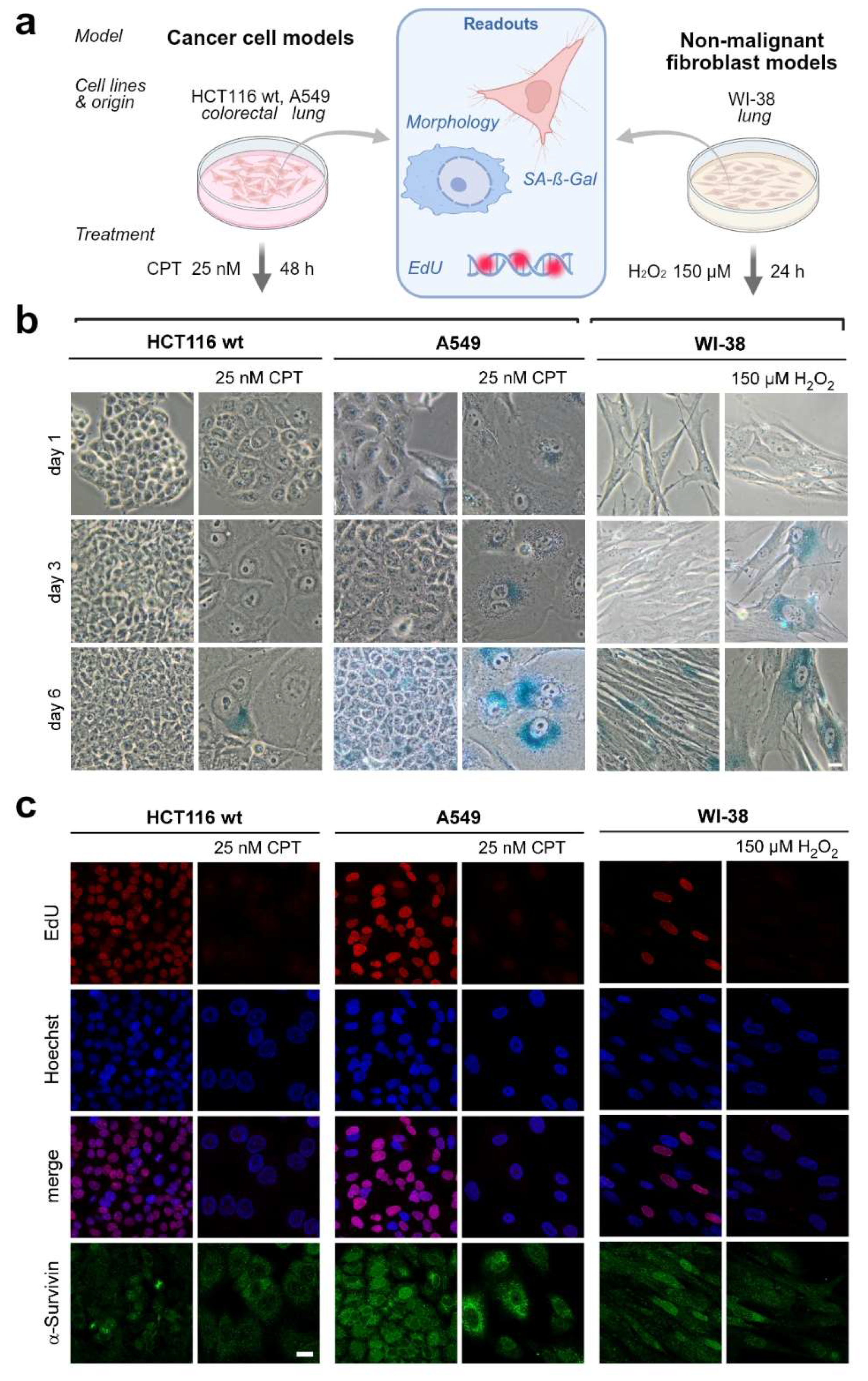
Figure 2.
Changes in Survivin and cell-cycle regulatory proteins during senescence induction. a-c. HCT116 wt and A549 cells were treated with camptothecin (CPT; 25 nM, 48 h). Three days after treatment, cells were immunostained for CDK1 and Survivin (a), p21 and Survivin (b), or CDK1 and phosphorylated Survivin (SurvT34ph; c). DNA was counterstained with Hoechst. Untreated cells served as controls. Representative confocal images with enlarged insets are shown. Scale bars, 25 µm. d. Schematic representation of cell-cycle regulation by cyclin-CDK complexes and the CDK inhibitors p16 and p21, highlighting CDK1-mediated phosphorylation of Survivin at Thr34 during mitosis. e. Immunoblot analysis of CDK1, p21 and phosphorylated ATM (pATM) in HCT116 wt and A549 cells following CPT treatment (25 nM, 48 h). GAPDH served as a loading control. Relative band intensities are indicated below the corresponding blots.
Figure 2.
Changes in Survivin and cell-cycle regulatory proteins during senescence induction. a-c. HCT116 wt and A549 cells were treated with camptothecin (CPT; 25 nM, 48 h). Three days after treatment, cells were immunostained for CDK1 and Survivin (a), p21 and Survivin (b), or CDK1 and phosphorylated Survivin (SurvT34ph; c). DNA was counterstained with Hoechst. Untreated cells served as controls. Representative confocal images with enlarged insets are shown. Scale bars, 25 µm. d. Schematic representation of cell-cycle regulation by cyclin-CDK complexes and the CDK inhibitors p16 and p21, highlighting CDK1-mediated phosphorylation of Survivin at Thr34 during mitosis. e. Immunoblot analysis of CDK1, p21 and phosphorylated ATM (pATM) in HCT116 wt and A549 cells following CPT treatment (25 nM, 48 h). GAPDH served as a loading control. Relative band intensities are indicated below the corresponding blots.
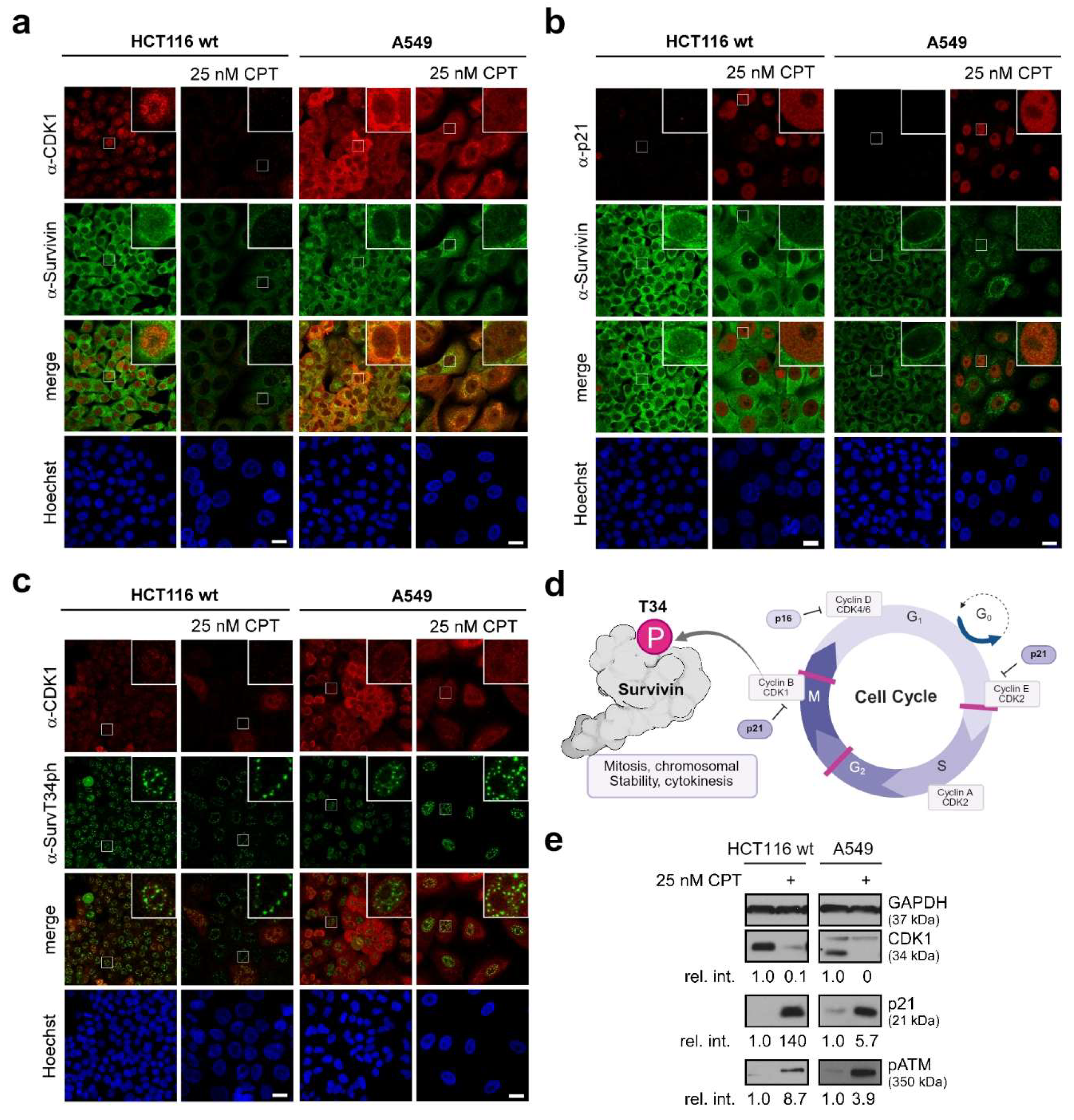
Figure 3.
Phenotypic and molecular consequences of Survivin depletion. a. Representative bright-field images of senescence-associated β-galactosidase (SA-β-gal) staining and phase-contrast images of HCT116 wt and A549 cells 72 h after transfection with non-targeting siRNA (siNT) or Survivin-specific siRNA (siSurv). Insets show enlarged regions. Scale bars, 100 µm. b. Schematic overview of the experimental workflow used for the immunoblot analyses in c-e. Cells were transfected with siNT or siSurv and subsequently treated with camptothecin (CPT; 25 nM, 48 h) or left untreated. X indicates sampling time points, whereas (X) denotes additional siRNA transfections. c. Immunoblot analysis of Survivin, p53, CDK1 and p21 in A549 cells at 72, 144 and 216 h after siRNA transfection, with or without subsequent CPT treatment. d. Time-course immunoblot analysis of Survivin, p53, CDK1 and p21 in HCT116 wt cells at 24, 48, 72, 144 and 216 h after siRNA transfection, with or without CPT treatment. e. Immunoblot analysis of Survivin, p53, CDK1, p21 and phosphorylated ATM (pATM) in HCT116 wt cells at 72, 144 and 216 h after siRNA transfection, with or without CPT treatment. GAPDH served as a loading control in all immunoblot analyses.
Figure 3.
Phenotypic and molecular consequences of Survivin depletion. a. Representative bright-field images of senescence-associated β-galactosidase (SA-β-gal) staining and phase-contrast images of HCT116 wt and A549 cells 72 h after transfection with non-targeting siRNA (siNT) or Survivin-specific siRNA (siSurv). Insets show enlarged regions. Scale bars, 100 µm. b. Schematic overview of the experimental workflow used for the immunoblot analyses in c-e. Cells were transfected with siNT or siSurv and subsequently treated with camptothecin (CPT; 25 nM, 48 h) or left untreated. X indicates sampling time points, whereas (X) denotes additional siRNA transfections. c. Immunoblot analysis of Survivin, p53, CDK1 and p21 in A549 cells at 72, 144 and 216 h after siRNA transfection, with or without subsequent CPT treatment. d. Time-course immunoblot analysis of Survivin, p53, CDK1 and p21 in HCT116 wt cells at 24, 48, 72, 144 and 216 h after siRNA transfection, with or without CPT treatment. e. Immunoblot analysis of Survivin, p53, CDK1, p21 and phosphorylated ATM (pATM) in HCT116 wt cells at 72, 144 and 216 h after siRNA transfection, with or without CPT treatment. GAPDH served as a loading control in all immunoblot analyses.
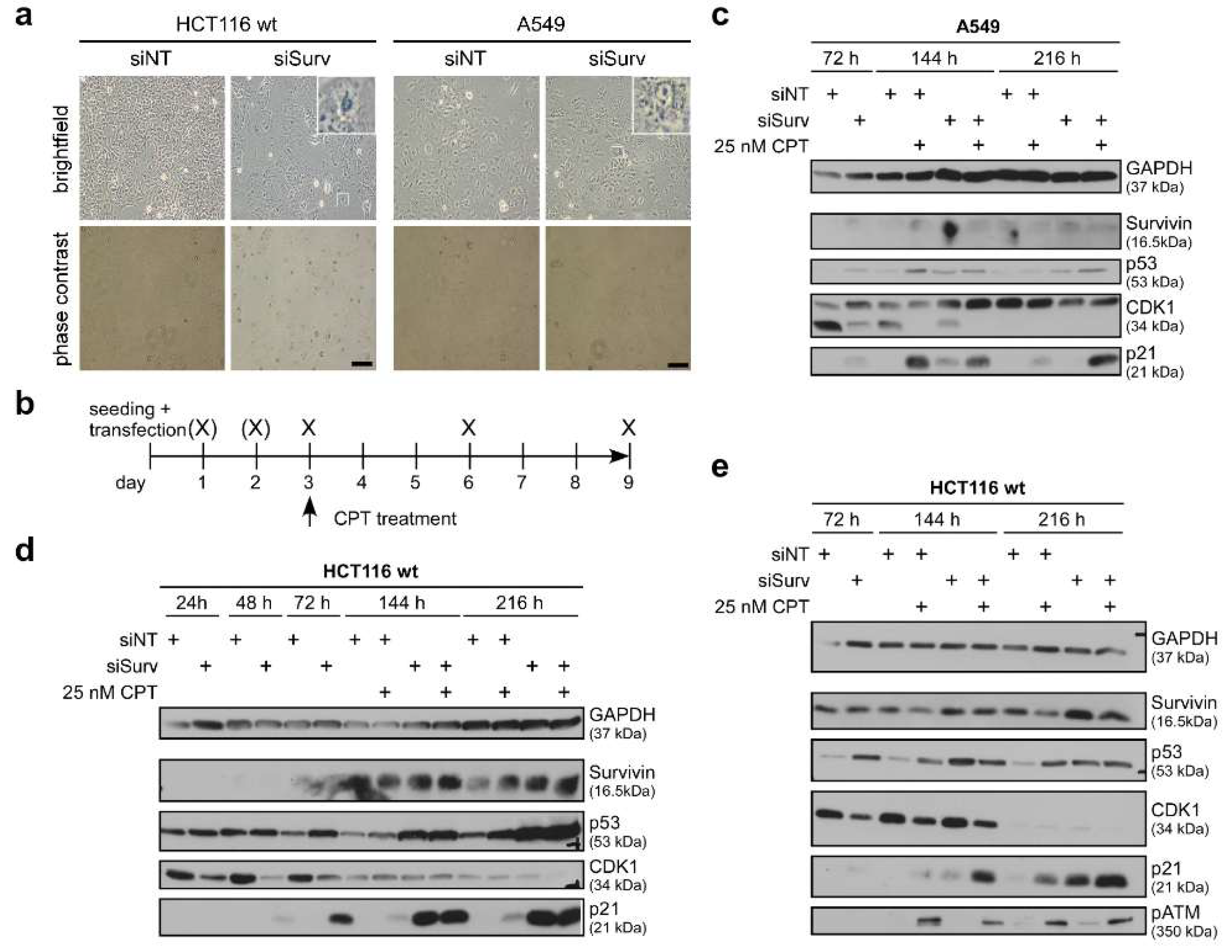
Figure 4.
Heterochromatin organization during cellular senescence. a. Immunofluorescence analysis of Survivin and Aurora B in relation to H3K9me2/3 in HCT116 wt cells following senescence induction. b. Immunofluorescence analysis of CRM1 and INCENP in relation to HP1α in HCT116 wt cells. For a and b, cells were treated with camptothecin (CPT; 25 nM, 48 h) and analyzed three days after treatment. DNA was counterstained with Hoechst. Representative confocal images with enlarged insets are shown. Scale bars, 7.5 µm. c. Schematic illustration of heterochromatin organization, highlighting centromeric heterochromatin and H3K9-methylated, HP1-associated chromatin, as well as changes in local chromatin interactions associated with senescence-associated heterochromatin foci (SAHF). c. Schematic representation of heterochromatin reorganization during cellular senescence, illustrating the redistribution of heterochromatin from the nuclear periphery and the formation of SAHF.
Figure 4.
Heterochromatin organization during cellular senescence. a. Immunofluorescence analysis of Survivin and Aurora B in relation to H3K9me2/3 in HCT116 wt cells following senescence induction. b. Immunofluorescence analysis of CRM1 and INCENP in relation to HP1α in HCT116 wt cells. For a and b, cells were treated with camptothecin (CPT; 25 nM, 48 h) and analyzed three days after treatment. DNA was counterstained with Hoechst. Representative confocal images with enlarged insets are shown. Scale bars, 7.5 µm. c. Schematic illustration of heterochromatin organization, highlighting centromeric heterochromatin and H3K9-methylated, HP1-associated chromatin, as well as changes in local chromatin interactions associated with senescence-associated heterochromatin foci (SAHF). c. Schematic representation of heterochromatin reorganization during cellular senescence, illustrating the redistribution of heterochromatin from the nuclear periphery and the formation of SAHF.
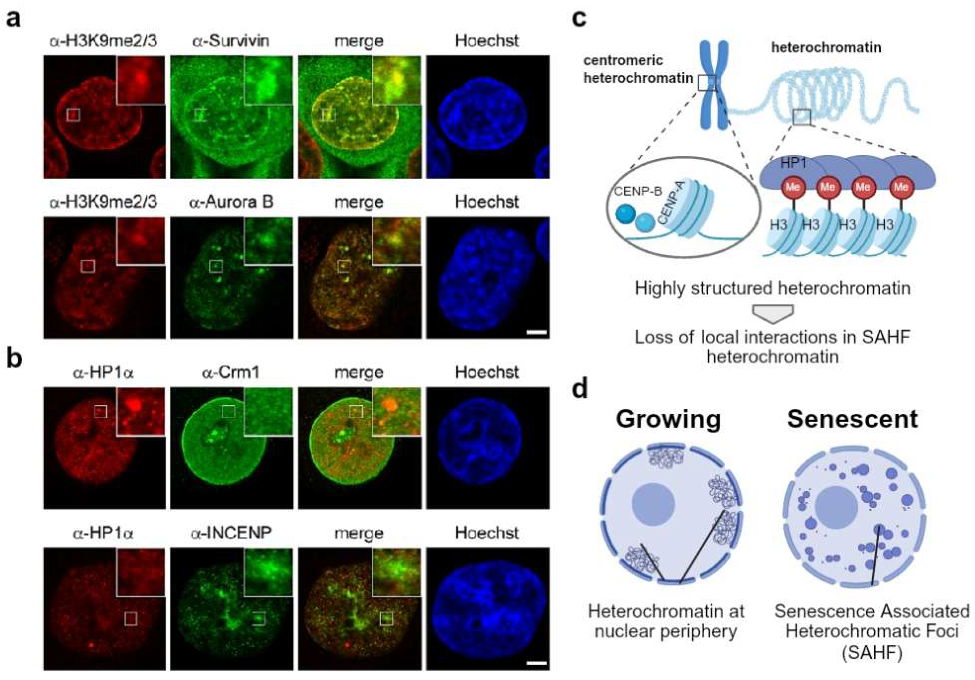
Figure 5.
Schematic overview of Survivin-associated processes during the transition from proliferation to cellular senescence. Proposed model integrating the findings of the present study with the established roles of Survivin in cell-cycle progression and chromosomal passenger complex (CPC) function. In proliferating cells, Survivin contributes to mitotic competence as a component of the CPC together with Aurora B, INCENP and Borealin. Its potential involvement in replication-associated processes and chromatin organization is also illustrated. Senescence-inducing stress is associated with activation of the DNA damage response, including ATM phosphorylation and the recruitment of DNA damage-associated factors such as γH2AX and 53BP1. Activation of p53 and induction of the CDK inhibitor p21 contribute to reduced cyclin-CDK activity and cell-cycle arrest. These changes accompany the transition towards a senescence-associated phenotype, characterized by reduced proliferative capacity and altered cellular morphology. The schematic integrates established molecular pathways with potential Survivin-associated functions.
Figure 5.
Schematic overview of Survivin-associated processes during the transition from proliferation to cellular senescence. Proposed model integrating the findings of the present study with the established roles of Survivin in cell-cycle progression and chromosomal passenger complex (CPC) function. In proliferating cells, Survivin contributes to mitotic competence as a component of the CPC together with Aurora B, INCENP and Borealin. Its potential involvement in replication-associated processes and chromatin organization is also illustrated. Senescence-inducing stress is associated with activation of the DNA damage response, including ATM phosphorylation and the recruitment of DNA damage-associated factors such as γH2AX and 53BP1. Activation of p53 and induction of the CDK inhibitor p21 contribute to reduced cyclin-CDK activity and cell-cycle arrest. These changes accompany the transition towards a senescence-associated phenotype, characterized by reduced proliferative capacity and altered cellular morphology. The schematic integrates established molecular pathways with potential Survivin-associated functions.
Disclaimer/Publisher’s Note: The statements, opinions and data contained in all publications are solely those of the individual author(s) and contributor(s) and not of MDPI and/or the editor(s). MDPI and/or the editor(s) disclaim responsibility for any injury to people or property resulting from any ideas, methods, instructions or products referred to in the content. |
© 2026 by the authors. Licensee MDPI, Basel, Switzerland. This article is an open access article distributed under the terms and conditions of the Creative Commons Attribution (CC BY) license.
Copyright: This open access article is published under a Creative Commons CC BY 4.0 license, which permit the free download, distribution, and reuse, provided that the author and preprint are cited in any reuse.