

Submitted:
31 October 2026
Posted:
03 November 2026
You are already at the latest version
Abstract
Viral infections influence every stage of the kidney transplantation continuum, shaping immunologic risk, graft outcomes, and long term patient survival. Pre transplant serologic assessments guide critical decisions, including vaccinations, the avoidance of lymphocyte depleting induction in Epstein-Barr virus (EBV) mismatched candidates and the selection of Cytomegalovirus (CMV) prophylaxis tailored to donor–recipient serostatus. Acceptance of hepatitis C viremic donor organs has significantly reduced wait times, while human immunodeficiency virus (HIV) positive donor organs remain restricted to HIV positive recipients. Post transplant, viral pathogens are major drivers of morbidity: CMV functions as an immunomodulator that increases susceptibility to rejection and opportunistic fungal infections, and routine surveillance for CMV and BK viremia is essential, especially following rejection therapies. BK virus nephropathy, adenovirus nephropathy, and JC virus nephropathy can lead to progressive allograft dysfunction. Additionally, COVID 19 and parvovirus infections have been associated with collapsing focal segmental glomerulosclerosis (FSGS) and severe graft injury. Persistent viral infections and virus associated glomerulopathies contribute to chronic graft loss, while EBV driven post transplant lymphoproliferative disorder (PTLD) remains a significant cause of cancer related mortality. Transplant recipients are also at increased risk for persistent human papillomavirus infection (HPV) and progression to anogenital and oropharyngeal malignancies. Finally, donor derived viral infections, including rabies, West Nile virus (WNV), and lymphocytic choriomeningitis virus (LCMV), though rare, are well described in the literature. Collectively, these viral complications substantially impact morbidity, graft survival, and overall mortality in kidney transplant recipients.
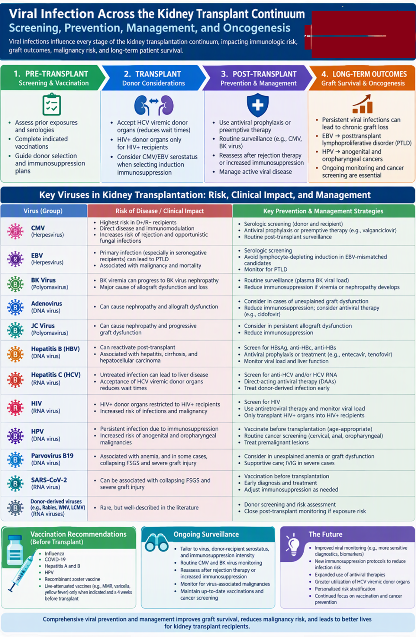
Keywords:
transplantation
; BK virus
; CMV infection
; parvovirus B19
; malignancy
; immunosuppression
Introduction
Solid organ transplant recipients (SOTRs) are at increased risk for viral infections because immunosuppression (IS) can reactivate latent infections acquired before transplantation, facilitate donor-derived transmission, and increase susceptibility to newly acquired community infections. Viral infections may result in direct tissue-invasive disease or exert indirect effects through immune modulation. Immunomodulatory viruses, including CMV, EBV, human herpesvirus 6 (HHV-6), hepatitis B virus (HBV), and hepatitis C virus (HCV), can alter inflammatory and antiviral immune pathways [1,2,3,4,5], thereby increasing the risk of opportunistic infections, allograft dysfunction, and virus-associated malignancies.
Mitigating these risks requires a comprehensive and integrated approach that includes pretransplant screening and vaccination, individualized antiviral prophylaxis or preemptive therapy, and ongoing post-transplant surveillance. Pretransplant evaluation typically involves a detailed assessment of prior infectious exposures and comprehensive virologic screening of both donors and recipients [6]. The resulting donor-recipient serologic profile, considered in conjunction with the anticipated intensity of induction and maintenance IS, informs decisions regarding organ acceptance, risk stratification, antiviral prophylaxis, preemptive therapy, and post-transplant monitoring.
Vaccination status should also be carefully reviewed, and all indicated vaccines should be administered before transplantation whenever feasible, as immune responses are generally more robust prior to transplantation and live-attenuated vaccines may still be administered safely during this period [7]. Following transplantation, surveillance strategies should be tailored to the specific virus, donor-recipient serostatus, and the recipient's net state of IS. Virus-specific prevention and monitoring strategies should be reassessed after treatment for rejection or other clinically significant increases in IS, particularly for CMV and BK virus infections [8,9].
Vaccination
Vaccination status should be reviewed early during kidney transplant evaluation, as vaccine responses are generally more robust before transplantation, and live-attenuated vaccines are typically contraindicated once IS has been initiated. Age-appropriate viral immunizations, including influenza, COVID-19, hepatitis A and B, human papillomavirus (HPV), and recombinant zoster vaccines, should be completed before transplantation whenever feasible, with accelerated or catch-up schedules used when necessary [7,10]. Administration of non-live vaccines does not require temporary inactivation from the deceased-donor waitlist; however, vaccination at least 2 weeks before transplantation is preferred. Live-attenuated viral vaccines, including measles-mumps-rubella, varicella, and yellow fever vaccines, should be administered only when clinically indicated and at least 4 weeks before transplantation to ensure resolution of vaccine-virus replication

Table 1.
showing vaccine considerations in the pre transplant phase.
| Non-live vaccines | Influenza virus COVID 19 virus Hepatitis A virus Hepatitis B virus Human Papilloma virus Varicella Zoster virus- recombinant vaccine |
| Live attenuated vaccines | Mumps-Measles-Rubella Varicella Zoster virus Yellow fever virus |
Pretransplant vaccination and cancer screening are particularly important for oncogenic viruses such as HPV, as post-transplant IS increases the risk of persistent infection and HPV-associated malignancies. Although HPV infection or treated premalignant lesions alone should not be considered equivalent to active malignancy, transplantation is generally deferred in candidates with active cancer. Under the 2020 KDIGO framework, adequately treated cervical carcinoma in situ does not require an additional waiting period, whereas suggested intervals following completion of curative treatment are at least 2 years for localized cervical cancer and at least 5 years for invasive disease, with individualized multidisciplinary assessment [10].
Pre-Transplant Viral Screening and Significance
Clinically important viruses in transplantation span multiple genomic and taxonomic groups, including DNA viruses such as herpesviruses, polyomaviruses, and HBV, as well as RNA viruses such as HIV and HCV [11]. These viral characteristics influence replication, latency, diagnostic testing, and antiviral susceptibility. Viral infections that are untreated or unrecognized before transplantation may become clinically manifest after transplantation as a consequence of IS. Accordingly, pretransplant viral screening is essential for identifying infections that may influence transplant candidacy, donor selection, antiviral treatment, prophylaxis, or post-transplant surveillance. Among the most clinically significant assessments are donor and recipient CMV and EBV serostatus, which inform risk stratification and post-transplant prevention and monitoring strategies and, in selected circumstances, may influence the choice of induction IS [10].
CMV seroprevalence is estimated at 83% in the general population and approximately 86% among women of reproductive age and among blood or organ donors [12]. Both donors and recipients should undergo CMV IgG testing before transplantation. CMV-seronegative recipients of kidneys from CMV-seropositive donors (D+/R−) are at the highest risk for primary CMV infection and disease. Seropositive recipients, whether D+/R+ or D−/R+, are generally considered at intermediate risk because of the potential for viral reactivation or reinfection, whereas D−/R− recipients have the lowest transplant-associated risk [10].
Recommended EBV testing includes viral capsid antigen (VCA) IgG and Epstein-Barr nuclear antigen (EBNA) IgG [10]. EBV D+/R− transplantation places recipients at increased risk for primary EBV infection and EBV-associated PTLD. Consequently, EBV serostatus may influence the selection of induction therapy. In an observational study of 6,620 adult EBV D+/R− kidney transplant recipients, rATG and alemtuzumab were associated with higher adjusted risks of PTLD compared with basiliximab (adjusted sub-distribution hazard ratio [aSHR] = 1.98, 95% confidence interval [CI] 1.29-3.04, P = .002 for ATG and aSHR = 1.80, 95% CI 1.04-3.11, P = .04 for alemtuzumab); however, PTLD risk did not differ significantly between rATG and alemtuzumab [13].
Clinically relevant human herpesviruses include HSV-1, HSV-2, varicella zoster virus (VZV), HHV-6, HHV-7, and HHV-8. HSV and VZV serologic testing can identify prior exposure and, in the case of VZV, candidates who may benefit from vaccination before transplantation [10]. Routine screening for HHV-6 and HHV-7 is not recommended in asymptomatic candidates, and neither routine prophylaxis nor preemptive therapy is indicated [14]. HHV-6 may interact with CMV and other pathogens, although the clinical significance of these interactions in transplantation remains incompletely understood [15].
HHV-8, also known as Kaposi sarcoma–associated herpesvirus (KSHV), is the etiologic agent of Kaposi sarcoma (KS), primary effusion lymphoma, multicentric Castleman disease, and KSHV inflammatory cytokine syndrome. It has also been associated with hemophagocytic syndromes and bone marrow failure [14,16]. Between 2021 and 2025, the CDC investigated organs from 46 deceased donors suspected of transmitting KSHV, approximately fivefold higher than the nine suspected donor-derived events reported during 2016-2020. Two-thirds of implicated donors had a history of nonmedical inhaled or injected drug use [17]. Among 153 recipients of organs from implicated donors, 74 (48%) developed post-transplant KSHV infection. Of these, 45 (61%) developed KS, including 10 who also developed a KSHV-associated lymphoproliferative disorder and 6 who developed KSHV inflammatory cytokine syndrome. These findings support maintaining a high index of clinical suspicion and pursuing targeted testing when donor risk factors are present or when KSHV infection is identified in another recipient of organs from the same donor.
Kidney transplant candidates with HIV should generally have a CD4 count of at least 200 cells/µL, suppressed HIV RNA levels, and a stable antiretroviral regimen, with transplant eligibility determined collaboratively with an HIV specialist [10]. The HIV Organ Policy Equity (HOPE) Act and its subsequent regulatory implementation initially permitted transplantation of organs from donors with HIV into recipients with HIV under research protocols and specified safeguards [18]. In a multicenter observational study, kidney transplantation from donors with HIV to recipients with HIV was noninferior to transplantation from donors without HIV [19]. The primary safety endpoint was a composite of death from any cause, graft loss, serious adverse events, HIV breakthrough infection, persistent failure of HIV treatment, or opportunistic infection. The incidence of this composite outcome did not differ between recipients of kidneys from donors with HIV and those receiving kidneys from donors without HIV (adjusted HR 1.00, 95% CI 0.73-1.38), demonstrating noninferiority. Furthermore, secondary safety analyses evaluating cancer, surgical complications, and vascular complications revealed no significant differences, supporting the overall safety of this approach [19]. Subsequent mechanistic studies have evaluated inflammatory and immunologic profiles among kidney donors with and without HIV, although the clinical implications of these findings remain uncertain [20]. Access to kidneys from donors with HIV was also associated with a higher transplantation rate among HOPE candidates than among non-HOPE candidates (70% versus 43%) and a substantially shorter median time to transplantation (10.3 versus 60.8 months) [21].
HCV screening should include an anti-HCV immunoassay followed by nucleic acid test (NAT) when the antibody result is positive [10]. Direct NAT should also be considered when recent exposure or immunocompromise raises concern for a false-negative antibody result. The THINKER and EXPANDER trials provided early evidence supporting transplantation of kidneys from HCV-viremic donors into recipients without HCV infection when direct-acting antiviral (DAA) therapy was provided [22,23]. The multicenter MYTHIC trial subsequently demonstrated the efficacy and safety of early treatment with an 8-week course of glecaprevir-pibrentasvir following transplantation of kidneys from HCV-viremic donors into HCV-negative recipients [24]. A single-center study similarly demonstrated comparable 1-year allograft function and rejection outcomes between recipients of kidneys from HCV-viremic and HCV-nonviremic donors [25]. KDIGO recommends consideration of kidneys from HCV-infected donors regardless of recipient HCV status, provided informed consent, education, and timely access to DAA therapy are ensured [26]. A systematic review conducted for the KDIGO guideline reported sustained virologic response rates exceeding 97%, with excellent 1-year patient and allograft survival [27].
Pretransplant HBV screening should include HBsAg, anti-HBs, and total anti-HBc. HBV DNA testing is indicated when either HBsAg or anti-HBc is positive [10]. Candidates with chronic HBV infection may undergo isolated kidney transplantation provided they do not have decompensated cirrhosis and receive appropriate antiviral therapy and specialist follow-up. Candidates with resolved HBV infection, defined by negative HBsAg and positive anti-HBc, have a lower but persistent risk of reactivation (<5%), particularly in the absence of anti-HBs or following intensified IS. Monitoring should therefore be individualized rather than uniformly restricted to the first post-transplant year [10].
Donor HBV risk should be stratified according to HBsAg, anti-HBc, and HBV NAT results. In a quantitative review of kidneys from HBsAg-negative, anti-HBc-positive donors, seroconversion to anti-HBc and/or anti-HBs occurred in 3.24% of recipients, whereas HBsAg seroconversion occurred in only 0.28% [28]. More recently, transplantation of kidneys from HBV NAT-positive donors into selected recipients, using entecavir initiated perioperatively and continued for 52 weeks, was associated with negative HBV PCR results at both 9 and 12 months [29]. With respect to long-term outcomes, a recent analysis of the SRTR database demonstrated that transplantation of kidneys from HBV NAT-positive donors was not associated with inferior graft or patient survival compared with HBV NAT-negative donors (p≥0.188) [30]. In addition, waitlist time was significantly shorter among recipients of HBV NAT-positive donor kidneys than among recipients of HBV NAT-negative donor kidneys (9.1 [IQR, 18.7] vs. 19.0 [IQR, 32] months; p<0.001) [30]. Although standardized viral risk stratification and established safety data for all categories of "HBV-positive" donors remain limited, these findings support the carefully selected use of kidneys from anti-HBc-positive and HBV NAT-positive donors within structured institutional antiviral prophylaxis and monitoring protocols.
Table 2.
summarizing pre transplant viral screening and transplant considerations.
| Viral screening | Clinical implication |
| Recipient and donor Cytomegalovirus serostatus | Determination of post-transplant prophylaxis |
| Recipient and donor Ebstein-Barr virus serostatus | Determination of transplant induction regimen |
| HIV positive recipient | Consideration for HIV positive organ donor (HOPE Act) |
| Donor Hepatitis (B&C) virus Nucleic Acid Test positivity | Recipients wait list time reduction |
Considerations in the Perioperative Period
The perioperative period represents the transition from pretransplant viral risk stratification to the implementation of induction therapy, initial maintenance IS, antiviral prophylaxis, and post-transplant monitoring strategies. Because IS is most intense during this period, close coordination among transplant, infectious diseases, pharmacy, and HIV specialists is essential to optimize outcomes and minimize infectious complications.
Cytomegalovirus
CMV may manifest as asymptomatic DNAemia, a mononucleosis-like syndrome, or tissue-invasive disease, after which the virus establishes lifelong latency with the potential for subsequent reactivation. Among SOTRs receiving valganciclovir or ganciclovir prophylaxis, one study reported that 38 of 585 recipients (6.5%) developed CMV infection or disease while receiving prophylaxis. Recipients with D+/R− serostatus were significantly more likely to develop breakthrough infection than D+/R+ and D−/R+ recipients (16.1% vs. 4.6% and 2.2%, respectively). D+/R− mismatch was independently associated with CMV breakthrough infection [31].
Valganciclovir 900 mg daily, adjusted for kidney function, remains a standard prophylactic option. In kidney transplantation, 6 months of prophylaxis is recommended for D+/R− recipients, whereas for seropositive recipients, either 3 months of prophylaxis or a preemptive monitoring strategy may be employed according to the recipient’s overall risk profile [8]. CMV serostatus may also be considered when selecting induction therapy. Among D+/R− recipients, rATG has been associated with a higher risk of CMV infection than basiliximab, particularly when antiviral prophylaxis is not consistently administered [32]. Similarly, when alemtuzumab was compared with basiliximab, alemtuzumab induction was associated with a significantly increased risk of CMV viremia (OR 3.15, 95% CI 1.6-6.5; P<0.001) [33]. These findings are biologically plausible because effective CMV control depends predominantly on cell-mediated immunity, which is substantially depleted by rATG and alemtuzumab but relatively preserved with basiliximab. In the INTAC trial, among high-risk patients who received either alemtuzumab or rATG, 3-year rates of CMV infection were similar (11% vs. 12%; P=1.00) [34].
Letermovir is an alternative to valganciclovir for CMV prophylaxis in high-risk D+/R− kidney transplant recipients. In a randomized trial, letermovir administered for 200 days was noninferior to valganciclovir for the prevention of CMV disease through 52 weeks (10.4% vs. 11.8%) and was associated with substantially lower rates of leukopenia or neutropenia (26% vs. 64%) [35]. A post hoc analysis demonstrated that impaired kidney function adversely affected valganciclovir exposure and adherence, whereas letermovir efficacy was maintained across varying levels of kidney function [36]. Because letermovir does not require renal dose adjustment, it may offer a particular advantage during periods of unstable allograft function. For intermediate-risk recipients (R+), switching to letermovir has been suggested in cases of valganciclovir intolerance; however, the supporting evidence remains limited [8]. Additional data are needed to better define the role of letermovir in R+ SOTRs. Letermovir inhibits CYP3A and CYP2C8 and may increase exposure to tacrolimus and cyclosporine, necessitating close therapeutic drug monitoring of [37]. The interaction with cyclosporine is bidirectional; when the agents are coadministered, the letermovir dose should be reduced to 240 mg daily and cyclosporine concentrations monitored closely [37].
Epstein-Barr Virus
EBV D+/R− mismatch should be considered when selecting induction therapy because rATG and alemtuzumab have been associated with a higher risk of PTLD than basiliximab in this population [13]. When lymphocyte-depleting induction is required because of substantial immunologic risk, intensified EBV DNA monitoring should be considered. Experimental data suggest that mTOR inhibition may exert antiproliferative effects against EBV-transformed B cells while preserving certain aspects of T-cell function, whereas CNIs suppress T-cell cytotoxicity directed against EBV-infected cells [38].
Human Immunodeficiency Virus
In recipients with HIV, perioperative management requires uninterrupted anti-retroviral therapy (ART), maintenance of viral suppression, and careful management of interactions between antiretroviral and immunosuppressive medications. Tenofovir alafenamide is generally preferred over tenofovir disoproxil fumarate, when clinically appropriate, because of its more favorable renal and bone safety profile [39]. Unboosted second-generation integrase strand transfer inhibitor regimens containing bictegravir or dolutegravir are preferred because they have fewer interactions with CNIs and mTOR inhibitors. Ritonavir- or cobicistat-boosted protease inhibitor regimens should generally be avoided because potent CYP3A4 inhibition can markedly increase tacrolimus, cyclosporine, or sirolimus exposure. Therapeutic drug monitoring of immunosuppressive agents is therefore essential, particularly when ART is initiated, discontinued, or modified [39].
HIV RNA and CD4 counts should be assessed before transplantation and monitored after transplantation in collaboration with an HIV specialist. NIH guidance emphasizes the importance of maintaining viral suppression, avoiding interruptions in ART, and closely monitoring HIV RNA following transplantation [39]. For patients receiving stable suppressive ART, HIV RNA is generally measured every 3 to 4 months and may be extended to every 6 months after more than 1 year of sustained viral suppression and clinical stability. Monitoring should be intensified following ART interruption or modification or when virologic failure, nonadherence, impaired absorption, or clinically significant drug interactions are suspected. CD4 monitoring should be individualized according to baseline CD4 count, degree of IS, virologic control, and risk of opportunistic infection [39].
CD4 counts frequently decline after transplantation, particularly following lymphocyte-depleting induction. A CD4 count below 200 cells/µL at 4 weeks after kidney transplantation has been associated with an increased risk of serious infection during the first 6 months post-transplant [40]. CD4 trends should therefore inform decisions regarding opportunistic infection prophylaxis and evaluation for excessive IS. Maintenance therapy should remain individualized based on rejection risk, graft function, infection history, leukopenia, and therapeutic drug levels.
Empiric reduction of IS based solely on a low CD4 count should be avoided because recipients with HIV already have an increased risk of rejection. This heightened risk is thought to be multifactorial and may reflect reduced exposure to immunosuppressive agents due to drug interactions, infection of the allograft, prior alloimmunization, and chronic immune activation [41]. Contemporary data continue to demonstrate this rejection signal. In a UK registry study, crude 12-month acute rejection rates among recipients with versus without HIV were 24.8% versus 14.2% following deceased-donor transplantation and 37.5% versus 15.3% following living-donor transplantation [42]. After adjustment, the association remained significant in the living-donor cohort but not in the deceased-donor cohort, suggesting that some of the historically reported excess risk may be attributable to confounding demographic and socioeconomic factors. Nevertheless, meticulous management of immunosuppressant exposure and avoidance of ART-related pharmacokinetic interactions remain critical.
Post-transplant HIV viral breakthrough may occur, particularly in the setting of ART interruption or subtherapeutic drug exposure. In the HOPE in Action study, viral breakthrough occurred in 10% of HIV D+/R+ recipients and 4% of HIV D−/R+ recipients, corresponding to an incidence-rate ratio of 3.14 [19]. However, no recipient developed persistent virologic failure, and only one potential donor-derived HIV superinfection was identified [19]. These findings underscore the importance of maintaining ART and monitoring HIV RNA after transplantation. The phenomenon is more appropriately described as HIV viral breakthrough rather than “HIV resurgence.” In this study, breakthrough was defined as HIV RNA >200 copies/mL or a single HIV RNA measurement >1000 copies/mL [19].
Hepatitis C Virus
HCV-negative recipients of kidneys from HCV-viremic donors should receive prophylactic or preemptive pan-genotypic DAA therapy. In the MYTHIC trial, an 8-week course of glecaprevir-pibrentasvir initiated 2 to 5 days after transplantation achieved sustained virologic response in all treated recipients [24]. In a separate phase 3, single-center study, 30 recipients of non-liver organs, including 10 kidney recipients and 1 kidney-pancreas recipient, received glecaprevir-pibrentasvir plus ezetimibe beginning before transplantation and continuing for 7 days afterward; none developed chronic HCV infection [43].
AASLD-IDSA guidance recommends initiating prophylactic therapy immediately before or on the day of transplantation, or preemptive therapy between post-transplant days 0 and 7 once the recipient is clinically stable, without requiring confirmation of recipient viremia [44]. For non-liver recipients, the recommended regimen is 8 weeks of glecaprevir-pibrentasvir when therapy is initiated within the first post-transplant week; treatment should be extended to 12 weeks if initiation is delayed beyond day 7 [44]. A separate pilot study demonstrated that a 7-day course of glecaprevir-pibrentasvir initiated before transplantation prevented chronic HCV infection in 10 non-liver organ recipients, including 6 kidney recipients [45]. The abbreviated regimens reported by Feld et al. [43] and Ramirez-Sanchez et al. [45] demonstrate the feasibility of shorter peri-transplant DAA strategies; however, the optimal duration and role of abbreviated therapy require further investigation.
Donor-Derived Neurotropic Viruses
Donor-derived LCMV, WNV, and rabies may present early after transplantation and should be considered in recipients with unexplained febrile illness, hepatitis, encephalitis, or multiorgan failure. LCMV typically presents within the first month after transplantation with fever, hepatitis, neurologic manifestations, and progressive multiorgan failure. Across six reported donor-derived transmission clusters, 15 of 21 recipients (71%) died, frequently with prominent hepatitis [46].
WNV has a typical incubation period of approximately 6 days and may present with fever, myalgias, gastrointestinal symptoms, meningitis, or encephalitis [47]. Transplant recipients receiving IS are at increased risk for neuroinvasive disease. Encephalitis was reported in approximately 70% of published transplant-associated cases reviewed by AST IDCOP, although this estimate may reflect preferential reporting of severe disease [46].
Rabies is a nearly universally fatal encephalomyelitis caused by viruses within the genus Lyssavirus [48]. Three transplant-associated rabies transmission events in the United States between 1978 and 2013 affected nine organ or tissue recipients. An additional transmission event was identified in 2025 after a kidney recipient died from rabies acquired from a donor with previously unrecognized infection following skunk exposure [49]. A high index of suspicion and rapid coordination with public health authorities are essential because these uncommon donor-derived infections may initially affect multiple recipients of organs from the same donor.
Viral Infections of Significance in the Post-Transplant Period
The post-transplant period is associated with a broad spectrum of viral complications, the timing and severity of which are influenced by donor and recipient serostatus, antiviral prophylaxis strategies, viral exposures, and the recipient's net state of immunosuppression. CMV functions as an immunomodulator that increases susceptibility to rejection and opportunistic fungal infections, and routine surveillance for CMV and BK viremia is essential, especially following rejection therapies.
Cytomegalovirus
CMV remains one of the most common opportunistic viral infections after SOT, particularly among CMV-seronegative recipients of organs from CMV-seropositive donors. CMV quantitative nucleic acid amplification testing (CMV-QNAT) is the preferred laboratory method for diagnosing and monitoring CMV infection in blood, whereas CMV IgM and IgG serologies should not be used to diagnose active CMV infection or disease [50]. Although interpretation depends on the assay used and the clinical context, changes in CMV DNAemia exceeding 0.5 log10 IU/mL (approximately threefold) or 0.7 log10 IU/mL (approximately fivefold) when the initial viral load is <3 log10 IU/mL may represent clinically significant changes [8].
The VICTOR trial demonstrated comparable long-term outcomes with oral valganciclovir and intravenous ganciclovir for the treatment of CMV disease in SOTRs [51]. Oral valganciclovir is generally preferred for patients with mild-to-moderate disease who can reliably absorb oral therapy, whereas intravenous ganciclovir is recommended for severe, life-threatening, or sight-threatening disease. Maribavir and foscarnet are important alternatives for refractory or drug-resistant CMV infection. In the SOLSTICE trial, maribavir was superior to investigator-assigned therapy in achieving CMV clearance with sustained symptom control and was associated with fewer treatment-limiting toxicities [52]. Maribavir does not cause the clinically significant myelosuppression or nephrotoxicity associated with conventional anti-CMV therapies, although dysgeusia is common. Maribavir may increase tacrolimus exposure; therapeutic drug monitoring is therefore recommended when maribavir is initiated or discontinued [37].
Foscarnet may be preferred in clinically unstable patients with high-level CMV DNAemia; however, its use is limited by substantial nephrotoxicity, including acute kidney injury, electrolyte wasting, and acute tubulointerstitial nephritis, as well as overall tolerability concerns [53]. Regarding its renal adverse effects, electrolyte disturbances are among the most frequently reported toxicities. In one report involving a patient with chronic tubulointerstitial disease and acute tubular necrosis, renal biopsy demonstrated crystal deposits containing foscarnet [53].
Polyomavirus
Among the polyomaviruses of clinical relevance after transplantation, BK virus and JC virus are the most important. BK polyomavirus (BKPyV) is the predominant polyomavirus affecting kidney transplant recipients. Viral replication typically progresses from viruria to DNAemia and, in a subset of recipients, to BK polyomavirus-associated nephropathy (BKPyVAN). BKPyVAN occurs in approximately 1%-10% of kidney transplant recipients and can result in progressive allograft dysfunction and graft loss [54].
Higher plasma BKPyV viral loads are associated with both an increased likelihood and greater histologic severity of BKPyVAN. Following the onset of viruria, nearly 50% of kidney transplant recipients develop viremia within 2-6 weeks. Plasma BK viral loads >4 log10 copies/mL have been associated with higher rates of biopsy-proven BKPyVAN, whereas levels >6 log10 copies/mL are predictive of extensive BKPyVAN pathology characterized by positive SV40 immunohistochemistry and significant inflammatory infiltrates [54].
The Second International Consensus Guidelines recommend monthly plasma BKPyV screening through month 9 after transplantation, followed by screening every 3 months through 2 years post-transplant [9]. Reduction of maintenance IS remains the cornerstone of treatment for sustained BKPyV DNAemia and biopsy-proven BKPyVAN. Kidney allograft biopsy is not routinely required before reducing IS in adults with BKPyV DNAemia and stable allograft function unless immunologic risk is considered high [9].
BKPyV presents a unique therapeutic challenge in multiorgan transplant recipients because reducing IS to control viral replication may increase the risk of rejection in the accompanying organ allograft. A recent three-center study found that BKPyV DNAemia and BKPyVAN were common after simultaneous heart-kidney transplantation and were associated with an increased risk of kidney allograft rejection, although rates of heart allograft rejection were not increased [55]. These patients therefore require coordinated monitoring of both allografts with IS modifications. Similar concerns apply to simultaneous pancreas-kidney transplantation, in which IS reduction to treat BKPyV DNAemia may increase the risk of pancreas allograft rejection [56]. This strategy highlights a gap in BKpyVAN management. Historically, other adjunctive therapies utilized included quinolones, cidofovir, leflunomide, and Intravenous immunoglobulin (IVIG) [9,54,57]. Cidofovir and leflunomide have largely fallen out of favor after a systematic review with pooled analysis found no improvement in graft survival compared with reduction of immunosuppression alone [57].
For patients with persistent BKPyV DNAemia or BKPyVAN despite appropriate reduction of immunosuppression, BK-neutralizing antibodies in IVIG preparations have been used as adjunctive or rescue therapy in observational studies [58,59], although evidence remains insufficient to support their routine use [9]. Emerging therapies include BKPyV-neutralizing monoclonal antibodies [60], including MAU868, which demonstrated greater reductions in BKPyV DNAemia than placebo in a small, randomized phase 2 trial [61]. Potravitug, a targeted therapy for BKPyV infection in kidney transplant recipients, completed a phase 1 trial in healthy adults [62] and was subsequently evaluated in a randomized phase 2 trial in kidney transplant recipients [63]. Phase 2 data demonstrated reductions in BKPyV DNAemia and biopsy-proven BKPyVAN by week 20, although larger studies are needed to establish effects on long-term allograft outcomes. In a randomized phase 2 trial, posoleucel was well tolerated and associated with greater reductions in BKPyV DNAemia than placebo [64]. Larger studies are needed to determine whether these therapies improve BKPyV-associated nephropathy and allograft outcomes.
JC polyomavirus (JCPyV) is considerably less common than BKPyV but can cause JC polyomavirus-associated nephropathy (JCPyVAN) or progressive multifocal leukoencephalopathy. JCPyV replication may remain clinically silent, and one surveillance study detected asymptomatic JCPyV DNAemia in approximately 2.7% of kidney transplant recipients [65]. Compared with BKPyVAN, JCPyVAN generally presents later after transplantation and is characterized histologically by relatively sparse viral cytopathic changes accompanied by inflammation and fibrosis [66]. Although JCPyVAN may result in allograft dysfunction, progression to graft loss appears to be uncommon.
Adenovirus
Adenovirus is another important cause of post-transplant infection and may manifest as fever, hemorrhagic cystitis, nephritis, pneumonitis, enterocolitis, or disseminated disease. In a systematic review of adenovirus nephritis among adult kidney transplant recipients, the median interval from transplantation to infection was approximately 168 days, with fever, dysuria, diarrhea, and hematuria among the most common presenting manifestations [67].
Allograft biopsy typically demonstrates tubulointerstitial nephritis. Initial management consists of supportive care and reduction of IS [68]. Although prospective randomized trials evaluating antiviral therapy are lacking, intravenous cidofovir is commonly used for severe, progressive, or disseminated adenovirus disease, with careful monitoring for nephrotoxicity [68].
IVIG may be considered as adjunctive therapy, particularly in patients with hypogammaglobulinemia, although evidence supporting its efficacy remains limited. A retrospective series reported successful treatment with combined cidofovir and IVIG in five SOTRs, including four kidney recipients and one heart recipient with invasive adenovirus disease [69]. Because all patients received combination therapy, however, the independent therapeutic contribution of IVIG cannot be determined.
Parvovirus B19
Parvovirus B19 is the most clinically important and best-characterized human pathogen within the Parvoviridae family and is a recognized cause of persistent or recurrent erythropoietin-resistant anemia with reticulocytopenia following SOT, often necessitating repeated blood transfusions [70].
The anemia and reticulocytopenia result from viral replication within erythroid progenitor cells, leading to lysis of infected cells and downregulation of erythropoietin receptor expression. Other clinical manifestations include fever, rash, arthropathy, and tissue-invasive disease, including carditis, hepatitis, pneumonitis, kidney transplant glomerulopathy, focal segmental glomerulosclerosis (FSGS) including collapsing variant, and neurologic disease [70].
Diagnosis is often delayed; therefore, a high index of clinical suspicion should prompt testing for parvovirus B19 DNA using quantitative PCR [70]. When the diagnosis remains uncertain, bone marrow examination may reveal giant pronormoblasts with finely granular cytoplasm and characteristic glassy intranuclear inclusions surrounded by a clear halo ("lantern cells"), along with an absence of late normoblasts. In situ hybridization and immunohistochemical staining may also aid diagnosis when PCR testing or serologic studies are negative. Treatment generally consists of cautious reduction of IS, including reduction or temporary discontinuation of the antiproliferative agent. IVIG may be considered in patients with severe, symptomatic, or persistent infection, typically at a total dose of 2 g/kg, as no specific antiviral therapy is currently available for parvovirus B19 infection [70].
COVID-19
COVID-19 has been associated with a diverse spectrum of kidney allograft pathology, including acute rejection, acute tubular injury, and podocytopathies. In a series of 18 kidney transplant recipients undergoing allograft biopsy after SARS-CoV-2 infection, acute rejection was identified in 5 of 11 (45%) biopsies performed within 1 month of infection, while other findings included collapsing glomerulopathy, acute tubular injury, infarction, and transplant glomerulopathy; most patients had persistent allograft dysfunction during follow-up [71]. A separate series of seven recipients demonstrated multiple forms of FSGS, including collapsing, perihilar, tip-lesion, and secondary variants, frequently accompanied by acute tubular injury and patients with collapsing or perihilar FSGS experienced progression of chronic allograft nephropathy [72]. Neither study demonstrated evidence of direct SARS-CoV-2 infection within the allograft.
COVID-19 has been associated with substantial morbidity and mortality in kidney transplant recipients. In a cohort of 912 recipients with COVID-19, 3-month survival was 82.3% overall but only 49% among the patients requiring intensive care [73]. In patients who were admitted to the ICU, 10.6% experienced irreversible loss of graft function within 3-months after presentation [73]. Vaccination was associated with improved long-term outcomes in a subsequent cohort of 129 kidney transplant recipients followed for a median of 50 months after SARS-CoV-2 infection [74]. Vaccinated recipients had significantly higher graft survival (85% vs. 52%) and patient survival (99% vs. 83%) than unvaccinated recipients, while lack of vaccination independently predicted graft failure [74]. These observational findings support the importance of COVID-19 vaccination in kidney transplant recipients.
Viral Enteritis
Viral pathogens are important causes of diarrhea after solid organ transplantation, with CMV and norovirus among the most frequently recognized viral etiologies; less common viruses include adenovirus, rotavirus, sapovirus, astrovirus, and enteroviruses [75,76]. CMV may cause tissue-invasive enterocolitis, whereas adenovirus can produce enteritis as part of localized or disseminated disease; both viruses are discussed elsewhere in this review. In addition to direct gastrointestinal morbidity, diarrhea in solid organ transplant recipients may lead to dehydration, altered exposure to immunosuppressive medications, drug toxicity, and allograft dysfunction, underscoring the importance of timely etiologic diagnosis.
Norovirus is an important cause of acute and chronic diarrhea in solid organ transplant recipients. Unlike the typically self-limited illness observed in immunocompetent hosts, infection in transplant recipients may persist for weeks to months and is diagnosed by stool-specific or multiplex PCR testing [77]. In a retrospective cohort of 79 solid organ transplant recipients with noroviral diarrhea, 70% required hospitalization and 85% experienced improvement or resolution within 6 months; chronic diarrhea preceding diagnosis was associated with persistent symptoms [77]. Management is primarily supportive and may include cautious reduction of immunosuppression, particularly reduction or discontinuation of mycophenolate when clinically appropriate. Nitazoxanide with or without intravenous immunoglobulin may be used as adjunctive therapies [77,78,79], although efficacy remains uncertain and no established antiviral therapy is currently available. In the cohort reported by Nair et al., outcomes were not significantly improved with nitazoxanide or IVIG, although all 12 recipients in whom mycophenolate was discontinued improved [77].
Enteroviruses are less well characterized as gastrointestinal pathogens after solid organ transplantation but may cause diarrhea, abdominal pain, and vomiting, particularly in immunosuppressed recipients. In a Dutch cohort of 264 patients with enterovirus infection, including 38 transplant recipients, gastrointestinal manifestations were significantly more common among transplant recipients than among nontransplant patients, occurring in 71% of infected pediatric and 46% of infected adult transplant recipients [80]. Transplant recipients were also more likely to experience persistent symptoms and recurrent enterovirus infection, although overall disease severity was not greater than in nontransplant patients [80]. Because enteroviruses may be asymptomatically detected in stool [80], attribution of gastrointestinal symptoms to enterovirus requires excluding other possible causes.
Table 3.
summarizing post-transplant viral infections and clinical manifestations.
| Name of the virus | Clinical manifestation |
| Cytomegalovirus (CMV) | CMV DNAemia, CMV tissue invasive disease |
| BK virus | BK viremia, BK Polyoma virus nephropathy |
| Adenovirus | Adenovirus hemorrhagic cystitis, nephritis, disseminated disease |
| Parvo virus B19 | Reticulocytopenia, anemia, podocytopathy |
| COVID 19 infection | Acute tubular injury, podocytopathy, pneumonia |
| Norovirus | Enteritis, failure to thrive |
Monitoring Strategies Post-Transplantation
Kidney transplantation is the preferred treatment for eligible patients with end-stage kidney disease; however, lifelong IS is required to prevent allograft rejection. Clinical management therefore requires a careful balance between insufficient IS, which increases the risk of rejection, and excessive IS, which predisposes recipients to opportunistic infections, malignancy, and drug-related toxicity.
Conventional monitoring primarily relies on immunosuppressant drug concentrations, kidney allograft function, proteinuria, donor-specific antibodies, and, when indicated, allograft biopsy. However, drug concentrations do not fully capture a recipient's net state of IS and do not reliably predict infectious or alloimmune complications.
Torque teno virus (TTV), a highly prevalent nonenveloped DNA virus belonging to the Anelloviridae family, has not been conclusively associated with human disease [81]. TTV replication increases with increasing intensity of IS and is not substantially affected by conventional antiviral therapies [82]. Consequently, plasma TTV viral load has emerged as a potential dynamic biomarker of the net state of IS rather than a marker of a specific immune cell population.
In a prospective observational study of kidney transplant recipients, TTV viral loads between approximately 1 × 10⁶ and 1 × 10⁸ copies/mL were associated with the lowest combined risk of infection and rejection [83]. A subsequent systematic review and meta-analysis similarly demonstrated that higher TTV loads were associated with infection, whereas lower loads were associated with rejection following SOT [84].
These findings support the potential use of serial TTV measurements as a complement to conventional therapeutic drug monitoring. Proposed thresholds vary according to assay methodology, specimen type, immunosuppressive regimen, and time from transplantation. TTV levels increase dynamically during the first 3-4 months after transplantation and may not reach a sufficiently stable state for reliable interpretation during this period [85]. The TTVguideIT multicenter randomized trial which enrolled stable, low immunologic risk kidney transplant recipients after the third post-transplant month, demonstrated noninferiority of TTV-guided tacrolimus dosing (86)
Consequently, TTV-guided IS management should currently be viewed as complementary to, rather than a replacement for, established clinical, pharmacologic, virologic, and allograft monitoring strategies until its utility is validated across broader transplant populations.
Virus-specific immune assays may provide an additional individualized approach to infection risk assessment. CMV-specific cell-mediated immunity assays, including interferon-γ release assays and enzyme-linked immunospot assays, may help identify recipients at increased risk for CMV infection following discontinuation of prophylaxis and may assist in selected decisions regarding the duration of secondary prophylaxis. However, variability among available assays and the absence of standardized thresholds currently limit their universal application. Accordingly, these assays should not replace CMV-QNAT surveillance when virologic monitoring is otherwise clinically indicated [8].
Virus-Associated Malignancies After SOT
SOTRs have an approximately twofold higher overall cancer risk than the general population, although risk varies by cancer type, transplanted organ, recipient characteristics, and the duration and intensity of IS [88]. Virus-associated malignancies account for a disproportionate share of this excess risk. Major oncogenic viruses include EBV, which causes most early PTLD and rare EBV-associated smooth muscle tumors; KSHV/HHV-8, which causes KS, primary effusion lymphoma, and multicentric Castleman disease; HPV, which is associated with cervical, vulvar, vaginal, anal, penile, and oropharyngeal cancers; Merkel cell polyomavirus (MCPyV), which is linked to a subset of Merkel cell carcinomas; and HBV and HCV, which contribute to hepatocellular carcinoma [89,90,91,92,93,94]. Human T-cell lymphotropic virus type-1 is an additional consideration in recipients from endemic regions, including the Caribbean and parts of Africa, South America, Asia, and Oceania, where it is associated with adult T-cell leukemia/lymphoma, although the impact of post-transplant IS on disease progression remains incompletely characterized [95].
Post-transplant viral oncogenesis reflects the interplay of viral persistence, impaired immune surveillance, direct viral oncogenic effects, chronic inflammation, and host and environmental factors. Immunosuppressive therapy impairs cytotoxic T-cell and natural killer cell control of viral replication, facilitating primary infection, donor-derived transmission, or reactivation of latent oncogenic viruses [89]. Several viruses also directly alter cellular growth pathways. EBV latent proteins promote B-cell survival and proliferation [96], HHV-8 proteins drive angiogenesis, proliferation, immune evasion, and inflammatory signaling [91], high-risk HPV types E6 and E7 oncoproteins disrupt the p53 and retinoblastoma pathways [97], and MCPyV promotes Merkel cell carcinoma through viral DNA integration and oncogenic protein expression [98]. HBV contributes to hepatocarcinogenesis through viral DNA integration, genomic instability, HBx protein expression, and chronic inflammation, whereas HCV promotes carcinogenesis through chronic inflammation, oxidative stress, fibrosis, and cirrhosis [94].
Cancer risk reflects both cumulative IS exposure and the immunosuppressive regimen used [89,90]. In addition to suppressing antitumor immunity, CNIs may exert direct protumorigenic effects through increased transforming growth factor-β and vascular endothelial growth factor expression that lead to tumor cell invasiveness and tumor angiogenesis [99,100]. Azathioprine metabolites induce mutagenic oxidative DNA damage following ultraviolet A exposure [101]. In contrast, mTOR inhibitors possess antiproliferative and antiangiogenic properties and have been associated with reductions in selected post-transplant malignancies, particularly nonmelanoma skin cancer and KS [90]. However, their use must be balanced against adverse effects including proteinuria, dyslipidemia, cytopenias, impaired wound healing, edema, oral ulcers, and pneumonitis, as well as higher rates of treatment discontinuation and rejection in selected patients [90,102].
Prevention and management require an integrated strategy encompassing pretransplant risk assessment, vaccination when available, post-transplant viral and cancer surveillance, modification of IS when feasible, antiviral therapy, and tumor-specific treatment.
Epstein-Barr Virus and Post-Transplant Lymphoproliferative Disorder
PTLD comprises a spectrum ranging from nondestructive lymphoid proliferations to polymorphic and monomorphic lymphomas, most commonly diffuse large B-cell lymphoma. Approximately 60%-70% of PTLD cases are EBV-associated, although late-onset disease is more frequently EBV-negative [91,95,103]. Among adult kidney and kidney-pancreas recipients, cumulative PTLD incidence is approximately 1% at 5 years and 2% at 10 years, with substantially higher rates following intestinal and thoracic transplantation [103]. Major risk factors include EBV donor-positive/recipient-negative serostatus, primary EBV infection, younger age, greater cumulative IS exposure, and T-cell-depleting induction therapy [13,91].
PTLD commonly involves extranodal sites, including the gastrointestinal tract, allograft, and central nervous system. Diagnosis requires tissue biopsy with assessment of morphology, immunophenotype, clonality, and EBV-encoded RNA. Although EBV DNAemia may support clinical suspicion and monitoring, it lacks sufficient sensitivity and specificity to establish or exclude the diagnosis. Prognosis depends on histologic subtype, disease stage, performance status, extranodal or central nervous system involvement, and treatment response. In a contemporary cohort, overall survival was approximately 68% at 1 year and 51% at 5 years [103].
IS reduction is the initial therapeutic intervention when feasible [104]. For systemic CD20-positive B-cell PTLD, response-adapted therapy is preferred, typically consisting of four weekly rituximab doses followed by additional rituximab in complete responders or R-CHOP in those without complete remission [104,105]. This approach resulted in a median overall survival of 6.6 years in the PTLD-1 trial [105]. Localized disease may be managed with surgery or radiotherapy, whereas central nervous system, T-cell, Hodgkin-type, and relapsed or refractory PTLD require subtype-specific therapy. Although mTOR inhibition may suppress EBV-associated B-cell proliferation while maintaining immunosuppression [38,90], it should be considered an adjunctive modification of maintenance IS rather than a substitute for lymphoma-directed therapy.
HHV-8/KSHV-Associated Diseases
KS is the most common HHV-8/KSHV-associated malignancy after SOT, although KSHV may also cause primary effusion lymphoma, multicentric Castleman disease, and KSHV inflammatory cytokine syndrome. Post-transplant KS most often results from reactivation of latent infection but may also be donor-derived. Disease may be limited to the skin or involve lymph nodes and visceral organs, with disseminated disease associated with poorer outcomes [91,106,107].
Management begins with reduction of IS when feasible. Conversion from a CNI to sirolimus is particularly attractive because it maintains IS while inhibiting mTOR-dependent proliferation and angiogenesis. In a European cohort of 145 SOTRs with KS, 83% responded by 6 months and 40% achieved complete remission following IS reduction, mTOR inhibitor conversion, and/or chemotherapy [106]. Systemic therapy, most commonly pegylated liposomal doxorubicin, is generally reserved for extensive, rapidly progressive, symptomatic, or visceral disease [107].
In a prospective study of 15 kidney transplant recipients with cutaneous KS, conversion from cyclosporine to sirolimus resulted in complete clinical regression by 3 months and histologic remission by 6 months without deterioration in allograft function [108]. NCCN guidelines list sirolimus and liposomal doxorubicin as preferred first-line systemic options, with paclitaxel as an alternative [109]. Everolimus has also been associated with KS regression in case reports, although evidence remains substantially more limited than for sirolimus [110,111]. Management should be individualized through multidisciplinary collaboration among transplant, infectious diseases, oncology, and dermatology specialists.
HPV-Associated Malignancies
Persistent HPV infection is more common in SOTRs because chronic IS impairs HPV-specific immunity and viral clearance. Oncogenic HPV types are associated with cervical, anal, vulvar, vaginal, penile, and oropharyngeal cancers and their precursor lesions [92,112,113]. In a US registry study of 187,649 transplant recipients, rates of HPV-associated cancers were substantially higher than in the general population, particularly vulvar, anal, vaginal, and penile cancers [92]. Incidence increased with time after transplantation, emphasizing the value of HPV vaccination before transplantation and continued surveillance afterward [92,112,113].
Cutaneous β-HPV types have been implicated in nonmelanoma skin cancer, particularly cSCC. Prospective studies have demonstrated that β-HPV seropositivity, infection with multiple β-HPV types, and greater viral burden may predict cSCC development following transplantation [114,115], potentially through interference with apoptosis and cellular responses to ultraviolet-induced DNA damage [116]. However, β-HPV is common in normal skin, and transcriptionally active HPV was detected in only 15 of 345 post-transplant nonmelanoma skin cancers in one study [117]. Consequently, β-HPV is best regarded as a facilitator of ultraviolet-mediated carcinogenesis rather than a proven direct cause of cSCC. Importantly, currently available HPV vaccines target selected mucosal α-HPV types and do not adequately protect against cutaneous β-HPV types [118]. Nonmelanoma skin cancer is the most common malignancy after SOT, with cSCC predominating over basal cell carcinoma, the reverse of the pattern observed in the general population. SOTRs have an approximately 50- to 100-fold increased risk of cSCC and are more likely to develop multiple tumors, recurrence, metastasis, and disease-specific mortality [119]. Management is primarily surgical, using standard excision or Mohs micrographic surgery, while radiation therapy may be considered when surgery is not feasible or as adjuvant treatment for selected high-risk tumors. Preventive strategies include photoprotection, regular dermatologic surveillance, treatment of premalignant lesions, systemic acitretin, and modification of IS in recipients who develop multiple or high-risk cSCCs [120]. In the TUMORAPA trial, conversion from CNI-based therapy to sirolimus reduced the incidence of new cSCC at 2 years (22% vs. 39%) and prolonged the median time to a new lesion from 7 to 15 months [121]. However, serious adverse events were more common, and 23% of recipients discontinued sirolimus because of toxicities. Five-year follow-up demonstrated sustained reductions in subsequent skin cancers without significant differences in graft function, patient survival, or graft survival, with the greatest benefit observed among recipients converted after their first cSCC [122].
Table 4.
summarizing viruses with oncogenic potential.
| Name of the virus | Cancer condition |
| Ebstein-Barr virus | Post transplant lymphoproliferative disease |
| Human Papilloma Virus | Genital, anal, oropharyngeal cancers |
| Huma Herpes virus-8 | Kaposi sarcoma |
| Hepatitis B virus | Hepatocellular carcinoma |
| Hepatitis C virus | Hepatocellular carcinoma |
| Merkel cell Polyoma virus | Merkel cell carcinoma |
Conclusion
Viral infections remain a major cause of morbidity, graft dysfunction, graft loss, and virus-associated malignancy after SOT. Optimal outcomes depend on careful pretransplant risk stratification, appropriate vaccination, antiviral prophylaxis, and virus-specific post-transplant surveillance. Early diagnosis and individualized management, including judicious modification of IS and timely antiviral therapy, are essential to balance effective infection control against the risk of allograft rejection.
Although most transplant centers have developed institution-specific protocols based on available evidence, continued research is needed to refine prevention, monitoring, and treatment strategies as transplantation practices, immunosuppressive regimens, and antiviral therapies continue to evolve.
Author Contributions
SS, AR: contributed equally to the manuscript writing, editing, generation of tables and graphical abstract; SS, SA: article conceptualization; BGA, HK, SA, WC, BK, RLH: manuscript review, expert guidance and final review.
Funding
No external funding was received in the preparation of this review article.
Conflicts of Interest
B. Tanriover: investigator-initiated grant relationship with Veloxis Pharmaceuticals.
References
- Mocarski, E.S., Jr. Immunomodulation by cytomegaloviruses: manipulative strategies beyond evasion. Trends Microbiol. 2002, 10, 332–339. [Google Scholar] [CrossRef]
- Šimičić, P.; Batović, M.; Stojanović Marković, A.; Židovec-Lepej, S. Deciphering the Role of Epstein-Barr Virus Latent Membrane Protein 1 in Immune Modulation: A Multifaced Signalling Perspective. Viruses 2024, 16, 564. [Google Scholar] [CrossRef]
- Dagna, L.; Pritchett, J.C.; Lusso, P. Immunomodulation and immunosuppression by human herpesvirus 6A and 6B. Future Virol. 2013, 8, 273–287. [Google Scholar] [CrossRef]
- Ashuo, A.; Liu, J.; Yuan, Z.; Chen, J. Interferon-α for Immune Modulation in Chronic Hepatitis B Toward Functional Cure. Viruses 2025, 17, 1358. [Google Scholar] [CrossRef]
- Fernández-Ponce, C.; Dominguez-Villar, M.; Muñoz-Miranda, J.P.; Arbulo-Echevarria, M.M.; Litrán, R.; Aguado, E.; García-Cozar, F. Immune modulation by the hepatitis C virus core protein. J. Viral Hepat. 2017, 24, 350–356. [Google Scholar] [CrossRef]
- Nanayakkara, D.D.; Schaenman, J. Screening of donors and recipients for infections prior to solid organ transplantation. Curr. Opin. Organ Transplant. 2019, 24, 456–464. [Google Scholar] [CrossRef]
- Danziger-Isakov, L.; Kumar, D.; AST ID Community of Practice. Vaccination of solid organ transplant candidates and recipients: Guidelines from the American society of transplantation infectious diseases community of practice. Clin. Transplant. 2019, 33, e13563. [Google Scholar] [CrossRef]
- Kotton, C.N.; Kumar, D.; Manuel, O.; Chou, S.; Hayden, R.T.; Danziger-Isakov, L.; Asberg, A.; Tedesco-Silva, H.; Humar, A.; Transplantation Society International CMV Consensus Group. The Fourth International Consensus Guidelines on the Management of Cytomegalovirus in Solid Organ Transplantation. Transplantation 2025, 109, 1066–1110. [Google Scholar] [CrossRef]
- Kotton, C.N.; Kamar, N.; Wojciechowski, D.; Eder, M.; Hopfer, H.; Randhawa, P.; Sester, M.; Comoli, P.; Tedesco Silva, H.; Knoll, G.; et al. The Second International Consensus Guidelines on the Management of BK Polyomavirus in Kidney Transplantation. Transplantation 2024, 108, 1834–1866. [Google Scholar] [CrossRef]
- Chadban, S.J.; Ahn, C.; Axelrod, D.A.; Foster, B.J.; Kasiske, B.L.; Kher, V.; Kumar, D.; Oberbauer, R.; Pascual, J.; Pilmore, H.L.; et al. KDIGO Clinical Practice Guideline on the Evaluation and Management of Candidates for Kidney Transplantation. Transplantation 2020, 104, S11–S103. [Google Scholar] [CrossRef]
- Lefkowitz, E.J.; Dempsey, D.M.; Hendrickson, R.C.; Orton, R.J.; Siddell, S.G.; Smith, D.B. Virus taxonomy: the database of the International Committee on Taxonomy of Viruses (ICTV). Nucleic Acids Res. 2018, 46, D708–D717. [Google Scholar] [CrossRef]
- Zuhair, M.; Smit, G.S.A.; Wallis, G.; Jabbar, F.; Smith, C.; Devleesschauwer, B.; Griffiths, P. Estimation of the worldwide seroprevalence of cytomegalovirus: A systematic review and meta-analysis. Rev. Med. Virol. 2019, 29, e2034. [Google Scholar] [CrossRef]
- Attieh, R.M.; Wadei, H.M.; Mao, M.A.; Mao, S.A.; Pungpapong, S.; Taner, C.B.; Jarmi, T.; Cheungpasitporn, W.; Leeaphorn, N. The impact of induction therapy on the risk of posttransplant lymphoproliferative disorder in adult kidney transplant recipients with donor-recipient serological Epstein-Barr virus mismatch. Am. J. Transplant. 2024, 24, 1486–1494. [Google Scholar] [CrossRef]
- Pellett Madan, R.; Hand, J.; AST Infectious Diseases Community of Practice. Human herpesvirus 6, 7, and 8 in solid organ transplantation: Guidelines from the American Society of Transplantation Infectious Diseases Community of Practice. Clin. Transplant. 2019, 33, e13518. [Google Scholar] [CrossRef]
- Campadelli-Fiume, G.; Mirandola, P.; Menotti, L. Human Herpesvirus 6: An Emerging Pathogen. Emerg. Infect. Dis. 1999, 5, 353–366. [Google Scholar] [CrossRef]
- Mularoni, A.; Cona, A.; Mikulska, M.; Pecoraro, F.; Piazza, C.; De Vita, E.; Pietrosi, G.; Bulati, M.; Lazzarotto, T.; Luppi, M. HHV-8/KSHV in Solid Organ Transplantation: Current Gaps of Knowledge and Future Directions. Transpl. Infect. Dis. 2026, 28, e70179. [Google Scholar] [CrossRef]
- Kracalik, I.; Annambhotla, P.; McCormick, D.W.; Geller, A.I.; McDavid, K.; Griffin, I.; Lynch, R.; Doby, B.; Natori, Y.; Tokman, S.; et al. Kaposi Sarcoma–Associated Herpesvirus Infection and Complications Among Solid Organ Transplant Recipients — United States, January 2021–September 2025. MMWR Morb. Mortal. Wkly. Rep. 2026, 75, 99–105. [Google Scholar] [CrossRef]
- Health Resources and Services Administration (HRSA); Department of Health and Human Services (HHS). Organ procurement and transplantation: Implementation of the HIV Organ Policy Equity Act. Final rule. Fed. Regist. 2015, 80, 26464–26467. [Google Scholar]
- Durand, C.M.; Massie, A.; Florman, S.; Liang, T.; Rana, M.M.; Friedman-Moraco, R.; Gilbert, A.; Stock, P.; Mehta, S.A.; Mehta, S.; et al. Safety of Kidney Transplantation from Donors with HIV. N. Engl. J. Med. 2024, 391, 1390–1401. [Google Scholar] [CrossRef]
- Zaman, F.; Zhu, X.; Hunt, J.H.; Rozek, G.; Eby, Y.; Hussain, S.; Desai, N.M.; Florman, S.; Rana, M.M.; Friedman-Moraco, R.; et al. Inflammation among kidney transplant donors with and without HIV: Multicenter HOPE in Action Consortium. Clin. Immunol. 2025, 280, 110563. [Google Scholar] [CrossRef]
- Motter, J.D.; Hussain, S.; Brown, D.M.; Florman, S.; Rana, M.M.; Friedman-Moraco, R.; Gilbert, A.J.; Stock, P.; Mehta, S.; Mehta, S.A.; et al. Wait Time Advantage for Transplant Candidates With HIV Who Accept Kidneys From Donors With HIV Under the HOPE Act. Transplantation 2024, 108, 759–767. [Google Scholar] [CrossRef]
- Goldberg, D.S.; Abt, P.L.; Blumberg, E.A.; Van Deerlin, V.M.; Levine, M.; Reddy, K.R.; Bloom, R.D.; Nazarian, S.M.; Sawinski, D.; Porrett, P.; et al. Trial of Transplantation of HCV-Infected Kidneys into Uninfected Recipients. N. Engl. J. Med. 2017, 376, 2394–2395. [Google Scholar] [CrossRef]
- Durand, C.M.; Bowring, M.G.; Brown, D.M.; Chattergoon, M.A.; Massaccesi, G.; Bair, N.; Wesson, R.; Reyad, A.; Naqvi, F.F.; Ostrander, D.; et al. Direct-Acting Antiviral Prophylaxis in Kidney Transplantation From Hepatitis C Virus-Infected Donors to Noninfected Recipients: An Open-Label Nonrandomized Trial. Ann. Intern. Med. 2018, 168, 533–540. [Google Scholar] [CrossRef]
- Sise, M.E.; Goldberg, D.S.; Kort, J.J.; Schaubel, D.E.; Alloway, R.R.; Durand, C.M.; Fontana, R.J.; Brown, R.S., Jr.; Friedewald, J.J.; Prenner, S.; et al. Multicenter Study to Transplant Hepatitis C-Infected Kidneys (MYTHIC): An Open-Label Study of Combined Glecaprevir and Pibrentasvir to Treat Recipients of Transplanted Kidneys from Deceased Donors with Hepatitis C Virus Infection. J. Am. Soc. Nephrol. 2020, 31, 2678–2687. [Google Scholar] [CrossRef]
- Molnar, M.Z.; Azhar, A.; Tsujita, M.; Talwar, M.; Balaraman, V.; Bhalla, A.; Podila, P.S.B.; Kothadia, J.; Agbim, U.A.; Maliakkal, B.; et al. Transplantation of Kidneys From Hepatitis C Virus-Infected Donors to Hepatitis C Virus-Negative Recipients: One-Year Kidney Allograft Outcomes. Am. J. Kidney Dis. 2021, 77, 739–747. [Google Scholar] [CrossRef]
- Kidney Disease: Improving Global Outcomes (KDIGO) Hepatitis C Work Group. KDIGO 2022 Clinical Practice Guideline for the Prevention, Diagnosis, Evaluation, and Treatment of Hepatitis C in Chronic Kidney Disease. Kidney Int. 2022, 102, S129–S205.
- Gordon, C.E.; Adam, G.P.; Jadoul, M.; Martin, P.; Balk, E.M. Kidney Transplantation From Hepatitis C Virus-Infected Donors to Uninfected Recipients: A Systematic Review for the KDIGO 2022 Hepatitis C Clinical Practice Guideline Update. Am. J. Kidney Dis. 2023, 82, 410–418. [Google Scholar] [CrossRef]
- Mahboobi, N.; Tabatabaei, S.V.; Blum, H.E.; Alavian, S.M. Renal grafts from anti-hepatitis B core-positive donors: a quantitative review of the literature. Transpl. Infect. Dis. 2012, 14, 445–451. [Google Scholar] [CrossRef]
- Lopez-Soler, R.I.; Joyce, C.; Cotiguala, L.; Aguirre, O.; Samra, M.; Trotter, C.; Zingraf, G.; Sorensen, J.; Sodhi, R.; Thorndyke, A. Utilization of Hepatitis B viremic donors (NAT+) leads to improved kidney transplant access for older adult recipients with little to no wait time. Transpl. Infect. Dis. 2024, 26, e14295. [Google Scholar] [CrossRef]
- Rogers, J.L.; Rega, S.; Feurer, I.; Concepcion, B.P.; Reese, P.; Forbes, R.C. Outcomes of Hepatitis B Viremic Donor Kidney Transplants in Seronegative Recipients: An Analysis of National Registry Data. Am. J. Transplant. 2025, 25, S83. [Google Scholar] [CrossRef]
- Khurana, M.P.; Lodding, I.P.; Mocroft, A.; Sørensen, S.S.; Perch, M.; Rasmussen, A.; Gustafsson, F.; Lundgren, J.D. Risk Factors for Failure of Primary (Val)ganciclovir Prophylaxis Against Cytomegalovirus Infection and Disease in Solid Organ Transplant Recipients. Open Forum Infect. Dis. 2019, 6, ofz215. [Google Scholar] [CrossRef]
- Luan, F.L.; Samaniego, M.; Kommareddi, M.; Park, J.M.; Ojo, A.O. Choice of induction regimens on the risk of cytomegalovirus infection in donor-positive and recipient-negative kidney transplant recipients. Transpl. Infect. Dis. 2010, 12, 473–479. [Google Scholar] [CrossRef]
- Chukwu, C.A.; Kalra, P.A.; Lowe, M.; Poulton, K.; Augustine, T.; Rao, A. Outcomes of basiliximab vs alemtuzumab induction in kidney allograft recipients with matched immunological Profiles: A retrospective cohort study. World J. Transplant. 2026, 16, 111959. [Google Scholar] [CrossRef]
- Hanaway, M.J.; Woodle, E.S.; Mulgaonkar, S.; Peddi, V.R.; Kaufman, D.B.; First, M.R.; Croy, R.; Holman, J. INTAC Study Group. Alemtuzumab induction in renal transplantation. N. Engl. J. Med. 2011, 364, 1909–1919. [Google Scholar] [CrossRef]
- Limaye, A.P.; Budde, K.; Humar, A.; Vincenti, F.; Kuypers, D.R.J.; Carroll, R.P.; Stauffer, N.; Murata, Y.; Strizki, J.M.; Teal, V.L.; et al. Letermovir vs valganciclovir for prophylaxis of cytomegalovirus in high-risk kidney transplant recipients: a randomized clinical trial. JAMA 2023, 330, 33–42. [Google Scholar] [CrossRef]
- Budde, K.; Kamar, N.; Crespo, M.; Small, C.B.; Stauffer, N.; Broyde, N.; Gilbert, C.L.; Moise, P.; Haber, B. Impact of kidney function on 200 days of antiviral prophylaxis for cytomegalovirus disease in cytomegalovirus-seronegative recipients of cytomegalovirus-seropositive donor kidneys: Post hoc analysis of a randomized, phase 3 trial of letermovir vs valganciclovir prophylaxis. Am. J. Transplant. 2025, 25, 2621–2633. [Google Scholar] [CrossRef]
- Burger, D.M.; Nijboer, L.; Ghobreyal, M.; Maertens, J.; Blijlevens, N.; Hilbrands, L.; Baas, M.C.; Ljungman, P.; Brüggemann, R.J.M. Drug-Drug Interaction Management with the Novel Anti-Cytomegalovirus Agents Letermovir and Maribavir: Guidance for Clinicians. Clin. Pharmacokinet. 2024, 63, 1529–1546. [Google Scholar] [CrossRef]
- Thieme, C.J.; Schulz, M.; Wehler, P.; Anft, M.; Amini, L.; Blàzquez-Navarro, A.; Stervbo, U.; Hecht, J.; Nienen, M.; Stittrich, A.B.; et al. In vitro and in vivo evidence that the switch from calcineurin to mTOR inhibitors may be a strategy for immunosuppression in Epstein-Barr virus-associated post-transplant lymphoproliferative disorder. Kidney Int. 2022, 102, 1392–1408. [Google Scholar] [CrossRef]
- HHS Panel on Antiretroviral Guidelines for Adults and Adolescents—A Working Group of the Office of AIDS Research Advisory Council (OARAC). Guidelines for the Use of Antiretroviral Agents in Adults and Adolescents With HIV. Available online: https://www.ncbi.nlm.nih.gov/books/NBK586306/ (accessed on 30 September 2026).
- Suarez, J.F.; Rosa, R.; Lorio, M.A.; Morris, M.I.; Abbo, L.M.; Simkins, J.; Guerra, G.; Roth, D.; Kupin, W.L.; Mattiazzi, A.; et al. Pretransplant CD4 Count Influences Immune Reconstitution and Risk of Infectious Complications in Human Immunodeficiency Virus-Infected Kidney Allograft Recipients. Am. J. Transplant. 2016, 16, 2463–2472. [Google Scholar] [CrossRef]
- Zheng, X.; Gong, L.; Xue, W.; Zeng, S.; Xu, Y.; Zhang, Y.; Hu, X. Kidney transplant outcomes in HIV-positive patients: a systematic review and meta-analysis. AIDS Res. Ther. 2019, 16, 37. [Google Scholar] [CrossRef]
- Gray, M.L.; Tingle, S.J.; Elzawahry, M.; Hurn, E.; Mahendran, B.; Post, F.A.; Hilton, R. The Impact of Recipient HIV Status on Kidney Transplant Outcomes in the United Kingdom: Have Previous Studies Overestimated Risk? Transplantation 2026, 110, e670–e679. [Google Scholar] [CrossRef]
- Feld, J.J.; Cypel, M.; Kumar, D.; Dahari, H.; Pinto Ribeiro, R.V.; Marks, N.; Kamkar, N.; Bahinskaya, I.; Onofrio, F.Q.; Zahoor, M.A.; et al. Short-course, direct-acting antivirals and ezetimibe to prevent HCV infection in recipients of organs from HCV-infected donors: a phase 3, single-centre, open-label study. Lancet Gastroenterol. Hepatol. 2020, 5, 649–657. [Google Scholar] [CrossRef]
- Bhattacharya, D.; Aronsohn, A.; Price, J.; Lo Re, V. AASLD-IDSA HCV Guidance Panel. Hepatitis C Guidance 2023 Update: AASLD-IDSA Recommendations for Testing, Managing, and Treating Hepatitis C Virus Infection. Clin. Infect. Dis. 2023, ciad319. [Google Scholar] [CrossRef]
- Ramirez-Sanchez, C.; Kozuch, J.; Shah, M.M.; et al. A Pilot Trial for Prevention of Hepatitis C Virus Transmission From Donor to Organ Transplant Recipient With Short-Course Glecaprevir/Pibrentasvir. Open Forum Infect. Dis. 2022, 9, ofac550. [Google Scholar] [CrossRef]
- Anesi, J.A.; Silveira, F.P. the AST Infectious Diseases Community of Practice. Arenaviruses and West Nile Virus in solid organ transplant recipients: Guidelines from the American Society of Transplantation Infectious Diseases Community of Practice. Clin. Transplant. 2019, 33, e13576. [Google Scholar] [CrossRef]
- Dauphin, G.; Zientara, S.; Zeller, H.; Murgue, B. West Nile: worldwide current situation in animals and humans. Comp. Immunol. Microbiol. Infect. Dis. 2004, 27, 343–355. [Google Scholar] [CrossRef]
- Warrell, M.J.; Warrell, D.A. Rabies: the clinical features, management and prevention of the classic zoonosis. Clin. Med. (Lond.) 2015, 15, 78–81. [Google Scholar] [CrossRef]
- Earnest, R.; Carter, K.K.; Margrey, S.F.; Wicker, V.V.; Betz, R.; Reik, R.; Shiltz, E.; Khalil, B.; Palinski, B.; Jordan, B.; et al. Human-to-Human Rabies Transmission via Solid Organ Transplantation from a Donor with Undiagnosed Rabies—United States, October 2024–February 2025. MMWR Morb. Mortal. Wkly. Rep. 2025, 74, 600–605. [Google Scholar] [CrossRef]
- Razonable, R.R.; Humar, A. Cytomegalovirus in solid organ transplant recipients-Guidelines of the American Society of Transplantation Infectious Diseases Community of Practice. Clin. Transplant. 2019, 33, e13512. [Google Scholar] [CrossRef]
- Asberg, A.; Humar, A.; Jardine, A.G.; Rollag, H.; Pescovitz, M.D.; Mouas, H.; Bignamini, A.; Töz, H.; Dittmer, I.; Montejo, M.; et al. Long-term outcomes of CMV disease treatment with valganciclovir versus IV ganciclovir in solid organ transplant recipients. Am. J. Transplant. 2009, 9, 1205–1213. [Google Scholar] [CrossRef]
- Avery, R.K.; Alain, S.; Alexander, B.D.; Blumberg, E.A.; Chemaly, R.F.; Cordonnier, C.; Duarte, R.F.; Florescu, D.F.; Kamar, N.; Kumar, D.; et al. Maribavir for Refractory Cytomegalovirus Infections With or Without Resistance Post-Transplant: Results From a Phase 3 Randomized Clinical Trial. Clin. Infect. Dis. 2022, 75, 690–701. [Google Scholar] [CrossRef]
- Pierce, B.; Richardson, C.L.; Lacloche, L.; Allen, A.; Ison, M.G. Safety and efficacy of foscarnet for the management of ganciclovir-resistant or refractory cytomegalovirus infections: A single-center study. Transpl. Infect. Dis. 2018, 20, e12852. [Google Scholar] [CrossRef]
- Kant, S.; Dasgupta, A.; Bagnasco, S.; Brennan, D.C. BK virus nephropathy in kidney transplantation: A state-of-the-art review. Viruses 2022, 14, 1616. [Google Scholar] [CrossRef]
- Nica, A.; Misra, S.; Steidley, D.E.; Hentz, J.; Graf, E.; Smith, M.; Patel, P.; Riad, S.; Wadei, H.M.; Mour, G. Outcomes of BK in simultaneous heart kidney transplant: A 3 center experience. J. Heart Lung Transplant. 2026, 45, 475–484. [Google Scholar] [CrossRef]
- Westphal, S.G.; Lyden, E.R.; Langewisch, E.D.; Miles, C.D. BK viremia surveillance and outcomes in simultaneous pancreas-kidney transplant recipients. Clin. Transplant. 2017, 31, e13010. [Google Scholar] [CrossRef]
- Johnston, O.; Jaswal, D.; Gill, J.S.; Doucette, S.; Fergusson, D.A.; Knoll, G.A. Treatment of polyomavirus infection in kidney transplant recipients: a systematic review. Transplantation 2010, 89, 1057–1070. [Google Scholar] [CrossRef]
- Vu, D.; Shah, T.; Ansari, J.; Naraghi, R.; Min, D. Efficacy of intravenous immunoglobulin in the treatment of persistent BK viremia and BK virus nephropathy in renal transplant recipients. Transplant. Proc. 2015, 47, 394–398. [Google Scholar] [CrossRef]
- Sener, A.; House, A.A.; Jevnikar, A.M.; Boudville, N.; McAlister, V.C.; Muirhead, N.; Rehman, F.; Luke, P.P. Intravenous immunoglobulin as a treatment for BK virus associated nephropathy: one-year follow-up of renal allograft recipients. Transplantation 2006, 81, 117–120. [Google Scholar] [CrossRef]
- Helle, F.; Aubry, A.; Morel, V.; Descamps, V.; Demey, B.; Brochot, E. Neutralizing Antibodies Targeting BK Polyomavirus: Clinical Importance and Therapeutic Potential for Kidney Transplant Recipients. J. Am. Soc. Nephrol. 2024, 35, 1425–1433. [Google Scholar] [CrossRef]
- Jordan, S.; Limaye, A.P.; Fischbach, B.; Sood, P.; Collette, S.; Gasink, L.; Fordyce, M.W.; Lin, C.J.; Hodges, M.; Brennan, D.C. A Randomized Phase 2 Study of MAU868 vs Placebo to Treat BK Viremia in Kidney Transplant Recipients. Am. J. Transplant. 2022, 22. Available online: https://atcmeetingabstracts.com/abstract/a-randomized-phase-2-study-of-mau868-vs-placebo-to-treat-bk-viremia-in-kidney-transplant-recipients/ (accessed on 27 September 2026).
- May, M.; Bialek-Waldmann, J.K.; Wright, A.; Gruver, S.; Grant, J.; Eicher, B.; Seidenberg, J.; Parzmair, G.P.; Beck, J. First-In-Human, Randomized, Placebo-Controlled, SAD and MAD Trial to Evaluate Safety, Tolerability, and PK/PD Modeling of Potravitug in Healthy Adults. Clin. Transl. Sci. 2025, 18, e70316. [Google Scholar] [CrossRef]
- Wojciechowski, D.; Haririan, A.; Asch, W.S.; Dadhania, D.M.; Kew, C.E.; Pavlakis, M.; Fowler, K.J.; Beck, J.; Costa, N. Potravitug for the Treatment of BK Polyomavirus Infection in Kidney Transplant Recipients: A Phase II, Randomized, Double-Blind, Placebo-Controlled Clinical Trial: TH-PO1227. J. Am. Soc. Nephrol. 2025, 36. [Google Scholar] [CrossRef]
- Chandraker, A.; Regmi, A.; Gohh, R.; Sharma, A.; Woodle, E.S.; Ansari, M.J.; Nair, V.; Chen, L.X.; Alhamad, T.; Norman, S.; et al. Posoleucel in Kidney Transplant Recipients with BK Viremia: Multicenter, Randomized, Double-Blind, Placebo-Controlled Phase 2 Trial. J. Am. Soc. Nephrol. 2024, 35, 618–629. [Google Scholar] [CrossRef]
- Razonable, R.R.; Brown, R.A.; Humar, A.; Covington, E.; Alecock, E.; Paya, C.V. PV16000 Study Group. A longitudinal molecular surveillance study of human polyomavirus viremia in heart, kidney, liver, and pancreas transplant patients. J. Infect. Dis. 2005, 192, 1349–1354. [Google Scholar] [CrossRef]
- Drachenberg, C.B.; Hirsch, H.H.; Papadimitriou, J.C.; Gosert, R.; Wali, R.K.; Munivenkatappa, R.; Nogueira, J.; Cangro, C.B.; Haririan, A.; Mendley, S.; et al. Polyomavirus BK versus JC replication and nephropathy in renal transplant recipients: A prospective evaluation. Transplantation 2007, 84, 323–330. [Google Scholar] [CrossRef]
- Kiran, P.V.S.; Gupta, N.; Prabhu, A.R.; Sebastian, A.; Boodman, C.; Kumar, T.P. Adenovirus nephritis in adult kidney allograft recipients: A systematic review of literature. Infection 2025, 53, 25–37. [Google Scholar] [CrossRef]
- Florescu, D.F.; Schaenman, J.M.; AST Infectious Diseases Community of Practice. Adenovirus in solid organ transplant recipients: Guidelines from the American Society of Transplantation Infectious Diseases Community of Practice. Clin. Transplant. 2019, 33, e13527. [Google Scholar] [CrossRef]
- Permpalung, N.; Mahoney, M.V.; Alonso, C.D. Adjunctive use of cidofovir and intravenous immunoglobulin to treat invasive adenoviral disease in solid organ transplant recipients. Pharmacotherapy 2018, 38, 1260–1266. [Google Scholar] [CrossRef]
- Eid, A.J.; Ardura, M.I. AST Infectious Diseases Community of Practice. Human parvovirus B19 in solid organ transplantation: Guidelines from the American society of transplantation infectious diseases community of practice. Clin. Transplant. 2019, 33, e13535. [Google Scholar] [CrossRef]
- Daniel, E.; Sekulic, M.; Kudose, S.; Kubin, C.; Ye, X.; Shayan, K.; Patel, A.; Cohen, D.J.; E Ratner, L.; Santoriello, D.; et al. Kidney allograft biopsy findings after COVID-19. Am. J. Transplant. 2021, 21, 4032–4042. [Google Scholar] [CrossRef]
- Basic-Jukic, N.; Coric, M.; Bulimbasic, S.; Dika, Z.; Juric, I.; Furic-Cunko, V.; Katalinic, L.; Kos, J.; Fistrek, M.; Kastelan, Z.; et al. Histopathologic findings on indication renal allograft biopsies after recovery from acute COVID-19. Clin. Transplant. 2021, 35, e14486. [Google Scholar] [CrossRef]
- Duivenvoorden, R.; Vart, P.; Noordzij, M.; Soares Dos Santos, A.C., Jr.; Zulkarnaev, A.B.; Franssen, C.F.M.; Kuypers, D.; Demir, E.; Rahimzadeh, H.; Kerschbaum, J.; et al. Clinical, Functional, and Mental Health Outcomes in Kidney Transplant Recipients 3 Months After a Diagnosis of COVID-19. Transplantation 2022, 106, 1012–1023. [Google Scholar] [CrossRef]
- Dedinská, I.; Dadhania, D.M.; Li, C.; Hauser, N.; Lamba, P.; Lee, J.R.; Muthukumar, T.; Suthanthiran, M. Impact of COVID-19 vaccination on long-term patient and kidney allograft survival following SARS-CoV-2 infection. medRxiv 2026. [Google Scholar] [CrossRef]
- Angarone, M.; Snydman, D.R.; Community of Practice, A.S.T. I.D. Diagnosis and management of diarrhea in solid-organ transplant recipients: Guidelines from the American Society of Transplantation Infectious Diseases Community of Practice. Clin. Transplant. 2019, 33, e13550. [Google Scholar] [CrossRef]
- Abbas, A.; Zimmer, A.J.; Florescu, D. Viral Enteritis in Solid-Organ Transplantation. Viruses 2021, 13. [Google Scholar] [CrossRef]
- Nair, S.N.; Bhaskaran, A.; Chandorkar, A.; Fontana, L.; Obeid, K.M. Noroviral diarrhea in solid organ transplant recipients: An analysis of interventions and outcomes. Clin. Transplant. 2023, 37, e14855. [Google Scholar] [CrossRef]
- Hedvat, J.; Salerno, D.M.; Kovac, D.; Scheffert, J.L.; Corbo, H.; Chen, J.K.; Choe, J.Y.; Jennings, D.L.; Anamisis, A.; Liu, E.C.; et al. Nitazoxanide treatment for norovirus infection in solid organ transplant recipients. Clin. Transplant. 2022, 36, e14594. [Google Scholar] [CrossRef]
- Avery, R.K.; Lonze, B.E.; Kraus, E.S.; Marr, K.A.; Montgomery, R.A. Severe chronic norovirus diarrheal disease in transplant recipients: Clinical features of an under-recognized syndrome. Transpl. Infect. Dis. 2017, 19, e12674. [Google Scholar] [CrossRef]
- Cassidy, H.; van Leer-Buter, C.; Niesters, H.G.M. Enterovirus Infections in Solid Organ Transplant Recipients: a Clinical Comparison from a Regional University Hospital in the Netherlands. Microbiol. Spectr. 2022, 10, e0221521. [Google Scholar] [CrossRef]
- Focosi, D.; Antonelli, G.; Pistello, M.; Maggi, F. Torquetenovirus: the human virome from bench to bedside. Clin. Microbiol. Infect. 2016, 22, 589–593. [Google Scholar] [CrossRef]
- De Vlaminck, I.; Khush, K.K.; Strehl, C.; Kohli, B.; Luikart, H.; Neff, N.F.; Okamoto, J.; Snyder, T.M.; Cornfield, D.N.; Nicolls, M.R.; et al. Temporal response of the human virome to immunosuppression and antiviral therapy. Cell 2013, 155, 1178–1187. [Google Scholar] [CrossRef]
- Doberer, K.; Schiemann, M.; Strassl, R.; Haupenthal, F.; Dermuth, F.; Görzer, I.; Eskandary, F.; Reindl-Schwaighofer, R.; Puchhammer-Stöckl, E.; Böhmig, G.A.; et al. Torque teno virus for risk stratification of graft rejection and infection in kidney transplant recipients-A prospective observational trial. Am. J. Transplant. 2020, 20, 2081–2090. [Google Scholar] [CrossRef]
- van Rijn, A.L.; Roos, R.; Dekker, F.W.; Rotmans, J.I.; Feltkamp, M. Torque teno virus load as marker of rejection and infection in solid organ transplantation - A systematic review and meta-analysis. Rev. Med. Virol. 2023, 33, e2393. [Google Scholar] [CrossRef]
- Doberer, K.; Kapps, S.; Haupenthal, F.; Bond, G. Immune monitoring goes viral-Torque teno virus for immunologic risk stratification after kidney transplantation. Transpl. Int. 2025, 38, 15074. [Google Scholar] [CrossRef]
- Haupenthal, F.; Rahn, J.; Maggi, F.; Gelas, F.; Bourgeois, P.; Hugo, C.; Jilma, B.; Böhmig, G.A.; Herkner, H.; Wolzt, M.; et al. A multicentre, patient- and assessor-blinded, non-inferiority, randomised and controlled phase II trial to compare standard and torque teno virus-guided immunosuppression in kidney transplant recipients in the first year after transplantation: TTVguideIT. Trials 2023, 24, 213. [Google Scholar] [CrossRef]
- Haupenthal, F.; Herkner, F.; Kapps, S.; et al. Safety of Torque Teno Virus-Guided Immunosuppression in Kidney Transplant Recipients: A Randomised Controlled, Non-Inferiority, Phase 2 Trial. SSRN. [CrossRef]
- Engels, E.A.; Pfeiffer, R.M.; Fraumeni, J.F., Jr.; Kasiske, B.L.; Israni, A.K.; Snyder, J.J.; Wolfe, R.A.; Goodrich, N.P.; Bayakly, A.R.; Clarke, C.A.; et al. Spectrum of cancer risk among US solid organ transplant recipients. JAMA 2011, 306, 1891–1901. [Google Scholar] [CrossRef]
- Gutierrez-Dalmau, A.; Campistol, J.M. Immunosuppressive therapy and malignancy in organ transplant recipients: A systematic review. Drugs 2007, 67, 1167–1198. [Google Scholar] [CrossRef]
- de Fijter, J.W. Cancer and mTOR inhibitors in transplant recipients. Transplantation 2017, 101, 45–55. [Google Scholar] [CrossRef]
- Atamna, A.; Yahav, D.; Hirzel, C. Prevention of oncogenic Gammaherpesvirinae (EBV and HHV8) associated disease in solid organ transplant recipients. Transpl. Int. 2023, 36, 11856. [Google Scholar] [CrossRef]
- Madeleine, M.M.; Finch, J.L.; Lynch, C.F.; Goodman, M.T.; Engels, E.A. HPV-related cancers after solid organ transplantation in the United States. Am. J. Transplant. 2013, 13, 3202–3209. [Google Scholar] [CrossRef]
- Clarke, C.A.; Robbins, H.A.; Tatalovich, Z.; Lynch, C.F.; Pawlish, K.S.; Finch, J.L.; Hernandez, B.Y.; Fraumeni, J.F., Jr.; Madeleine, M.M.; Engels, E.A. Risk of Merkel cell carcinoma after solid organ transplantation. J. Natl. Cancer Inst. 2015, 107, dju382. [Google Scholar] [CrossRef]
- Shen, C.; Jiang, X.; Li, M.; Luo, Y. Hepatitis virus and hepatocellular carcinoma: Recent advances. Cancers 2023, 15, 533. [Google Scholar] [CrossRef]
- Kaul, D.R.; Sharma, T.S.; AST Infectious Diseases Community of Practice. Human T-cell lymphotrophic virus in solid-organ transplant recipients: Guidelines from the American Society of Transplantation Infectious Diseases Community of Practice. Clin. Transplant. 2019, 33, e13575. [Google Scholar] [CrossRef]
- Nijland, M.L.; Kersten, M.J.; Pals, S.T.; Bemelman, F.J.; Ten Berge, I.J. Epstein-Barr virus–positive posttransplant lymphoproliferative disease after solid organ transplantation: Pathogenesis, clinical manifestations, diagnosis, and management. Transplant. Direct 2016, 2, e48. [Google Scholar] [CrossRef]
- Pal, A.; Kundu, R. Human papillomavirus E6 and E7: The cervical cancer hallmarks and targets for therapy. Front. Microbiol. 2020, 10, 3116. [Google Scholar] [CrossRef]
- Becker, J.C.; Stang, A.; DeCaprio, J.A.; Cerroni, L.; Lebbé, C.; Veness, M.; Nghiem, P. Merkel cell carcinoma. Nat. Rev. Dis. Primers 2017, 3, 17077. [Google Scholar] [CrossRef]
- Hojo, M.; Morimoto, T.; Maluccio, M.; Asano, T.; Morimoto, K.; Lagman, M.; Shimbo, T.; Suthanthiran, M. Cyclosporine induces cancer progression by a cell-autonomous mechanism. Nature 1999, 397, 530–534. [Google Scholar] [CrossRef]
- Basu, A.; Contreras, A.G.; Datta, D.; Flynn, E.; Zeng, L.; Cohen, H.T.; Briscoe, D.M.; Pal, S. Overexpression of vascular endothelial growth factor and the development of post-transplantation cancer. Cancer Res. 2008, 68, 5689–5698. [Google Scholar] [CrossRef]
- O'Donovan, P.; Perrett, C.M.; Zhang, X.; Montaner, B.; Xu, Y.Z.; Harwood, C.A.; McGregor, J.M.; Walker, S.L.; Hanaoka, F.; Karran, P. Azathioprine and UVA light generate mutagenic oxidative DNA damage. Science 2005, 309, 1871–1874. [Google Scholar] [CrossRef]
- Knoll, G.A.; Kokolo, M.B.; Mallick, R.; Beck, A.; Buenaventura, C.D.; Ducharme, R.; Barsoum, R.; Bernasconi, C.; Blydt-Hansen, T.D.; Ekberg, H.; et al. Effect of sirolimus on malignancy and survival after kidney transplantation: Systematic review and meta-analysis of individual patient data. BMJ 2014, 349, g6679. [Google Scholar] [CrossRef]
- Vergote, V.K.J.; Deroose, C.M.; Fieuws, S.; Laleman, W.; Sprangers, B.; Uyttebroeck, A.; Van Cleemput, J.; Verhoef, G.; Vos, R.; Tousseyn, T.; et al. Characteristics and outcome of post-transplant lymphoproliferative disorders after solid organ transplantation: A single center experience of 196 patients over 30 years. Transpl. Int. 2022, 35, 10707. [Google Scholar] [CrossRef]
- Shah, N.; Eyre, T.A.; Tucker, D.; Kassam, S.; Parmar, J.; Featherstone, C.; Andrews, P.; Asgari, E.; Chaganti, S.; Menne, T.F.; et al. Front-line management of post-transplantation lymphoproliferative disorder in adult solid organ recipient patients: A British Society for Haematology guideline. Br. J. Haematol. 2021, 193, 727–740. [Google Scholar] [CrossRef]
- Trappe, R.U.; Dierickx, D.; Zimmermann, H.; Morschhauser, F.; Mollee, P.; Zaucha, J.M.; Dreyling, M.H.; Dührsen, U.; Reinke, P.; Verhoef, G.; et al. Response to rituximab induction is a predictive marker in B-cell post-transplant lymphoproliferative disorder and allows successful stratification into rituximab or R-CHOP consolidation in an international, prospective, multicenter phase II trial. J. Clin. Oncol. 2017, 35, 536–543. [Google Scholar] [CrossRef]
- Delyon, J.; Rabate, C.; Euvrard, S.; Harwood, C.A.; Proby, C.; Güleç, A.T.; Seçkin, D.; Del Marmol, V.; Bouwes-Bavinck, J.N.; Ferrándiz-Pulido, C.; et al. Management of Kaposi sarcoma after solid organ transplantation: A European retrospective study. J. Am. Acad. Dermatol. 2019, 81, 448–455. [Google Scholar] [CrossRef]
- Mularoni, A.; Cona, A.; Piazza, C.; Pecoraro, F.; Barozzi, P.; Luppi, M. Kaposi sarcoma in solid organ transplant recipients: Updates in epidemiology, diagnosis, treatment and prevention. Front. Immunol. 2026, 17, 1698179. [Google Scholar] [CrossRef]
- Stallone, G.; Schena, A.; Infante, B.; Di Paolo, S.; Loverre, A.; Maggio, G.; Ranieri, E.; Gesualdo, L.; Schena, F.P.; Grandaliano, G. Sirolimus for Kaposi’s sarcoma in renal-transplant recipients. N. Engl. J. Med. 2005, 352, 1317–1323. [Google Scholar] [CrossRef]
- National Comprehensive Cancer Network. NCCN Clinical Practice Guidelines in Oncology: Kaposi Sarcoma, Version 1. 2025. Available online: https://www.nccn.org/guidelines/guidelines-detail?category=1&id=1485 (accessed on 23 August 2026).
- Detroyer, D.; Deraedt, K.; Schöffski, P.; Hauben, E.; Lagrou, K.; Naesens, M.; Delforge, M.L.; Kuypers, D. Resolution of diffuse skin and systemic Kaposi’s sarcoma in a renal transplant recipient after introduction of everolimus: a case report. Transpl. Infect. Dis. 2015, 17, 303–307. [Google Scholar] [CrossRef]
- Holdaas, H.; De Simone, P.; Zuckermann, A. Everolimus and malignancy after solid organ transplantation: a clinical update. J. Transplant. 2016, 2016, 4369574. [Google Scholar] [CrossRef]
- Chin-Hong, P.V.; Reid, G.E.; AST Infectious Diseases Community of Practice. Human papillomavirus infection in solid organ transplant recipients: guidelines from the American Society of Transplantation Infectious Diseases Community of Practice. Clin. Transplant. 2019, 33, e13590. [Google Scholar] [CrossRef]
- Cui, J.; Mukherjee, A.; Patel, P.G.; Junkins, A.; Agarwal, G.; Giri, S.; Griffin, R.; Sudenga, S.L.; Shrestha, S. Sex and organ-specific risk and temporal trends of human papillomavirus-associated anogenital cancer among solid organ transplant recipients in the United States. Int. J. Cancer 2026, 158, 1507–1516. [Google Scholar] [CrossRef]
- Bouwes Bavinck, J.N.; Feltkamp, M.C.W.; Green, A.C.; Fiocco, M.; Euvrard, S.; Harwood, C.A.; Nasir, S.; Thomson, J.; Proby, C.M.; Naldi, L.; et al. Human papillomavirus and posttransplantation cutaneous squamous cell carcinoma: a multicenter, prospective cohort study. Am. J. Transplant. 2018, 18, 1220–1230. [Google Scholar] [CrossRef]
- Genders, R.E.; Mazlom, H.; Michel, A.; Plasmeijer, E.I.; Quint, K.D.; Pawlita, M.; van der Meijden, E.; Waterboer, T.; de Fijter, H.; Claas, F.H.; et al. The presence of betapapillomavirus antibodies around transplantation predicts the development of keratinocyte carcinoma in organ transplant recipients: a cohort study. J. Investig. Dermatol. 2015, 135, 1275–1282. [Google Scholar] [CrossRef]
- Rollison, D.E.; Viarisio, D.; Amorrortu, R.P.; Gheit, T.; Tommasino, M. An emerging issue in oncogenic virology: the role of β-human papillomavirus types in the development of cutaneous squamous cell carcinoma. J. Virol. 2019, 93, e01003–18. [Google Scholar] [CrossRef]
- Arroyo Mühr, L.S.; Hultin, E.; Dillner, J. Transcription of human papillomaviruses in nonmelanoma skin cancers of the immunosuppressed. Int. J. Cancer 2021, 149, 1341–1347. [Google Scholar] [CrossRef]
- Mariz, F.C.; Balz, K.; Dittrich, M.; Zhang, Y.; Yang, F.; Zhao, X.; Bolchi, A.; Ottonello, S.; Müller, M. A broadly protective vaccine against cutaneous human papillomaviruses. npj Vaccines 2022, 7, 116. [Google Scholar] [CrossRef]
- Gómez-Tomás, Á.; González-Cruz, C.; García-Patos, V.; Ferrándiz-Pulido, C. Immunosuppressive regimens and skin cancer risk in solid-organ transplant recipients. Br. J. Dermatol. 2026, 194, 747–757. [Google Scholar] [CrossRef]
- Massey, P.R.; Schmults, C.D.; Li, S.J.; Arron, S.T.; Asgari, M.M.; Bouwes Bavinck, J.N.; Billingsley, E.; Blalock, T.W.; Blasdale, K.; Carroll, B.T.; et al. Consensus-based recommendations on the prevention of squamous cell carcinoma in solid organ transplant recipients: a Delphi consensus statement. JAMA Dermatol. 2021, 157, 1219–1226. [Google Scholar] [CrossRef]
- Euvrard, S.; Morelon, E.; Rostaing, L.; Goffin, E.; Brocard, A.; Tromme, I.; Broeders, N.; del Marmol, V.; Chatelet, V.; Dompmartin, A.; et al. Sirolimus and secondary skin-cancer prevention in kidney transplantation. N. Engl. J. Med. 2012, 367, 329–339. [Google Scholar] [CrossRef]
- Dantal, J.; Morelon, E.; Rostaing, L.; Goffin, E.; Brocard, A.; Tromme, I.; Broeders, N.; Del Marmol, V.; Chatelet, V.; Dompmartin, A.; et al. Sirolimus for secondary prevention of skin cancer in kidney transplant recipients: 5-year results. J. Clin. Oncol. 2018, 36, 2612–2620. [Google Scholar] [CrossRef]
Disclaimer/Publisher’s Note: The statements, opinions and data contained in all publications are solely those of the individual author(s) and contributor(s) and not of MDPI and/or the editor(s). MDPI and/or the editor(s) disclaim responsibility for any injury to people or property resulting from any ideas, methods, instructions or products referred to in the content. |
© 2026 by the authors. Licensee MDPI, Basel, Switzerland. This article is an open access article distributed under the terms and conditions of the Creative Commons Attribution (CC BY) license.
Copyright: This open access article is published under a Creative Commons CC BY 4.0 license, which permit the free download, distribution, and reuse, provided that the author and preprint are cited in any reuse.