
Submitted:
30 October 2026
Posted:
03 November 2026
You are already at the latest version
Abstract
The optimal extent of lymphadenectomy for colorectal cancer has been debated for over a century. Ultraradical operations did not improve survival, and three regional approaches later defined surgical quality in different ways: station-based D3 dissection in Japan, the number of examined nodes in the United States, and complete mesocolic excision with central vascular ligation in Europe. Recent studies suggest that any benefit of extended central dissection may be confined to a minority of patients. Yet the extent of dissection is still decided on the basis of vascular landmarks, without seeing the lymphatic drainage of the individual tumor. This narrative review places fluorescence lymph node mapping (FLNM) with indocyanine green (ICG) within this debate. Because colonic lymph drains through a web-like network with skip metastasis, the sentinel node concept of breast cancer is not directly applicable. We propose that FLNM should map the D3 territory and drainage pathway of the individual tumor, show where central dissection may be extended, and check for residual fluorescent nodes, without reducing dissection below the extent we propose for the clinical stage. ICG is a drainage map rather than a metastasis marker, and tumor-replaced nodes may not fluoresce. We also review ongoing trials and emerging technologies.
Keywords:
colorectal neoplasms
; lymph node excision
; indocyanine green
; fluorescence
; lymphatic mapping
; complete mesocolic excision
; D3 lymphadenectomy
; near-infrared fluorescence imaging
; fluorescence-guided surgery
1. Introduction
Surgical quality in colorectal cancer rests on two pillars: the plane of dissection and the extent of lymphadenectomy. The first has been largely standardized. Total mesorectal excision showed that dissection along the embryological plane can reduce local recurrence in rectal cancer [1], and complete mesocolic excision (CME) applied the same principle to the colon [2]. The second pillar remains contested. Surgeons still disagree on how far central dissection should extend, and for whom.
Recent evidence has focused this debate on which patients benefit. In the randomized RELARC trial, CME did not significantly improve 3-year disease-free survival over D2 dissection for right-sided colon cancer [3]. At 5 years, overall survival was also similar, although exploratory analyses suggested a benefit in stage III disease, particularly in patients with pN2 disease or lymphovascular invasion [4]. In the international T-REX cohort of 3647 patients, central lymph node metastasis was detected in only about 3% of patients, and the investigators concluded that central lymphadenectomy should be selective rather than routine [5]. Together, these findings suggest that any benefit of extended dissection may be confined to the few patients with metastasis in the central nodes or extensive nodal involvement.
Identifying these patients before or during surgery is difficult. Preoperative computed tomography (CT) detects nodal involvement in colon cancer with a sensitivity of only 71% and a specificity of 67% [6]. During surgery, the extent of central dissection is decided by the surgeon on the basis of vascular landmarks, because lymphatic drainage cannot be seen under white light. Fluorescence lymph node mapping (FLNM) with indocyanine green (ICG) offers a real-time, patient-specific view of the nodal basin [7,8]. In breast cancer and melanoma, nodal mapping is used to find a sentinel node and to limit surgery. In the colon, lymph flows through a multidirectional network, and metastasis does not always follow the order of the nodal stations. We therefore propose that the main role of FLNM in colorectal cancer is to map and check the territory of D3 dissection and to show along which vascular pedicle lymph from the individual tumor drains. This supports a strategy that we call selection and focus: the dissection that we propose for the clinical stage (the floor) is always performed, FLNM may show where central dissection could be extended in advanced tumors, and it helps to check that no fluorescent node has been left behind in the dissected territory. In this sense, tailored lymphadenectomy means that dissection beyond the floor is adapted to the drainage of the individual tumor; outside trials, it does not mean a dissection smaller than the floor. Key terms, including the floor, selection and focus, are defined in Box 1.
Several reviews and meta-analyses of ICG lymphatic mapping in colorectal cancer have appeared since 2023, summarizing detection rates, technical variables and node yield [14,15,16,17,18,19,20,21]. This review differs in three respects: it places FLNM within the century-long debate on the extent of lymphadenectomy; it argues, as others have also suggested [20], that the purpose of mapping in the mesocolon should change from sentinel node detection to D3-territory mapping; and it proposes a location-specific framework, stating which parts are supported by data and which remain hypotheses. The aim of this review is to define the role of FLNM in determining the extent of lymphadenectomy for colorectal cancer, together with its current limits.
Box 1. Terminology used in this review

| Term | Definition Used in This Review |
| D1, D2, D3 dissection | Removal of pericolic (D1), intermediate (D2) and main (D3) nodes along the feeding arteries, as defined by the Japanese Society for Cancer of the Colon and Rectum (JSCCR) [9,10] |
| D3 (main) nodes | Nodes at the roots of the feeding arteries: Nos. 203 (ileocolic), 213 (right colic), 223 (middle colic) and 253 (inferior mesenteric artery) [9]; nodes on the anterior surface of the SMA at the origins of the ileocolic, right colic and middle colic arteries are main nodes (Nos. 203, 213 and 223). The feeding arteries are determined by the position of the tumor relative to the colic arteries rather than by the named segment [9]. In this review, No. 223 includes the nodes behind the origin of the middle colic artery, up to the inferior border of the pancreas (zone 3c); this inclusion is our convention. Nodes further along the SMA toward its root, behind the pancreas, are No. 214 (superior mesenteric arterial root) nodes; they are not regional nodes, metastasis there is classified as distant (M1) [9], and they are not dissected in this framework |
| Central nodes | Used in Western and international studies (e.g., T-REX) for nodes near the origin of the feeding vessels; definitions vary between studies and only partly overlap with JSCCR D3 nodes [5,11,12] |
| CME with central vascular ligation (CVL) | Dissection along the embryological mesocolic plane with ligation of the feeding vessels at their origin [2] |
| Sentinel node mapping | Identification of the first-echelon node(s) to predict the status of the whole basin, usually with injection shortly before or during surgery |
| D3-territory mapping | Visualization of the nodal territory that drains the tumor, up to the D3 nodes, to guide and check D3 dissection; usually with injection on the day before surgery |
| Drainage pathway | The vascular pedicle along which fluorescent lymphatics run from the tumor toward the root of a colic artery; in right-sided colon cancer, ileocolic, middle colic or dual (both) [13]. The classification may change once the mesentery is opened (Section 7.1). The pathway describes drainage and directs the completeness check; it is not used as a trigger for extension |
| Selection and focus | The strategy proposed in this review, made up of the floor, selection and focus, defined in the next three rows |
| Floor | The dissection set in this framework for the clinical stage and performed regardless of fluorescence: D3 dissection for cT2–4 or clinically node-positive tumors and whenever advanced disease or nodal metastasis is suspected during surgery, and D2 dissection for T1 tumors, assessed endoscopically or confirmed after endoscopic resection. For cT2 tumors, JSCCR recommends at least D2 dissection and permits D3 [10]; the D3 floor from cT2 and the medial boundary below are our proposal, not a validated definition of complete D3 dissection (Section 7). In right-sided cancer, the feeding arteries are ligated at their origins, the anterior surface of the SMV is exposed and the tissue between the SMV and the SMA is removed en bloc up to the right side of the SMA sheath, which is not opened (Section 7) |
| Selection | In cT3–4 or clinically node-positive tumors, including tumors found to be more advanced during surgery, dissection is extended beyond the floor to a root that is not a D3 station for the tumor (zone 3b; e.g., the middle colic root, No. 223, in ascending colon cancer fed by the ileocolic and right colic arteries) when it contains suspicious nodes and may be extended when it contains fluorescent nodes. Fluorescent nodes along the anterior surface of the SMA between the arterial origins (zone 3a) are removed only within research protocols and are labeled with the root they join. Extension on fluorescence is our proposal, not a guideline-based indication, and its benefit has not been tested (Section 7) |
| Focus | Tissue within the floor and any extension is removed en bloc regardless of fluorescence. Focus is the completeness check: the roots on the drainage pathway are re-inspected for residual fluorescent nodes and photographed under recorded imaging conditions (Section 5.3). The check shows only whether fluorescent nodes remain, not that the dissection is anatomically or oncologically complete |
| Residual fluorescent node | A discrete, node-shaped signal that persists after irrigation and suction of leaked dye, viewed at close range in the standard fluorescence display of the system; diffuse signal without a discrete focus does not count. The judgment is made by the operating surgeon, and this definition has not been tested for reproducibility |
| Main nodes outside the plane of the floor | In D3 dissection, the main nodes on the anterior surface of the SMA at the origins of the feeding arteries and, whenever No. 223 is dissected, those in zone 3c. They are often not visible in white light and are removed without opening the sheath when fluorescent or suspicious. Non-fluorescent nodes at these sites that are not suspicious are left in place, as in our control groups [13] and in conventional lymph node dissection with the medial boundary at the right margin of the SMV; whether this matters is unknown (Section 7.1). The floor, like conventional lymph node dissection, therefore does not ensure the removal of all main nodes, and FLNM is used to find those outside its plane |
| Suspicious node | A node that is enlarged on preoperative CT or is enlarged, firm or irregular at surgery (Section 7). Suspicious nodes within the regional nodes are removed whether or not they fluoresce, as in conventional surgery; the limits by stage and zone in this box apply only to the use of fluorescence |
| Limits | FLNM is not used to reduce dissection below the floor outside trials, and the absence of fluorescence does not show that a node is free of metastasis (Section 8.2). Fluorescence alone does not justify clearance of the anterior surface or circumference of the SMA (as distinct from the removal of individual nodes without opening the sheath), dissection along the jejunal and ileal arteries, or extension along atypical routes, such as toward the iliac vessels, which are recorded |
2. Methods
This is a narrative review, prepared with reference to the SANRA scale for narrative reviews (Supplementary Table S3). References were assembled during writing from the authors’ knowledge of the field, the reference lists of key articles, targeted searches that were not recorded, historical archives, guidelines, regulatory documents and trial registries, and were selected for their importance to the questions discussed rather than for completeness.
Structured searches were used to check and supplement the coverage of this list, not to define it by eligibility criteria. Six PubMed searches and one Embase search, restricted to records not indexed in MEDLINE, were run on 15 October 2026, after most references had been assembled, and repeated on 16 October 2026. The titles and abstracts were screened by two separate runs of a large language model (Claude Opus 5.5, Anthropic), and the model compared the flagged records with a draft of the manuscript that it had helped to write. The abstracts of the records that the model selected as possibly changing or qualifying a statement were read independently by two authors (G.M.S. and I.Y.L.; agreement 69%, κ = 0.20), and decisions on records selected by only one of them were made by a third author who is not an author of the primary FLNM studies but is a CONSTELLARE investigator (S.Y.L.). Conference abstracts and articles not published in English, which the screening protocol had not excluded, were not added at this step. Before submission, on 20 October 2026, the corresponding author re-examined the nine A records that had not been added and added two of them, a randomized trial of ICG guidance after neoadjuvant chemotherapy in gastric cancer and a study of chylous ascites after para-aortic lymphadenectomy, in which ICG guidance was a risk factor; one of these additions reversed the third author’s decision, and no record was removed. In total, 35 articles and trial registrations were added. In an audit of 30 randomly sampled records that the model had rated as adding little or as not needed, G.M.S., who added records only when essential, judged that none would have been added (0 of 30; two-sided exact 95% CI 0–11.6%), and the remaining 286 were not read; this audit compared the model’s ratings with the authors’ judgment of what the manuscript needed, not the sensitivity of the searches. In a random 10% sample of the records excluded at screening, the same two authors, who knew that the records had been excluded, judged all 51 to have been correctly excluded (0 of 51 misclassified; two-sided exact 95% CI 0–7.0%). The search strings, procedure and audits are given in Supplementary Table S1 and Text S1, and all records and decisions in Supplementary Table S2; 116 of the 235 cited references were among the retrieved records (Supplementary Figure S1). Numerical data were checked by the authors against the original abstracts or, where available, full texts. Because the references were not selected by predefined eligibility criteria, this is not a systematic review, and the evidence was synthesized narratively, without pooled analysis or formal risk-of-bias assessment; a systematic review of FLNM with a registered protocol is planned. The language model was also used for drafting, language editing and figure preparation (Acknowledgments).
3. A Century of Debate on the Extent of Lymphadenectomy
3.1. From Anatomical Mapping to the Limits of Radical Surgery
The anatomical basis of nodal dissection was laid with a dye; we have reviewed this history in the context of CME [22] and summarize here how the extent of dissection came to be defined. Using specimens injected with Prussian blue, Jamieson and Dobson named four groups of colonic nodes (epicolic, paracolic, intermediate and main), the basis of the D1–D3 terminology, and held that no cancer operation was complete without removal of the related lymphatic area [23,24,25]. They also noted that many lymphatics from the right colon bypassed the paracolic nodes [23], anticipating what is now recognized as skip spread. Moynihan advocated high ligation of the feeding vessels [26], Miles described upward, lateral and downward spread in rectal cancer [27], and in 1932 Dukes placed nodal status at the center of the pathological staging of rectal cancer [28].
The belief, formulated by Halsted for breast cancer, that cancer spreads in order from the primary tumor to the regional nodes [29] encouraged extended resections. Grinnell, who described retrograde spread when the lymphatics were blocked by tumor [30,31], advocated ligation of the inferior mesenteric artery at the aorta [32,33]. His own results were sobering: almost all patients with metastases at or above the origin of the left colic artery died of their disease, and high ligation showed no definite survival benefit [31,33]. Although he still advised high ligation in favorable cases, he concluded that the extremely poor prognosis of patients with retrograde spread and lymphatic block "should discourage the use of ultraradical operations except where specifically indicated" [31]. High ligation later did not improve 5-year survival in 1370 patients [34], and a systematic review found no conclusive survival benefit [35]. Central node involvement came to be regarded as a marker of systemic disease, a view that a recent multicenter cohort has questioned [36] (Section 3.3). In the following decades, the NSABP B-04 trial, led by Fisher, found no survival advantage of radical mastectomy [37], and adjuvant chemotherapy prolonged survival in stage III colon cancer [38,39]. The emphasis of treatment moved from ever wider resection toward systemic control.
3.2. Three Approaches to Surgical Quality
Interest in the quality of surgery returned when total mesorectal excision showed that the operation itself affects local recurrence [1]. From the late twentieth century, three regional approaches defined the quality of lymphadenectomy in different ways: by anatomical station, by the number of examined nodes and by the plane of dissection (Table 1).
Japan: station-based D3 dissection. The Japanese classification, developed since 1977, grades the extent of lymph node dissection by the nodal groups along the feeding arteries [9]. The 2024 guidelines of the Japanese Society for Cancer of the Colon and Rectum (JSCCR), the latest edition available in English, recommend D3 dissection when nodal metastasis is recognized or suspected; for cT2 cancer, at least D2 dissection is recommended, and D3 dissection may be performed because about 1% of these tumors have main node metastasis [10]. The supporting evidence is observational: in 3425 propensity-matched pairs with pT3–4 colon cancer treated in 1985–1994, D3 dissection was associated with better overall survival than D2 dissection (hazard ratio [HR] 0.814, 95% confidence interval [CI] 0.734–0.904) [51]. The JCOG0404 trial compared laparoscopic with open D3 dissection, not D3 with D2 [52].
The United States: the number of examined nodes. Consensus guidelines recommended examination of at least 12 nodes [40], a pathological metric later adopted as a hospital quality measure. Survival rose with the number of examined nodes [42,43,44], but hospitals with higher 12-node rates did not have better adjusted survival [41], and from 1988 to 2008 the proportion of patients with 12 or more examined nodes rose from 34.6% to 73.6% without any change in the proportion of node-positive cancers [45]. The node count therefore reflects the quality of surgery and pathology rather than a direct therapeutic effect.
Europe: CME with central vascular ligation. Hohenberger and colleagues combined dissection along the embryological mesocolic plane with ligation of the supplying vessels at their origin; in Erlangen, 5-year local recurrence fell from 6.5% to 3.6% and cancer-related survival rose from 82.1% to 89.1% [2]. Mesocolic plane surgery was associated with better survival [46]. CME specimens contained more nodes than Japanese D3 specimens (median 32 vs 18) but not more tumor-involved nodes [47], as also found in a randomized comparison in left-sided colon cancer [54]. Danish population-based studies associated CME with higher disease-free survival [48,49], and regional implementation of laparoscopic CME was followed by a lower 5-year cumulative incidence of recurrence (12.5% vs 16.1%) and fewer severe complications [55], whereas the German RESECTAT study found no general benefit [50]. Consensus statements have since standardized the technique and reporting of CME [56,57], and its benefit may come from standardization of the operation rather than from wider dissection [58]. In all three approaches, the extent of dissection was set by anatomical or stage-based templates rather than by the drainage of the individual tumor.
3.3. Contemporary Trials: Radicality for Whom?
Randomized evidence on the extent of central dissection has appeared only recently. In the RELARC trial of right-sided colon cancer (995 patients), 3-year disease-free survival was 86.1% after CME and 81.9% after D2 dissection (HR 0.74, 95% CI 0.54–1.02; p = 0.06) [3]. At 5 years, overall survival did not differ significantly (HR 0.74, 95% CI 0.51–1.07), but CME was associated with better overall survival in stage III disease, pN2 disease and lymphovascular invasion [4]. These subgroup results are exploratory, and pN category and lymphovascular invasion are determined after surgery and can be influenced by the extent of dissection. The definition of CME in RELARC has also been debated, so the comparison is not identical to Japanese D3 versus D2 dissection [59]. Meta-analyses associated CME with better survival without more overall or major morbidity, but with more vascular injury and low certainty of evidence [60,61,62,63]. These analyses compare CME, which combines a defined mesocolic plane with central ligation, with surgery that was not standardized, so the effect of the extent of dissection cannot be separated from that of plane quality. RELARC is the only completed trial with a defined D2 comparator that has reported survival, and its primary endpoint was not met; in a Bayesian meta-analysis of the three randomized trials of CME versus D2 dissection in right-sided cancer (1378 patients), CME retrieved more nodes and took longer, without more complications [64]. Other randomized trials have reported only short-term outcomes [65,66], a multicenter cohort reported higher 5-year disease-free survival after D3 than after D2 dissection [67], and the RICON trial of D2 versus D3 dissection, with 5-year overall survival as the primary endpoint, is ongoing [68]. Figure 1 shows the reported hazard ratios side by side; because they come from different designs and periods, they cannot be compared directly or pooled.
Large observational studies show how rarely central nodes are involved. In T-REX, which prospectively mapped nodes in 3647 patients from 31 hospitals in six countries, central node metastasis was detected in 114 patients (about 3%), from 0.2% in pT1 to 7% in pT4 tumors; because level-C (central) dissection was performed in 2767 of them (76%), this is a detected rather than a true frequency [5]. In Japanese studies of right-sided colon cancer, metastasis around the superior mesenteric vein (SMV) occurred in 1.1–7.5% of patients, depending on tumor location [71], and nodes in front of the SMV near the arterial origins were involved in 3.8%, with a therapeutic index about one-tenth that of the pericolic area [72]. Reported rates of central metastasis in right-sided cancer range from 1% to 22%; differences in the definition of nodal location are one of several sources of this heterogeneity [12], and a meta-analysis of CME with D3 dissection gave a pooled prevalence of 8.6% for D3-territory metastasis and 2.2% for skip metastasis [73]. Main-node status also carries prognostic information: in a Japanese database of 6866 patients with stage III colorectal cancer, a category combining main and lateral pelvic node involvement improved stratification over the TNM classification, although only marginally over the Japanese classification [74]. Central node metastasis is not always a sign of systemic disease: in a multicenter trial cohort whose D3 volume extended to the left side of the SMA and behind the mesenteric vessels, 42 patients had metastatic central nodes, and projected 5-year overall and disease-free survival were 72.9% and 73.1% in the 29 who underwent R0 resection [36].
Data on the medial border of D3 dissection are mixed. In a propensity-matched study, moving the medial border from the left side of the SMV to the left margin of the superior mesenteric artery (SMA) detected more metastatic main nodes (p = 0.005) but caused more chylous leakage (9.4% vs 2.6%), with at most a trend toward better survival in stage III disease (p = 0.056) [75]. A meta-analysis found more positive nodes but no survival improvement [76]. In a small randomized trial, SMA-side dissection did not increase the number of positive nodes but increased postoperative diarrhea (39.5% vs 18.4%) and chylous leakage (21.1% vs 5.3%) [77]; in another, complications did not differ between open SMA-side and laparoscopic SMV-side dissection, although the approach also differed [78]. In sigmoid cancer, extending CME to the territory of the inferior mesenteric vein added a median of one node, none of them positive, did not change 2-year survival and worsened the recovery of urinary function in men in a multicenter randomized trial [79].
Taken together, wider central dissection finds more metastatic central nodes but does not benefit every patient. Its morbidity is uncertain: pooled analyses found no more complications [61,64] but more vascular injury [60,63], and in a prospective multicenter study of 1854 right hemicolectomies, D3 dissection was independently associated with 30-day morbidity, although not with anastomotic leak; confounding by indication cannot be excluded, because D3 dissection is chosen for more advanced tumors [80]. Because the patients who may benefit cannot be identified reliably before surgery, surgeons need an intraoperative tool that shows where lymph from the individual tumor drains and whether fluorescent nodes remain after its dissection. FLNM can show both, within the limits described in Section 8.
4. Why the Colon Is Not the Breast: Lymphatic Architecture and the Limits of the Sentinel Concept
4.1. Sequential and Web-like Drainage
In breast cancer and melanoma, lymph generally drains first to one or a few first-echelon nodes, and sentinel node biopsy has replaced routine regional dissection or been tested against observation in randomized trials [81,82,83,84,85]; ICG-based sentinel mapping is also standard in endometrial and early cervical cancer [86,87,88]. In these cancers, mapping is a tool for limiting surgery safely (Table 2). The lymphatic anatomy of the colon is different (Figure 2). Lymph flows along the marginal vessels as well as toward the root of the mesentery, many tumors lie in watershed areas supplied by more than one artery, and under long arcades, as in the transverse colon, lymphatics run parallel to the bowel before reaching an arterial trunk [23,107]. About 10% of patients with transverse colon cancer had metastasis along the right colic artery [108], the distribution of D3-area nodes depends on the course of the ileocolic artery [109], and aberrant drainage outside the expected basin is detected in mapping studies, more often after preoperative than after intraoperative injection [110]. Metastasis also does not always proceed in order: molecular analysis identified anatomical skip lesions in 10 of 54 patients [105], and phylogenetic analyses showed that many lymphatic metastases arise from other nodal metastases and that lymphatic and distant metastases often arise from independent subclones [111,112]. The first node to receive lymph is therefore not necessarily the first or only node to harbor metastasis.
4.2. Sentinel Node Mapping in Colon Cancer: Lessons from Failure
These features help explain why sentinel node mapping, after it had proved itself in breast cancer and melanoma, performed poorly when it was applied to colon cancer. In a US multicenter study with blue dye, the false-negative rate was 54% even after multilevel sectioning [113], and in a German multicenter study of 315 patients, the false-negative rate for identifying node-positive patients was 46% (38 of 82) [94]. Meta-analyses later reported that the sentinel node predicted the nodal status of the basin with a pooled sensitivity of 76% [95], and meta-analyses of ICG reported a pooled sensitivity of 64.3–71% for the detection of metastatic nodes [96,114]. They also showed where the concept fails: in a meta-analysis of sentinel node procedures with fluorescent tracers, the sensitivity of the sentinel node for nodal status was 80% in T1–2 but only 30% in T3–4 tumors [97], the tumors for which central dissection matters most. A recent systematic review found that ICG has not been shown to be superior to blue dye and that protocols vary widely [16]. Gastric cancer, which also has multidirectional drainage, followed a similar course: the JCOG0302 trial was suspended because of a false-negative rate of 46% on intraoperative frozen section; with paraffin sections the rate was 14%, so the failure reflected the single-plane frozen section as much as the drainage pattern [89]. Sentinel node navigation surgery also did not meet the non-inferiority criterion in the SENORITA trial [90]. Nodes extensively replaced by tumor may not take up ICG (Section 8.2) [106,115]. Sentinel-node-based reduction of lymphadenectomy is therefore not a standard approach in colon cancer; it is being explored mainly for organ-preserving resection of early tumors, as in the SENTRY trial and its pilot study [98,116].
4.3. Redefining the Purpose: From Sentinel Detection to D3-Territory Mapping
For colon cancer staged cT2–4 or node-positive, we suggest a different question: which nodal territory drains the tumor, and does any fluorescent node remain after its dissection? The behavior of ICG suits this purpose. After interstitial injection, ICG binds to proteins; premixed with albumin, it has a hydrodynamic diameter of about 7 nm [117], whereas radiocolloids are particles that are largely retained in the first node [118]. Size alone does not explain the difference, since albumin nanocolloid has a similar mean particle size (about 8 nm) [119], and macrophages take up both tracers. That the dissolved dye moves on beyond the first nodes is therefore a hypothesis; the longer interval between injection and imaging probably matters more. When ICG is injected on the day before surgery, the image shows the cumulative distribution of dye across the basin rather than the first step of drainage. A recent single-arm trial adopted similar reasoning, using ICG to define the lymphatic collector territory; metastatic nodes lay within the fluorescent territory in 44 of 46 node-positive patients (95.6%, a patient-level measure), although this did not reach the prespecified sensitivity threshold [99].
In this setting, FLNM serves three purposes: (1) to show along which pedicle the lymph drains when more than one is possible; (2) to show where, in advanced tumors, central dissection may be extended beyond the floor; and (3) to check for residual fluorescent nodes after dissection [13,53]. FLNM is used during the dissection proposed for the clinical stage (the floor; Box 1), which is still guided by anatomy and by visual and tactile assessment; it is not used to reduce that dissection.
5. Principles of D3-Oriented FLNM and a Proposed Workflow
5.1. Optical and Lymphatic Behavior of ICG
ICG is a tricarbocyanine dye that absorbs near-infrared light at about 780–805 nm and emits at about 810–835 nm; the peaks shift with the solvent and when the dye binds to plasma proteins [118,120]. After injection into the submucosa or subserosa, ICG binds to albumin and lipoproteins in the interstitial fluid, enters the lymphatic capillaries and travels with the lymph to regional nodes, where it is retained in the sinuses and taken up by macrophages [118]. The dye is thought to move beyond the first nodes to subsequent echelons within hours (Section 4.3). This is a disadvantage for sentinel node biopsy but an advantage for mapping a whole nodal territory.
Three optical properties shape what the surgeon sees. First, detection depth is limited. In tissue-mimicking phantoms, clinical laparoscopes detected ICG through 1.6–2.4 cm of material with high system settings [121], but in a lipid-emulsion phantom the signal at 5 mm fell to about one-eighth of its surface value [122]; no in vivo measurement for nodes is available. Nodes in a thick, fatty mesentery may become visible only after dissection, and apparent brightness reflects depth as well as dye content. Second, fluorescence does not increase in proportion to concentration. ICG fluorescence is brightest at about 8–30 µg/mL in plasma and albumin solutions [123] and is quenched at higher concentrations, above about 80 µg/mL (about 0.10 mM) in whole blood [124]. A typical injectate of 0.25 mg/mL (about 0.32 mM) is about three times this threshold, so the benefit of low-dose protocols probably comes from dilution in tissue and lymph and from less diffuse background staining, not from the concentration of the injectate itself; concentrations in tissue and lymph have not been measured. Third, after interstitial injection the signal reflects lymphatic transport, not the presence of cancer cells (Section 8).
5.2. Protocol Variables and Lack of Standardization
Published protocols differ in route, timing, dose and imaging platform (Table 3), and these differences partly explain the heterogeneity among studies [19].
Route and timing. In a meta-analysis of prospective studies, preoperative injection traced a higher proportion of nodes than intraoperative injection (30.1% vs 14.1%) and detected aberrant drainage more often, whereas the injection layer had no significant effect [110]; in a phase 3 gastric cancer trial, subserosal and submucosal injection also did not differ significantly in 3-year overall or disease-free survival (secondary outcomes) [129]. Intraoperative injection allows real-time observation of the direction of flow and suits sentinel node studies. Preoperative injection allows the dye to accumulate across several echelons and suits territory mapping.
Dose and concentration. Dilution and injection volumes differ between studies, so total doses are not directly comparable and are not always reported (Table 3). In a randomized trial, 25 mg was dissolved in 10 mL (2.5 mg/mL), the concentration used for conventional endoscopic tattooing [126], and about 2–3 mL was injected at each of two to four sites [125]; the GREENLIGHT trial used 0.5 mg/mL × 1.5 mL × 4 sites (3 mg) [53]. In our dose-optimization study of 192 patients injected 12–18 h before surgery, the concentration was reduced stepwise from 2.5 to 0.2 mg/mL; FLNM success was highest at a total dose of 0.5–1 mg (84.0%, vs 64.4% below 0.5 mg, calculated from the published table), a range that also preserved tumor localization and later ICG angiography, and 0.25 mg/mL × 1 mL × 2 sites (0.5 mg) was proposed as the optimal protocol (Figure 3B) [126]. That study, however, included mainly left-sided cancers (150 of 192) and three imaging systems, and a xenon light source and obesity were also independently associated with failure [126]. In our later right-colon series, with one laser-based system and two injection sites at the distal tumor margin, 0.25 mg (0.25 mg/mL, 0.5 mL per site) achieved a mapping success rate of 98.6% [13], although mapping success was defined as definite circular or crescent-shaped fluorescent nodes in [126] and was not formally defined in [13], so the two rates are not directly comparable; the total volume was later increased to 1–2 mL (0.25–0.5 mg) [127], and the protocol proposed in [126] is used in CONSTELLARE (Section 9.1). Dose therefore cannot be separated from tumor location, injection technique and camera, and it should be reported as concentration × volume per site × number of sites, together with the diluent, which is seldom stated (sterile water in GREENLIGHT [53]; saline in CONSTELLARE, Section 9.1). Excess India ink from a previous tattoo can also block lymphatic drainage [130].
Imaging platform. Commercially available systems differ in excitation source, filters, sensor and image processing. Figure 3A shows how three laparoscopic systems display fluorescent nodes [126]. Fluorescence intensity also depends on working distance, angle, depth and gain. Most clinical systems adjust gain automatically, and in a phantom study this made the visual impression of contrast disagree with measured contrast [121,131]. Intensity is therefore a relative, device-dependent measure. Intensity-based discrimination has been reported in single studies (Section 8.2), but no validated, device-independent threshold for a "fluorescent node" exists, and most studies rely on the surgeon’s visual impression. Consensus statements have recognized the need for quantification and standardized reporting [132,133], and in an intercontinental Delphi survey, the only consensus on lymph node assessment was that two to four injection points are needed, and no consensus was reached on the value of sentinel node mapping [134].
Safety and practical aspects. Serious reactions to ICG are rare: after intravenous injection, severe reactions occurred in 0.05% of angiograms (1 of 1923) [135], and no anaphylaxis was reported in two large series of interstitial injection [136,137]. Extravasation at the injection site can cause mapping to fail [138]. ICG degrades in aqueous solution [139]; we therefore prepare the solution immediately before injection. The same low-dose injection can be used for tumor localization, and a later intravenous bolus for perfusion assessment [126,140]. The added cost and operating time of FLNM have not been formally evaluated.
5.3. A Proposed Workflow for D3-Territory Mapping
Based on our experience in laparoscopic right hemicolectomy, we propose the following workflow (Figure 4) [13,127]. It is presented as a framework for study rather than as a validated standard.
1. Injection. On the day before surgery, diluted ICG is injected endoscopically into the submucosa at two sites near the distal margin of the tumor.
2. Mapping. Before dissection, the near-infrared camera is used to identify the drainage pathway and fluorescent nodes in the D3 area, with the working distance noted; the pathway may be reclassified once the mesentery is opened (Box 1).
3. Boundary setting. The floor is dissected in every patient; main nodes outside its plane are removed when fluorescent or suspicious, and dissection beyond the floor follows the selection rule (Box 1; Section 7).
4. Completeness check. After irrigation and suction of leaked dye, the roots on the drainage pathway are re-examined for residual fluorescent nodes (Box 1) and photographed, as before dissection, under recorded imaging conditions. This is a qualitative check: with automatic gain, faint residual signal is amplified once the brightest nodes have been removed, leakage of dye from transected lymphatics adds background, depth attenuation hides deeper nodes, and non-fluorescent, tumor-replaced nodes cannot be detected.
5. Labeling. On the back table, D3 nodes are labeled by station (Nos. 203, 213 and 223), with fluorescent and non-fluorescent nodes separated; nodes removed from zone 3a are submitted separately, and those from zone 3c are labeled No. 223.
In a series using intraoperative lymph flow imaging, such information changed the plan of lymphadenectomy in 23.5% of patients [7].
6. Clinical Evidence
6.1. Gastric Cancer: A Signal from Randomized Trials
The strongest randomized evidence for ICG-guided lymphadenectomy comes from gastric cancer (Table 4). In the single-center FUGES-012 trial, ICG-guided gastrectomy retrieved more nodes (primary endpoint) and reduced noncompliance, the absence of nodes from stations that should have been dissected [91]; as secondary endpoints, 5-year overall and disease-free survival were better and locoregional recurrence was lower (1.6% vs 7.8%) in the ICG group [69]. In the multicenter CLASS-11 trial of 1006 patients analyzed per protocol, ICG increased node retrieval, and the negative predictive value of non-fluorescent stations was 93.9%; these were secondary short-term outcomes, and the primary endpoint, 3-year disease-free survival, has not been reported [92]. A meta-analysis of three studies with reconstructed individual patient data (6325 patients) found no significant difference in overall survival and a difference of 1.3 months in restricted mean disease-free survival in favor of ICG [93]. Two meta-analyses of mixed designs found about six to seven more retrieved nodes without more complications, with inconsistent survival findings [149,150].
One plausible, untested explanation for the survival signal in FUGES-012 is that ICG improved the completeness of station-based dissection. A colorectal analogue of noncompliance, such as the absence of nodes from the D3 stations relevant to the tumor, could serve as a quality metric.
6.2. Colorectal Cancer
Evidence in colorectal cancer remains limited to one small randomized trial and cohort studies (Table 4). In the randomized trial of 66 patients with sigmoid and rectal cancer, ICG increased the median number of harvested D3 nodes (7 vs 5) without changing the number of positive nodes [125]. In matched cohorts of colon cancer, ICG increased the number of central or apical nodes, but 3-year relapse-free survival did not differ [8,147,148]. In left-sided and rectal cancer, ICG-guided surgery retrieved more nodes in prospective non-randomized studies and was independently associated with overall survival in one of them [70,151]. In the GREENLIGHT interim analysis, fluorescent nodes outside the standard basin were found in 41.4% of patients and the extent of lymphadenectomy changed in 50% [53].
Our own data come from a single institution and overlapping cohorts, the framework proposed in Section 7 rests mainly on them, and they should be judged by the standard we applied to the RELARC subgroups. In a study of laparoscopic right hemicolectomy (73 FLNM vs 130 control patients, 2018–2022), published as a case–control study but by design a cohort of consecutive patients with predominantly historical and partly concurrent controls, FLNM was associated with about 1.5 times as many harvested D3 nodes, and D3 metastasis was found in 10 of 73 versus 11 of 130 patients overall and in pathological stage III disease in 10 of 16 versus 11 of 41 (p = 0.012, Pearson’s χ2 test; p = 0.017, Fisher’s exact test, calculated) [13]; the overall difference was not significant (p = 0.24, Fisher’s exact test, calculated from the published counts). In an extended cohort (86 vs 132 patients, 2018–2023), FLNM improved the agreement between CT and pathological D3 staging; D3 metastasis was found in 12 of 86 versus 11 of 132 patients overall (p = 0.26, Fisher’s exact test, calculated from the published counts), in 11 of 35 versus 7 of 74 cT3–4 tumors (31.4% vs 9.5%; p = 0.006, Fisher’s exact test, calculated) and in 1 of 51 versus 4 of 58 cT1–2 tumors (p = 0.37, Fisher’s exact test, calculated; [127], its Table 3). At pathological labeling in both cohorts, nodes proximal to the division of the middle colic artery into its right and left branches were labeled No. 223 and those distal to it No. 222; because the first branches of the ileocolic and right colic arteries arise far from their origins, the node basin at the root, about 1.5–2 cm long from the origin of the artery in front of the SMV, was labeled No. 203 or 213 and the segment beyond it No. 202 or 212 (unpublished information from the authors); JSCCR does not give a distance for this boundary [9]. The subgroup rates concern these main nodes in advanced disease and are higher than other estimates, such as about 3% overall and 7% in pT4 tumors in T-REX [5] and a pooled 8.6% for D3-territory metastasis [73]; this long main-node segment at the ileocolic and right colic roots may partly explain the difference. Five limitations apply. First, FLNM was used in 15.5% of operations in 2018–2019 and in 90.9% in 2020–2022 [13], and patients who did not undergo FLNM after 2020 were included as controls [127], so era effects and selection by indication cannot be excluded. Second, the stage III comparison is a post-operative subgroup, subject to the same stage migration as the RELARC subgroups, and the groups differed at baseline (clinically node-positive tumors 34.2% vs 60.8% [13] and 44.2% vs 73.5% [127]; cT3–4 tumors 42.5% vs 55.4% and 40.7% vs 56.1%); the proportion of well-differentiated tumors (46.6% vs 20.8%) also suggests a change in pathological reporting [13]. Third, only in the FLNM group were fluorescent nodes harvested under near-infrared guidance and submitted separately, so the intensity of the search differed. Fourth, the extent and definition of the dissected area differed. In the control group, the medial boundary was that of the floor proposed here, the right side of the SMA sheath (described in [13] as a line in the gap between the SMV and the SMA), and the deep tissue between the middle colic root and the pancreas was not routinely removed. In FLNM patients of any stage, fluorescent nodes on the anterior surface of the SMA were also removed whenever present [13] and labeled as D3 (Nos. 203, 213 or 223) nodes without separate reporting (unpublished information from the authors), and, when fluorescent nodes were seen behind the middle colic root, the deep tissue between its posterior aspect and the inferior margin of the pancreas was removed and its nodes labeled No. 223 [13]. The FLNM protocol was therefore wider than the framework proposed here. The total number of harvested nodes did not differ (41.1 vs 39.5, p = 0.499), and fewer pericolic nodes were counted with FLNM (25.7 vs 29.2, p = 0.080; Table S1 in the Supplementary Materials of [13]), so the difference in D3 node yield partly reflects reclassification. In stage III disease, the difference in D3 metastasis arose in the ileocolic group (8 of 16 vs 6 of 41 patients), which in [13] combined the ileocolic and right colic nodes and included fluorescent nodes beyond the control boundary labeled No. 203 or 213, and not in the middle colic group (4 of 16 vs 7 of 41) [13]. These differences may therefore reflect the definition and extent of the dissected area, a form of stage migration, rather than more complete clearance of the same area; specimen length and the rate of complete CME were similar [13]. Fifth, the improvement in CT accuracy may partly reflect a change in the pathological reference standard, and whether residual disease is reduced can be judged only from recurrence data, which will be difficult to obtain: in a systematic review of 24 studies (13,521 patients), locoregional, potentially preventable nodal recurrence after colon cancer surgery occurred in fewer than 1% of patients and was not affected by the extent of lymphadenectomy [152].
Meta-analyses found more harvested nodes and, where reported, no difference in 3-year survival or local recurrence (Table 4) [146,153,154]; a meta-analysis of fluorescence-guided surgery, including targeted agents, found no difference in the lymph node yield ratio in three comparative studies [15]. No randomized trial has yet reported survival outcomes for colorectal FLNM.
6.3. Node Yield Is Not Oncological Benefit
A higher node count may reflect stage migration rather than therapeutic benefit. A small randomized trial illustrates the mechanism: injection of carbon nanoparticles as a tracer increased the node count and the proportion of stage III patients (15 of 40 vs 6 of 39) [155]. When fluorescent nodes are submitted separately, pathologists may search more thoroughly, which inflates node counts independently of the surgery, and more intensive examination also changes stage: in a meta-analysis, ultrastaging of all nodes by serial sectioning and immunohistochemistry upstaged 6% of patients with colon cancer [156]. Upstaging could benefit the individual patient if it leads to adjuvant chemotherapy, but the benefit of chemotherapy was shown in conventionally staged stage III disease [38,39], and whether patients upstaged by more intensive examination or more extensive dissection gain the same benefit is uncertain [156]; whether the cohort as a whole benefits can be shown only by randomized trials. In our cohort, 21 of the 23 patients with D3 metastasis also had pericolic involvement and would have been node-positive without the D3 nodes [127]. The published data do not show how often the additional D3 nodes changed the pN category, the stage or adjuvant treatment. "D3 nodes" may not mean the same thing in different reports, because the boundary between D2 and D3 territory varies [11,47], and most studies were single-center, non-randomized and unblinded, biases that favor FLNM. In our series, the correlation between the numbers of harvested and metastatic D3 nodes was reversed beyond 15 harvested D3 nodes, a post hoc observation that needs confirmation [13].
We therefore suggest that FLNM should be judged not by the total number of nodes but by three outcomes: the detection of metastatic D3 nodes, the completeness of D3 dissection, judged by criteria that do not depend on fluorescence and apply to both arms, and, ultimately, locoregional recurrence and survival. The detection of D3 metastasis is vulnerable to detection bias unless pathological examination is standardized in both arms of a trial, and recurrence unless follow-up is the same.
7. Drainage Zones and the Drainage Pathway: A Framework for Location-Specific Use of FLNM
Most segments of the colon, from the ascending colon to the descending colon, lie between two vascular territories, whereas the cecum and the sigmoid colon drain mainly along a single pedicle (Figure 5, Table 5). In these dual drainage zones, tumor location alone does not show which route predominates in an individual patient.
What is supported and what is proposed. Real-time studies with intraoperative injection show the direction of lymph flow: in splenic flexure cancer, no patient showed flow to both the left colic artery and the left branch of the middle colic artery [128]; after injection into the normal wall of the ascending colon, away from the tumor, lymph flowed within about 2 cm of the ileocolic artery (median 1.9 cm; flow seen in 15 of 17 patients) [167]; and metastatic nodes lay only along fluorescent pedicles in 12 of 14 node-positive patients in another study [168]. Our series reported the drainage pathway by right-sided tumor location after day-before injection [13]; another day-before tracer study reported the distribution of stained central nodes [169]. These data support the use of FLNM to show where central dissection may be extended or checked; whether this improves outcomes remains a hypothesis (Table 5). Metastases were also found along non-fluorescent pedicles in 2 of 14 patients [168], and, in our series, 5 of 10 patients with D3 involvement had at least one metastatic non-fluorescent D3 node, 2 of them only in non-fluorescent D3 nodes [13]. In [13], non-fluorescent metastatic nodes were described as often larger than 1 cm, firm and irregular; how reliably such nodes are recognized in laparoscopic or robotic surgery is unknown.
The floor is always performed, and suspicious nodes within the regional nodes are removed whether or not they fluoresce (Box 1). The stage criteria follow JSCCR, which recommends D2 dissection for cT1 cancer (or pT1 cancer after endoscopic resection) and permits D3 dissection for cT2 tumors, partly because the preoperative diagnosis of the depth of invasion is not very accurate [10]; because the accuracy of CT in distinguishing T1 from T2 tumors was not reported in a meta-analysis [6], a tumor is treated as T1 only when it is so assessed endoscopically, with endoscopic ultrasound where available, or after endoscopic resection. In T1 tumors, FLNM is not needed for the floor; if used, it serves only the completeness check. In our cohort, D3 metastasis was found in none of 41 CT-staged T1 tumors, in 5 of 68 cT2 tumors (7.4%; 1 of 27 in the FLNM group and 4 of 41 in the control group) and in 1 of 71 cT1–2N0 tumors (1.4%, a control patient); all of these cT2 tumors also had pericolic involvement, so none had isolated D3 metastasis ([127], its Table 3 and Table 4). The rate in cT2 tumors is several times the rate of about 1% given by JSCCR for T2 tumors, which suggests that many CT-staged T2 tumors were more advanced; it is consistent with the D3 dissection that JSCCR permits for cT2 tumors. The excess was also present in the control group (4 of 41), so it is not explained by fluorescence-guided labeling, and it may partly reflect how main nodes were defined at labeling (Section 6.2). These post hoc, single-cohort observations neither establish the benefit of fluorescence-guided extension nor validate a stage threshold; we set the floor at D3 from cT2 and propose restricting extension beyond the floor to cT3–4 or node-positive tumors for prospective evaluation. Our earlier suggestion of fluorescence-guided D3 node excision instead of radical D3 dissection in cT1–2N0 tumors [127] aimed, like the floor, to remove the D3 nodes without fully exposing the central vessels; the floor does so by en bloc dissection within its plane regardless of fluorescence, which is used to extend and check the dissection and to find main nodes outside its plane (Box 1), not to decide which nodes within the floor are removed.
The floor itself has a cost. It requires exposure of the SMV and clearance of the tissue between the SMV and the SMA in every patient with a cT2–4 tumor, which goes beyond the boundaries for which comparative complication data exist: extending dissection to the left side of the SMV increased complications (16.7% vs 1.7%) without changing nodal staging [144], CME was associated with threefold higher odds of vascular injury [63], and D3 dissection was independently associated with overall morbidity in an observational study [80], although CME did not increase overall or severe complications over D2 dissection in a meta-analysis of randomized trials [64]. No comparative data address the morbidity of the floor proposed here, particularly in cT2 tumors. Complications are recorded in both arms of CONSTELLARE, which use the same medial boundary (Section 9.1), and they should be measured in any trial that adopts the floor.
The medial boundary of central dissection is defined differently across studies (Table 6), and JSCCR does not define it in anatomical terms [9]. The floor proposed here, which is the boundary of our control groups [13,127], extends to the right side of the SMA sheath: it includes the tissue between the SMV and the SMA with the nodes at the origins of the feeding arteries to the right of the sheath, but the sheath is not opened. Cranially, the floor extends up to the point where the right branch of the middle colic artery is divided, or up to the origin of the middle colic artery when No. 223 is dissected. The sheath is exposed only where the colic arteries are ligated. When the ileocolic artery passes behind the SMV, it is ligated at its origin with the nodes along it. The tissue behind the SMV is not dissected routinely, so nodes there may be left behind; dissection behind the SMV may be needed when nodes there are enlarged, when the ileocolic or right colic artery passes behind the SMV with an enlarged node at its root, or when fluorescent nodes continue behind the SMV, and its benefit is unknown. The floor lies between the two arms of RELARC [141] and between the SMV and SMA groups of [75], and is therefore wider than the boundary that the authors of [75] described as current mainstream practice (Table 6). Clearing the tissue anterior to the SMA detected more metastatic main nodes in some studies (Section 3.3) but increased chylous leakage and diarrhea, without a proven survival benefit [75,76,77]; in a day-before tracer study, none of the tracer-marked nodes there (present in 16 of 27 patients) was metastatic, and the authors recommended preserving the SMA sheath [169]; and transection of the superior mesenteric nerve plexus was associated with more frequent bowel movements in a non-randomized comparison [170]. The floor itself may be more than some patients need (Section 3.3) [3,72]. We therefore do not clear the anterior surface of the SMA and do not enter it on fluorescence alone; fluorescent or suspicious nodes at the origins of the feeding arteries are main nodes and are removed with the roots without opening the sheath (Box 1), so that at these origins fluorescence or suspicion decides whether a main node is removed. Nodes removed individually outside the en bloc plane may be taken without the surrounding lymphatic tissue, and their capsule may be breached; whether this affects oncological outcome is unknown. Whether the fluorescent nodes along the SMA between the origins should also be removed, which may cause less chylous leakage and diarrhea than clearing the nerve plexus around the SMA, should be studied within research protocols; in a single-center retrospective series of laparoscopic or robotic para-aortic lymphadenectomy for left-sided colorectal cancer, ICG guidance was also independently associated with chylous ascites (odds ratio 2.92), although confounding by the extent of dissection cannot be excluded [171]. In the experimental arm of CONSTELLARE, all visible fluorescent nodes in the D3 area, including these, are removed without clearing the plexus, so that none is left behind, and are labeled with the root they join (Section 9.1).
How often extension would be used is unknown. In our series, the drainage pathway reached the middle colic root in 30 of 43 ascending colon cancers [13], so, in tumors not fed by the right branch of the middle colic artery, fluorescent nodes at this root, and hence extension, may be frequent rather than exceptional. The patients in whom RELARC suggested a benefit, those with stage III or pN2 disease [4], may be those in whom tumor-replaced, non-fluorescent nodes are most frequent [106,115]; in bulky or clinically node-positive disease, extension may need to be decided on clinical findings rather than on fluorescence.
In our experience, the visible fluorescent route from the ileocolic root usually ascends along the anterior surface of the SMV and between the SMV and the SMA to the middle colic root, but fluorescent nodes may also continue behind the SMV, particularly when the ileocolic or right colic artery passes behind it. This observation is unquantified: tissue behind the SMV was not routinely dissected and tissue to the left of the SMA was not dissected, fluorescence is attenuated with depth and may be absent from tumor-replaced nodes, and a diffuse signal cannot be localized in front of or behind the vessels in a two-dimensional view. Nodes and lymphatic vessels behind the SMV have been found in cadavers, particularly when the ileocolic artery passes behind it, and in a tracer study [109,169,172], whereas lymphatic vessels visible on CT angiography ran in front of the SMA [173]. Whether the functional route depends on the course of the ileocolic artery needs to be studied directly.
In practice, the medial boundary is sometimes placed at the right margin of the SMV, as in the D2 arm of RELARC [141], partly because exposure of the SMV has been associated with vascular injury: 3% after CME and 1% after D2 dissection in RELARC, although the CME arm also cleared the surface of the SMA and had fewer Clavien–Dindo grade III–IV complications (1% vs 3%) [142], and more SMV injuries in one CME center than in three conventional centers in Denmark [143]. Full exposure of the SMV was not achieved in 13% of patients even in a quality-controlled multicenter study [174], intended central ligation was often not achieved on postoperative CT [175], and in an Italian multicenter study nodes at the origins of the ileocolic, right colic and middle colic vessels were harvested in only 42.4%, 31.1% and 20.3% of patients [176]. A recorded D3 dissection may therefore be incomplete, and no objective indicator shows whether nodes remain at the roots; FLNM may offer a way to check, and to record photographically, whether a residual fluorescent node is visible. Whether FLNM improves the completeness of standard D3 dissection, where the middle colic root is a D3 station, or whether fluorescence-guided extension is beneficial, where it is not (Section 7.1), has not been shown. A registered multicenter randomized trial of using the absence of nodal fluorescence to divide the tributaries of the gastrocolic trunk away from its root has not updated its record since 2021 [177].
7.1. Cecum, Ascending Colon and Hepatic Flexure
In our right-sided series, the drainage pathway, classified from the connection of fluorescent lymphatics to the ileocolic or the middle colic root nodes (dual when both were reached), differed by tumor location [13]. The pathway was classified by the operating surgeon on near-infrared imaging 12–18 h after injection, before dissection; because nodes in a thick mesentery may become visible only after dissection (Section 5.1), the classification may change once the mesentery is opened, and agreement between observers is unknown. Cecal cancers drained mainly along the ileocolic route (7 of 8; middle colic route in 1). Ascending colon cancers drained along the ileocolic route in 13 of 43, the middle colic route in 11 of 43 and both routes in 19 of 43. Hepatic flexure and proximal transverse colon cancers drained mainly toward the middle colic artery (12 of 18 and 3 of 4) [13]. These are descriptive observations in small groups. Pathological series also show that the main feeding artery determines the distribution of nodal metastasis [71,107,108].
In cecal cancer, and in ascending colon cancer fed only by the ileocolic (and right colic) artery, the main nodes are those at the ileocolic and, when present, right colic roots, and the right branch of the middle colic artery is divided; when that branch is also a feeding artery, No. 223 is part of standard D3 dissection [9], and FLNM is used to check its clearance. FLNM sometimes shows fluorescent nodes at the middle colic root in tumors in which it is not a D3 station. In cecal cancer, metastasis to this root occurred in 4 of 224 patients (1.8%), all with extensive nodal disease, although involvement of No. 223 was the only independent factor for disease-free survival in that series (hazard ratio 4.59, 95% CI 1.18–17.86) [160]; metastasis around the SMA occurred in 1.9% of cecal cancers in another series [71]. In our right-sided cohort, skip metastasis to D3 nodes without pericolic involvement was found in 2 of 218 patients (0.9%), corresponding to 2 of 61 node-positive patients (3.3%) and 2 of 23 patients with D3 metastasis (8.7%), both cT3–4 tumors in the FLNM group [127]. Under the selection rule (Box 1), the middle colic root is dissected in these tumors only in cT3–4 or node-positive cancer with fluorescent or suspicious nodes there, and the benefit is uncertain; the trunk and left branch of the middle colic artery may then be preserved or divided. The station number alone does not decide whether a positive node there is regional; this depends on the relation between the tumor and the feeding arteries in the classification used [9]. Fluorescent nodes are also seen behind the origin of the middle colic artery, continuous with those in front of and beside it (zone 3c). In this review, they are classified as No. 223. In our experience, they extend only just cranial to the origin, up to the inferior border of the pancreas. In a multicenter trial cohort in which the mesentery was removed en bloc in front of and behind the mesenteric vessels, the D3 volume of the specimen was defined with a similar cranial border, 5 mm cranial to the origin of the middle colic artery [36]. In conventional lymph node dissection with the medial boundary at the right margin of the SMV (Figure 4B, left panel), these nodes are often not visible in white light and are usually not removed; in our control groups, the deep tissue between the middle colic root and the pancreas was not routinely removed [13]. FLNM shows them, although whether they are seen depends on depth and imaging conditions (Section 5.1). When fluorescent or suspicious, they are removed with No. 223 without opening the SMA sheath, as fluorescent nodes there were in our FLNM patients (Section 6.2) [13]. Their dissection requires care to avoid injury to the gastrocolic trunk and the pancreas and may increase chylous leakage. Dissection is not extended behind the pancreas toward the root of the SMA (No. 214); complete removal of these nodes would require a much deeper dissection, and metastasis there is classified as distant (M1) [9]. Hepatic flexure cancers may also drain outside the mesocolon: infrapyloric nodal metastasis was found in 11.8% of patients who underwent dissection of these nodes [157], and gastroepiploic involvement in 4.0% [161,162]. These nodes lie outside the mesocolic D3 area and are not part of the floor; in our practice, the gastroepiploic nodes near the tumor are removed with the adjacent greater omentum, usually preserving the infrapyloric vessels, and the infrapyloric nodes are removed when suspicious. Whether FLNM can identify the patients who need such extramesocolic dissection remains to be tested.
7.2. Distal Transverse Colon, Splenic Flexure and Descending Colon
These tumors lie in the watershed between the superior and inferior mesenteric systems, and an international Delphi consensus reached only moderate agreement on the definition and the extent of resection of splenic flexure cancer [164]. In a multicenter study of 153 splenic flexure cancers, metastasis at the left middle colic and left colic stations occurred in only 1.6% and 1.0% of patients, and an accessory middle colic artery (41.8%) also harbored metastatic nodes [158]. Nodes along the right colic artery were positive in 9% of patients who underwent subtotal colectomy for splenic flexure cancer [163]. In real-time imaging of 31 clinically node-negative splenic flexure cancers, lymph flowed toward the root of the inferior mesenteric vein in 61.3%, and all metastatic nodes lay within the fluorescent flow areas [128]; central metastasis at the splenic flexure was rare (0.5%) in T-REX [5]. These small series suggest that FLNM could help to choose which pedicle to divide and to limit the resection to a segment, but this is a hypothesis to be tested within trials; outside trials, the main nodes of both feeding arteries are dissected as part of the floor proposed here. Nodes completely replaced by tumor did not fluoresce in this region, whereas 8 of 10 partly involved nodes did [106], so suspicious nodes along the other pedicle should still be removed.
7.3. Sigmoid Colon and Upper Rectum
Sigmoid and upper rectal cancers drain mainly toward the root of the inferior mesenteric artery. ICG mapping showed that most apical nodes lay within about 2 cm of this root, although atypical pathways, including toward the iliac region, were seen in some patients [70]. Here, FLNM is used not to choose between routes but to check the clearance of the apical (No. 253) nodes and to detect atypical drainage. In a retrospective two-center propensity-matched cohort of rectal cancer, ICG guidance increased the number of retrieved nodes (21 vs 17) and the proportion of patients in whom metastatic nodes along the inferior mesenteric artery were found (15.6% vs 5.7%) [178]. Preservation of the left colic artery with No. 253 dissection was associated with fewer overall complications (9.6% vs 21.6%) in an exploratory analysis of JCOG0404 in which the procedure was classified retrospectively from operative photographs [166]. Preservation concerns the artery, not the extent of nodal dissection; whether FLNM helps in this decision has not been studied, and the absence of fluorescence around the origin of the left colic artery is not a reason to omit the dissection of nodes there.
7.4. Lower Rectum: Upward and Lateral Drainage
Low rectal cancer drains upward and laterally to the internal iliac and obturator nodes, which were visualized in 92.9% of patients after ICG injection at the dentate line or the anal margin of the tumor [159]. In JCOG0212, non-inferiority of mesorectal excision alone was not confirmed, and lateral dissection reduced local recurrence (7.4% vs 12.6%, a secondary endpoint) [179], and with 7-year follow-up non-inferiority was again not confirmed; in a subgroup analysis, recurrence-free survival was better after lateral dissection in clinical stage III disease [180]; in the Lateral Node Study, lateral local recurrence in patients with lateral nodes of 7 mm or more was 19.5% without and 5.7% with lateral dissection [181]. The settings differ: JCOG0212 enrolled patients without lateral node enlargement who underwent surgery without preoperative treatment, whereas the cited comparison in the Lateral Node Study concerned patients who had received neoadjuvant (chemo)radiotherapy, with node size measured on pretreatment MRI. ICG-guided lateral dissection was associated with more harvested lateral nodes in non-randomized studies and systematic reviews [100,101,102,182,183]. In one propensity-matched study, more positive nodes were found in the obturator compartment, but the total number of positive lateral nodes did not differ significantly [101]; in another, of 58 matched pairs, the estimated 3-year lateral local recurrence was 0% with ICG versus 9.3% without, with no significant difference in relapse-free or overall survival [100]. Lateral sentinel node biopsy has been explored to select patients for lateral dissection [103,104]; whether the lateral compartment behaves as a sequential basin, draining in order from one node station to the next, has not been established, and lateral mapping studies included patients after neoadjuvant (chemo)radiotherapy without reporting their mapping separately [101]. In lower rectal cancer, the floor in this framework is mesorectal excision with dissection of the nodes at the root of the inferior mesenteric artery (No. 253), and lateral nodes (internal iliac, Nos. 263P and 263D; obturator, No. 283) are dissected according to established indications, based either on tumor location and depth, as in the JSCCR guidelines (tumors whose lower border lies below the peritoneal reflection and that invade beyond the muscularis propria) [10], or on lateral node size on MRI [181]. ICG mapping does not replace these indications, and the absence of fluorescence does not cancel them.
8. Limitations of Current ICG-Based FLNM
8.1. A Drainage Map, Not a Metastasis Marker
After interstitial injection, ICG is not taken up selectively by cancer cells; it shows where lymph flows, not where cancer has spread [118]. (After intravenous injection, ICG can accumulate in some tumors, but this is a different use [184].) Most fluorescent nodes are therefore benign. Fluorescent nodes outside the standard field were not metastatic in 29 patients with such nodes in the GREENLIGHT interim analysis [53] or in 9 patients with such nodes in another series [168], although such nodes were metastatic in both of the 2 patients with them in a 10-patient pilot study [130], and in a multicenter registry of gastrointestinal cancers (77.7% colorectal) that combined sentinel navigation and lymphography, fluorescent nodes outside the standard lymphatic stations were identified in 52.1% of cases, 43.8% of which were reported as positive for metastasis, without stating whether this proportion refers to patients or nodes [185]. In advanced gastric cancer, ICG imaging had a sensitivity of 85.9% and a negative predictive value of 96% for metastatic nodes, but a specificity of only 33.4% [186]; in a pooled analysis of two randomized trials, fluorescence detected 86.8% of metastatic node stations, with a negative predictive value of 92.2% for non-fluorescent stations [187]; and in a meta-analysis of 15 studies in oesophageal and gastric cancer, the pooled sensitivity and specificity for metastatic nodes were 69.1% and 47.4%, with lower sensitivity after neoadjuvant chemotherapy [188]. A fluorescent node should therefore be read as "within the drainage territory", not as "metastatic" [17].
8.2. False-Negative Nodes and False Reassurance
The more important limitation is that a metastatic node may not fluoresce. Nodes completely occupied by tumor did not fluoresce on ex vivo examination in splenic flexure cancer, whereas 8 of 10 partly involved nodes did [106]. In another series of 155 patients, metastasis was more frequent in fluorescence-negative than in fluorescence-positive nodes (5.8% vs 2.8%), and fluorescence was absent when cancer cells occupied more than 90% of the node [115]. In an ex vivo analysis of pericolic nodes, metastatic nodes showed weaker fluorescence than non-metastatic nodes (area under the curve 0.931), a finding that needs confirmation with correction for node size and depth [189]. In our series, 5 of 10 patients with D3 involvement had at least one metastatic non-fluorescent D3 node, 2 of them only in non-fluorescent nodes, so a stopping rule based on fluorescence alone, without the floor, could have left them behind [13]; and in the ISCAPE trial metastatic nodes lay outside the fluorescent territory in 2 of 46 node-positive patients [99]. Lymphatic obstruction by advanced tumors may also divert lymph flow [190]. In our protocol, ICG is injected only at the distal margin of the tumor, which may not reflect drainage from its proximal part, so D3 staining may be incomplete in locally advanced tumors [13]. The nodes that matter most may thus be the ones that FLNM misses, and the absence of fluorescence must never be used to justify a dissection smaller than the floor.
To our knowledge, mapping after preoperative treatment has not been studied in colon cancer. Preoperative treatment is standard for locally advanced rectal cancer, and in locally advanced colon cancer, neoadjuvant chemotherapy improved 2-year disease control in a randomized trial [191] and neoadjuvant immunotherapy produced a pathological response in 98% of mismatch repair-deficient tumors in a phase 2 study [192]. Fibrosis, lymphatic obliteration and nodal regression after chemotherapy or radiotherapy may change lymphatic drainage and dye uptake. In oesophageal and gastric cancer, ICG detected metastatic nodes less sensitively after neoadjuvant chemotherapy [188]; in a randomized trial in locally advanced gastric cancer, ICG guidance after neoadjuvant chemotherapy still increased node retrieval and reduced noncompliance (secondary endpoints), with no significant improvement in the small subgroup of patients with measurable nodes at baseline that had not responded completely [193]. In this framework, the floor and any extension are therefore set by the pretreatment stage, and the absence of fluorescence after treatment is not used to reduce dissection.
8.3. Absence of Survival Evidence and Regulatory Status
No randomized trial has shown that FLNM improves survival in colorectal cancer (Section 6.2). In the United States, ICG is approved for lymphatic mapping in breast, cervical and uterine cancer [194], and in Japan for sentinel node identification in breast cancer and melanoma and, since 2026, cervical and endometrial cancer [195], but not for colorectal cancer in either country. In Korea, near-infrared ICG lymph node mapping during colorectal cancer surgery was listed as a new health technology in 2024, and ICG may be used for it only when its off-label use has been approved [196]. That assessment was based on safety, node yield and the absence of residual nodes, not on survival, and regulatory acceptance is not proof of oncological benefit.
9. Overcoming the Limitations: Ongoing Research
Table 7 summarizes ongoing trials and emerging approaches, together with the limitation that each addresses.
9.1. Ongoing Clinical Trials
Two randomized trials are testing FLNM in right-sided colon cancer (Table 7). A single-center trial of 1200 patients with clinically node-positive disease (NCT06485609), with 3-year disease-free survival as the primary endpoint, would be the first large randomized test of the oncological value of FLNM in colon cancer [197].
CONSTELLARE (NCT07060443), led by our group, is a multicenter, open-label, randomized phase II trial in patients undergoing laparoscopic or robotic right hemicolectomy for locally advanced right-sided colon cancer (cT3–4N0 or cTanyN1–2) [198]. In the experimental arm, ICG (0.25 mg/mL in saline, 1 mL at each of two sites; 0.5 mg in total) is injected endoscopically on the day before surgery, and D3 dissection is guided by FLNM and checked for residual fluorescent nodes; the control arm undergoes D3 dissection without FLNM. By the investigators’ working surgical standard, which the protocol describes more briefly, in both arms the medial boundary is the line between the SMV and the SMA, at the right side of the SMA sheath as in the floor (Box 1), and dissection follows the anterior surface of the SMV up to the origins of the ileocolic and middle colic arteries, so No. 223 is dissected in every patient; this is wider than the floor proposed here for tumors not fed by the right branch of the middle colic artery. In both arms, the surgeon removes nodes behind the origin of the middle colic artery and on the anterior surface of the SMA at the arterial origins when they are judged suspicious; the experimental arm differs in that fluorescence is also used, so that all visible fluorescent nodes in the D3 area, including those at these sites and on the SMA between the origins, are removed even when they are not suspicious; the SMA is not exposed, and its nerve plexus is not cleared. Nodes on the anterior surface of the SMA are labeled with the root they join (Nos. 203, 213 or 223), following the protocol’s labeling by vessel, so the trial will not give separate nodal data for zone 3a (Figure 4B); because these nodes, and zone 3c nodes labeled No. 223, count as D3 nodes, the primary endpoint also reflects any difference in the extent of dissection between the arms, and, because No. 223 is dissected in both arms, the trial does not test the selection rule as proposed in Box 1. The completeness check is qualitative: it records whether residual fluorescent nodes are present, and the protocol does not fix the working distance, display mode or gain. The primary endpoint is the pathological detection rate of D3 nodal metastasis (planned sample size 186); secondary endpoints include node yield by station, an exploratory, unvalidated three-point completeness score, which, being based on fluorescent nodes, can be recorded only in the experimental arm (1, residual fluorescent nodes in the D3 area; 2, a residual fluorescent node judged by the surgeon not to need resection, for example outside the D3 area; 3, no residual fluorescent node), complications, and 3-year recurrence and disease-free survival. Nodes are labeled by station in both arms, and fluorescent nodes are also submitted separately in the experimental arm. This handling is part of the strategy being tested, and pathologists are therefore not blinded to allocation; the primary endpoint compares FLNM-guided dissection and node handling, together, with standard D3 dissection, so it cannot separate the contribution of dissection from that of node handling (Section 6.3). The registry record (last updated on 11 July 2025; an update is planned) gives the design, eligibility, sample size, primary endpoint, ICG concentration and diluent, and the completeness score; the injection volume and the station labeling in both arms are given in the study protocol (version 3.0, April 2026; not published). As a phase II trial, CONSTELLARE can show whether the FLNM-guided strategy increases the pathological detection of D3 nodal metastasis, not whether it improves survival; its effect on the pN category and treatment would need separate assessment.
Other single-center trials are testing ICG-guided lymphadenectomy in sigmoid and stage III colorectal cancer [199,200]; two smaller trials, of No. 253 dissection with preservation of the left colic artery [226] and of carbon nanoparticles versus ICG [227], have not updated their records since 2023 and 2021. The long-term results of CLASS-11 in gastric cancer are awaited [92]. The SENTRY trial of sentinel-node-guided organ preservation in early colon tumors [98] addresses a different question from D3-territory mapping.
9.2. Tumor-Targeted Fluorescent Agents
An intravenous agent that binds to cancer cells could, in principle, overcome the lack of specificity of ICG and label metastatic nodes whose lymphatic inflow is blocked; nine such tracers have entered clinical trials in gastrointestinal cancer [228]. In colorectal cancer, SGM-101, an anti-carcinoembryonic antigen (CEA) antibody conjugated to a 700-nm dye [201,202], detected cancer lesions with high sensitivity but moderate specificity (Table 7); false-positive lesions included lymph nodes containing CEA-positive histiocytes, and a conglomerate of three metastatic nodes was missed [203]. A phase 3 trial (SGM-CLIN03; 242 patients) did not meet its primary endpoint and was stopped early for futility, and no nodal outcomes have been reported [204]. Other agents have been tested mainly for the primary tumor and margins [205,206,207]. Nodal data are scarce: with the c-Met-targeted agent EMI-137, no fluorescent nodes were detected although five of nine patients had c-Met-positive nodal metastases [208], and poor perfusion and necrosis of tumor-replaced nodes may also limit the uptake of intravenous agents. The best nodal evidence comes from head and neck cancer, where ex vivo imaging after panitumumab-IRDye800CW had a high negative but a low positive predictive value [209,210].
These limitations suggest a "map plus marker" strategy: interstitial ICG shows the drainage territory, and an intravenous tumor-targeted agent might identify metastatic nodes within it (Figure 6). Two practical problems must be solved. First, the two agents must be imaged separately. IRDye800CW-based conjugates emit in almost the same window as ICG and cannot be separated from it with current single-channel near-infrared (NIR-I) systems, whereas a 700-nm agent such as SGM-101 can be imaged in a separate channel. At 700 nm, however, tissue autofluorescence is higher than at 800 nm, as the authors of [203] noted, so the marker may be detected only at shallower depths than ICG, and cross-talk from bright ICG-laden nodes into the 700-nm channel must be measured. Systems with 700-nm and 800-nm channels have been used sequentially in colorectal surgery [203,230], simultaneous imaging of SGM-101 and ICG has been shown only in mice [231], and most installed systems have a single 800-nm channel. Second, antibody conjugates are given several days before surgery, whereas ICG for territory mapping is injected on the day before. No clinical study of this dual-tracer approach has been reported in colorectal cancer, and after the negative phase 3 trial of SGM-101, it remains a hypothesis.
9.3. Rapid Examination of Excised Nodes
Probes excited by visible or ultraviolet light penetrate tissue poorly but suit excised nodes on the back table (Table 7). Examples are protoporphyrin IX fluorescence after oral 5-aminolevulinic acid [211,212,213], the sprayable probe γ-glutamyl hydroxymethyl rhodamine green [214,215] and deep-ultraviolet microscopy with a neural network [216]; intraoperative laparoscopic detection with 5-aminolevulinic acid was poor [213]. These methods examine only the cut surface and may miss metastases elsewhere in the node; they are best used alongside frozen section, or to select fluorescent or suspicious D3 nodes for it.
9.4. Label-Free, Lifetime and Second Near-Infrared Window Imaging
Hyperspectral imaging distinguished colorectal cancer from normal mucosa in resected specimens without a contrast agent, but it images only superficial tissue and has not been tested in nodes [217,218]. Fluorescence lifetime imaging distinguished tumors after intravenous ICG largely independently of system settings, depth and dye amount [184], so it could overcome the device dependence of intensity, but it has not been tested in nodes after interstitial injection. In the second near-infrared window (1000–1700 nm), ICG can be imaged at higher contrast [219], and imaging in this window improved tumor detection in open liver surgery after intravenous ICG [220], but a gain in detection depth has not been shown (in a lipid-emulsion phantom the signal at 5 mm fell further than in the first window [122]). No commercial laparoscopic system offers NIR-II imaging, and no clinical nodal data exist.
9.5. Artificial Intelligence
Quantitative image analysis and artificial intelligence may help to select patients for mapping and to reduce the subjectivity of FLNM. A CT radiomics nomogram predicted nodal status in colorectal cancer (C-index 0.778 in temporal validation), but not the location of central nodes [229]. A deep learning model based on white-light, fluorescence and pseudo-color images of excised colorectal nodes predicted metastasis better than single-mode models [221], whereas a model based on near-infrared images of breast sentinel nodes had a false-negative rate of 18.75%, too high for decisions on the extent of dissection [222]. Because images depend on the device, gain and display mode, such models must be validated across systems and centers and reported according to STARD-AI and TRIPOD+AI [232,233]. Circulating tumor DNA may also change the role of nodal staging and shift the purpose of lymphadenectomy toward local control of confirmed central metastasis [224,225].
10. Conclusions and Future Directions
10.1. Future Directions
Five steps are needed before FLNM can become a standard part of colorectal cancer surgery (Figure 6).
First, survival trials should be enriched for patients most likely to benefit. Because central nodal metastasis is rare overall [5] and the RELARC subgroups were defined after surgery [4], enrichment should rely on preoperative variables, such as cT3–4 or cN-positive disease, CT-suspected central nodes and tumor location, and possibly molecular features such as BRAF mutation, which was associated with central metastasis in a T-REX sub-study [234].
Second, the protocol and reporting of FLNM should be standardized: device, excitation source and filters, working distance, gain mode (fixed or automatic, with the setting recorded), display mode, time from injection to imaging, and dose as concentration × volume per site × number of sites, with the diluent. The working definition of a residual fluorescent node (Box 1) should first be tested for agreement between observers on paired photographs taken before and after dissection and against ex vivo fluorescence of the resected specimen; a quantitative definition, such as a node-to-background ratio with a stated threshold, and calibration targets for multicenter trials are needed before a completeness check can serve as an endpoint, and pathological handling should include station-based labeling, with fluorescent nodes from zone 3a submitted separately and the same node retrieval and counting protocol in all study arms [132,133].
Third, outcomes other than node yield should be used: the detection rate of D3 metastasis, the completeness of D3 dissection, a colorectal analogue of station noncompliance, locoregional recurrence and survival, with photographic assessment of the mesocolic plane and of central radicality as an objective record of surgical quality, as in the central photographic review of D3 dissection in JCOG0404 [235] and a Korean multicenter study [174].
Fourth, selection and focus should be tested formally: whether extending dissection to fluorescent roots in cT3–4 or node-positive cancer reduces locoregional recurrence, whether a completeness check improves the clearance of the floor, and what the floor itself costs in morbidity [63,80,144]. Priority areas are the completeness of middle colic root dissection in hepatic flexure and transverse colon cancer, the functional route in relation to the course of the ileocolic artery, segmental versus extended resection at the splenic flexure, selective lateral dissection in low rectal cancer, and mapping after neoadjuvant treatment in colon and rectal cancer.
Fifth, if a metastasis marker, such as a spectrally distinct tumor-targeted agent, a rapid back-table probe or lifetime imaging (Section 9), shows nodal accuracy, the drainage map could be combined with it, with cost-effectiveness evaluated alongside clinical outcomes.
10.2. Conclusions
More than a century after Jamieson and Dobson traced the colonic lymphatics with Prussian blue in specimens, fluorescence imaging with ICG lets the surgeon see them in the individual patient during surgery. Yet the extent of lymphadenectomy is still decided on the basis of vascular landmarks, without seeing the lymphatic drainage of the individual tumor. By making this drainage visible and mapping the D3 territory and the drainage pathway of each tumor, FLNM may help the surgeon to adapt the extent of dissection to the individual tumor: to extend central dissection in advanced tumors where fluorescent nodes lie beyond the floor and to check that no fluorescent node has been left behind in the dissected territory. In D3 dissection, it is not a tool to find a sentinel node or to reduce dissection below the floor proposed for the clinical stage, which is not a validated definition of complete D3 dissection. Its limits are that ICG shows drainage, not metastasis, that nodes replaced by tumor may not fluoresce, and that no randomized trial has yet shown a survival benefit. Ongoing trials are evaluating whether FLNM increases the detection of D3 nodal metastasis and, in the larger trials, whether it improves survival. Further studies will show whether, combined with a metastasis marker, it can move colorectal surgery from radical to tailored lymphadenectomy.
Supplementary Materials
Table S1: Search strategies, dates, numbers of records and screening procedure. Table S2 (spreadsheet): Records retrieved by the structured searches and screening decisions. Table S3: SANRA checklist. Figure S1: Sources of the cited references. Text S1: Screening protocols given to the language model.
Author Contributions
Conceptualization, G.M.S.; methodology, G.M.S.; investigation, G.M.S., I.Y.L., S.B.P., H.-M.A., S.Y.L., S.Y.P., E.J.P. and G.W.H.; writing—original draft preparation, G.M.S., I.Y.L., S.B.P., H.-M.A., S.Y.L., S.Y.P., E.J.P. and G.W.H.; writing—review and editing, G.M.S., I.Y.L., S.B.P., E.J.H., H.-M.A., S.Y.L., S.Y.P., E.J.P. and G.W.H.; visualization, I.Y.L.; supervision, G.M.S. All authors have read and agreed to the published version of the manuscript.
Funding
This research received no external funding.
Institutional Review Board Statement
Not applicable to this review. The reproduced patient images come from previously published studies approved by the Institutional Review Board of Pusan National University Yangsan Hospital (05-2018-182).
Informed Consent Statement
Not applicable to this review. Written informed consent was obtained from all patients in the original studies from which the images in Figure 3 and 4A are reproduced.
Data Availability Statement
No new datasets were generated in this study. Operative and labeling details of the authors’ published series that were not reported in the original articles are identified in the text as unpublished information from the authors. Values derived from published tables are identified as such in the text. The search records and screening decisions are provided in Supplementary Table S2.
Acknowledgments
During the preparation of this manuscript, the authors used Claude Opus 5.5 (Anthropic, San Francisco, CA, USA) for title and abstract screening of the records retrieved by the structured searches (two separate runs with a written protocol), comparison of the flagged records with the manuscript and a first reading of the abstracts in the audit (Methods and Supplementary Text S1), drafting, language editing and the preparation of the schematic figures (PptxGenJS) and the forest plot (Python, matplotlib). The searches were run by the authors. The authors made the final selection of the literature, checked the cited and plotted data against the original sources, revised the text and figures, and take full responsibility for the content of this publication.
Conflicts of Interest
All authors are investigators in the CONSTELLARE trial (NCT07060443), which is discussed in this review; G.M.S. is its principal investigator. CONSTELLARE is supported by a research grant from the Korean Society of Surgical Oncology (KSSO-1); this review was not funded by that grant. G.M.S., I.Y.L., S.B.P., H.-M.A., S.Y.P. and G.W.H. are authors of primary studies on fluorescence lymph node mapping cited in this review, and S.Y.L. is an author of other cited studies. G.M.S. served as a content expert in advisory meetings on the Korean new health technology assessment of near-infrared ICG lymph node mapping and played a leading advisory role during the application, but was not responsible for the application procedure. G.M.S. is also an inventor of patents and patent applications held by Pusan National University (and Pusan National University Hospital) related to intraoperative perfusion assessment and colorectal anastomosis; perfusion assessment after the same injection is mentioned in Section 5.2. He has received no royalties or licensing income related to these patents. G.M.S. is a Guest Editor of the Special Issue to which this manuscript has been submitted and has no role in its editorial handling and will have no access to its peer review. The authors declare no other conflicts of interest.
References
- MacFarlane, J.K.; Ryall, R.D.; Heald, R.J. Mesorectal excision for rectal cancer. Lancet 1993, 341, 457–460. [Google Scholar] [CrossRef] [PubMed]
- Hohenberger, W.; Weber, K.; Matzel, K.; Papadopoulos, T.; Merkel, S. Standardized surgery for colonic cancer: Complete mesocolic excision and central ligation—technical notes and outcome. Colorectal Dis. 2009, 11, 354–364. [Google Scholar] [CrossRef] [PubMed]
- Lu, J.; Xing, J.; Zang, L.; Zhang, C.; Xu, L.; Zhang, G.; He, Z.; Sun, Y.; Feng, Y.; Du, X.; et al. Extent of lymphadenectomy for surgical management of right-sided colon cancer: The randomized phase III RELARC trial. J. Clin. Oncol. 2024, 42, 3957–3966. [Google Scholar] [CrossRef] [PubMed]
- Li, K.; Li, H.; Wu, A.; Zang, L.; Zhang, G.; Xu, L.; Lu, J.; Xing, J.; Feng, B.; Sun, Y.; et al. Long-term survival on extent of lymphadenectomy for right-sided colon cancer: Five-year follow-up results of a randomized controlled trial (RELARC trial). Ann. Surg. online ahead of print. 2025. [Google Scholar] [CrossRef] [PubMed]
- Ueno, H.; Kim, N.K.; Kim, J.C.; Tsarkov, P.; Hohenberger, W.; Grützmann, R.; Samalavičius, N.E.; Dulskas, A.; Liang, J.T.; Quirke, P.; et al. Lymph node mapping-based optimal bowel-resection margin and central radicality in colon cancer surgery: An international, prospective, observational cohort study. ESMO Gastrointest. Oncol. 2025, 9, 100231. [Google Scholar] [CrossRef] [PubMed]
- Nerad, E.; Lahaye, M.J.; Maas, M.; Nelemans, P.; Bakers, F.C.; Beets, G.L.; Beets-Tan, R.G. Diagnostic accuracy of CT for local staging of colon cancer: A systematic review and meta-analysis. AJR Am. J. Roentgenol. 2016, 207, 984–995. [Google Scholar] [CrossRef] [PubMed]
- Nishigori, N.; Koyama, F.; Nakagawa, T.; Nakamura, S.; Ueda, T.; Inoue, T.; Kawasaki, K.; Obara, S.; Nakamoto, T.; Fujii, H.; et al. Visualization of lymph/blood flow in laparoscopic colorectal cancer surgery by ICG fluorescence imaging (Lap-IGFI). Ann. Surg. Oncol. 2016, 23 (Suppl. 2), S266–S274. [Google Scholar] [CrossRef] [PubMed]
- Park, S.Y.; Park, J.S.; Kim, H.J.; Woo, I.T.; Park, I.K.; Choi, G.S. Indocyanine green fluorescence imaging-guided laparoscopic surgery could achieve radical D3 dissection in patients with advanced right-sided colon cancer. Dis. Colon Rectum 2020, 63, 441–449. [Google Scholar] [CrossRef] [PubMed]
- Japanese Society for Cancer of the Colon and Rectum. Japanese classification of colorectal, appendiceal, and anal carcinoma: The 3d English edition [secondary publication]. J. Anus Rectum Colon 2019, 3, 175–195. [Google Scholar] [CrossRef] [PubMed]
- Kinugasa, Y.; Uehara, K.; Yamaguchi, K.; Saito, Y.; Murofushi, K.; Sugai, T.; Ishiguro, M.; Ishihara, S.; Ueno, H.; Oka, S.; et al. Japanese Society for Cancer of the Colon and Rectum (JSCCR) guidelines 2024 for the treatment of colorectal cancer. Int. J. Clin. Oncol. 2025, 30, 2410–2463. [Google Scholar] [CrossRef] [PubMed]
- Sica, G.S.; Vinci, D.; Siragusa, L.; Sensi, B.; Guida, A.M.; Bellato, V.; García-Granero, Á.; Pellino, G. Definition and reporting of lymphadenectomy and complete mesocolic excision for radical right colectomy: A systematic review. Surg. Endosc. 2023, 37, 846–861. [Google Scholar] [CrossRef] [PubMed]
- Bertelsen, C.A.; Kirkegaard-Klitbo, A.; Nielsen, M.; Leotta, S.M.; Daisuke, F.; Gögenur, I. Pattern of colon cancer lymph node metastases in patients undergoing central mesocolic lymph node excision: A systematic review. Dis. Colon Rectum 2016, 59, 1209–1221. [Google Scholar] [CrossRef] [PubMed]
- Son, G.M.; Yun, M.S.; Lee, I.Y.; Im, S.B.; Kim, K.H.; Park, S.B.; Kim, T.U.; Shin, D.H.; Nazir, A.M.; Ha, G.W. Clinical effectiveness of fluorescence lymph node mapping using ICG for laparoscopic right hemicolectomy: A prospective case–control study. Cancers 2023, 15, 4927. [Google Scholar] [CrossRef] [PubMed]
- Saeidishahri, S.; Wagner, L.; Emile, S.H.; Wexner, S.D. Navigating technical variations and outcomes in indocyanine green-guided lymph node mapping for colorectal cancer. Surgery 2026, online ahead of print, 110576. [Google Scholar] [CrossRef] [PubMed]
- Fadel, M.G.; Zonoobi, E.; Rodríguez-Luna, M.R.; Mishima, K.; Ris, F.; Diana, M.; Vahrmeijer, A.L.; Perretta, S.; Ashrafian, H.; Fehervari, M. Efficacy and safety of fluorescence-guided surgery compared to conventional surgery in the management of colorectal cancer: A systematic review and meta-analysis. Cancers 2024, 16, 3377. [Google Scholar] [CrossRef] [PubMed]
- Litchinko, A.; Meyer, J.; Buhler, L.; Ris, F.; Adamina, M. Fluorescence indocyanine green (ICG) for sentinel-lymph-node mapping in colorectal cancer: A systematic review. Langenbecks Arch. Surg. 2025, 410, 202. [Google Scholar] [CrossRef] [PubMed]
- Ruse, F.-A.; Badiu, D.-C.; Popescu, C.-G.; Treteanu, A.-R.; Zgura, A.; Andronic, O. Fluorescence-guided surgery in colorectal cancer: State-of-the-art and translational perspectives. Curr. Oncol. 2026, 33, 160. [Google Scholar] [CrossRef] [PubMed]
- Lee, K.-H. Fluorescence-guided surgery in colorectal cancer: Current evidence, quantitative advances, and future perspectives. Ann. Coloproctol. 2026, 42, 58–71. [Google Scholar] [CrossRef] [PubMed]
- Shevchenko, I.; Serban, D.; Dascalu, A.M.; Tribus, L.; Alius, C.; Cristea, B.M.; Suceveanu, A.I.; Voiculescu, D.; Dumitrescu, D.; Bobirca, F.; et al. Factors affecting the efficiency of near-infrared indocyanine green (NIR/ICG) in lymphatic mapping for colorectal cancer: A systematic review. Cureus 2024, 16, e55290. [Google Scholar] [CrossRef] [PubMed]
- Ueda, K.; Ushijima, H.; Kawamura, J. Lymphatic flow mapping during colon cancer surgery using indocyanine green fluorescence imaging. Minim. Invasive Ther. Allied Technol. 2023, 32, 233–239. [Google Scholar] [CrossRef] [PubMed]
- Baldari, L.; Boni, L.; Cassinotti, E. Lymph node mapping with ICG near-infrared fluorescence imaging: Technique and results. Minim. Invasive Ther. Allied Technol. 2023, 32, 213–221. [Google Scholar] [CrossRef] [PubMed]
- Son, G.M.; Lee, I.Y.; Lee, Y.S.; Kye, B.H.; Cho, H.M.; Jang, J.H.; Kim, C.N.; Lee, K.Y.; Lee, S.H.; Kim, J.G.; et al. Is laparoscopic complete mesocolic excision and central vascular ligation really necessary for all patients with right-sided colon cancer? Ann. Coloproctol. 2021, 37, 434–444. [Google Scholar] [CrossRef] [PubMed]
- Jamieson, J.K.; Dobson, J.F. Lymphatics of the colon: With special reference to the operative treatment of cancer of the colon. Ann. Surg. 1909, 50, 1077–1090. [Google Scholar] [CrossRef] [PubMed]
- Jamieson, J.K.; Dobson, J.F. The lymphatics of the colon. Proc. R. Soc. Med. 1909, 2, 149–174. [Google Scholar] [CrossRef] [PubMed]
- Jamieson, J.K.; Dobson, J.F. On the injection of lymphatics by Prussian blue. J. Anat. Physiol. 1910, 45, 7–10. [Google Scholar] [PubMed]
- Moynihan, B.G. The surgical treatment of cancer of the sigmoid flexure and rectum. Surg. Gynecol. Obstet. 1908, 6, 463–466. [Google Scholar]
- Miles, W.E. A method of performing abdomino-perineal excision for carcinoma of the rectum and of the terminal portion of the pelvic colon. Lancet 1908, 172, 1812–1813. [Google Scholar] [CrossRef]
- Dukes, C.E. The classification of cancer of the rectum. J. Pathol. Bacteriol. 1932, 35, 323–332. [Google Scholar] [CrossRef]
- Halsted, W.S. The results of operations for the cure of cancer of the breast performed at the Johns Hopkins Hospital from June, 1889, to January, 1894. Ann. Surg. 1894, 20, 497–555. [Google Scholar] [CrossRef] [PubMed]
- Grinnell, R.S. Lymphatic metastases of carcinoma of the colon and rectum. Ann. Surg. 1950, 131, 494–506. [Google Scholar] [CrossRef] [PubMed]
- Grinnell, R.S. Lymphatic block with atypical and retrograde lymphatic metastasis and spread in carcinoma of the colon and rectum. Ann. Surg. 1966, 163, 272–280. [Google Scholar] [CrossRef] [PubMed]
- Grinnell, R.S.; Hiatt, R.B. Ligation of the inferior mesenteric artery at the aorta in resections for carcinoma of the sigmoid and rectum. Surg. Gynecol. Obstet. 1952, 94, 526–534. [Google Scholar] [PubMed]
- Grinnell, R.S. Results of ligation of inferior mesenteric artery at the aorta in resections of carcinoma of the descending and sigmoid colon and rectum. Surg. Gynecol. Obstet. 1965, 120, 1031–1036. [Google Scholar] [PubMed]
- Pezim, M.E.; Nicholls, R.J. Survival after high or low ligation of the inferior mesenteric artery during curative surgery for rectal cancer. Ann. Surg. 1984, 200, 729–733. [Google Scholar] [CrossRef] [PubMed]
- Titu, L.V.; Tweedle, E.; Rooney, P.S. High tie of the inferior mesenteric artery in curative surgery for left colonic and rectal cancers: A systematic review. Dig. Surg. 2008, 25, 148–157. [Google Scholar] [CrossRef] [PubMed]
- Banipal, G.S.; Stimec, B.V.; Andersen, S.N.; Edwin, B.; Nesgaard, J.M.; Šaltytė Benth, J.; Ignjatovic, D. RCC Study Group. Are metastatic central lymph nodes (D3 volume) in right-sided colon cancer a sign of systemic disease? A sub-group analysis of an ongoing multicenter trial. Ann. Surg. 2024, 279, 648–656. [Google Scholar] [CrossRef] [PubMed]
- Fisher, B.; Jeong, J.H.; Anderson, S.; Bryant, J.; Fisher, E.R.; Wolmark, N. Twenty-five-year follow-up of a randomized trial comparing radical mastectomy, total mastectomy, and total mastectomy followed by irradiation. N. Engl. J. Med. 2002, 347, 567–575. [Google Scholar] [CrossRef] [PubMed]
- Moertel, C.G.; Fleming, T.R.; Macdonald, J.S.; Haller, D.G.; Laurie, J.A.; Goodman, P.J.; Ungerleider, J.S.; Emerson, W.A.; Tormey, D.C.; Glick, J.H. Levamisole and fluorouracil for adjuvant therapy of resected colon carcinoma. N. Engl. J. Med. 1990, 322, 352–358. [Google Scholar] [CrossRef] [PubMed]
- André, T.; Boni, C.; Navarro, M.; Tabernero, J.; Hickish, T.; Topham, C.; Bonetti, A.; Clingan, P.; Bridgewater, J.; Rivera, F.; et al. Improved overall survival with oxaliplatin, fluorouracil, and leucovorin as adjuvant treatment in stage II or III colon cancer in the MOSAIC trial. J. Clin. Oncol. 2009, 27, 3109–3116. [Google Scholar] [CrossRef] [PubMed]
- Nelson, H.; Petrelli, N.; Carlin, A.; Couture, J.; Fleshman, J.; Guillem, J.; Miedema, B.; Ota, D.; Sargent, D. National Cancer Institute Expert Panel. Guidelines 2000 for colon and rectal cancer surgery. J. Natl. Cancer Inst. 2001, 93, 583–596. [Google Scholar] [CrossRef] [PubMed]
- Wong, S.L.; Ji, H.; Hollenbeck, B.K.; Morris, A.M.; Baser, O.; Birkmeyer, J.D. Hospital lymph node examination rates and survival after resection for colon cancer. JAMA 2007, 298, 2149–2154. [Google Scholar] [CrossRef] [PubMed]
- Swanson, R.S.; Compton, C.C.; Stewart, A.K.; Bland, K.I. The prognosis of T3N0 colon cancer is dependent on the number of lymph nodes examined. Ann. Surg. Oncol. 2003, 10, 65–71. [Google Scholar] [CrossRef] [PubMed]
- Le Voyer, T.E.; Sigurdson, E.R.; Hanlon, A.L.; Mayer, R.J.; Macdonald, J.S.; Catalano, P.J.; Haller, D.G. Colon cancer survival is associated with increasing number of lymph nodes analyzed: A secondary survey of intergroup trial INT-0089. J. Clin. Oncol. 2003, 21, 2912–2919. [Google Scholar] [CrossRef] [PubMed]
- Chang, G.J.; Rodriguez-Bigas, M.A.; Skibber, J.M.; Moyer, V.A. Lymph node evaluation and survival after curative resection of colon cancer: Systematic review. J. Natl. Cancer Inst. 2007, 99, 433–441. [Google Scholar] [CrossRef] [PubMed]
- Parsons, H.M.; Tuttle, T.M.; Kuntz, K.M.; Begun, J.W.; McGovern, P.M.; Virnig, B.A. Association between lymph node evaluation for colon cancer and node positivity over the past 20 years. JAMA 2011, 306, 1089–1097. [Google Scholar] [CrossRef] [PubMed]
- West, N.P.; Morris, E.J.; Rotimi, O.; Cairns, A.; Finan, P.J.; Quirke, P. Pathology grading of colon cancer surgical resection and its association with survival: A retrospective observational study. Lancet Oncol. 2008, 9, 857–865. [Google Scholar] [CrossRef] [PubMed]
- West, N.P.; Kobayashi, H.; Takahashi, K.; Perrakis, A.; Weber, K.; Hohenberger, W.; Sugihara, K.; Quirke, P. Understanding optimal colonic cancer surgery: Comparison of Japanese D3 resection and European complete mesocolic excision with central vascular ligation. J. Clin. Oncol. 2012, 30, 1763–1769. [Google Scholar] [CrossRef] [PubMed]
- Bertelsen, C.A.; Neuenschwander, A.U.; Jansen, J.E.; Wilhelmsen, M.; Kirkegaard-Klitbo, A.; Tenma, J.R.; Bols, B.; Ingeholm, P.; Rasmussen, L.A.; Jepsen, L.V.; et al. Disease-free survival after complete mesocolic excision compared with conventional colon cancer surgery: A retrospective, population-based study. Lancet Oncol. 2015, 16, 161–168. [Google Scholar] [CrossRef] [PubMed]
- Bertelsen, C.A.; Neuenschwander, A.U.; Jansen, J.E.; Tenma, J.R.; Wilhelmsen, M.; Kirkegaard-Klitbo, A.; Iversen, E.R.; Bols, B.; Ingeholm, P.; Rasmussen, L.A.; et al. 5-year outcome after complete mesocolic excision for right-sided colon cancer: A population-based cohort study. Lancet Oncol. 2019, 20, 1556–1565. [Google Scholar] [CrossRef] [PubMed]
- Benz, S.R.; Feder, I.S.; Vollmer, S.; Tam, Y.; Reinacher-Schick, A.; Denz, R.; Hohenberger, W.; Lippert, H.; Tannapfel, A.; Stricker, I. Complete mesocolic excision for right colonic cancer: Prospective multicentre study. Br. J. Surg. 2023, 110, 98–105. [Google Scholar] [CrossRef] [PubMed]
- Kotake, K.; Mizuguchi, T.; Moritani, K.; Wada, O.; Ozawa, H.; Oki, I.; Sugihara, K. Impact of D3 lymph node dissection on survival for patients with T3 and T4 colon cancer. Int. J. Colorectal Dis. 2014, 29, 847–852. [Google Scholar] [CrossRef] [PubMed]
- Kitano, S.; Inomata, M.; Mizusawa, J.; Katayama, H.; Watanabe, M.; Yamamoto, S.; Ito, M.; Saito, S.; Fujii, S.; Konishi, F.; et al. Survival outcomes following laparoscopic versus open D3 dissection for stage II or III colon cancer (JCOG0404): A phase 3, randomised controlled trial. Lancet Gastroenterol. Hepatol. 2017, 2, 261–268. [Google Scholar] [CrossRef] [PubMed]
- Ribero, D.; Mento, F.; Sega, V.; Lo Conte, D.; Mellano, A.; Spinoglio, G. ICG-guided lymphadenectomy during surgery for colon and rectal cancer—interim analysis of the GREENLIGHT trial. Biomedicines 2022, 10, 541. [Google Scholar] [CrossRef] [PubMed]
- Feng, H.; Zhao, X.W.; Zhang, Z.; Han, D.P.; Mao, Z.H.; Lu, A.G.; Thasler, W.E. Laparoscopic complete mesocolic excision for stage II/III left-sided colon cancers: A prospective study and comparison with D3 lymph node dissection. J. Laparoendosc. Adv. Surg. Tech. A 2016, 26, 606–613. [Google Scholar] [CrossRef] [PubMed]
- Haug, T.; Valentin, J.B.; Ørntoft, M.W.; Iversen, L.H.; Johnsen, S.P.; Kennedy, R.; Miskovic, D.; Madsen, A.H. Five-year recurrence and postoperative complications after laparoscopic complete mesocolic excision: A population-based, multicentred study. Colorectal Dis. 2026, 28, e70380. [Google Scholar] [CrossRef] [PubMed]
- Tejedor, P.; Francis, N.; Jayne, D.; Hohenberger, W.; Khan, J.; CME Project Working Group. Consensus statements on complete mesocolic excision for right-sided colon cancer—technical steps and training implications. Surg. Endosc. 2022, 36, 5595–5601. [Google Scholar] [CrossRef] [PubMed]
- Sica, G.S.; Anania, G.; Fiorani, C.; Siragusa, L.; Vinci, D.; Caricato, M.; Delrio, P.; Agrusa, A.; Baldazzi, G.; Reddavid, R.; et al. RRoC-STAR Collaborative Group. Standardization of the surgical technique and reporting for radical right colectomy with central vascular ligation and complete mesocolic excision (RRoC-STAR): Delphi consensus. BJS Open 2025, 9, zraf066. [Google Scholar] [CrossRef] [PubMed]
- Brown, K.G.M.; Shepherd, T.; Solomon, M.J. Standardization before radicality: Current evidence for complete mesocolic excision. J. Gastrointest. Surg. 2026, 30, 102486. [Google Scholar] [CrossRef] [PubMed]
- Jongsma, W.A.; Grüter, A.A.J.; Toorenvliet, B.R.; Tuynman, J.B.; Tanis, P.J. Comment on the definition and interpretation of complete mesocolic excision in the RELARC trial. J. Clin. Oncol. 2025, 43, 763–764. [Google Scholar] [CrossRef] [PubMed]
- Hayashi, K.; Passera, R.; Meroni, C.; Dallorto, R.; Marafante, C.; Ammirati, C.A.; Arezzo, A. Complete mesocolic excision (CME) impacts survival only for stage III right-sided colon cancer: A systematic review and meta-analysis. Minim. Invasive Ther. Allied Technol. 2024, 33, 323–333. [Google Scholar] [CrossRef] [PubMed]
- Kitaguchi, D.; Forgione, A.; Innocenzi, C.; Yang, Y.; Espínola, F.; Giménez, M.; Oda, T.; Marescaux, J. The role of complete mesocolic excision with central vascular ligation in colon cancer surgery: A systematic review and meta-analysis of prospective trials. Eur. J. Surg. Oncol. 2026, 52, 111516. [Google Scholar] [CrossRef] [PubMed]
- Tzanis, A.A.; Carrano, F.M.; Perivoliotis, K.; Kumar, S.S.; Christogiannis, C.; Mavridis, D.; Huo, B.; Bouvy, N.; Christou, N.; Dore, S.; et al. A systematic review, meta-analysis and GRADE assessment of the evidence on complete mesocolic excision for right-sided colon cancer with SAGES and ESCP participation. Surg. Endosc. 2025, 39, 3466–3473. [Google Scholar] [CrossRef] [PubMed]
- Kong, J.C.; Prabhakaran, S.; Choy, K.T.; Larach, J.T.; Heriot, A.; Warrier, S.K. Oncological reasons for performing a complete mesocolic excision: A systematic review and meta-analysis. ANZ J. Surg. 2021, 91, 124–131. [Google Scholar] [CrossRef] [PubMed]
- Pompeu, B.F.; Delgado, L.M.; Barone, G.L.; Pilon, G.B.; Garcia, G.L.; Viana, P.; Theis, C.; Poli de Figueiredo, S.M.; Formiga, F.B. Complete mesocolic excision versus D2 lymphadenectomy for right colon cancer: A Bayesian meta-analysis of randomized trials assessing surgical and perioperative outcomes. J. Laparoendosc. Adv. Surg. Tech. A 2026, 36, 650–663. [Google Scholar] [CrossRef] [PubMed]
- Degiuli, M.; Aguilar, A.H.R.; Solej, M.; Azzolina, D.; Marchiori, G.; Corcione, F.; Bracale, U.; Peltrini, R.; Di Nuzzo, M.M.; Baldazzi, G.; et al. A randomized phase III trial of complete mesocolic excision compared with conventional surgery for right colon cancer: Interim analysis of a nationwide multicenter study of the Italian Society of Surgical Oncology Colorectal Cancer Network (CoME-in trial). Ann. Surg. Oncol. 2024, 31, 1671–1680. [Google Scholar] [CrossRef] [PubMed]
- Karachun, A.; Panaiotti, L.; Chernikovskiy, I.; Achkasov, S.; Gevorkyan, Y.; Savanovich, N.; Sharygin, G.; Markushin, L.; Sushkov, O.; Aleshin, D.; et al. Short-term outcomes of a multicentre randomized clinical trial comparing D2 versus D3 lymph node dissection for colonic cancer (COLD trial). Br. J. Surg. 2020, 107, 499–508. [Google Scholar] [CrossRef] [PubMed]
- Yoon, S.; Ji, W.B.; Kim, J.S.; Hong, K.D.; Um, J.W.; Min, B.W.; Lee, S.I.; Kang, S.; Ju, Y.; Kim, J.; et al. Long-term oncologic outcome of D3 lymph node dissection for clinical stage 2/3 right-sided colon cancer. Int. J. Colorectal Dis. 2023, 38, 42. [Google Scholar] [CrossRef] [PubMed]
- Balaban, V.; Mutyk, M.; Bondarenko, N.; Zolotukhin, S.; Sovpel, O.; Sovpel, I.; Zykov, D.; Rublevskiy, I.; Klochkov, M.; Prado, A.P.; et al. Comparison of D2 vs D3 lymph node dissection for RIght COloN cancer (RICON): Study protocol for an international multicenter open-label randomized controlled trial. Trials 2024, 25, 438. [Google Scholar] [CrossRef] [PubMed]
- Zhong, Q.; Wu, D.; Liu, Z.Y.; Shang-Guan, Z.X.; Huang, Z.N.; Zhang, Z.Q.; Qiu, T.Y.; Chen, J.Y.; Jiang, Y.M.; Lin, G.T.; et al. Long-term oncological outcomes of indocyanine green fluorescence imaging-guided laparoscopic lymphadenectomy for gastric cancer: 5-year outcomes from the FUGES-012 randomized clinical trial. BMC Med. 2025, 23, 497. [Google Scholar] [CrossRef] [PubMed]
- Bao, Y.; Gao, Z.; Shen, K.; Cao, J.; Yin, M.; Jiang, K.; Liang, B.; Yang, X.; Xie, Q.; Ye, Y.; et al. Indocyanine green fluorescence-guided lymphatic mapping improves central nodes dissection and survival in left-sided colon cancer and rectal cancer: A prospective cohort study. Int. J. Surg. 2026, 112, 3348–3359. [Google Scholar] [CrossRef] [PubMed]
- Tsukamoto, S.; Ouchi, A.; Komori, K.; Shiozawa, M.; Yasui, M.; Ohue, M.; Nogami, H.; Takii, Y.; Moritani, K.; Kanemitsu, Y. A multicenter prospective observational study of lymph node metastasis patterns and short-term outcomes of extended lymphadenectomy in right-sided colon cancer. Ann. Gastroenterol. Surg. 2023, 7, 940–948. [Google Scholar] [CrossRef] [PubMed]
- Sahara, K.; Watanabe, J.; Ishibe, A.; Goto, K.; Takei, S.; Suwa, Y.; Suwa, H.; Ota, M.; Kunisaki, C.; Endo, I. Optimal extent of central lymphadenectomy for right-sided colon cancers: Is lymphadenectomy beyond the superior mesenteric vein meaningful? Surg. Today 2021, 51, 268–275. [Google Scholar] [CrossRef] [PubMed]
- Balciscueta, Z.; Balciscueta, I.; Uribe, N.; Pellino, G.; Frasson, M.; García-Granero, E.; García-Granero, Á. D3-lymphadenectomy enhances oncological clearance in patients with right colon cancer. Results of a meta-analysis. Eur. J. Surg. Oncol. 2021, 47, 1541–1551. [Google Scholar] [CrossRef] [PubMed]
- Shinto, E.; Hida, J.I.; Ike, H.; Kobayashi, H.; Hashiguchi, Y.; Hase, K.; Ueno, H.; Sugihara, K. A new N staging system for colorectal cancer in the era of extended lymphadenectomy. Ann. Surg. Oncol. 2018, 25, 3891–3897. [Google Scholar] [CrossRef] [PubMed]
- Zhou, L.; Diao, D.; Ye, K.; Feng, Y.; Yi, X.; Tong, W.; Xu, J.; Su, H.; Wang, Y.; He, L.; et al. The medial border of laparoscopic D3 lymphadenectomy for right colon cancer: Results from an exploratory pilot study. Dis. Colon Rectum 2021, 64, 1286–1296. [Google Scholar] [CrossRef] [PubMed]
- Wang, M.; Li, Z.; Fan, X.; Chen, J.; Han, H. Exploration of the medial border for laparoscopic D3 lymph node dissection in right hemi-colon cancer: A systematic review and meta-analysis. Front. Surg. 2025, 12, 1651549. [Google Scholar] [CrossRef] [PubMed]
- Zhou, X.; Shang, Z.; Tian, F.; Liu, J.; Zhang, B.; Bai, M.; Lian, G.; Li, L.; Jing, C.; Chen, Y. Impact of D3 lymph node dissection extent on postoperative bowel function and nutritional status in right-sided colon cancer: A prospective randomized controlled trial. Int. J. Surg. 2026, 112, 1030–1038. [Google Scholar] [CrossRef] [PubMed]
- Lygre, K.B.; Eide, G.E.; Forsmo, H.M.; Dicko, A.; Storli, K.E.; Pfeffer, F. Complications after open and laparoscopic right-sided colectomy with central lymphadenectomy for colon cancer: Randomized controlled trial. BJS Open 2023, 7, zrad074. [Google Scholar] [CrossRef] [PubMed]
- Planellas, P.; Marinello, F.; Elorza, G.; Golda, T.; Farrés, R.; Espín-Basany, E.; Enríquez-Navascués, J.M.; Kreisler, E.; Cornejo, L.; Codina-Cazador, A. Extended versus standard complete mesocolon excision in sigmoid colon cancer: A multicenter randomized controlled trial. Ann. Surg. 2022, 275, 271–280. [Google Scholar] [CrossRef] [PubMed]
- Gao, J.; Gu, X.; Pang, M.; Zhang, H.; Lian, Y.; Zhou, L.; Feng, B.; Wang, G.; Zhang, Z.; Huang, H.; et al. Risk factors for anastomotic leak and postoperative morbidity after right hemicolectomy for colon cancer: Results from a prospective, multi-centre, snapshot study in China. Br. J. Surg. 2024, 111, znad316. [Google Scholar] [CrossRef] [PubMed]
- Krag, D.N.; Anderson, S.J.; Julian, T.B.; Brown, A.M.; Harlow, S.P.; Costantino, J.P.; Ashikaga, T.; Weaver, D.L.; Mamounas, E.P.; Jalovec, L.M.; et al. Sentinel-lymph-node resection compared with conventional axillary-lymph-node dissection in clinically node-negative patients with breast cancer: Overall survival findings from the NSABP B-32 randomised phase 3 trial. Lancet Oncol. 2010, 11, 927–933. [Google Scholar] [CrossRef] [PubMed]
- Giuliano, A.E.; Ballman, K.V.; McCall, L.; Beitsch, P.D.; Brennan, M.B.; Kelemen, P.R.; Ollila, D.W.; Hansen, N.M.; Whitworth, P.W.; Blumencranz, P.W.; et al. Effect of axillary dissection vs no axillary dissection on 10-year overall survival among women with invasive breast cancer and sentinel node metastasis: The ACOSOG Z0011 (Alliance) randomized clinical trial. JAMA 2017, 318, 918–926. [Google Scholar] [CrossRef] [PubMed]
- Gentilini, O.D.; Botteri, E.; Sangalli, C.; Galimberti, V.; Porpiglia, M.; Agresti, R.; Luini, A.; Viale, G.; Cassano, E.; Peradze, N.; et al. Sentinel lymph node biopsy vs no axillary surgery in patients with small breast cancer and negative results on ultrasonography of axillary lymph nodes: The SOUND randomized clinical trial. JAMA Oncol. 2023, 9, 1557–1564. [Google Scholar] [CrossRef] [PubMed]
- Reimer, T.; Stachs, A.; Veselinovic, K.; Kühn, T.; Heil, J.; Polata, S.; Marmé, F.; Müller, T.; Hildebrandt, G.; Krug, D.; et al. Axillary surgery in breast cancer—primary results of the INSEMA trial. N. Engl. J. Med. 2025, 392, 1051–1064. [Google Scholar] [CrossRef] [PubMed]
- Morton, D.L.; Thompson, J.F.; Cochran, A.J.; Mozzillo, N.; Nieweg, O.E.; Roses, D.F.; Hoekstra, H.J.; Karakousis, C.P.; Puleo, C.A.; Coventry, B.J.; et al. Final trial report of sentinel-node biopsy versus nodal observation in melanoma. N. Engl. J. Med. 2014, 370, 599–609. [Google Scholar] [CrossRef] [PubMed]
- Rossi, E.C.; Kowalski, L.D.; Scalici, J.; Cantrell, L.; Schuler, K.; Hanna, R.K.; Method, M.; Ade, M.; Ivanova, A.; Boggess, J.F. A comparison of sentinel lymph node biopsy to lymphadenectomy for endometrial cancer staging (FIRES trial): A multicentre, prospective, cohort study. Lancet Oncol. 2017, 18, 384–392. [Google Scholar] [CrossRef] [PubMed]
- Frumovitz, M.; Plante, M.; Lee, P.S.; Sandadi, S.; Lilja, J.F.; Escobar, P.F.; Gien, L.T.; Urbauer, D.L.; Abu-Rustum, N.R. Near-infrared fluorescence for detection of sentinel lymph nodes in women with cervical and uterine cancers (FILM): A randomised, phase 3, multicentre, non-inferiority trial. Lancet Oncol. 2018, 19, 1394–1403. [Google Scholar] [CrossRef] [PubMed]
- Cibula, D.; Marnitz, S.; Jarkovský, J.; Kocián, R.; Dundr, P.; Klát, J.; Zapardiel, I.; Arencibia, O.; Landoni, F.; Presl, J.; et al. Sentinel lymph node biopsy without systematic pelvic lymphadenectomy in females with early-stage cervical cancer: Final outcome of the SENTIX prospective, single-arm, noninferiority, international trial. Nat. Cancer 2025, 6, 1585–1594. [Google Scholar] [CrossRef] [PubMed]
- Miyashiro, I.; Hiratsuka, M.; Sasako, M.; Sano, T.; Mizusawa, J.; Nakamura, K.; Nashimoto, A.; Tsuburaya, A.; Fukushima, N. Gastric Cancer Surgical Study Group (GCSSG) in the Japan Clinical Oncology Group (JCOG). High false-negative proportion of intraoperative histological examination as a serious problem for clinical application of sentinel node biopsy for early gastric cancer: Final results of the Japan Clinical Oncology Group multicenter trial JCOG0302. Gastric Cancer 2014, 17, 316–323. [Google Scholar] [CrossRef] [PubMed]
- Kim, Y.W.; Min, J.S.; Yoon, H.M.; An, J.Y.; Eom, B.W.; Hur, H.; Lee, Y.J.; Cho, G.S.; Park, Y.K.; Jung, M.R.; et al. Laparoscopic sentinel node navigation surgery for stomach preservation in patients with early gastric cancer: A randomized clinical trial. J. Clin. Oncol. 2022, 40, 2342–2351. [Google Scholar] [CrossRef] [PubMed]
- Chen, Q.Y.; Xie, J.W.; Zhong, Q.; Wang, J.B.; Lin, J.X.; Lu, J.; Cao, L.L.; Lin, M.; Tu, R.H.; Huang, Z.N.; et al. Safety and efficacy of indocyanine green tracer-guided lymph node dissection during laparoscopic radical gastrectomy in patients with gastric cancer: A randomized clinical trial. JAMA Surg. 2020, 155, 300–311. [Google Scholar] [CrossRef] [PubMed]
- Zheng, C.H.; Chen, Y.B.; Yu, W.B.; Cai, L.S.; Wang, Q.; Sun, Y.H.; Yan, S.; He, X.L.; Xu, Z.K.; Li, G.X.; et al. Safety and efficacy of indocyanine green-guided laparoscopic lymphadenectomy for locally advanced gastric cancer: The CLASS-11 clinical trials. Cell Rep. Med. 2025, 6, 102136. [Google Scholar] [CrossRef] [PubMed]
- Calì, M.; Aiolfi, A.; Sato, S.; Hwang, J.; Bonitta, G.; Albanesi, F.; Bonavina, G.; Cavalli, M.; Campanelli, G.; Biondi, A.; et al. Effect of indocyanine green-guided lymphadenectomy during gastrectomy on survival: Individual patient data meta-analysis. Cancers 2025, 17, 980. [Google Scholar] [CrossRef] [PubMed]
- Bembenek, A.E.; Rosenberg, R.; Wagler, E.; Gretschel, S.; Sendler, A.; Siewert, J.R.; Nährig, J.; Witzigmann, H.; Hauss, J.; Knorr, C.; et al. Sentinel lymph node biopsy in colon cancer: A prospective multicenter trial. Ann. Surg. 2007, 245, 858–863. [Google Scholar] [CrossRef] [PubMed]
- van der Pas, M.H.; Meijer, S.; Hoekstra, O.S.; Riphagen, I.I.; de Vet, H.C.; Knol, D.L.; van Grieken, N.C.; Meijerink, W.J. Sentinel-lymph-node procedure in colon and rectal cancer: A systematic review and meta-analysis. Lancet Oncol. 2011, 12, 540–550. [Google Scholar] [CrossRef] [PubMed]
- Villegas-Tovar, E.; Jimenez-Lillo, J.; Jimenez-Valerio, V.; Diaz-Giron-Gidi, A.; Faes-Petersen, R.; Otero-Piñeiro, A.; De Lacy, F.B.; Martinez-Portilla, R.J.; Lacy, A.M. Performance of indocyanine green for sentinel lymph node mapping and lymph node metastasis in colorectal cancer: A diagnostic test accuracy meta-analysis. Surg. Endosc. 2020, 34, 1035–1047. [Google Scholar] [CrossRef] [PubMed]
- Burghgraef, T.A.; Zweep, A.L.; Sikkenk, D.J.; van der Pas, M.H.G.M.; Verheijen, P.M.; Consten, E.C.J. In vivo sentinel lymph node identification using fluorescent tracer imaging in colon cancer: A systematic review and meta-analysis. Crit. Rev. Oncol. Hematol. 2021, 158, 103149. [Google Scholar] [CrossRef] [PubMed]
- van de Laar, B.C.T.; Sikkenk, D.J.; Tanis, P.J.; Verheijen, P.M.; Schwartz, M.P.; Akol, H.; Arensman, R.; Voskens, F.J.; Moons, L.M.G.; de Vos Tot Nederveen Cappel, W.H.; et al. Rationale and design of the SENTRY trial: Sentinel node and organ-preserving surgery versus segmental resection in stage I colon cancer. BJS Open 2026, 10, zrag075. [Google Scholar] [CrossRef] [PubMed]
- Panaiotti, L.; Karachun, A.; Muravtseva, A.; Golovanova, T.; Khaetskaya, M.; Shkatov, M.; Petrov, A. Indocyanine green (ICG) fluorescence guided lymph node mapping for determination of resection margins in colon cancer—ISCAPE trial. Tech. Coloproctol. 2025, 29, 179. [Google Scholar] [CrossRef] [PubMed]
- Watanabe, J.; Ohya, H.; Sakai, J.; Suwa, Y.; Goto, K.; Nakagawa, K.; Ozawa, M.; Ishibe, A.; Suwa, H.; Kunisaki, C.; et al. Long-term outcomes of indocyanine green fluorescence imaging-guided laparoscopic lateral pelvic lymph node dissection for clinical stage II/III middle-lower rectal cancer: A propensity score-matched cohort study. Tech. Coloproctol. 2023, 27, 759–767. [Google Scholar] [CrossRef] [PubMed]
- Qiu, W.; Niu, H.; Hu, G.; Mei, S.; Li, Y.; Wang, Y.; Mei, L.; Zhao, W.; Liu, J.; Tang, J.; et al. Fluorescence lymph node mapping using ICG improves lateral lymph node dissection for mid-low rectal cancer: A propensity score-matched cohort. Tech. Coloproctol. 2025, 29, 139. [Google Scholar] [CrossRef] [PubMed]
- Kehagias, D.; Lampropoulos, C.; Bellou, A.; Kehagias, I. The use of indocyanine green for lateral lymph node dissection in rectal cancer—preliminary data from an emerging procedure: A systematic review of the literature. Tech. Coloproctol. 2024, 28, 53. [Google Scholar] [CrossRef] [PubMed]
- Sueda, T.; Yasui, M.; Nishimura, J.; Kagawa, Y.; Kitakaze, M.; Mori, R.; Noura, S.; Omori, T.; Miyata, H.; Ohue, M. Prognostic impact of lateral sentinel lymph node biopsy using indocyanine green on oncological outcomes for clinical stage II/III lower rectal cancer without suspected lateral lymph node metastasis. Langenbecks Arch. Surg. 2024, 409, 311. [Google Scholar] [CrossRef] [PubMed]
- Qiu, W.; Zhou, C.; Zhao, W.; Mei, S.; Liu, Q. ICG fluorescence-guided sentinel lymph node biopsy for decision-making in lateral lymph node dissection in local advanced rectal cancer: A retrospective study. Updat. Surg. 2025, 77, 2355–2363. [Google Scholar] [CrossRef] [PubMed]
- Merrie, A.E.; Phillips, L.V.; Yun, K.; McCall, J.L. Skip metastases in colon cancer: Assessment by lymph node mapping using molecular detection. Surgery 2001, 129, 684–691. [Google Scholar] [CrossRef] [PubMed]
- Kakizoe, M.; Watanabe, J.; Suwa, Y.; Nakagawa, K.; Suwa, H.; Ozawa, M.; Ishibe, A.; Masui, H.; Nagahori, K. The histopathological evaluation based on the indocyanine green fluorescence imaging of regional lymph node metastasis of splenic flexural colon cancer by near-infrared observation. Int. J. Colorectal Dis. 2021, 36, 717–723. [Google Scholar] [CrossRef] [PubMed]
- Toyota, S.; Ohta, H.; Anazawa, S. Rationale for extent of lymph node dissection for right colon cancer. Dis. Colon Rectum 1995, 38, 705–711. [Google Scholar] [CrossRef] [PubMed]
- Park, I.J.; Choi, G.S.; Kang, B.M.; Lim, K.H.; Jun, S.H. Lymph node metastasis patterns in right-sided colon cancers: Is segmental resection of these tumors oncologically safe? Ann. Surg. Oncol. 2009, 16, 1501–1506. [Google Scholar] [CrossRef] [PubMed]
- Spasojevic, M.; Stimec, B.V.; Dyrbekk, A.P.; Tepavcevic, Z.; Edwin, B.; Bakka, A.; Ignjatovic, D. Lymph node distribution in the D3 area of the right mesocolon: Implications for an anatomically correct cancer resection. A postmortem study. Dis. Colon Rectum 2013, 56, 1381–1387. [Google Scholar] [CrossRef] [PubMed]
- Lucas, K.; Melling, N.; Giannou, A.D.; Reeh, M.; Mann, O.; Hackert, T.; Izbicki, J.R.; Perez, D.; Grass, J.K. Lymphatic mapping in colon cancer depending on injection time and tracing agent: A systematic review and meta-analysis of prospective designed studies. Cancers 2023, 15, 3196. [Google Scholar] [CrossRef] [PubMed]
- Zhang, C.; Zhang, L.; Xu, T.; Xue, R.; Yu, L.; Zhu, Y.; Wu, Y.; Zhang, Q.; Li, D.; Shen, S.; et al. Mapping the spreading routes of lymphatic metastases in human colorectal cancer. Nat. Commun. 2020, 11, 1993. [Google Scholar] [CrossRef] [PubMed]
- Naxerova, K.; Reiter, J.G.; Brachtel, E.; Lennerz, J.K.; van de Wetering, M.; Rowan, A.; Cai, T.; Clevers, H.; Swanton, C.; Nowak, M.A.; et al. Origins of lymphatic and distant metastases in human colorectal cancer. Science 2017, 357, 55–60. [Google Scholar] [CrossRef] [PubMed]
- Bertagnolli, M.; Miedema, B.; Redston, M.; Dowell, J.; Niedzwiecki, D.; Fleshman, J.; Bem, J.; Mayer, R.; Zinner, M.; Compton, C. Sentinel node staging of resectable colon cancer: Results of a multicenter study. Ann. Surg. 2004, 240, 624–630. [Google Scholar] [CrossRef] [PubMed]
- Emile, S.H.; Elfeki, H.; Shalaby, M.; Sakr, A.; Sileri, P.; Laurberg, S.; Wexner, S.D. Sensitivity and specificity of indocyanine green near-infrared fluorescence imaging in detection of metastatic lymph nodes in colorectal cancer: Systematic review and meta-analysis. J. Surg. Oncol. 2017, 116, 730–740. [Google Scholar] [CrossRef] [PubMed]
- Sato, Y.; Satoyoshi, T.; Okita, K.; Kyuno, D.; Hamabe, A.; Okuya, K.; Nishidate, T.; Akizuki, E.; Ishii, M.; Yamano, H.O.; et al. Snapshots of lymphatic pathways in colorectal cancer surgery using near-infrared fluorescence, in vivo and ex vivo. Eur. J. Surg. Oncol. 2021, 47, 3130–3136. [Google Scholar] [CrossRef] [PubMed]
- Sikkenk, D.J.; Sterkenburg, A.J.; Burghgraef, T.A.; Akol, H.; Schwartz, M.P.; Arensman, R.; Verheijen, P.M.; Nagengast, W.B.; Consten, E.C.J. Robot-assisted fluorescent sentinel lymph node identification in early-stage colon cancer. Surg. Endosc. 2023, 37, 8394–8403. [Google Scholar] [CrossRef] [PubMed]
- Mieog, J.S.; Troyan, S.L.; Hutteman, M.; Donohoe, K.J.; van der Vorst, J.R.; Stockdale, A.; Liefers, G.-J.; Choi, H.S.; Gibbs-Strauss, S.L.; Putter, H.; et al. Toward optimization of imaging system and lymphatic tracer for near-infrared fluorescent sentinel lymph node mapping in breast cancer. Ann. Surg. Oncol. 2011, 18, 2483–2491. [Google Scholar] [CrossRef] [PubMed]
- Schaafsma, B.E.; Mieog, J.S.; Hutteman, M.; van der Vorst, J.R.; Kuppen, P.J.; Löwik, C.W.; Frangioni, J.V.; van de Velde, C.J.; Vahrmeijer, A.L. The clinical use of indocyanine green as a near-infrared fluorescent contrast agent for image-guided oncologic surgery. J. Surg. Oncol. 2011, 104, 323–332. [Google Scholar] [CrossRef] [PubMed]
- Jimenez, I.R.; Roca, M.; Vega, E.; García, M.L.; Benitez, A.; Bajén, M.; Martín-Comín, J. Particle sizes of colloids to be used in sentinel lymph node radiolocalization. Nucl. Med. Commun. 2008, 29, 166–172. [Google Scholar] [CrossRef] [PubMed]
- Alander, J.T.; Kaartinen, I.; Laakso, A.; Pätilä, T.; Spillmann, T.; Tuchin, V.V.; Venermo, M.; Välisuo, P. A review of indocyanine green fluorescent imaging in surgery. Int. J. Biomed. Imaging 2012, 2012, 940585. [Google Scholar] [CrossRef] [PubMed]
- Sikkenk, D.J.; Sterkenburg, A.J.; Schmidt, I.; Gorpas, D.; Nagengast, W.B.; Consten, E.C.J. Detection of tumour-targeted IRDye800CW tracer with commercially available laparoscopic surgical systems. Diagnostics 2023, 13, 1591. [Google Scholar] [CrossRef] [PubMed]
- Starosolski, Z.; Bhavane, R.; Ghaghada, K.B.; Vasudevan, S.A.; Kaay, A.; Annapragada, A. Indocyanine green fluorescence in second near-infrared (NIR-II) window. PLoS ONE 2017, 12, e0187563. [Google Scholar] [CrossRef] [PubMed]
- Cheon, H.; Kim, S.A.; Kim, B.; Jeon, J.Y. Investigation of optimizing indocyanine green solution for in vivo lymphatic research using near-infrared fluorescence indocyanine green lymphangiography. Sci. Rep. 2023, 13, 14966. [Google Scholar] [CrossRef] [PubMed]
- Mordon, S.; Devoisselle, J.M.; Soulie-Begu, S.; Desmettre, T. Indocyanine green: Physicochemical factors affecting its fluorescence in vivo. Microvasc. Res. 1998, 55, 146–152. [Google Scholar] [CrossRef] [PubMed]
- Wan, J.; Wang, S.; Yan, B.; Tang, Y.; Zheng, J.; Ji, H.; Hu, Y.; Zhuang, B.; Deng, H.; Yan, J. Indocyanine green for radical lymph node dissection in patients with sigmoid and rectal cancer: Randomized clinical trial. BJS Open 2022, 6, zrac151. [Google Scholar] [CrossRef] [PubMed]
- Ahn, H.M.; Son, G.M.; Lee, I.Y.; Shin, D.H.; Kim, T.K.; Park, S.B.; Kim, H.W. Optimal ICG dosage of preoperative colonoscopic tattooing for fluorescence-guided laparoscopic colorectal surgery. Surg. Endosc. 2022, 36, 1152–1163. [Google Scholar] [CrossRef] [PubMed]
- Son, G.M.; Kim, T.U.; Yun, M.S.; Kim, C.; Lee, I.Y.; Park, S.B.; Shin, D.H.; Ha, G.W. Effect of fluorescence lymph node mapping on improving diagnostic values of CT D3 lymph node staging for right-sided colon cancer. Cancers 2024, 16, 3496. [Google Scholar] [CrossRef] [PubMed]
- Watanabe, J.; Ota, M.; Suwa, Y.; Ishibe, A.; Masui, H.; Nagahori, K. Evaluation of lymph flow patterns in splenic flexural colon cancers using laparoscopic real-time indocyanine green fluorescence imaging. Int. J. Colorectal Dis. 2017, 32, 201–207. [Google Scholar] [CrossRef] [PubMed]
- Zhong, Q.; Shang-Guan, Z.X.; Liu, Z.Y.; Wu, D.; Huang, Z.N.; Wang, H.G.; Chen, J.Y.; Wu, J.X.; Li, P.; Xie, J.W.; et al. Comparison of a submucosal and subserosal approach in ICG-guided laparoscopic lymphadenectomy in gastric cancer patients: Long-term outcomes of a phase 3 randomized clinical trial. Int. J. Surg. 2025, 111, 2558–2569. [Google Scholar] [CrossRef] [PubMed]
- Chand, M.; Keller, D.S.; Joshi, H.M.; Devoto, L.; Rodriguez-Justo, M.; Cohen, R. Feasibility of fluorescence lymph node imaging in colon cancer: FLICC. Tech. Coloproctol. 2018, 22, 271–277. [Google Scholar] [CrossRef] [PubMed]
- Dalli, J.; Jindal, A.; Gallagher, G.; Epperlein, J.P.; Hardy, N.P.; Malallah, R.; O’Donoghue, K.; Cantillon-Murphy, P.; Mac Aonghusa, P.G.; Cahill, R.A. Evaluating clinical near-infrared surgical camera systems with a view to optimizing operator and computational signal analysis. J. Biomed. Opt. 2023, 28, 035002. [Google Scholar] [CrossRef] [PubMed]
- Cassinotti, E.; Al-Taher, M.; Antoniou, S.A.; Arezzo, A.; Baldari, L.; Boni, L.; Bonino, M.A.; Bouvy, N.D.; Brodie, R.; Carus, T.; et al. European Association for Endoscopic Surgery (EAES) consensus on indocyanine green (ICG) fluorescence-guided surgery. Surg. Endosc. 2023, 37, 1629–1648. [Google Scholar] [CrossRef] [PubMed]
- van Dam, M.A.; Bijlstra, O.D.; Faber, R.A.; Warmerdam, M.I.; Achiam, M.P.; Boni, L.; Cahill, R.A.; Chand, M.; Diana, M.; Gioux, S.; et al. Consensus conference statement on fluorescence-guided surgery (FGS) ESSO course on fluorescence-guided surgery. Eur. J. Surg. Oncol. 2024, 50, 107317. [Google Scholar] [CrossRef] [PubMed]
- Wexner, S.; Abu-Gazala, M.; Boni, L.; Buxey, K.; Cahill, R.; Carus, T.; Chadi, S.; Chand, M.; Cunningham, C.; Emile, S.H.; et al. Use of fluorescence imaging and indocyanine green during colorectal surgery: Results of an intercontinental Delphi survey. Surgery 2022, 172, S38–S45. [Google Scholar] [CrossRef] [PubMed]
- Hope-Ross, M.; Yannuzzi, L.A.; Gragoudas, E.S.; Guyer, D.R.; Slakter, J.S.; Sorenson, J.A.; Krupsky, S.; Orlock, D.A.; Puliafito, C.A. Adverse reactions due to indocyanine green. Ophthalmology 1994, 101, 529–533. [Google Scholar] [CrossRef] [PubMed]
- Zammarrelli, W.A.; Afonso, A.M.; Broach, V.; Sonoda, Y.; Zivanovic, O.; Mueller, J.J.; Leitao, M.M.; Chan, A.; Abu-Rustum, N.R. Sentinel lymph node biopsy in patients with endometrial cancer and an indocyanine green or iodinated contrast reaction—A proposed management algorithm. Gynecol. Oncol. 2021, 162, 262–267. [Google Scholar] [CrossRef] [PubMed]
- Capasso, I.; Cucinella, G.; Volcheck, G.; McGree, M.; Fought, A.J.; Chuzhyk, O.; De Vitis, L.A.; Schivardi, G.; Fumagalli, D.; Occhiali, T.; et al. Let go of the myth: Safety of indocyanine green for sentinel lymph node mapping in endometrial cancer. Int. J. Gynecol. Cancer 2024, 34, 80–87. [Google Scholar] [CrossRef] [PubMed]
- Ho, M.F.; Futaba, K.; Mak, T.W.C.; Ng, S.S.M. Personalized laparoscopic resection of colon cancer with the use of indocyanine green lymph node mapping: Technical and clinical outcomes. Asian J. Endosc. Surg. 2022, 15, 563–568. [Google Scholar] [CrossRef] [PubMed]
- Saxena, V.; Sadoqi, M.; Shao, J. Degradation kinetics of indocyanine green in aqueous solution. J. Pharm. Sci. 2003, 92, 2090–2097. [Google Scholar] [CrossRef] [PubMed]
- Son, G.M.; Ahn, H.M.; Lee, I.Y.; Ha, G.W. Multifunctional indocyanine green applications for fluorescence-guided laparoscopic colorectal surgery. Ann. Coloproctol. 2021, 37, 133–140. [Google Scholar] [CrossRef] [PubMed]
- Lu, J.Y.; Xu, L.; Xue, H.D.; Zhou, W.X.; Xu, T.; Qiu, H.Z.; Wu, B.; Lin, G.L.; Xiao, Y. The Radical Extent of lymphadenectomy—D2 dissection versus complete mesocolic excision of LAparoscopic Right Colectomy for right-sided colon cancer (RELARC) trial: Study protocol for a randomized controlled trial. Trials 2016, 17, 582. [Google Scholar] [CrossRef] [PubMed]
- Xu, L.; Su, X.; He, Z.; Zhang, C.; Lu, J.; Zhang, G.; Sun, Y.; Du, X.; Chi, P.; Wang, Z.; et al. Short-term outcomes of complete mesocolic excision versus D2 dissection in patients undergoing laparoscopic colectomy for right colon cancer (RELARC): A randomised, controlled, phase 3, superiority trial. Lancet Oncol. 2021, 22, 391–401. [Google Scholar] [CrossRef] [PubMed]
- Bertelsen, C.A.; Neuenschwander, A.U.; Jansen, J.E.; Kirkegaard-Klitbo, A.; Tenma, J.R.; Wilhelmsen, M.; Rasmussen, L.A.; Jepsen, L.V.; Kristensen, B.; Gögenur, I. Short-term outcomes after complete mesocolic excision compared with ’conventional’ colonic cancer surgery. Br. J. Surg. 2016, 103, 581–589. [Google Scholar] [CrossRef] [PubMed]
- Chi, X.; Li, X.; Liang, Q.; Huang, P.; Liu, J. Tailored lymph node dissection in right hemicolectomy: A retrospective study focusing on the anterior tissue of the superior mesenteric vein surgical trunk. PeerJ 2025, 13, e19290. [Google Scholar] [CrossRef] [PubMed]
- Chen, Q.Y.; Zhong, Q.; Liu, Z.Y.; Li, P.; Lin, G.T.; Zheng, Q.L.; Wang, J.B.; Lin, J.X.; Lu, J.; Cao, L.L.; et al. Indocyanine green fluorescence imaging-guided versus conventional laparoscopic lymphadenectomy for gastric cancer: Long-term outcomes of a phase 3 randomised clinical trial. Nat. Commun. 2023, 14, 7413. [Google Scholar] [CrossRef] [PubMed]
- Guo, H.; Luo, Y.; Fu, Z.; Wang, D. Indocyanine green fluorescence imaging for lymph node detection and long-term clinical outcomes in colorectal cancer surgery: A systematic review and meta-analysis. World J. Surg. 2024, 48, 2818–2830. [Google Scholar] [CrossRef] [PubMed]
- Daibo, S.; Watanabe, J.; Suwa, H.; Sato, S.; Suwa, Y.; Ozawa, M.; Ishibe, A.; Endo, I. Short-term and mid-term outcomes of indocyanine green fluorescence imaging-guided laparoscopic right-sided colectomy: A propensity score-matched cohort study. Dis. Colon Rectum 2024, 67, 82–89. [Google Scholar] [CrossRef] [PubMed]
- Sato, Y.; Miyo, M.; Ishii, M.; Miura, R.; Okuya, K.; Toyota, M.; Fujino, H.; Okamoto, K.; Takemasa, I. Real-time indocyanine green fluorescence imaging enhances the precision of lymph node dissection in laparoscopic colon cancer surgery: A propensity score-matched cohort study. Surg. Endosc. 2026, 40, 5702–5712. [Google Scholar] [CrossRef] [PubMed]
- Afridi, A.; Qadri, M.; Sajjad, F.; Habib, H.; Khan, I.; Shahid, I.; Saleem, Y.; Khattak, F.; Nisa, F.; Khan, H.; et al. Comparing ICG-guided vs. conventional laparoscopic lymphadenectomy in gastric cancer: A systematic review and meta-analysis. J. Gastrointest. Cancer 2025, 56, 202. [Google Scholar] [CrossRef] [PubMed]
- Deidda, L.; Pisanu, A.; Ielpo, B.; Podda, M. Guiding role of indocyanine green fluorescence lymphography compared to standard techniques in lymphadenectomy for gastric cancer during minimally invasive surgery: A systematic review and meta-analysis. Updat. Surg. online ahead of print. 2026. [Google Scholar] [CrossRef] [PubMed]
- Qiu, W.; Hu, G.; Mei, S.; Li, Y.; Quan, J.; Niu, H.; Mei, L.; Jin, S.; Liu, Q.; Tang, J. Indocyanine green highlights the lymphatic drainage pathways, enhancing the effectiveness of radical surgery for mid-low rectal cancer: A non-randomized controlled prospective study. Eur. J. Surg. Oncol. 2025, 51, 109520. [Google Scholar] [CrossRef] [PubMed]
- Struys, M.; Ceelen, W. Incidence of lymph node recurrence after primary surgery for non-metastatic colon cancer: A systematic review. Eur. J. Surg. Oncol. 2022, 48, 1679–1684. [Google Scholar] [CrossRef] [PubMed]
- Kapsampelis, P.; Calabrese, E.C.; Kumar, S.S.; Shehata, D.; Bansal, V.; Carsky, K.; Eason, A.; Khataniar, H.; Scholz, S.; Rodríguez-Luna, M.R.; et al. SAGES 2025 systematic review and meta-analysis for fluorescence image-guided gastrointestinal surgery using indocyanine green. Surg. Endosc. 2025, 39, 7860–7904. [Google Scholar] [CrossRef] [PubMed]
- Negrut, R.L.; Cote, A.; Feder, B.; Bodog, F.D.; Maghiar, A.M. Enhanced lymph node detection in colon cancer using indocyanine green fluorescence: A systematic review of studies from 2020 onwards. J. Pers. Med. 2025, 15, 54. [Google Scholar] [CrossRef] [PubMed]
- Tang, L.; Sun, L.; Zhao, P.; Kong, D. Effect of activated carbon nanoparticles on lymph node harvest in patients with colorectal cancer. Colorectal Dis. 2019, 21, 427–431. [Google Scholar] [CrossRef] [PubMed]
- van de Laar, B.C.T.; Sikkenk, D.J.; Kats-Ugurlu, G.; de Groot, D.J.A.; Nagengast, W.B.; Consten, E.C.J. Histopathological ultrastaging of mesocolic lymph nodes after colon cancer resection: A systematic review and meta-analysis. Crit. Rev. Oncol. Hematol. 2026, 218, 105089. [Google Scholar] [CrossRef] [PubMed]
- Pan, T.; Liang, X.W.; Wen, J.; Yang, H.; Zheng, Y.C.; Yan, J.; Liu, C.; Hu, H. The value of infrapyloric lymph nodes dissection in right hemicolectomy for hepatic flexure colon cancer: A multicenter analysis based on propensity score matching. Dis. Colon Rectum 2025, 68, 338–350. [Google Scholar] [CrossRef] [PubMed]
- Watanabe, J.; Kanemitsu, Y.; Suwa, H.; Kakeji, Y.; Ishihara, S.; Shinto, E.; Ozawa, H.; Suto, T.; Kawamura, J.; Fujita, F.; et al. A multicenter cohort study on mapping of lymph node metastasis for splenic flexural colon cancer. Ann. Gastroenterol. Surg. 2023, 7, 265–271. [Google Scholar] [CrossRef] [PubMed]
- Ishizuka, M.; Nagata, H.; Takagi, K.; Iwasaki, Y.; Kubota, K. Fluorescence imaging visualizes three sets of regional lymph nodes in patients with lower rectal cancer. Hepatogastroenterology 2012, 59, 1381–1384. [Google Scholar] [CrossRef] [PubMed]
- Yu, L.; Liu, Z.; Chen, Z.; Wang, X.; Xu, Z.; Jiang, W.; Huang, Y.; Lin, H.; Chi, P. Pathways of lymph node metastasis and prognosis after right hemicolectomy for cecal cancer: Results from a retrospective single center. World J. Surg. Oncol. 2023, 21, 281. [Google Scholar] [CrossRef] [PubMed]
- Wang, X.; Huang, S.; Lu, X.; Huang, Y.; Chi, P. Incidence of and risk factors for gastroepiploic lymph node involvement in patients with cancer of the transverse colon including the hepatic flexure. World J. Surg. 2021, 45, 1514–1525. [Google Scholar] [CrossRef] [PubMed]
- Perrakis, A.; Weber, K.; Merkel, S.; Matzel, K.; Agaimy, A.; Gebbert, C.; Hohenberger, W. Lymph node metastasis of carcinomas of transverse colon including flexures. Consideration of the extramesocolic lymph node stations. Int. J. Colorectal Dis. 2014, 29, 1223–1229. [Google Scholar] [CrossRef] [PubMed]
- Manceau, G.; Mori, A.; Bardier, A.; Augustin, J.; Breton, S.; Vaillant, J.C.; Karoui, M. Lymph node metastases in splenic flexure colon cancer: Is subtotal colectomy warranted? J. Surg. Oncol. 2018, 118, 1027–1033. [Google Scholar] [CrossRef] [PubMed]
- Benlice, C.; Parvaiz, A.; Baca, B.; Hohenberger, W.; Miskovic, D.; Stocchi, L.; Steele, S.; Kim, S.-H.; Holm, T.; Spinelli, A.; et al. Splenic Flexure Cancer Delphi Consensus Study Group. Standardization of the definition and surgical management of splenic flexure carcinoma by an international expert consensus using the Delphi technique: Room for improvement? Dis. Colon Rectum 2023, 66, 805–815. [Google Scholar] [CrossRef] [PubMed]
- Lee, S.Y.; Yeom, S.S.; Kim, C.H.; Kim, Y.J.; Kim, H.R. Distribution of lymph node metastasis and the extent of lymph node dissection in descending colon cancer patients. ANZ J. Surg. 2019, 89, E373–E378. [Google Scholar] [CrossRef] [PubMed]
- Akagi, T.; Inomata, M.; Hara, T.; Mizusawa, J.; Katayama, H.; Shida, D.; Ohue, M.; Ito, M.; Kinugasa, Y.; Saida, Y.; et al. Clinical impact of D3 lymph node dissection with left colic artery (LCA) preservation compared to D3 without LCA preservation: Exploratory subgroup analysis of data from JCOG0404. Ann. Gastroenterol. Surg. 2020, 4, 163–169. [Google Scholar] [CrossRef] [PubMed]
- Uemura, M.; Kusunoki, C.; Nakajo, S.; Osaki, M.; Kusafuka, H.; Sekido, Y.; Takeda, M.; Hata, T.; Hamabe, A.; Ogino, T.; et al. Intraoperative visualization of lymphatic flow along the ileocolic artery using ICG fluorescence imaging and its implications for right-sided colon cancer surgery. Surg. Endosc. 2025, 39, 5872–5878. [Google Scholar] [CrossRef] [PubMed]
- Kinoshita, H.; Kawada, K.; Itatani, Y.; Okamura, R.; Oshima, N.; Okada, T.; Hida, K.; Obama, K. Timing of real-time indocyanine green fluorescence visualization for lymph node dissection during laparoscopic colon cancer surgery. Langenbecks Arch. Surg. 2023, 408, 38. [Google Scholar] [CrossRef] [PubMed]
- Feng, X.; Li, H.; Lu, X.; Yi, X.; Wan, J.; Liao, W.; Wang, J.; Ke, Y.; Tan, P.; Chen, J.; et al. Regional lymph nodes distribution pattern in central area of right-sided colon cancer: In-vivo detection and the update on the clinical exploration. Am. J. Cancer Res. 2021, 11, 2095–2105. [Google Scholar] [PubMed]
- Thorsen, Y.; Stimec, B.; Andersen, S.N.; Lindstrom, J.C.; Pfeffer, F.; Oresland, T.; Ignjatovic, D. RCC Study Group. Bowel function and quality of life after superior mesenteric nerve plexus transection in right colectomy with D3 extended mesenterectomy. Tech. Coloproctol. 2016, 20, 445–453. [Google Scholar] [CrossRef] [PubMed]
- Sun, Y.; Tang, Z.; Wang, X.; Xu, Z.; Huang, Y.; Chi, P. Focusing on chylous ascites: A noteworthy complication after laparoscopic/robotic para-aortic lymphadenectomy in left-sided colorectal cancer in a high-volume single center. Tech. Coloproctol. 2025, 29, 78. [Google Scholar] [CrossRef] [PubMed]
- Nesgaard, J.M.; Stimec, B.V.; Soulie, P.; Edwin, B.; Bakka, A.; Ignjatovic, D. Defining minimal clearances for adequate lymphatic resection relevant to right colectomy for cancer: A post-mortem study. Surg. Endosc. 2018, 32, 3806–3812. [Google Scholar] [CrossRef] [PubMed]
- Stimec, B.V.; Ignjatovic, D. Visible lymph affluents in the D3 volume: An MDCTA pictorial essay. Diagnostics 2022, 12, 2441. [Google Scholar] [CrossRef] [PubMed]
- Yang, S.Y.; Kim, M.J.; Kye, B.H.; Han, Y.D.; Cho, M.S.; Park, J.W.; Jeong, S.Y.; Song, S.H.; Park, J.S.; Park, S.Y.; et al. Surgical quality assessment for the prospective study of oncologic outcomes after laparoscopic modified complete mesocolic excision for nonmetastatic right colon cancer (PIONEER study). Int. J. Surg. 2024, 110, 1484–1492. [Google Scholar] [CrossRef] [PubMed]
- Munkedal, D.L.E.; Rosenkilde, M.; Nielsen, D.T.; Sommer, T.; West, N.P.; Laurberg, S. Radiological and pathological evaluation of the level of arterial division after colon cancer surgery. Colorectal Dis. 2017, 19, O238–O245. [Google Scholar] [CrossRef] [PubMed]
- Anania, G.; Chiozza, M.; Campagnaro, A.; Bagolini, F.; Resta, G.; Azzolina, D.; Silecchia, G.; Cirocchi, R.; Agrusa, A.; Cuccurullo, D.; et al. Laparoscopic right hemicolectomy: A SICE (Società Italiana di Chirurgia Endoscopica e Nuove tecnologie) network prospective study on the approach to right colon lymphadenectomy in Italy: Is there a standard?—CoDIG 2 (ColonDx Italian Group). Surg. Endosc. 2024, 38, 1432–1441. [Google Scholar] [CrossRef] [PubMed]
- ClinicalTrials.gov. Evaluation the safety of indocyanine green guided treatment of Henle trunk using endoscopic linear stapler in laparoscopic radial hemicolectomy of colon cancer—a prospective randomized controlled multicenter clinical study (SIGHELC). Identifier NCT05125393. Available online: https://clinicaltrials.gov/study/NCT05125393 (accessed on 17 October 2026).
- Chen, B.; Zheng, S.; Ning, L.; Jun, Z.; Zhong, L.; Zhuang, J.; Liu, X.; Guan, G.; Zhao, Y. Indocyanine green fluorescence-guided imaging enhances the efficiency and accuracy of inferior mesenteric artery lymph node dissection in rectal cancer. Surg. Endosc. 2025, 39, 6530–6540. [Google Scholar] [CrossRef] [PubMed]
- Fujita, S.; Mizusawa, J.; Kanemitsu, Y.; Ito, M.; Kinugasa, Y.; Komori, K.; Ohue, M.; Ota, M.; Akazai, Y.; Shiozawa, M.; et al. Mesorectal excision with or without lateral lymph node dissection for clinical stage II/III lower rectal cancer (JCOG0212): A multicenter, randomized controlled, noninferiority trial. Ann. Surg. 2017, 266, 201–207. [Google Scholar] [CrossRef] [PubMed]
- Tsukamoto, S.; Fujita, S.; Ota, M.; Mizusawa, J.; Shida, D.; Kanemitsu, Y.; Ito, M.; Shiomi, A.; Komori, K.; Ohue, M.; et al. Long-term follow-up of the randomized trial of mesorectal excision with or without lateral lymph node dissection in rectal cancer (JCOG0212). Br. J. Surg. 2020, 107, 586–594. [Google Scholar] [CrossRef] [PubMed]
- Ogura, A.; Konishi, T.; Cunningham, C.; Garcia-Aguilar, J.; Iversen, H.; Toda, S.; Lee, I.K.; Lee, H.X.; Uehara, K.; Lee, P.; et al. Neoadjuvant (chemo)radiotherapy with total mesorectal excision only is not sufficient to prevent lateral local recurrence in enlarged nodes: Results of the multicenter Lateral Node Study of patients with low cT3/4 rectal cancer. J. Clin. Oncol. 2019, 37, 33–43. [Google Scholar] [CrossRef] [PubMed]
- Dai, J.Y.; Han, Z.J.; Wang, J.D.; Liu, B.S.; Liu, J.Y.; Wang, Y.C. Short-term outcomes of near-infrared imaging using indocyanine green in laparoscopic lateral pelvic lymph node dissection for middle-lower rectal cancer: A propensity score-matched cohort analysis. Front. Med. 2022, 9, 1039928. [Google Scholar] [CrossRef] [PubMed]
- Brollo, P.P.; Stecca, T.; Di Giacomo, A.; Massani, M.; Bresadola, V. Near-infrared imaging with indocyanine green fluorescence for intraoperative lymphatic assessment in rectal cancer surgery: A systematic review. Minerva Surg. 2025, 80, 86–94. [Google Scholar] [CrossRef] [PubMed]
- Pal, R.; Lwin, T.M.; Krishnamoorthy, M.; Collins, H.R.; Chan, C.D.; Prilutskiy, A.; Nasrallah, M.P.; Dijkhuis, T.H.; Shukla, S.; Kendall, A.L.; et al. Fluorescence lifetime of injected indocyanine green as a universal marker of solid tumours in patients. Nat. Biomed. Eng. 2023, 7, 1649–1666. [Google Scholar] [CrossRef] [PubMed]
- Picchetto, A.; Cinelli, L.; Bannone, E.; Baiocchi, G.L.; Morales-Conde, S.; Casali, L.; Spinoglio, G.; Franzini, C.; Santi, C.; D’Ambrosio, G.; et al. Fluorescence-based sentinel lymph node mapping and lymphography evaluation: Results from the IHU-IRCAD-EAES EURO-FIGS registry. Surg. Endosc. 2023, 37, 5472–5481. [Google Scholar] [CrossRef] [PubMed]
- Lv, C.B.; Tong, L.Y.; Sun, Y.Q.; Huang, R.J.; Han, H.H.; Chen, Q.X.; Cai, L.S. Beneficial impact of indocyanine green fluorescence imaging on lymphadenectomy in laparoscopic total gastrectomy for advanced upper gastric cancer. Front. Oncol. 2025, 15, 1588048. [Google Scholar] [CrossRef] [PubMed]
- Zhong, Q.; Chen, Q.Y.; Huang, X.B.; Lin, G.T.; Liu, Z.Y.; Chen, J.Y.; Wang, H.G.; Weng, K.; Li, P.; Xie, J.W.; et al. Clinical implications of indocyanine green fluorescence imaging-guided laparoscopic lymphadenectomy for patients with gastric cancer: A cohort study from two randomized, controlled trials using individual patient data. Int. J. Surg. 2021, 94, 106120. [Google Scholar] [CrossRef] [PubMed]
- Slim, N.; Anbu, D.; Darzi, A.; Elson, D.S.; Peters, C.J. The use of indocyanine green and near-infrared fluorescence in the detection of metastatic lymph nodes during oesophageal and gastric cancer resection: A systematic review and meta-analysis. Surg. Endosc. 2025, 39, 3525–3538. [Google Scholar] [CrossRef] [PubMed]
- Lin, W.; Li, Q.; Sheng, J.; Zhao, Y.; Cui, W. Quantitative analysis of peri-intestinal lymph node metastasis using indocyanine green fluorescence imaging technology. Medicine 2024, 103, e39240. [Google Scholar] [CrossRef] [PubMed]
- Ushijima, H.; Kawamura, J.; Ueda, K.; Yane, Y.; Yoshioka, Y.; Daito, K.; Tokoro, T.; Hida, J.I.; Okuno, K. Visualization of lymphatic flow in laparoscopic colon cancer surgery using indocyanine green fluorescence imaging. Sci. Rep. 2020, 10, 14274. [Google Scholar] [CrossRef] [PubMed]
- Morton, D.; Seymour, M.; Magill, L.; Handley, K.; Glasbey, J.; Glimelius, B.; Palmer, A.; Seligmann, J.; Laurberg, S.; Murakami, K.; et al. Preoperative chemotherapy for operable colon cancer: Mature results of an international randomized controlled trial. J. Clin. Oncol. 2023, 41, 1541–1552. [Google Scholar] [CrossRef] [PubMed]
- Chalabi, M.; Verschoor, Y.L.; Tan, P.B.; Balduzzi, S.; Van Lent, A.U.; Grootscholten, C.; Dokter, S.; Büller, N.V.; Grotenhuis, B.A.; Kuhlmann, K.; et al. Neoadjuvant immunotherapy in locally advanced mismatch repair-deficient colon cancer. N. Engl. J. Med. 2024, 390, 1949–1958. [Google Scholar] [CrossRef] [PubMed]
- Huang, Z.N.; Tang, Y.H.; Zhong, Q.; Li, P.; Xie, J.W.; Wang, J.B.; Lin, J.X.; Lu, J.; Cao, L.L.; Lin, M.; et al. Assessment of laparoscopic indocyanine green tracer-guided lymphadenectomy after neoadjuvant chemotherapy for locally advanced gastric cancer: A randomized controlled trial. Ann. Surg. 2024, 279, 923–931. [Google Scholar] [CrossRef] [PubMed]
- U.S. Food and Drug Administration. SPY AGENT GREEN (indocyanine green for injection), prescribing information, revised 2023. Available online: https://www.accessdata.fda.gov/drugsatfda_docs/label/2023/211580s006lbl.pdf (accessed on 15 October 2026).
- Pharmaceuticals and Medical Devices Agency. Diagnogreen for Injection 25 mg (indocyanine green), package insert, 3rd ed.; revised February 2026. Available online: https://www.pmda.go.jp/PmdaSearch/iyakuDetail/430574_7224400A1034_1_11 (accessed on 16 October 2026).
- Ministry of Health and Welfare; Republic of Korea. Notice No. 2024-127; Partial amendment of the notice on the results of safety and effectiveness assessment of new health technologies; Appendix 1, item 947, near-infrared indocyanine green lymph node mapping during colorectal cancer surgery. 27 June 2024. Available online: https://www.law.go.kr/LSW/admRulInfoP.do?admRulSeq=2100000245944 (accessed on 16 October 2026).
- ClinicalTrials.gov. A prospective, randomized, open, parallel-controlled, superiority clinical study of radical right hemicolectomy versus radical right hemicolectomy combined with indocyanine green fluorescence imaging lymphatic tracer dissection for the treatment of right colon cancer. Identifier NCT06485609. Available online: https://clinicaltrials.gov/study/NCT06485609 (accessed on 15 October 2026).
- ClinicalTrials.gov. Clinico-oncological safety and efficacy of real-time fluorescence lymph node mapping (FLNM) in laparoscopic and robotic right hemicolectomy for patients with locally advanced right-sided colon cancer; multicenter phase II open labelled randomized controlled trial (CONSTELLARE). Identifier NCT07060443. Available online: https://clinicaltrials.gov/study/NCT07060443 (accessed on 17 October 2026).
- ClinicalTrials.gov. A prospective, randomized, open, parallel-controlled, superior-efficacy clinical study of radical sigmoidectomy for sigmoid cancer versus radical sigmoidectomy combined with indocyanine green fluorescence imaging lymphatic tracing dissection in the treatment of sigmoid cancer. Identifier NCT06396806. Available online: https://clinicaltrials.gov/study/NCT06396806 (accessed on 17 October 2026).
- ClinicalTrials.gov. A single center randomized controlled study of the prognosis after indocyanine green fluorescence-guided radical surgery in colorectal cancer patients of stage III. Identifier NCT06583564. Available online: https://clinicaltrials.gov/study/NCT06583564 (accessed on 17 October 2026).
- Gutowski, M.; Framery, B.; Boonstra, M.C.; Garambois, V.; Quenet, F.; Dumas, K.; Scherninski, F.; Cailler, F.; Vahrmeijer, A.L.; Pèlegrin, A. SGM-101: An innovative near-infrared dye-antibody conjugate that targets CEA for fluorescence-guided surgery. Surg. Oncol. 2017, 26, 153–162. [Google Scholar] [CrossRef] [PubMed]
- Boogerd, L.S.F.; Hoogstins, C.E.S.; Schaap, D.P.; Kusters, M.; Handgraaf, H.J.M.; van der Valk, M.J.M.; Hilling, D.E.; Holman, F.A.; Peeters, K.C.M.J.; Mieog, J.S.D.; et al. Safety and effectiveness of SGM-101, a fluorescent antibody targeting carcinoembryonic antigen, for intraoperative detection of colorectal cancer: A dose-escalation pilot study. Lancet Gastroenterol. Hepatol. 2018, 3, 181–191. [Google Scholar] [CrossRef] [PubMed]
- de Valk, K.S.; Deken, M.M.; Schaap, D.P.; Meijer, R.P.; Boogerd, L.S.; Hoogstins, C.E.; van der Valk, M.J.; Kamerling, I.M.; Bhairosingh, S.S.; Framery, B.; et al. Dose-finding study of a CEA-targeting agent, SGM-101, for intraoperative fluorescence imaging of colorectal cancer. Ann. Surg. Oncol. 2021, 28, 1832–1844. [Google Scholar] [CrossRef] [PubMed]
- EU Clinical Trials Register. EudraCT 2018-000151-40; summary of results first published on 12 March 2026; ClinicalTrials.gov identifier NCT03659448; Multicenter, semi-blinded, randomized, controlled, parallel arms study of SGM-101 for the delineation of primary and recurrent tumor and metastases in patients undergoing surgery for colorectal cancer (SGM-CLIN03). Available online: https://www.clinicaltrialsregister.eu/ctr-search/trial/2018-000151-40/results (accessed on 17 October 2026).
- de Valk, K.S.; Deken, M.M.; Handgraaf, H.J.M.; Bhairosingh, S.S.; Bijlstra, O.D.; van Esdonk, M.J.; Terwisscha van Scheltinga, A.G.T.; Valentijn, A.R.P.M.; March, T.L.; Vuijk, J.; et al. First-in-human assessment of cRGD-ZW800-1, a zwitterionic, integrin-targeted, near-infrared fluorescent peptide in colon carcinoma. Clin. Cancer Res. 2020, 26, 3990–3998. [Google Scholar] [CrossRef] [PubMed]
- de Jongh, S.J.; Tjalma, J.J.J.; Koller, M.; Linssen, M.D.; Vonk, J.; Dobosz, M.; Jorritsma-Smit, A.; Kleibeuker, J.H.; Hospers, G.A.P.; Havenga, K.; et al. Back-table fluorescence-guided imaging for circumferential resection margin evaluation using bevacizumab-800CW in patients with locally advanced rectal cancer. J. Nucl. Med. 2020, 61, 655–661. [Google Scholar] [CrossRef] [PubMed]
- Voskuil, F.J.; Steinkamp, P.J.; Zhao, T.; van der Vegt, B.; Koller, M.; Doff, J.J.; Jayalakshmi, Y.; Hartung, J.P.; Gao, J.; Sumer, B.D.; et al. Exploiting metabolic acidosis in solid cancers using a tumor-agnostic pH-activatable nanoprobe for fluorescence-guided surgery. Nat. Commun. 2020, 11, 3257. [Google Scholar] [CrossRef] [PubMed]
- Armstrong, G.R.; Khot, M.I.; Portal, C.; West, N.P.; Perry, S.L.; Maisey, T.I.; Tiernan, J.P.; Hughes, T.A.; Tolan, D.J.; Jayne, D.G. A novel fluorescent c-met targeted imaging agent for intra-operative colonic tumour mapping: Translation from the laboratory into a clinical trial. Surg. Oncol. 2022, 40, 101679. [Google Scholar] [CrossRef] [PubMed]
- Nishio, N.; van den Berg, N.S.; van Keulen, S.; Martin, B.A.; Fakurnejad, S.; Teraphongphom, N.; Chirita, S.U.; Oberhelman, N.J.; Lu, G.; Horton, C.E.; et al. Optical molecular imaging can differentiate metastatic from benign lymph nodes in head and neck cancer. Nat. Commun. 2019, 10, 5044. [Google Scholar] [CrossRef] [PubMed]
- Krishnan, G.; van den Berg, N.S.; Nishio, N.; Juniper, G.; Pei, J.; Zhou, Q.; Lu, G.; Lee, Y.J.; Ramos, K.; Iagaru, A.H.; et al. Metastatic and sentinel lymph node mapping using intravenously delivered panitumumab-IRDye800CW. Theranostics 2021, 11, 7188–7198. [Google Scholar] [CrossRef] [PubMed]
- Harada, K.; Harada, Y.; Beika, M.; Koizumi, N.; Inoue, K.; Murayama, Y.; Kuriu, Y.; Nakanishi, M.; Minamikawa, T.; Yamaoka, Y.; et al. Detection of lymph node metastases in human colorectal cancer by using 5-aminolevulinic acid-induced protoporphyrin IX fluorescence with spectral unmixing. Int. J. Mol. Sci. 2013, 14, 23140–23152. [Google Scholar] [CrossRef] [PubMed]
- Matsumoto, T.; Murayama, Y.; Matsuo, H.; Okochi, K.; Koshiishi, N.; Harada, Y.; Tanaka, H.; Takamatsu, T.; Otsuji, E. 5-ALA-assistant automated detection of lymph node metastasis in gastric cancer patients. Gastric Cancer 2020, 23, 725–733. [Google Scholar] [CrossRef] [PubMed]
- Andrew, H.; Gossedge, G.; Croft, J.; Corrigan, N.; Brown, J.M.; West, N.; Quirke, P.; Tolan, D.; Cahill, R.; Jayne, D.G. Next Generation intraoperative Lymph node staging for Stratified colon cancer surgery (GLiSten): A multicentre, multinational feasibility study of fluorescence in predicting lymph node-positive disease. Effic. Mech. Eval. 2016, 3(6). [Google Scholar] [CrossRef] [PubMed]
- Shinden, Y.; Ueo, H.; Tobo, T.; Gamachi, A.; Utou, M.; Komatsu, H.; Nambara, S.; Saito, T.; Ueda, M.; Hirata, H.; et al. Rapid diagnosis of lymph node metastasis in breast cancer using a new fluorescent method with γ-glutamyl hydroxymethyl rhodamine green. Sci. Rep. 2016, 6, 27525. [Google Scholar] [CrossRef] [PubMed]
- Kubo, H.; Hanaoka, K.; Kuriki, Y.; Komatsu, T.; Ueno, T.; Kojima, R.; Kamiya, M.; Murayama, Y.; Otsuji, E.; Urano, Y. Rapid detection of metastatic lymph nodes of colorectal cancer with a gamma-glutamyl transpeptidase-activatable fluorescence probe. Sci. Rep. 2018, 8, 17781. [Google Scholar] [CrossRef] [PubMed]
- Matsumoto, T.; Niioka, H.; Kumamoto, Y.; Sato, J.; Inamori, O.; Nakao, R.; Harada, Y.; Konishi, E.; Otsuji, E.; Tanaka, H.; et al. Deep-UV excitation fluorescence microscopy for detection of lymph node metastasis using deep neural network. Sci. Rep. 2019, 9, 16912. [Google Scholar] [CrossRef] [PubMed]
- Jansen-Winkeln, B.; Barberio, M.; Chalopin, C.; Schierle, K.; Diana, M.; Köhler, H.; Gockel, I.; Maktabi, M. Feedforward artificial neural network-based colorectal cancer detection using hyperspectral imaging: A step towards automatic optical biopsy. Cancers 2021, 13, 967. [Google Scholar] [CrossRef] [PubMed]
- Collins, T.; Maktabi, M.; Barberio, M.; Bencteux, V.; Jansen-Winkeln, B.; Chalopin, C.; Marescaux, J.; Hostettler, A.; Diana, M.; Gockel, I. Automatic recognition of colon and esophagogastric cancer with machine learning and hyperspectral imaging. Diagnostics 2021, 11, 1810. [Google Scholar] [CrossRef] [PubMed]
- Carr, J.A.; Franke, D.; Caram, J.R.; Perkinson, C.F.; Saif, M.; Askoxylakis, V.; Datta, M.; Fukumura, D.; Jain, R.K.; Bawendi, M.G.; et al. Shortwave infrared fluorescence imaging with the clinically approved near-infrared dye indocyanine green. Proc. Natl. Acad. Sci. USA 2018, 115, 4465–4470. [Google Scholar] [CrossRef] [PubMed]
- Hu, Z.; Fang, C.; Li, B.; Zhang, Z.; Cao, C.; Cai, M.; Su, S.; Sun, X.; Shi, X.; Li, C.; et al. First-in-human liver-tumour surgery guided by multispectral fluorescence imaging in the visible and near-infrared-I/II windows. Nat. Biomed. Eng. 2020, 4, 259–271. [Google Scholar] [CrossRef] [PubMed]
- Zhu, X.; Sun, H.; Wang, Y.; Hu, G.; Shao, L.; Zhang, S.; Liu, F.; Chi, C.; He, K.; Tang, J.; et al. Prediction of lymph node metastasis in colorectal cancer using intraoperative fluorescence multi-modal imaging. IEEE Trans. Med. Imaging 2025, 44, 1568–1580. [Google Scholar] [CrossRef] [PubMed]
- Fan, X.Q.; Bai, J.W.; Yu, S.L.; Shen, X.; Niu, L.; Huang, W.H.; Wang, G.M.; Du, Z.C.; Zhao, X.; Zhang, F.H.; et al. Intraoperative evaluation of metastatic SLNs with NIRF imaging assisted by artificial intelligence in breast cancers. Int. J. Surg. 2026, 112, 527–536. [Google Scholar] [CrossRef] [PubMed]
- Yeung, T.M.; Wang, L.M.; Colling, R.; Kraus, R.; Cahill, R.; Hompes, R.; Mortensen, N.J. Intraoperative identification and analysis of lymph nodes at laparoscopic colorectal cancer surgery using fluorescence imaging combined with rapid OSNA pathological assessment. Surg. Endosc. 2018, 32, 1073–1076. [Google Scholar] [CrossRef] [PubMed]
- Kotani, D.; Oki, E.; Nakamura, Y.; Yukami, H.; Mishima, S.; Bando, H.; Shirasu, H.; Yamazaki, K.; Watanabe, J.; Kotaka, M.; et al. Molecular residual disease and efficacy of adjuvant chemotherapy in patients with colorectal cancer. Nat. Med. 2023, 29, 127–134. [Google Scholar] [CrossRef]
- Tie, J.; Wang, Y.; Lo, S.N.; Lahouel, K.; Cohen, J.D.; Wong, R.; Shapiro, J.D.; Harris, S.J.; Khattak, A.; Burge, M.E.; et al. Circulating tumor DNA analysis guiding adjuvant therapy in stage II colon cancer: 5-year outcomes of the randomized DYNAMIC trial. Nat. Med. 2025, 31, 1509–1518. [Google Scholar] [CrossRef] [PubMed]
- ClinicalTrials.gov. Applications of near-infrared fluorescence imaging guided lymph node dissection and fluorescence angiography of inferior mesenteric artery assisted left colic artery preservation: A prospective randomized controlled study. Identifier NCT06033794. Available online: https://clinicaltrials.gov/study/NCT06033794 (accessed on 17 October 2026).
- ClinicalTrials.gov. A multicentre randomized controlled clinical study of the efficacy of carbon nanoparticles versus indocyanine green in surgical treatment of colorectal cancer. Identifier NCT04759820. Available online: https://clinicaltrials.gov/study/NCT04759820 (accessed on 17 October 2026).
- Darai, A.; Graus, E.H.M.; van der Stroom, F.J.A.; Rijpkema, M.; Vahrmeijer, A.; Hilling, D.E.; De Wilt, J.H.W.; Hutteman, M. Tumour-targeted fluorescence-guided surgery in gastrointestinal cancer: A systematic review of preclinical and clinical research. Clin. Transl. Med. 2026, 16, e70615. [Google Scholar] [CrossRef] [PubMed]
- Huang, Y.Q.; Liang, C.H.; He, L.; Tian, J.; Liang, C.S.; Chen, X.; Ma, Z.L.; Liu, Z.Y. Development and validation of a radiomics nomogram for preoperative prediction of lymph node metastasis in colorectal cancer. J. Clin. Oncol. 2016, 34, 2157–2164. [Google Scholar] [CrossRef] [PubMed]
- Polom, W.; Migaczewski, M.; Skokowski, J.; Swierblewski, M.; Cwalinski, T.; Kalinowski, L.; Pedziwiatr, M.; Matuszewski, M.; Polom, K. Multispectral imaging using fluorescent properties of indocyanine green and methylene blue in colorectal surgery—initial experience. J. Clin. Med. 2022, 11, 368. [Google Scholar] [CrossRef] [PubMed]
- Nishino, H.; Turner, M.A.; Amirfakhri, S.; Hollandsworth, H.M.; Lwin, T.M.; Hosseini, M.; Framery, B.; Cailler, F.; Pèlegrin, A.; Hoffman, R.M.; et al. Proof of concept of improved fluorescence-guided surgery of colon cancer liver metastasis using color-coded imaging of a tumor-labeling fluorescent antibody and indocyanine green restricted to the adjacent liver segment. Surgery 2022, 172, 1156–1163. [Google Scholar] [CrossRef] [PubMed]
- Sounderajah, V.; Guni, A.; Liu, X.; Collins, G.S.; Karthikesalingam, A.; Markar, S.R.; Golub, R.M.; Denniston, A.K.; Shetty, S.; Moher, D.; et al. The STARD-AI reporting guideline for diagnostic accuracy studies using artificial intelligence. Nat. Med. 2025, 31, 3283–3289. [Google Scholar] [CrossRef] [PubMed]
- Collins, G.S.; Moons, K.G.M.; Dhiman, P.; Riley, R.D.; Beam, A.L.; Van Calster, B.; Ghassemi, M.; Liu, X.; Reitsma, J.B.; van Smeden, M.; et al. TRIPOD+AI statement: Updated guidance for reporting clinical prediction models that use regression or machine learning methods. BMJ 2024, 385, e078378. [Google Scholar] [CrossRef] [PubMed]
- Kuliavas, J.; Marcinkeviciute, K.; Vaicekauskaite, I.; Sabaliauskaite, R.; Bausys, A.; Dulskas, A.; Mickys, U.; Stulpinas, R.; Strupas, K. Increased risk of central mesocolic lymph node metastases in BRAF-mutated stage I–III colon cancer. J. Clin. Med. 2026, 15, 2766. [Google Scholar] [CrossRef] [PubMed]
- Nakajima, K.; Inomata, M.; Akagi, T.; Etoh, T.; Sugihara, K.; Watanabe, M.; Yamamoto, S.; Katayama, H.; Moriya, Y.; Kitano, S. Quality control by photo documentation for evaluation of laparoscopic and open colectomy with D3 resection for stage II/III colorectal cancer: Japan Clinical Oncology Group Study JCOG 0404. Jpn. J. Clin. Oncol. 2014, 44, 799–806. [Google Scholar] [CrossRef] [PubMed]
Figure 1.
Reported hazard ratios for survival after more extensive or ICG-guided lymphadenectomy. (A) Colon cancer, extent of central lymphadenectomy: D3 versus D2 dissection for pT3–4 colon cancer in a propensity-matched registry analysis of patients treated in 1985–1994 [51]; CME versus D2 dissection in the randomized RELARC trial, for the 3-year primary analysis [3] and for 5-year overall survival with exploratory subgroups [4]. Subgroups (open circles) were defined by pathological variables after surgery and are hypothesis-generating. (B) Gastric cancer, ICG-guided versus conventional laparoscopic lymphadenectomy in the single-center FUGES-012 trial at 5 years (secondary endpoints) [69]. These are adjusted, per-protocol estimates (258 of 266 randomized patients): the hazard ratio for overall survival comes from a model adjusted for stage and adjuvant chemotherapy (Table 2 of [69]), that for disease-free survival from the full multivariable model (Table 3 of [69]), and that for recurrence is the subdistribution hazard ratio with death as a competing event reported in the text and eTable 6 of [69]. Because stage and adjuvant chemotherapy are determined after surgery, the adjusted estimates do not represent the whole effect of randomized allocation. The estimates are displayed side by side for orientation only and were not pooled. No randomized survival data are available for ICG-guided lymphadenectomy in colorectal cancer; observational associations (e.g., [70]) are not shown. Squares indicate whole-population estimates, and horizontal lines indicate 95% confidence intervals. CI, confidence interval; CME, complete mesocolic excision; DFS, disease-free survival; HR, hazard ratio; ICG, indocyanine green; OS, overall survival; PSM, propensity score matching; RCT, randomized controlled trial. Original figure drawn from the published data (Python 3, matplotlib).
Figure 1.
Reported hazard ratios for survival after more extensive or ICG-guided lymphadenectomy. (A) Colon cancer, extent of central lymphadenectomy: D3 versus D2 dissection for pT3–4 colon cancer in a propensity-matched registry analysis of patients treated in 1985–1994 [51]; CME versus D2 dissection in the randomized RELARC trial, for the 3-year primary analysis [3] and for 5-year overall survival with exploratory subgroups [4]. Subgroups (open circles) were defined by pathological variables after surgery and are hypothesis-generating. (B) Gastric cancer, ICG-guided versus conventional laparoscopic lymphadenectomy in the single-center FUGES-012 trial at 5 years (secondary endpoints) [69]. These are adjusted, per-protocol estimates (258 of 266 randomized patients): the hazard ratio for overall survival comes from a model adjusted for stage and adjuvant chemotherapy (Table 2 of [69]), that for disease-free survival from the full multivariable model (Table 3 of [69]), and that for recurrence is the subdistribution hazard ratio with death as a competing event reported in the text and eTable 6 of [69]. Because stage and adjuvant chemotherapy are determined after surgery, the adjusted estimates do not represent the whole effect of randomized allocation. The estimates are displayed side by side for orientation only and were not pooled. No randomized survival data are available for ICG-guided lymphadenectomy in colorectal cancer; observational associations (e.g., [70]) are not shown. Squares indicate whole-population estimates, and horizontal lines indicate 95% confidence intervals. CI, confidence interval; CME, complete mesocolic excision; DFS, disease-free survival; HR, hazard ratio; ICG, indocyanine green; OS, overall survival; PSM, propensity score matching; RCT, randomized controlled trial. Original figure drawn from the published data (Python 3, matplotlib).
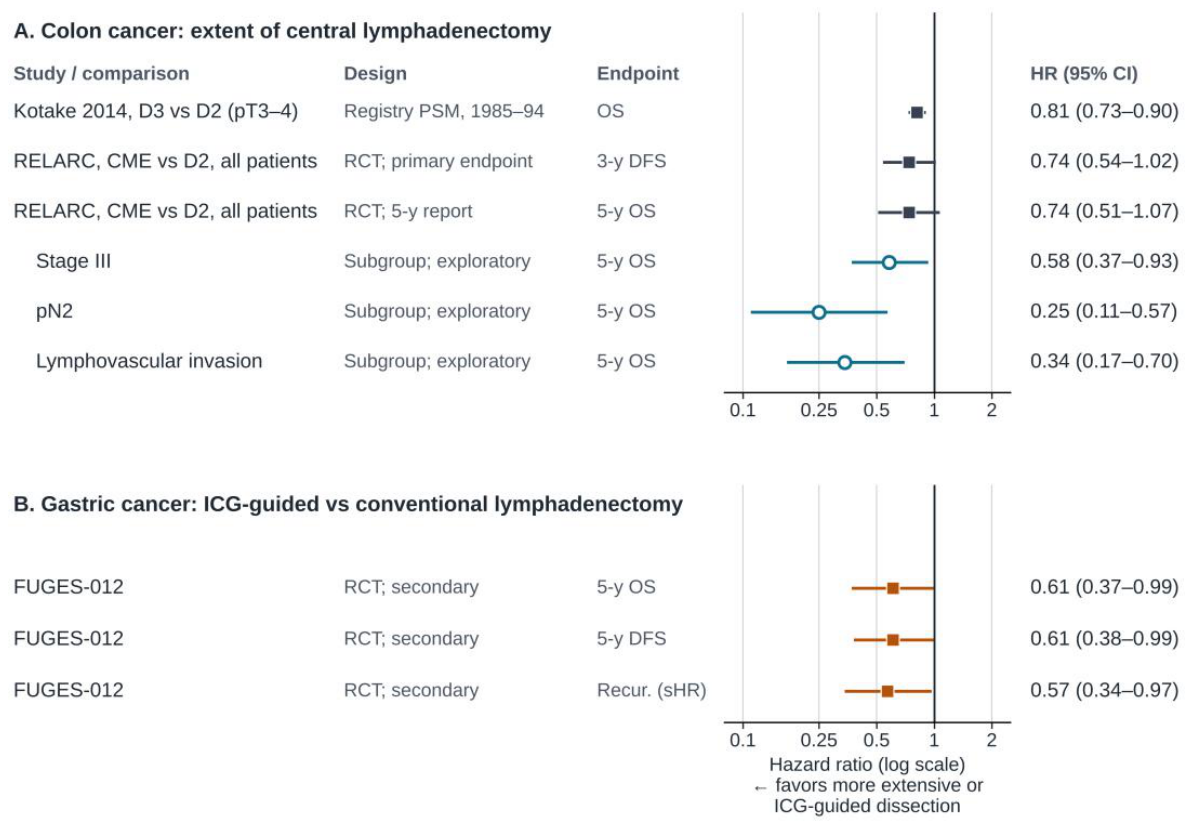
Figure 2.
Lymphatic architecture and the purpose of nodal mapping in breast and colon cancer. (A) In breast cancer and melanoma, lymph drains sequentially through one or a few first-echelon (sentinel) nodes, so a negative sentinel node predicts a negative basin and mapping is used to limit surgery [81,82,83,84,85]. (B) In colon cancer, lymph flows along the marginal vessels and toward the roots of two or more vascular pedicles through a web-like network. Skip metastasis, aberrant drainage and tumor-replaced nodes that do not take up tracer make the sentinel concept unreliable in the mesocolon [23,94,105,106]. Mapping is therefore used to define the D3 territory and the drainage pathway and to check for residual fluorescent nodes after dissection. In our right-sided series, dual drainage was seen in 19 of 43 ascending colon cancers and none of 8 cecal cancers [13]. D1, D2 and D3, pericolic, intermediate and main nodes. Original schematic.
Figure 2.
Lymphatic architecture and the purpose of nodal mapping in breast and colon cancer. (A) In breast cancer and melanoma, lymph drains sequentially through one or a few first-echelon (sentinel) nodes, so a negative sentinel node predicts a negative basin and mapping is used to limit surgery [81,82,83,84,85]. (B) In colon cancer, lymph flows along the marginal vessels and toward the roots of two or more vascular pedicles through a web-like network. Skip metastasis, aberrant drainage and tumor-replaced nodes that do not take up tracer make the sentinel concept unreliable in the mesocolon [23,94,105,106]. Mapping is therefore used to define the D3 territory and the drainage pathway and to check for residual fluorescent nodes after dissection. In our right-sided series, dual drainage was seen in 19 of 43 ascending colon cancers and none of 8 cecal cancers [13]. D1, D2 and D3, pericolic, intermediate and main nodes. Original schematic.
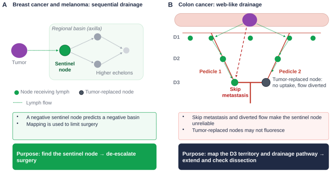
Figure 3.
Fluorescence lymph node mapping (FLNM) across imaging systems and mapping success by the total dose of indocyanine green (ICG) in a mixed colorectal cohort. (A) Fluorescent lymph nodes in different patients imaged with three laparoscopic near-infrared systems: Stryker 1588 AIM (laser excitation; Stryker, Kalamazoo, MI, USA; top row: white light, Endoscopic Near-Infrared Visualization [ENV] mode and a fluorescent node isolated from the specimen), Karl Storz IMAGE1 S (xenon light source; Karl Storz, Tuttlingen, Germany; middle row: white light, conventional ICG mode and Spectra mode) and Olympus CLV-S200-IR (xenon light source; Olympus, Tokyo, Japan; bottom row: white light, overlay in narrow-band imaging mode and infrared mode). The same type of signal is displayed differently depending on the system and display mode, and because the patients differ, the panels cannot be used to compare sensitivity between systems. (B) Success rates of gross localization, fluorescence localization, FLNM and ICG angiography according to the total dose of ICG injected endoscopically into the submucosa before surgery. Mapping success, defined as definite circular or crescent-shaped fluorescent lymph nodes [126], was highest (84.0%) at a total dose of 0.5–1 mg in this mixed, mainly left-sided population. The numbers of patients in the dose categories were 3 (>12 mg), 10 (1–12 mg), 75 (0.5–1 mg), 32 (0.3–0.5 mg) and 72 (<0.3 mg), so rates in the highest-dose categories rest on few patients. Panels A and B are reproduced from Figure 2 and Figure 3 of Ahn et al. [126], published under a Creative Commons Attribution (CC BY 4.0) license; panel titles, row and column labels and the numbers of patients per dose category were added, the letters A–I in panel A are those of the original figure, and the photographs and graph are otherwise unchanged.
Figure 3.
Fluorescence lymph node mapping (FLNM) across imaging systems and mapping success by the total dose of indocyanine green (ICG) in a mixed colorectal cohort. (A) Fluorescent lymph nodes in different patients imaged with three laparoscopic near-infrared systems: Stryker 1588 AIM (laser excitation; Stryker, Kalamazoo, MI, USA; top row: white light, Endoscopic Near-Infrared Visualization [ENV] mode and a fluorescent node isolated from the specimen), Karl Storz IMAGE1 S (xenon light source; Karl Storz, Tuttlingen, Germany; middle row: white light, conventional ICG mode and Spectra mode) and Olympus CLV-S200-IR (xenon light source; Olympus, Tokyo, Japan; bottom row: white light, overlay in narrow-band imaging mode and infrared mode). The same type of signal is displayed differently depending on the system and display mode, and because the patients differ, the panels cannot be used to compare sensitivity between systems. (B) Success rates of gross localization, fluorescence localization, FLNM and ICG angiography according to the total dose of ICG injected endoscopically into the submucosa before surgery. Mapping success, defined as definite circular or crescent-shaped fluorescent lymph nodes [126], was highest (84.0%) at a total dose of 0.5–1 mg in this mixed, mainly left-sided population. The numbers of patients in the dose categories were 3 (>12 mg), 10 (1–12 mg), 75 (0.5–1 mg), 32 (0.3–0.5 mg) and 72 (<0.3 mg), so rates in the highest-dose categories rest on few patients. Panels A and B are reproduced from Figure 2 and Figure 3 of Ahn et al. [126], published under a Creative Commons Attribution (CC BY 4.0) license; panel titles, row and column labels and the numbers of patients per dose category were added, the letters A–I in panel A are those of the original figure, and the photographs and graph are otherwise unchanged.
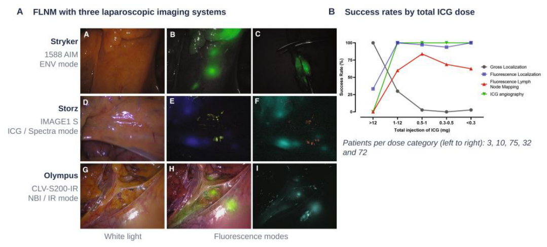
Figure 4.
D3-territory-oriented FLNM in laparoscopic right hemicolectomy. (A) Standard procedure, shown in six photographs that follow the workflow in Section 5.3: A, endoscopic submucosal ICG injection on the day before surgery (step 1); B, exploration of the drainage pathway (step 2); C, dissection of fluorescent nodes (step 3); D, check for residual fluorescence after D3 dissection (step 4); E, harvesting of nodes on the back table (step 5); F, pathological evaluation of a metastatic and a non-metastatic fluorescent node. Adapted from Figure 2 of Son et al. [13], published under a Creative Commons Attribution (CC BY 4.0) license; panel labels were shortened, the two photographs of steps 4 and 5 were exchanged to follow the workflow, and one arrow was repositioned. (B) The medial boundary in right-sided colon cancer; the same dashed line marks the medial boundary in the three panels. The depicted cancer, in the middle of the ascending colon, is assumed to be fed by the ileocolic and right colic arteries, so that the middle colic root is not a D3 station, and to drain along both the ileocolic and middle colic routes. Left: conventional lymph node dissection, with the medial boundary at the right margin of the SMV, as in the D2 arm of RELARC [141]; nodes on the SMV and at the arterial origins may not be removed, and SMV exposure has been associated with vascular injury [142,143,144]. Middle: the floor proposed in this review (Box 1), which extends to the right side of the SMA sheath without opening it and is also, by the authors’ description, the boundary of the control groups of our studies [13,127]; the dotted line marks the left border of the SMA, the boundary of the SMA group of [75]. Right: FLNM-guided dissection on the same floor. Numbers mark what FLNM adds: 1, the drainage pathway (arrows); 2, fluorescent nodes within the floor, removed en bloc with it and checked for residual signal (focus); 3, extension zones (shaded) for cT3–4 or node-positive tumors (Box 1): (a) fluorescent nodes along the anterior surface of the SMA between the arterial origins, removed without opening the SMA sheath and within research protocols only, and (b) the middle colic root, not a D3 station for the depicted tumor, dissected when it contains fluorescent or suspicious nodes; zone 3c, behind the origin of the middle colic artery up to the inferior border of the pancreas, is part of No. 223 and is not shown in a frontal projection; 4, the completeness check, photographed under recorded imaging conditions. The dashed green line is the route usually observed in our experience; it has not been quantified, and fluorescent nodes may also continue behind the SMV (Section 7). Non-fluorescent nodes within the floor (open circles) and enlarged nodes that may be replaced by tumor (dark circles; non-fluorescent with FLNM) are removed en bloc with the dissected tissue; suspicious nodes elsewhere are removed whether or not they fluoresce (Box 1). That FLNM prevents nodes from being left behind or improves safety is a hypothesis. The panels are frontal projections; nodes drawn between the SMV and the SMA, or on the SMA, lie on the anterior surface of the vessels. FLNM, fluorescence lymph node mapping; ICA, ileocolic artery; ICG, indocyanine green; MCA, middle colic artery; RCA, right colic artery; SMA, superior mesenteric artery; SMV, superior mesenteric vein. Original schematic.
Figure 4.
D3-territory-oriented FLNM in laparoscopic right hemicolectomy. (A) Standard procedure, shown in six photographs that follow the workflow in Section 5.3: A, endoscopic submucosal ICG injection on the day before surgery (step 1); B, exploration of the drainage pathway (step 2); C, dissection of fluorescent nodes (step 3); D, check for residual fluorescence after D3 dissection (step 4); E, harvesting of nodes on the back table (step 5); F, pathological evaluation of a metastatic and a non-metastatic fluorescent node. Adapted from Figure 2 of Son et al. [13], published under a Creative Commons Attribution (CC BY 4.0) license; panel labels were shortened, the two photographs of steps 4 and 5 were exchanged to follow the workflow, and one arrow was repositioned. (B) The medial boundary in right-sided colon cancer; the same dashed line marks the medial boundary in the three panels. The depicted cancer, in the middle of the ascending colon, is assumed to be fed by the ileocolic and right colic arteries, so that the middle colic root is not a D3 station, and to drain along both the ileocolic and middle colic routes. Left: conventional lymph node dissection, with the medial boundary at the right margin of the SMV, as in the D2 arm of RELARC [141]; nodes on the SMV and at the arterial origins may not be removed, and SMV exposure has been associated with vascular injury [142,143,144]. Middle: the floor proposed in this review (Box 1), which extends to the right side of the SMA sheath without opening it and is also, by the authors’ description, the boundary of the control groups of our studies [13,127]; the dotted line marks the left border of the SMA, the boundary of the SMA group of [75]. Right: FLNM-guided dissection on the same floor. Numbers mark what FLNM adds: 1, the drainage pathway (arrows); 2, fluorescent nodes within the floor, removed en bloc with it and checked for residual signal (focus); 3, extension zones (shaded) for cT3–4 or node-positive tumors (Box 1): (a) fluorescent nodes along the anterior surface of the SMA between the arterial origins, removed without opening the SMA sheath and within research protocols only, and (b) the middle colic root, not a D3 station for the depicted tumor, dissected when it contains fluorescent or suspicious nodes; zone 3c, behind the origin of the middle colic artery up to the inferior border of the pancreas, is part of No. 223 and is not shown in a frontal projection; 4, the completeness check, photographed under recorded imaging conditions. The dashed green line is the route usually observed in our experience; it has not been quantified, and fluorescent nodes may also continue behind the SMV (Section 7). Non-fluorescent nodes within the floor (open circles) and enlarged nodes that may be replaced by tumor (dark circles; non-fluorescent with FLNM) are removed en bloc with the dissected tissue; suspicious nodes elsewhere are removed whether or not they fluoresce (Box 1). That FLNM prevents nodes from being left behind or improves safety is a hypothesis. The panels are frontal projections; nodes drawn between the SMV and the SMA, or on the SMA, lie on the anterior surface of the vessels. FLNM, fluorescence lymph node mapping; ICA, ileocolic artery; ICG, indocyanine green; MCA, middle colic artery; RCA, right colic artery; SMA, superior mesenteric artery; SMV, superior mesenteric vein. Original schematic.
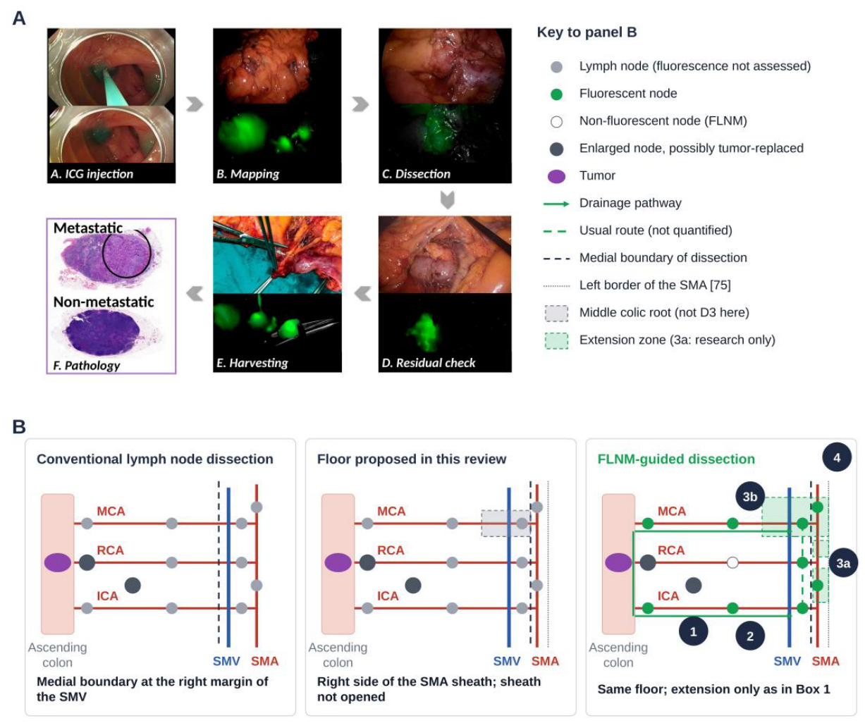
Figure 5.
Drainage zones and the drainage pathway in colorectal cancer. The routes of the five zones are described in the panels and in Section 7.1, Section 7.2, Section 7.3 and Section 7.4. In dual drainage zones, FLNM shows the drainage pathway of the individual tumor; selection and focus is applied as defined in Box 1, and suspicious nodes are removed whether or not they fluoresce. The location-specific uses are hypotheses that require prospective evaluation; the underlying data are summarized in Table 5 [13,70,100,128,157,158,159]. FLNM, fluorescence lymph node mapping; ICA, ileocolic artery; IMA, inferior mesenteric artery; IMV, inferior mesenteric vein; LCA, left colic artery; MCA, middle colic artery; RCA, right colic artery; SMA, superior mesenteric artery. Original schematic.
Figure 5.
Drainage zones and the drainage pathway in colorectal cancer. The routes of the five zones are described in the panels and in Section 7.1, Section 7.2, Section 7.3 and Section 7.4. In dual drainage zones, FLNM shows the drainage pathway of the individual tumor; selection and focus is applied as defined in Box 1, and suspicious nodes are removed whether or not they fluoresce. The location-specific uses are hypotheses that require prospective evaluation; the underlying data are summarized in Table 5 [13,70,100,128,157,158,159]. FLNM, fluorescence lymph node mapping; ICA, ileocolic artery; IMA, inferior mesenteric artery; IMV, inferior mesenteric vein; LCA, left colic artery; MCA, middle colic artery; RCA, right colic artery; SMA, superior mesenteric artery. Original schematic.
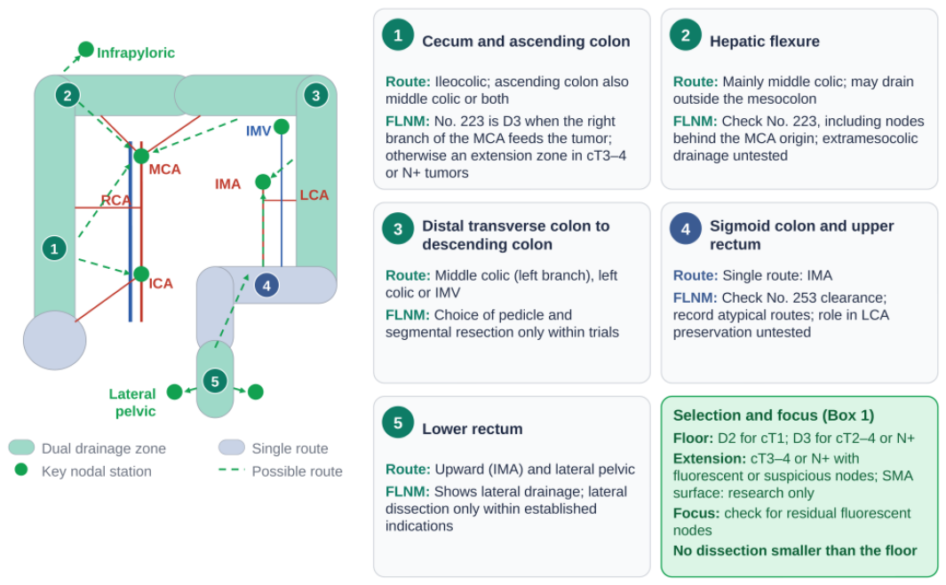
Figure 6.
Proposed framework for tailored lymphadenectomy: from drainage map to metastasis marker. Before surgery, endoscopy and CT staging set the floor and whether extension may be considered (Box 1), and tumor location indicates the likely drainage zone. On the day before surgery, interstitial ICG is injected to map the drainage territory. In the future, a spectrally distinct tumor-targeted agent (for example, a 700-nm antibody conjugate given several days before surgery) may label metastatic nodes in a separate channel; this is a proposal, and nodal false positives and the shallower detection depth at 700 nm are unresolved. During surgery, FLNM shows the drainage pathway and the D3 territory, selection and focus is applied (Box 1), and the field is photographed under recorded imaging conditions. CT radiomics may help select patients [229], rapid back-table probes may classify excised nodes, and circulating tumor DNA may add systemic risk information. All components are investigational, and the framework as a whole is not under randomized evaluation; components in dashed boxes have not been validated for D3 or central nodes in colorectal cancer. CT, computed tomography; FLNM, fluorescence lymph node mapping; ICG, indocyanine green. Original schematic.
Figure 6.
Proposed framework for tailored lymphadenectomy: from drainage map to metastasis marker. Before surgery, endoscopy and CT staging set the floor and whether extension may be considered (Box 1), and tumor location indicates the likely drainage zone. On the day before surgery, interstitial ICG is injected to map the drainage territory. In the future, a spectrally distinct tumor-targeted agent (for example, a 700-nm antibody conjugate given several days before surgery) may label metastatic nodes in a separate channel; this is a proposal, and nodal false positives and the shallower detection depth at 700 nm are unresolved. During surgery, FLNM shows the drainage pathway and the D3 territory, selection and focus is applied (Box 1), and the field is photographed under recorded imaging conditions. CT radiomics may help select patients [229], rapid back-table probes may classify excised nodes, and circulating tumor DNA may add systemic risk information. All components are investigational, and the framework as a whole is not under randomized evaluation; components in dashed boxes have not been validated for D3 or central nodes in colorectal cancer. CT, computed tomography; FLNM, fluorescence lymph node mapping; ICG, indocyanine green. Original schematic.
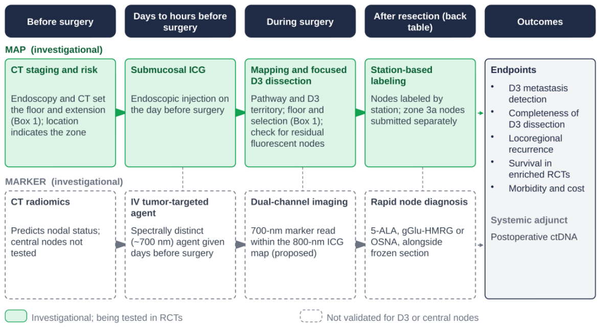
Table 1.
Milestones in the debate on the extent of lymphadenectomy for colorectal cancer. Years are publication years of the cited sources or, for sentinel node mapping, of the multicenter studies discussed in Section 4.2.
Table 1.
Milestones in the debate on the extent of lymphadenectomy for colorectal cancer. Years are publication years of the cited sources or, for sentinel node mapping, of the multicenter studies discussed in Section 4.2.
| Years | Milestone | Key Contribution | Ref. |
| 1908 | Moynihan; Miles | High ligation of feeding vessels; abdominoperineal excision based on upward, lateral and downward lymphatic spread | [26,27] |
| 1909–1910 | Jamieson and Dobson | Mapped colonic lymphatics with Prussian blue injection; named epicolic, paracolic, intermediate and main node groups; required removal of the related lymphatic area | [23,24,25] |
| 1932 | Dukes | Nodal status placed at the center of the pathological staging of rectal cancer | [28] |
| 1950–1966 | Grinnell | Retrograde spread with lymphatic block; no definite 5-year survival benefit of high IMA ligation; warned against ultraradical operations | [30,31,32,33] |
| 1984–2008 | High vs low IMA ligation | No 5-year survival difference in 1370 patients; no conclusive benefit in a systematic review | [34,35] |
| 2001–2011 | ≥12 examined nodes (USA) | Node count as a pathological quality measure, not a defined extent of dissection; later questioned as a causal factor | [40,41,42,43,44,45] |
| 2004–2007 | Sentinel node mapping (colon) | False-negative rates of 46–54% in multicenter studies; not a standard basis for reducing dissection | Section 4.2 |
| 2008–2022 | CME with CVL (Europe) | Mesocolic plane plus central ligation; lower recurrence in Danish cohorts; no general benefit in RESECTAT | [2,46,47,48,49,50] |
| 2014–2025 | Station-based D3 dissection (Japan) | Classification developed since 1977 [9]; D1–D3 dissection; D3 associated with better survival in registry data from 1985–1994 (HR 0.814) | [10,51,52] |
| 2016–present | ICG mapping of lymph flow and the D3 territory | Real-time, patient-specific visualization of lymph flow and the nodal territory | [7,8,13,53] |
| 2024–2025 | RELARC (RCT) | No significant overall DFS or OS benefit of CME over D2; exploratory OS benefit in stage III, pN2 and LVI-positive disease | [3,4] |
| 2025 | T-REX (prospective, 3647 patients) | Central node metastasis detected in about 3% (central dissection in 76%); central dissection should be selective | [5] |
Abbreviations: CME, complete mesocolic excision; CVL, central vascular ligation; DFS, disease-free survival; HR, hazard ratio; ICG, indocyanine green; IMA, inferior mesenteric artery; LVI, lymphovascular invasion; OS, overall survival; RCT, randomized controlled trial.
Table 2.
Status of lymphatic mapping across cancer types.
| Cancer Type | Drainage Pattern | Role of Nodal Mapping | Current Status | Key Evidence |
| Breast | Sequential (axilla) | Sentinel node biopsy to omit axillary dissection; omission of biopsy in selected patients | Standard | NSABP B-32, ACOSOG Z0011, SOUND, INSEMA [81,82,83,84] |
| Cutaneous melanoma | Sequential to regional basin | Sentinel node biopsy for staging | Standard | MSLT-I [85] |
| Endometrial cancer | Pelvic, largely predictable | ICG sentinel node mapping for staging | Standard | FIRES, FILM [86,87] |
| Cervical cancer (early) | Pelvic, level I predominant | Sentinel node biopsy with ultrastaging | Standard or conditional | FILM, SENTIX [87,88] |
| Gastric cancer: sentinel navigation | Multidirectional | Sentinel-based stomach-preserving surgery | Investigational (non-inferiority not shown) | JCOG0302, SENORITA [89,90] |
| Gastric cancer: ICG-guided D2 | Multidirectional | ICG to improve completeness of D2 lymphadenectomy | Promising (single-center survival signal; meta-analysis of reconstructed data: no significant OS difference, small DFS difference; multicenter survival data pending) | FUGES-012, CLASS-11 [69,91,92,93] |
| Colon cancer: sentinel node | Web-like, multidirectional | Sentinel-based limited resection | Investigational (early tumors and organ preservation only) | [16,94,95,96,97,98] |
| Colon cancer: D3-territory mapping | Web-like, multidirectional | ICG to show the drainage pathway, show where dissection may be extended and check for residual fluorescent nodes | Investigational (node yield increased; survival unproven; randomized trials ongoing) | [8,13,53,99] |
| Rectal cancer: lateral pelvic nodes | Upward and lateral routes | ICG-guided lateral dissection; lateral sentinel node biopsy | Investigational (no RCT) | [100,101,102,103,104] |
Abbreviations: ICG, indocyanine green; RCT, randomized controlled trial.
Table 3.
Representative protocols for ICG lymphatic mapping in colorectal cancer. Doses are given as concentration × volume per site × number of sites = total dose, where reported.
Table 3.
Representative protocols for ICG lymphatic mapping in colorectal cancer. Doses are given as concentration × volume per site × number of sites = total dose, where reported.
| Study | Population | Route and Timing | Dose | Main Mapping Finding |
| Wan et al., 2022 [125] | Sigmoid and rectal cancer (RCT, n = 66) | Endoscopic submucosal, day before surgery | 25 mg dissolved in 10 mL (2.5 mg/mL), injected at 2–4 sites (about 2–3 mL per site); total dose per patient not stated | D3 node yield 7 vs 5 |
| Ribero et al., 2022 (GREENLIGHT) [53] | Robotic colorectal resection (interim, n = 70) | Endoscopic submucosal, 24 h before surgery by protocol (72 h in 21 of 70) | 0.5 mg/mL × 1.5 mL × 4 sites = 3 mg | Nodes outside the standard basin in 41.4%; extent changed in 50% |
| Ahn et al., 2022 [126] | Colorectal cancer (n = 192; 150 left-sided) | Endoscopic submucosal, 12–18 h before surgery | 0.2–2.5 mg/mL; optimal 0.5–1 mg in total; proposed protocol 0.25 mg/mL × 1 mL × 2 sites = 0.5 mg | FLNM success 84.0% at 0.5–1 mg vs 64.4% below 0.5 mg (calculated); laser light source, non-obesity and a dose of at least 0.5 mg independent factors; multiple sites not significant in multivariable analysis |
| Son et al., 2023 [13] | Right-sided colon cancer (73 FLNM vs 130 control) | Endoscopic submucosal, 12–18 h before surgery | 0.25 mg/mL × 0.5 mL × 2 sites = 0.25 mg | Mapping success 98.6% (73 of 74); the patient with failed mapping was excluded, and 73 were analyzed; ileocolic, middle colic or dual pathway |
| Son et al., 2024 [127] | Right-sided colon cancer (86 FLNM vs 132 control) | Endoscopic submucosal, 12–18 h before surgery | 0.25 mg/mL, 1–2 mL in total, 2 sites = 0.25–0.5 mg | Improved agreement between CT and pathological D3 staging |
| Watanabe et al., 2017 [128] | Splenic flexure cancer, cN0 (n = 31) | Submucosal; flow observed 30 min after injection | 2.5 mg | Flow to the IMV root in 61.3% |
| Qiu et al., 2025 [101] | Mid-low rectal cancer, lateral dissection (PSM cohort, 67 ICG vs 156 control; 51 matched pairs) | Endoscopic submucosal around the tumor, 0.5 h before surgery | 1.25 mg/mL × 0.2 mL × 4 sites = 1 mg | More lateral nodes harvested, mainly obturator and internal iliac |
| ISCAPE, 2025 [99] | Colon cancer (single-arm phase II, n = 101) | Subserosal at the start of surgery | 2.5 mg/mL (volume not stated) | Metastatic nodes within the fluorescent territory in 44 of 46 node-positive patients; prespecified sensitivity threshold not met |
Abbreviations: CT, computed tomography; FLNM, fluorescence lymph node mapping; ICG, indocyanine green; IMV, inferior mesenteric vein; PSM, propensity score matching; RCT, randomized controlled trial.
Table 4.
Key clinical studies of ICG-guided lymphadenectomy in gastric and colorectal cancer.
| Study | Design and Population | n for the Reported Result | Node Yield | Metastatic Node Detection | Survival or Recurrence |
| FUGES-012 [69,91,145] | Single-center RCT, gastric cancer (primary endpoint: node yield) | 258 of 266 randomized (per protocol) | 50.5 vs 42.0; noncompliance 31.8% vs 57.4% | — | 5-year OS 80.6% vs 66.7%; DFS 77.5% vs 64.3% (secondary endpoints, per protocol) |
| CLASS-11 [92] | Multicenter RCT, gastric cancer | 1006 (per protocol) | Higher with ICG | NPV of non-fluorescent stations 93.9% (secondary outcome) | Not yet reported (primary endpoint: 3-year DFS) |
| Calì 2025 [93] | Meta-analysis of 3 studies with reconstructed IPD, gastric cancer | 6325 | — | — | OS not significantly different; RMST difference for DFS 1.3 months in favor of ICG |
| Wan 2022 [125] | RCT, sigmoid and rectal cancer | 66 | D3 7 vs 5 | No difference | — |
| Guo 2024 [146] | Meta-analysis of mainly observational studies, colorectal | 1552 (survival: 289 vs 289) | 23.5 vs 18.9 | — | No difference in 3-year OS, RFS or local recurrence |
| Fadel 2024 [15] | Meta-analysis, fluorescence-guided surgery (including targeted agents), colorectal | 3 comparative studies (review: 35 studies, 3217 patients) | Yield ratio: no difference | — | — |
| GREENLIGHT [53] | Prospective non-randomized trial, robotic colorectal resection (interim) | 70 | — | No metastasis in out-of-basin fluorescent nodes (29 patients) | — |
| FLICC [130] | Prospective pilot, colon cancer (intraoperative subserosal injection) | 10 | — | Out-of-margin fluorescent nodes in 2 patients, both metastatic | — |
| Park SY 2020 [8] | Matched cohort, right colon (cT3–4) | 75 | Median central 14 vs 7; total 39 vs 30 | No difference | — |
| Son 2023 [13] | Single-surgeon cohort of consecutive patients, right colon; predominantly historical, partly concurrent controls (published as case–control); FLNM extension in all stages; overlaps with [127] | 73 vs 130 | D3 about 1.5-fold; total 41.1 vs 39.5 | D3 metastasis 10/73 vs 11/130 overall; stage III 10/16 vs 11/41 | — |
| Son 2024 [127] | Extended cohort, right colon; predominantly historical, partly concurrent controls (after 2020, patients without FLNM for various reasons); overlaps with [13] | 86 vs 132 | — | D3 metastasis 12/86 vs 11/132 overall; cT3–4 11/35 vs 7/74 (31.4% vs 9.5%); cT1–2 1/51 vs 4/58 | — |
| Daibo 2024 [147] | Multicenter PSM cohort, right colon | 231 vs 231 | Median central 6 vs 4; total 31 vs 27 | — | 3-year RFS 88.8% vs 89.4% |
| Sato 2026 [148] | PSM cohort, colon | 50 vs 50 | Mean apical 6.8 vs 3.3 | — | 3-year RFS 80.5% vs 80.1% |
| Bao 2026 [70] | Prospective PSM cohort, left colon and rectum | 60 vs 120 | 20.8 vs 16.3 | — | ICG independently associated with OS |
Abbreviations: DFS, disease-free survival; ICG, indocyanine green; IPD, individual patient data; NPV, negative predictive value; OS, overall survival; PSM, propensity score matching; RCT, randomized controlled trial; RFS, relapse-free survival; RMST, restricted mean survival time.
Table 5.
Drainage patterns by tumor location and the proposed use of FLNM. All proposed uses are hypotheses that require prospective evaluation; the floor and the selection rule are defined in Box 1.
Table 5.
Drainage patterns by tumor location and the proposed use of FLNM. All proposed uses are hypotheses that require prospective evaluation; the floor and the selection rule are defined in Box 1.
| Tumor Location | Main Vascular Pedicles | Drainage Pattern | Key Data (Study design) | Proposed Use of FLNM |
| Cecum | Ileocolic (± right colic) | Mainly single | Ileocolic route in 7 of 8, middle colic in 1 [13] (single-center, descriptive); No. 223 metastasis 4/224, all with extensive nodal disease [160] (retrospective) | Confirm the ileocolic route; the middle colic root is an extension zone (Box 1) |
| Ascending colon | Ileocolic, right colic, right branch of middle colic | Dual | Ileocolic 13/43, middle colic 11/43, both 19/43 [13] (single-center, descriptive) | Identify the pathway; No. 223 is a D3 station when the right branch of the middle colic artery is a feeding artery, and otherwise an extension zone (Box 1) |
| Hepatic flexure and proximal transverse colon | Right branch of middle colic, right colic; gastroepiploic and infrapyloric nodes | Dual, with possible extramesocolic drainage | Middle colic route 12/18 (hepatic flexure) and 3/4 (proximal transverse) [13] (descriptive); infrapyloric metastasis 11.8% [157]; gastroepiploic 4.0% [161,162] (retrospective) | No. 223 is part of the floor; fluorescent or suspicious nodes behind the middle colic origin are removed with it: check for residual fluorescent nodes after its clearance (Box 1); role in extramesocolic drainage to be tested |
| Distal transverse colon, splenic flexure and descending colon | Left branch of middle colic, accessory middle colic, left colic; IMV | Dual | Splenic flexure: flow to the IMV root in 19/31, cN0 only [128] (real-time, single-center); accessory middle colic artery present in 41.8%, metastasis at Nos. 222-lt and 232 in 1.6% and 1.0% (n = 153) [158] (multicenter retrospective); right colic involvement 9% after subtotal colectomy [163] (retrospective); central metastasis 0.5% [5] (prospective cohort); Delphi consensus [164] (expert opinion). Descending colon: no IMA-root metastasis (0/26) [165] (retrospective) | Within trials: select the pedicle and test segmental resection; outside trials, the main nodes of both feeding arteries are dissected |
| Sigmoid colon and upper rectum | Inferior mesenteric artery | Mainly single | Most apical nodes within about 2 cm of the IMA root; occasional atypical routes [70] (prospective cohort) | Check No. 253 clearance; record atypical routes (Box 1); role in LCA preservation untested [166] |
| Lower rectum | Inferior mesenteric system; internal iliac and obturator nodes | Dual (upward and lateral) | Lateral nodes visualized in 13/14 after injection at the dentate line or the anal margin of the tumor [159] (case series); 3-year lateral local recurrence 0% with ICG vs 9.3% without in 58 matched pairs [100] (non-randomized) | Show lateral drainage; selective lateral dissection within established indications, based on tumor location and depth (JSCCR) or on MRI, which fluorescence does not replace [103,104] |
Abbreviations: FLNM, fluorescence lymph node mapping; ICG, indocyanine green; IMA, inferior mesenteric artery; IMV, inferior mesenteric vein; LCA, left colic artery.
Table 6.
Medial boundary of central dissection in right-sided colon cancer in the main sources cited in this review.
Table 6.
Medial boundary of central dissection in right-sided colon cancer in the main sources cited in this review.
| Source | Medial Boundary and Ligation | Tissue on the Anterior Surface of the SMA | Relevant Findings |
| JSCCR classification and guidelines [9,10] | Main nodes at the origins of the feeding arteries | Medial boundary not defined anatomically | — |
| RELARC, D2 arm [141] | Vessels ligated at the right side of the SMV; tissue covering the SMV left intact | Not removed | Comparator: CME did not significantly improve 3-year DFS (HR 0.74, p = 0.06) [3] |
| RELARC, CME arm [141] | Vessels ligated at their origins; lymphoadipose tissue on the surfaces of the SMV and SMA removed | Removed | 3-year DFS not significantly different [3]; exploratory 5-year OS benefit in stage III disease [4]; intraoperative vascular injury 3% vs 1% and Clavien–Dindo grade III–IV complications 1% vs 3% [142] |
| Zhou et al., SMV group [75] | Left side of the SMV, described by the authors as current mainstream practice | Not removed | Fewer main nodes; less chylous leakage |
| Zhou et al., SMA group [75] | Left border of the SMA | Removed | More main nodes (3.70 vs 1.81) and metastatic main nodes (p = 0.005); chylous leakage 9.4% vs 2.6%; survival not significantly different (trend in stage III DFS, p = 0.056). The authors considered dissection along the left side of the SMV insufficient for D3 under the JSCCR definition |
| Randomized trial of SMA-side vs SMV-side dissection [77] | SMA side vs SMV side | Removed vs not removed | More diarrhea and chylous leakage; positive nodes not different |
| Randomized trial of open vs laparoscopic central lymphadenectomy [78] | Left border of the SMA (open) vs along the SMV (laparoscopic) | Removed vs not removed | Complications 42.8% vs 38.4% (not significant); approach differed between arms |
| D3 extended mesenterectomy vs D2 [170] | Soft tissue around the superior mesenteric vessels removed, with transection of the nerve plexus | Removed | Non-randomized comparison of consecutive cohorts: higher bowel frequency after D3 extended mesenterectomy; total bowel-function and quality-of-life scores not different |
| Meta-analysis [76] | SMA side vs SMV side | — | More positive nodes (p = 0.04); no difference in OS, RFS or total complications; the analysis contains internal inconsistencies (e.g., [75] classified as randomized) |
| Chi et al. [144] | Right margin vs left side of the SMV (propensity-matched, different periods) | Not removed | Left side: more main nodes (16.6 vs 9.0) and complications (16.7% vs 1.7%) without a change in N stage; the authors favored the right margin |
| Sahara et al. [72] | Pathological study of central lymphadenectomy | — | Nodes anterior to the SMV near the arterial origins involved in 3.8%; therapeutic index about one-tenth that of the pericolic area |
| Nesgaard et al. [172] | Post-mortem study | — | Lymphatic vessels crossed the SMV both anteriorly and posteriorly in 12 of 16 cadavers and only anteriorly in 4; the authors recommended clearance to the left side of the SMA |
| Feng et al. [169] | Day-before tracer (carbon nanoparticles or ICG; mapping failed in 4 of 27) | — | Tracer-marked nodes anterior to the SMA in 16 of 27 patients (none metastatic), between the SMV and the SMA in 22 and behind the SMV in 6 (ileocolic artery behind the SMV in 5 of these 6); the authors recommended preserving the vascular sheath of the SMA |
| Tsukamoto et al. [71] | Prospective pathological study | — | Metastasis around the SMA in 1.9% of cecal cancers; in transverse colon cancer, 2.5% at the middle colic and 6.7% at the right colic root; 17% of tumors fed mainly by the ileocolic artery had metastasis along the middle or right colic artery; routine dissection around the SMA judged unnecessary in cecal and ascending colon cancer but possibly necessary in transverse colon cancer or when the middle colic artery feeds the tumor |
| Our control groups [13,127] | Line between the SMV and the SMA (at the right side of the SMA sheath, which is not opened, by the authors’ description; same as the floor); middle colic root exposed and its right branch divided | Not removed | — |
| Floor proposed in this review (Box 1) | Same as our control groups | Not cleared; fluorescent or suspicious main nodes at the origins removed | No comparative data; complications recorded in CONSTELLARE (Section 9.1) |
Abbreviations: CME, complete mesocolic excision; DFS, disease-free survival; ICG, indocyanine green; JSCCR, Japanese Society for Cancer of the Colon and Rectum; OS, overall survival; RFS, relapse-free survival; SMA, superior mesenteric artery; SMV, superior mesenteric vein.
Table 7.
Ongoing trials and emerging approaches, and the limitations of ICG-based FLNM that they address.
Table 7.
Ongoing trials and emerging approaches, and the limitations of ICG-based FLNM that they address.
| Category | Approach (Example) | Limitation Addressed | Current Evidence | Status/Relevance to Colorectal Nodes |
| Randomized trials | NCT06485609 [197] | Lack of survival evidence | Right hemicolectomy ± ICG-guided dissection; planned n = 1200; primary endpoint 3-year DFS | Recruiting; single center, clinically node-positive; completion expected 2031 |
| CONSTELLARE (NCT07060443) [198] | Lack of randomized staging data | FLNM-guided D3 dissection vs D3 dissection without FLNM; planned n = 186; primary endpoint D3 metastasis detection rate | Recruiting (phase II); no results yet | |
| Other RCTs of ICG-guided lymphadenectomy (NCT06396806; NCT06583564) [199,200] | Lack of survival evidence | Sigmoid cancer, single center, planned n = 1072, primary endpoint 3-year DFS; stage III colorectal cancer, single center, planned n = 192, primary endpoint DFS | Enrolling by invitation; no results yet | |
| CLASS-11 [92] | Lack of multicenter survival data (gastric) | n = 1006 per protocol; node yield increased | Long-term results awaited; indirect relevance | |
| Tumor-targeted agents (intravenous) | SGM-101 (anti-CEA; 700-nm channel) [201,202,203,204] | No cancer specificity; blocked lymphatics | 10-mg cohort, dosed 4 days (17 patients) or 6 days (2) before surgery (n = 19): lesion-level sensitivity 96%, specificity 63%, mean in vivo TBR 1.9; whole study (n = 37): plan change 32% (warranted in 24%) | Phase 3 (SGM-CLIN03) stopped early for futility; no nodal outcomes (sponsor summary, March 2026); nodal false positives; separable from ICG at 700 nm, with shallower detection |
| cRGD-ZW800-1, bevacizumab-800CW, ONM-100 [205,206,207] | No cancer specificity | Feasibility for primary tumor and margins | No nodal data | |
| EMI-137 (anti-c-Met) [208] | No cancer specificity | No fluorescent nodes despite c-Met-positive metastases | Negative pilot | |
| Panitumumab-IRDye800CW (head and neck) [209,210] | No cancer specificity | Ex vivo specimen imaging, nodes preselected by intensity: sensitivity 84.6%, specificity 94.0%, NPV 99.3%, PPV 36.2% | Indirect; overlaps spectrally with ICG | |
| Visible-light probes (excised nodes) | 5-ALA/protoporphyrin IX (excitation ~405 nm) [211,212,213] | No cancer specificity; need for rapid diagnosis | Excised colorectal nodes: sensitivity 88.3%, specificity 92.0% (with spectral unmixing); intraoperative laparoscopic detection of node-positive colon cancer: sensitivity 0–11% | Ex vivo, surface only; poor intraoperative performance |
| gGlu-HMRG spray (excitation ~480–500 nm) [214,215] | Need for rapid diagnosis | Excised breast nodes: sensitivity 97%, specificity 79%, NPV 99%; colorectal: mouse model only, with target enzyme expression in human metastatic nodes on immunohistochemistry | Ex vivo; surface only; no human colorectal imaging data | |
| UV-excitation microscopy | Deep-UV excitation with a neural network [216] | Need for rapid diagnosis | Gastric cancer nodes | Experimental |
| Label-free, lifetime and NIR-II imaging | Hyperspectral imaging [217,218] | No cancer specificity | Colorectal cancer vs mucosa on resected specimens ex vivo: sensitivity 86%, specificity 95%; AUC 0.93 | Not tested in nodes; superficial |
| Fluorescence lifetime imaging of ICG [184] | Device dependence of intensity | IV ICG in solid tumors: Accuracy >97%; largely independent of system, depth and dose; tumor-infiltrated nodes imaged after IV ICG | Not tested in nodes after interstitial injection | |
| NIR-II imaging of ICG [219,220] | Limited contrast and resolution | Liver tumors after IV ICG: sensitivity 100% vs 90.6% (NIR-I) | No commercial laparoscopic system; no clinical nodal data | |
| Quantification | Fluorescence intensity of excised nodes [189] | Subjective interpretation | Weaker fluorescence in metastatic pericolic nodes; AUC 0.931 | Single-center; not corrected for size and depth |
| Artificial intelligence | Multimodal deep learning [221,222] | Subjective interpretation | Colorectal nodes: better than single-mode models; breast sentinel nodes: AUC 0.898 | Single-center development; device-specific |
| Molecular and biological adjuncts | OSNA of fluorescent nodes; ctDNA [223,224,225] | Staging accuracy; systemic risk | OSNA of fluorescent nodes matched the final nodal stage in 3 of 6 patients (pilot); ctDNA HR 10.0 for recurrence | Complementary |
Abbreviations: 5-ALA, 5-aminolevulinic acid; AUC, area under the curve; CEA, carcinoembryonic antigen; ctDNA, circulating tumor DNA; DFS, disease-free survival; FLNM, fluorescence lymph node mapping; gGlu-HMRG, γ-glutamyl hydroxymethyl rhodamine green; HR, hazard ratio; ICG, indocyanine green; IV, intravenous; NIR, near-infrared; NPV, negative predictive value; OSNA, one-step nucleic acid amplification; TBR, tumor-to-background ratio; UV, ultraviolet.
Disclaimer/Publisher’s Note: The statements, opinions and data contained in all publications are solely those of the individual author(s) and contributor(s) and not of MDPI and/or the editor(s). MDPI and/or the editor(s) disclaim responsibility for any injury to people or property resulting from any ideas, methods, instructions or products referred to in the content. |
© 2026 by the authors. Licensee MDPI, Basel, Switzerland. This article is an open access article distributed under the terms and conditions of the Creative Commons Attribution (CC BY) license.
Copyright: This open access article is published under a Creative Commons CC BY 4.0 license, which permit the free download, distribution, and reuse, provided that the author and preprint are cited in any reuse.