
Submitted:
31 October 2026
Posted:
03 November 2026
You are already at the latest version
Abstract
Ex vivo expansion is needed to produce human myogenic cell products, but culture can alter the molecular states that govern engraftment and regeneration. This critical narrative review reappraised 28 primary reports of tissue-derived human myogenic cells, yielding 39 question-specific records. We separated production yield, administered dose, donor persistence, human fiber contribution, later regenerative activity, and recipient function. In documented adult donor sources, transient p38 mitogen-activated protein kinase inhibition and extracellular matrix culture increased histological graft output at matched final doses. SETD7 inhibition increased cell production and postgraft reporter signal, although the administered human dose was unreported. Brief Notch-ligand exposure changed myogenic markers without a detected gain in human fiber output. Population selection, senolytic removal, delivery geometry, and recipient support also altered selected outcomes. Expanded preparations retained later graft activity in some studies, but durable adult-source functional recovery remained insufficiently tested. The evidence supports a product-level framework that links molecular state to defined graft endpoints while evaluating dose, spatial delivery, host context, and indication-specific function separately.
Keywords:
human muscle stem cells
; satellite cells
; ex vivo culture
; molecular cell state
; stem cell niche
; cell transplantation
; skeletal muscle regeneration
1. Introduction
Muscle satellite cells occupy the niche between the myofiber membrane and basal lamina and provide the principal stem-cell source for postnatal skeletal muscle repair [1]. Their quiescence, activation, self-renewal, and differentiation are shaped by molecular signals from the niche and by the physical and metabolic conditions encountered after isolation. Human satellite-cell preparations can generate donor-derived fibers in conditioned recipient muscle and can leave donor-derived precursors capable of later regenerative activity [2,3]. Therapeutic development is constrained by the small amount of starting tissue, which makes ex vivo expansion attractive. Expansion changes signaling, metabolism, adhesion, cell-cycle state, and population composition. The relevant unit is therefore the final administered preparation.
The central translational problem is that a molecular state favoring cell accumulation in culture may not support engraftment. A manipulation can increase yield, maintain a marker, or delay differentiation without improving repair. In mouse models, directly isolated satellite cells can outperform expanded cells under defined transplantation conditions [4]. Lineage and single-cell graft experiments further separate immediate fiber formation from precursor maintenance and self-renewal [5,6]. Human product assessment requires the same distinctions. Persistent myopathy requires donor activity after later injury, whereas reconstruction of a focal defect requires repair across the treated volume and an appropriate functional endpoint.
Previous reviews have summarized myogenic cell transplantation in muscle disease [7], examined links between engraftment and functional recovery [8], discussed recipient conditioning and graft heterogeneity [9], and surveyed culture strategies across mouse and human systems [10]. The unresolved issue is which molecular or culture-induced changes improve a defined human graft endpoint when dose, host context, and outcome differ. Selected human graft studies are therefore compared at the administered-product level. Production yield is separated from performance at a matched dose, marker retention from repair, donor persistence from later regenerative activity, and local histology from recipient function.
This endpoint-specific approach tests how far each molecular finding can be extended. More human-derived fibers at a matched final dose support progression to tests of later regeneration or function. Increased culture yield with an unresolved graft dose requires dose attribution before the performance of the administered population can be assigned. Table 1 summarizes the transplant conditions underlying these judgments. Figure 1 links the principal molecular and experimental findings to the next product-level comparisons.
2. Review Scope and Evidence Appraisal
We conducted a critical narrative review of tissue-derived human satellite cells and their myogenic progeny. Study selection was purposive. A targeted PubMed search was rerun on 30 October 2026 and identified no additional eligible primary graft report beyond the reserve-cell study identified in the 11 October search [11]. Among 82 cited sources, 28 primary human-cell graft reports were reappraised using their Results, Methods, figure legends, and relevant supplementary files. Supplementary File S1 provides the search records, source dispositions, extraction rules, and reproducible coding files. Because the original discovery history is incomplete, this review does not claim exhaustive or systematic screening.
The 28 reports yielded 39 question-specific comparison or demonstration records. For each record, we retained the cell preparation, administered dose when available, denominator, recipient conditions, timing, statistics, and source location. Records can share animals, cohorts, or controls and were not treated as independent effect sizes. We distinguished observed benefit, no detected improvement, untested function, and unresolved attribution. A nonsignificant result was not interpreted as equivalence, and values were extracted from source text and legends without graph digitization.
Donor provenance was assessed at tissue donation. The primary adult restriction required a documented entirely adult cohort or an explicitly adult graft source without contradictory information. A cohort mean age or a parental relationship contributed only to a contextual sensitivity analysis. Mixed-age cohorts were excluded from the primary adult restriction when adult results could not be separated. These rules evaluate applicability to adult-derived products and do not test a biological age effect. The accompanying code reproduces the sensitivity-analysis outputs but does not replace independent source adjudication.
3. Molecular and Cellular Determinants of Graft Performance after Ex Vivo Culture
3.1. Matched-Dose Evidence for p38 MAPK, Extracellular Matrix, and SETD7 Modulation
Administering the same number of cells in a within-study comparison excludes a larger delivered dose as the sole explanation for greater graft output. It does not equalize postgraft survival, spatial spread, population composition, or differentiation, each of which may mediate a useful culture effect. A fiber count therefore characterizes the administered population but does not isolate activity per surviving cell. Comparisons that fix the starting culture instead combine production yield with the behavior of the resulting preparation.
Charville and colleagues studied a donor cohort aged 35–82 years and used 10 μM SB203580 to reversibly delay differentiation. Fresh, untreated activated, and inhibitor-treated preparations were transplanted at 30,000 cells per graft into NOD scid gamma (NSG) tibialis anterior muscle injured with barium chloride 48 hours earlier. At four weeks, treated cells produced 221 ± 20 human-containing fibers per section, compared with 49 ± 14 for fresh cells and 21 ± 13 for untreated activated cells. Errors are standard deviations, and each treated-versus-control comparison had P < 0.001. Donor-derived sublaminar cells expressing the satellite cell transcription factor PAX7 were also observed [12]. Equal dose excludes a larger administered graft as the explanation. Supplemental images show human dystrophin contribution at 56 days, extending the observation window without testing a second injury or direct force.
Zhao and colleagues studied human satellite cells from donors aged 40–56 years. After a five-day comparison on decellularized extracellular matrix or uncoated surfaces, they grafted 25,000 cells from each condition into athymic nude tibialis anterior muscle injured with cardiotoxin one day earlier. At four weeks, matrix-cultured cells produced more human dystrophin-positive fibers and donor-derived PAX7-positive cells. Methods specify six tibialis anterior muscles per group [13]. Together with the p38 experiment, this is direct evidence that a culture intervention can improve the output of an equal-sized human graft.
SETD7 inhibition connects production with a human reporter endpoint. Judson and colleagues started cultures with 15,000 cells from donors aged 15–53 years and compared seven days of 4 μM PFI-2 with vehicle. Cells were reporter-transduced and delivered to NSG tibialis anterior muscle in a 1.2% barium chloride suspension. Bioluminescence was followed for eleven days [14]. Human cell accumulation and postgraft signal increased. The published Methods, Figure 7, and supplementary Figure S1–S5 of that report do not identify the final administered human cell number. Consequently, production and performance per administered cell cannot be separated. Matched-dose fiber, recovered stem cell, and force experiments elsewhere in the paper used mouse donors and cannot supply those human conclusions.
The SETD7 result calls for a human graft comparison with a stated, matched final dose and a parallel measurement of usable yield. By contrast, the p38 and matrix results already support an improvement in histological output of an equal-sized graft. Their next tests concern later regeneration or indication-specific function. Fiber counts from different host preparations cannot rank the two culture processes.
3.2. Cell Selection, Senescence, and Expansion History
Population composition can change graft output without an expansion intervention. Equal grafts of 500 caveolin-1 (CAV1)-positive or CAV1-negative human satellite cells produced different fiber outputs in an irradiated and injured NSG recipient, favoring the positive fraction [18]. The result supports selection of a functionally favorable population, but induction of CAV1 in an individual cell remains untested. An equal-dose culture benefit can similarly reflect enrichment of a favorable fraction or altered behavior of its constituent cells.
Conte and colleagues removed a senescent fraction from myotonic dystrophy type 1 (DM1) myoblast cultures using A1155463. Equal grafts of 200,000 cells from three patient lines produced greater human dystrophin-positive fiber contribution in NSG muscle (the source paper's Figure 6j, P = 0.014). Cardiotoxin preceded grafting by one day, and harvest occurred 21 days after injury, or twenty days after grafting [20]. The equal dose attributes the improvement to the treated population and excludes a larger administered graft as its sole explanation. Because the Results and Methods differ in exposure duration, a single exact treatment interval is not assigned. Selection against defective cells remains compatible with the outcome. Rejuvenation of every surviving cell was not demonstrated.
CAV1 selection favors a fresh fraction, while senolytic treatment removes defective cultured cells. Both results concern the retained preparation. Manufacturing assessment must also retain yield lost during selection.
Culture duration must be linked to the transplanted preparation. In the p38 study, a three-week growth curve and a two-week expansion followed by drug withdrawal addressed proliferation and reversible differentiation, respectively. The matched-dose transplantation comparison used 30,000 cells and a four-week endpoint, but its specific culture interval is not stated in the examined main and supplemental procedures. A separate GFP-labeling graft workflow began with ten days of expansion, adenoviral labeling, and more than three additional passages [12]. This supports graft activity after extended processing without assigning that duration to every experiment in the report.
Cooper and colleagues provide a within-donor example of a culture-history effect. Each freeze-injured immunodeficient muscle received 500,000 myoblasts. At eight weeks, 84% of remaining human nuclei were located within fibers after early infant culture, compared with 21% after extensive amplification of the same infant source. The 71-year-old donor population reached 37%, exceeding the late infant population [26]. These percentages describe nuclear location, not retention of that fraction of the injected cells. One donor at each age and three recipient mice per condition prevent generalization of an age effect. The infant comparison makes processing history a product-selection variable. Cross-age differences remain confounded by donor identity.
Human age comparisons also depend on the preparation and comparator. Corbu and colleagues studied cells from six donors aged 74–76 years against three pediatric controls aged four months to sixteen years. Growth, passage tolerance, and differentiation kinetics differed. This was not an older-versus-young-adult graft comparison [37]. Human tissue xenografts retained regeneration across donor ages extending into later life, preserving architecture and associated cells [38]. Serially passaged human cultures showed reduced fusion and several metabolic responses. Insulin responsiveness was endpoint-specific: glucose-uptake fold response did not change detectably, whereas glycogen-synthesis and oxidation responses declined [39]. These findings distinguish chronological age, culture history, and recipient-supported regeneration without assigning an intrinsic age ranking to final products.
Surface-marker work separates CD82/CD318-associated myogenic populations from CD201-associated interstitial cells and tests effects of CD82 perturbation on cell behavior [40]. Source identity still requires a graft-specific readout. Orbicularis oculi cells from an adult female cohort were induced for three weeks and compared with noninduced cells at 200,000 cells per nude-mouse tibialis anterior. At one week, CM-DiI-positive fibers were detected in induced grafts [31]. The reported recovery measure counts labeled fibers, not surviving donor cells. The source/induction result warrants human-specific donor identification and later functional testing before it supports lasting replacement.
3.3. Mechanobiology, Metabolism, and Manufacturing Scale
Mouse mechanisms motivate human culture candidates. Soft substrates preserved self-renewal and regeneration [41]. Combined soft support and transient p38 inhibition improved aged-mouse regeneration and strength [42]. Independent work linked altered p38 activity to self-renewal defects [43]. Compression and Piezo1 perturbation connect mechanical sensing to cell state [44,45]. Mouse strength findings do not supply a human graft endpoint.
Human myoblast media altered cell accumulation, marker expression, and in vitro fusion across passages [46]. Greater usable yield from a biopsy remains valuable even without improved function per administered cell. These production findings do not determine graft reserve or recipient force.
Insulin-like growth factor 2 (IGF2) perturbation altered proliferation, differentiation, and mitochondrial bioenergetics in human cultures [47]. PGC-1α overexpression improved early tissue formation in vivo [48]. These adjustable properties do not establish a metabolic threshold predicting reserve or strength across manufacturing routes.
CDK4 and telomerase expression produced clonal human myogenic lines for disease research [49]. Scalable production comparisons did not uniformly retain proliferation and differentiation [50]. Laboratory expansion remains useful, while clinical manufacture requires validation of the specific population delivered.
Microcarrier production provides a positive example of processing compatibility with a narrow inferential boundary. Cells from a thirteen-month-old donor grown on microcarriers or in static flasks were administered at two million per contralateral tibialis anterior in two conditioned SCID mice. Both routes produced human dystrophin-positive fibers at four weeks. Similar numbers were described without quantitative counts or an equivalence analysis [30]. The comparison is compatible with microcarrier manufacture and leaves equivalence to an adult product unresolved. Calpeptin-mediated fusion control was tested in culture, and the examined report does not identify a graft comparison of calpeptin-treated material. A clinical-grade flask process instead demonstrates animal-free production with human platelet lysate and defined release measurements [51]. Those measurements identify usable material, while matched-dose functional comparisons determine its repair claim.
Across culture strategies, molecular perturbation and product composition act at different levels. p38 inhibition and extracellular matrix culture improved matched-dose histological output [12,13], whereas SETD7 inhibition combined increased production with an unresolved final dose [14]. CAV1 selection and senolytic treatment changed the composition of the transplanted preparation [18,20]. Soft matrices and p38 signaling in mouse systems identify plausible state-control variables [41,42,43]. Compression and Piezo1 studies further implicate mechanosensing [44,45]. Together, these findings support a hierarchy from molecular state to product attribute to graft endpoint. Movement across levels requires a controlled human comparison.
4. Molecular Cell-State Markers and Donor Persistence as Incomplete Surrogates for Repair
4.1. Notch Signaling and the Dissociation between Cell State and Fiber Output
Sakai and colleagues treated expanded, previously frozen human myoblasts with Jagged 1 (JAG1) or Delta-like ligand 4 (DLL4) for 24 hours. MYOD decreased, and the PAX7-positive/MYOD-negative fraction increased, although human PAX7 expression did not increase. Each NOD-SCID tibialis anterior received 100,000 cells after cardiotoxin injury. At two weeks, no increase was detected in the maximum number of human spectrin-positive fibers per injected muscle. The JAG1 comparison used five muscles per condition, and the DLL4 comparison used three [16].
The brief Notch-ligand regimen changed a measured cell-state profile without a detected gain in human fiber output. The small graft groups do not establish equivalence, but the marker result alone does not justify selecting this treatment for fiber replacement. Sustained support from isolation is a distinct intervention that would require its own graft comparison.
Mouse loss-of-function experiments established a requirement for canonical Notch signaling in resting satellite cell maintenance [52,53]. A Notch-linked collagen V circuit further supported niche maintenance [54]. The associated 2025 correction replaced a misassembled receptor-screening image. The authors reported no change in interpretation [55]. These necessity experiments motivate culture strategies. They address a different question from whether brief ligand treatment is sufficient to improve an already expanded human graft.
4.2. Reserve Cells, Reporter Persistence, and Donor Compartment
Reserve cells are the nonfusing fraction recovered after two days of differentiation of expanded myoblasts. Their selection changes graft endpoints unequally. Laumonier and colleagues grafted 500,000 reserve cells or myoblasts into lacerated NOG gastrocnemius. At day twenty-one, reporter activity was 54.1 ± 5.2% versus 31.5 ± 4.8% of each graft's three-hour baseline (n = 12; P < 0.05). The spectrin-positive fiber count divided by human lamin A/C-positive nuclei was 27.1 ± 4.6% versus 23.1 ± 7.0% (n = 6; P = 0.6); errors are standard errors. The human PAX7-positive fraction was also higher [17].
Both groups in the 2017 study underwent reporter and histological evaluation. Higher late signal and a larger PAX7-positive fraction favor the reserve-cell route for further evaluation, but neither substitutes for a direct later-regeneration endpoint. The nonsignificant fiber-to-nucleus ratio cannot demonstrate equivalent absolute fiber production. Yield after generating and selecting the reserve fraction remains part of the manufacturing comparison.
A later reserve-cell study compares autofluorescence-high and -low fractions, a different comparison from reserve cells versus myoblasts. Equal grafts of 200,000 cells showed no detected difference in day-twenty-eight reporter signal or human PAX7-positive fraction. Cardiotoxin reinjury on day twenty-eight was followed by reporter imaging on day thirty-two [11]. The challenge was performed, but the reporter increase does not directly measure new fiber repair or recipient force. The Results describe nonsignificance while that report's Figure 6C legend reports P < 0.05. Donor ages are not reported in the examined main text. The study extends the tested trajectory while leaving direct adult-source later repair and equivalence between fractions unresolved.
A controlled Duchenne study directly separates donor protein contribution from strength. Twelve boys received 110 million donor myoblasts per biceps in each of six monthly procedures, with contralateral sham injections. Exon-specific staining confirmed donor-derived dystrophin in four recipients. One biopsy contained 420 positive fibers among 4,080 examined, or 10.3%, without a treated-arm strength advantage. No strength advantage was detected at the measured times through twelve months [33]. The biopsy fraction does not quantify repaired whole-muscle volume. Protein restoration supplies a necessary molecular observation for this strategy, but it cannot replace the physiological endpoint.
Longitudinal reporter measurements and tissue localization provide complementary information. Bioluminescence follows donor-associated reporter activity and is not a direct cell count. Human-specific DNA establishes human material in a sample, while staining locates nuclei in fibers or precursor compartments. Combined molecular and histological assays have been applied to human muscle xenografts [56]. This combination is particularly useful when increased signal might reflect more fused nuclei instead of a larger unfused reserve. A higher reporter signal can reflect different donor compartments, so normalization and tissue localization are needed before assigning a precursor-persistence claim.
Quarta and colleagues tested human cells after approximately one day in quiescence-supporting conditions. They followed equal grafts of 1,000 cells with reporter imaging for thirty days, with greater signal under optimized conditions [15]. Reporter activity integrates viable reporter-expressing material, proliferation, differentiation, and reporter regulation. The experiment supports brief preservation but leaves extensive human expansion untested. Its positive reporter result and the limiting Notch comparison involve different starting material, processing histories, and measured functions.
4.3. Quiescence, Senescence, and Reversible Activation
Serum-replacement conditions induced a quiescence-associated cell-cycle profile in expanded human myoblasts while retaining subsequent differentiation [57]. Retinoic acid suppressed differentiation and increased specification-associated gene expression in human progenitors [58]. These culture results identify controllable states. Their translational value depends on whether the resulting preparations retain productive repair or later precursor activity after delivery, which cannot be assigned from reversible differentiation alone.
Isolation itself begins the change in cell state. Mouse studies using in situ fixation or in vivo transcript labeling showed that dissociation can induce transcriptional responses before cells are placed in an experimental culture [59,60]. A human preparation described as fresh is therefore an operational category, not proof that every transcript retains its resting in vivo abundance. In a study of dual p38 and JNK inhibition during purification, the transplantation improvement was established in mouse cells, while human experiments supported improved recovery and a less activated transcriptional profile [61]. This species distinction matters when evaluating preservation from the earliest processing step.
Noncycling cells are not necessarily reversibly quiescent. Geriatric mouse satellite cells acquired a senescence program that impaired subsequent activation. Silencing p16INK4a restored activation and regenerative function in mouse experiments [62]. In human primary cultures, p16INK4a silencing increased BrdU incorporation and reduced senescence-associated β-galactosidase positivity [62]. The human arm did not test graft rescue. Basal autophagy helped prevent senescence in mouse cells, with similar observations in human geriatric samples [63]. Nutrient utilization also influenced SIRT1-dependent epigenetic regulation during mouse activation [64]. For a culture signal that suppresses proliferation, subsequent re-entry and differentiation are therefore necessary interpretive tests. Marker retention alone cannot distinguish preserved reserve from impaired activation.
Hypoxic culture improved later graft performance in a mouse myoblast model [65]. In human myoblast xenografts, delaying differentiation was associated with greater proliferation, distribution, and later fiber contribution [66]. These results motivate a precursor interval that permits expansion and spread before fusion. They do not establish an optimal timing sequence for durable adult human repair. The relevant unresolved comparison would hold cell dose and delivery constant while measuring both first repair and subsequent precursor availability.
5. Durable Regenerative Activity and Functional Recovery
5.1. Later Repair, Reinjury, and Secondary Transplantation
Durable repair is a separate product requirement from initial engraftment. Reinjury tests whether donor contribution can recur, whereas secondary transplantation tests whether graft-derived cells can be recovered and remain graft active. Fresh human satellite cells and expanded postnatal myoblasts have supported these endpoints under different conditions. The available experiments demonstrate later activity but do not constitute a controlled fresh-versus-expanded efficacy comparison.
Fresh-cell experiments define functions that an expanded product can be tested against. Xu and colleagues isolated CD56-positive/CD29-positive human cells that formed fibers and occupied the satellite cell niche [2]. Garcia and colleagues used lineage exclusion and CXCR4-based selection and recovered cells capable of secondary transplantation [3]. These are distinct preparations, not interchangeable standards. Their benchmark value lies in the functions demonstrated under specified recipient conditions.
Xu and colleagues demonstrated human contribution after reinjury of conditioned recipient muscle [2]. Garcia and colleagues recovered 200–1,400 human cells per four-mouse group ten weeks after initial grafts of 2,500–5,000 cells. The recovered population formed human fibers and occupied the satellite compartment in secondary recipients [3]. Recovery averaged approximately 7% of the starting graft and did not establish net expansion in the host. The authors considered persistent quiescent cells as an alternative explanation. Their five-month cryopreservation comparison also retained graft activity, with no significant fiber-count difference (P = 0.361). This supports the preservation route but does not prove equivalence or applicability to heavily expanded material. Reinjury and secondary transplantation demonstrate later competence but leave the self-renewing lineage history unresolved.
Expanded postnatal myoblasts also show evidence of later regeneration. Skuk and colleagues used cells from a 13-month-old infant and adults aged 39 and 43 years, with two to three million cells delivered to irradiated SCID muscles. Recovered cells formed human fibers after further culture and secondary transplantation, and human contribution was present after injury two months following the initial graft [22]. The reinjured muscles had a higher mean maximum human fiber count. However, the paired comparison did not reach the conventional significance threshold (P = 0.06). The report therefore supports later regenerative activity but does not demonstrate a quantitative advantage over uninjured grafts or fresh cells. Initial culture duration differs between Methods and Discussion. Product-specific replication at a defined dose and culture history is required before extending this finding to a durable-repair strategy.
These experiments keep both fresh and expanded preparations in consideration for continuing repair. A fair product comparison would specify donor provenance and final processing, match the administered dose and host conditions, and report initial and later donor contribution separately. Table 2 compares the distinct evidence supplied by reinjury, secondary transplantation, and recipient-force measurements.
5.2. Preservation, Bioengineering, and Disease-Corrected Products
Marg and colleagues transplanted human muscle fiber fragments after five to seven days of culture. The graft subset comprised six donors aged 44–64 years, and researchers observed human fibers and niche-associated PAX7-positive cells at three or seven weeks in locally irradiated NOG recipients [25]. The broader tissue cohort was aged 20–80 years. The preparation retained native structures and associated cells, and robust graft formation depended on recipient conditioning. This is a positive adult culture result for a fiber-associated preparation. It does not establish equivalent activity after purification and numerical expansion of the cells, because both the retained tissue support and recipient environment contribute to the tested route.
Preservation acts on different starting materials. Fresh human satellite cell purification and preservation protocols build on validated graft preparations [67]. Cultures derived from frozen biopsies instead address cell recovery from archived tissue [68]. A frozen biopsy, a cryopreserved sorted population, and a frozen expanded product differ in composition and processing stress. A preservation result supports the tested route. Changing the material requires a corresponding functional comparison.
Wu and colleagues demonstrated a physiological outcome with freshly isolated human cells in a 7 × 2 × 2 mm volumetric tibialis anterior defect. A decellularized matrix scaffold releasing basic fibroblast growth factor and insulin-like growth factor 1 was compared with a plain dECM scaffold carrying human cells. Recipient contractile performance improved at six weeks, with four replicates across three donor populations [24]. The comparator supports a benefit of programmed support in the human-cell combination. It does not isolate the force generated by human fibers or establish the necessity of human cells through a complete cell-free factorial comparison. Because the cells were delivered immediately after isolation, the result serves as a benchmark for an expanded product but provides no evidence that expansion preserves force-restoring capacity. Human loading dose and donor ages were not identified in the examined main text, Supplementary Information, or Reporting Summary.
Improved vascular and neuromuscular junction-associated features accompanied the scaffold's recipient-force benefit [24]. Their separate causal contributions were not isolated. For an expanded product, the informative comparison is fresh versus expanded human cells at matched loading in the same growth-factor-releasing scaffold and recipient model. Component controls would address the added human-cell contribution. These comparisons test whether the supported reconstruction route preserves the force gain without attributing it to expanded cells or human fibers alone.
Genetic correction adds a disease-specific manufacturing requirement. Escobar and colleagues grafted SGCA-corrected cells from a 13-year-old male patient into irradiated NSG tibialis anterior muscle. Two injections delivered 25,000 cells each, for a total of 50,000 cells per muscle. Irradiation preceded grafting by two days, and evaluation occurred at 19 days. Corrected protein was present in human-derived fibers, and human PAX7-positive compartment cells were observed [21]. The examined human graft arm demonstrates editing-compatible activity. Superiority over a matched unedited graft was not tested in this arm. The pediatric donor, short observation window, and absence of a direct force endpoint limit extrapolation to an adult product that supports continuing repair.
mRNA delivery of gene-editing tools subsequently enabled editing in primary human cells while retaining tested myogenic attributes [69]. Template-free repair of a CAPN3 mutation supplied another disease-specific correction route [70]. These results should be connected to their individual endpoints: molecular correction, retained culture phenotype, and in vivo repair are different validation steps. For an autologous product, the final corrected and expanded preparation is the relevant unit, including any selection, freezing, and recovery that occur between editing and delivery.
Engineered human myobundles can generate measurable electrically evoked force and drug responses [71]. Engineered muscles from pluripotent-cell-derived cells have also recovered contractility after injury [72]. These assays evaluate tissue formation under controlled conditions but do not reproduce the delivery and recipient barriers of tissue-derived grafts. Mini-MEndR uses a human myoblast template with mouse muscle stem cells. A regenerative response in that mixed system cannot be attributed solely to adult human precursors [73]. Nonhuman engineered-muscle implantation studies further demonstrate integration and contractile maturation [74]. Each platform provides a defined functional assay but cannot substitute for transplantation testing of the intended product.
Macroporous matrix hydrogels improved mouse cell expansion and functional recovery, with scale-up examined in rats [75]. Aligned constructs containing murine myoblasts and endothelial cells improved vascular organization and perfusion after implantation [76]. These studies motivate delivery architecture for human products. Their donor species and multicellular composition prevent attribution of an equivalent force-restoring effect to expanded adult human satellite cells.
Histological contribution by a corrected product, recoverable donor precursors, and whole-recipient force are distinct results. None substitutes for testing the final preparation under the recipient conditions of its intended use. Figure 2 summarizes how donor provenance and product processing narrow the conclusions available from the graft literature.
6. Delivery Geometry and the Recipient Microenvironment
6.1. Dose Distribution and Early Donor Retention
Spatial coverage can change the preferred intervention. Garcia and colleagues found no significant fiber-count improvement when a single-site fresh-cell dose rose from 1,000 to 5,000 or 10,000. At the same total dose of 2,000 cells, multiple sites yielded a reported mean of 155 human fibers versus 40 after one-site delivery [3]. This finding supports evaluating spatial distribution before further single-site dose escalation in this model. It establishes neither a universal saturation dose nor equivalence across the tested doses.
Bentzinger and colleagues treated primary human myoblasts with Wnt7a for three hours, washed the cells, and transplanted 100,000 cells into injured, FK506-immunosuppressed mdx muscle. The source paper's Figure 7 specifies assessment at 21 days. Human fiber contribution, fiber size, and maximum cluster distance increased [19]. This matched-dose result tests spatial deployment after a short pretreatment and does not address preservation during prolonged expansion. The strength experiments used mouse donor cells. Its interpretation complements the p38 and matrix results while identifying distribution as another possible source of greater human fiber output.
Garcia isolated the effect of delivery geometry at a fixed total cell dose. Wnt7a pretreatment altered a different variable before transplantation. Both findings make spatial coverage a plausible development target, but neither establishes whole-muscle benefit from an expanded human product.
AG490 provides a direct comparison of delivery routes. Each graft contained 500,000 expanded myoblasts from an adult donor. Local coinjection increased day-five residual radiolabeled DNA from 5.5% of day-zero signal in controls to 8.0%, 7.3%, and 7.8% at the tested concentrations. Twenty-four-hour pretreatment and systemic recipient treatment did not improve this endpoint. The highest pretreatment concentration reduced it [27]. The readout is retained radioactivity, not a direct live-cell count. Local coinjection is the supported route for an early retention claim, and later repair remains to be tested.
Dextran sulfate defines a temporal boundary. In pediatric myoblast grafts, bioluminescence normalized to each graft's three-hour baseline was 77.9 ± 10.1% versus 39.4 ± 4.9% at day one (P = 0.0009). At day three, normalized signal was 38.1 ± 8.5% versus 15.1 ± 3.4% (P = 0.01). Errors are standard errors, and n = 7. The day-seven difference was not significant (P = 0.06), and both signals reached assay background at day fourteen [28]. Early reporter protection is supported. The result establishes neither lasting function nor death of every donor cell at the limit of detection.
6.2. Host Conditioning and Stromal Support
The grafted cell state is expressed within a recipient niche that can amplify or suppress its regenerative potential. Fresh-cell benchmark experiments used immunodeficient muscle with acute injury and, in several cases, irradiation [2,3,18]. Within-study comparisons remain informative under these conditions, but irradiation changes competition from endogenous precursors and acute injury supplies a regenerative opportunity. Similar four- or five-week fiber endpoints therefore do not make the p38, matrix, and CAV1 experiments equivalent dose-response assays.
Striedinger and colleagues administered recipient Bindarit for seven days after grafting 2,000 fresh human cells into irradiated, bupivacaine-injured NSG tibialis anterior muscle. Human fiber formation and size improved at three weeks [23]. Because the drug was delivered to the recipient, the experiment identifies an environmental intervention and does not test improved ex vivo maintenance. Its broad pharmacological action leaves several donor and host processes within the effect. The small fresh-cell dose and conditioned host also distinguish it from expanded myoblast grafts administered at hundreds of thousands of cells.
A dominant-negative activin type IIB receptor expressed in human myoblasts increased donor-derived dystrophin-positive fibers in a dystrophic recipient [77]. Systemic delivery of a soluble receptor targeting related ligands also improved human myoblast transplantation [78]. The two approaches intervene in donor signaling and the broader recipient environment, respectively. They support modulation of postgraft growth and differentiation without isolating a shared donor-intrinsic mechanism.
Mouse studies identified fibro-adipogenic progenitors that support myogenic differentiation after injury and mesenchymal progenitors that can contribute to ectopic fat [79,80]. Host stromal response can therefore influence repair without generating donor muscle fibers. This supports separating myogenic purity from associated-cell support and from the fibrosis or fatty replacement encountered in a recipient. It does not establish which stromal process explains a particular human culture comparison.
6.3. Anatomical Scale and Clinical Interpretation
Local histology and whole-muscle function sample different extents of repair. A biopsy near an injection tract can show substantial donor contribution while representing only a small part of the treated muscle. Conversely, a whole-muscle measurement can dilute a useful local effect. This is an interpretive consequence of spatially restricted delivery, not a measured explanation for a particular trial. Dose comparisons should therefore retain the treated volume, injection distribution, and anatomical scale wherever those parameters are available.
Adult oculopharyngeal muscular dystrophy (OPMD) transplantation links donor-site selection to a local therapeutic goal. Cultures from affected cricopharyngeal muscle showed premature arrest, whereas clinically unaffected donor muscle was a more favorable source [81]. In the subsequent 12-patient study, autologous myoblasts underwent 21 days of good manufacturing practice expansion, and a median of 178 million cells was delivered during cricopharyngeal myotomy [29]. Swallowing assessments showed favorable changes, but pharyngeal propulsion was unchanged overall at two years. The study had no myotomy-only comparator, and the nonrandomized dose association does not isolate a causal cell benefit. The study supports adult manufacturing and local delivery feasibility and illustrates why procedural improvement and cell-mediated restoration of muscle function require separate attribution.
Earlier controlled studies contain both limiting and favorable findings. Karpati and colleagues delivered 55 million father-derived cells after 16–18 population doublings to each of eight treated biceps. Three recipients showed a statistically significant relative treated-side force advantage, and two also increased from their own baseline. No perceptible daily-function benefit was reported [32]. Low overall efficiency therefore coexists with individual local responses. In the San Francisco study, ten boys received 100 million cells in one tibialis anterior and placebo in the other. Force increased in both legs, beginning after cyclosporine treatment and before implantation, without a detected between-leg difference [34]. The timing limits attribution to cells but does not establish drug causality without a drug-free comparison.
In a five-boy triple-blind trial without immunosuppression, HLA-matched sibling cells were clonally expanded through approximately 24 divisions. Treated biceps received 102–240 million cells. The contralateral biceps received sham injections. No strength advantage was detected at two to six months. At sixteen to eighteen months, the reported mean decline was larger on the cell side than on the sham side (29.2% versus 5.0%; P < 0.05) [35]. Early tibialis anterior biopsies showed 0–36% dystrophin-positive fibers, generally with weak expression. Later biceps biopsies sampled different muscle and could not locate the exact injection sites. These observations cannot establish longitudinal protein loss within one muscle. Serum reactivity and complement-dependent lysis in two recipients support an immune concern, while the cause of strength loss and antigen identity remain unresolved. Donor ages were not reported.
A genetically identical twin case further shows anatomical dependence. A fourteen-year-old donor supplied approximately 704 million expanded cells for biceps and extensor carpi radialis grafts. Reported wrist-extension gains at four observations from one to six months exceeded pretreatment intervals, while elbow flexion did not improve [36]. This is positive local evidence in a single pediatric case, not a group estimate of efficacy. The muscle, baseline variability, and paired control define the claim. Taken together, these clinical reports preserve possible local responses while requiring a controlled functional difference before attributing therapeutic benefit to a manufactured product.
The clinical studies show why manufacturing feasibility, local donor expression, and functional efficacy require separate conclusions. When myotomy and cells are delivered together, an improvement in swallowing cannot establish the added benefit of cells without an appropriate surgery-only comparison. Local donor protein or force responses should be interpreted at the measured anatomical scale.
7. Applicability to Adult Donor-Derived Products
Applying the documented donor-provenance restriction retained seven of 28 reports and 13 of 39 question-specific records. Five reports contained mixed-age sources, four were pediatric, and 12 did not establish an entirely adult source for the relevant comparison. Table 3 shows which conclusions survive this restriction. These counts describe the selected corpus and should not be interpreted as the prevalence of adult evidence in the field or as evidence of a biological age effect.
The matched-dose p38 and matrix findings remain adult-source culture results [12,13]. CAV1 selection retains a human histological advantage, although its adult RNA-sequenced cohort cannot be assigned to the separately described graft arm [18]. The orbicularis induction study remains limited by adult-source feasibility and dye-based identity [31]. The separable arm from the 71-year-old donor further showed donor nuclei within fibers after expansion [26]. That descriptive arm is retained without counting the mixed-age comparison as an adult-only age effect. Adult restriction therefore supports selected graft-active cultured preparations but does not identify a superior manufacturing route.
Garcia's report [3] still supports adult-source secondary transplantation and fixed-dose distribution findings. Its primary source explicitly describes adult tissue and adult-source serial grafts, although individual arm ages are unavailable. The mixed-age Xu and Skuk reports remain useful demonstrations of later human activity [2,22]. They do not separately establish an adult expanded-product result in this extraction. The 2026 reserve-cell challenge also lacks confirmed adult provenance [11]. The broader conclusion that both fresh and expanded human preparations can retain later activity consequently survives, while its adult-only reach narrows.
The adult AG490 route comparison retains its early-window benefit [27]. Dextran sulfate and microcarrier manufacture remain pediatric evidence [28,30]. Wu's human-cell scaffold provides a physiological finding but lacks donor demographics, so it cannot support a confirmed adult-source rescue [24]. Adult OPMD manufacture and combined clinical treatment remain supported. The surgery-only comparator is absent [29]. These distinctions locate an unmet adult-product claim without converting unconfirmed age or an unmeasured function into biological failure.
The contextual sensitivity adds the reserve-cell cohort reported only by mean age and the father-derived Karpati grafts [17,32]. It retains nine reports and fifteen records without verifying individual donor ages. The Notch observations remain outside the primary restriction [16]. Mendell, mixed-age Miller, and the pediatric twin case also retain their broader human observations [33,34,36]. This separation makes the adult applicability of a finding explicit while preserving broader human evidence for candidate selection. Unknown age requires better source documentation, not an inference of biological failure.
8. Molecular-to-Functional Translation and Evidence Gaps
8.1. Matched-Dose and Component-Controlled Comparisons
The next experiment should isolate the unresolved variable in a defined final product (Table 4). Matched-dose histological gains warrant later functional testing. Increased yield with an unknown administered dose requires a dose-controlled graft comparison. A fixed-dose distribution effect calls for spatial-delivery testing, whereas a force result from a cell-scaffold combination requires component controls. Culture, delivery, and host support can interact, but the present evidence neither ranks these processes across studies nor validates a clinical treatment algorithm.
Recent multi-omic analysis identified promoter- and isoform-specific NFIX programs associated with quiescent, committed, and regenerative myogenic states. Corresponding promoter-accessibility patterns were also examined in human satellite-cell and adult-muscle datasets [82]. These data provide a molecular state map, but the study did not test an ex vivo product after transplantation. A candidate state marker becomes translationally informative only when it predicts a defined graft endpoint under controlled dose and host conditions.
8.2. Indication-Specific Validation
A product intended for persistent myopathy needs evidence of donor activity after later injury. Adult-source fresh cells provide a serial-transplant benchmark [3]. Expanded human preparations have also shown later activity, but their adult-specific applicability remains unresolved [22]. Testing defined adult final products under a common later challenge would separate initial graft output from subsequent donor activity.
In a focal defect, fresh human cells combined with a growth-factor-releasing matrix improved recipient force compared with cells in a plain matrix [24]. The result supports testing the complete delivery construct. It does not isolate the added effect of human cells or establish that an expanded adult product will work in the same construct. Matched loading, component controls, and donor-age documentation would address those questions.
Disease-corrected autologous cells must retain the relevant function after editing, expansion, selection, preservation, and delivery. Molecular correction and graft histology validate particular steps, not the entire product. The adult oculopharyngeal muscular dystrophy study established manufacture and combined treatment with myotomy [29]. A surgery-only comparator is needed to attribute a swallowing benefit to the transplanted cells.
8.3. Limitations of the Evidence Reappraisal
This reappraisal is limited by purposive selection, a partly unrecoverable discovery history, shared cohorts, incomplete donor-to-arm mapping, heterogeneous recipient conditions, and source-reporting gaps. Supplementary File S1 reproduces the coding and aggregation, but no second adjudicator independently re-extracted the primary-source data. We make no field-wide effect estimate or cross-product efficacy ranking.
9. Conclusions
Human graft studies support a limited set of molecular and cellular strategies. Within the reviewed evidence, no single state marker was shown to predict durable function. In documented adult donor sources, transient p38 mitogen-activated protein kinase inhibition and extracellular matrix culture improved histological graft output at matched final doses. Cell selection and senolytic removal altered the performance of retained populations, whereas Notch-associated marker changes alone did not predict greater fiber production. Evidence for later donor activity exists in fresh and expanded preparations, but durable adult-source functional recovery remains insufficiently tested. Future studies should define the final cell product, report production yield and administered dose separately, control delivery geometry and host support, and measure both later regenerative activity and indication-specific function. Molecular modulation becomes translationally informative when it is connected to the endpoint that a human myogenic therapy is intended to achieve.
Supplementary Materials
The following supporting information can be downloaded with this article: Supplementary File S1, Human myogenic cell graft evidence reappraisal package, including evidence annotations, question-specific extraction records, donor-provenance coding, source dispositions, the reappraisal protocol, targeted search replays, reference mapping, input hashes, and the reproduce_reappraisal.py script.
Author Contributions
Conceptualization, W.L.; investigation, T.H.P.N. and W.L.; methodology, W.L.; visualization, T.H.P.N. and W.L.; writing—original draft preparation, W.L.; writing—review and editing, W.L.; funding acquisition, W.L. All authors have read and agreed to the published version of the manuscript.
Funding
This work was supported in part by the National Research Foundation of Korea (NRF), funded by the Korean government (MSIT) (Grant No. RS-2026-25480711).
Institutional Review Board Statement
Not applicable. This review reports no new experiments involving humans or animals.
Informed Consent Statement
Not applicable.
Data Availability Statement
No new experimental data were generated. The source publications are identified in the reference list. Evidence annotations, question-specific extraction records, donor-provenance coding, source dispositions, the reappraisal protocol, targeted search replays, sensitivity outputs, and the reproduction script are provided in Supplementary File S1.
Acknowledgments
During the preparation of this manuscript, the authors used OpenAI ChatGPT (GPT-Sol 5.6, OpenAI, San Francisco, CA, USA) for the purposes of preparing preliminary figure schematics and checking language, structure, citation placement, and internal consistency. The preliminary schematics were subsequently redrawn in BioRender. The authors have reviewed and edited the output and take full responsibility for the content of this publication.
Conflicts of Interest
The authors declare no conflicts of interest. The funder had no role in the design of this review; the selection, analysis, or interpretation of the literature; the writing of the manuscript; or the decision to publish.
References
- Mauro, A. Satellite cell of skeletal muscle fibers. J. Biophys. Biochem. Cytol. 1961, 9, 493–495. [Google Scholar] [CrossRef] [PubMed]
- Xu, X.; Wilschut, K. J.; Kouklis, G.; Tian, H.; Hesse, R.; Garland, C.; Sbitany, H.; Hansen, S.; Seth, R.; Knott, P. D.; et al. Human Satellite Cell Transplantation and Regeneration from Diverse Skeletal Muscles. Stem Cell Rep. 2015, 5, 419–434. [Google Scholar] [CrossRef] [PubMed]
- Garcia, S. M.; Tamaki, S.; Lee, S.; Wong, A.; Jose, A.; Dreux, J.; Kouklis, G.; Sbitany, H.; Seth, R.; Knott, P. D.; et al. High-Yield Purification, Preservation, and Serial Transplantation of Human Satellite Cells. Stem Cell Rep. 2018, 10, 1160–1174. [Google Scholar] [CrossRef] [PubMed]
- Montarras, D.; Morgan, J.; Collins, C.; Relaix, F.; Zaffran, S.; Cumano, A.; Partridge, T.; Buckingham, M. Direct isolation of satellite cells for skeletal muscle regeneration. Science 2005, 309, 2064–2067. [Google Scholar] [CrossRef] [PubMed]
- Kuang, S.; Kuroda, K.; Le Grand, F.; Rudnicki, M. A. Asymmetric self-renewal and commitment of satellite stem cells in muscle. Cell 2007, 129, 999–1010. [Google Scholar] [CrossRef] [PubMed]
- Sacco, A.; Doyonnas, R.; Kraft, P.; Vitorovic, S.; Blau, H. M. Self-renewal and expansion of single transplanted muscle stem cells. Nature 2008, 456, 502–506. [Google Scholar] [CrossRef] [PubMed]
- Boyer, O.; Butler-Browne, G.; Chinoy, H.; Cossu, G.; Galli, F.; Lilleker, J. B.; Magli, A.; Mouly, V.; Perlingeiro, R. C. R.; Previtali, S. C.; et al. Myogenic Cell Transplantation in Genetic and Acquired Diseases of Skeletal Muscle. Front. Genet. 2021, 12, 702547. [Google Scholar] [CrossRef] [PubMed]
- Azzag, K.; Perlingeiro, R. C. R. Progress on cell therapy for skeletal muscle disorders. Adv. Drug Deliv. Rev. 2026, 233, 115859. [Google Scholar] [CrossRef] [PubMed]
- Hekmatnejad, B.; Rudnicki, M. A. Transplantation to study satellite cell heterogeneity in skeletal muscle. Front. Cell Dev. Biol. 2022, 10, 902225. [Google Scholar] [CrossRef] [PubMed]
- Kumari, P.; Raval, A.; Rana, P.; Kaushik, R. R.; Sahi, A. K.; Gundu, S.; Mahto, S. K. Revisiting culture conditions of satellite cells in vitro for skeletal muscle regeneration and therapeutic applications. Ann. Anat. 2026, 153381. [Google Scholar] [CrossRef] [PubMed]
- Bouche, A.; Michel, D.; Castets, P.; Hannouche, D.; Laumonier, T. Intracellular autofluorescence enables the isolation of viable, functional human muscle reserve cells with distinct Pax7 levels and stem cell states. Stem Cell Res. Ther. 2026, 17, 12. [Google Scholar] [CrossRef] [PubMed]
- Charville, G. W.; Cheung, T. H.; Yoo, B.; Santos, P. J.; Lee, G. K.; Shrager, J. B.; Rando, T. A. Ex Vivo Expansion and In Vivo Self-Renewal of Human Muscle Stem Cells. Stem Cell Rep. 2015, 5, 621–632. [Google Scholar] [CrossRef] [PubMed]
- Zhao, S.; Sood, S.; Yu, R.; Arvizu Gutierrez, L.; Ayupova, T.; Taiyari, S.; Miles, J.; Hamilton, S.; Cerletti, M. Isolation of human satellite cells using CD29/CDH15 and a nanoparticle-based live Pax7 mRNA detection system, and their expansion with extracellular matrix and its derivatives. Stem Cell Res. Ther. 2026, 17, 345. [Google Scholar] [CrossRef] [PubMed]
- Judson, R. N.; Quarta, M.; Oudhoff, M. J.; Soliman, H.; Yi, L.; Chang, C. K.; Loi, G.; Vander Werff, R.; Cait, A.; Hamer, M.; et al. Inhibition of Methyltransferase Setd7 Allows the In Vitro Expansion of Myogenic Stem Cells with Improved Therapeutic Potential. Cell Stem Cell 2018, 22, 177–190.e7. [Google Scholar] [CrossRef] [PubMed]
- Quarta, M.; Brett, J. O.; DiMarco, R.; De Morree, A.; Boutet, S. C.; Chacon, R.; Gibbons, M. C.; Garcia, V. A.; Su, J.; Shrager, J. B.; et al. An artificial niche preserves the quiescence of muscle stem cells and enhances their therapeutic efficacy. Nat. Biotechnol. 2016, 34, 752–759. [Google Scholar] [CrossRef] [PubMed]
- Sakai, H.; Fukuda, S.; Nakamura, M.; Uezumi, A.; Noguchi, Y. T.; Sato, T.; Morita, M.; Yamada, H.; Tsuchida, K.; Tajbakhsh, S.; et al. Notch ligands regulate the muscle stem-like state ex vivo but are not sufficient for retaining regenerative capacity. PLoS ONE 2017, 12, e0177516. [Google Scholar] [CrossRef] [PubMed]
- Laumonier, T.; Bermont, F.; Hoffmeyer, P.; Kindler, V.; Menetrey, J. Human myogenic reserve cells are quiescent stem cells that contribute to muscle regeneration after intramuscular transplantation in immunodeficient mice. Sci. Rep. 2017, 7, 3462. [Google Scholar] [CrossRef] [PubMed]
- Barruet, E.; Garcia, S. M.; Striedinger, K.; Wu, J.; Lee, S.; Byrnes, L.; Wong, A.; Xuefeng, S.; Tamaki, S.; Brack, A. S.; et al. Functionally heterogeneous human satellite cells identified by single cell RNA sequencing. eLife 2020, 9, e51576. [Google Scholar] [CrossRef] [PubMed]
- Bentzinger, C. F.; von Maltzahn, J.; Dumont, N. A.; Stark, D. A.; Wang, Y. X.; Nhan, K.; Frenette, J.; Cornelison, D. D.; Rudnicki, M. A. Wnt7a stimulates myogenic stem cell motility and engraftment resulting in improved muscle strength. J. Cell Biol. 2014, 205, 97–111. [Google Scholar] [CrossRef] [PubMed]
- Conte, T. C.; Duran-Bishop, G.; Orfi, Z.; Mokhtari, I.; Deprez, A.; Côté, I.; Molina, T.; Kim, T. Y.; Tellier, L.; Roussel, M. P.; et al. Clearance of defective muscle stem cells by senolytics restores myogenesis in myotonic dystrophy type 1. Nat. Commun. 2023, 14, 4033. [Google Scholar] [CrossRef] [PubMed]
- Escobar, H.; Krause, A.; Keiper, S.; Kieshauer, J.; Müthel, S.; de Paredes, M. G.; Metzler, E.; Kühn, R.; Heyd, F.; Spuler, S. Base editing repairs an SGCA mutation in human primary muscle stem cells. JCI Insight 2021, 6, e145994. [Google Scholar] [CrossRef] [PubMed]
- Skuk, D.; Paradis, M.; Goulet, M.; Chapdelaine, P.; Rothstein, D. M.; Tremblay, J. P. Intramuscular transplantation of human postnatal myoblasts generates functional donor-derived satellite cells. Mol. Ther. 2010, 18, 1689–1697. [Google Scholar] [CrossRef] [PubMed]
- Striedinger, K.; Barruet, E.; Atamaniauc, E.; Linquist, K.; Knott, C.; Brack, A.; Pomerantz, J. H. Control of quiescence and activation of human muscle stem cells by cytokines. PLoS ONE 2025, 20, e0327701. [Google Scholar] [CrossRef] [PubMed]
- Wu, D.; Eugenis, I.; Hu, C.; Kim, S.; Kanugovi, A.; Yue, S.; Wheeler, J. R.; Fathali, I.; Feeley, S.; Shrager, J. B.; et al. Bioinstructive scaffolds enhance stem cell engraftment for functional tissue regeneration. Nat. Mater. 2025, 24, 1364–1374. [Google Scholar] [CrossRef] [PubMed]
- Marg, A.; Escobar, H.; Gloy, S.; Kufeld, M.; Zacher, J.; Spuler, A.; Birchmeier, C.; Izsvák, Z.; Spuler, S. Human satellite cells have regenerative capacity and are genetically manipulable. J. Clin. Investig. 2014, 124, 4257–4265. [Google Scholar] [CrossRef] [PubMed]
- Cooper, R. N.; Thiesson, D.; Furling, D.; Di Santo, J. P.; Butler-Browne, G. S.; Mouly, V. Extended amplification in vitro and replicative senescence: key factors implicated in the success of human myoblast transplantation. Hum. Gene Ther. 2003, 14, 1169–1179. [Google Scholar] [CrossRef] [PubMed]
- Gérard, C.; Dufour, C.; Goudenege, S.; Skuk, D.; Tremblay, J. P. AG490 improves the survival of human myoblasts in vitro and in vivo. Cell Transplant. 2012, 21, 2665–2676. [Google Scholar] [CrossRef] [PubMed]
- Laumonier, T.; Pradier, A.; Hoffmeyer, P.; Kindler, V.; Menetrey, J. Low molecular weight dextran sulfate binds to human myoblasts and improves their survival after transplantation in mice. Cell Transplant. 2013, 22, 1213–1226. [Google Scholar] [CrossRef] [PubMed]
- Périé, S.; Trollet, C.; Mouly, V.; Vanneaux, V.; Mamchaoui, K.; Bouazza, B.; Marolleau, J. P.; Laforêt, P.; Chapon, F.; Eymard, B.; et al. Autologous myoblast transplantation for oculopharyngeal muscular dystrophy: a phase I/IIa clinical study. Mol. Ther. 2014, 22, 219–225. [Google Scholar] [CrossRef] [PubMed]
- Boudreault, P.; Tremblay, J. P.; Pépin, M. F.; Garnier, A. Scale-up of a myoblast culture process. J. Biotechnol. 2001, 91, 63–74. [Google Scholar] [CrossRef] [PubMed]
- Liu, G.; Liao, C.; Chen, X.; Xu, Y.; Tan, J.; Han, F.; Ye, X. Identification and Characterization of Skeletal Muscle Stem Cells from Human Orbicularis Oculi Muscle. Tissue Eng. Part C Methods 2018, 24, 486–493. [Google Scholar] [CrossRef] [PubMed]
- Karpati, G.; Ajdukovic, D.; Arnold, D.; Gledhill, R. B.; Guttmann, R.; Holland, P.; Koch, P. A.; Shoubridge, E.; Spence, D.; Vanasse, M. Myoblast transfer in Duchenne muscular dystrophy. Ann. Neurol. 1993, 34, 8–17. [Google Scholar] [CrossRef] [PubMed]
- Mendell, J. R.; Kissel, J. T.; Amato, A. A.; King, W.; Signore, L.; Prior, T. W.; Sahenk, Z.; Benson, S.; McAndrew, P. E.; Rice, R. Myoblast transfer in the treatment of Duchenne's muscular dystrophy. N. Engl. J. Med. 1995, 333, 832–838. [Google Scholar] [CrossRef] [PubMed]
- Miller, R. G.; Sharma, K. R.; Pavlath, G. K.; Gussoni, E.; Mynhier, M.; Lanctot, A. M.; Greco, C. M.; Steinman, L.; Blau, H. M. Myoblast implantation in Duchenne muscular dystrophy: the San Francisco study. Muscle Nerve 1997, 20, 469–478. [Google Scholar] [CrossRef]
- Tremblay, J. P.; Malouin, F.; Roy, R.; Huard, J.; Bouchard, J. P.; Satoh, A.; Richards, C. L. Results of a triple blind clinical study of myoblast transplantations without immunosuppressive treatment in young boys with Duchenne muscular dystrophy. Cell Transplant. 1993, 2, 99–112. [Google Scholar] [CrossRef] [PubMed]
- Tremblay, J. P.; Bouchard, J. P.; Malouin, F.; Théau, D.; Cottrell, F.; Collin, H.; Rouche, A.; Gilgenkrantz, S.; Abbadi, N.; Tremblay, M. Myoblast transplantation between monozygotic twin girl carriers of Duchenne muscular dystrophy. Neuromuscul. Disord. 1993, 3, 583–592. [Google Scholar] [CrossRef] [PubMed]
- Corbu, A.; Scaramozza, A.; Badiali-DeGiorgi, L.; Tarantino, L.; Papa, V.; Rinaldi, R.; D'Alessandro, R.; Zavatta, M.; Laus, M.; Lattanzi, G.; et al. Satellite cell characterization from aging human muscle. Neurol. Res. 2010, 32, 63–72. [Google Scholar] [CrossRef] [PubMed]
- Novak, J. S.; Mázala, D. A. G.; Nearing, M.; Hindupur, R.; Uapinyoying, P.; Habib, N. F.; Dickson, T.; Ioffe, O. B.; Harris, B. T.; Fidelia-Lambert, M. N.; et al. Human muscle stem cells are refractory to aging. Aging Cell 2021, 20, e13411. [Google Scholar] [CrossRef] [PubMed]
- Nehlin, J. O.; Just, M.; Rustan, A. C.; Gaster, M. Human myotubes from myoblast cultures undergoing senescence exhibit defects in glucose and lipid metabolism. Biogerontology 2011, 12, 349–365. [Google Scholar] [CrossRef] [PubMed]
- Uezumi, A.; Nakatani, M.; Ikemoto-Uezumi, M.; Yamamoto, N.; Morita, M.; Yamaguchi, A.; Yamada, H.; Kasai, T.; Masuda, S.; Narita, A.; et al. Cell-Surface Protein Profiling Identifies Distinctive Markers of Progenitor Cells in Human Skeletal Muscle. Stem Cell Rep. 2016, 7, 263–278. [Google Scholar] [CrossRef] [PubMed]
- Gilbert, P. M.; Havenstrite, K. L.; Magnusson, K. E.; Sacco, A.; Leonardi, N. A.; Kraft, P.; Nguyen, N. K.; Thrun, S.; Lutolf, M. P.; Blau, H. M. Substrate elasticity regulates skeletal muscle stem cell self-renewal in culture. Science 2010, 329, 1078–1081. [Google Scholar] [CrossRef] [PubMed]
- Cosgrove, B. D.; Gilbert, P. M.; Porpiglia, E.; Mourkioti, F.; Lee, S. P.; Corbel, S. Y.; Llewellyn, M. E.; Delp, S. L.; Blau, H. M. Rejuvenation of the muscle stem cell population restores strength to injured aged muscles. Nat. Med. 2014, 20, 255–264. [Google Scholar] [CrossRef] [PubMed]
- Bernet, J. D.; Doles, J. D.; Hall, J. K.; Kelly Tanaka, K.; Carter, T. A.; Olwin, B. B. p38 MAPK signaling underlies a cell-autonomous loss of stem cell self-renewal in skeletal muscle of aged mice. Nat. Med. 2014, 20, 265–271. [Google Scholar] [CrossRef] [PubMed]
- Tao, J.; Choudhury, M. I.; Maity, D.; Kim, T.; Sun, S. X.; Fan, C. M. Mechanical compression creates a quiescent muscle stem cell niche. Commun. Biol. 2023, 6, 43. [Google Scholar] [CrossRef] [PubMed]
- Peng, Y.; Du, J.; Günther, S.; Guo, X.; Wang, S.; Schneider, A.; Zhu, L.; Braun, T. Mechano-signaling via Piezo1 prevents activation and p53-mediated senescence of muscle stem cells. Redox Biol. 2022, 52, 102309. [Google Scholar] [CrossRef] [PubMed]
- Jarocha, D.; Stangel-Wojcikiewicz, K.; Basta, A.; Majka, M. Efficient myoblast expansion for regenerative medicine use. Int. J. Mol. Med. 2014, 34, 83–91. [Google Scholar] [CrossRef] [PubMed]
- Voshtani, R.; Hou, P.; Liu, Z.; Cao, L.; Feng, C.; Shao, C.; Shi, Y.; Fang, J. IGF2 regulates proliferation, differentiation, and mitochondrial bioenergetics in human satellite cells. Biol. Direct 2025, 20, 115. [Google Scholar] [CrossRef] [PubMed]
- Haralampieva, D.; Salemi, S.; Dinulovic, I.; Sulser, T.; Ametamey, S. M.; Handschin, C.; Eberli, D. Human Muscle Precursor Cells Overexpressing PGC-1α Enhance Early Skeletal Muscle Tissue Formation. Cell Transplant. 2017, 26, 1103–1114. [Google Scholar] [CrossRef] [PubMed]
- Massenet, J.; Gitiaux, C.; Magnan, M.; Cuvellier, S.; Hubas, A.; Nusbaum, P.; Dilworth, F. J.; Desguerre, I.; Chazaud, B. Derivation and Characterization of Immortalized Human Muscle Satellite Cell Clones from Muscular Dystrophy Patients and Healthy Individuals. Cells 2020, 9, 1780. [Google Scholar] [CrossRef] [PubMed]
- Chua, M. J.; Yildirim, E. D.; Tan, J. E.; Chua, Y. B.; Low, S. C.; Ding, S. L. S.; Li, C. W.; Jiang, Z.; Teh, B. T.; Yu, K.; et al. Assessment of different strategies for scalable production and proliferation of human myoblasts. Cell Prolif. 2019, 52, e12602. [Google Scholar] [CrossRef] [PubMed]
- Lee-Wing, M.; Szwajcer, D.; Lockwood, A.; Flynn, A.; Anjos, K.; Tulloch, M.; Giftakis, A.; Guan, Q. Manufacturing autologous myoblast for regenerative medicine applications. Cytotechnology 2020, 72, 605–614. [Google Scholar] [CrossRef] [PubMed]
- Bjornson, C. R.; Cheung, T. H.; Liu, L.; Tripathi, P. V.; Steeper, K. M.; Rando, T. A. Notch signaling is necessary to maintain quiescence in adult muscle stem cells. Stem Cells 2012, 30, 232–242. [Google Scholar] [CrossRef] [PubMed]
- Mourikis, P.; Sambasivan, R.; Castel, D.; Rocheteau, P.; Bizzarro, V.; Tajbakhsh, S. A critical requirement for notch signaling in maintenance of the quiescent skeletal muscle stem cell state. Stem Cells 2012, 30, 243–252. [Google Scholar] [CrossRef] [PubMed]
- Baghdadi, M. B.; Castel, D.; Machado, L.; Fukada, S. I.; Birk, D. E.; Relaix, F.; Tajbakhsh, S.; Mourikis, P. Reciprocal signalling by Notch-Collagen V-CALCR retains muscle stem cells in their niche. Nature 2018, 557, 714–718. [Google Scholar] [CrossRef] [PubMed]
- Baghdadi, M. B.; Castel, D.; Machado, L.; Fukada, S. I.; Birk, D. E.; Relaix, F.; Tajbakhsh, S.; Mourikis, P. Author Correction: Reciprocal signalling by Notch-Collagen V-CALCR retains muscle stem cells in their niche. Nature 2025, 647, E4. [Google Scholar] [CrossRef] [PubMed]
- Bensalah, M.; Klein, P.; Riederer, I.; Chaouch, S.; Muraine, L.; Savino, W.; Butler-Browne, G. S.; Trollet, C.; Mouly, V.; Bigot, A.; et al. Combined methods to evaluate human cells in muscle xenografts. PLoS ONE 2019, 14, e0211522. [Google Scholar] [CrossRef] [PubMed]
- Gudagudi, K. B.; d'Entrèves, N. P.; Woudberg, N. J.; Steyn, P. J.; Myburgh, K. H. In vitro induction of quiescence in isolated primary human myoblasts. Cytotechnology 2020, 72, 189–202. [Google Scholar] [CrossRef] [PubMed]
- El Haddad, M.; Notarnicola, C.; Evano, B.; El Khatib, N.; Blaquière, M.; Bonnieu, A.; Tajbakhsh, S.; Hugon, G.; Vernus, B.; Mercier, J.; et al. Retinoic acid maintains human skeletal muscle progenitor cells in an immature state. Cell. Mol. Life Sci. 2017, 74, 1923–1936. [Google Scholar] [CrossRef] [PubMed]
- Machado, L.; Esteves de Lima, J.; Fabre, O.; Proux, C.; Legendre, R.; Szegedi, A.; Varet, H.; Ingerslev, L. R.; Barrès, R.; Relaix, F.; et al. In Situ Fixation Redefines Quiescence and Early Activation of Skeletal Muscle Stem Cells. Cell Rep. 2017, 21, 1982–1993. [Google Scholar] [CrossRef] [PubMed]
- van Velthoven, C. T. J.; de Morree, A.; Egner, I. M.; Brett, J. O.; Rando, T. A. Transcriptional Profiling of Quiescent Muscle Stem Cells In Vivo. Cell Rep. 2017, 21, 1994–2004. [Google Scholar] [CrossRef] [PubMed]
- Ozturk, T.; Mignot, J.; Gattazzo, F.; Gervais, M.; Relaix, F.; Rouard, H.; Didier, N. Dual inhibition of P38 MAPK and JNK pathways preserves stemness markers and alleviates premature activation of muscle stem cells during isolation. Stem Cell Res. Ther. 2024, 15, 179. [Google Scholar] [CrossRef] [PubMed]
- Sousa-Victor, P.; Gutarra, S.; García-Prat, L.; Rodriguez-Ubreva, J.; Ortet, L.; Ruiz-Bonilla, V.; Jardí, M.; Ballestar, E.; González, S.; Serrano, A. L.; et al. Geriatric muscle stem cells switch reversible quiescence into senescence. Nature 2014, 506, 316–321. [Google Scholar] [CrossRef] [PubMed]
- García-Prat, L.; Martínez-Vicente, M.; Perdiguero, E.; Ortet, L.; Rodríguez-Ubreva, J.; Rebollo, E.; Ruiz-Bonilla, V.; Gutarra, S.; Ballestar, E.; Serrano, A. L.; et al. Autophagy maintains stemness by preventing senescence. Nature 2016, 529, 37–42. [Google Scholar] [CrossRef] [PubMed]
- Ryall, J. G.; Dell'Orso, S.; Derfoul, A.; Juan, A.; Zare, H.; Feng, X.; Clermont, D.; Koulnis, M.; Gutierrez-Cruz, G.; Fulco, M.; et al. The NAD(+)-dependent SIRT1 deacetylase translates a metabolic switch into regulatory epigenetics in skeletal muscle stem cells. Cell Stem Cell 2015, 16, 171–183. [Google Scholar] [CrossRef] [PubMed]
- Liu, W.; Wen, Y.; Bi, P.; Lai, X.; Liu, X. S.; Liu, X.; Kuang, S. Hypoxia promotes satellite cell self-renewal and enhances the efficiency of myoblast transplantation. Development 2012, 139, 2857–2865. [Google Scholar] [CrossRef] [PubMed]
- Riederer, I.; Negroni, E.; Bencze, M.; Wolff, A.; Aamiri, A.; Di Santo, J. P.; Silva-Barbosa, S. D.; Butler-Browne, G.; Savino, W.; Mouly, V. Slowing down differentiation of engrafted human myoblasts into immunodeficient mice correlates with increased proliferation and migration. Mol. Ther. 2012, 20, 146–154. [Google Scholar] [CrossRef] [PubMed]
- Striedinger, K.; Barruet, E.; Pomerantz, J. H. Purification and preservation of satellite cells from human skeletal muscle. STAR Protoc. 2021, 2, 100302. [Google Scholar] [CrossRef] [PubMed]
- Suárez-Calvet, X.; Fernández-Simón, E.; Piñol-Jurado, P.; Alonso-Pérez, J.; Carrasco-Rozas, A.; Lleixà, C.; López-Fernández, S.; Pons, G.; Soria, L.; Bigot, A.; et al. Isolation of human fibroadipogenic progenitors and satellite cells from frozen muscle biopsies. FASEB J. 2021, 35, e21819. [Google Scholar] [CrossRef] [PubMed]
- Stadelmann, C.; Di Francescantonio, S.; Marg, A.; Müthel, S.; Spuler, S.; Escobar, H. mRNA-mediated delivery of gene editing tools to human primary muscle stem cells. Mol. Ther. Nucleic Acids 2022, 28, 47–57. [Google Scholar] [CrossRef] [PubMed]
- Müthel, S.; Marg, A.; Ignak, B.; Kieshauer, J.; Escobar, H.; Stadelmann, C.; Spuler, S. Cas9-induced single cut enables highly efficient and template-free repair of a muscular dystrophy causing founder mutation. Mol. Ther. Nucleic Acids 2023, 31, 494–511. [Google Scholar] [CrossRef] [PubMed]
- Madden, L.; Juhas, M.; Kraus, W. E.; Truskey, G. A.; Bursac, N. Bioengineered human myobundles mimic clinical responses of skeletal muscle to drugs. eLife 2015, 4, e04885. [Google Scholar] [CrossRef] [PubMed]
- Shahriyari, M.; Islam, M. R.; Sakib, S. M.; Rinn, M.; Rika, A.; Krüger, D.; Kaurani, L.; Gisa, V.; Winterhoff, M.; Anandakumar, H.; et al. Engineered skeletal muscle recapitulates human muscle development, regeneration and dystrophy. J. Cachexia Sarcopenia Muscle 2022, 13, 3106–3121. [Google Scholar] [CrossRef] [PubMed]
- Gulati, N.; Davoudi, S.; Xu, B.; Rjaibi, S. T.; Jacques, E.; Pham, J.; Fard, A.; McGuigan, A. P.; Gilbert, P. M. Mini-MEndR: a miniaturized 96-well predictive assay to evaluate muscle stem cell-mediated repair. BMC Methods 2024, 1, 5. [Google Scholar] [CrossRef] [PubMed]
- Juhas, M.; Engelmayr, G. C.; Fontanella, A. N.; Palmer, G. M.; Bursac, N. Biomimetic engineered muscle with capacity for vascular integration and functional maturation in vivo. Proc. Natl. Acad. Sci. USA 2014, 111, 5508–5513. [Google Scholar] [CrossRef] [PubMed]
- Eugenis, I.; Wu, D.; Hu, C.; Chiang, G.; Huang, N. F.; Rando, T. A. Scalable macroporous hydrogels enhance stem cell treatment of volumetric muscle loss. Biomaterials 2022, 290, 121818. [Google Scholar] [CrossRef] [PubMed]
- Nakayama, K. H.; Quarta, M.; Paine, P.; Alcazar, C.; Karakikes, I.; Garcia, V.; Abilez, O. J.; Calvo, N. S.; Simmons, C. S.; Rando, T. A.; et al. Treatment of volumetric muscle loss in mice using nanofibrillar scaffolds enhances vascular organization and integration. Commun. Biol. 2019, 2, 170. [Google Scholar] [CrossRef] [PubMed]
- Fakhfakh, R.; Michaud, A.; Tremblay, J. P. Blocking the myostatin signal with a dominant negative receptor improves the success of human myoblast transplantation in dystrophic mice. Mol. Ther. 2011, 19, 204–210. [Google Scholar] [CrossRef] [PubMed]
- Fakhfakh, R.; Lee, S. J.; Tremblay, J. P. Administration of a soluble activin type IIB receptor promotes the transplantation of human myoblasts in dystrophic mice. Cell Transplant. 2012, 21, 1419–1430. [Google Scholar] [CrossRef] [PubMed]
- Joe, A. W.; Yi, L.; Natarajan, A.; Le Grand, F.; So, L.; Wang, J.; Rudnicki, M. A.; Rossi, F. M. Muscle injury activates resident fibro/adipogenic progenitors that facilitate myogenesis. Nat. Cell Biol. 2010, 12, 153–163. [Google Scholar] [CrossRef] [PubMed]
- Uezumi, A.; Fukada, S.; Yamamoto, N.; Takeda, S.; Tsuchida, K. Mesenchymal progenitors distinct from satellite cells contribute to ectopic fat cell formation in skeletal muscle. Nat. Cell Biol. 2010, 12, 143–152. [Google Scholar] [CrossRef] [PubMed]
- Périé, S.; Mamchaoui, K.; Mouly, V.; Blot, S.; Bouazza, B.; Thornell, L. E.; St Guily, J. L.; Butler-Browne, G. Premature proliferative arrest of cricopharyngeal myoblasts in oculo-pharyngeal muscular dystrophy: Therapeutic perspectives of autologous myoblast transplantation. Neuromuscul. Disord. 2006, 16, 770–781. [Google Scholar] [CrossRef] [PubMed]
- Rovetta, G.; Ferrari, G.; Ronzio, M.; Gallo, A.; Epis, R.; Bonfanti, C.; Dolfini, D.; Careccia, G.; Messina, G. Dynamic NFIX promoter usage shapes stem and committed myogenic states. Int. J. Mol. Sci. 2026, 27, 7367. [Google Scholar] [CrossRef] [PubMed]
Figure 1.
Molecular and experimental determinants linking ex vivo cell state to human myogenic graft outcomes. (A) Separate adult-source studies found greater histological graft output after transient p38 inhibition and extracellular matrix (ECM) culture at matched final doses, each relative to its own control. No direct p38-versus-ECM comparison was performed [12,13]. (B) Twenty-four-hour exposure to Jagged 1 (JAG1) or Delta-like ligand 4 (DLL4) changed myogenic marker expression in expanded, previously frozen human myoblasts without a detected increase in human fiber output. Donor ages were unconfirmed [16]. (C) At matched doses, reserve-cell grafts showed higher late bioluminescence imaging (BLI) signal and a larger donor PAX7-positive fraction than expanded-myoblast grafts, but no increase was detected in normalized fiber output. Only the cohort mean age was reported [17]. A separate autofluorescence-fraction study used BLI after reinjury but did not directly measure new fiber formation or recipient force; its Results section and Figure 6C legend conflict on statistical significance [11]. (D) Fresh human satellite cells retained activity after later injury [2]. Adult-source serial grafting studies using fresh cells and mixed-age studies using expanded myoblasts also demonstrated later activity under different conditions [3,22]. These studies do not provide a direct fresh-versus-expanded adult comparison. (E) Local AG490 coinjection increased retained DNA radiolabel at five days, whereas pretreatment and systemic recipient treatment did not improve this readout [27]. Retained radiolabel is an early retention measure and does not establish viable-cell number or durable repair. (F) Fresh human cells delivered in a dECM scaffold that released basic fibroblast growth factor and insulin-like growth factor 1 improved recipient force relative to cells delivered in plain dECM [24]. This finding applies to the tested cell–scaffold combination. Donor ages and the human-cell loading dose were unreported, and expanded products were not tested. Teal labels denote measured findings, and amber dashed arrows and boxes denote proposed next comparisons. Schematic dimensions indicate neither effect size nor rank. A nonsignificant result does not establish equivalence. Created in BioRender. Lee, W. (2026) https://BioRender.com/dao84mw.
Figure 1.
Molecular and experimental determinants linking ex vivo cell state to human myogenic graft outcomes. (A) Separate adult-source studies found greater histological graft output after transient p38 inhibition and extracellular matrix (ECM) culture at matched final doses, each relative to its own control. No direct p38-versus-ECM comparison was performed [12,13]. (B) Twenty-four-hour exposure to Jagged 1 (JAG1) or Delta-like ligand 4 (DLL4) changed myogenic marker expression in expanded, previously frozen human myoblasts without a detected increase in human fiber output. Donor ages were unconfirmed [16]. (C) At matched doses, reserve-cell grafts showed higher late bioluminescence imaging (BLI) signal and a larger donor PAX7-positive fraction than expanded-myoblast grafts, but no increase was detected in normalized fiber output. Only the cohort mean age was reported [17]. A separate autofluorescence-fraction study used BLI after reinjury but did not directly measure new fiber formation or recipient force; its Results section and Figure 6C legend conflict on statistical significance [11]. (D) Fresh human satellite cells retained activity after later injury [2]. Adult-source serial grafting studies using fresh cells and mixed-age studies using expanded myoblasts also demonstrated later activity under different conditions [3,22]. These studies do not provide a direct fresh-versus-expanded adult comparison. (E) Local AG490 coinjection increased retained DNA radiolabel at five days, whereas pretreatment and systemic recipient treatment did not improve this readout [27]. Retained radiolabel is an early retention measure and does not establish viable-cell number or durable repair. (F) Fresh human cells delivered in a dECM scaffold that released basic fibroblast growth factor and insulin-like growth factor 1 improved recipient force relative to cells delivered in plain dECM [24]. This finding applies to the tested cell–scaffold combination. Donor ages and the human-cell loading dose were unreported, and expanded products were not tested. Teal labels denote measured findings, and amber dashed arrows and boxes denote proposed next comparisons. Schematic dimensions indicate neither effect size nor rank. A nonsignificant result does not establish equivalence. Created in BioRender. Lee, W. (2026) https://BioRender.com/dao84mw.
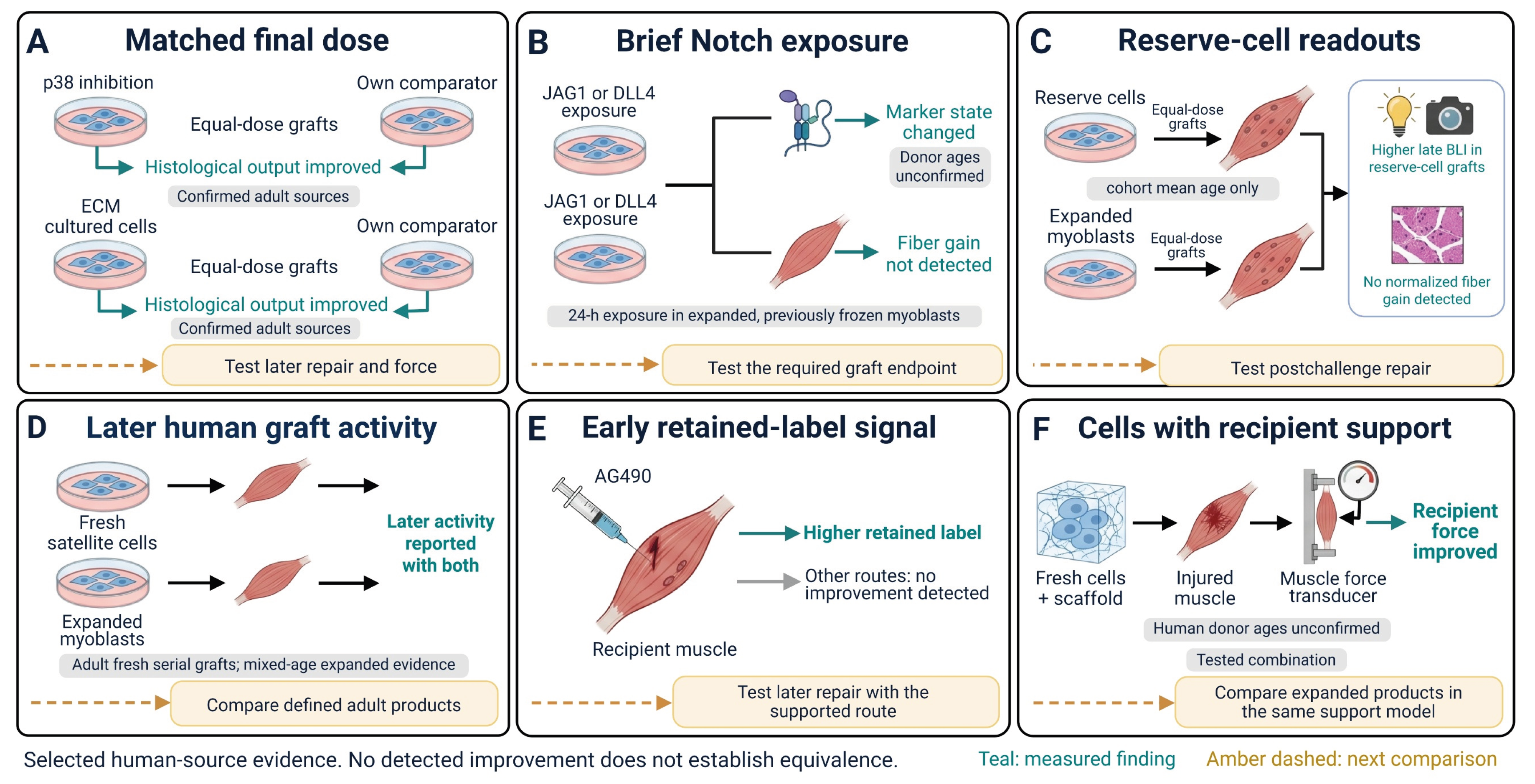
Figure 2.
Donor provenance and the limits of the evidence reappraisal. (A) Donor source and age, processing history and yield, final dose and distribution, and recipient conditioning and local support define the tested graft. The mouse icons represent recipient context and endpoint assessment, not separate cohorts. Donor-associated signal, fiber contribution, later repair or reserve, and recipient function are distinct outcomes. Signal is not a viable-cell count, niche occupancy does not establish self-renewal, and recipient force includes host contributions. (B) The selected corpus comprises 28 reports and 39 question-specific records: seven adult-confirmed, five mixed-age, four pediatric, and twelve age-unconfirmed reports. The primary adult analysis retains seven reports and thirteen records. Cohort mean age and father-derived donor sources add two reports in the contextual sensitivity analysis, giving nine reports and fifteen records without verifying individual donor ages. Recipient age does not establish donor age. (C) Source methods, results, figure legends, and supplementary materials anchor extraction of dose, denominator, recipient context, timing, and statistics. The released package contains 28 donor-provenance annotations, 39 question-specific records, and source dispositions for 82 cited references. It reproduces the coding and aggregation, but no second adjudicator independently verified the primary-source extraction. Incomplete discovery and shared cohorts or controls preclude pooled efficacy estimates, independent-effect counts, and product rankings. A nonsignificant result does not establish equivalence. Created in BioRender. Lee, W. (2026) https://BioRender.com/3veihtt.
Figure 2.
Donor provenance and the limits of the evidence reappraisal. (A) Donor source and age, processing history and yield, final dose and distribution, and recipient conditioning and local support define the tested graft. The mouse icons represent recipient context and endpoint assessment, not separate cohorts. Donor-associated signal, fiber contribution, later repair or reserve, and recipient function are distinct outcomes. Signal is not a viable-cell count, niche occupancy does not establish self-renewal, and recipient force includes host contributions. (B) The selected corpus comprises 28 reports and 39 question-specific records: seven adult-confirmed, five mixed-age, four pediatric, and twelve age-unconfirmed reports. The primary adult analysis retains seven reports and thirteen records. Cohort mean age and father-derived donor sources add two reports in the contextual sensitivity analysis, giving nine reports and fifteen records without verifying individual donor ages. Recipient age does not establish donor age. (C) Source methods, results, figure legends, and supplementary materials anchor extraction of dose, denominator, recipient context, timing, and statistics. The released package contains 28 donor-provenance annotations, 39 question-specific records, and source dispositions for 82 cited references. It reproduces the coding and aggregation, but no second adjudicator independently verified the primary-source extraction. Incomplete discovery and shared cohorts or controls preclude pooled efficacy estimates, independent-effect counts, and product rankings. A nonsignificant result does not establish equivalence. Created in BioRender. Lee, W. (2026) https://BioRender.com/3veihtt.
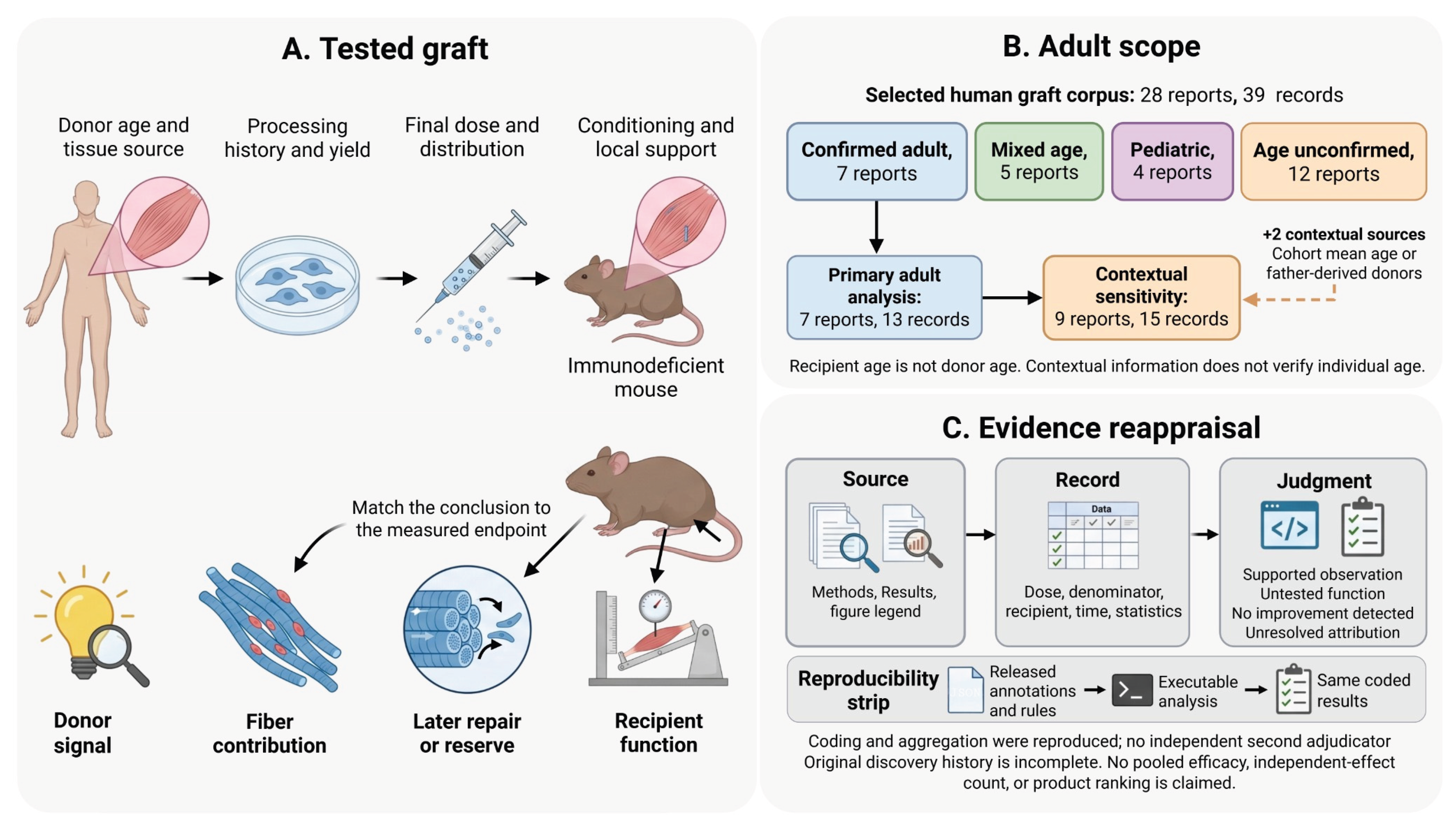

Table 1.
Human cell graft studies and their interpretable comparisons.
| Study and preparation | Dose and comparison | Observed result and denominator | Development implication and limit |
|---|---|---|---|
| Charville et al. 2015 [12] p38 inhibition; cohort 35–82 y |
30,000 each; treated cells, activated-cell controls, and fresh cells; injured NSG; 4 wk | 221 ± 20 versus 21 ± 13 and 49 ± 14 fibers/section (SD); both P < 0.001. Niche/recovery benefit also supported. | Administered-population improvement; matched-arm culture interval unresolved. Later repair and force untested. |
| Zhao et al. 2026 [13] Matrix culture; adults 40–56 y |
25,000 each; five-day matrix versus uncoated culture; injured nude mice; 4 wk | Human fibers and donor-derived PAX7-positive cells increased; Methods report six TA muscles per group. | Population improvement at equal dose; ATPase staining does not establish contractile force. |
| Judson et al. 2018 [14] SETD7 inhibition; 15–53 y |
15,000 starting cells; seven-day culture; final human dose unresolved; 11-day BLI | Production and reporter signal increased. Matched-final-dose output cannot be assigned. | Retain supply benefit; distinguish increased supply from performance of the administered population. |
| Quarta et al. 2016 [15] Artificial niche; ages unresolved |
1,000 each; approximately one-day preservation; injured recipients; 30-day BLI | Greater reporter-associated activity with combined niche and medium support. | Brief preservation supported; extensive human expansion and direct force untested. |
| Sakai et al. 2017 [16] Brief Notch rescue; ages unresolved |
100,000 each; 24-hour JAG1/DLL4 after expansion/freezing; injured NOD-SCID; 2 wk | Marker state changed; fiber improvement not detected. JAG1: five muscles/group; DLL4: three. | Marker response does not validate fiber replacement; nonsignificance does not establish equivalence. |
| Laumonier et al. 2017 [17] Reserve fraction; cohort mean 25.6 ± 1 y |
500,000 each; reserve cells versus myoblasts; injured NOG; 21 d | Late BLI and donor PAX7 fraction increased. Fibers/human nuclei: 27.1 ± 4.6% versus 23.1 ± 7.0% (SEM), P = 0.6. | Retain precursor-associated advantage; equivalence in absolute fiber output and later repair were not established. |
| Barruet et al. 2020 [18] CAV1 selection; graft ages unresolved |
500 each; positive versus negative fresh fractions; conditioned NSG; 5 wk | CAV1-positive fraction produced more human fibers. | Composition-associated benefit; not a causal CAV1-induction or expansion-rescue comparison. |
| Bentzinger et al. 2014 [19] Wnt7a; ages unresolved |
100,000 each; short pretreatment versus control; immunosuppressed mdx; 21 d | Human fiber contribution, size, and distribution increased. | Deployment benefit supported. Direct force arm used mouse cells; human expansion history unresolved. |
| Conte et al. 2023 [20] DM1 senolytic selection |
200,000 each; three lines, three mice/line; injured NSG; 20 d after graft | Human fiber contribution increased (P = 0.014). Treatment-duration statements conflict. | Population improvement retained; individual-cell rejuvenation and later force not established. |
| Escobar et al. 2021 [21] SGCA-edited cells; 13-y donor |
50,000 per muscle; conditioned NSG; 19 d; no matched unedited graft comparator | Corrected protein, human fibers and niche cells demonstrated; two reported grafts. | Editing-compatible activity; controlled superiority and sustained adult function were not tested. |
| Xu et al. 2015 [2] Fresh satellite cells; 17–81 y |
5,000 in later-challenge arm; conditioned NSG; reinjury at 5 wk; assessment at 10 wk | Human fibers, niche contribution and human participation after reinjury. | Later availability supported. Self-renewing divisions and whole-muscle force unestablished. |
| Garcia et al. 2018 [3] Adult-source fresh/cryopreserved satellite cells |
Fixed 2,000-cell distribution test; serial arm 2,500–5,000 initially; conditioned NSG | Multisite mean 155 versus 40 fibers; single-site escalation showed no gain. Secondary graft and tested cryopreserved-cell activity retained. | Optimize coverage; retain later competence and tested cryopreservation compatibility without equivalence claims. |
| Skuk et al. 2010 [22] Expanded cells; 13 mo, 39/43 y |
2–3 million initially; conditioned SCID; recovery/reculture and secondary graft; reinjury at 2 mo | Secondary human fibers and post-reinjury activity demonstrated. Reinjury count comparison P = 0.06. | Expanded route retains later activity. Mixed ages, high dose, and a culture-duration discrepancy preclude superiority. |
| Striedinger et al. 2025 [23] Recipient chemokine intervention |
2,000 fresh cells; recipient treatment versus vehicle; conditioned NSG; 3 wk | Human fiber formation and size increased. | Recipient support demonstrated; not a cultured-donor rescue or donor-specific mechanism. |
| Wu et al. 2025 [24] Fresh cells with programmed scaffold |
Same support model with/without programmed growth factors; human loading dose unresolved; 6 wk | Recipient contractile performance and structural repair improved in the supported human-cell graft. | Physiological combination benefit; expanded loading and separate added-cell effect require comparisons. |
| Marg et al. 2014 [25] Fiber fragments; graft donors 44–64 y |
Six adult graft sources; five- to seven-day culture; recipient-conditioning comparison | Human fibers and niche contribution depend on recipient context; associated architecture retained. | Adult short-culture activity; not a matched-dose single-cell or extensive-expansion comparison. |
| Cooper et al. 2003 [26] Expanded cells; 5 d, 17/71 y |
500,000 each; early/late infant amplification and older donors; 8 wk | Remaining human nuclei within fibers: 84% early infant, 21% late infant, 37% oldest source. | Within-infant processing effect supported; cross-donor age inference limited. These fractions do not represent retention of injected cells. |
| Gérard et al. 2012 [27] AG490; adult donor, 23 y |
500,000 each; coinjection, pretreatment or systemic routes; 5 d | Coinjection residual radiolabel 8.0/7.3/7.8% versus 5.5% control; other routes showed no benefit. | Route-specific early retained-label signal; a direct live-cell count and lasting repair remain untested. |
| Laumonier et al. 2013 [28] Dextran sulfate; 15 mo–4 y |
1 million each; pretreatment versus control; own three-hour BLI baseline | Day-one/day-three signal improved; day-seven P = 0.06; both at assay background by day fourteen. | Early reporter protection retained. Neither durable benefit nor death of all donor cells follows. |
| Périé et al. 2014 [29] Adult OPMD myoblasts |
Twelve adults; 21-day expansion; median 178 million; myotomy plus cells; 2 y | Manufacture/delivery feasible; some swallowing measures improved, propulsion globally unchanged. | Combined clinical observations; surgery-only comparator needed for added cell efficacy. |
| Boudreault et al. 2001 [30] Microcarriers; 13-mo donor |
2 million each; paired microcarrier/static grafts in two conditioned SCID mice; 4 wk | Human dystrophin fibers after both routes; similar numbers described without counts or equivalence test. | Manufacturing compatibility retained. Adult equivalence and calpeptin-treated graft function not established. |
| Liu et al. 2018 [31] Orbicularis cells; adults 18–51 y |
200,000 each; three-week induction versus noninduced; paired nude-mouse TA; 1 wk | CM-DiI-positive fibers in induced grafts; reported recovery counts labeled fibers. | Early source/induction feasibility. Human-specific identity, surviving-cell count and durable function unestablished. |
| Karpati et al. 1993 [32] Father-derived expanded cells |
Eight DMD boys; 55 million per biceps; paired placebo; up to 12 mo | Relative treated-side force advantage in 3/8; absolute baseline gain in two; no perceived daily-function gain. | Individual local responses coexist with low overall efficiency; neither uniform failure nor general efficacy. |
| Mendell et al. 1995 [33] Expanded father/brother cells |
Twelve DMD boys; 110 million per biceps × six; paired sham; 12 mo | Donor dystrophin confirmed in four; one biopsy 10.3% positive, without strength gain. No treated-side force advantage detected. | Protein-to-function dissociation; biopsy contribution does not measure repaired whole-muscle volume. |
| Miller et al. 1997 [34] Mixed-age donor myoblasts |
Ten DMD boys; 100 million per TA; paired placebo; cyclosporine; 6 mo | Both legs gained force; no between-leg advantage. Increase began before implantation. | Cell effect not demonstrated; treatment timing limits attribution without proving drug causality. |
| Tremblay et al. 1993 [35] Sibling cells; donor ages unreported |
Five boys; 102–240 million per BB; paired sham; no immunosuppression; 2–18 mo | No early strength advantage; larger late decline (29.2% vs 5.0%, reported P < 0.05). Local TA dystrophin is weak. | The TA and BB assessments differ in muscle, timing, and biopsy location; immune causality remains unresolved. |
| Tremblay et al. 1993 [36] Identical twin case; 14 y |
One pair; 704 million across biceps and wrist extensor; paired saline; 1–6 mo | Wrist gains 31/19/12/26% exceeded pretreatment intervals; elbow flexion did not improve. | An anatomically localized positive response is retained; a single pediatric case cannot establish population efficacy. |
| Bouche et al. 2026 [11] AF-selected reserve cells; ages unreported |
200,000 each; paired NSG gastrocnemius; fixed left/right allocation; day 28; CTX reinjury day 28, BLI day 32 | No detected fraction difference in BLI or PAX7 proportion. Reinjury reporter test performed; Results/legend significance conflict. | Retain the performed later challenge. Reporter increase is not direct postchallenge repair or force; adult provenance unconfirmed. |
Twenty-eight reports supply 39 question-specific records that may share cohorts and controls. Source anchors and full extraction accompany the manuscript. SD, standard deviation; SEM, standard error; BLI, bioluminescence imaging; TA, tibialis anterior; NSG/NOG/SCID, immunodeficient mouse strains; DM1, myotonic dystrophy type 1; OPMD, oculopharyngeal muscular dystrophy. Nonsignificance does not establish equivalence.
Table 2.
Human graft evidence for later regeneration and recipient function.
| Study and intervention | Graft and recipient context | Function demonstrated | Boundary of interpretation |
|---|---|---|---|
| Xu et al. 2015 [2] Fresh satellite cells; graft sources, 17–81 years |
5,000 CD56+/CD29+ cells in reinjury arm; NSG TA; irradiation on graft day and bupivacaine co-injection; five- and ten-week observations, with notexin reinjury. | Human fibers, niche occupancy, and human participation after reinjury. | Later availability in a conditioned xenograft. Mixed adolescent/adult graft sources; self-renewing divisions and whole-muscle force not established. |
| Garcia et al. 2018 [3] Fresh and cryopreserved satellite cells |
Serial arm: 2,500–5,000 initial cells; recovery at ten weeks and secondary NSG graft evaluation five weeks later. 18-Gy irradiation one day before graft; bupivacaine co-injection. | Human fibers and niche contribution in secondary grafts; tested fresh-cell cryopreservation retained activity. | Recovered cells were fewer than initial grafts. Secondary capacity does not distinguish dividing lineages from persistent quiescent cells. |
| Skuk et al. 2010 [22] Expanded myoblasts; 13 months, 39 and 43 years |
2–3 million cells; irradiated SCID TA. Initial culture: four weeks in Methods, 21 days in Discussion. Recovery at four weeks, 32-day reculture, secondary graft assessed four weeks later. | Human fibers after secondary transplantation and reinjury at two months; donor PAX7-positive niche cells. | Mixed-age, high-dose conditioned model. Reinjury count comparison P = 0.06. Later activity demonstrated; superiority and recipient force not established. |
| Striedinger et al. 2025 [23] Recipient chemokine intervention |
2,000 fresh cells; irradiated NSG TA with bupivacaine injury; recipient Bindarit for seven days; evaluated at three weeks. | Human fiber formation and size improved after recipient treatment. | Supports an environmental intervention. Broad pharmacological action does not isolate a donor-specific mechanism; direct force not tested. |
| Wu et al. 2025 [24] Growth factor-releasing dECM scaffold |
Fresh cells immediately after isolation; young NSG TA; 7 × 2 × 2 mm defect; three donor populations and four replicates; evaluated at six weeks. | Structural repair and recipient contractile performance improved versus a plain dECM scaffold carrying human cells. | Combined cell/scaffold benefit; expanded products not tested. Human loading dose and donor ages not identified in examined main text, Supplementary Information, and Reporting Summary. |
The studies address different functions and are not ranked. Reinjury tests later availability. Secondary transplantation tests recoverable graft capacity. Force measures recipient performance, including host tissue. Human PAX7-positive niche occupancy alone does not establish self-renewing divisions.
Table 3.
Scope of human graft findings under the adult donor restriction.
| Claim | Primary adult restriction | Contextual sensitivity | Retained scope and limit |
|---|---|---|---|
| Matched-dose culture output [12,13] | Adult p38 and matrix comparisons retain a histological benefit at matched final dose. | — | No intrinsic single-cell rejuvenation, later-challenge or force claim follows. |
| SETD7 supply and graft signal [14] | The mixed-age supply result remains useful; adult-specific performance is not isolated. | — | The missing final dose prevents separation of production yield from performance per administered cell, irrespective of age. |
| Marker and output dissociation [16,17,33] | These human limiting comparisons are not age-confirmed adult-only demonstrations. | Adds the cohort-mean reserve-cell comparison; other adult provenance remains unconfirmed. | Contextual sensitivity includes the reserve-cell marker/output dissociation; it does not establish adult Notch or clinical protein-to-force evidence. |
| Fresh and expanded later activity [2,3,11,22] | Adult-source serial fresh-cell activity is retained. An adult-only expanded later-activity comparison is not established in this corpus. | — | Mixed-age expanded activity and the age-unconfirmed reserve-cell challenge remain affirmative context, without product superiority. |
| Spatial distribution [3] | Adult-source fixed-dose multisite improvement remains supported. | — | No universal saturation dose or unconditioned-host claim is assigned. |
| Route and early window [27,28] | Adult AG490 route findings remain; dextran sulfate is pediatric context. | — | Early retained label cannot establish sustained repair. |
| Scalable manufacture compatibility [30] | Microcarrier graft compatibility is pediatric; adult equivalence is untested in this selected set. | — | A feasible manufacturing route remains a candidate rather than a failed adult product. |
| Recipient contractile performance [24] | The human-cell scaffold force finding lacks confirmed adult donor provenance. | — | Recipient function belongs to the tested combination; this row does not count other clinical force observations. |
| Clinical efficacy attribution [29,32,33,34,36] | Adult OPMD manufacture and combined treatment remain; isolated cell efficacy is unresolved. | Adds father-derived local force responses; numerical donor ages remain unreported. | Contextual father-derived Karpati grafts retain local positive responses. Other clinical reports remain age-unconfirmed, mixed or pediatric. The nonimmunosuppressed trial lacks confirmed donor ages [35]. |
Primary adult eligibility retains seven reports and thirteen records. Contextual sensitivity adds a cohort reported only by mean age and father-derived donor sources, giving nine reports and fifteen records without verifying individual ages. Mixed, pediatric, and age-unconfirmed studies remain in the broader review. Counts are not independent effects or field prevalence. Cooper’s separable 71-year arm remains descriptive context. Source coding and outputs accompany the manuscript. A dash indicates that no additional age-eligible report changes the claim.
Table 4.
Evidence-linked comparisons for developing human myogenic cell products.
| Human comparison | Development choice supported now | Comparison that would extend or limit the claim |
|---|---|---|
| Matched-dose p38 and matrix culture [12,13] | Retain culture routes with demonstrated adult-source histological gains. Advance beyond initial fiber output. | Test the defined final preparation at the same dose against its own control for later repair or recipient force. These studies do not rank p38 against matrix culture. |
| SETD7 inhibition [14] | Retain the production gain. Because the final human graft dose is unresolved, performance per administered cell cannot be assigned. | Compare treated and control grafts at the same final dose while reporting yield from the starting tissue. No fixed-dose gain would limit the graft-performance claim, not invalidate the yield benefit. |
| Microcarrier manufacture [30] | Retain a graft-compatible scalable route. Both preparations formed human fibers in a small pediatric-source experiment. | Quantify defined adult final products at matched dose and recipient conditions. Similar reported fiber formation does not establish equivalence. |
| Brief Notch exposure [16] | Retain a state response without selecting this regimen for fiber replacement from marker changes alone. | Test the intended processing regimen against its own control at the required graft endpoint. The limiting result applies to the tested brief exposure. |
| Reserve-cell comparisons [11,17] | Retain higher late BLI and precursor-associated measurements in the 2017 comparison. A separate autofluorescence-fraction study performed a reinjury reporter test. | Resolve the source statistics and directly measure postchallenge repair or force. Include yield after reserve-cell selection. Reporter response does not establish new fiber repair. |
| Fixed-dose distribution [3] | Evaluate spatial deployment before further single-site dose escalation in this conditioned adult-source model. At 2,000 cells, multisite grafts gave 155 versus 40 mean fibers. | Compare injection distributions at equal total dose, recording repair coverage and recipient function. Replication would define the reach beyond this recipient model. |
| AG490 and dextran sulfate [27,28] | Retain the supported route or early window. Adult AG490 improves day-five retained radiolabel by coinjection; pediatric dextran sulfate improves early BLI. | Test later repair with the supported route and appropriate controls. A transient benefit would preserve the early-delivery claim while limiting durability. |
| Fresh and expanded later activity [2,3,22] | Keep both routes in contention for sustained repair. Adult-source fresh serial activity is retained; the expanded later comparisons include mixed-age sources. | Compare defined adult final products under the same later challenge. Relative potency cannot be assigned from the different reported doses and recipients. |
| Human-cell scaffold support [24] | Use fresh cells in growth-factor-releasing dECM as a recipient-force benchmark against cells in plain dECM. Donor age is unconfirmed. | Match fresh and expanded cell loading within the same support model. Component controls would establish the added human-cell contribution. Whole-muscle force includes host tissue. |
| Clinical contribution | Retain adult OPMD manufacture and combined-treatment feasibility [29], and local responses in other reports [32,36]. Donor protein alone does not establish functional efficacy [33]. | Use an appropriate surgery-only or paired control and prespecified muscle function. Preserve donor provenance and the spatial scale of the measured repair. |
Rows link human findings to development choices and the comparisons needed to extend them. Table 3 specifies adult applicability. Supplementary File S1 provides extraction and source anchors. These are development inferences, not a validated treatment algorithm.
Disclaimer/Publisher’s Note: The statements, opinions and data contained in all publications are solely those of the individual author(s) and contributor(s) and not of MDPI and/or the editor(s). MDPI and/or the editor(s) disclaim responsibility for any injury to people or property resulting from any ideas, methods, instructions or products referred to in the content. |
© 2026 by the authors. Licensee MDPI, Basel, Switzerland. This article is an open access article distributed under the terms and conditions of the Creative Commons Attribution (CC BY) license (http://creativecommons.org/licenses/by/4.0/).
Copyright: This open access article is published under a Creative Commons CC BY 4.0 license, which permit the free download, distribution, and reuse, provided that the author and preprint are cited in any reuse.