

Submitted:
30 October 2026
Posted:
03 November 2026
You are already at the latest version
Abstract
Poly(vinyl alcohol) (PVA) hydrogels are promising for meniscal replacement, but reproducing native meniscal anisotropy remains unsolved. We developed a fabrication strategy for imparting controlled anisotropy to PVA hydrogels. We hypothesized that modulus and alignment would increase with applied strain, and that circumferential strain would produce direction-dependence in a meniscal-like geometry. Dogbone specimens at three PVA concentrations were strained 0–200% during freeze–thaw processing. Meniscal-like specimens were fabricated at 27.5% PVA under 200% circumferential strain. Constructs were characterized by tensile testing, stress-relaxation compression, and polarized light microscopy. In dogbone specimens, tensile modulus rose exponentially and coherency linearly with applied strain (adjusted R² > 0.92; R² ≥ 0.96). In meniscal-like specimens, stretching increased circumferential peak stress (587 ± 173 vs 439 ± 96 kPa), and direction-dependence appeared only after stretching. Stretched constructs showed higher optical coherency (p = 0.002, r = 0.77), concentrated in the outer third of the cross-section. Compressive moduli trended lower in stretched constructs, reaching significance anteriorly. Strain applied during freeze–thaw processing programs direction-dependent stiffness and alignment into a homogeneous hydrogel, though absolute stiffness remains below native tissue. More strain during processing is the most direct route to a stiffer construct, and further development is required before functional replacement.
Keywords:
poly(vinyl alcohol)
; hydrogel
; meniscus
; meniscal replacement
; strain-directed processing
; fiber alignment
; polarized light microscopy
; fibrocartilage biomechanics
1. Introduction
Meniscal pathology is a major contributor to knee dysfunction, and meniscal procedures remain among the most frequently performed orthopaedic operations. Not all tears are amenable to repair, and partial meniscectomy remains the most common surgical treatment [1,2]. Approximately 985,000 knee arthroscopies were performed in the United States in 2006, with roughly half involving meniscal surgery [3]. While often necessary, removal of meniscal tissue disrupts the knee’s native load-sharing mechanics, increasing contact stresses across the articular cartilage. Partial meniscectomy is associated with an increased risk of incident radiographic osteoarthritis and worsening cartilage damage within the following year [4].
To address the mechanical deficits associated with meniscectomy, considerable effort has been devoted to the development of meniscal scaffolds and implants aimed at replicating native biomechanics. A primary design challenge is reproducing the complex, depth-dependent, and anisotropic collagen fiber architecture of the native meniscus, which is essential for effective hoop stress transmission [5]. Electrospun polycaprolactone (PCL) scaffolds and collagen-based structures allow partial control over fiber alignment but are limited by their reliance on cell-mediated matrix deposition and insufficient mechanical robustness [6,7]. Synthetic alternatives, including fiber-reinforced hydrogels and double-network constructs, improve mechanical performance but introduce interfaces between materials with dissimilar stiffness profiles, leading to potential delamination under cyclic loading [8,9].
Poly(vinyl alcohol) (PVA) hydrogels represent a promising platform for meniscal replacement owing to their biocompatibility, low coefficient of friction, high fatigue resistance, and highly tunable mechanical properties [10,11]. PVA can be physically crosslinked via repeated freeze–thaw cycling and readily processed under mechanical strain to induce uniaxial anisotropy [12,13]. While these attributes enablethe fabrication of homogeneous, anisotropic constructs without requiring composite layering or heterogeneous phases, the ability to generate the complex anisotropy of the meniscus has not been captured.
The objective of this study was to develop a fabrication strategy for imparting controlled anisotropy to PVA hydrogels by applying tensile strain during freeze–thaw processing. To achieve this objective, we took a step-wise approach where we first determined the optimal PVA concentration and strain on dogbone-shaped samples in uniaxial tension. We then applied the optimized strain and PVA concentration to a meniscal-like geometry to generate circumferentially oriented alignment. We hypothesized that tensile modulus and optical alignment would increase with the strain applied during freeze–thaw manufacture, and that applying circumferential strain to a meniscal-like geometry would produce direction-dependent tensile behavior and a depth-dependent alignment gradient.
The primary endpoints of this study were tensile modulus and optical coherency as functions of PVA concentration and applied strain in dogbone specimens, and in meniscal-like geometries, optical coherency and direction-dependenttensile behavior as assessed by peak stress and tangent modulus in circumferentially versus radially cut specimens. Yield stress, yield strain, peak strain, peak load, energy to failure, and compressive instantaneous and relaxation moduli were recorded as secondary descriptive outcomes.
2. Methods
2.1. PVA Hydrogel Preparation
PVA hydrogels were prepared at concentrations of 20%, 27.5%, and 30% (w/v in deionized H2O). Solutions were prepared by autoclaving the PVA and water mixture at 110 °C for 1 hour. Once the pressure was released, the solution was mixed and stored until use. Solutions were reautoclaved at 110 °C for 15 mins and allowed to cool to 60 °C before use. Solutions were cast into custom-designed, 3D-printed molds to form either dogbone-shaped specimens for mechanical characterization or pre-meniscal shapes for implantation using a custom injection system. All samples underwent three freeze–thaw cycles (−20 °C for 20 hours, followed by 20 °C for 4 hours per cycle) to achieve partial physical crosslinking.
2.2. Dogbone Samples
2.2.1. Sample Preparation
Following initial crosslinking, samples were randomly assigned to either an “Unstretched” (control n = 26) or “Stretched” (n = 74) group. For the “Stretched” condition, anisotropy was induced by mechanically stretching the samples during an additional three freeze–thaw cycles. Dogbone samples were uniaxially strained to 100%, 150%, or 200% of their original length and maintained at this strain for three days prior to further cycling (Figure 1a & b).
2.2.2. Mechanical Characterization
Tensile testing was performed on 100 total dogbone specimens across multiple PVA concentrations (20, 27.5, and 30%) as well as strain conditions using an MTS Acumen electrodynamic testing system (Table 1, Figure 1c). For tensile testing, samples were strained uniaxially at a constant rate of 0.1% strain/sec until failure, and the tensile modulus was calculated from the linear region of the stress–strain response (Figure 3). To understand the relationship between strain and tensile modulus, an exponential curve was fit to the tensile modulus vs strain data.
2.2.3. Alignment Characterization
To assess polymer orientation and induced anisotropy, all samples were imaged under a circular polarizer and coherency was quantified using the OrientationJ plugin for ImageJ (0 = isotropic, 1 = perfectly aligned in-plane) [14]. Tensile dogbone samples were sliced longitudinally and 100-μm sections were taken from the center of each sample following fabrication and freeze–thaw processing (Figure 1d). Coherency vs strain data was fit to a linear regression to understand if there was a relationship between strain and polymer alignment.
2.3. Meniscus-Like Specimens
2.3.1. Sample Preparation
Among the conditions tested, 27.5% (w/v) PVA at 200% applied strain was selected as the fabrication condition for meniscal-like geometries.Tensile modulus increased with polymer concentration while coherency decreased, so stiffness and alignment could not be maximized simultaneously (Figure 3a,b). The 27.5% (w/v) formulation achieved tensile moduli comparable to the 30% (w/v) formulation while retaining higher coherency at every strain level tested, and achieved substantially higher moduli than the 20% (w/v) formulation for only a modest reduction in coherency; it therefore represented the best available compromise between stiffness and alignment. Because both tensile modulus and coherency increased monotonically with applied strain in all formulations, 200% — the highest strain evaluated — was selected. Qualitative handling assessment by an experienced knee surgeon was consistent with this selection.
To generate meniscal samples, 27.5% PVA was injected into wedge shaped molds and partially physically crosslinked. Samples were then mounted around a 53.4 mm circumference spherical post and stretched to 200% circumferential strain using a custom radial straining device. During the final three freeze–thaw cycles, “Stretched” samples remained fixed in their deformed configurations, while “Unstretched” samples were cycled without strain applied (Figure 2a-c).
2.3.2. Mechanical Characterization
2.3.2.1. Circumferential and Radial Tensile Testing
PVA Meniscus specimens were prepared for uniaxial tensile testing in both the circumferential and radial direction (Figure 2d) as recommended by Peloquin et al.[15]. Unstretched and Stretched PVA Menisci samples were stored at 4 °C in phosphate buffered saline (PBS) until testing. Samples were cut from the center (mid-circumferential, mid-radial, and mid-axial) region of the PVA Meniscus. While whole, the meniscus was planed using a freezing-stage microtome (Leica) to obtain the desired thickness of 2 mm. The specimen testing shape was then cut out using a #21 scalpel using a 3D-printed (PLA material) guide for target dimensions. Dimensions were measured following preparation using a caliper.
Circumferential samples had a gauge length of 15.72 ± 3.03 mm, 7.11 ± 1.15 mm midsection width, and 2.22 ± 0.42 mm thickness. Radial samples had a gauge length of 11.47 ± 1.31 mm, 4.86 ± 0.60 mm width, and 2.0 ± 0.0 mm thickness. Tensile testing was performed on 9 circumferential and 8 radial specimens per processing condition.
Prepared samples were mounted in aluminum grips with serrated teeth in a servo electric load (Acumen, MTS) system using a 50-N load cell (Electroforce, TA). A double layer of 280 grit sandpaper with a drop of cyanoacrylate was used to protect the specimen from the grip teeth and remain fixed during tensile testing. Grips were hand tightened via bolts (2 per grip) and kept aligned with a rigid guide. Samples were kept damp with PBS except when being manipulated.
Each sample was first preloaded with 0.2-N and held for 10 seconds. Next, 5% strain was applied at 0.05 mm/sec for 10 cycles to precondition the sample. Loads and displacements were then zeroed out and a displacement-controlled ramp to failure was run at 0.5 mm/sec to represent quasi-static loading.
Force vs displacement data was used to calculate the peak stress and strain, yield stress and strain, tangent modulus, and energy to failure per sample and grouped based on circumferential or radial test and unstretched or stretched status using a custom MATLAB script. The tangent modulus was calculated for the region between 25% and 100% peak strain. Peak stress and tangent modulus were pre-specified as the primary tensile outcomes and were compared between unstretched and stretched specimens, and between radial and circumferential specimens, using two-sample t-tests with statistical significance defined at p ≤ 0.05. Yield stress, yield strain, peak strain, peak load, and energy to failure were treated as secondary descriptive outcomes; comparisons for these endpoints were not corrected for multiple testing and are therefore reported as exploratory. All data are reported as mean ± standard deviation, unless otherwise indicated.
2.3.2.2. Compression Testing
PVA Meniscus specimens were prepared for uniaxial compression testing in the anterior, posterior, and central (Figure 2d) regions for both unstretched and stretched samples [16]. Samples were stored at 4 °C in PBS until testing. While whole, the meniscus was planed using a freezing-stage sledge microtome (Leica) to obtain the desired thickness and biopsy punched for the desired diameter. Dimensions were measured following preparation using a caliper.
Compression testing was performed on 9 Unstretched and 8 Stretched specimens per anatomical region per processing condition. Samples were 2.26 ± 0.52 mm tall and 4.65 ± 0.46 mm in diameter. No significant differences in dimensions were observed between posterior, central, and anterior regions. Prepared samples were mounted in 60-mm petri dishes and kept centered with a drop of cyanoacrylate. 2-mL 1x PBS was used to keep the samples hydrated during testing in the same servo electric load frame. Each sample was first preloaded with 0.2-N and held for 10 seconds. Next, 5% strain was applied at 0.05 mm/sec for 10 cycles to precondition the sample. Loads and displacements were then zeroed out prior to stress relaxation steps. Using displacement control, 3 sequential stress relaxation steps (5%, 10%, and 15% strain) were each applied at a near-instantaneous rate of 1 mm/sec then held for 30 minutes.
Using stress versus time data, the instantaneous and relaxation moduli of the viscoelastic samples were estimated using Fung’s 2-term exponential model, a common model for determining biomechanical properties of human menisci [17]. The instantaneous and relaxation moduli were each compared between unstretched and stretched specimens within each anatomical region using t-tests, with statistical significance set to p ≤ 0.05. Because these comparisons span three independent anatomical regions and two compressive strain levels, they were not corrected for multiple testing and are reported as exploratory.
2.3.3. Alignment Characterization
Similar to the dogbone samples, meniscus-like specimens were imaged under a circular polarizer and coherency was quantified using the OrientationJ plugin for ImageJ (0 = isotropic, 1 = perfectly aligned in-plane) [14]. Meniscal samples were sliced longitudinally into 100-μm sections. For meniscal samples, coherency was mapped across the full arc of each cross-section and summarized as a normalized depth metric (0 = outer surface, 1 = inner core), with the arc divided into three radial zones: Outer (0–0.33), Middle (0.33–0.67), and Inner (0.67–1.0) (Figure 2e). Coherency values were averaged per sample and compared between stretched and unstretched groups using the two-tailed Mann–Whitney U test. Zone-level comparisons (Outer, Middle, Inner) were performed using the same test with Benjamini–Hochberg correction applied across the three zones.
3. Results
3.1. PVA Dogbone Samples
Application of uniaxial strain during the fabrication process significantly influenced the mechanical properties of PVA hydrogels across all concentrations tested. Tensile modulus increased exponentially with strain for each formulation. For 20%, 27.5%, and 30% (w/v) PVA, the relationship between modulus and applied strain was well-approximated by exponential functions with adjusted R2 values exceeding 0.92. The modulus of 20% PVA ranged from 0.27 to 1.64 MPa across the strain range tested (0–200%), while 27.5% and 30% PVA exhibited modulus ranges of 0.75 to 3.70 MPa and 0.66 to 3.88 MPa, respectively (Figure 3a).
3.1.1. Alignment Quantification
To assess structural anisotropy, apparent in-plane alignment was quantified using polarized light microscopy and coherency analysis. Coherency increased linearly with applied strain in all formulations. Notably, for a given strain, lower-concentration PVA hydrogels exhibited higher coherency values, indicating greater optical alignment. Linear regression analysis yielded strong correlations between strain and coherency for all groups: R2 = 0.96 for 20% PVA, R2 = 0.97 for 27.5% PVA, and R2 = 1.00 for 30% PVA.
3.2. Meniscus-Like Specimen Geometry
For all sample types, stress-strain curves displayed nonlinear responses, typical of fibrous soft tissue. As expected due to altered polymer orientation when stretched, circumferential samples showed significantly higher strength (p < 0.05), as measured by peak stress (stretched: 586.82 ± 172.52 kPa and unstretched: 438.90 ± 96.48 kPa) and tangent modulus (stretched: 752.92 ± 179.33 kPa and unstretched: 508.23 ± 64.61 kPa) (Figure 4a). Interestingly, the radial yield stress was significantly lower for the stretched samples (stretched: 58.79 ± 18.99 kPa and unstretched: 112.43 ± 46.11 kPa) (Figure 4b). There was also a significant difference in yield stress and peak stress between the stretched samples when comparing the circumferential to radial directions that is not seen in the unstretched samples.
For the compressive testing, some of the samples were unable to be fit using the Fung model [17] and were excluded from the analysis with the sample size used for the analysis given in Figure 5. For the remaining samples fit well across all sample tests (R2 = 0.91 ± 0.10). Instantaneous and relaxation moduli from 10% and 15% strain levels were assessed. The 5% strain level resulted in values that were below the load cell threshold and were not used for analysis. Interestingly, the compressive moduli of the stretched samples trended lower in moduli values than unstretched counterparts with a significant decrease in the anterior region in the relaxation moduli (p < 0.05) (Figure 5).
3.2.1. Alignment Quantification
Stretched PVA meniscal constructs exhibited significantly higher coherency than unstretched constructs (Mann–Whitney U, two-tailed: p = 0.002, rank-biserial r = 0.77; Figure 6), indicating that circumferential stretching during freeze–thaw processing reliably induced preferential in-plane alignment in the meniscal geometry. The large effect size reflects that stretched sample coherency values ranked above those of unstretched samples in approximately 88% of pairwise comparisons.
Zone-level analysis revealed that the coherency difference was concentrated in the Outer zone (p = 0.017, r = 0.69, BH-FDR adjusted; Supplemental Figure 1), corresponding to the outermost third of the cross-sectional depth. The Middle (p = 0.13, r = 0.46) and Inner (p = 0.2, r = 0.33) zones did not reach statistical significance after correction for multiple comparisons, though both effect sizes were positive and directionally consistent with the Outer zone finding (Table 2, Supplemental Figure 1). The depth profile (Figure 7) shows that the greatest separation between groups occurred near the outer surface, diminishing progressively toward the inner core.
4. Discussion
This study demonstrates that mechanically induced anisotropy in PVA hydrogels can be modulated through strain-directed processing and polymer concentration. Across all concentrations evaluated, tensile modulus increased exponentially with applied strain, while optical coherency increased linearly. These findings are consistent with physical theory governing semi-crystalline polymer networks and suggest that chain alignment during the freeze–thaw process can be leveraged to tune the biomechanical behavior of homogeneous hydrogels.
In the meniscus-like specimens, testing in the circumferential direction demonstrated consistent strain-dependent increases in the yield and tangent moduli (Figure 4) supporting the observed trends under tensile loading in the dogbone samples. The observed inverse relationship between polymer concentration and coherency at a given strain likely reflects increased crosslinking density and reduced chain mobility in more concentrated solutions.
The observed strain-dependent alignment is consistent with previous studies demonstrating that mechanical deformation during PVA hydrogel processing promotes preferential orientation of polymer chains and crystalline domains along the loading axis [12,18]. In a similar vein, tunable anisotropy ratios in PVA hydrogels have also been seen through directional freezing [13,19,20].
Our results build upon these by demonstrating a quantitative relationship between applied mechanical strain and optical coherency measured using polarized light microscopy, suggesting a relatively simple strain-directed freeze–thaw processing method for tuning alignment within homogeneous hydrogels. Our findings are consistent with recent reviews of anisotropic hydrogel fabrication strategies, which highlight mechanical deformation as a powerful approach for generating oriented fibrillar structures and anisotropic mechanical behavior within soft materials [21,22].
We found that lower PVA concentrations produce higher coherency at equivalent strain levels, likely reflecting increased polymer chain mobility in less densely crosslinked networks. In contrast, the denser networks in higher polymer concentrations limit structural realignment despite increasing baseline stiffness. Similar relationships between polymer concentration, chain mobility, and network organization have been described in recent reviews of PVA hydrogel processing and mechanical behavior [23,24].
These findings have important implications for meniscus tissue engineering. The native meniscus possesses a highly organized collagen architecture – reproducing this circumferential, anisotropic architecture remains one of the central challenges in designing functional meniscal replacements [5,25]. Previous work in generating alignment in meniscal tissue engineered constructs focus on anchoring the tissue engineered meniscus horns and applying forces seen during tissue loading led to reorganize the collagen fibers through cellular contraction and matrix deposition [26]. While they were able to generate tissue engineered menisci with equivalent compressive properties, the circumferential and radial moduli were below native meniscus levels. The approach taken in this study applies strains to the material to simulate the process that occurs during organism growth. This approach has been shown in neuronal engineering to allow for the extension of axons over large distances and may be a promising approach to mechanically generate alignment not through cellular tractive forces [27]. Our strain-engineering strategy presented here enables the generation of anisotropic mechanical properties within a single homogeneous hydrogel material, offering potential advantages in manufacturing simplicity, mechanical continuity, and resistance to delamination under cyclic loading. The ability to program anisotropy through controlled mechanical deformation during fabrication may therefore provide a promising pathway for producing biomimetic meniscal constructs without requiring complex multi-material assemblies.
The mechanical properties observed in this study remain below those of native meniscal tissue, which exhibits circumferential tensile moduli reported in the range of approximately 100–300 MPa depending on region and testing conditions [28].The constructs reported here therefore remain approximately two orders of magnitude below native circumferential tensile modulus. The contribution of this work is not a mechanically equivalent substitute, but a demonstration that directional stiffness and alignment can be programmed into a single-phase hydrogel through processing alone. Closing the remaining mechanical gap will likely require higher applied strain, alternative base polymers, or hierarchical reinforcement strategies [18].
The tensile behavior of the meniscal-like PVA specimens was qualitatively consistent with native fibrocartilage mechanics [15]. The characteristic toe region followed by linear stiffening mirrors the J-shaped stress-strain response of native meniscal tissue, arising from progressive engagement of aligned fibrous elements under increasing load. The significantly higher peak stress in circumferentially oriented stretched samples relative to radial counterparts is mechanically expected given the induced circumferential chain alignment; loading parallel to the dominant polymer orientation recruits the aligned network more efficiently, consistent with the relationship between fiber angle and tensile modulus in unidirectional fiber systems [29]. When comparing the tensile dogbones to the circumferential tangential modulus of the meniscus-like PVA specimens, the 200% strain applied to both types of specimens yielded different moduli, with the dogbone specimens having 3 times higher modulus values compared to the meniscus-like PVA specimens. This suggests that additional strain may need to be applied to the circumferential samples to generate a similar modulus to the tensile dogbones. Interestingly, there was a significant decrease in compressive relaxation modulus observed in the anterior region of stretched samples warrants further investigation and may reflect regional variation in the constraint geometry during circumferential stretch, producing non-uniform local strain — and therefore non-uniform alignment — across the anterior, central, and posterior positions of the construct. The lower compressive modulus compared to the unstretched samples may be related to the alignment of the chains in the circumferential direction and suggests there is a need to actively apply a compressive force through the straining device.
Stretching produced a robust and spatially non-uniform increase in optical alignment in the meniscal-shaped PVA specimens, with the overall coherency difference between stretched and unstretched groups reaching statistical significance with a large effect size (p = 0.002, rank-biserial r = 0.77). This finding is consistent with the strain-dependent chain and crystallite alignment observed in the dogbone formulations and confirms that circumferential strain-directed freeze–thaw processing translates to the meniscal geometry. Notably, the alignment effect was concentrated in the outermost third of the cross-section (Outer zone: p = 0.017, r = 0.69), with Middle and Inner zones trending in the same direction but not reaching significance after multiple-comparisons correction. This surface-dominant spatial gradient likely reflects the radially inhomogeneous strain field within a cylindrical body under circumferential loading, in which outer regions experience greater local deformation than the inner core — analogous to the well-documented depth-dependent collagen fiber organization of the native meniscus, where circumferentially oriented fibers predominate in the superficial and peripheral zones.
This depth-dependent gradient also mirrors the known architecture of native meniscus, in which the deep central layer is dominated by highly aligned circumferential fiber bundles while the superficial and lamellar layers contain predominantly randomly oriented fibrils — a hierarchy that X-ray diffraction and second harmonic generation imaging have consistently demonstrated across human, bovine, and porcine menisci [16,30,31]. Taken together, the coherency data provide evidence that the 27.5% PVA formulation subjected to 200% circumferential strain achieved a favorable balance of stiffness and alignment, making it well-suited for further evaluation.
Despite these promising results, the study has several limitations. The applied boundary strains during freeze–thaw processing represent nominal bulk values; local strains at the polymer chain level were not independently quantified and may deviate from the applied value due to viscoelastic relaxation during the freezing cycle. A single uniform PVA concentration was used for all meniscal-like specimens, whereas native meniscal tissue exhibits marked spatial heterogeneity: the outer vascularized zone is predominantly fibrous while the inner avascular zone is fibrocartilaginous, with corresponding gradients in proteoglycan content, water fraction, and mechanical stiffness. Reproducing this zonal architecture would require a concentration or compositional gradient within the construct. The strain-processing approach generates predominantly uniaxial circumferential alignment, whereas the native meniscus features a three-dimensional fiber hierarchy — circumferential bundles in the bulk interspersed with radial tie fibers and randomly-oriented superficial and lamellar layers — that the present fabrication method does not fully replicate. Coherency was derived from two-dimensional polarized light images and therefore reflects apparent in-plane optical anisotropy rather than three-dimensional fiber architecture; out-of-plane orientations are not captured, and the metric should not be interpreted as a direct measure of three-dimensional structural organization. All mechanical testing was conducted under quasi-static loading; dynamic viscoelastic behavior at physiological gait frequencies was not characterized. Finally, this study focused exclusively on mechanical and microstructural characterization; no biological evaluation regarding cell viability, protein adsorption, or tissue integration was performed, as PVA is biologically inert without surface modification and would require additional functionalization for tissue engineering applications. Sample sizes for the coherency analysis were modest (n = 16 stretched, n = 8 unstretched), and the Middle and Inner zone effects, while directionally consistent, may reach significance with larger cohorts. Group sizes were also unequal across the mechanical testing conditions, and the secondary mechanical endpoints were analyzed without correction for multiple testing; those comparisons are therefore exploratory and would require confirmation in an appropriately powered study designed to test them directly.
Follow-up studies are planned to evaluate the mechanical durability and surgical viability of the PVA constructs. These include suture pull-out testing to assess fixation strength, crack propagation experiments to examine long-term fatigue behavior, and dynamic simulator trials using gait-like loading patterns. Further, this approach is not limited to PVA and other polymers with more favorable starting mechanical properties will be explored using this technique.
5. Conclusion
This work establishes a reproducible method for fabricating mechanically anisotropic PVA hydrogels with tunable properties. By controlling strain and polymer concentration during freeze–thaw processing, we generated constructs with moduli and alignment profiles that reproduce the direction-dependence of native meniscal tissue, though not its absolute stiffness. Because both stiffness and alignment increased with applied strain in every formulation, the most direct route to a stiffer construct is more strain during processing rather than a different material.
Author Contributions
Conceptualization, TC, SM. Methodology, TC, JW, CZ; software, JW, TC, CZ; formal analysis, JW, MG, TC, CZ; investigation, JW, CM, AK, MG, RY; resources, TC, SM.; data curation, JW, CM, AK, MG, RY; writing—original draft preparation, JW, MG, CZ; writing—review and editing, TC, SM; visualization, JW, CZ. All authors have read and agreed to the published version of the manuscript.
Funding
this work received no external funding.
Institutional Review Board Statement
Not applicable.
Informed Consent Statement
Not applicable.
Data Availability Statement
The data presented in this study are available on request from the corresponding author.
Acknowledgments
The authors thank Dr. Russell Warren for guidance on formulation selection and surgical handling requirements. The authors acknowledge the use of Claude Opus 5.5 to improve the clarity and readability of the manuscript. All generated edits were carefully reviewed, verified, and modified by the authors, who take full responsibility for the final content.
Conflicts of Interest
The authors declare no conflicts of interest.
References
- Espejo-Reina, A.; Sevillano-Pérez, E.; Espejo-Reina, M.J.; Lombardo-Torre, M.; Pérez-Blanca, A.; Espejo-Baena, A. The Proportion of Meniscus Tears Considered Repairable, and Thus Repaired, Increased During a Single Surgeon’s Practice of 20 Years. Arthrosc. Sports Med. Rehabil. 2023, 5, 100778. [Google Scholar] [CrossRef] [PubMed]
- Boutin, R.D.; Fritz, R.C.; Marder, R.A. Magnetic Resonance Imaging of the Postoperative Meniscus: Resection, Repair, and Replacement. Magn. Reson Imaging Clin. N Am. 2014, 22, 517–555. [Google Scholar] [CrossRef] [PubMed]
- Kim, S.; Bosque, J.; Meehan, J.P.; Jamali, A.; Marder, R. Increase in Outpatient Knee Arthroscopy in the United States: A Comparison of National Surveys of Ambulatory Surgery, 1996 and 2006. J. Bone Jt. Surg. Am. 2011, 93, 994–1000. [Google Scholar] [CrossRef] [PubMed]
- Roemer, F.W.; Kwoh, C.K.; Hannon, M.J.; Hunter, D.J.; Eckstein, F.; Grago, J.; Boudreau, R.M.; Englund, M.; Guermazi, A. Partial Meniscectomy Is Associated with Increased Risk of Incident Radiographic Osteoarthritis and Worsening Cartilage Damage in the Following Year. Eur. Radiol. 2017, 27, 404–413. [Google Scholar] [CrossRef] [PubMed]
- Makris, E.A.; Hadidi, P.; Athanasiou, K.A. The Knee Meniscus: Structure-Function, Pathophysiology, Current Repair Techniques, and Prospects for Regeneration. Biomaterials 2011, 32, 7411–7431. [Google Scholar] [CrossRef] [PubMed]
- Baker, B.M.; Handorf, A.M.; Ionescu, L.C.; Li, W.-J.; Mauck, R.L. New Directions in Nanofibrous Scaffolds for Soft Tissue Engineering and Regeneration. Expert Rev. Med. Devices 2009, 6, 515–532. [Google Scholar] [CrossRef] [PubMed]
- Ozeki, N.; Seil, R.; Krych, A.J.; Koga, H. Surgical Treatment of Complex Meniscus Tear and Disease: State of the Art. J. ISAKOS 2021, 6, 35–45. [Google Scholar] [CrossRef] [PubMed]
- Shemesh, M.; Shefy-Peleg, A.; Levy, A.; Shabshin, N.; Condello, V.; Arbel, R.; Gefen, A. Effects of a Novel Medial Meniscus Implant on the Knee Compartments: Imaging and Biomechanical Aspects. Biomech. Model Mechanobiol. 2020, 19, 2049–2059. [Google Scholar] [CrossRef] [PubMed]
- Nonoyama, T.; Gong, J.P. Tough Double Network Hydrogel and Its Biomedical Applications. Annu Rev. Chem. Biomol. Eng. 2021, 12, 393–410. [Google Scholar] [CrossRef] [PubMed]
- Spiller, K.L.; Maher, S.A.; Lowman, A.M. Hydrogels for the Repair of Articular Cartilage Defects. Tissue Eng. Part B Rev. 2011, 17, 281–299. [Google Scholar] [CrossRef] [PubMed]
- Baker, M.I.; Walsh, S.P.; Schwartz, Z.; Boyan, B.D. A Review of Polyvinyl Alcohol and Its Uses in Cartilage and Orthopedic Applications. J. BioMed Mater. Res. B Appl. Biomater. 2012, 100, 1451–1457. [Google Scholar] [CrossRef] [PubMed]
- Chen, Y.; Jiao, C.; Peng, X.; Liu, T.; Shi, Y.; Liang, M.; Wang, H. Biomimetic Anisotropic Poly(Vinyl Alcohol) Hydrogels with Significantly Enhanced Mechanical Properties by Freezing–Thawing under Drawing. J. Mater. Chem. B 2019, 7, 3243–3249. [Google Scholar] [CrossRef]
- Duan, S.; Liu, Z.; Wu, S.; Hua, M.; He, X. Tuning Structural and Mechanical Anisotropy of PVA Hydrogels. Mech. Mater. 2022, 172, 104411. [Google Scholar] [CrossRef]
- Püspöki, Z.; Storath, M.; Sage, D.; Unser, M. Transforms and Operators for Directional Bioimage Analysis: A Survey. In Focus on Bio-Image Informatics; Advances in Anatomy, Embryology and Cell Biology; De Vos, W.H., Munck, S., Timmermans, J.-P., Eds.; Springer International Publishing: Cham, 2016; Vol. 219, pp. 69–93. ISBN 978-3-319-28547-4. [Google Scholar]
- Peloquin, J.M.; Santare, M.H.; Elliott, D.M. Advances in Quantification of Meniscus Tensile Mechanics Including Nonlinearity, Yield, and Failure. J. Biomech. Eng. 2016, 138, 021002. [Google Scholar] [CrossRef] [PubMed]
- Proctor, C.S.; Schmidt, M.B.; Whipple, R.R.; Kelly, M.A.; Mow, V.C. Material Properties of the Normal Medial Bovine Meniscus. J. Orthop. Res. 1989, 7, 771–782. [Google Scholar] [CrossRef] [PubMed]
- Fung, Y.C. Biomechanics; Springer: New York, NY, 1981; ISBN 978-1-4757-1754-9. [Google Scholar]
- Li, H.; Wu, H.; Guan, C.; Hu, W.; Su, W.; Chen, D.; Gao, J. Hierarchical Crack-Resistant, Tissue-Mimetic Hydrogels Enabled by Progressive Nanocrystallization of Anisotropic Polymer Networks. Nat. Commun. 2025, 16, 10890. [Google Scholar] [CrossRef] [PubMed]
- Sharifi, F.; Bai, Z.; Montazami, R.; Hashemi, N. Mechanical and Physical Properties of Poly(Vinyl Alcohol) Microfibers Fabricated by a Microfluidic Approach. RSC Adv. 2016, 6, 55343–55353. [Google Scholar] [CrossRef]
- Sanhueza Ortega, J.; Shanmuganathan, K.; Poole-Warren, L.; Beier, S.; Aregueta Robles, U. Physicochemical Reinforcement Unlocks Sterilization-St Anisotropic Hydrogels for Cell-Compatible Mock Arteries. Adv. Healthc. Mater. 2026, 15, e00010. [Google Scholar] [CrossRef] [PubMed]
- Zhang, F.; Gao, Z.; Sun, L.; Jin, Z.; Liu, Y.; Tong, M.; Zhao, Y.; Jiang, X.; Gu, X. Programmable Anisotropic Hydrogels for Biomedicine: From Precise Design to Advanced Applications. ACS Appl. Mater. Interfaces 2025, 17, 62860–62890. [Google Scholar] [CrossRef] [PubMed]
- Abalymov, A.; Pinchasik, B.-E.; Akasov, R.A.; Lomova, M.; Parakhonskiy, B.V. Strategies for Anisotropic Fibrillar Hydrogels: Design, Cell Alignment, and Applications in Tissue Engineering. Biomacromolecules 2023, 24, 4532–4552. [Google Scholar] [CrossRef] [PubMed]
- Liang, X.; Zhong, H.-J.; Ding, H.; Yu, B.; Ma, X.; Liu, X.; Chong, C.-M.; He, J. Polyvinyl Alcohol (PVA)-Based Hydrogels: Recent Progress in Fabrication, Properties, and Multifunctional Applications. Polymers 2024, 16, 2755. [Google Scholar] [CrossRef] [PubMed]
- Bercea, M. Recent Advances in Poly(Vinyl Alcohol)-Based Hydrogels. Polymers 2024, 16, 2021. [Google Scholar] [CrossRef] [PubMed]
- Zhou, Z.; Wang, J.; Jiang, C.; Xu, K.; Xu, T.; Yu, X.; Fang, J.; Yang, Y.; Dai, X. Advances in Hydrogels for Meniscus Tissue Engineering: A Focus on Biomaterials, Crosslinking, Therapeutic Additives. Gels 2024, 10, 114. [Google Scholar] [CrossRef] [PubMed]
- Puetzer, J.L.; Bonassar, L.J. Physiologically Distributed Loading Patterns Drive the Formation of Zonally Organized Collagen Structures in Tissue-Engineered Meniscus. Tissue Eng. Part A 2016, 22, 907–916. [Google Scholar] [CrossRef] [PubMed]
- Pfister, B.J.; Iwata, A.; Meaney, D.F.; Smith, D.H. Extreme Stretch Growth of Integrated Axons. J. Neurosci. 2004, 24, 7978–7983. [Google Scholar] [CrossRef] [PubMed]
- Schwer, J.; Ignatius, A.; Seitz, A.M. The Biomechanical Properties of Human Menisci: A Systematic Review. Acta Biomater. 2024, 175, 1–26. [Google Scholar] [CrossRef] [PubMed]
- Wang, H.W.; Zhou, H.W.; Gui, L.L.; Ji, H.W.; Zhang, X.C. Analysis of Effect of Fiber Orientation on Young’s Modulus for Unidirectional Fiber Reinforced Composites. Compos. Part B Eng. 2014, 56, 733–739. [Google Scholar] [CrossRef]
- Aspden, R.M.; Yarker, Y.E.; Hukins, D.W. Collagen Orientations in the Meniscus of the Knee Joint. J. Anat. 1985, 140 Pt 3, 371–380. [Google Scholar] [PubMed]
- Rasheed, B.; Ayyalasomayajula, V.; Schaarschmidt, U.; Vagstad, T.; Schaathun, H.G. Region- and Layer-Specific Investigations of the Human Menisci Using SHG Imaging and Biaxial Testing. Front Bioeng. Biotechnol. 2023, 11, 1167427. [Google Scholar] [CrossRef] [PubMed]
Figure 1.
Manufacturing process for Dogbone and Meniscus samples. Dogbone samples were cast in 3D-printed molds and underwent three freeze-thaw cycles. a) Dogbone samples stretched in a custom tensioning system to 100, 150, and 200% strain. Strains were held as samples underwent three more freeze-thaw cycles. b) Dogbone unstretched (left most) and stretched (right) samples. Dogbone samples either c) tested mechanically in tension or d) processed and imaged for polymer orientation.
Figure 1.
Manufacturing process for Dogbone and Meniscus samples. Dogbone samples were cast in 3D-printed molds and underwent three freeze-thaw cycles. a) Dogbone samples stretched in a custom tensioning system to 100, 150, and 200% strain. Strains were held as samples underwent three more freeze-thaw cycles. b) Dogbone unstretched (left most) and stretched (right) samples. Dogbone samples either c) tested mechanically in tension or d) processed and imaged for polymer orientation.
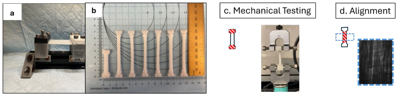
Figure 2.
Manufacturing process and quantifications for Meniscus-like specimens. a) meniscal samples were cast into linear molds and underwent three freeze-thaw cycles before b) stretching to 200% circumferential strain using a custom stretching device. After completion of all freeze-thaw cycles c) finished meniscal samples were either d) prepared for mechanical testing in tension (circumferentially or radially) or compression, or e) processed and imaged for polymer orientation.
Figure 2.
Manufacturing process and quantifications for Meniscus-like specimens. a) meniscal samples were cast into linear molds and underwent three freeze-thaw cycles before b) stretching to 200% circumferential strain using a custom stretching device. After completion of all freeze-thaw cycles c) finished meniscal samples were either d) prepared for mechanical testing in tension (circumferentially or radially) or compression, or e) processed and imaged for polymer orientation.
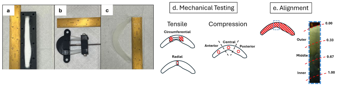
Figure 3.
Results of dogbone mechanical and structural characterization. a) Tensile modulus as a function of applied strain for 20%, 27.5%, and 30% (w/v) PVA, fit with exponential functions (adjusted R2 > 0.92). b) Coherency as a function of applied strain for each formulation, fit with linear regression (R2 = 0.96–0.97). Error bars represent standard deviation.
Figure 3.
Results of dogbone mechanical and structural characterization. a) Tensile modulus as a function of applied strain for 20%, 27.5%, and 30% (w/v) PVA, fit with exponential functions (adjusted R2 > 0.92). b) Coherency as a function of applied strain for each formulation, fit with linear regression (R2 = 0.96–0.97). Error bars represent standard deviation.
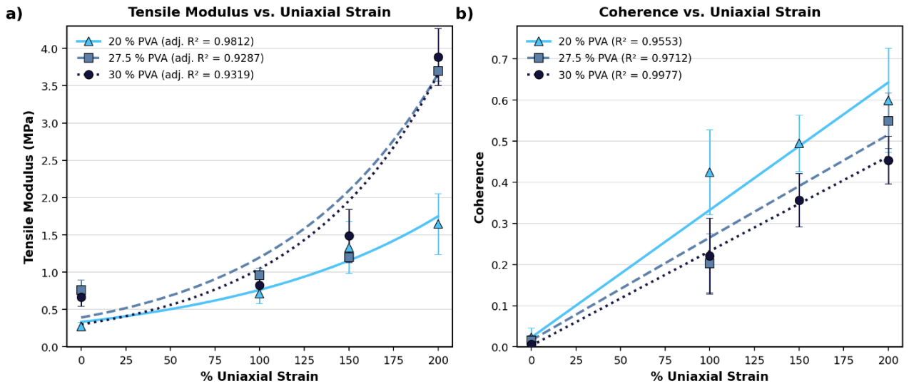
Figure 4.
Circumferential and radial stretched sample (a) yield stress (b) yield strain (c) peak stress (d) peak strain (e) tangent modulus (f) peak load and (g) energy to failure results.
Figure 4.
Circumferential and radial stretched sample (a) yield stress (b) yield strain (c) peak stress (d) peak strain (e) tangent modulus (f) peak load and (g) energy to failure results.
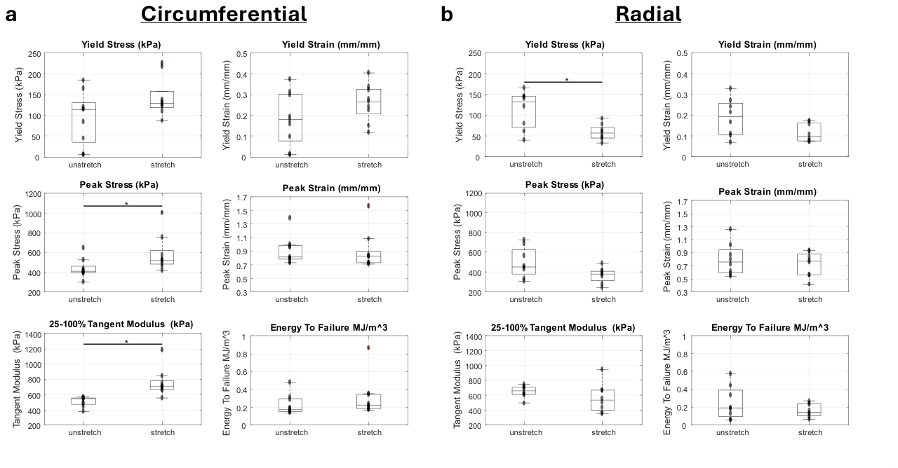
Figure 5.
Instantaneous (top row) and relaxation (bottom row) moduli for anterior, central, and posterior samples at (a) 10% strain and (b) 15% strain for unstretched (US) and stretched (S) samples.
Figure 5.
Instantaneous (top row) and relaxation (bottom row) moduli for anterior, central, and posterior samples at (a) 10% strain and (b) 15% strain for unstretched (US) and stretched (S) samples.
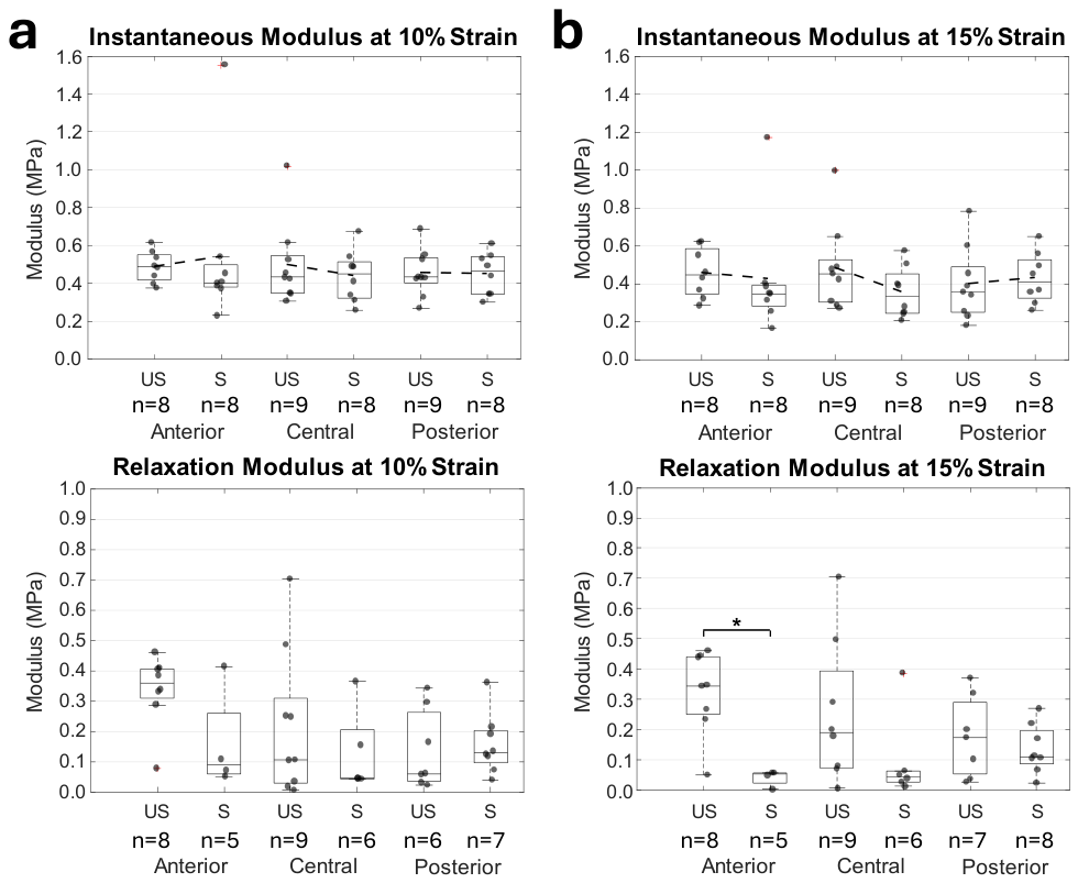
Figure 6.
PVA optical coherency by condition. Boxplots of per-biological-sample mean coherency for stretched (n = 16) and unstretched (n = 8) groups. Box: IQR; whiskers: 1.5×IQR; points: individual samples beyond whiskers. p = 0.002, r = 0.77 (Mann–Whitney U, two-tailed).
Figure 6.
PVA optical coherency by condition. Boxplots of per-biological-sample mean coherency for stretched (n = 16) and unstretched (n = 8) groups. Box: IQR; whiskers: 1.5×IQR; points: individual samples beyond whiskers. p = 0.002, r = 0.77 (Mann–Whitney U, two-tailed).
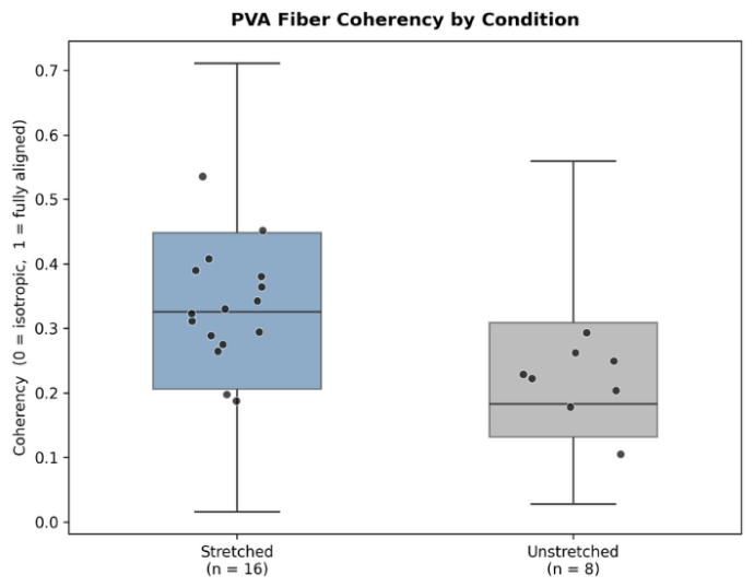
Figure 7.
Optical coherency in meniscus-like specimens. A) Coherency as a function of normalized cross-sectional depth. Each cross-sectional arc was folded about its midpoint so that both halves map onto a common depth axis (0 = outer surface, 1 = inner core). Mean coherency (± SD) shown for stretched (blue) and unstretched (grey) groups. Illustrative samples of b) stretched sample outer surface, c) stretched sample inner surface, d) unstretched sample outer surface, e) unstretched sample inner surface.
Figure 7.
Optical coherency in meniscus-like specimens. A) Coherency as a function of normalized cross-sectional depth. Each cross-sectional arc was folded about its midpoint so that both halves map onto a common depth axis (0 = outer surface, 1 = inner core). Mean coherency (± SD) shown for stretched (blue) and unstretched (grey) groups. Illustrative samples of b) stretched sample outer surface, c) stretched sample inner surface, d) unstretched sample outer surface, e) unstretched sample inner surface.
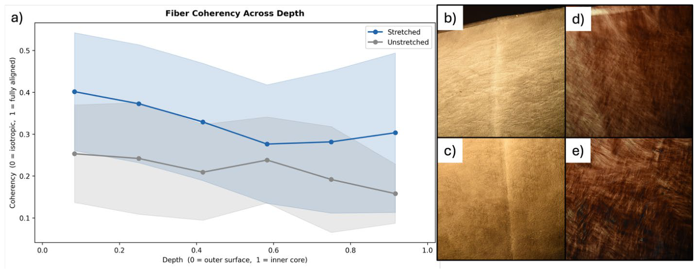

Table 1.
Tensile testing sample size by PVA percentage and strain percentage.
| Strain (%) | 20% PVA | 27.5% PVA | 30% PVA | Total |
|---|---|---|---|---|
| 0 | 12 | 7 | 7 | 26 |
| 100 | 17 | 6 | 6 | 29 |
| 150 | 6 | 6 | 6 | 18 |
| 200 | 17 | 6 | 4 | 27 |
| Total | 52 | 25 | 23 | 100 |
Table 2.
Statistical summary of coherency analysis in meniscal-like specimens.
| Region | n Stretched | n Unstretched | p-value | r (rank-biserial) |
|---|---|---|---|---|
| Overall | 16 | 8 | 0.002** | 0.77 |
| Outer zone# | 16 | 8 | 0.017* | 0.69 |
| Middle zone# | 16 | 8 | 0.13 | 0.46 |
| Inner zone# | 16 | 8 | 0.2 | 0.33 |
Mann-Whitney U, two-tailed. Zones: Outer = depth 0–0.33, Middle = 0.33–0.67, Inner = 0.67–1.0. Per-biological-sample mean coherency used as the unit of analysis. * p < 0.05, ** p < 0.01. #Benjamini-Hochberg FDR correction applied.
Disclaimer/Publisher’s Note: The statements, opinions and data contained in all publications are solely those of the individual author(s) and contributor(s) and not of MDPI and/or the editor(s). MDPI and/or the editor(s) disclaim responsibility for any injury to people or property resulting from any ideas, methods, instructions or products referred to in the content. |
© 2026 by the authors. Licensee MDPI, Basel, Switzerland. This article is an open access article distributed under the terms and conditions of the Creative Commons Attribution (CC BY) license (http://creativecommons.org/licenses/by/4.0/).
Copyright: This open access article is published under a Creative Commons CC BY 4.0 license, which permit the free download, distribution, and reuse, provided that the author and preprint are cited in any reuse.