
Submitted:
31 October 2026
Posted:
02 November 2026
You are already at the latest version
Abstract
Background/Objectives: Total cell-free DNA (cfDNA) is not tumour-specific and may be affected by peri-operative injury and inflammation. We evaluated peri-operative plasma cfDNA dynamics and explored associations with recurrence and mortality in resectable non-small cell lung cancer (NSCLC). Methods: This prospective pilot cohort included 35 patients undergoing lobectomy with systematic nodal dissection and 30 healthy controls. Samples were collected pre-operatively, from the tumour-draining pulmonary vein before major manipulation, peripherally after resection, and on postoperative day 14. Analyses included group comparisons, receiver operating characteristic analysis, logistic regression and Cox regression. Results: Pre-operative cfDNA was higher in patients than controls (mean 1.96 versus 1.08 ng/µL; p = 0.002). Discrimination was modest (AUC 0.67, 95% CI 0.54–0.80), with sensitivity 49% and specificity 93% at an internally selected cut-off. cfDNA differed across timepoints (overall p = 0.008), with higher intra-operative and post-resection values than pre-operative values. Among 34 patients with complete three-year outcomes, 12 recurred and 13 died. Pre-operative cfDNA was associated with recurrence in multivariable logistic regression (adjusted OR 2.47, 95% CI 1.07–5.67; p = 0.034). Day-14 cfDNA was higher among patients who died (5.35 versus 2.17 ng/µL; p = 0.008). Conclusions: Total cfDNA showed modest diagnostic discrimination and exploratory outcome associations. Small sample size, potential model overfitting and incomplete analytical documentation limit interpretation. These hypothesis-generating findings require independent validation with standardised methods, peri-operative covariates and tumour-informed circulating tumour DNA assays.
Keywords:
non-small cell lung cancer
; liquid biopsy
; cell-free DNA
; circulating tumour DNA
; peri-operative sampling
; pulmonary vein
; recurrence
; survival
1. Introduction
Lung cancer remains a major cause of cancer mortality worldwide [1]. Low-dose computed tomography and wider use of cross-sectional imaging have increased opportunities to diagnose potentially resectable disease, while also increasing the clinical burden of false-positive findings and indeterminate pulmonary nodules [2,3].
For operable NSCLC, anatomical resection with appropriate lymph-node assessment remains a principal curative treatment. Lobectomy remains standard for many patients, although randomised trials support anatomical segmentectomy or sublobar resection in carefully selected patients with peripheral, node-negative tumours measuring 2 cm or less [4,5]. Recurrence nevertheless occurs after complete resection, and stage alone does not fully account for heterogeneity in biological behaviour. The present cohort was staged using the eighth TNM edition [6]; the ninth-edition stage-group proposals were published subsequently [7]. Histological classification and pathological risk features provide additional prognostic information [8].
Liquid biopsy enables repeated, minimally invasive biomarker assessment. In advanced NSCLC, plasma circulating tumour DNA (ctDNA) can complement tissue genotyping, particularly when tissue is insufficient or difficult to obtain, although a negative plasma result does not exclude a targetable alteration and should prompt tissue testing where feasible [9,10]. In resectable disease, a major translational objective is detection of molecular residual disease after definitive treatment and improved postoperative risk stratification [11,12,13,14].
ctDNA assays seek tumour-derived genomic alterations and are therefore more specific than measurement of total cfDNA. By contrast, quantitative cfDNA includes DNA released from malignant and non-malignant cells. Meta-analyses and cohort studies report higher average cfDNA concentrations in NSCLC than in controls and associations between higher concentrations and adverse outcomes, but estimates vary substantially because of differences in disease stage, specimen type, pre-analytical handling, analytical method and threshold selection [15,16,17,18,19].
Peri-operative sampling introduces additional confounding because surgery can increase circulating cfDNA through tissue injury and inflammatory activation [20]. Sampling compartment may also matter. Pulmonary venous circulating tumour cell studies demonstrate enrichment in tumour-draining blood and changes during manipulation [21,22,23,24], but those findings cannot be assumed to apply directly to total cfDNA concentration.
We therefore conducted a prospective pilot study of quantitative plasma cfDNA in patients undergoing curative-intent lobectomy for NSCLC. The primary objectives were to characterise peri-operative cfDNA dynamics and to explore associations with recurrence and mortality. A secondary objective was to examine whether tumour-proximal pulmonary venous sampling provided a stronger signal than peripheral sampling. Because the assay measured total cfDNA rather than tumour-informed ctDNA, all prognostic analyses were considered exploratory.
2. Materials and Methods
2.1. Study Design and Setting
This was a prospective, non-interventional pilot cohort conducted in two thoracic surgery units in Athens, Greece. Consecutive eligible patients undergoing curative-intent lobectomy for NSCLC were enrolled. Clinical management followed contemporaneous multidisciplinary practice. No formal hypothesis-driven sample-size calculation was performed; the sample size was feasibility-based and constrained by prospective recruitment and laboratory resources.
2.2. Participants and Controls
Adults aged 18 years or older with resectable NSCLC (clinical stage I–IIIA) who were scheduled to undergo lobectomy with systematic mediastinal lymph-node dissection were eligible for inclusion. Exclusion criteria were incomplete resection, receipt of neoadjuvant treatment, and synchronous or previous malignancy. The study was conducted in the Thoracic Surgery Department of the Sotiria General Hospital of Thoracic Diseases and the Thoracic Surgery Department of NIMTS. Patient recruitment took place from January 2020 to November 2021 and was substantially affected by the COVID-19 pandemic. Thirty healthy controls, aged 18–80 years and with no history of malignancy, were included for the exploratory diagnostic comparison. Recruitment began in January 2020 following authorisation. Publication of the official approval document from the Scientific Council of the Sotiria General Hospital of Thoracic Diseases was delayed by the COVID-19 pandemic; the document was published on 12 June 2020 (identifier: ΩΥΣ9469069-66Ξ). All patients and healthy controls provided written informed consent before enrolment.
2.3. Clinical Assessment and Pathology
Staging investigations included computed tomography of the chest and abdomen, brain imaging, and positron-emission tomography-computed tomography where available. Stage was assigned according to the eighth edition of the TNM classification [6]. The cohort was not reclassified for this analysis using the subsequently published ninth-edition stage groups [7]. Histological subtype was assigned according to the contemporaneous World Health Organization classification [8].
2.4. Sampling Schedule
Blood was collected at four timepoints: (i) pre-operative peripheral blood at admission; (ii) intra-operative blood from the tumour-draining pulmonary vein before major manipulation; (iii) peripheral blood after completion of lobectomy and removal of the specimen; and (iv) peripheral blood on postoperative day 14 (Figure 1). Sampling time and anatomical compartment were recorded in structured laboratory worksheets.
2.5. Plasma Processing and Pre-Analytical Handling
Blood was collected into EDTA tubes and processed using two-step centrifugation to reduce cellular contamination: 10 min at 1900 × g at 4 °C, transfer of plasma without disturbing the buffy coat, followed by 10 min at 16,000 × g at 4 °C. Plasma was processed on the same day where feasible, held at 2–8 °C until extraction. Low-bind consumables and aerosol-resistant tips were used.
2.6. cfDNA Isolation
cfDNA was extracted using a silica-membrane circulating nucleic acid workflow with vacuum processing (QIAamp Circulating Nucleic Acid Kit and QIAvac manifold; QIAGEN, Hilden, Germany) [25]. The workflow included proteinase K and carrier RNA during lysis, binding under chaotropic conditions, sequential membrane washing, membrane drying and elution in low-salt buffer. Extraction batches included negative controls.
2.7. cfDNA Quantification
Circulating cell-free DNA (cfDNA) was extracted from plasma using the QIAamp Circulating Nucleic Acid Kit (QIAGEN, Cat. No. 55114) according to the manufacturer's protocol. Plasma samples were processed using QIAamp Mini columns on a QIAvac 24 Plus vacuum manifold. Carrier RNA was incorporated into the extraction procedure; this does not establish which quantification method was used. The original laboratory documentation available for this manuscript did not specify the assay target, primer/probe sequences or commercial quantification assay, amplification platform, calibration procedure, replicate strategy, or assay-specific limits of detection and quantification. cfDNA concentrations were measured in the extracted eluate and are reported in ng/µL of eluate, rather than per unit volume of the original plasma.
2.8. Outcomes and Follow-Up
Outcome assessment was completed at three years or until death, whichever occurred first, for 34 of 35 patients. Recurrence was defined as radiologically and/or pathologically confirmed relapse following R0 resection and was assessed from the date of surgery until death or completion of follow-up. Patients were contacted by telephone every 6 months during the first year after surgery and annually thereafter. Overall survival was defined as time from diagnosis to death from any cause, with the same time origin used for the Cox regression analysis. Deaths were ascertained through the treating surgeon involved in the patient’s clinical care.
2.9. Statistical Analysis
Statistical analyses were performed using IBM SPSS Statistics version 21.0. Continuous variables were summarised as mean ± standard deviation or median (interquartile range), as appropriate, and categorical variables as counts and percentages. Distributional normality was assessed before analysis. Between-group comparisons used Welch's t-test for transformed continuous data and the Mann–Whitney U test for non-parametric comparisons; categorical variables were compared using Fisher's exact test as appropriate. Longitudinal cfDNA measurements were analysed after log10 transformation of the numerical concentrations expressed in ng/µL of extracted eluate. One-way repeated-measures ANOVA was used to assess changes across the four sampling timepoints, with Bonferroni-adjusted pairwise comparisons. Exploratory subgroup analyses used two-way mixed ANOVA to evaluate time, subgroup and time-by-subgroup interaction effects, with Bonferroni adjustment for pairwise comparisons. Associations between continuous variables were assessed using Spearman's rank correlation coefficient.
Diagnostic discrimination was assessed using receiver operating characteristic (ROC) analysis, with area under the curve (AUC) and 95% confidence intervals (CIs) reported. Exploratory cut-offs were selected by maximising the sum of sensitivity and specificity within the study cohort and were considered internally derived. Associations with recurrence and mortality status at completion of follow-up were explored using univariable logistic regression. Multivariable logistic regression for recurrence was performed using the enter method and included age, sex, baseline cfDNA, histological subtype, tumour diameter, surgical approach, number of chemotherapy cycles and SUVmax. Time-to-death was analysed using univariable and multivariable Cox proportional hazards regression. The multivariable Cox model included age, sex, day-14 cfDNA, histological subtype, surgical approach, clinical stage, pathological stage, tumour diameter, number of chemotherapy cycles and SUVmax. Analyses were performed using complete cases, with no formal imputation for missing data. All statistical tests were two-sided, with p < 0.05 considered statistically significant.
2.10. Use of Generative Artificial Intelligence
During the preparation of this manuscript, the authors used OpenAI ChatGPT (GPT-5.6 Luna; accessed October 2026) to assist with language editing, structural reorganisation and journal formatting. The tool was not used to analyse patient-level data, perform statistical analyses, or generate study results. All AI-assisted output was reviewed and edited by the authors, who take full responsibility for the content of this publication.
3. Results
3.1. Cohort and Diagnostic Comparison
Thirty-five patients with resectable NSCLC and 30 healthy controls were included. No statistically significant between-group difference was observed in the reported age or sex comparisons; this should not be interpreted as evidence of equivalence. Pre-operative cfDNA was higher in patients than controls (mean 1.96 versus 1.08 ng/µL; p=0.002) (Table 1). Raw age data were not available to recalculate medians and interquartile ranges; mean and standard deviation are therefore retained as reported.
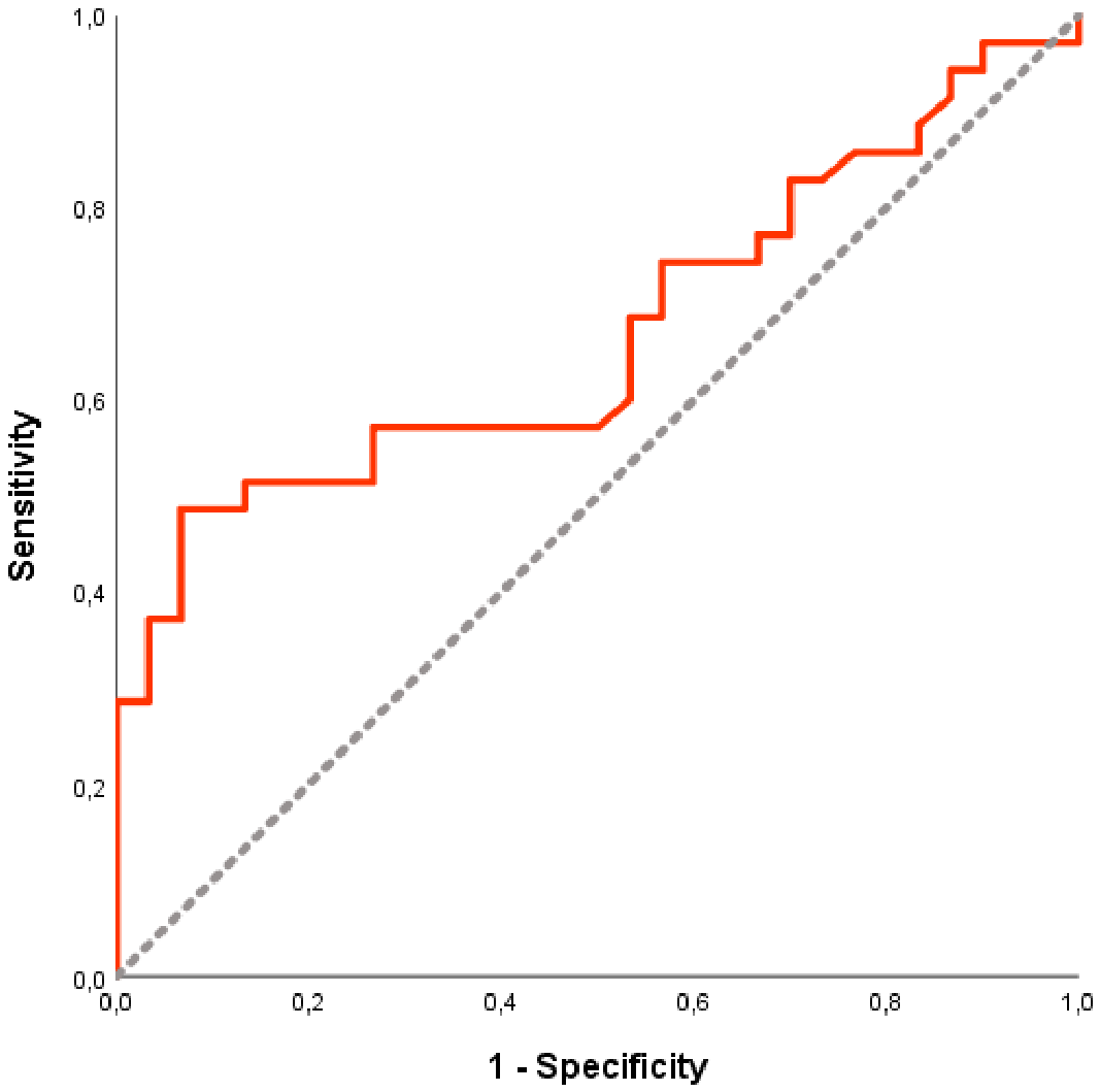
The pre-operative measurement produced an AUC of 0.67 (95% CI 0.54-0.80). At the internally selected cut-off of 1.56 ng/µL, sensitivity was 49% and specificity 93% (Table 2). These values describe apparent performance in the derivation cohort and should not be interpreted as validated screening or rule-in performance. The positive and negative predictive values are additionally dependent on the artificial case-control prevalence and are not directly transferable to a screening or diagnostic population.
Figure 2.
Receiver operating characteristic (ROC) curve for pre-operative plasma cfDNA distinguishing patients with resectable NSCLC from healthy controls (AUC 0.670, 95% CI 0.538–0.802).
Figure 2.
Receiver operating characteristic (ROC) curve for pre-operative plasma cfDNA distinguishing patients with resectable NSCLC from healthy controls (AUC 0.670, 95% CI 0.538–0.802).
3.2. Peri-Operative cfDNA Dynamics
cfDNA differed across the four peri-operative timepoints after log10 transformation (overall p=0.008). Mean log10 cfDNA increased from 0.22 ± 0.30 pre-operatively to 0.32 ± 0.23 in pulmonary venous blood and 0.39 ± 0.26 immediately after resection, before falling to 0.33 ± 0.32 on postoperative day 14 (Table 3). Bonferroni-adjusted pairwise comparisons showed higher values in pulmonary venous blood before major manipulation than in the pre-operative peripheral sample (p=0.027), and higher immediate post-resection peripheral values than pre-operative values (p<0.005). No other pairwise comparison reached statistical significance.
Exploratory subgroup analyses by sex, histological subtype, tumour location and clinical stage did not identify statistically significant time-by-subgroup interactions. Clinical stage showed a time-independent difference on the log10 scale (overall p=0.033), driven by higher values in clinical stage III than stage I (Bonferroni-adjusted p=0.031); given the very small subgroup sizes, this finding was considered exploratory.
Concentrations refer to extracted eluate; log10 values were calculated from concentrations expressed in ng/µL. Overall repeated-measures comparison on the log10 scale: p=0.008. Bonferroni-adjusted pairwise comparisons: pulmonary venous vs pre-operative p=0.027; immediate post-resection vs pre-operative p<0.005; all other pairwise comparisons were not statistically significant.
3.3. Recurrence
Complete three-year outcome data were available for 34 patients: 12 (35.3%) developed recurrence and 22 remained recurrence-free. Mean pre-operative cfDNA was higher in patients who recurred than in those who did not (2.93 versus 1.60 ng/µL; p=0.040), as was the immediate post-resection value (3.88 versus 2.34 ng/µL; p=0.047).
In univariable logistic regression, recurrence was associated with higher pre-operative cfDNA (OR 2.00, 95% CI 1.18–3.59; p=0.020), pulmonary venous cfDNA before manipulation (OR 1.92, 95% CI 1.01–3.63; p=0.045), and peripheral cfDNA after resection (OR 1.92, 95% CI 1.06–3.45; p=0.030). In the multivariable model, pre-operative cfDNA remained associated with recurrence (adjusted OR 2.47, 95% CI 1.07–5.67; p=0.034), as did number of chemotherapy cycles (adjusted OR 1.80 per cycle, 95% CI 1.06–3.06; p=0.029) (Table 4). The chemotherapy association should be interpreted cautiously because treatment exposure is likely to reflect underlying pathological risk and confounding by indication.
The post-resection peripheral measurement had the highest exploratory discrimination for recurrence (AUC 0.720, 95% CI 0.520–0.920; p=0.037). At the internally selected cut-off of 2.72 ng/µL, sensitivity was 67% and specificity 73%. For the pre-operative measurement, a cut-off of 2.76 ng/µL yielded an AUC of 0.678 (95% CI 0.456–0.901), sensitivity of 58% and specificity of 86%; recurrence occurred in 5/24 (20.8%) patients below this threshold compared with 7/10 (70.0%) above it (p=0.015). These thresholds were derived and evaluated within the same small cohort and are therefore exploratory.
3.4. Mortality
Thirteen of 34 patients (38.2%) died during follow-up. Day-14 cfDNA was higher among patients who died than among those alive at the end of follow-up (5.35 vs 2.17 ng/µL; p=0.008). In univariable logistic regression, higher day-14 cfDNA was associated with death (OR 1.48, 95% CI 1.01–2.17; p=0.042). In multivariable Cox regression, higher day-14 cfDNA remained associated with time to death (adjusted HR 1.20, 95% CI 1.03–1.40; p=0.021) (Table 4).
4. Discussion
This prospective pilot cohort examined total plasma cfDNA across four peri-operative timepoints, including tumour-draining pulmonary venous blood. Three findings merit cautious consideration. First, pre-operative cfDNA was higher in resectable NSCLC patients than in healthy controls, but discrimination was modest and sensitivity was low. Second, cfDNA changed during the peri-operative period, consistent with substantial non-tumour contributions from operative tissue injury and inflammation. Third, several peri-operative measurements were associated with recurrence, and day-14 values were higher among patients who died; however, the small number of events, extensive multivariable adjustment and multiple testing limit the strength of inference.
The diagnostic comparison is consistent with quantitative cfDNA and broader liquid-biopsy literature showing higher average concentrations or discriminatory signals in NSCLC than in controls, while also highlighting the methodological and clinical challenges of translating such signals into practice [16,17,26,27,28]. Nevertheless, pooled estimates from heterogeneous studies can overstate applicability to early-stage disease, because advanced cancers generally shed more tumour-derived DNA and because total cfDNA is influenced by non-malignant conditions. In the present cohort, the apparent specificity of 93% was achieved at a cut-off selected within the same data used to estimate performance. Without external validation and comparison with clinically relevant benign pulmonary disease controls, this threshold should not be proposed for screening, diagnosis or clinical triage.
The peri-operative rise is biologically plausible. A lung-cancer surgery cohort similarly demonstrated higher postoperative cfDNA and inflammatory cytokines [20]. These observations reinforce that the timing of a total cfDNA measurement changes its meaning: an immediate postoperative increase cannot be assumed to indicate residual malignant burden. The day-14 result may be less dominated by acute operative release, but it could still reflect postoperative inflammation, complications, comorbidity or delayed recovery. Because these covariates were not systematically captured, a tumour-specific interpretation is not justified.
The recurrence associations are directionally consistent with studies linking total cfDNA burden to outcome [18,19] and with tumour-informed ctDNA studies showing strong postoperative prognostic value [12,13,14,15,29,30,31,32]. The distinction is important. Total cfDNA may function as a composite marker of tumour burden, host response and tissue turnover, whereas ctDNA-based molecular residual disease assays identify tumour-derived variants. Even for ctDNA, professional recommendations consider molecular residual disease detection clinically valid as a prognostic marker in several settings but do not support routine treatment escalation or de-escalation outside trials without evidence of clinical utility [10].
Pulmonary venous sampling was technically incorporated into the protocol and is a distinctive feature of the study. Pulmonary venous CTC studies support enrichment of tumour-derived cells in tumour-draining blood and demonstrate changes during manipulation [21,22,23,24]. In this cohort, however, total cfDNA in the pulmonary venous sample did not show clear incremental prognostic performance over peripheral measurements. This should not be interpreted as evidence against tumour-proximal sampling; rather, total cfDNA concentration may be insufficiently specific to detect compartmental enrichment. A future study should test the hypothesis using mutation-informed ctDNA or other tumour-specific analytes and formally quantify incremental value over peripheral sampling.
The reported association between number of chemotherapy cycles and recurrence is particularly vulnerable to confounding by indication: patients with higher pathological risk are more likely to receive adjuvant treatment. In a small observational cohort, treatment exposure should therefore be regarded as a marker of baseline clinical risk rather than a causal determinant of recurrence. The estimate is exploratory and should not be interpreted as a causal treatment effect.
This study also has several important strengths. The prospective design enabled serial cfDNA sampling at predefined peri-operative timepoints, providing insight into dynamic changes across the surgical pathway rather than relying on a single measurement. A particular strength was the inclusion of tumour-draining pulmonary venous sampling before major tumour manipulation, allowing direct comparison of tumour-proximal and peripheral cfDNA measurements within the same patients. The cohort was clinically characterised with pathological staging and three-year outcome assessment, permitting exploratory evaluation of both recurrence and mortality. The inclusion of healthy controls also enabled the diagnostic performance of pre-operative cfDNA to be considered alongside its potential prognostic value. Taken together, the combination of prospective serial sampling, a rarely studied intra-operative sampling compartment, and longitudinal clinical outcomes provides a distinctive dataset and a useful basis for further investigation of peri-operative cfDNA as a biomarker in resectable NSCLC.
This study also has limitations of note. It was a two-centre, single-country pilot with 35 patients and 30 healthy controls. Only 12 recurrences and 13 deaths occurred, creating risk of unstable multivariable coefficients and overfitting. Multiple timepoints, subgroup analyses, outcomes and ROC analyses were examined without a study-wide multiplicity correction, despite Bonferroni adjustment for selected pairwise comparisons, so some nominally significant results may represent type I error. The ROC thresholds were data-derived and lack internal resampling or external validation. cfDNA concentrations were measured in ng/µL of extracted eluate. The exact quantification assay and sample-specific plasma input and elution volumes were not documented in the material available for this manuscript, limiting reproducibility and cross-study comparison. Total cfDNA is non-specific, and postoperative complications, inflammatory biomarkers, renal function and other potential determinants of cfDNA were not systematically included. Detailed follow-up duration and censoring information were not available for this manuscript. Finally, healthy controls do not reproduce the intended clinical differential diagnosis of patients with benign or inflammatory pulmonary lesions.
The study should therefore be viewed as hypothesis-generating. Its main contribution is the structured peri-operative sampling design and the demonstration that total cfDNA varies materially across sampling times and compartments. Validation should use a prespecified analysis plan, harmonised pre-analytics, complete reporting of analytical performance, adequate events per model parameter, correction or prioritisation for multiple hypotheses, clinically relevant control groups, and external validation. Integrating tumour-informed ctDNA with total cfDNA and structured postoperative covariates may help distinguish tumour signal from host injury and recovery. Recent work in resectable NSCLC also supports the need for assay standardisation and prospective evidence of clinical utility before routine implementation [31,32,33].
5. Conclusions
In this small prospective cohort, quantitative plasma cfDNA was higher in patients with resectable NSCLC than in healthy controls, but diagnostic discrimination was modest and sensitivity was insufficient for screening. cfDNA changed significantly across the peri-operative period, emphasising the influence of sampling time and non-tumour biological processes. Pre-operative, pulmonary venous and post-resection measurements showed exploratory associations with recurrence, while day-14 values were higher among patients who died. These findings do not establish clinical utility and should be regarded as hypothesis-generating, requiring confirmation in larger, independently validated cohorts with a fully specified assay, rigorous time-to-event methods, prespecified modelling and tumour-informed ctDNA measurement. Total cfDNA should not be interpreted as a surrogate for tumour-specific molecular residual disease without further validation.
Pulmonary venous sampling remains a biologically plausible research strategy, but this study does not demonstrate that it improves prognostic performance when total cfDNA concentration is used. Its incremental value should be tested directly using tumour-specific assays and paired peripheral samples.
Author Contributions
Conceptualization, S.G., D.G. and N.S.; Methodology, S.G., D.G., E.K. and A.C.; Formal Analysis, S.G. and D.G.; Investigation, S.G., V.K., T.S., E.K. and A.C.; Data Curation, S.G. and D.G.; Writing—Original Draft Preparation, S.G., C.F; Writing—Review & Editing, S.G., D.G., V.K., T.S.,C.F., E.K., L.D., A.C. and N.S.; Supervision, N.S.; Project Administration, S.G. All authors have read and agreed to the published version of the manuscript.
Funding
This research received no external funding. No study-related charges were incurred by the participating hospitals.
Institutional Review Board Statement
The study was authorised by the Scientific Council of the Sotiria General Hospital of Thoracic Diseases before recruitment commenced in January 2020. Publication of the official approval document was delayed by the COVID-19 pandemic until 12 June 2020 (document identifier: ΩΥΣ9469069-66Ξ).
Informed Consent Statement
Written informed consent was obtained from all patients and healthy controls before enrolment.
Data Availability Statement
Requests for access to anonymised data supporting the findings of this study may be directed to the corresponding author. Any data sharing will be subject to participant consent, applicable ethical approvals and institutional data protection requirements.
Acknowledgments
During the preparation of this manuscript, the authors used OpenAI ChatGPT (GPT-5.6 Luna; accessed October 2026) for language editing, structural reorganisation and journal formatting. The authors have reviewed and edited the output and take full responsibility for the content of this publication.
Conflicts of Interest
The authors declare no conflicts of interest.
Abbreviations
ANOVA, analysis of variance; AUC, area under the curve; cfDNA, cell-free DNA; CI, confidence interval; CTC, circulating tumour cell; ctDNA, circulating tumour DNA; HR, hazard ratio; IQR, interquartile range; NSCLC, non-small cell lung cancer; OR, odds ratio; ROC, receiver operating characteristic; SD, standard deviation; SUVmax, maximum standardised uptake value; TNM, tumour–node–metastasis.
References
- Li, C.; Lei, S.; Ding, L.; Xu, Y.; Wu, X.; Wang, H.; et al. Global burden and trends of lung cancer incidence and mortality. Chin. Med. J. (Engl) 2023, 136(13), 1583–1590. [Google Scholar] [CrossRef]
- Knight, S.B.; Crosbie, P.A.; Balata, H.; Chudziak, J.; Hussell, T.; Dive, C. Progress and prospects of early detection in lung cancer. Open Biol. 2017, 7(9), 170070. [Google Scholar] [CrossRef]
- Ostrin, E.J.; Sidransky, D.; Spira, A.; Hanash, S.M. Biomarkers for lung cancer screening and detection. Cancer Epidemiol. Biomark. Prev. 2020, 29(12), 2411–2415. [Google Scholar] [CrossRef]
- Saji, H.; Okada, M.; Tsuboi, M.; et al. Segmentectomy versus lobectomy in small-sized peripheral non-small-cell lung cancer (JCOG0802/WJOG4607L): a multicentre, open-label, phase 3, randomised, controlled, non-inferiority trial. Lancet 2022, 399(10335), 1607–1617. [Google Scholar] [CrossRef]
- Altorki, N.; Wang, X.; Kozono, D.; et al. Lobar or sublobar resection for peripheral stage IA non-small-cell lung cancer. N Engl. J. Med. 2023, 388(6), 489–498. [Google Scholar] [CrossRef]
- Detterbeck, F.C.; Boffa, D.J.; Kim, A.W.; Tanoue, L.T. The eighth edition lung cancer stage classification. Chest 2017, 151(1), 193–203. [Google Scholar] [CrossRef]
- Rami-Porta, R.; Nishimura, K.K.; Giroux, D.J.; et al. The International Association for the Study of Lung Cancer Lung Cancer Staging Project: proposals for revision of the TNM stage groups in the forthcoming ninth edition of the TNM classification for lung cancer. J. Thorac. Oncol. 2024, 19(7), 1007–1027. [Google Scholar] [CrossRef]
- Nicholson, A.G.; Tsao, M.S.; Beasley, M.B.; et al. The 2021 WHO classification of lung tumors: impact of advances since 2015. J. Thorac. Oncol. 2022, 17(3), 362–387. [Google Scholar] [CrossRef]
- Rolfo, C.; Mack, P.; Scagliotti, G.V.; et al. Liquid biopsy for advanced NSCLC: a consensus statement from the International Association for the Study of Lung Cancer. J. Thorac. Oncol. 2021, 16(10), 1647–1662. [Google Scholar] [CrossRef]
- Pascual, J.; Attard, G.; Bidard, F.C.; et al. ESMO recommendations on the use of circulating tumour DNA assays for patients with cancer: a report from the ESMO Precision Medicine Working Group. Ann. Oncol. 2022, 33(8), 750–768. [Google Scholar] [CrossRef]
- Abbosh, C.; Hodgson, D.R.; Doherty, G.J.; et al. Implementing circulating tumor DNA as a prognostic biomarker in resectable non-small cell lung cancer. Trends Cancer 2024, 10(7), 643–654. [Google Scholar] [CrossRef]
- Chaudhuri, A.A.; Chabon, J.J.; Lovejoy, A.F.; et al. Early detection of molecular residual disease in localized lung cancer by circulating tumor DNA profiling. Cancer Discov. 2017, 7(12), 1394–1403. [Google Scholar] [CrossRef] [PubMed]
- Qiu, B.; Guo, W.; Zhang, F.; et al. Dynamic recurrence risk and adjuvant chemotherapy benefit prediction by ctDNA in resected NSCLC. Nat. Commun. 2021, 12(1), 6770. [Google Scholar] [CrossRef]
- Xia, L.; Mei, J.; Kang, R.; et al. Perioperative ctDNA-based molecular residual disease detection for non-small cell lung cancer: a prospective multicenter cohort study (LUNGCA-1). Clin. Cancer Res. 2022, 28(15), 3308–3317. [Google Scholar] [CrossRef]
- Hu, W.; Yang, Y.; Zhang, L.; et al. Post surgery circulating free tumor DNA is a predictive biomarker for relapse of lung cancer. Cancer Med. 2017, 6(5), 962–974. [Google Scholar] [CrossRef]
- Jiang, T.; Zhai, C.; Su, C.; Ren, S.; Zhou, C. The diagnostic value of circulating cell free DNA quantification in non-small cell lung cancer: a systematic review with meta-analysis. Lung Cancer 2016, 100, 63–70. [Google Scholar] [CrossRef]
- Chen, Z.; Miao, H.; Zeng, Q.; Xu, S.; Chen, Z.; Liu, K. Circulating cell-free DNA as a diagnostic and prognostic biomarker for non-small-cell lung cancer: a systematic review and meta-analysis. Biomark. Med. 2020, 14(7), 587–597. [Google Scholar] [CrossRef]
- van der Drift, M.A.; Prinsen, C.F.M.; Hol, B.E.A.; et al. Circulating DNA is a non-invasive prognostic factor for survival in non-small cell lung cancer. Lung Cancer 2010, 68(2), 283–287. [Google Scholar] [CrossRef]
- Cargnin, S.; Canonico, P.L.; Genazzani, A.A.; Terrazzino, S. Quantitative analysis of circulating cell-free DNA for correlation with lung cancer survival: a systematic review and meta-analysis. J. Thorac. Oncol. 2017, 12(1), 43–53. [Google Scholar] [CrossRef]
- Katopodis, P.; Anikin, V.; Kishore, U.; et al. Circulating tumour cells and circulating cell-free DNA in patients with lung cancer: a comparison between thoracotomy and video-assisted thoracoscopic surgery. BMJ Open Respir. Res. 2021, 8(1), e000917. [Google Scholar] [CrossRef]
- Reddy, R.M.; Murlidhar, V.; Zhao, L.; et al. Pulmonary venous blood sampling significantly increases the yield of circulating tumor cells in early-stage lung cancer. J. Thorac. Cardiovasc Surg. 2016, 151(3), 852–858. [Google Scholar] [CrossRef]
- Hashimoto, M.; Tanaka, F.; Yoneda, K.; et al. Significant increase in circulating tumour cells in pulmonary venous blood during surgical manipulation in patients with primary lung cancer. Interact. Cardiovasc Thorac. Surg. 2014, 18(6), 775–783. [Google Scholar] [CrossRef]
- Hashimoto, M.; Tanaka, F.; Yoneda, K.; et al. Positive correlation between postoperative tumor recurrence and changes in circulating tumor cell counts in pulmonary venous blood during surgical manipulation in non-small cell lung cancer. J. Thorac. Dis. 2018, 10(1), 298–306. [Google Scholar] [CrossRef]
- Duan, X.; Zhu, Y.; Cui, Y.; et al. Circulating tumor cells in the pulmonary vein increase significantly after lobectomy: a prospective observational study. Thorac. Cancer 2019, 10(2), 163–169. [Google Scholar] [CrossRef]
- QIAGEN. QIAamp Circulating Nucleic Acid Handbook; QIAGEN: Hilden, Germany, 2019. [Google Scholar]
- Chae, Y.K.; Oh, M.S. Detection of Minimal Residual Disease Using ctDNA in Lung Cancer: Current Evidence and Future Directions. J. Thorac. Oncol. 2019, 14(1), 16–24. [Google Scholar] [CrossRef]
- Rijavec, E.; Coco, S.; Genova, C.; Rossi, G.; Longo, L.; Grossi, F. Liquid Biopsy in Non-Small Cell Lung Cancer: Highlights and Challenges. Cancers 2020, 12(1), 17. [Google Scholar] [CrossRef]
- Wang, S.; Meng, F.; Li, M.; et al. Multidimensional Cell-Free DNA Fragmentomic Assay for Detection of Early-Stage Lung Cancer. Am. J. Respir. Crit. Care Med. 2023, 207(9), 1203–1213. [Google Scholar] [CrossRef]
- Waldeck, S.; Mitschke, J.; Wiesemann, S.; et al. Early assessment of circulating tumor DNA after curative-intent resection predicts tumor recurrence in early-stage and locally advanced non-small-cell lung cancer. Mol. Oncol. 2022, 16(2), 527–537. [Google Scholar] [CrossRef]
- Fu, R.; Huang, J.; Tian, X.; et al. Postoperative circulating tumor DNA can refine risk stratification in resectable lung cancer: results from a multicenter study. Mol. Oncol. 2023, 17(5), 825–838. [Google Scholar] [CrossRef] [PubMed]
- Tian, X.; Liu, X.; Wang, K.; et al. Postoperative ctDNA in indicating the recurrence risk and monitoring the effect of adjuvant therapy in surgical non-small cell lung cancer. Thorac. Cancer 2024, 15(10), 797–807. [Google Scholar] [CrossRef]
- Normanno, N.; Morabito, A.; Rachiglio, A.M.; et al. Circulating tumour DNA in early stage and locally advanced NSCLC: ready for clinical implementation? Nat. Rev. Clin. Oncol. 2025, 22(3), 215–231. [Google Scholar] [CrossRef]
- Vandekerckhove, O.; Cuppens, K.; Pat, K.; Du Pont, B.; Froyen, G.; Maes, B. Liquid Biopsy in Early-Stage Lung Cancer: Current andFuture Clinical Applications. Cancers 2023, 15(10), 2702. [Google Scholar] [CrossRef]
Figure 1.
Peri-operative blood sampling schedule.
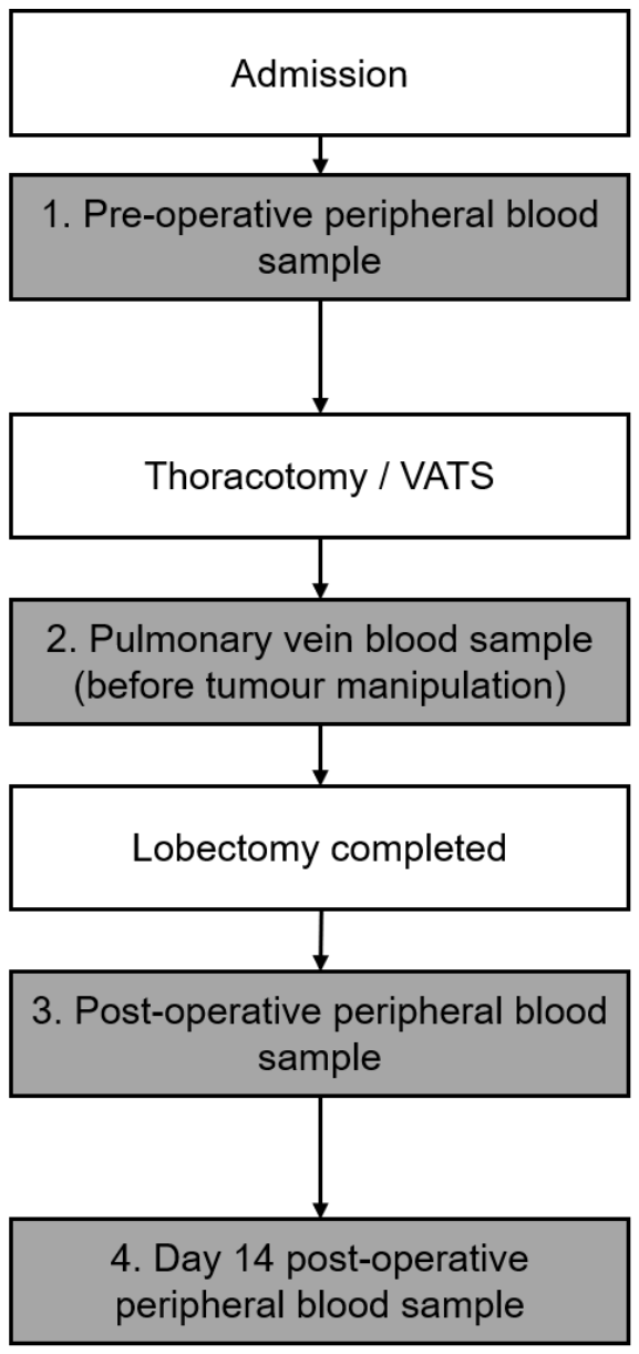

Table 1.
Reported baseline comparison between healthy controls and patients with resectable NSCLC.
| Characteristic | Healthy controls (n=30) | NSCLC patients (n=35) | p value |
| Age, years, mean +/- SD | 65.93 +/- 9.31 | 67.71 +/- 8.17 | 0.415 |
| Male/Female | 53.3%/46.7% | 57.1%/42.9% | 0.806 |
| Pre-operative cfDNA (ng/µL), mean ± SD* | 1.08 +/- 0.46 | 1.96 +/- 1.45 | 0.002 |
* cfDNA concentrations are expressed in ng/µL of extracted eluate.
Table 2.
Exploratory diagnostic performance of pre-operative cfDNA for distinguishing NSCLC patients from healthy controls.
Table 2.
Exploratory diagnostic performance of pre-operative cfDNA for distinguishing NSCLC patients from healthy controls.
| Metric | Estimate | 95% CI |
| AUC | 0.67 | 0.54-0.80 |
| Internally selected cut-off (ng/µL) | 1.56 | Not applicable |
| Sensitivity | 49% | 32-66% |
| Specificity | 93% | 77-99% |
| Positive predictive value | 89% | 65-98% |
| Negative predictive value | 61% | 45-75% |
Table 3.
Peri-operative plasma cfDNA measurements in patients with resectable NSCLC.
| Sampling timepoint | Mean ± SD (log10) | Median (IQR), ng/µL | n |
| Pre-operative peripheral | 0.22 ± 0.30 | 1.61 (1.94) | 35 |
| Pulmonary venous (before tumour manipulation) | 0.32 ± 0.23 | 1.94 (1.63) | 35 |
| Immediate post-resection peripheral | 0.39 ± 0.26 | 2.62 (1.44) | 35 |
| Postoperative day 14 peripheral | 0.33 ± 0.32 | 1.85 (3.00) | 34 |
Table 4.
Associations between plasma cfDNA measurements and clinical outcomes.
| Outcome | Measurement | Analysis | Effect estimate (95% CI) | p value |
| Recurrence | Pre-operative cfDNA | Univariable logistic regression | OR 2.00 (1.18-3.59) | 0.020 |
| Recurrence | Pulmonary venous cfDNA before manipulation | Univariable logistic regression | OR 1.92 (1.01-3.63) | 0.045 |
| Recurrence | Post-resection peripheral cfDNA | Univariable logistic regression | OR 1.92 (1.06-3.45) | 0.030 |
| Recurrence | Pre-operative cfDNA | Multivariable logistic regression | Adjusted OR 2.47 (1.07-5.67) | 0.034 |
| Recurrence | Chemotherapy cycles | Multivariable logistic regression | Adjusted OR 1.80 (1.06–3.06) | 0.029 |
| Mortality | Day-14 cfDNA | Univariable logistic regression | OR 1.48 (1.01–2.17) | 0.042 |
| Overall survival | Day-14 cfDNA | Multivariable Cox regression | Adjusted HR 1.20 (1.03–1.40) | 0.021 |
© 2026 by the authors. Licensee MDPI, Basel, Switzerland. This article is an open access article distributed under the terms and conditions of the Creative Commons Attribution (CC BY) license.
Copyright: This open access article is published under a Creative Commons CC BY 4.0 license, which permit the free download, distribution, and reuse, provided that the author and preprint are cited in any reuse.