
Submitted:
30 October 2026
Posted:
02 November 2026
You are already at the latest version
Abstract
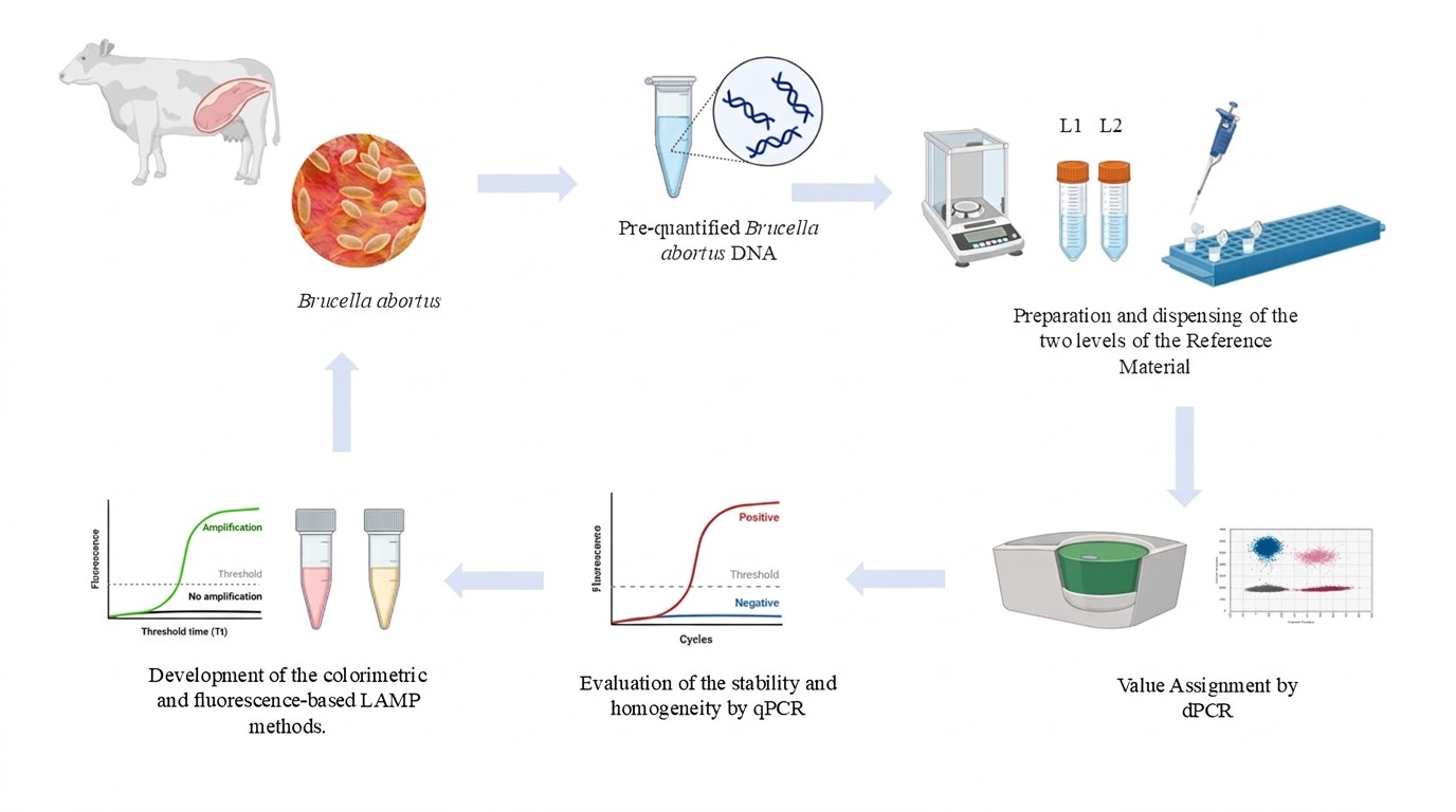
Analytical standardization of Brucella molecular assays requires reference materials with assigned values and defined uncertainty. We produced and characterized intralaboratory a candidate genomic DNA reference material from Brucella abortus RB51 for PCR-based detection of Brucella spp., and evaluated its preliminary use in fluorescence-based and colorimetric LAMP assays. DNA was extracted, assessed by qPCR and dPCR, and diluted to two levels. Homogeneity and stability were evaluated by duplex qPCR targeting ctrA and nikR, whereas property values were assigned by duplex dPCR. qPCR efficiencies were 95.3% for ctrA and 92.1% for nikR. The material fulfilled the homogeneity criterion, with relative homogeneity uncertainties between 0.37% and 1.41%. Assigned values (levels 1 and 2) were 32 989 ± 5 475 and 285 ± 47 copies/µL for ctrA, and 29 722 ± 3 263 and 286 ± 42 copies/µL for nikR. Short-term stability at 4 °C was supported for 8 weeks and was included in the uncertainty budget. ctrA-based LAMP assays were optimized at 67 °C for 45 min and amplified reproducibly in both formats. Amplification was consistent from 2411 to 6.34 copies/µL. This material provides a metrologically characterized tool for quality control, method verification, and optimization of PCR- and LAMP-based assays for Brucella spp.
Keywords:
bovine brucellosis
; quantitative PCR (qPCR)
; digital PCR (dPCR)
; metrological tool
; loop-mediated isothermal amplification (LAMP)
1. Introduction
Bovine brucellosis (BB) is a zoonotic disease primarily caused by Brucella abortus. However, Brucella melitensis and Brucella suis may also infect cattle under specific epidemiological conditions involving contact with sheep, goats, or pigs [1]. The disease remains a major concern for animal health, public health, and livestock productivity because infected animals may develop reproductive disorders, including abortion, infertility, birth of weak offspring, and decreased milk production [2]. Its economic impact is associated with reproductive failure, reduced productive performance, restrictions on animal movement, and culling of infected animals; in some scenarios, losses due to the culling of infected animals may represent between 30% and 50% of the difference between the market value of a healthy animal and the value obtained at slaughter [3]. Infected cattle may shed B. abortus through the placenta, fetal fluids, vaginal secretions, milk, semen, seminal fluid, and urine. In contrast, human infection is mainly associated with occupational exposure or consumption of unpasteurized dairy products [1]. These epidemiological and productive implications make reliable detection a central requirement for brucellosis surveillance, control, and eradication programs.
Brucellosis in animals is controlled through a combination of measures, including the isolation and, when necessary, euthanasia of infected animals, restrictions on animal movement, and vaccination. Vaccination with live strains, such as Brucella abortus strains 19 and RB51, Brucella suis S-2, the rough B. melitensis strain M111, and the attenuated B. melitensis Rev.1 strain, is considered one of the most effective strategies for the prevention and control of brucellosis. Preventive measures also include the pasteurization of dairy products, thorough cooking of meat, careful handling and disposal of biological materials associated with animal parturition, and the use of protective clothing in high-risk settings [3].
Current diagnostic strategies for bovine brucellosis commonly rely on serological screening tests, including the Rose Bengal test, milk ring test, standard tube agglutination test, complement fixation test, and ELISA. These methods are valuable for surveillance programs, but their interpretation may be affected by cross-reactivity, vaccination status, infection stage, and the indirect nature of antibody detection [3]. Direct approaches, such as bacteriological culture and Polymerase Chain Reaction (PCR), enable detection of the infectious agent or its genetic material and can therefore complement serological diagnosis. However, culture is constrained by slow bacterial growth, biosafety requirements, and specialized infrastructure, while molecular assays require standardized procedures, appropriate controls, and well-characterized materials to support reliable analytical interpretation and comparability of results [4].
Loop-mediated isothermal amplification (LAMP) has been explored as an alternative nucleic acid amplification strategy for Brucella spp. detection because it enables DNA amplification under isothermal conditions and can be coupled with colorimetric or fluorescence-based readouts. These characteristics may reduce equipment requirements compared with conventional thermocycling methods and make LAMP attractive for applied diagnostic workflows [5]. Previous studies have reported LAMP assays for Brucella spp. and B. abortus using targets such as IS711, bcsp31, omp25, and other species-associated sequences, demonstrating the feasibility of rapid molecular detection in different sample types [6,7,8]. Nevertheless, differences in primer design, target selection, amplification chemistry, template quality, and signal detection methods can substantially influence analytical performance, making direct comparison between assays difficult. Consequently, the development of robust LAMP assays requires not only the optimization of reaction conditions but also the availability of Reference Materials (RM) that enable a traceable evaluation of assay performance.
In colorimetric formats, weak or delayed amplification, pH-dependent color transitions, and borderline template concentrations may further complicate result interpretation. Under these conditions, a well-characterized genomic DNA RM provides a reliable basis for defining reproducible amplification ranges, analytical limits of detection, and objective interpretation criteria [9,10,11,12]. Conversely, the absence of an appropriate RM may lead laboratories to rely on in-house DNA preparations, plasmids, synthetic fragments, or positive controls with limited metrological characterization, thereby reducing interlaboratory comparability and increasing the risk of inaccurate estimation of analytical sensitivity. For pathogen detection, genomic DNA RM additionally preserves the native genomic context of target sequences and more closely resembles routine analytical samples than plasmid- or synthetic DNA-based controls. Consequently, they provide a more representative and traceable basis for assay development, optimization, performance evaluation, and interlaboratory comparison of LAMP-based molecular methods. For the detection of Brucella spp. there is a RM based on antibodies against Brucella spp. developed for Instituto Colombiano Agropecuario (ICA). However, there is still a lack of RM based on Brucella spp. nucleic acids for use in molecular detection methods. This gap highlights the need for well-characterized materials that can support the development and evaluation of molecular assays.
RMs are fundamental tools for analytical quality assurance because they provide sufficiently homogeneous and stable materials with assigned property values and associated uncertainty for a defined intended use. In molecular microbiology, nucleic acid-based RMs support method validation, quality control, estimation of limits of detection, and comparison of results across laboratories and measurement platforms [12]. The production of RMs requires a metrological workflow that includes preparation of the candidate material, assessment of homogeneity, stability evaluation, value assignment, and estimation of uncertainty in accordance with internationally accepted guidance [9,11].
In nucleic acid measurements, digital PCR (dPCR) has become a valuable approach for value assignment because it enables absolute quantification of target copy number concentration without reliance on external calibration curves. When appropriately validated and controlled, dPCR can support the assignment of property values to DNA-based RM and strengthen the measurement traceability in molecular assays. In contrast, real-time PCR (qPCR) remains valuable for assessing amplification performance, homogeneity, and stability trends. Together, these complementary approaches provide a robust framework for the characterization of nucleic acid RM intended for molecular diagnostic applications.
In this study, we produced and characterized, at the intralaboratory level, the first candidate RM based on genomic DNA from Brucella abortus RB51 for PCR-based detection of Brucella spp. The material was prepared at two concentration levels, evaluated for homogeneity and stability by duplex qPCR [12] targeting ctrA and nikR [13,14], and assigned copy number concentration values by duplex dPCR. The qPCR and dPCR assays were used as measurement tools for RM characterization rather than as newly developed diagnostic methods. As a preliminary proof of use, the characterized RM was applied to the optimization of fluorescence-based and colorimetric LAMP assays. Therefore, the primary contribution of this work is the production and intralaboratory characterization of a candidate genomic DNA RM, while LAMP optimization is presented as a controlled analytical application of the material.
2. Materials and Methods
2.1. Brucella Abortus Culture and Genomic DNA Extraction
Brucella abortus RB51 was initially cultured on Brucella Agar (Millipore, Germany, cat. no. 18795) and incubated for 72-96 h at 37 °C under a 5% CO₂ atmosphere. Colonies were then inoculated into Brucella broth base (Millipore, Germany, cat. no. B3051) in a vented cell culture flask (NEST Biotechnology, China, cat. no. 727003) and incubated under the same conditions until reaching an optical density (OD) of 1.0 (starting point for genomic DNA extraction). DNA extraction was performed according to the protocols described by [15,16] with minor modifications. The culture was centrifuged at 8 500 rpm for 15 min at 4 °C, and the resulting pellet was washed three times with 10 mL of 1X PBS under the same centrifugation conditions. The pellet was resuspended in 1 mL of 1X PBS and centrifuged at 10 000 rpm for 5 min at 4 °C, and the supernatant was discarded. Cell lysis was performed by adding 800 µL of lysis buffer [2% (w/v) CTAB, 100 mM Tris-HCl, 1.4 M NaCl, 20 mM EDTA, and 0.3% (w/v) LiCl, pH 8.0] supplemented with proteinase K at a final concentration of 100 µg/mL. The suspension was incubated for 1 h at 65 °C to promote cell disruption and protein digestion.
Genomic DNA was extracted by adding 1.5 volumes of chloroform: isoamyl alcohol (24:1), followed by centrifugation at 12 000 rpm for 12 min at 4 °C. The aqueous phase was recovered, treated with RNase at a final concentration of 50 µg/mL, and incubated at 37 °C for 30 min. Lithium chloride (LiCl) was then added to a final concentration of 3%, and a second extraction was performed with 1.5 volumes of chloroform: isoamyl alcohol (24:1), followed by centrifugation at 12 000 rpm for 12 min at 4 °C.
DNA was precipitated by adding sodium acetate to a final concentration of 2.5 M and one volume of cold isopropanol, followed by overnight incubation at -20 °C. The precipitated DNA was recovered by centrifugation at 12 000 rpm for 15 min at 4 °C. The pellet was washed twice with 70% ethanol under the same centrifugation conditions, and the supernatant was discarded. Finally, the pellet was air-dried and resuspended in 1X TE buffer (10 mM Tris-HCl; 1 mM EDTA, pH 8.0). DNA concentration was determined by spectrophotometry using a NanoDrop 2000 Spectrophotometer (Thermo Fisher Scientific, USA), and DNA integrity was assessed by 1% agarose gel electrophoresis. This CTAB-based extraction method was selected because the purpose of this work was to obtain high concentration, high molecular weight genomic DNA suitable for producing a candidate reference material, rather than use expensive kits.
2.2. Assessment of Initial DNA Quality and Sequencing
Genomic DNA was sequenced using an Oxford Nanopore MinION platform (Oxford Nanopore Technologies, Oxford, UK). Sequencing reads were filtered and assembled de novo to obtain a draft genome sequence. The identity of the strain was confirmed at the species level through comparison with reference genomes and by calculating Average Nucleotide Identity (ANI) values against publicly available Brucella genomes.
Real-time PCR (qPCR) was used to evaluate the amplification efficiency of a validated duplex assay targeting the ctrA and nikR genes (Table 1). The ctrA and nikR genes were selected as molecular targets for RM characterization based on their biological relevance in B. abortus and their suitability for amplification using qPCR and dPCR. ctrA encodes a cell-cycle regulator involved in bacterial cell division, outer membrane organization, and lipopolysaccharide synthesis, whereas nikR encodes a nickel-responsive transcriptional regulator associated with nickel homeostasis [13,14]. The use of two independent genomic targets allowed the candidate material to be evaluated through a duplex molecular measurement approach, providing complementary information for copy number assignment, homogeneity assessment, and stability evaluation. For the LAMP application, both targets were initially assessed; however, only the ctrA-based primer set was retained for subsequent optimization because it produced more reproducible amplification under the tested conditions.
Six serial tenfold dilutions (1:10) of the extracted DNA were prepared. qPCR reactions were performed in triplicate in a final volume of 11 µL using iTaq Universal Probes Supermix (2X) (Bio-Rad, USA, cat. no. 1725131). Primers for each target were used at a final concentration of 500 nM, and probes at 250 nM. Amplification was carried out in a CFX96 Touch Real-Time PCR Detection System (Bio-Rad, USA, cat. no. 1855196) under the following conditions: initial denaturation at 95 °C for 5 min, followed by 40 cycles of denaturation at 95 °C for 15 s and annealing at 58 °C for 30 s, with a final cooling step at 20 °C for 5 min. Amplification efficiency was evaluated using Equation 1 (Table S1). Acceptance criteria were defined as a coefficient of determination (R²) > 0.98, amplification efficiency between 90% and 110%, and a coefficient of variation (CV%) < 5%. ERM-AD623 Certified Reference Material (CRM) was included as a quality amplification control to verify the correct performance of the qPCR assay.
Absolute quantification of genomic DNA was subsequently performed by digital PCR (dPCR) using the same primer and probe sets described in Table 1. A DNA dilution yielding a Ct value > 22 in the qPCR assay was selected as the template. dPCR Reactions were carried out in duplex format in a final volume of 22 µL using ddPCR Multiplex Supermix (4X) (Bio-Rad, USA, cat. no. 12005910). Primers for each target were used at a final concentration of 900 nM, and probes at 250 nM.
Droplets were generated using the QX200 Droplet Generator (Bio-Rad, USA, cat. no. 1864002) with Droplet Generation Oil for Probes (Bio-Rad, USA, cat. no. 1863005). After droplet generation, amplification was conducted in a CFX96 Touch Real-Time PCR Detection System (Bio-Rad, USA, cat. no. 1855196) under the following conditions: enzyme activation at 95 °C for 10 min, followed by 50 cycles of denaturation at 94 °C for 30 s and annealing/extension at 56 °C for 1 min. This was followed by enzyme inactivation at 98 °C for 10 min and a final hold at 4 °C for 5 min. After thermal cycling, droplets were analyzed using the QX600 Droplet Reader (Bio-Rad, USA, cat. no. 12013328), employing ddPCR Droplet Reader Oil (Bio-Rad, USA, cat. no. 1863004) for droplet reading and absolute quantification. Copy number concentrations were estimated using Equations 2 and 3 (Table S1). ERM-AD623 CMR was used as a quality control to assess the proper performance of the dPCR assay.
2.3. Production of the Candidate Reference Material
The candidate Reference Material (RM) was prepared from a stock solution of genomic DNA (6.7 × 10⁷ copies/µL) extracted from B. abortus RB51 cell culture, through gravimetric dilutions in 1X TE buffer (10 mM Tris-HCl; 1 mM EDTA, pH 8.0) to produce two concentration levels with a nominal concentration of 35 000 copies/µL and 350 copies/µL, the solutions were homogenized using an MX-RD-PRO rotary mixer (BIOBASE, China) for 15 min. The two RM levels were dispensed into pre-labeled 500 µL polypropylene screw-cap vials, with a final volume of 100 µL. Dispensing was performed using a HandyStep S pipette (BRAND, Germany) to ensure volumetric accuracy across units. Fifty units were planned for each batch.
2.4. Homogeneity Assessment
Homogeneity study was performed using a stratified random sampling design, selecting 10% of the units from each batch. Each unit was measured in triplicate using the validated duplex qPCR method targeting ctrA and nikR. Data were evaluated using the Shapiro-Wilk test for normality, Levene's test for homoscedasticity, and Grubbs' test for one outlier detection. All tests were conducted at 0.05 significance level. After confirming the normality of the data, a one-way ANOVA was performed to compare between-unit and within-unit variability (repeatability). A simple linear regression model was used to assess potential trends associated with the filling order. A p-value < 0.05 was considered statistically significant. As an acceptance criterion, the relative uncertainty due to homogeneity was required to be < 3%, consistent with other studies indicating that in qPCR, experimental factors such as pipetting can contribute uncertainties of 3% or greater [12].
2.5. Stability Assessment
Short-term stability was evaluated over 8 weeks using an isochronous design to minimize the influence of between-run analytical variability. For each concentration level, two units stored at 4 °C, defined as the study temperature, were transferred weekly to −30 °C, defined as the reference temperature. At the end of the experimental period, 16 units per concentration level were analyzed, along with two reference units stored at -30 °C from time zero. Long-term stability was evaluated over 5 and 9 months using a classical design, in which, at each corresponding time point, two units stored at 4 °C and one reference unit stored at -30 °C were analyzed for each concentration level. All samples were measured in triplicate using a validated duplex qPCR method. Results were analyzed by linear regression of the measured values against storage time to identify potential degradation trends in the RM and to estimate the uncertainty contribution associated with stability.
2.6. Characterization and Value Assignment
Characterization was performed using three randomly selected units (also included in the homogeneity study); for both concentration levels, each one was measured in triplicate using a validated duplex dPCR method. The assigned concentration value corresponded to the weighted means of the obtained results, according to Equations 2, 3, and 4 (Table S1). Uncertainties associated with characterization, homogeneity, stability, and the combined standard uncertainty were estimated using the equations provided in Table S1. The expanded uncertainty of the material was obtained by multiplying the combined standard uncertainty by a coverage factor (k), corresponding to a 95% confidence level, in accordance with the Guide to the expression of Uncertainty in Measurement (GUM).
2.7. Preliminary LAMP Application Using the Candidate RM as Controlled Genomic DNA Template
The developed genomic DNA RM was used as the template for the development and optimization of two LAMP detection formats: a fluorescence-based real-time assay and a colorimetric endpoint assay. LAMP primer sets targeting ctrA and nikR were designed in this study, and their sequences are listed in Table 2. Both detection formats were evaluated using the same primer sets, template input volume, and experimental dilution scheme. This design allowed the RM to serve as a controlled material for assessing amplification behavior, reproducibility, and analytical detection range under comparable conditions, while the assays differed in detection chemistry, instrumentation, and result interpretation.
For each format and target, a LAMP primer mix was prepared at final stock concentrations of 16 µM for the inner primers FIP and BIP, 2 µM for the outer primers F3 and B3, and 4 µM for the loop primers LF and LB. The preliminary amplification condition for both detection formats was set at 65 °C for 45 min and subsequently optimized using a temperature gradient of 63 °C, 65 °C, 66 °C, and 67 °C.
The colorimetric LAMP assay was performed using the WarmStart Colorimetric LAMP 2X Master Mix (DNA & RNA) (New England Biolabs, USA, cat. no. M1800L) in a ProFlex™ 96-well PCR System (Applied Biosystems, USA). Each 25 µL reaction contained 2 µL of DNA template, 2.5 µL of primer mix, 8 µL of nuclease-free water, and 12.5 µL of 2X colorimetric master mix. Endpoint results were interpreted visually according to the reaction color: yellow was considered positive, red was considered negative, and orange was considered inconclusive.
The fluorescence-based real-time LAMP assay was performed using the WarmStart LAMP Kit (DNA & RNA) (New England Biolabs, USA, cat. no. E1700L) in a CFX96 Touch Real-Time PCR Detection System (Bio-Rad, USA, cat. no. 1855196). Each 25 µL reaction contained 2 µL of DNA template, 2.5 µL of primer mix, 0.5 µL of fluorescent dye, 7.5 µL of nuclease-free water, and 12.5 µL of WarmStart LAMP master mix. Amplification times were used to classify the fluorescence-based results as follows: < 20 min, clear amplification; 20-30 min, delayed amplification; and 30-45 min, low-reliability amplification.
After temperature optimization, analytical performance was evaluated using eight serial dilutions prepared from RM Levels, ranging from 2411 to 0.10 copies/µL in the reaction mix. Each dilution was tested over three independent days, in triplicate, using both LAMP detection formats. For both assays, a concentration level was considered positive when at least two of three replicates showed amplification. This RM-based dilution scheme provided a controlled framework for comparing the two LAMP formats and defining the reproducible amplification range under the tested conditions.
3. Results
3.1. The Starting Genomic DNA Stock Was Suitable for RM Production and Molecular Analysis
Genomic DNA extracted from B. abortus RB51 showed suitable quality for candidate RM preparation. Spectrophotometric assessment yielded A260/280 and A260/230 ratios of 1.90 and 2.20, respectively, with an average DNA concentration of 527.2 ng/µL. Agarose gel electrophoresis showed a single high-molecular-weight band with no visible smearing or degradation (Figure S1), confirming the integrity of the extracted DNA and its suitability as starting material for the production of RMs and LAMP assay development. These results supported the suitability of the CTAB-based extraction strategy for obtaining concentrated and high molecular weight genomic DNA required for candidate RM preparation.
Genome sequencing followed by ANI analysis confirmed the identity of the strain as Brucella abortus. The assessment genome exhibited an ANI of 99.9881% relative to the reference genome assembly ASM814v1 (GCF_000008145.1).
Linear regression analysis showed a high correlation between instrumental response (Ct values) and the logarithm of the DNA dilution series for both targets, with R2 > 0.99 (Figure 1). Amplification efficiencies were 95.3% for ctrA and 92.1% for nikR. Additionally, CV% among replicates were 3.6% for ctrA and 3.8% for nikR, meeting the established acceptance criteria and confirming the DNA quality for RM preparation.
Once the efficiency of the starting DNA material was verified, absolute quantification was performed by digital PCR (dPCR), yielding an average concentration of 6.7 × 10⁷ copies/µL for the starting material.
3.2. The Candidate RM Was Produced at Two Traceable Genomic DNA Concentration Levels
The B. abortus RB51 candidate RM was produced at two concentration levels to cover the expected working range of the subsequent molecular assays. The high concentration level, coded INM-043-1, yielded 59 units, whereas the low concentration level, coded INM-043-2, yielded 56 units. Each unit contained 100 µL of the solution dispensed into polypropylene screw-cap vials, which were pre-labeled with laminated P4 paper labels, including the following information: Reference Material name, code, batch number, vial volume, and unit number (U-00X) (Figure 2).
3.3. The Candidate RM Showed Suitable Between-Unit Homogeneity
Units U-008, U-014, U-030, U-035, and U-042 from Level 1, and units U-006, U-016, U-023, U-035, and U-043 from Level 2, were selected for homogeneity study. According to Grubbs' test, one replicate from unit U-030 of Level 1 for the nikR gene was identified as an outlier and excluded from the corresponding statistical analysis. The remaining replicates were retained for subsequent evaluation (Table S2). After outlier assessment, the datasets fulfilled the assumptions of normality and homogeneity of variances, as shown by the Shapiro-Wilk and Levene tests, supporting the use of ANOVA for homogeneity evaluation.
The distribution of Ct values across the selected units is shown in Figure 3. Overall, the between-unit variation was limited, with mean differences close to one Ct unit across the evaluated conditions. Slightly higher dispersion was observed for nikR, particularly in the low-concentration level, which is consistent with the greater variability expected at lower template concentrations. Regression analysis against filling order did not show significant trends for either concentration level or target gene (Figure S2), indicating that the dispensing sequence did not measurably affect the analytical response. Therefore, no additional uncertainty contribution associated with the filling order was included.
According to ANOVA, the homogeneity uncertainty ranged from 0.37% to 1.41%. The homogeneity contribution remained below the predefined acceptance criterion of 3% for all evaluated conditions; therefore, the B. abortus RM was considered sufficiently homogeneous for its intended analytical use (Table 3).
3.4. Stability Assessment Supported the Intended Analytical Use of the Candidate RM
The stability of the Brucella abortus RB51 genomic DNA candidate RM was evaluated under refrigerated storage using short-term and long-term stability studies. Long-term stability was assessed over nine months by regression analysis of Ct values obtained for both concentration levels and both genomic targets, ctrA and nikR (Figure 4). No significant time-dependent trend was observed for nikR at either concentration level. For ctrA, a significant trend was detected; however, Ct values decreased over time rather than increased, which is not consistent with progressive degradation of the genomic DNA template. A similar behavior was observed in the 8-week short-term stability study for Level 1 using the ctrA target (Figure S3). According to Grubbs' test, four outliers were excluded from the analysis: three for nikR in three vials at both concentration levels and one for ctrA in level 2 (Table S3).
Across the evaluated conditions, the observed variation in instrumental response did not exceed 2 Ct units and remained within the magnitude of variability observed during the homogeneity assessment. Therefore, the detected changes were treated conservatively as part of the stability-related uncertainty contribution rather than as evidence of material degradation. Overall, the stability data supported the suitability of the candidate RM for its intended analytical use, including dPCR-based value assignment and subsequent LAMP assay optimization.
3.5. dPCR-Based Value Assignment Confirmed Two Concentration Levels for Candidate RM
Units U-008, U-030, and U-042 for Level 1, and U-006, U-023, and U-043 for Level 2 were selected for dPCR characterization; the value assignment was based on the mean copy number concentration values from characterization, homogeneity, and stability results (Table 3). The standard uncertainty tends to be higher, around 8% for the material, with the nikR gene giving the smallest values; it is like other DNA reference materials, such as ERM-AD623 CRM [17], with 5% combined standard uncertainty. Regarding the nominal concentration value, there is a bias of 5.7% and 15.0% for L1 and 18.6% for L2 with ctrA and nikR genes, respectively, from the planning stage nominal values, because we began from a very high concentration sample to prepare the materials, with the need to perform several serial gravimetric dilutions to get the nominal concentration value. However, based on similar materials, it is considered fit for being used as an RM for a LAMP optimization method.
3.6. Development and Optimization of LAMP Assays
The RM supported optimization of a reproducible ctrA-based LAMP workflow
The developed B. abortus RB51 genomic DNA RM was used as a controlled quantitative template to evaluate LAMP amplification using two detection formats: fluorescence-based real-time detection and colorimetric endpoint detection. Units U-003 from both concentration levels were initially used to assess amplification of the two candidate targets, ctrA and nikR, at 65 °C for 45 min. Under the tested conditions, nikR showed nonspecific amplification during initial evaluations, leading to inconsistent results across repeated assays, and it was therefore not retained for subsequent optimization. In contrast, the ctrA-based LAMP assay produced positive amplification in both detection formats, with fluorescence signal generation in the real-time assay and a clear yellow color change in the colorimetric assay, while the non-template control remained negative (Figure 5).
Temperature optimization of the ctrA-based LAMP assay was then performed using the RM as template. For the colorimetric format, amplification was evaluated at 63 °C, 66 °C, and 67 °C, in addition to the preliminary condition of 65 °C, using both RM concentration levels and a non-template control (Figure 6). Among the evaluated temperatures, 67 °C produced the most consistent positive colorimetric response, with clear discrimination between positive reactions and the non-template control (NTC). The fluorescence-based format also showed a more stable amplification profile at 67 °C than at the preliminary condition of 65 °C. Based on these results, 67 °C for 45 min was selected as the final amplification condition for both detection formats.
The analytical detection range was subsequently evaluated using eight serial dilutions prepared from RM Levels, ranging from 2411 to 0.10 copies/µL in the reaction mix. Each dilution was tested over three consecutive days, in triplicate, under the same experimental conditions, allowing the reproducibility of both LAMP detection formats to be assessed (Figure 7, Table S4 and Table S5). Higher concentration levels showed consistent amplification in both fluorescence-based and colorimetric formats. In contrast, lower concentration levels showed increased variability, including orange or borderline colorimetric reactions and delayed amplification times (Tt) in the fluorescence-based assay. Using the predefined criterion that a concentration level was considered positive when at least two of three replicates showed amplification, reproducible ctrA detection was observed from 2411 and 6.34 copies/µL in the reaction mix. Therefore, 6.34 copies/µL was established as the lowest tested concentration showing consistent amplification in both LAMP detection formats. Below this concentration, amplification became less consistent, with greater variability and longer amplification times.
4. Discussion
This study reports the production and intralaboratory characterization of a candidate genomic DNA Reference Material from Brucella abortus RB51 for PCR-based molecular applications. The characterization workflow included the assessment of genomic DNA quality, molecular evaluation by qPCR and dPCR, homogeneity and stability studies, dPCR-based value assignment, and estimation of uncertainty contributions. These elements support the analytical suitability of the candidate material under the conditions evaluated. LAMP was incorporated as a preliminary proof-of-use application, using the RM as a controlled genomic DNA template to explore amplification behavior, reaction conditions, and reproducible detection ranges. Therefore, the LAMP results should be interpreted as an analytical application of the RM and not as evidence of diagnostic validation.
Nucleic acid-based RMs are increasingly recognized as essential tools for molecular diagnostics because assigned values, uncertainty estimates, and defined fitness for use, ensuring the comparability of results across different measurements and laboratories, enabling accurate and consistent diagnostic outcomes [12]. This role is consistent with internationally accepted principles for RM production and evaluation, including ISO 17034:2016 [9], which establishes general requirements for the competence of RM producers, and ISO 33405:2024 [11], which guides characterization and assessment of homogeneity and stability.
The candidate RM was produced at two concentration levels for PCR-based analytical applications targeting Brucella spp. A CTAB-based extraction method was selected to obtain concentrated, high molecular weight genomic DNA suitable for candidate RM production. Although commercial kits are useful for routine diagnostic workflows, their yield, binding capacity, elution volume, and cost may be limited when larger amounts of genomic DNA are required for RM preparation. In this context, the in-house standardized extraction strategy provided a practical balance between DNA quality, concentration, integrity, and resource optimization. A set of different tests was performed over the starting material, including DNA sequencing, DNA electrophoresis, qPCR efficiency, and dPCR quantification to ensure high quality and purity to avoid any interference during the amplification process [12].
Following these considerations, the starting material was evaluated using previous validated duplex qPCR and dPCR methods for the ctrA and nikR genes, yielding efficiencies of 95.3% and 92.1%, respectively, and an average concentration of 6.7 × 10⁷ copies/µL, suitable to produce different RM concentrations. Several gravimetric dilutions were needed to prepare the target concentration levels at 35 000 and 350 copies/µL, influencing the observed bias between these and the experimental results. An improvement in the preparation process could be to start with a near copy number concentration stock material. However, as these materials will be used to prepare a calibration curve, the intermediate concentration values could be prepared by adjusting the dilution. In this sense, the material is considered fit for the expected purpose.
Although the RM value was assigned in copy number concentration, with a dPCR reference measurement method, the homogeneity and stability were measured with a qPCR measurement method, in which the Ct value is the property directly observed by the measurement system, the same one that users of the material could use, in this regard, it provides a more direct estimate of the material variability, reducing the introduction of additional uncertainties arising from other factors, such as the calibration curve. Furthermore, qPCR is considered sufficiently precise [25] to detect differences or changes in the material resulting from preparation or storage conditions, which is the objective of this type of study according to ISO 17034 [9]. Finally, there are other logistical or administrative factors, such as cost, a qPCR study is more cost-effective than a dPCR study, especially with regard to dPCR reagents or consumables.
In reference to the homogeneity study, although variations for nikR (~2 Ct) tend to be higher than ctrA gene (~ 1 Ct unit) (Figure 3), this could be considered a normal variation in a qPCR measurement, especially in the measurement of two sequences in the same reaction as in a duplex assay. After statistical analysis, the calculated relative homogeneity uncertainties for two concentration levels were lower enough (less than 2%) to meet the homogeneity uncertainty criteria (3%), which is considered consistent with accepted limits in biological RM characterization and the recommendations of ISO Guide 17034:2016 [9]. This distinction is important: the observed repeatability reflects the analytical variability of replicate qPCR measurements, whereas the homogeneity contribution reflects between-unit variability. The low homogeneity contribution indicates that the candidate RM was suitable for subsequent stability assessment, dPCR-based value assignment, and use as a controlled template for LAMP assays.
A short- and long-term stability study is a requirement of ISO 17034:2016 [9] because it aims to evaluate if there is a significant trend in the material property value (copy number concentration in this case), during transport or storage conditions that can affect the intended use. Although genomic RM can be more stable at temperatures like -30°C or -80°C [19,20], we selected 4°C for short- and long-term stability studies (using -30°C as a reference temperature) because it is a better handling temperature, considering the infrastructure and logistical requirements for transportation and storage for user laboratories. We evaluated 8 weeks for the short-term stability study, given its potential use in a proficiency test exercise, where materials are prepared, characterized, and sent to laboratories for measurement within a similar timeframe. qPCR results showed that Ct values for ctrA and nikR genes remained stable at both concentration levels throughout the 8-week evaluation period. In contrast, long-term stability assessment revealed a significant trend only for the ctrA gene, showing a decrease in Ct values over the 9 months. This could be related to the DNA source, as B. abortus has two chromosomes; ctrA comes from the largest (Chr I), and nikR from the shortest (Chr 2). Given that the instrumental response change over the nine months did not exceed 2 Ct values, which could be considered a normal variation between qPCR measurements, this trend was included in the uncertainty budget for stability uncertainty.
dPCR has been widely used for RM characterization and value assignment, as well as method validation, demonstrating high specificity and sensitivity in genomic sequence quantification. In this study, the assigned value corresponded to the mean concentration values along with their absolute expanded uncertainty, considering the contributions specified in Table 3 and long-term stability. The obtained values were 32 989 ± 5 475 and 285 ± 47 copies/µL for the ctrA gene, and 29 722 ± 3 263 and 286 ± 42 copies/µL for the nikR gene. These two concentration levels provide a useful working range for downstream molecular assay development. The relative expanded uncertainties were within an acceptable range for a genomic DNA material intended for analytical development and method optimization. Importantly, the material should be interpreted according to its intended use: it provides a characterized genomic DNA template for analytical evaluation, not a clinical diagnostic calibrator validated across specimen matrices.
The application of the RM in LAMP assays demonstrates its potential as a controlled genomic DNA template for preliminary assay optimization. The RM allowed the evaluation of two candidate genetic targets and two detection formats under defined concentration conditions. The differences observed between the ctrA- and nikR-based LAMP assays highlight the influence of primer design, target sequence context, and reaction chemistry. Although both genes were suitable for qPCR and dPCR characterization of the RM, they did not show the same performance in LAMP, which requires coordinated binding of multiple primers within a restricted target region. Under the evaluated conditions, the ctrA primer set produced more reproducible amplification in both fluorescence-based and colorimetric formats, whereas nikR did not generate consistent positive amplification. Therefore, nikR should not be considered unsuitable as a molecular marker in general; rather, its performance was not adequate within the specific LAMP design and experimental conditions assessed in this study [5,13,14].
In this context, the selection of ctrA for subsequent LAMP optimization should be interpreted as an experimentally supported decision within a preliminary analytical workflow, not as definitive evidence of superiority over other possible Brucella spp. targets. Further optimization of nikR-based primers or evaluation of additional targets could be considered in future studies.
Previous studies have shown that LAMP can be a useful approach for Brucella spp. detection, with different genetic targets, detection chemistries, and sample types. Pérez-Sancho et al. described an IS711-based LAMP assay for Brucella spp. detection in clinical samples, with a reported detection limit of 100 fg/µL, whereas Karthik et al. developed a LAMP assay for rapid detection of B. abortus in cattle with a detection limit of 7 pg/µL [6,7]. Additional LAMP assays have also been described for B. abortus detection, supporting the applicability of isothermal amplification for rapid molecular detection [8]. More recently, probe-assisted and lateral-flow LAMP formats have been developed to improve specificity, visual interpretation, and field-oriented detection of Brucella spp. or B. abortus DNA [21,22]. In parallel, recent qPCR studies based on large-scale whole-genome analyses have highlighted the importance of inclusivity, exclusivity, broad strain panels, and non-target panels when diagnostic validation is intended [23].
Although these studies provide useful benchmarks, direct comparison with the present work should be interpreted cautiously because of differences in molecular targets, template type, detection chemistry, sample matrix, replicate design, and criteria used to define analytical sensitivity. Importantly, the present study was not designed to demonstrate diagnostic superiority over previously reported LAMP or qPCR assays. Instead, its main contribution is the production and intralaboratory characterization of a candidate genomic DNA RM, which was subsequently used as a controlled template to evaluate LAMP amplification behavior, support target selection, optimize reaction conditions, and define a reproducible amplification range under assigned copy-number conditions. Therefore, comparisons with previous molecular assays should be interpreted in terms of analytical framework, template characterization, and controlled assay evaluation, rather than direct diagnostic performance.
Using RM-based serial dilutions, reproducible ctrA amplification was observed from 2411 to 6.34 copies/µL in the reaction mixture, corresponding to 7.66 to 0.02 pg/µL. Under the replicate design used in this study, 6.34 copies/µL represented the lowest tested concentration showing consistent amplification in both LAMP detection formats. However, this value should not be interpreted as a formal diagnostic LoD, since robust LoD estimation requires a larger number of replicates and statistical modelling of detection probability, commonly reported as the concentration detected with 95% probability. This interpretation is consistent with current qPCR reporting guidance, which emphasizes the need to report detection limits and dynamic ranges according to the selected quantification approach, and to distinguish analytical detection from quantification performance [24]. At lower concentrations, amplification became less consistent, with delayed fluorescence amplification times and borderline colorimetric reactions.
This study has limitations that should be considered. First, the candidate RM was produced from genomic DNA of B. abortus RB51 and evaluated under controlled analytical conditions rather than with naturally contaminated clinical or field samples. Therefore, the results do not address sample preparation, extraction efficiency, inhibitors, matrix effects, or diagnostic sensitivity and specificity in real specimens. Second, although two genomic targets were initially evaluated during LAMP assay development, the final workflow was based on ctrA. Analytical specificity was not experimentally assessed against a broad panel of Brucella species, closely related bacteria, or other microorganisms relevant to veterinary diagnostic samples. Consequently, the LAMP results should be interpreted as a preliminary analytical application of the RM, not as evidence of diagnostic validation. Third, the material was characterized within a single production and evaluation workflow, and interlaboratory performance has not yet been assessed. Future studies should evaluate the RM in external laboratories, include broader strain and matrix panels, and determine its utility in proficiency testing or method-comparison exercises.
Overall, the developed B. abortus RB51 genomic DNA RM provides a characterized and uncertainty-supported material for analytical development of molecular assays. Its application to LAMP formats demonstrated that RM-based optimization can support target selection, definition of amplification conditions, and establishment of a reproducible detection range. These findings support the value of genomic DNA RMs as tools for improving analytical standardization and comparability in Brucella spp. molecular testing.
5. Conclusions
A genomic DNA Reference Material from Brucella abortus RB51 was successfully produced at two concentration levels, demonstrating homogeneity, short-term stability, and traceability of its assigned value. These results support that RM is a reliable and essential tool for validation, calibration, and quality control of PCR-based techniques, ensuring accurate, reliable, and comparable diagnostic results across laboratories, and serving as a strategic resource for surveillance and control of bovine brucellosis.
The developed RM enabled the optimization of a ctrA-based LAMP workflow using fluorescence-based and colorimetric detection formats. Both formats showed reproducible amplification over a range of 2411 and 6.34 copies/µL, corresponding to 7.66 and 0.02 pg/µL; 6.34 copies/µL was established as the lowest tested concentration showing consistent amplification in both LAMP detection formats under the tested conditions. These findings highlight the potential of LAMP as a rapid, sensitive, and practical alternative complementary to conventional PCR-based methods. Overall, this work provides a metrologically characterized genomic DNA RM and an analytical framework that may support further standardization, quality control, and method-comparison studies for Brucella molecular detection.
Supplementary Materials
The following supporting information can be downloaded at the website of this paper posted on Preprints.org.
Author Contributions
Conceptualization J.L. and C.Y.S.; formal analysis, M.P.J., J.L. and C.Y.S; funding acquisition, J.L. and C.Y.S.; investigation, M.P.J., S.D., J.L. and C.Y.S; methodology, M.P.J., S.D., J.L. and C.Y.S.; data curation, M.P.J., S.D., J.L.; supervision, J.L. and C.Y.S.; writing—original draft preparation, M.P.J., S.D., J.L. and C.Y.S.; writing—review and editing, M.P.J. S.D., J.L. and C.Y.S. All authors have read and agreed to the published version of the manuscript.
Data Availability Statement
The original contributions presented in this study are included in the article and/or supplementary material. Further inquiries can be directed to the corresponding author(s).
Acknowledgments
This work was supported by the Ministry of Science, Technology and Innovation - MINCIENCIAS and the Colombian Institute of Educational Credit and Technical Studies Abroad “Mariano Ospina Pérez” - ICETEX through the grant CD 890-82615 - CT ICETEX 2023-0809; and by the Bogotá Research Division (DIB) of the National University of Colombia: grant 49269.
Conflicts of Interest
The authors declare that there are no financial or personal conflicts of interest associated with the publication of this article.
Declaration of generative AI and AI-assisted technologies in the manuscript preparation process
During the preparation of this work, the author(s) used ChatGPT for redaction purposes. The author(s) reviewed and edited the output as needed and took full responsibility for the content of the published article.
Abbreviations
The following abbreviations are used in this manuscript:
| BB | Bovine brucellosis |
| CTAB | Cetyltrimethylammonium bromide |
| DNA | Deoxyribonucleic acid |
| dPCR | Digital PCR |
| qPCR | Quantitative PCR |
References
- World Organisation for Animal Health (WOAH). Brucellosis (infection with Brucella abortus, B. melitensis and B. suis), in Manual of Diagnostic Tests and Vaccines for Terrestrial Animals; WOAH: Paris, France, 2022. [Google Scholar]
- Gonçalves, M.S.; Dorneles, E.M.S. Does lipopolysaccharide morphology (smooth or rough) of Brucella abortus vaccine strains influence the potency or efficacy of the vaccine? Vet. Immunol. Immunopathol. 2025, 110950. [Google Scholar] [CrossRef] [PubMed]
- Rehman, S.; Ullah, S.; Kholik, K.; Munawaroh, M.; Sukri, A.; Malik, M.I.U.; Pradana, M.; Berliana, E.G.; Haddar, M.A.; Dharmawibawa, I.D.; Manzoor, R.; Sucipto, T.H. A detailed review of bovine brucellosis. Open Vet. J. 2025, 15, 1520–1535. [Google Scholar] [CrossRef] [PubMed]
- Instituto Colombiano Agropecuario (ICA). Programa Nacional de Prevención y Control de la Brucelosis Bovina; ICA: Bogotá, Colombia, 2024. [Google Scholar]
- Yang, N.; Zhang, H.; Han, X.; Liu, Z.; Lu, Y. Advancements and applications of loop-mediated isothermal amplification technology: a comprehensive overview. Front. Microbiol. 2024, 15, 1406632. [Google Scholar] [CrossRef] [PubMed]
- Pérez-Sancho, M.; García-Seco, T.; Arrogante, L.; García, N.; Martinez, I.; Diez-Guerrier, A.; Perales, A.; Goyache, J.; Domínguez, L.; Álvarez, J. Development and evaluation of an IS711-based loop mediated isothermal amplification method (LAMP) for detection of Brucella spp. on clinical samples. Res. Vet. Sci. 2013, 95, 489–494. [Google Scholar] [CrossRef] [PubMed]
- Karthik, K.; Rathore, R.; Thomas, P.; Arun, T.R.; Viswas, K.N.; Agarwal, R.K.; Manjunathachar, H.V.; Dhama, K. Loop-mediated isothermal amplification (LAMP) test for specific and rapid detection of Brucella abortus in cattle. Vet. Q. 2014, 34, 174–179. [Google Scholar] [CrossRef] [PubMed]
- Kang, S.I.; Her, M.; Kim, J.Y.; Lee, J.J.; Lee, K.; Sung, S.R.; Jung, S.C. Rapid and specific identification of Brucella abortus using the loop-mediated isothermal amplification (LAMP) assay. Comp. Immunol. Microbiol. Infect. Dis. 2015, 40, 1–6. [Google Scholar] [CrossRef] [PubMed]
- International Organization for Standardization. ISO 17034:2016; General Requirements for the Competence of Reference Material Producers. ISO: Geneva, Switzerland, 2016.
- International Organization for Standardization. ISO 33403:2024; Reference Materials — Requirements and Recommendations for Use. ISO: Geneva, Switzerland, 2024.
- International Organization for Standardization. ISO 33405:2024; Reference Materials — Approaches for Characterization and Assessment of Homogeneity and Stability. ISO, Switzerland, 2024.
- Li, K.; Guo, Y.; Qi, X.; Li, F.; Li, X.; Yang, M.; Zhou, J.; Zhou, H.; Liu, G.; Li, L. Nucleic acid-based reference materials: a “ruler” for precision molecular diagnostics. TrAC Trends Anal. Chem. 2025, 118268. [Google Scholar] [CrossRef]
- Francis, N.; Poncin, K.; Fioravanti, A.; Vassen, V.; Willemart, K.; Ong, T.A.P.; Rappez, L.; Letesson, J.J.; Biondi, E.G.; De Bolle, X. CtrA controls cell division and outer membrane composition of the pathogen Brucella abortus. Mol. Microbiol. 2017, 103, 780–797. [Google Scholar] [CrossRef] [PubMed]
- Budnick, J.A.; Prado-Sanchez, E.; Caswell, C.C. Defining the regulatory mechanism of NikR, a nickel-responsive transcriptional regulator, in Brucella abortus. Microbiology 2018, 164, 1320–1325. [Google Scholar] [CrossRef] [PubMed]
- Atashpaz, S.; Khani, S.; Barzegari, A.; Barar, J.; Vahed, S.Z.; Azarbaijani, R.; Omidi, Y. A robust universal method for extraction of genomic DNA from bacterial species. Microbiology 2010, 79, 538–542. [Google Scholar] [CrossRef]
- Vallejo, C.V.; Tere, C.P.; Calderón, M.N.; Arias, M.M.; Leguizamón, J.E. Development of a genomic DNA reference material for Salmonella enteritidis detection using polymerase chain reaction. Mol. Cell. Probes. 2021, 55, 101690. [Google Scholar] [CrossRef] [PubMed]
- Deprez, L. ERM®-AD623: Certified Reference Material for the Quantification of BCR-ABL1 Transcripts; JRC Technical Report JRC96232; European Commission, Joint Research Centre: Geel, Belgium, 2015. [Google Scholar]
- White, H.E.; Matejtschuk, P.; Rigsby, P.; Gabert, J.; Lin, F.; Wang, Y.L.; et al. Establishment of the first World Health Organization international genetic reference panel for quantitation of BCR-ABL mRNA. Blood 2010, 116, e111–e117. [Google Scholar] [CrossRef] [PubMed]
- Chen, W.C.; Kerr, R.; May, A.; Ndlovu, B.; Sobalisa, A.; Duze, S.T.; Joseph, L.; Mathew, C.G.; de Villiers, C.B. The integrity and Yield of Genomic DNA Isolated from Whole Blood Following Long-Term Storage at -30 °C. Biopreserv. Biobank. 2018, 16, 106–113. [Google Scholar] [CrossRef] [PubMed]
- Babel, M.; Mamilos, A.; Seitz, S.; Niedermair, T.; Weber, F.; Anzeneder, T.; Ortmann, O.; Dietmair, W.; Brochhousen, C. Comparison of DNA and RNA quality of breast cancer biobanking samples after long-term storage protocols in -80 °C and liquid nitrogen. Sci. Rep. 2020, 10, 14404. [Google Scholar] [CrossRef] [PubMed]
- Chen, F.; Wang, Y.; Yang, X.; Mao, S.; Huang, J.; Wei, X.; et al. A one-pot, sequence-specific and sensitive loop-mediated isothermal amplification platform assisted by a double-strand probe for Brucella spp. detection. Sens. Actuators B Chem. 2025, 426, 137094. [Google Scholar] [CrossRef]
- Lee, J.E.; Cho, Y.S.; Mun, H.; Kim, H.J.; Kim, S.; Shim, W.B. A probe-corrected loop-mediated isothermal amplification–lateral flow assay platform for highly specific, point-of-care detection of Brucella abortus DNA. Microchem. J. 2025, 116153. [Google Scholar] [CrossRef]
- Hoover, E.; Chmara, J.; Duceppe, M.O.; Charron, P.; Lloyd, D.; Huang, H.; et al. Two novel real-time PCR assays for Brucella detection: a species-specific multiplex and a genus-level singleplex developed via large-scale whole genome sequencing data analysis. Microbiol. Spectr. 2026, 14, e02974-25. [Google Scholar] [CrossRef] [PubMed]
- Bustin, S.A.; Ruijter, J.M.; van den Hoff, M.J.; Kubista, M.; Pfaffl, M.W.; Shipley, G.L.; et al. MIQE 2.0: revision of the minimum information for publication of quantitative real-time PCR experiments guidelines. Clin. Chem. 2025, 71, 634–651. [Google Scholar] [CrossRef] [PubMed]
- Stowers, C.C.; Haselton, F.R.; Boczko, E.M. An Analysis of Quantitative PCR Reliability Through Replicates Using the Ct Method. J. BioMed Sci. Eng. 2010, 3, 459–469. [Google Scholar] [CrossRef] [PubMed]
Figure 1.
qPCR amplification and linear regression analysis of six serial dilution levels of Brucella abortus RB51 genomic DNA targeting ctrA (A) and nikR (B). The regression plots show the relationship between Ct values and the logarithm of the DNA dilution series used to estimate amplification efficiency.
Figure 1.
qPCR amplification and linear regression analysis of six serial dilution levels of Brucella abortus RB51 genomic DNA targeting ctrA (A) and nikR (B). The regression plots show the relationship between Ct values and the logarithm of the DNA dilution series used to estimate amplification efficiency.
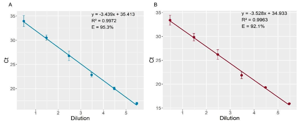
Figure 2.
Final presentation of the two-concentration level Brucella abortus RB51 genomic DNA candidate Reference Material, including labeled screw-cap tubes and their corresponding information.
Figure 2.
Final presentation of the two-concentration level Brucella abortus RB51 genomic DNA candidate Reference Material, including labeled screw-cap tubes and their corresponding information.
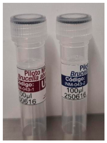
Figure 3.
Homogeneity assessment of the Brucella abortus RB51 genomic DNA candidate Reference material. Box and whisker plots show Ct values distributions obtained by the validated duplex qPCR method for selected units U-008, U-014, U-035, and U-042 (Level 1), and units U-006, U-016, U-023, U-035, and U-043 (Level 2) of the Brucella abortus RB51 Reference Material, for the ctrA (A and C) and nikR genes (B and D). The plots illustrate between-unit variability and repeatability across the evaluated units.
Figure 3.
Homogeneity assessment of the Brucella abortus RB51 genomic DNA candidate Reference material. Box and whisker plots show Ct values distributions obtained by the validated duplex qPCR method for selected units U-008, U-014, U-035, and U-042 (Level 1), and units U-006, U-016, U-023, U-035, and U-043 (Level 2) of the Brucella abortus RB51 Reference Material, for the ctrA (A and C) and nikR genes (B and D). The plots illustrate between-unit variability and repeatability across the evaluated units.
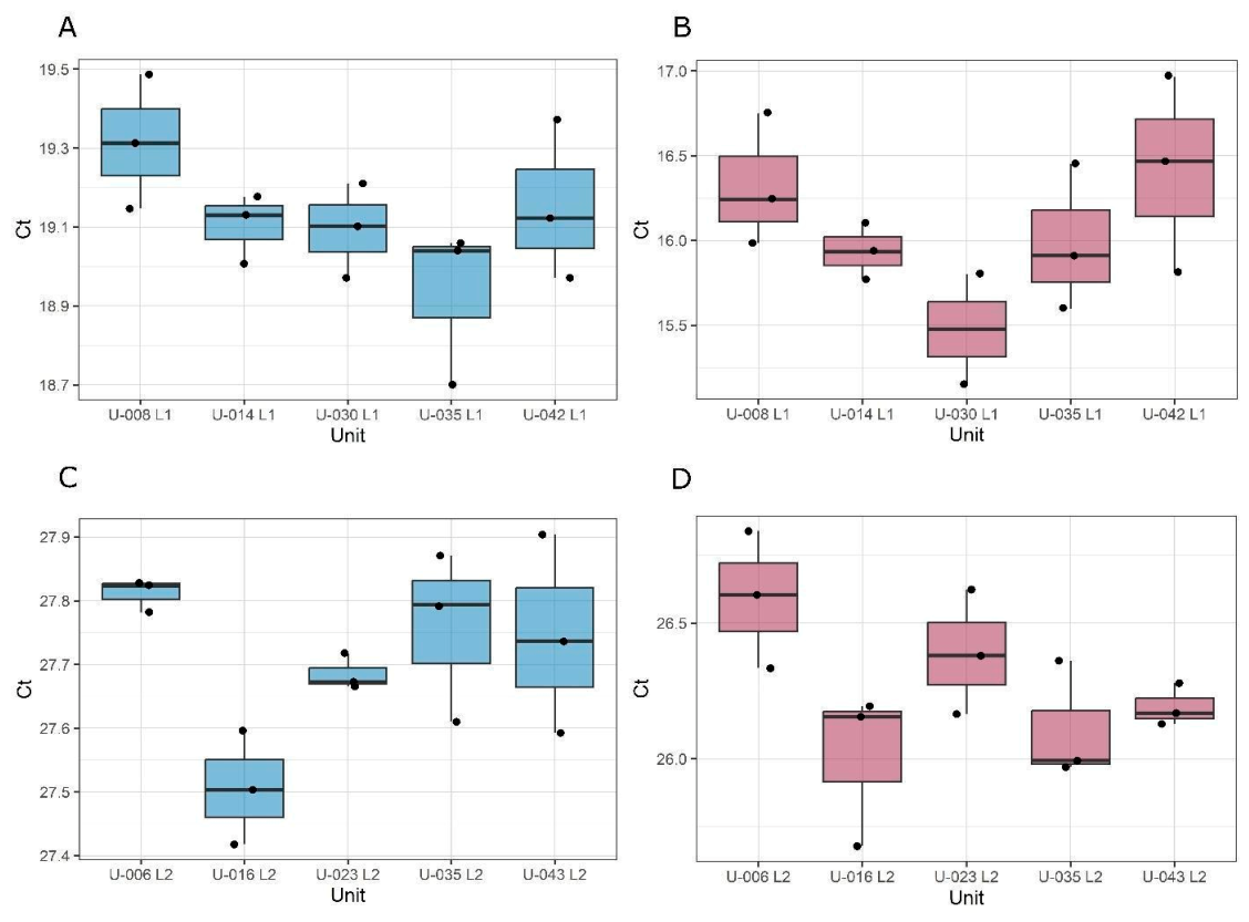
Figure 4.
Long-term stability study of the Brucella abortus RB51 candidate Reference Material. Regression analysis of Ct values obtained by the validated duplex qPCR method for ctrA and nikR during storage at 4 °C over nine months. (A and C) Levels 1 and 2 for the ctrA target. (C and D) Level 1 and 2 for nikR target. The analysis was used to identify potential time-dependent trends and to estimate the uncertainty contribution associated with stability.
Figure 4.
Long-term stability study of the Brucella abortus RB51 candidate Reference Material. Regression analysis of Ct values obtained by the validated duplex qPCR method for ctrA and nikR during storage at 4 °C over nine months. (A and C) Levels 1 and 2 for the ctrA target. (C and D) Level 1 and 2 for nikR target. The analysis was used to identify potential time-dependent trends and to estimate the uncertainty contribution associated with stability.
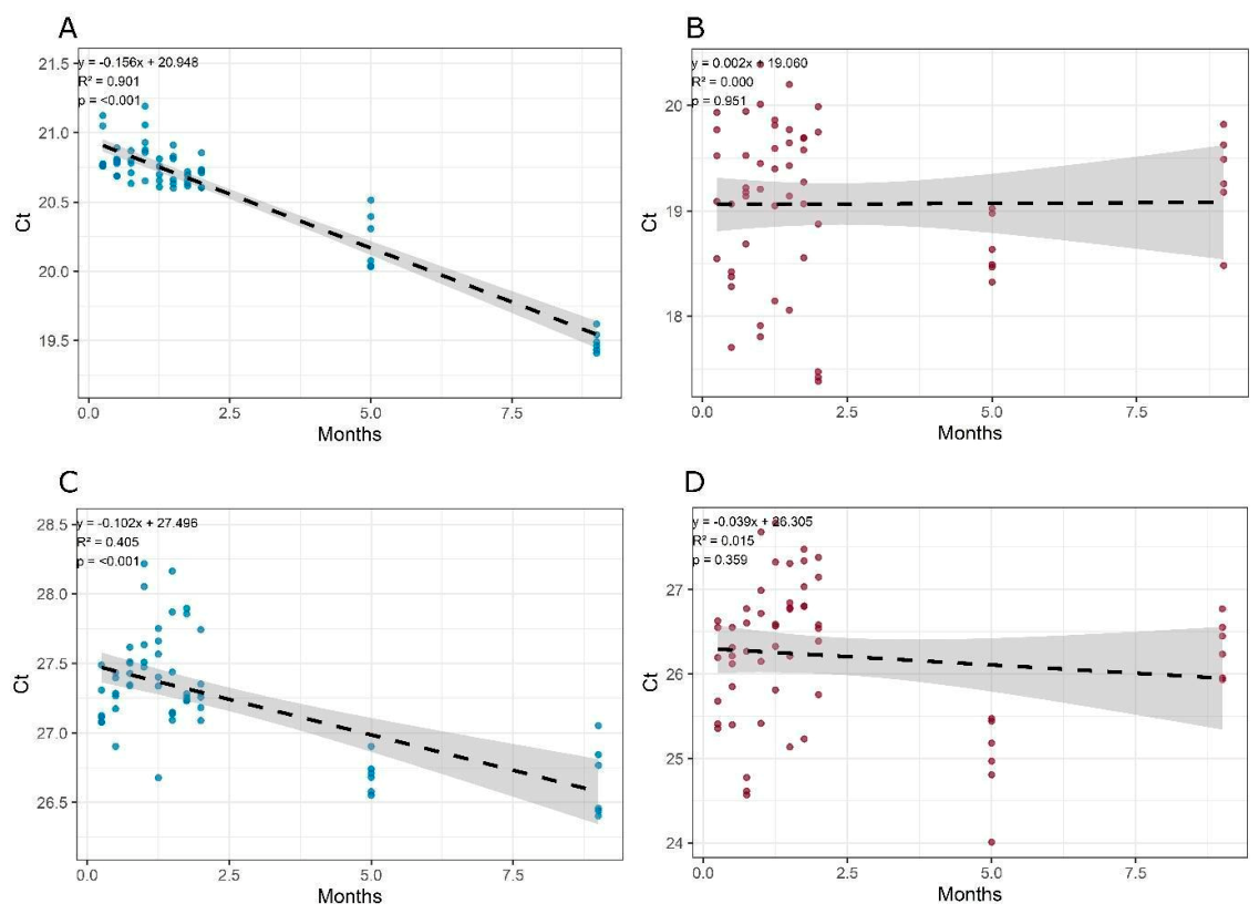
Figure 5.
Preliminary amplification of the ctrA gene by fluorescence (A) and colorimetric (B) LAMP methods using both RM levels concentration and non-template control (NTC). Positive colorimetric reactions were identified by a yellow color change, whereas negative reactions remained red.
Figure 5.
Preliminary amplification of the ctrA gene by fluorescence (A) and colorimetric (B) LAMP methods using both RM levels concentration and non-template control (NTC). Positive colorimetric reactions were identified by a yellow color change, whereas negative reactions remained red.
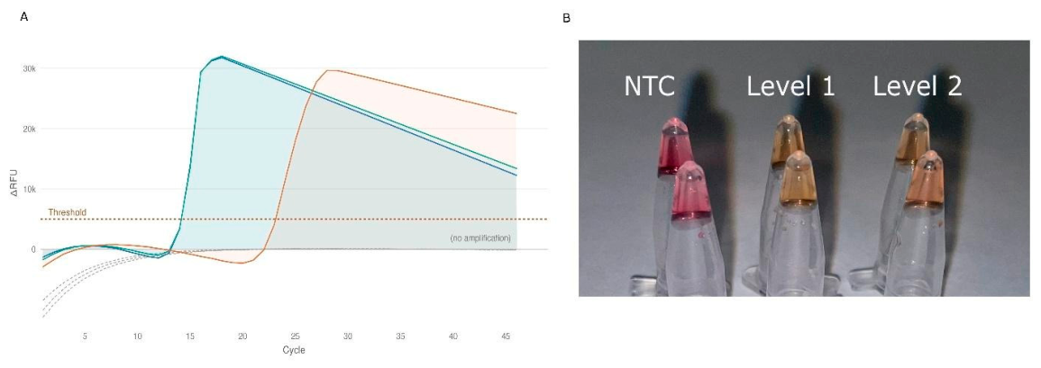
Figure 6.
Temperature optimization of the colorimetric ctrA-based LAMP assay. The assay was evaluated using Brucella abortus RB51 genomic DNA RM at both concentration levels and a non-template control (NTC). The evaluated temperatures were 63 °C, 66 °C, and 67 °C, in addition to the preliminary condition of 65 °C.
Figure 6.
Temperature optimization of the colorimetric ctrA-based LAMP assay. The assay was evaluated using Brucella abortus RB51 genomic DNA RM at both concentration levels and a non-template control (NTC). The evaluated temperatures were 63 °C, 66 °C, and 67 °C, in addition to the preliminary condition of 65 °C.
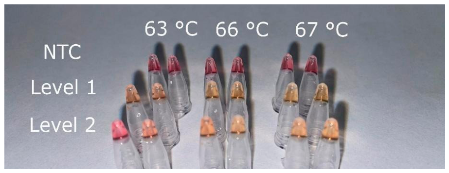
Figure 7.
Amplification curve obtained from eight concentration levels prepared from the Brucella abortus Reference Material and analyzed using colorimetric (A) and fluorescence-based (B) LAMP assays. NTC non-template control.
Figure 7.
Amplification curve obtained from eight concentration levels prepared from the Brucella abortus Reference Material and analyzed using colorimetric (A) and fluorescence-based (B) LAMP assays. NTC non-template control.
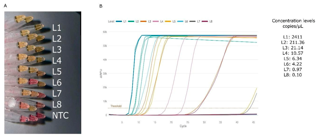

Table 1.
Primers and probe sequences used in duplex digital PCR and real-time PCR assays for the amplification of the ctrA and nikR genes of Brucella abortus RB51.
Table 1.
Primers and probe sequences used in duplex digital PCR and real-time PCR assays for the amplification of the ctrA and nikR genes of Brucella abortus RB51.
| Target | Primer | Sequence 5’ – 3’ | Amplicon size (bp) |
| ctrA | ctrA Forward | CCAAGCCGTTCCATAAGGAC | 183 |
| ctrA Reverse | GCAGCTCAAGCATCTGGTAT | ||
| ctrA Probe | FAM - AATCTGGATGCCAAGACGGTGGAA - BHQ1 | ||
| nikR | nikR Forward | TGGAAGAAGGCCAGATGGAA | 199 |
| nikR Reverse | TGCCTGACATCGTCCGTT | ||
| nikR Probe | ROX – TCATCACAACATCTCGGTCGCCAG - BHQ2 |
Table 2.
LAMP primer sequences targeting ctrA and nikR.
| Target | Primer | Sequence 5’ – 3’ |
| ctrA | F3 | CCAAGCCGTTCCATAAGGAC |
| B3 | GCAGCTCAAGCATCTGGTAT | |
| FIP | GATGACCGACTGGGCATGCCGAGCTGATTGCCCGTATCC | |
| BIP | GGCGACCTGGTGGTCAATCTGCCTTGCCGGTCAGATGAAC | |
| LF | TTGGAGCGGCGGACGAT | |
| LB | GATGCCAAGACGGTGGAAGTC | |
| nikR | F3 | TGGAAGAAGGCCAGATGGAA |
| B3 | TGCCTGACATCGTCCGTT | |
| FIP | AGCTTTTTGGAGAGGTCGCGGGCCTGTGTGGGAGTGCTA | |
| BIP | TCATCACAACATCTCGGTCGCCTGCCCTTGAGGATACTGACT | |
| LF | GCACTGTGATCATAGGTGTAGCT | |
| LB | GCATGCATGTGCATCTTGATCATG |
* F3/B3 (outer primers); FIP/BIP (inner primers); LF/LB (loop primers).
Table 3.
Summary of the characterization of the Brucella abortus RB51 genomic DNA candidate Reference Material.
Table 3.
Summary of the characterization of the Brucella abortus RB51 genomic DNA candidate Reference Material.
| Target | Level |
Mean copies copies/µL |
Mean Ct | uhom rel | ults rel | uchar rel | uRM rel | URM rel | Uabs |
| ctrA | 1 | 32 989 | 19.1 | 0.52 | 6.82 | 4.70 | 8.30 | 16.60 | 5 475 |
| nikR | 1 | 29 722 | 16.1 | 1.41 | 0.52 | 5.28 | 5.49 | 10.98 | 3 263 |
| ctrA | 2 | 285 | 27.7 | 0.37 | 3.37 | 7.56 | 8.29 | 16.57 | 47 |
| nikR | 2 | 286 | 26.3 | 0.74 | 0.42 | 7.45 | 7.50 | 15.00 | 42 |
* The table presents, for each target gene and level, the mean copies/µL, mean Ct, relative homogeneity uncertainty (uhom rel), relative long-term stability uncertainty (ults rel), relative characterization uncertainty (uchar rel), relative combined uncertainty of the RM (uRM rel), relative expanded uncertainty of the RM with coverage factor (k = 2, corresponding to a 95% confidence level) (the values were presented as %), and Uabs absolute expanded uncertainty (copies/µL).
Disclaimer/Publisher’s Note: The statements, opinions and data contained in all publications are solely those of the individual author(s) and contributor(s) and not of MDPI and/or the editor(s). MDPI and/or the editor(s) disclaim responsibility for any injury to people or property resulting from any ideas, methods, instructions or products referred to in the content. |
© 2026 by the authors. Licensee MDPI, Basel, Switzerland. This article is an open access article distributed under the terms and conditions of the Creative Commons Attribution (CC BY) license.
Copyright: This open access article is published under a Creative Commons CC BY 4.0 license, which permit the free download, distribution, and reuse, provided that the author and preprint are cited in any reuse.