

Submitted:
28 October 2026
Posted:
30 October 2026
You are already at the latest version
Abstract
Background. Mucosal vaccination offers the possibility of establishing immune protection directly at portals of pathogen entry, but its development remains constrained by biological, technological, and implementation barriers. Objective. To integrate the major challenges limiting mucosal vaccination within a common framework and examine emerging strategies capable of addressing several of these barriers simultaneously. Results and Discussion. We consider six interconnected challenges: incomplete recognition of the determinant role of adjuvants; scarcity of safe and accessible mucosal adjuvants; the need to overcome mucosal tolerance while preserving tissue homeostasis; dependence on coldchain logistics; reliance on multidose immunization schedules to achieve protective immunity; and limited breadth of vaccine-induced cross-reactive protection. To better capture the complexity of modern vaccine platforms, we discuss expand adjuvant activity as exogenous, self-adjuvanting, or endogenous according to the origin of the immunostimulatory signal. Particular emphasis is placed on proteoliposome- and cochleate-based systems and probiotic Bacillus subtilis spores as multifunctional platforms combining antigen delivery with intrinsic immunostimulatory properties. Preclinical studies support their capacity to induce systemic IgG, mucosal secretory IgA, cellular responses, and memory response, while their heat resistance may facilitate the development of more thermostable formulations. We also discuss the Single Time Vaccination Strategy as an approach to combined-route immunization and consider glycans and other microbial structures as potential contributors to antigen-specific and cross-reactive mucosal responses. Conclusions. Future mucosal vaccine development should consider adjuvants as tools for coordinated antigen delivery, innate sensing, immune programming with polarization activity, and mucosal memory rather than solely as immune potentiators and delivery system. Probiotic spores, cochleates, and combined-route strategies provide promising examples of this integrated approach, although their translational value will require rigorous demonstration of formulation stability, mechanisms of action, safety, protective breadth, and clinical efficacy.
Keywords:
adjuvants
; vaccines
; mucosa
; probiotic
; spores
; Bacillus subtilis
; cochleate
; SinTimVaS
; secretory IgA
; immune response
1. Introduction
It is generally accepted that vaccines are the main success of Immunology and the second most effective public health intervention after clean water consumption. However, a more critical analysis reveals a striking epidemiological-immunological mismatch: the vast majority of diseases covered by vaccines, including respiratory infections (influenza, SARS-CoV-2, respiratory syncytial virus, tuberculosis), enteric infections (cholera, rotavirus, and shigellosis), and sexually transmitted infections (human immunodeficiency virus and gonorrhea) are acquired exclusively through mucosal surfaces. Paradoxically, fewer than 10% of licensed vaccines are administered via mucosal routes to counter these mucosal pathogens. This disconnection is particularly problematic because parenteral immunization fails to establish the two fundamental pillars of frontline mucosal defense: (i) robust secretory IgA (sIgA), which acts as an immune barrier, and (ii) tissue-resident memory T (Trm) cells, which provide immediate, on-site cellular immunity upon pathogen re-encounter. While circulating IgG and central memory T cells offer systemic protection, they cannot compensate for the absence of Trm and sIgA at the portal of entry, critically compromising the first line of defense. This epidemiological-immunological disparity underscores the urgent, unmet need for effective mucosal adjuvants, a gap that our work directly addresses by providing platforms capable of inducing both humoral and cellular mucosal immunity. Nevertheless, there are hundreds of human infectious diseases without available vaccines. In 2012, Pérez et al. reported 90 registered prophylactic vaccines against. 31 bacterial or viral pathogens. Only out 9 of 90 vaccines against only 6 diseases were administered via mucosal routes: eight were oral vs. 5 diseases (poliovirus, cholera, Salmonella, rotavirus, and adenovirus) and one was nasally administered (flu vaccine) [1]. The COVID-19 pandemic directed the pharmaceutical industry mainly to protect against it with vaccine. So, in 2021, van der Ley et al. reported 112 vaccines in development against COVID-19 [2]. One year later, there were 371 COVID-19 candidates with 173 in clinical stages and 199 vaccines in the preclinical stage using 11 platforms: 32% (55) of protein subunits; 23% (40) of mRNA; 13% (23) of non-replicant viral vector (VVrn); 13% (22) of inactivated virus; 9% (16) of DNA; 4% (6) of virus like-particles; and around 1% of live-attenuated virus, replicant viral vectors (VVr), VVr plus antigen-presenting cells (APCs), VVrn plus APCs, and bacterial antigen-spore vector were reported [3]. Here, the first question appeared: the relationship between vaccine platforms and delivery systems (DS) requires clarification. Vaccine platform refers to the production method, and DS refers to how to target associated antigens to APCs. DS are usually nanoparticles, though some are also microparticles. So, some vaccine platforms described above serve as DS too. In addition, there are several lipid-based particles/formulations (liposomes, lipid nanoparticles, solid lipid particles, exosomes, virus-like particles, lipoplex, polyplex, polymersome, proteoliposomes, etc.); hydrogel and polymer nanoparticles; dendrimers, polymer particles; nano- and micro-emulsions; in situ gel; cell-penetrating peptides, plasmids; polylactide glycolipids [PLG] microparticles; immune stimulating complexes [ISCOM]; microneedles; cochleates; etc. Moreover, lipids and glycans, among other molecules included in DS, have immune potentiating activities, making the adjuvant more complex and difficult to define and characterize.
In 2024, over 180 vaccine candidates have entered COVID-19 clinical trials, but only 14 vaccines have received World Health Organization (WHO) approval for emergency use listing or pre-qualification status. It should be noted that none of the above mentioned WHO-approved vaccines were administered via the mucosal route, and the adjuvant, when present was usually concealed or masked because its importance is assumed to be secondary in the formulation. Going into a deeper analysis, only 5 adjuvants (Alum+Imidazoquinoline agonist of TLR7-8, Matrix-MTM [saponin based], SAO3 [adjuvant system squalene-based] and SQBA [squalene-based]); and 1 DS [liponanoparticles, in 2 vaccines]) in those 14 vaccines approved by WHO were properly identified [4,5,6,7]. Nevertheless, seven of vaccines mentioned before have intrinsic self-adjuvant characteristics included. Bearing this in mind, here we arrived at an important research question: could these vaccines components be considered as self-adjuvants? Some relevant components in the vaccine formulation are especially important because they not only exhibit robust immune activity, but also contain one or several immune potentiators with self-adjuvant properties and efficiently activate innate immune signaling pathways.
Generally, vaccine formulations contain antigens and excipients as preservatives, diluents, stabilizers, as well as and adjuvants. Gaston Ramón [8] is best known as the father of the term “adjuvant”, which was initially defined as “…substance used in combination with a specific antigen that produces a more robust immune response than the antigen alone…”. However, this definition currently faces several limitations when considering the potent adjuvant properties shown by attenuated, inactivated, and other vaccine platforms, due to self-adjuvant characteristics contained. So, it is known that vaccine effectiveness depends on the precise conformation of the vaccine formulation to be delivered to the appropriate immune cells.
While multiple adjuvant classifications have been used, an excellent reductionist classification of distinguishing immune potentiators and DS has been described, which is very helpful [9]. However, one element is important to raise; a pure DS is rare because most of them contain immune potentiator activities. We would suggest adding immune polarizator activity focused on programming the specific type of the protective immune responses required (Th [helper] 1, CTL [cytotoxic T lymphocytes], Th2, sIgA, etc.) [10,11]. In addition, categorizing adjuvants as facilitators of the immune signal is also interesting, because it goes deeper into the stimulation of the innate immune responses [12,13]. So, considerable diversity in classifying adjuvanted and non-adjuvanted vaccines is observed [14]. Most vaccines are seen as vaccines with self-adjuvant characteristics. Only plain carbohydrate-subunit vaccines are considered non-adjuvanted formulations. However, natural polysaccharides have attracted much attention due to their potential for intrinsic immunomodulation, biocompatibility, biodegradability, low toxicity, and safety, which have exhibited great potential for vaccine formulation (the adjuvant capacity of these structures will be discussed later).
Another issue is the diversity and fragmentation of the regulatory environment related to adjuvants. Guidelines to approve new adjuvants vary on a case-by-case basis and directly impact long approval times, making it one of the slowest processes in medicine [15]. Also, approved adjuvants are protected by the property rights of pharmaceutical companies (later discussed). In addition, another misconception is related to overcoming the biological barriers of mucosal surfaces. The development of a specific formulation strategies, depending on the specific targeted barrier, leads to the designing or identification of adequate delivery devices that allow access to the target inductive organ (drops or spray for nasal administration, drops for sublingual intake, inhalant or nebulizer for pulmonary diseases, tablet or capsule form for oral applications, etc.). Mucosal surfaces are not static and possess their own highly specialized networks. Understanding mucosal physiology and protective mucosal immunity helps to dispel misconceptions about drug delivery and immunological strategies at the mucosal level.
Nowadays, 21 SARS-CoV-2 mucosal vaccine candidates are in use or in clinical development. Of those, 12 candidates are in the phase I stage of clinical trials, 3 in phase II, and only 1 in phase III. For human use, 5 vaccines have received regional approval; however, none of them have received WHO approval [16] (see Table 1). The presence of exogenous adjuvant (the real adjuvant), as it was defined by Gaston, and self-adjuvants of these vaccines are summarized (see Tab. 2), including not only COVID vaccines, but also older approved mucosal vaccines like cholera, influenza, poliovirus, and rotavirus. Interestingly, among the mucosal vaccines, very few contain exogenous adjuvants in contrast with those platforms that have incorporated self-adjuvants in vaccine formulation. In addition, the description of self-adjuvants in the scientific literature as components used in vaccine formulation and co-administered with messenger RNA (mRNA) antigens, capable of inducing a robust immune activity and efficiently activating various innate immune signaling pathways, is a limited approach and, in our opinion, an incomplete view of the impact of those molecules at the mucosal surfaces. In fact, the idea of encapsulating immunostimulants such as different pathogen-associated molecular patterns (PAMPs) stimulating multiple Toll-like receptors (TLRs) in the cytosol, or at the endosome of APC, which can promote the recruitment and infiltration of innate immune cells deserves to be explored [17]. Studies have found that some self-adjuvant delivery materials can activate the Nlrp3 inflammasome, regulate innate immune cell infiltration to the injection site, and enhance immune responses [18]. Much controversy has existed about the increased risk of Bell's palsy after intranasal administration, which has been analyzed as a potential adverse event after mucosal routes of administration. The only vaccine ever confirmed to cause facial nerve palsy was Berna Biotech’s Nasalflu®, an inactivated intranasal influenza vaccine adjuvanted with Escherichia coli heat-labile toxin, which is no longer being produced [19]. However, this vision has changed mainly due to prioritizing the induction of protective immune responses at the entry point, the nose; the induction of mucosal immune responses in the upper respiratory tract, in particular the lung; and the need to prevent and interrupt the transmission of SARS-Co-V2. Despite the limitations of the studies developed, it has been found that COVID-19 vaccines were associated with rare oral adverse events that were predominantly similar to those emerging following seasonal influenza vaccines [20].
The aim of this review is therefore to integrate biological, technological, and implementation barriers to mucosal vaccination within a common conceptual framework and to examine emerging strategies capable of addressing these barriers. By linking an expanded concept of adjuvant activity with multifunctional platforms such as cochleates and probiotic spores, we propose that future mucosal vaccine development should move from viewing adjuvants simply as immune enhancers toward considering them as tools for coordinated antigen delivery, innate sensing, immune programming, and mucosal memory.
Particular emphasis is placed on proteoliposome- and cochleate-based adjuvant systems and probiotic Bacillus subtilis spores as examples of platforms that combine delivery and intrinsic immunostimulatory properties. Their potential thermostability and capacity to induce systemic and mucosal responses make them particularly relevant to mucosal vaccine development. We also discuss the Single Time Vaccination Strategy (SinTimVaS) as an alternative approach combining simultaneous immunization through different routes to reduce dependence on conventional multidose schedules while promoting both systemic and mucosal immunity. Finally, we consider how glycans and other microbial structures may contribute to antigen-specific and cross-reactive mucosal responses.
In this review, we consider six interconnected challenges that currently limit the development and implementation of mucosal vaccines: (1) incomplete recognition and understanding of the determinant role of adjuvants; (2) scarcity of safe and accessible mucosal adjuvants; (3) the need to overcome mucosal tolerance while preserving tissue homeostasis; (4) dependence on cold-chain logistics; (5) reliance on multidose immunization schedules to achieve protective immunity; and (6) limited breadth of vaccine-induced cross-reactive protection. The conceptual framework of the review, linking these challenges with adjuvant mechanisms, innovative platforms, immune programming, and translational advantages, is summarized in Figure 1, Figure 2, Figure 3 and Figure 4.
2. Incomplete Recognition of the Determinant Role of Adjuvants in Mucosal Immunity
An effective mucosal adjuvant should do more than simply increase the magnitude of the immune response or served as delivery molecules. Ideally, it should promote antigen uptake and presentation, activate appropriate innate immune pathways, and program the type and anatomical distribution of the adaptive response required for protection. Depending on the pathogen and route of infection, this may include systemic IgG, mucosal sIgA, Th1/Tc1 responses, cytotoxic T lymphocytes, and tissue-resident and B-cell memory. Adjuvant-mediated immune programming may also influence the breadth and persistence of vaccine-induced protection. Thus, mucosal vaccine development requires consideration of adjuvants not merely as immune potentiators, but as integral components linking antigen delivery, innate sensing, and the establishment of protective mucosal memory.
Adjuvants have traditionally been defined as exogenous substances added to vaccine formulations to enhance antigen-specific immune responses. However, this definition does not fully encompass the increasing complexity of modern vaccine platforms. Components intrinsic to live-attenuated microorganisms, viral or bacterial vectors, outer-membrane vesicles, proteoliposomes, nanoparticles, and other delivery systems can themselves activate innate immune pathways and contribute substantially to vaccine immunogenicity. Moreover, vaccination can induce endogenous danger signals derived from stressed, damaged, or dying host cells that further modulate innate and adaptive immune responses. We therefore propose a broader functional framework in which adjuvant activity can be considered exogenous, self-adjuvanting, or endogenous, depending on the origin of the immunostimulatory signal. This framework may be particularly useful for understanding mucosal vaccine platforms, in which delivery and innate immune activation are often closely interconnected.
It is well recognized that selecting the appropriate vaccine antigens are the central issue for new vaccine design. However, adjuvants play a key role in vaccine formulation, particularly mucosal adjuvants, as our group previously postulated11,12. There are the three main points for this contradiction;
1. , in the original definition, the adjuvant is considered as an exogenous component in the vaccine formulation. However, the complexity of several vaccine platforms, where self-components can prolong retention time, recruit immune cells, increase uptake of determinant antigens, and provide an inflammatory environment to activate adaptive immune responses, has led us to a broader definition: Exogenous adjuvants, Self-adjuvants, and Endogenous adjuvants. Self-Adjuvants are components that coexist in those vaccine platforms that use non-purified antigens and vectors, like live-attenuated or whole cells with the main antigens identified. Endogenous adjuvants are substances induced in the host during the vaccine application as a consequence of stressed, damaged, or necrotic cells [21,22] or antigen covalently conjugated to TLR or glycans. Nevertheless, if endogenous substances are added to vaccine formulation it must be considered as exogenous adjuvant.
2, the reduction of the multiple classifications of adjuvants to immune potentiators and DS10. Some or most DS have exhibited immune potentiator activities. So, these DS should also be considered immune potentiators; this simple separation complicates the understanding of the adjuvant definition; and
3, limiting adjuvant action just to increase the speed, intensity, and duration of the induced immune response. The role of the adjuvant should be seen in a broader scope than the classical view, as substances acting primarily as a passive DS (depot effect). A modern view has revealed that these components do much more:
- a, to activate and stimulate, immediately, the innate immune response acting over professional presenting cells, the DC and non-professional cells: macrophages (Mø), neutrophils (PMN), natural killer cells (NK), mast cells, or epithelial cells by mimicking danger signals and causing the immediate release of cytokines and chemokines, recruiting immune cells and establishing a local immunocompetent environment at the injection site;
- b, instead of clonal receptor selection of the acquired immune response, adjuvants act at a non-clonal level, recognizing broadly conserved, non-clonal microbial motifs and allowing identification of the main aggressors: viruses, bacteria, fungi, yeasts, protozoa, and helminths. Adjuvants can specifically recognize distinct structural molecular signatures like bacterial subdivisions or nucleic acid strandedness;
-
c, to direct and polarize to different subsets of helper T cells (Th1, Th2, Th3, Th9, Th17, Tfh [T follicular helper], or T regulatory cells), as part of the cell-mediated immune protection against pathogens transducing them in the induction of antibodies with:
- ○
- i, bactericidal and opsonophagocytic (IgG1 and IgG3 [in human], Th1), anaphylatoxin(IgE, Th2) or mucosal (sIgA) activities;
- ○
- ii, induction of CTL (CD8+ or CD4+ T cells). CD8 T memory response occurs only in the presence of Th1 assistance; and
- ○
- iii, neutralizing antibodies are not a consequence of immune polarization, because any antibody class and subclass are capable of neutralizing microorganisms. Furthermore, studies have demonstrated that the dose of a vector, the method of delivery, prior immunization, and the patient’s genetic predisposition may all impact the immune response and the formation of neutralizing antibodies;
- d, they permit vaccines to work at the extremes of age (newborns and elderly) and in immunocompromised people; the immature immune systems of newborns and the decline of the immune functions in the older adults can be overcome with the incorporation of adjuvants in the vaccine formulation. The adjuvant-specific activation of cell populations and the dissecting of innate cytokine responses by immunophenotyping may also help to identify age-specific adjuvants. A defined age-tailored adjuvantation system for newborns, tolder, and immunocompromised people might be beneficial to shape adaptive immunity in age-specific manners;
- e, they address vaccine efficacy through different routes, particularly the mucosal one, where sIgA antibody and Tmr responses are required;
- f, reshaping antigenic immunodominance in multiple vaccines by altering innate immune activation and antigen presentation kinetics. Adjuvants are capable of driving the immunodominant response toward the same antigen, resulting in differences in protective efficacy;
- g, the induction of the so-colled "trained immunity" through directly targeting PRRs and inducing epigenetic and metabolic reprogramming of innate immune cells can lead us to the modulation of innate signaling pathways, broader immune protection, and enhanced vaccine efficacy [23];
- h, they could induce a cross-protective response [24]. However, the effect of adjuvants on increasing the breadth of cross-reactivity has been poorly explored (it will be discussed in detail later); and
- I, some adjuvants have an antigen dose-sparing effect through creating a slow-release effect at the injection site and actively stimulating of the innate immune system, which allows reduced manufacturing costs, stretches limited supplies especially during pandemics and generates robust protection in patients with weaker immune systems.
Despite all this, most adjuvant mechanisms of action remain poorly understood. So, taking into account the above-discussed properties, we were able to design platforms that combine many of these characteristics, making those adjuvant system more precise, safer, and effective adjuvant platforms. Likewise, it was possible to understand better the pivotal role of adjuvants, in particular, self-adjuvants, in the vaccine formulations.
It is recognized that Alum, described by Glenny in 1026 [25], is the most common adjuvant (alone or in combination with other exogenous components) for almost a century. Alum salts are used in routine human vaccinations, although their mode of action remains unclear. When Alum is used alone, it induces a Th2 immune response [26] characterized by IgG4 and IgE antibodies that could be useful in the helminth vaccines (not much exploited yet) but not for those requiring a Th1/CTL response (like intracellular infections such as human immunodeficiency virus) or sIgA mucosal response reducing the risk of progressing to an active form of the disease like tuberculosis. Last but not least, exogenous adjuvant combination is the hallmark of vaccine formulations. Examples of theses combinations are the Adjuvant System (AS): AS01, AS03, and AS04 licensed since 2005 in several vaccines or Adjuvant Finlay Proteoliposome (AFPL)1 into Aluminum (Alum), licensed since 1988, etc.
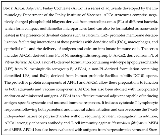
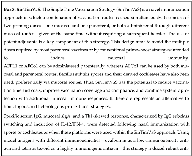
The AFPL platform contains self-adjuvants (lipopolysaccharide [LPS], porin proteins, and traces of bacterial DNA). On the one hand, AFPL can be combined with an exogenous adjuvant like alum to increase formulation stability, reduce pyrogenicity, create a deeper effect, and induce a polarization toward a Th1 pattern, thereby allowing it use by: parenteral route AFPL [AFPL1-Box 1] [13,27,28,29,30,31,32,33,34,35,36]. On the other hand, the Adjuvant Finlay cochleate-based platform (AFCo) was developed to avoid alum in the formulation and also has the potential to be used via the mucosal route[AFCo-Box 2] [29,37,38,39,40,41,42,43,44,45,46,47,48,49]. Moreover, AFCo has unique characteristics to induce systemic and mucosal immune responses similar to those responses induced by traditional mucosal and/or parenteral immunization schemes. We were able to develop a novel vaccination approach called Single Time Vaccination Strategy (SinTimVaS) where both parenteral and mucosal routes are combined in two simultaneously priming doses. SinTimVaS was not designed as a prime-boost strategy because a subsequent booster is not required; however, a timing booster dose would be possible if it is needed (SinTimVaS- Box 3) [50,51,52,53] Another AF (AFPL and AFCo) strength is that they can be used as vaccine candidates per se, due to the presence of immune-enhancing self-adjuvants and protective antigens in their structures.
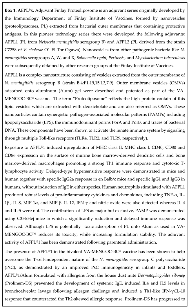
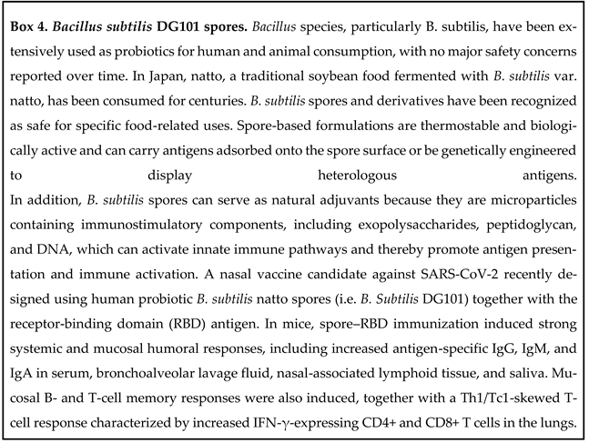
As AFPL and AFCo contain native LPS, a potent but toxic TLR4 agonist, which may be questioned by the regulatory authorities, we have used two strategies to overcome this challenge: the first strategy was essentially the absorption of PL onto Alum, allowing a masking effect by interaction between different formulation components increasing stability, overcoming Alum's Th2 response, and decreasing pyrogenicity, since LPS is absorbed into the Alum structure and is not free. In the case of AFCo, this approach was not required because LPS is embedded in the multilayer structure and no toxicity was detected [54] and the second strategy has been oriented towards the use of human probiotic Bacillus subtilis (Bs) cochleate or its spores for mucosal immunization and spores or vegetative forms of B. subtilis by parenteral routes (Bacillus subtilis-Box 4) [55,56,57]. Overall, these multiple functions of adjuvants permit a better understanding of their determinant role in vaccine formulation.
3. Scarcity of Safe and Accessible Mucosal Adjuvants
The limited number of licensed mucosal vaccines reflects the substantial biological and technological challenges associated with vaccination at mucosal surfaces. Unlike parenteral sites, mucosal tissues are continuously exposed to food antigens, environmental components, and commensal microorganisms and have therefore evolved mechanisms that favor immune homeostasis and tolerance. Vaccine antigens delivered through mucosal routes must overcome physical and enzymatic barriers, achieve sufficient retention and uptake, reach appropriate antigen-presenting cells, and generate an immune response of adequate magnitude, quality, and durability without causing excessive local inflammation. Consequently, effective mucosal vaccination depends not only on antigen selection but also on appropriate delivery systems and, particularly, safe and effective mucosal adjuvants.
Since 1990s, only five adjuvants: MF59 (squalene, Tween 80, and Span 85), AS01 (liposomes, monophosphoryl lipid A [MPL], and Quillaja saponaria [QS]-21); AS03 (squalene, α-tocopherol, [vitamine E] and Tween 80); AS04 (Alum and MPL), and CpG1018 (22-mer single-stranded DNA) have been approved for human use as part of licensed vaccines. Alum (Al(OH)3, Al4(OHPO4)3, or AlOHPO4SO4) remained the most common adjuvant used for more than 70 years. However, those points are not entirely accurate. Pulendram et al. [58] did not take into account the folowing adjuvants in their analysis: Virosome, Ca3(PO4)2, RC529, AS37, nor AFPL1. The latter mentioned is the main component of the Cuban anti-meningococcal bivalent (BC) vaccine (VA-MENGOC-BC®) since 1988 [59,60]. More than 70 million doses administrated to adults and more than 11 million of children aged 3 months old (two doses at 3 months and 6 weeks later) vaccinated. The adjuvant capacity of AFPL1 contained in this vaccine has been well described [27,30]. Studies developed in infants and toddlers with the proteoliposome and polysaccharide-based meningococcal vaccine have demonstrated long-lasting responses and subclass change pattern against polysaccharide C were observed. Certainly, AFPL1 can modulate the response to the non-covalently incorporated polysaccharide [61]. In addition, AFPL1 absorbed onto Alum has demonstrated its adjuvant and polarizing immune response capacities in a therapeutic vaccine formulation containing allergens from the House Dust Mite Dermatophagoides siboney (Prolinem-DS). The vaccine candidate has completed a phase I trial and has demonstrated its safety and the therapeutic effect in asthmatic patients sensitized to this mite species [32,33,34]. Interestingly, Prolinem-DS injected by subcutaneous route, with only three doses, was able to modulate the typical IgE/Th2 allergic response, inducing allergen-specific IgG1 and IgG4 antibodies, a rise in the IgG/IgE ratio, and a mixed IFN-gamma/IL-10 pattern downplaying IL-5 levels.
In terms of accessibility, Alum salt-based adjuvants are the most widely available and commonly used adjuvants in human vaccines, with millions of doses administered annually. Recently, companies like SEPPIC and InvivoGen have been developing a range of effective and affordable large-scale adjuvant manufacturing technologies to develop more accessible vaccines. Despite the progress that has been made and the evolution of vaccine platforms, more novel adjuvants are urgently needed. Likewise, large-scale manufacturing of adjuvants needs to be addressed to ensure a widespread use. Many promising vaccine antigens face delays in reaching clinical trials or even being rejected because they are not formulated an adequate adjuvant. Also, the complex regulatory framework for vaccine licensing frameworks constitutes a real challenge to guarantee vaccine accessibility. Vaccine market authorization must pass rigorous regulatory evaluation to ensure the safety, efficacy, and quality of each product. Likewise, pharmaceutical companies' intellectual property rights and trade secrets are barriers that limit vaccine access. Even if a patent application has already received a voluntary license, it often fails to translate into real production because crucial manufacturing know-how and formulation components, coupled with rigid technology transfer mechanisms, are not included.
Moreover, many mucosal pathogens and tumors breach mucosal barriers and possess the ability to establish, evade, or subvert host immune responses and colonize mucosal tissues, leading to a wide range of infections or malignancies. It is well recognized that mucosal adjuvant/vaccines should be able to induce sIgA, CD8+ T cells, and T/B resident memory (RM) cells for long-term protection following mucosal infection. Though, the field of mucosal immunization has lagged, some promising mucosal adjuvants have been described. The dmLT (E. coli double-mutant heat-labile toxin binding GM1 and DC) [62]; α-GalCer, (α-galactosylceramide, binding iNKT). α-GalCer adjuvanted intranasal live or inactivated influenza vaccines show promising safety because none of the vaccines redirect antigens into the brain) [63]; Chitosan nanoparticles have been used by the intranasally route by their ability to induce high IgA levels in different models; and also using cochleates [39]. Another promising adjuvant studied in three distinct anatomical tumor models (subcutaneous, lingual and intravaginal) is CTB+ DEC205+, specifically targeting DC, enhancing activation of Th1/Th17 responses, and promoting the formation of protective tissue-resident memory T cells.
Currently, some researchers are exploring new targets for inducing CD8+ T cells and TRM cells, as in the yellow fever 17D (YFV-17D), which is considered a gold standard for inducing multi-epitope specific CD8+ T cells, including rare stem cell-like memory cells that persist for over 25 years. Here are some examples highlighted [64]:
1, non-TLR agonists: RIG-I, retinoid acid-inducible gene [65]; STING, stimulator of IFN genes [66]; CLRs, C-type lectin receptors [67]; and NODs (nucleotide-binding oligomerization domain (NOD)-like) receptors [65];
2, metabolic adjuvants and nutrient sensors [68,69,70,71,72,73]: mToR, mechanistic target of rapamycin and GCn2, general control nondepresive;
Advances in systems biology (transcriptomics, proteomics, epigenomics, metabolomics, serology, and mass cytometry) are helping with adjuvant development, particularly on mucosal adjuvants
Overall, the major question to be highlighted in this part is the proposed classification of adjuvant into Exogenous, Self, and Endogenous Adjuvants, which will help to understand and classify the adjuvant properties of well-defined self-assembled molecules that are components of the vaccine formulation. In addition, the identifications of the type of immune response required for a specific vaccine and elucidating the mechanism of action of several adjuvants are important tasks.

Table 2.
Exogenous and Self-Adjuvants dependent of platform used to produce the vaccine. PAMP, pathogen-associated molecular pattern; PRR, paten recognition receptor; ds, double strand; DNA, deoxyribonucleic acid; CpG, cytosine guanine; TLR, toll-like receptor; NOD2, nucleotide-binding oligomerization domain-containing protein 2; NLR3, family pyrin domain containing 3; cGAS/STING, cytosolic DNA sensing pathway; RNA, ribonucleic acid; ss, single strand; RIG-I, retinoid acid inducible gen I; MDA5, melanoma differentiation-associate; st, structural proteins: H-N, hemagglutinin-neuraminidase; F, fusion; NP, nucleoprotein (NP); phosphoprotein (P); M, matrix, and L, high polymerase; PGN, peptidoglycan; EPS, exopolysaccharide; Matrix-M: two distinct fractions of saponins (Quillaja saponaria) + cholesterol and phospholipids to form 40-nm open cage-like nanoparticles; CL097, Imidazoquinone; RAS-01 (oil [sesame, olive and soybean] and Tween20-in-water emulsion); OMV, outer membrane vesicles: LPS, lipopolysaccharide; DC-SING, dendritic cell-specific intracellular adhesion molecule-3-grabbing non-integrin; scAd, single-cycle adenovirus or deletion-mutant adenovirus; I+O, Inaba+Ogawa. References. Mogensen TH, Pathogen Recognition and Inflammatory Signaling in Innate Immune Defens CLINICAL MICROBIOLOGY REVIEWS, 2009, 22(2): 240–273. doi:10.1128/CMR.00046-08. 2. Thermo Fisher Scientific - BR. Pattern Recognition Receptors Overview |https://share.google/2UOcVUrhYhcbpgQuQ.
Table 2.
Exogenous and Self-Adjuvants dependent of platform used to produce the vaccine. PAMP, pathogen-associated molecular pattern; PRR, paten recognition receptor; ds, double strand; DNA, deoxyribonucleic acid; CpG, cytosine guanine; TLR, toll-like receptor; NOD2, nucleotide-binding oligomerization domain-containing protein 2; NLR3, family pyrin domain containing 3; cGAS/STING, cytosolic DNA sensing pathway; RNA, ribonucleic acid; ss, single strand; RIG-I, retinoid acid inducible gen I; MDA5, melanoma differentiation-associate; st, structural proteins: H-N, hemagglutinin-neuraminidase; F, fusion; NP, nucleoprotein (NP); phosphoprotein (P); M, matrix, and L, high polymerase; PGN, peptidoglycan; EPS, exopolysaccharide; Matrix-M: two distinct fractions of saponins (Quillaja saponaria) + cholesterol and phospholipids to form 40-nm open cage-like nanoparticles; CL097, Imidazoquinone; RAS-01 (oil [sesame, olive and soybean] and Tween20-in-water emulsion); OMV, outer membrane vesicles: LPS, lipopolysaccharide; DC-SING, dendritic cell-specific intracellular adhesion molecule-3-grabbing non-integrin; scAd, single-cycle adenovirus or deletion-mutant adenovirus; I+O, Inaba+Ogawa. References. Mogensen TH, Pathogen Recognition and Inflammatory Signaling in Innate Immune Defens CLINICAL MICROBIOLOGY REVIEWS, 2009, 22(2): 240–273. doi:10.1128/CMR.00046-08. 2. Thermo Fisher Scientific - BR. Pattern Recognition Receptors Overview |https://share.google/2UOcVUrhYhcbpgQuQ.
| Platform | Adjuvants (Exogenous) | Self Adjuvant. These columns could be an incomplete record | Vaccine: | |
|---|---|---|---|---|
| PAMP | PRR | |||
| Adenovirus (Human and Chimpanzee) | None | dsDNA (CpG) | TLR9 | 1. AstraZeneca; 2. iosBio; 3. VaxArt; 4. OCU500; 5. Covaxin; 6 Convidecia; 7 Gamaleya; 8. Tetherex; |
| Cytosolic DNA | NOD2, NALP3, cGAS/STING | |||
| Cápside proteins | TLR4, TLR2 | |||
| RNA polimerase III | RIG-I | |||
| Flagelin | TLR5 | 8. Tetherex | ||
| Influenza virus (Parainfluenza 5) | None | ssRNA/dsRNA | RIG-I, MDA5 | 9. CyanVac LLC; 10. FluMist/Fluenz |
| ssRNA | TLR7/8 | |||
| dsRNA | TLR3 | |||
| Live-attenuated Respiratory syncytial virus | None | ssRNA | TLR7/8 | 11.Meissa; |
| ssRNA | RIG-I, MDA5 | |||
| Live-attenuated NewCastle Disease virus | None | ssRNA/dsRNA | TLR7/8, RIG-I, MDA5 | 12. CastleVax; |
| dsRNA | TLR3 | |||
| Sp: H-N, F, NP, P, M, L | ? | |||
| Recombinant bacterial probiotic, Bifidobacterium longum | None | PGN, teicoic acid, EPS | TLR2/4, NOD | 13. Symvivo; |
| Dowregulate | TLR4 | |||
| ADN (CpG) | TLR3 | |||
| Live-attenuated SARS-Co-V2 | None | ssRNA | TLR7/8 | 14. Codagenix; |
| dsRNA | TLR3 | |||
| DNA (CpG) | TLR9 | |||
| Sp: E | TLR2 | |||
| Spike | TLR4 | |||
| ssRNA/dsRNA | RIG-I, MDA5 | |||
| Live-attenuated, flu H1N1 (dNS1-RBD) | None | dsRNA | RIG-I | 15. Pneucolin; |
| ssRNA | TLR7/8 | |||
| dsRNA | TLR3 | |||
| Porten M2/ZBP1 | NALP3 | |||
| Protein-based, spike??? | Polimerosome | CpG7909 | TLR9 | 16. ACM Biolabs; |
| Lipids | TLR2 | |||
| None | Lipids | TLR2 | 17. BlueWillow; | |
| Sp | TLR4 | |||
| Alum | Deposit | NALP3 | 18. VaxForm; | |
| CL097 | TLR7/8 | |||
| Glucan | Dectin-1 | |||
| Alum, Poly I:C Matrix-Mtm |
Deposit | NALP3, TLR3, TLR2 | 19. Yisheng Bio; 20. Novavax; | |
| None | Sp: M, E | TLR2 | 21. Oravax; 22. Roamed; | |
| Spike | TLR4? | |||
| OMV Neisseria meningitidis | DNA traces? | TLR9 | 23. Intravacc; | |
| LPS | TLR4 | |||
| Porins | TLR2 | |||
| HBcAg | dsNRA | TLR3 | 24. Mambisa; | |
| ssRNA | TLR7/8 | |||
| DNA | TLR9 | |||
| Alum | Deposit | NALP3 | 25. Razi Vaccine; | |
| RAS-01 | Lipids | TLR2/4 | ||
| Protein-based (S) plus multiple synthetic CD4 and CD8 peptides | Sílica NP | NALP3 | 26. Multicovac; | |
| Mucoadhesive polymer | Hidratation, adhesion, penetrations, high viscosity | |||
| Poli (I:C) | TLR3, RIG-I, MAD5 | |||
| Inactivated | None | LPS | TLR4 | 27. Dokoral; 28. Euvichol |
| Flagelin | TLR5 | |||
| Pili | TLR2 | |||
| DNA (CpG) | TLR9 | |||
| rCTB | GM1 | 27. Dokoral | ||
| Live attenuated | None | LPS | TLR4 | 29. Vaxchora; |
| Flagelin | TLR5 | |||
| Pili | TLR2 | |||
| DNA (CpG) | TLR9 | |||
| Live attenuated/reassortant | None | LPS | TLR4 | 30. Typhi Vivotif; |
| Flagelin | TLR5 | |||
| Lipoprotein | TLR2 | |||
| NLR | ||||
| Live attenuated Polio | None | ssRNA | TLR7/8 | 31. Biopolio; |
| dsRNA | TLR3 | |||
| RNA citoplasme | RIG-I, MDA5 | |||
| Live Rotavirus | None | dsRNA | TLR3 | 32. Rotateq; 33. Rotarix. |
| ssRNA | TLR7/8 | |||
| RNA | RIG-I, MDA5 | |||
4. The Need to Overcome Mucosal Tolerance While Preserving Tissue Homeostasis
Mucosal surfaces are continuously exposed to a wide range of external substances -including non-threatening ones such as food components and commensal microbiota - thus the mucosal immune system at these sites tends to favor tolerance over activation through different mechanisms that distinguish between harmless and harmful components. This state of immunological tolerance plays a crucial role in preserving mucosal homeostasis and preventing excessive inflammatory responses. However, this same regulatory environment can also hinder the development of robust immune responses to vaccines or pathogens, making the mucosal immune system inherently biased toward tolerance rather than activation [80,81]. Also, growing knowledge of immune tolerance has been used to develop novel approaches for cancer immunotherapies. Patients who have antigen-expressing tumors have shown some degree of immune tolerance. Different strategies, including therapeutic cancer vaccines, have been used to break this tolerance. In recent years, the use of immunomodulators has become vital in regulating tumor progression and the metastasic environments, particularly mucosal immune dysregulation. Very small-size proteoliposomes (VSSP), a nanoparticulated adjuvant obtained by the combination of proteoliposome from Neisseria meningitidis (containing TLR2 and TLR4 agonists) and GM3 ganglioside, have proved sufficient to differentiate myeloid-derived suppressor cells into phenotypically mature dendritic cells [82]. More recently, ex vivo treatment with VSSP has shown induced M1-like polarization in tumor-associated macrophages from patients with metastatic ovarian cancer and variably abrogated their suppressor phenotype [83].
One of the key challenges in mucosal vaccination is the unintended induction of immunosuppressive regulatory responses. The mucosa contains regulatory T cells (Tregs), tolerogenic dendritic cells, and anti-inflammatory cytokines such as IL-10 and TGF-β [84,85]. While these elements are essential for maintaining immune homeostasis and preventing tissue damage, they can also suppress the desired vaccine-induced effector responses. As a result, mucosal vaccines may elicit suboptimal protection if they inadvertently promote regulatory pathways that dampen immune activation52. Therefore, understanding the complex interplay between the mucosal immune system’s regulatory and effector arms is critical to harnessing the full potential of mucosal immunization. Designing effective mucosal vaccines, therefore, requires a delicate balance between immunogenicity and immune regulation. To overcome this, the use of adjuvants appears to promote the effector cell responses over regulatory responses. Targeted delivery systems are encouraged, aiming to improve antigen uptake by immune stimulatory cells, as well as, formulations that avoid excessive activation of tolerogenic pathways [86]. Overall, breaking mucosal tolerance with adjuvants without perturbing mucosal homeostasis will permit obtaining better mucosal vaccines.
5. Dependence on Cold Chain Requirements
Beyond these immunological requirements, successful mucosal vaccines must overcome important practical and translational barriers. Dependence on the cold chain increases the complexity and cost of vaccine distribution, particularly in resource-limited settings. Preserving vaccines at low temperatures arise from the inherent lability of proteins and other biomolecules. Not all vaccines have the same cold chain requirements. In fact, the cold chain comprises a specific spatial distribution of dedicated cold chain equipment interconnected by temperature-controlled transportation logistics, under continuous management oversight for major deviations in temperature control, vaccine expiry, and stock distribution. Some vaccines need refrigerated temperatures (2-8ºC), others require frozen (50 to -15°C), or ultra-cold (-80 to -60°C) conditions. Cold chain logistics requirements comprise all vaccine manufacturing steps, protecting product integrity from the origin to the point of administration.
Several steps are essential: refrigerated storage with a monitoring system; the use of coolers and isolated containers for packaging and isolation; and the use of efficient transportation with a monitoring system until administration. Any change in temperature during storage or transportation can irreparably damage the product, affecting vaccine effectiveness. Cold chain management should consider not only the product itself, but also production levels, recipients, and vaccine distribution. Its maintenance has led to the accumulation of 80% of all the total costs of the vaccination programs [87]. Therefore, approaches to achieving thermostability in vaccine formulations, including adjuvant-containing vaccines, will help facilitate and increase vaccine affordability worldwide. So, termotolerant human probiotic spores (Bacillus subtilis-Box 4) and cochleate (Cochleate-Box 2) as heat-resistant adjuvants that preserve the antigen's effectiveness in the formulation have also been studied by our group. Overall, the development of heat-resistant vaccine formulations allows for increase shelf life, removes the need for a cold chain, and thus could facilitate access to vaccination programs of larger populations, particularly in low-development countries.
6. Reliance on Multidose Immunization Schedules to Achieve Protective Immunity
Multidose vaccination schedules increase logistical demands and can reduce completion rates. Furthermore, the limited availability of safe, accessible, and scalable mucosal adjuvants remains a major constraint. Platforms combining immunostimulatory activity, antigen delivery, thermostability, and compatibility with different mucosal routes could therefore address several of these limitations simultaneously. The multiple doses, where the first priming naīve cells and the second stimulates the few memory cells that were induced during the prime. Some vaccines also require one or several booster doses to maintain their effectiveness. Those administered by the parenteral route primarily stimulate systemic response (mainly specific IgG) rather than activate local mucosal-associated lymphoid tissue, consequently, failing to induce enough mucosal-specific sIgA response (e.g the pulmonary response against SARS-Co-V-2) [88,89,90]. A proven strategy for producing a long-lasting responses was homologous and heterologous prime-boost vaccination, consisting of one parenteral prime followed weeks later by a mucosal dose (most often using a live vector or attenuated vaccines) or vice-versa [91]. Nevertheless, mucosal unadjuvanted booster vaccines elicit local IgA responses by conversion of pre-existing immunity, at least in mice [92]. Recently, Chen et al. revealed a mechanism explaining how intranasal boosters re-stimulates memory B cells primed by intramuscular vaccination to undergo IgA class switching, somatic hypermutation, and clonal expansion. Single-cell RNA-seq (scRNA-seq) revealed that an intranasal booster upregulated the expression of mucosal homing receptors in spike-specific IgA-expressing B cells. This increase coincided with a transient increase in cytokines and chemokines that facilitate B-cell recruitment in the nasal mucosa. Their findings demonstrate that an intranasal booster can be an effective strategy for inducing upper respiratory mucosal sIgA and establishing mucosal immune protection [93]. The only concerning issue is that they use two doses of intranasal Adenovirus 5. It is a virus that frequently infects humans which requires further consideration, and it still requires multiple doses. The same occurs with widespread SARS-CoV-2 infection in humans; a mucosal imprint is established, analogous to a serological response, which has implications for an heterologous boosters delivered via mucosal routes. Besides, recent studies using mRNA and protein-based vaccines support the general principle that the quality of priming, rather than the boosting modality, determines the success of mucosal immunity [94]. The priming phase can induce a durable anatomical and functional program, that subsequently enable a mucosal recall response, which play a decisive role in programming the quality, durability, and anatomical distribution of the subsequent immune response. These findings redefine vaccine adjuvants as tools for immune programming, and can be rationally incorporated into heterologous prime-boost strategies.
With the intent of finding a way around a multiple injections regime, single-shot vaccines were designed. By combining a prime and booster dose in a single injection and using a microsphere-based formulation that encapsulates the antigen, this strategy enables booster immunizations that can be achieved through delayed antigen release [95]. Anyway, this may be a solution for parenteral immunization schemes but not for mucosal ones. Therefore, to reduce the number of doses, we developed two additional approaches. First, it is interesting to note that conventional allergen immunotherapy is a long-term medical treatment where multiple shots are frequently required, aiming to induce immunotolerance to allergens. Developing novel allergen vaccine formulations able to reduce the number of doses and the treatment duration is a promising research trend. We designed an allergen vaccine formulation based on House Dust Mite allergens and a potent adjuvant. The injection vaccine of Dermatophagoides siboney allergens and AFPL1 in Alum (Proplinem-DS) only needs a 3-dose immunization schedule to induce a mixed Th1/Treg pattern, decreasing the allergic response upon allergen challenge [32,33,34,53]. The second approach has been envisaged in order to reduce the number of doses further. With the application of SinTimVaS, where two simultaneous primes combination: one parenteral and one mucosal, or both mucosal primes by different routes, we were able to induce systemic and mucosal specific immune responses against several model antigens at least in mice (SinTimVaS-Box. 3). This innovative approach has the potential to reduce the duration and cost of vaccine campaigns, increasing coverage. It induces specific systemic and mucosal responses with only one mucosal dose, and probably induces herd immunity in humans. In addition, when two mucosal primes are used (nasal/oral combination or nasal/sublingual combination), an expansion of the mucosal target can be achieved due to mucosal response compartmentalization [96] helping to direct the immune response to a particular site where the infection can be effectively countered. This would not only be efficient, but also might avoid undesirable effects elsewhere [50,51,52,53]. Then, studies in mice have demonstrated that the SinTimVaS combination of Prolinem-Ds subcutaneous and the freeze-dried House Dust Mite extract VALLERGENTM administered intranasally may reduce the number of doses still more than Polinem-Ds subcutaneous, at least in mice [53]. However, more work is needed to determine the induced mucosal response. This simultaneous immunization approach was also used against Chlamydia trachomatis infection, but using several doses [97] and in a nicotine candidate vaccine [98,99]. Overall, this new strategy using simultaneous routes of priming could solve the multiple-dose problem of most vaccine schedules; importantly, inducing both systemic and mucosal immune responses. The understanding of the potentiating mechanism, as well as, the induction of memory cell response, and their function in humans are limitations that should be addressed. Nevertheless, similar mechanisms to those reported by Chen et al [94] is probably involved.
7. Limited Breadth of Vaccine-Induced Cross-Reactive Protection
Cross-reactive immune responses are the ability of antibodies to recognize the same or very similar antigens, mainly protein epitopes, from different pathogens, which is more frequently observed in related microorganisms. This is extended to Th lymphocytes, where people previously exposed to the common cold coronaviruses can improve the immune response to SARS-CoV-2 both during natural infection and after vaccination. It has been also observed that the quality of the immune response against SARS-CoV-2 is related to the number of cross-reactive cells present before COVID-19 acquisition. This cross-reactivity could help explain, in part, why older people are more susceptible to severe disease and why vaccine-induced immunity is often weaker than that of young people. In addition, cross-reactivity can provide a possible explanation of why the clinical features and pathophysiology of SARS-CoV-2 infection can vary widely among those who contract it [100]. Therefore, cross-reactivity may influence vaccine effectiveness by allowing antibodies and/or T cells generated by previous exposures to other pathogenic or non-pathogenic viruses and other parasites. So, the effectiveness of an adjuvant/vaccine depends on the immunological background of the population because the immune system relies on its memory to earlier detected epitopes rather than mounting another primary or secondary response to new epitopes. This is the so-called 'the original antigenic sin' [101]. In our experience, meningococcal group B and C nasopharyngeal carriers and patients recovered from meningococcal serogroup infections, who have been vaccinated with VA-MENGOC-BCTM, have shown better immune response, both at the first and at second doses. Serum and salivary specific IgA antibodies were also induced, whereas in naïve subjects they were not detected (Pérez O unpublished results). Nevertheless, the search of the antibody and T cross-reactive responses has focused on target proteins/peptides, as detected in serum samples and the response on mucosal tissue has been least explored, which could be related to the fact that evaluating mucosal responses is inherently challenging and there is a lack of good mucosal correlates of protection. Consequently, protein antigens have been the main targets of these observations.
Glycans are mono-, di- or poly-saccharides present in most of proteins, lipids, and even RNAs in cell membranes and cell walls of most living organisms. The majority of all human proteins are glycosylated, and many glycans become altered and contribute to the pathophysiology of major diseases, including cancer [102].
Pathogenic and non-pathogenic organisms can share similar glycan epitopes. So, cross-reactivity of antibodies to glycans between microorganisms and the host exists naturally, protecting against infectious diseases and it is involved in host homeostatic functions. Many glycans and derivatives are used as adjuvants. In addition, there are 6 adjuvants and their several derivatives containing glycans: Chitosan, Glucan, Mannose, Inulin, medicinal herbs polysaccharides [103], and a-GalCer (a synthetic glycolipid). Some of them are M/PAMPs recognized by specific PRRs of the innate immune system as well as by other receptors that stimulate the innate response (DCs, macrophages, etc.). In addition, polysaccharides administered via mucosal routes are recognized as T-cell independent antigens [104,105] inducing low-affinity sIgA, which is inherently more cross-reactive than serum IgG. Consequently, studies in young adults vaccinated in infancy with VA-MENGOC-BCTM against N. meningitidis serogroup B, which contains polysaccharide C, have shown influence on gonococcal incidence, with a clear decline after vaccine's incorporation into the expanded immunization program. Likewise, cross-reactive specific IgA in saliva was detected in carriers boosted with this vaccine was detected. In saliva samples, 2 gonococcal antigens were identified by Western blot [45,106,107]. Incidence rates of gonorrhea and other sexually transmitted diseases in Cuba were analyzed. After the VA-MENGOC-BCTM massive vaccination campaign, a sharp decrease in gonorrhea incidence was observed, in contrast with other sexually transmitted diseases [108]. Accordingly, nasal immunization with AFCo1 induces cross-reactive mucosal and systemic immune responses against N. gonorrhoeae antigens in mice [109].
Cross-reactivity is also common between allergens, particularly between carbohydrate allergens. About 20% of allergic patients generate specific anti-glycan IgE which is commonly cross-reactive. They come from plant and insect glycoproteins, where asparagine-linked carbohydrate moieties predominant as two (xylose and the core-3-linked fucose) independent epitopes. Plants contain both epitopes whereas only fucose is present in insects. They and other fucosylated epitopes are also found in helminths. Nevertheless, cross-reactive IgE does not appear to cause clinical symptoms in most, if not all patients, likely because repeated stimulation induces blocking antibodies [110]. Normally the blocking antibodies in allergies are of the IgG4 subclass, found in serum. In addition, sIgA is involved in mediating immune exclusion of antigens in the gut, and it also plays a role in the prevention of IgE-mediated allergies and induction of immune tolerance [111,112,113]. These sIgA are mainly induced against protein allergens and are of high avidity. In contrast, the sIgA against glycans are of low-avidity and more cross-reactive than the anti-proteins ones. The role of anti-glycans sIgA directed to cross-reactive allergens and evading clinical symptoms is not well explored yet. In addition to conventional subcutaneous allergen immunotherapy (SCIT) the sublingual route (SLIT) has gained relevance in recent years mostly for inhalant allergens such as pollens and House Dust Mites. Oral immunotherapy for food allergy is also better supported by clinical and immunological results including the protective role of allergen specific IgA [114,115,116,117,118]. Interestingly, SLIT using the recombinant weed pollen allergen rArt v 1 induces stronger specific IgA than SCIT in a mouse model of asthma [119] confirming a previous report of allergic humans using a protein from grass pollen, Phl p (Phleum pretense), in which an IgA responses were observed using SLIT [120].
Last but not least, lipids (as lipoproteins, lipoglycans, or free) are present in all cell membranes and they have self-adjuvant and antigenic activity [121]. Lipids with self-adjuvant activity reported are: anionic lipid-coated Ca2+ phosphate [122]; DLinDMA (ionizable cationic lipid) [123,124,125]; C1 (cationic lipid-like material) [126]; A2 (ionizable cationic lipid-like) [127]; DDA (a quaternary ammonium lipid) [128]; C12-TLRa (lipidoid) [129]; SAL12 (ionizable amino lipid) [130]; and a-GalCer (glycolipid) [131]. Lipids with antigen activity reported are: cardiolipine (an anionic phospholipid present in anti-phospholipid syndrome that produces thrombosis, abortion, etc.) and phosphatidylinositol which induce a serum antibody response and T response recognized by CD1. Moreover, the level of anti-lipid IgA in severe COVID-19 was lower than in mild and control cases. For seven evaluated lipids (phosphatidylcholine, -ehtanolamine, -inositol, -serine, sphingomyelin, sulphatides and cardiolipin) the IgA response was highest in control subjects (25% each of n=40) than in COVID-19 patients. So, the non-COVID-19 infected subjects have an anti-lipid IgA response, which is not highlight bye the authors [132]. Specific IPA was also present in tears [133]. Low serum anti-SARS-Co-V-2 IgA titers in early COVID-19 may indicate patients a higher death risk [134].
Overall, the inclusion of proteo- and lipo-glycans, only glycans, or lipids in a mucosal vaccines warrants detailed exploration. Recently, ex vivo studies of probiotic bacteria in intestinal mucus have revealed that morphological characteristics, chain and/or cluster forming ability; as well as the presence of surface exopolysaccharides, might have an impact on bacterial-adhesion [135]. Equally, the baseline immune response to pre-existing anti-glycan repertoire should be taken into account, particularly for allergen vaccines where the cross-reactive allergens are involved in allergic response to several products and perhaps also in SARS-Co-V2 vaccines. Therefore, B. subtilis spores, in particular, spore surface glycoproteins, and the exopolysaccharide produced (biofilm producing strain) emerge as a vehicle for heterologous antigen presentation and as well as a vehicle for bioactive macromolecules for the induction of mucosal IgA and B/T memory responses56,57 not needing the incorporation of recombinant antigens in the Bacillus strains [136].
Nevertheless, there are other potential limitations for mucosal vaccine development that can be added:
- (1)
- the translational gap between animal models and human immune responses. Although animal models provide valuable insights into immune mechanisms and safety, they often fail to accurately reproduce the complexity of the human immune system, including mucosal anatomy, microbiota composition, the balance between tolerance and inflammation, and realistic patterns of pathogen exposure [137];
- (2)
- regulatory and ethical hurdles. Because mucosal delivery often involves novel administration routes (nasal, oral, inhaled) and adjuvants that target mucosal immune systems, regulatory authorities demand rigorous safety data and well-defined immunological correlates of protection, both of which are often lacking for mucosal platforms [138];
- (3)
- 3), difficulty in inducing long-lasting mucosal memory responses. While systemic immunization can generate circulating memory B and T cells, establishing long-lived tissue-resident memory lymphocytes and sIgA at the mucosa is far more complex, as reviewed by Bemark et al. 2016 [139]; and
- (4)
- variability in mucosal microenvironments and resident microbiota interactions contributes significantly to the challenge of designing effective mucosal vaccines. Different mucosal surfaces differ greatly in their anatomical structure, epithelial cell types, mucus composition, resident immune cells, and microbial communities. For instance, the intestinal mucosa varies widely along its length in mucus layer thickness, epithelial cell functions, and microbial density, and these features are further shaped by diet, age, geography, antibiotic use and host genetics. The microbiota itself not only modulates local barrier and immune functions (such as influencing mucus production or dendritic cell activation) but also influences the magnitude and quality of vaccine-induced responses; one review states that “microbiota composition can significantly affect vaccination potency” in oral vaccine contexts [140,141,142].
It is also important to highlight some limitations and prevailing barriers in the developed Adjuvant Platform:
- (1)
- the cochleate platform has not been tested in humans;
- (2)
- the SinTimVaS immune potentiation mechanism remains unknown, as does the secondary immune organ where both routes interact. The mechanism probably be the same as reported by Chen et al [93]; but further studies are needed to reveal it;
- (3)
- the SinTimVaS has not yet been tested in humans; and
- (4)
- the adjuvant function of the exopolysaccharide per se produced by B. subtilis is still unraveled.
8. Perspective and Translational Research Priorities
The preceding limitations define the next phase of research. Contemporary vaccinology evaluates antigen selection, innate sensing, delivery, tissue targeting, durability, manufacturability, and access as an integrated system. The proposed exogenous, self and endogenous categories can be useful if each component is linked to a measurable mechanism rather than treated as a label. Non-TLR sensors, metabolic pathways, cell-death signals, epigenetic mechanisms, and trained immunity are plausible dimensions of adjuvant biology, and systems approaches may identify signatures that separate protective programming from reactogenicity [23,68,69,70,71,72,74,75,76,77,78,79,86].
Anatomical precision is a second priority. Serum antibody titres alone do not describe a mucosal vaccine response. Future SinTimVaS studies should measure sIgA, mucosal memory B cells, TRM phenotypes, local recall and functional protection, and should identify where signals delivered by simultaneous routes converge. Direct comparison with sequential prime-boost regimens is necessary to determine whether simultaneous administration creates a distinct response or merely combines two conventional exposures [51,92,93,94,98].
Breadth is a third priority. Glycan- and lipid-containing structures, cochleates and probiotic spore surfaces may broaden immune recognition, but breadth must be distinguished from non-specific reactivity. Studies should map epitopes, neutralisation, Fc-mediated functions, IgA clonality and protection against antigenically distinct strains. Trained innate effects should be evaluated separately from adaptive cross-reactivity, with duration, specificity, and safety defined prospectively [24,100,101,102,103,104,105,106,107,108,109,110,111,112,113,131].
Host context is a fourth priority. Age, diet, antibiotic exposure, prior infection or vaccination, mucosal inflammation, microbiota composition, and baseline anti-glycan repertoires may alter responses to probiotic or spore-based platforms. Prospective microbiome and mucosal immune profiling could identify response phenotypes, but exploratory correlations will require validation across populations and sites [57,135,140,141,142].
Translation is the fifth priority. The potential advantages of heat-resistant spores, cochleates, and simplified schedules must be demonstrated through formal stability studies, potency assays after storage, batch-consistency testing, toxicological thresholds, and comparative regimen studies. Cost, delivery devices, intellectual property, and technology transfer should be considered early because they determine whether an immunogenic formulation can become an accessible product [50,51,52,53,56,57,87,97,98,99,136].
Together, these priorities move the manuscript from a catalogue of obstacles towards a testable research program. The central questions are why each platform works, where protective memory is established, how long it persists, in which populations it is effective, and whether the platform can be manufactured reproducibly and used safely across antigens and mucosal sites [93,94,136,138,140,141,142].
9. Concluding Remarks
Mucosal vaccination has the potential to complement systemic immunity with protective responses at the portals of pathogen entry, but its development remains limited by interconnected biological, technological, and implementation barriers. In this review, we identify six major challenges: incomplete recognition of the determinant role of adjuvants; scarcity of safe and accessible mucosal adjuvants; the need to overcome mucosal tolerance while preserving homeostasis; dependence on cold-chain logistics; reliance on multidose schedules; and limited breadth of vaccine-induced protection. Although some of these challenges are shared with non-mucosal vaccines, considering them together, rather than independently, provides a more useful framework for the rational design of future mucosal vaccines.
Within this framework, adjuvants should not be viewed solely as additives that increase immune-response magnitude. The conceptual distinction proposed here among exogenous, self-adjuvanting, and endogenous adjuvant activity highlights that immunostimulatory signals may arise from added components, from the vaccine platform itself, or from host-derived signals generated after vaccination. Such a broader view is particularly relevant at mucosal surfaces, where antigen delivery, innate sensing, epithelial responses, immune-cell recruitment, immune polarization and the generation of local and systemic memory are closely interconnected.
Probiotic B. subtilis spores-based vaccine platforms and proteoliposome - or cochleate-based systems illustrate the potential of multifunctional technologies capable of combining antigen delivery with intrinsic immunostimulatory properties. Preclinical studies support their capacity to induce antigen-specific systemic and mucosal responses, including serum IgG, mucosal secretory IgA, cellular immunity, and memory response. Their heat resistance is also attractive for the development of formulations with reduced cold-chain dependence. However, thermostability must be demonstrated for the complete vaccine formulation, and the available evidence is not equivalent across platforms or disease models.
The SinTimVaS provides a complementary experimental strategy aimed at simplifying conventional immunization schedules by administering mucosal and parenteral vaccine components simultaneously through different routes. Experimental studies suggest that this approach can promote systemic and mucosal antigen-specific immunity, but it should not yet be interpreted as demonstrating that subsequent booster doses are unnecessary. The number and timing of doses required are likely to depend on antigen characteristics, adjuvant potency, route, host factors, and the durability of the resulting immune response. Likewise, the possibility that glycans, lipids, microbial-associated molecular patterns and spore-associated structures may broaden vaccine-induced responses is scientifically attractive, but genuine protective breadth must be distinguished from nonspecific or low-affinity cross-reactivity and linked to meaningful outcomes such as reduced infection, disease, or transmission. Claims of broader protection should ultimately be supported by functional outcomes such as neutralisation, reduced pathogen acquisition or replication, decreased disease severity, diminished transmission, or protection against heterologous strains.
Translation therefore remains the critical next step. Future studies should define mechanisms of action, biodistribution, route-specific safety and tolerability, dose-response relationships, formulation requirements, durability of mucosal memory, and the contribution of individual platform components. Immunological evaluation should extend beyond conventional serum-antibody measurements to include secretory IgA, local memory B cells, tissue-resident T cells, neutralization and Fc-mediated effector functions, epitope breadth, stability, ease of administration, and implementation feasibility. Comparative formulation studies, systems vaccinology, single-cell approaches, and standardized mucosal sampling may help identify reproducible correlates of protection and guide platform selection according to pathogen biology and the target mucosal tissue. Importantly, biomarkers identified in experimental systems will require validation in appropriately designed human studies before they can be regarded as predictive correlates of clinical protection.
Overall, the principal opportunity is to move from considering mucosal adjuvants as isolated immune enhancers toward designing integrated vaccine systems in which antigen delivery, innate activation, adaptive immune programming, mucosal and systemic memory, formulation stability, and practical deployment are addressed together. B. subtilis spore-based platforms, cochleates, and proteoliposome systems, and combined-route immunization strategies such as SinTimVaS provide valuable experimental models for this integrated approach. Their ultimate contribution to human vaccination will depend on rigorous mechanistic, toxicological, pharmaceutical, and clinical evidence demonstrating that their theoreticaland preclinical advantages translate into safe, reproducible, durable, and clinical meaningful protection at mucosal portals of infection.
Author Contributions
Conceptualization, O.P., B.R., and L.M.R; methodology, O.P., B.R., and L.M.R; investigation, M.L., R.R.P., J.B., D.A.P., M.L.R., A.L., O.S.-T. L.M.R, B.R., R.G, O.P.; formal analysis, B.R., L.M.R., R.G., and O.P.; resources, O.P., A,L., R.G., L.M.R., R.R.P., and B.R.; writing-original draft preparation, O.P., B.R., and L.M.R.; writing-review and editing, O.P., B.R., and L.M.R., G.D., A.V., R.G., O.S.-T., D.A.P, M.L.R., A.L., J.B., ; I.T., R.V., P.G.; funding acquisition, O.P. and L.M.R.;. supervision O.P., R.V., U.W. and R.G. All authors have read and agreed to the published version of the manuscript.
Funding
This research was funded by the Instituto de Ciencias Básicas y Preclínicas “Victoria de Girón, Universidad de Ciencias Médicas de La Habana, Cuba.
Informed Consent Statement
Not applicable.
Data Availability Statement
The data presented in this study are available upon request from the corresponding authors.
Acknowledgments
We would like to thank Dr. Alexander Batista-Duharte for his suggestions.
Conflict: of interest statement
Roberto Grau is employed by the company Kyojin S.A. The remaining authors declare no conflicts of interest. Kyojin and the funder had no role in the design or execution of the study; in the collection, analyses, or interpretation of data; in the preparation or writing of the manuscript; or in the decision to publish the results.
References
- Pérez, O.; Batista-Duharte, A.; González, E.; Zayas, C,; Balboa, J.; Cuello, M.; Cabrera, O.; Lastre, M.; Schijns, V.E. Human prophylactic vaccine adjuvants and their determinant role in new vaccine formulations. Braz J Med Biol Res. 2012; 45(8):681-92. [CrossRef]
- van der Ley, P.A.; Zariri, A.; van Riet, E.; Oosterhoff, D.; Kruiswijk, C,P. An Intranasal OMV-Based Vaccine Induces High Mucosal and Systemic Protecting Immunity Against a SARS-CoV-2 Infection. Front Immunol. 2021; 17;12:781280. [CrossRef]
- Bouazzaoui, A.; Abdellatif A.A.H. Vaccine delivery systems and administration routes: Advanced biotechnological techniques to improve the immunization efficacy. Vaccine X. 2024; 24;19:100500. [CrossRef]
- World Health Organization. COVID-19 vaccine tracker and landscape 30 March 2023. https://www.who.int/teams/blueprint/covid-19/covid-19-vaccine-tracker-and-landscape.
- Mambelli, F.; de Araujo, A.C.V.S.C., Farias, JP.; de Andrade, K.Q.; Ferreira, L.C.S.; Minoprio, P.; Leite, L.C.C.; Oliveira, S.C. An Update on Anti-COVID-19 Vaccines and the Challenges to Protect Against New SARS-CoV-2 Variants. Pathogens. 2025; 1;14(1):23. [CrossRef]
- Hossain, M.K. Hassanzadeganroudsari, M.; Feehan, J.; Apostolopoulos, V. The race for a COVID-19 vaccine: where are we up to? Expert Rev Vaccines. 2022 Mar; 21(3):355-376. [CrossRef]
- WHO EUL/PQ status of COVID-19 Vaccines. 2024 https://extranet.who.int/prequal/sites/default/files/document_files/summary-status-of-covid-19-vaccines-within-who-eul-pq-evaluation-process-09-december-2024.pdf. Accessed January 20, 2025.
- Gaston, R. On diphtheria toxin and toxoid [French]. Ann. Inst. Pasteur 38, 13 (1924).
- Singh, M.; O’Hagan, D.T. Recent advances in vaccine adjuvants. Pharm Res. 2002;19(6):715-28. [CrossRef]
- Pérez, O,; Lastre, M,; Bracho, G.; del Campo, J.; Zayas, C.; Acevedo, R.; Gil, D.; Mora, N.; González, D.; Balboa, J.; et al. Natural Neisseria derived proteoliposome and Cochleate as potent vaccine adjuvants Pharmacologyonline 2006;3:762-764.
- Pérez, O.; Reyes, L.M.; Batista-Duhartre, A.; Ramos Pupo, R.; Pérez Nicado, R.; Borrego, Y.; Pérez Lastre, D.; Grau, R.R.; Lastre González, M.S.J.B. Challenging the adjuvant´s Gaston definition. Frontier in Immunology, Immuno Mexico 2018 ISBN: 978-2-88945-511-9. DOI: 10.3389/978-2-88945-511-9. [CrossRef]
- Schijns, V.; Tartour, E.; Michalek, J.; Stathopoulos, A.; Dobrovolskienė, N.T.; Strioga M.M. Immune adjuvants as critical guides directing immunity triggered by therapeutic cancer vaccines. Cytotherapy. 2014; 16(4):427-39. [CrossRef]
- Pérez, O.; Romeu, B.; Cabrera, O.; González, E.; Batista-Duharte, A.; Labrada, A.; Pérez, R.; Lastre, M. Adjuvants are Key Factors for the Development of Future Vaccines: Lessons from the Finlay Adjuvant Platform. Front Immunol. 2013; 4:407. [CrossRef]
- Di Pasquale, A.; Preiss, S.; Tavares Da Silva, F.; Garçon, N. Vaccine Adjuvants: from 1920 to 2015 and Beyond. Vaccines (Basel). 2015;3(2):320-43. [CrossRef]
- De Gregorio, E.; Tritto, E.; Rappuoli, R. Alum adjuvanticity: unraveling a century old mystery. Eur J Immunol. 2008 ;38(8):2068-71. [CrossRef]
- Knisely, JM.; Buyon, L.E.; Mandt, R.; Farkas, R.; Balasingam, S.; Bok, K.; Buchholz, U.J.; D’Souza, M.P.; Gordon, J.L.; King, D.F.L.; et al. Mucosal vaccines for SARS-CoV-2: scientific gaps and opportunities-workshop report. NPJ Vaccines. 2023; 8(1):53. [CrossRef]
- Roth, G.A.; Saouaf, O.M.; Smith, A.A.A.; Gale, E.C.; Hernández, M.A.; Idoyaga, J.; Appel. E.A. Prolonged Codelivery of Hemagglutinin and a TLR7/8 Agonist in a Supramolecular Polymer-Nanoparticle Hydrogel Enhances Potency and Breadth of Influenza Vaccination. ACS Biomater Sci Eng. 2021 May 10;7(5):1889-1899. [CrossRef]
- Li, W.A.; Lu, B.Y.; Gu, L.; Choi, Y.; Kim, J.; Mooney, D.J. The effect of surface modification of mesoporous silica micro-rod scaffold on immune cell activation and infiltration. Biomaterials. 2016 Mar;83:249-56. [CrossRef]
- Mutsch, M.; Zhou, W.; Rhodes, P.; Bopp, M.; Chen, R.T.; Linder, T.; Spyr, C.; Steffen, R. Use of the inactivated intranasal influenza vaccine and the risk of Bell’s palsy in Switzerland. The New England journal of medicine 2004; 350(9):896-903.
- Riad, A.; Põld, A.; Kateeb, E.; Attia, S. Oral Adverse Events Following COVID-19 Vaccination: Analysis of VAERS Reports. Front Public Health. 2022 Jul 11;10:952781. [CrossRef]
- Rock, K.L.; Hearn, A.; Chen, C.J. Natural endogenous adjuvants. Springer Semin Immun 26, 231–246 (2005). https://doi.org/10.1007/s00281-004-0173-3. [CrossRef]
- Bolhassani, A.; Talebi, S.; Anvar, A. Endogenous and Exogenous Natural Adjuvants for Vaccine Development. Mini Rev Med Chem. 2017;17(15):1442-1456. [CrossRef]
- Netea, M.G.; Joosten, L.A.; Latz, E.; Mills, K.H.; Natoli, G.; Stunnenberg, H.G.; O’Neill, L.A.; Xavier, R.J. Trained immunity: A program of innate immune memory in health and disease. Science. 2016; 352(6284):aaf1098. [CrossRef]
- Li, Z.; Zhao, Y.; Li, Y.; Chen, X. Adjuvantation of Influenza Vaccines to Induce Cross-Protective Immunity. Vaccines (Basel). 2021; 21;9(2):75. [CrossRef]
- Glenny, A.T.; Pope, C.G. Waddington, H. & Wallace, U. Immunological notes. XVII–XXIV. J. Pathol. Bacteriol. 1926; 29, 31-40.
- Marichal, T.; Ohata, K.; Bedoret, D.; Mesnil, C.; Sabatel, C.; Kobiyama, K. et al. DNA released from dying host cells mediates aluminum adjuvant activity. Nat Med. 2011; 17(8):996-1002. [CrossRef]
- Pérez, O.; Lastre, M.; Lapinet, J.; Bracho, G.; Padrón, J.; Díaz, M.; Zayas, C.; Taboada, C.; Sierra, G. Immune Response Induction and New Effector Mechanisms Possibly Involved in Protection of Cuban Anti-Meningococcal BC Vaccine. Infect Immun. 2001, 69(72001):4502-4508. [CrossRef]
- Pérez, O.; Lastre, M. Proteoliposome: VA-MENGOC-BC® core and adjuvant platform. VacciMonitor. 2013. 22:1-3.
- Pérez Martín, O.G.; Lastre González, M.S.J.B.; Rodríguez Ramírez, T.; Bracho Granado, G.R.; Mastroeni, P.; del Campo Alonso, J.M. Proteoliposomas y sus derivados como adyuvantes inductores de respuesta citotóxica y las formulaciones resultantes. Oficina Española de Patentes No. 2 301 418.
- Pérez, O.; Lastre, M.; Lapinet, J.; Pérez, A.; Díaz, M.; Zayas, C.; Batista, A.; Quintero, Y.; Aguiar, F.; Sánchez R.; Sierra, G. Long-lasting cellular immune response in babies, children, and pre-teenagers vaccinated with a proteoliposome based anti-meningococcal BC vaccine. Inmunología 2001, 20(4):177-183.
- Pérez, O.; Romeu, B.; del Campo, J.; Zayas, C.; Lastre, M. Proteoliposome and Polysaccharide-Based Meningococcal Vaccine Are Immunogenic in Infants and Toddlers and Primes for Memory against Serogroup C Polysaccharide. World Journal of Vaccines, 2013, 3, 77-87. [CrossRef]
- Lastre González, M.S.J.B.; Pérez Martín, O.G.; Labrada Rosado, A.; Bidot Martínez, I.; Bracho Granado, G.R.; del Campo Alonso, J.M.; Pérez Lastre, D.A.; Facenda Ramos, E.; Zayas Vignier, C.; Rodríguez Martínez, C.; et al. PCT/CU03/00007. Allergy Vaccine composition, production method therefor and use the same in allergy treatment. US Patent 7648709 (2010); EP 1 520 587 B1 (2011).
- Ramírez, W.; Bourg, V.; Torralba, D.; Facenda, E.; Tamargo, V.; González V.O.; Sierra, G.; Pérez, O.; Pérez-Llanio Y.; Labrada, A. Safety of a proteoliposome from Neisseria meningitides as adjuvant for a house dust mite allergy vaccine. J Immunotoxicology, 2017;14(10):152–159. [CrossRef]
- Ramírez, W.; Torralba, D.; Bourg, V.; Lastre, M.; Pérez, O.; Jacquet, A.; Labrada, A. Immunogenicity of a novel anti-allergic vaccine based on house dust mite purified allergens and a combination adjuvant in a murine prophylactic model. Frontiers in allergy. 2022, 17;3:1040076.
- Romeu, B.; Lastre, M.; García, L.; Cedré B, Mandariotri, A.; Fariñas, M.; Oliva, R.; Rosenqvist, E.; Pérez, O. Combined meningococcal serogroup A and W135 outer-membrane vesicles activate cell-mediated immunity and long-term memory responses against non-covalent capsular polysaccharide A. Immunol Res. 2014;58(1):75-85. [CrossRef]
- Pérez-Rodríguez, S.; Rodríguez-González, M.C.; Ochoa-Azze, R.; Climent-Ruiz, Y.; González-Delgado, C.A.; Paredes-Moreno, B.; Valenzuela-Silva, C.; Rodríguez-Noda L.; Pérez-Nicado, R.; González-Mujica, R. et al. A randomized, double-blind phase I clinical trial of two recombinant dimeric RBD COVID-19 vaccine candidates: Safety, reactogenicity and immunogenicity. Vaccine 2022, 40:2068–2075.
- Pérez, O.; Lastre, M.; Cabrera, O.; del Campo, J.; Bracho, G.; Cuello, M.; Balboa, J.; Acevedo, R.; Zayas, C.; Gil, D; et al. New vaccines require potent adjuvants like AFPL1 and AFCo1. Scand J Immunol. 2007; 66(2-3):271-7. [CrossRef]
- Pérez, O.; Bracho, G.; Lastre, M.; Mora, N.; del Campo, J.; Gil, D.; Zayas, C.; Acevedo, R.; González, D.; López, J.A.; et al. Novel adjuvant based on a proteoliposome-derived cochleate structure containing native lipopolysaccharide as a pathogen-associated molecular pattern. Immunol Cell Biol. 2004; 82(6):603-10. [CrossRef]
- Pérez Martín, O.G.; Bracho Grandos, G.R.;, Lastre González, M.S.J.B., Sierra González, V.G.; Campa Huergo, C.; Mora González, N.; et al. Método de obtención de estructuras cocleares. Composiciones vacúnales y adyuvantes basados en estructuras cocleares y sus intermediarios. http://www.ocpi.cu/boletines/boletin_244_ago_2008.pdf.
- Acevedo, R.; Callicó, A.; del Campo, J.; González, E.; Cedré, B.; González, L.; Romeu, B.; Zayas, C,; Lastre, M.; Fernández, S.; et al. Intranasal administration of proteoliposome-derived cochleates from Vibrio cholerae O1 induce mucosal and systemic immune responses in mice. Methods 2009;49(4):309-15.
- Pérez Martín, O.G.; González Aznar, E.; Cabrera Blanco, O.; Zayas Vignier, C.; Sifontes Rodríguez, S.; Balboa González, J.; Cuello Pérez, M.; Romeu Alvarez, B.; Lastre González, M.S.J.B.; Rodriguez Negrin, Z.; et al. Cocleato con solo MAMP. Apply OCPI, 2011; EP 2 689 775 A1.
- Pérez Martín, O.G.; Romeu Alvarez, B.; Lastre González, M.S.J.B.; Zayas Vignier, C.; González Aznar, E.; Balboa González, J.; Cabrera Blanco, O.; Cuello Pérez, M.; García Imia, L.G.; Acevedo Grogués, R.; et al. Adyuvantes para vacunas Polisacarídicas (Adjuvants for Polysaccharides vaccines). CU-P-2011-2002. WO 2013/064129 A1.
- Pérez Martín, O.G.; Cuello Pérez, M.; Cabrera Blanco, O.; Balboa González, J.; Lastre González, M.S.J.B.; Zayas Viognier, C.; González Aznar, E.; Romeu Alvarez, B. Adjuvanted tolerogens as malaria vaccines. WO 2010/137876 A2.
- Romeu, B.; González, E.; del Campo, J.; Acevedo, R.; Zayas, C.; Valdez, Y.; Cabrera, O.; Cuello, M.; Balboa, J.; Lastre, M.; Pérez, O. Mucosal and systemic immune response of mice to tetanus toxoid coadministered nasally with AFCo1. Cand J. Microbiol 2011, 57;256-261.
- Romeu, B.; González, E.; Zayas, C.; del Campo, J.; Acevedo, R.; Cuello, M.; Valdes, Y.; Balboa, J.; Cabrera, O.; Lastre, M.; Pérez, O. AFCo1 as nasal adjuvant of capsular polysaccharide from Neisseria meningitidis serogroup C induces systemic and mucosa Immune responses. Scand J Infect Dis 2011, 43:809-813.
- Romeu, B.; Lastre, M.; García, L.; Cedré, B.; Mandariote, A.; Fariñas, M.; Oliva, R.; at al. Can mucosal adjuvants contribute to the induction of immunological memory induced via unconjugated T-cell-independent antigens? J Drug Target. 2012; 20(6):502-8. [CrossRef]
- Bracho, G.; Zayas, C.; Wang, L.; Coppel, R.; Pérez, O.; Petrovsky, N. AFCo1, a meningococcal B-derived cochleate adjuvant strongly enhances antibody and T-cell immunity against Plasmodium falciparum merozoite surface protein 4 and 5. Malaria Journal, 8:35, 2009.
- del Campo, J.D.; Lindqvist, M.; Cuello, M.; Backström M, Cabrera, O.; Person, J.; Pérez, O.; Harandi, A. Intranasal immunization with a proteoliposome-derived cochleate containing recombinant gD2 protein confers protective immunity against genital herpes in mice. Vaccine 28 (6):1193-1200, 2010.
- Cabrera, O.; Cuello, M.; Thörn, K.; Peerson, J.; Lindqvist, M.; Lastre, M.; González, E.; Zayas, C.; Balboa, J.; Romeu, B.; et al. La gD2 coadministrada con el AFCo1 por vía intranasal induce inmunidad protectora contra virus de herpes simple tipo 2 en ratones. VacciMonitor 21(3):19-25, 2012.
- Pérez Martín, O.G.; González Aznar, E.; Romeu Alvarez, B.; del Campo Alonso, J.M.; Acevedo Grogues, R.; Lastre González, M.SJB.; Zayas Vignier, C.; Cuello Pérez, M.; Cabrera Blanco, O.; Nuñez Gutierrez, N.; Balboa González, J.A. Vacunas unitemporales. (Single-Time Vaccines). OCPI, CU/P/2008/215. WO 2010/057447 A1 (27.05.2010 Gazatte 2010/21).
- Gonzalez Aznar, E.; Romeu, B.; Lastre, M.; Zayas, C.; Cuello, M.; Cabrra, O.; Valdez, Y.; Fariñas, M.; Pérez, O. Mucosal and systemic immune response against Neisseria meningitidis B induced by single time vaccination strategy. VacciMonitor 2009, 18(2):73-75.
- Gonzalez Aznar, E.; Romeu, B.; Lastre, M.; Zayas, C.; Cuello, M.; Cabrra, O.; Valdez, Y.; Fariñas, M.; Pérez, O. Mucosal and systemic immune responses induced by a single time vaccination strategy in mice. Can J Microbio 2015, 61(8):531-8. [CrossRef]
- Lastre González, M.SJB.; Pérez Fumero, Y.; Reyez Diaz, L.M.; Ramirez, W.; Reyes Zamora, M.C.; Borrego González , Y.; Pérez Lastre, D.A.; Ramos Pupo, R.; Labrada, A.; Grau, R.; et al. Alternatives to multiple doses of allergy ́s immunotherapy. Vaccines. Immuno Mexico 2018: 995-98.
- Infante, J.F.; Sifontes, S.; Pérez, V.; Bracho, G.; Hernández, T.; Zayas, C.; López, Y.; Díaz, D.; Acevedo, R.; Rodríguez, N.; et al. Ensayo de inmunogenicidad y toxicidad local del cocleato de Neisseria meningitidis en ratas Sprague Dawley. Vaccimonitor, 2009; 18(1):1-7.
- Argañaraz F.; Marquez, A.; Bauman, C.; Gauffin, C.; Paola , M.; Grau, R. Bacillus subtilis a novel probiotic to improve innate immunity and host health parameters. Frontier in Immunology, Immuno Mexico 2018 ISBN: 978-2-88945-511-9. DOI: 10.3389/978-2-88945-511-9. [CrossRef]
- Tub, F.; Reyes-Díaz, L.M.; Vega-García, I.G.; González-Aznar, E.; Otero-Alfaro, O.; Lumpuy-Castillo, J.; Ricardo-Grau, R.; Pérez, O. Acción Adyuvante de esporas de Bacillus subtilis por vía mucosa VacciMonitor 2016:25(1):19-29.
- Ramos Pupo, R.; Reyes Diaz, L.M.; Suarez Formigo, G.M.; Borrego Gonzalez, Y.; Lastre Gonzalez, M.; Saavedra Hernandez, D.; Crombet Ramos, T.; Sanchez Ramirez, B.; Grau, R.; Hellings, N.; Stinissen P.; Pérez O.; Bogie J.F.J. Mucosal vaccination against SARS-CoV-2 using human probiotic Bacillus subtilis spores as an adjuvant induces potent systemic and mucosal immunity. Vaccines 2025, 13,772. Doi.org/10.3390/ vaccines13070772.
- Pulendran, B.S.; Arunachalam, P.; O’Hagan, D.T. Emerging concepts in the science of vaccine adjuvants. Nat Rev Drug Discov. 2021; 20(6):454-475. [CrossRef]
- Campa Huergo, C.; Sierra Gonzalez, V.G.; Gutiérrez Vázquez, M.M.; Bisset Jorrin, G.; Garcia Imia, L.G.; de la Caridad Puentes Rizo, G.; et al. Method of Producing Neisseria meningitidis B Vaccine, and Vaccine Produced by Method. Granted US Patent No. 5597572 (1997).
- Sierra, G.V., Campa, H.C.; Varcacel, N.M.; Garcia Imia, L.; Izquierdo, P.L.; Sotolongo, P.F.; et al. Vaccine against group B Neisseria meningitidis: protection trial and mass vaccination results in Cuba. NIPH Ann. 1991; 14(2):195-207; discussion 208-10. PMID: 1812432.
- World Journal of Vaccines, 2013, 3, 77-87. (http://www.scirp.org/journal/wjv.
- Clements, J.D.; Norton, E.B. The Mucosal Vaccine Adjuvant LT(R192G/L211A) or dmLT. mSphere. 2018 Jul 25;3(4):e00215-18. [CrossRef]
- Youn, H.J.; Ko, S.Y.; Lee, K.A.; Ko, H.J.; Lee, Y.S. Fujihashi, K.; Boyaka, PN.; Kim, SH.; Horimoto, T.; Kweon, M.N.; Kang, C.Y. A single intranasal immunization with inactivated influenza virus and alpha-galactosylceramide induces long-term protective immunity without redirecting antigen to the central nervous system. Vaccine. 2007 Jul 9;25(28):5189-98. [CrossRef]
- Akondy, R.S.; Johnson, P.L.; Nakaya, H.I.; Edupuganti, S.; Mulligan, M.J.; Lawson, B.; et al. Initial viral load determines the magnitude of the human CD8 T cell response to yellow fever vaccination. Proc Natl Acad Sci U S A. 2015; 112(10):3050-5. [CrossRef]
- Yong, H.Y.; Luo, D. RIG-I-Like Receptors as Novel Targets for Pan-Antivirals and Vaccine Adjuvants Against Emerging and Re-Emerging Viral Infections. Front Immunol. 2018; 9:1379. [CrossRef]
- Dubensky, T.W. Jr. Kanne, D.B.; Leong, M.L. Rationale, progress and development of vaccines utilizing STING-activating cyclic dinucleotide adjuvants. Ther Adv Vaccines. 2013; 1(4):131-43. [CrossRef]
- Chiffoleau, E. C-Type Lectin-Like Receptors As Emerging Orchestrators of Sterile Inflammation Represent Potential Therapeutic Targets. Front Immunol. 2018; 9:227. [CrossRef]
- Pearce, E.J. Everts, B. Dendritic cell metabolism. Nat Rev Immunol. 2015 Jan;15(1):18-29. [CrossRef]
- Sinclair, C.; Bommakanti, G.; Gardinassi, L.; Loebbermann, J.; Johnson, M.J.; Hakimpour, P.; et al. mTOR regulates metabolic adaptation of APCs in the lung and controls the outcome of allergic inflammation. Science. 2017; 357(6355):1014-1021. [CrossRef]
- Cao, W.; Manicassamy, S.; Tang, H.; Kasturi, S.P.; Pirani, A.; Murthy, N.; et al. Toll-like receptor-mediated induction of type I interferon in plasmacytoid dendritic cells requires the rapamycin-sensitive PI(3)K-mTOR-p70S6K pathway. Nat Immunol. 2008; 9(10):1157-64. [CrossRef]
- Ravindran, R.; Khan, N.; Nakaya, H.I.; Li, S.; Loebbermann, J.; Maddur, M.S.; et al. Vaccine activation of the nutrient sensor GCN2 in dendritic cells enhances antigen presentation. Science. 2014; 343(6168):313-317. [CrossRef]
- Ravindran, R.; Loebbermann, J.; Nakaya, H.I.; Khan, N.; Ma, H.; Gama, L.; et al. The amino acid sensor GCN2 controls gut inflammation by inhibiting inflammasome activation. Nature. 2016 Mar 24;531(7595):523-527. [CrossRef]
- Halaby, M.J.; Hezaveh, K.; Lamorte, S.; Ciudad, M.T.; Kloetgen, A.; MacLeod, BL.; et al. GCN2 drives macrophage and MDSC function and immunosuppression in the tumor microenvironment. Sci Immunol. 2019; 4(42):eaax8189. [CrossRef]
- Bevan, M.J. Cross-priming for a secondary cytotoxic response to minor H antigens with H-2 congenic cells which do not cross-react in the cytotoxic assay. J Exp Med. 1976 May 1;143(5):1283-8. [CrossRef]
- Kim, E.H.; Woodruff, M.C.; Grigoryan, L.; Maier, B.; Lee, S.H.; Mandal, P.; et al. Squalene emulsion-based vaccine adjuvants stimulate CD8 T cell, but not antibody responses, through a RIPK3-dependent pathway. Elife. 2020; 9:e52687. [CrossRef]
- Netea, M.G.; Latz, E.; Mills, K.H.; O’Neill, L.A. Innate immune memory: a paradigm shift in understanding host defense. Nat Immunol. 2015; 16(7):675-9. [CrossRef]
- Cerwenka, A.; Lanier, L.L. Natural killer cell memory in infection, inflammation and cancer. Nat Rev Immunol. 2016 Feb;16(2):112-23. [CrossRef]
- Kleinnijenhuis, J.; Quintin, J.; Preijers, F.; Joosten, L.A.; Ifrim, D.C.; Saeed, S.; et al. Bacille Calmette-Guerin induces NOD2-dependent nonspecific protection from reinfection via epigenetic reprogramming of monocytes. Proc Natl Acad Sci U S A. 2012 Oct 23;109(43):17537-42. [CrossRef]
- Arts, R.J.W.; Moorlag, S.J.C.F.M.; Novakovic, B.; Li, Y.; Wang, S.Y.; Oosting, M.; et al. BCG Vaccination Protects against Experimental Viral Infection in Humans through the Induction of Cytokines Associated with Trained Immunity. Cell Host Microbe. 2018 Jan 10;23(1):89-100.e5. [CrossRef]
- Mestecky, J.; Russell, M.W.; Elson, C.O. Perspectives on mucosal vaccines: is mucosal tolerance a barrier? J Immunol. 2007 Nov 1;179(9):5633-8. [CrossRef]
- Tsai, C.J.Y.; Loh, J.M.S.; Fujihashi, K.; Kiyono, H. Mucosal vaccination: onward and upward. Expert Rev Vaccines. 2023 Jan-Dec;22(1):885-899. [CrossRef]
- Fernández, A.; Oliver, L.; Alvarez, R.; Hernández, A.; Raymond, J.; Fernández, L.E.; Mesa, C. Very small size proteoliposomes abrogate cross-presentation of tumor antigens by myeloid-derived suppressor cells and induce their differentiation to dendritic cells. J Immunother Cancer. 2014 Mar 11;2:5. [CrossRef]
- Khan, A.N.H.; Emmons, T.R.; Magner, W.J.; Algassim, E.; Singel, K.L.; Ricciuti, J.; Eng , K.H.; Odunsi , K.; Tomasi, T.B.; Lee, K.; et al. VSSP abrogates murine ovarian tumor-associated myeloid cell-driven immune suppression and induces M1 polarization in tumor-associated macrophages from ovarian cancer patients. Cancer Immunol Immunother 71, 2355–2369 (2022). https://doi.org/10.1007/s00262-022-03156-x. [CrossRef]
- Ballke, C.; Gran, E.; Baekkevold, E.S.; Jahnsen, F.L. Characterization of Regulatory T-Cell Markers in CD4+ T Cells of the Upper Airway Mucosa. PLoS One. 2016 Feb 11;11(2):e0148826. [CrossRef]
- Traxinger, B.R.; Richert-Spuhler, L.E.; Lund, J.M. Mucosal tissue regulatory T cells are integral in balancing immunity and tolerance at portals of antigen entry. Mucosal Immunol. 2022 Mar;15(3):398-407. [CrossRef]
- Baker, J.R.Jr.; Farazuddin, M.; Wong, P.T. O’Konek, J.J. The unfulfilled potential of mucosal immunization. J Allergy Clin Immunol. 2022;150(1):1-11. [CrossRef]
- Vikas, T.; Md Kamal, H.; Sachin Kumar, M. Factors to vaccine cold chain management for sustainable and resilient healthcare delivery, Journal of Cleaner Production, Vol 434, 2024,140116,ISSN 0959-6526, https://doi.org/10.1016/j.jclepro.2023.140116. [CrossRef]
- Lapuente, D.; Fuchs, J.; Willar, J.; Vieira Antão, A.; Eberlein, V.; Uhlig, N.; et al. Protective mucosal immunity against SARS-CoV-2 after heterologous systemic prime-mucosal boost immunization. Nat Commun. 2021 Nov 26;12(1):6871. [CrossRef]
- Yoshimura, M.; Sakamoto, A.; Ozuru, R.; Kurihara, Y.; Itoh, R.; Ishii, K.; et al. Insufficient anti-spike RBD IgA responses after triple vaccination with intramuscular mRNA BNT162b2 vaccine against SARS-CoV-2. Heliyon. 2023 Dec 13;10(1):e23595. [CrossRef]
- Lasrado, N.; Rowe, M.; McMahan, K.; Hachmann, N.P.; Miller, J.; Jacob-Dolan, C.; et al. SARS-CoV-2 XBB.1.5 mRNA booster vaccination elicits limited mucosal immunity. Sci Transl Med. 2024 Oct 23;16(770):eadp8920. [CrossRef]
- Kardani, K.; Bolhassani, A.; Shahbazi, S. Prime-boost vaccine strategy against viral infections: Mechanisms and benefits. Vaccine 2016; 20;34(4):413-423. [CrossRef]
- Kwon, D.I.; Mao, T.; Israelow, B.; Santos Guedes de Sá, K.; Dong, H.; Iwasaki, A. Mucosal unadjuvanted booster vaccines elicit local IgA responses by conversion of pre-existing immunity in mice. Nat Immunol. 2025 Jun;26(6):908-919. [CrossRef]
- Chen, S.; Zhang, Z.; Lin, Z.; Yin, L.; Ning, L.; Liu, W.; Wang, Q.; Yang, C.; Feng, B.; Feng, Y.; et al. Intranasal booster drives class switching and homing of memory B cells for mucosal IgA response. JCI Insight. 2026;11(3):e198045. https://doi.org/10.1172/jci.insight.198045. [CrossRef]
- Uematsu, S. Programming systemic and mucosal immunity through co-adjuvant-based prime-boost vaccination. Curr Opin Virol. 2026 Apr 4;76:101525. [CrossRef]
- Anjaneyulu, V.; Siva Anka Rao, A. Single shot vaccine for multi-use: An overview. World Journal of Advanced Research and Reviews, 2023; 18(02), 095-102. DOI: https://doi.org/10.30574/wjarr.2023.18.2.0775. [CrossRef]
- Holmgren, J.; Czerkinsky, C. Mucosal immunity and vaccines. Nat Med. 2005 Apr;11(4 Suppl):S45-53. [CrossRef]
- Wern, J.E.; Sorensen, M.R.; Olsen, A.W.; Andersen, P.; Follmann, F. Simultaneous Subcutaneous and Intranasal Administration of a CAF01-Adjuvanted Chlamydia Vaccine Elicits Elevated IgA and Protective Th1/Th17 Responses in the Genital Tract. Front Immunol. 2017 May 17;8:569. [CrossRef]
- Fraleigh, N.L.; Oliva, R.; Lewicky, J.D.; Martel, A.L.; Acevedo, R.; Dagmar, G-R.; et al. Assessing the immunogenicity and toxicity of the AFPL1-conjugate nicotine vaccine using heterologous and homologous vaccination routes. PLoS ONE 2019;14(8): e0221708. https://doi.org/10.1371/journal.pone.0221708. [CrossRef]
- Oliva, R.; Nya, L.; Fraleigh.; Jordan, D.; Lewicky, J.D.; et al. Repeat-Dose Toxicity Study Using the AFPL1-Conjugate Nicotine Vaccine in Male Sprague Dawley Rats. Pharmaceutics 2019,11,626. http://dx.doi.org/10.3390/pharmaceutics11120626. [CrossRef]
- Loyal, L.; Braun, J.; Henze, L.; Kruse, B.; Dingeldey, M.; Reimer, U.; et al. Cross-reactive CD4+ T cells enhance SARS-CoV-2 immune responses upon infection and vaccination. Science. 2021 Oct 8;374(6564):eabh1823. [CrossRef]
- Monto, A.S.; Malosh, R.E.; Petrie, J.G.; Martin, E.T. The Doctrine of Original Antigenic Sin: Separating Good From Evil. J Infect Dis. 2017 Jun 15;215(12):1782-1788. [CrossRef]
- Pinho, S.S.; Reis, C.A. Glycosylation in cancer: mechanisms and clinical implications. Nat Rev Cancer 2015; 15, 540-555. 10.1038/nrc3982. [CrossRef]
- Liang, X.; Zhou, J.; Wang, M.; Wang, J.; Song, H.; Xu, Y.; Li, Y. Progress and prospect of polysaccharides as adjuvants in vaccine development. Virulence. 2024 Dec;15(1):2435373. [CrossRef]
- Lastre González, M.S.J.; Reyes Díaz, L.M.; Borrego, Y Ramos Pupo, R.; González Ramírez, V.E.; Vega García, I.; Pérez Lastre, D.; Paradoa Pérez, M.L.; Batista-Duharte, A.; Bauman, C.; et al. Polysaccharides as target of thymus-independent mucosal immune responses and possible cross protection. Frontier in Immunology, Immuno Mexico 2018 ISBN: 978-2-88945-511-9. DOI: 10.3389/978-2-88945-511-9. [CrossRef]
- Reyes Díaz, L.M.; Lastre González, M.S.J.B.; Bauman, C.; Borrego González, Y.; Ramos Pupo, R.; González Ramírez, V.E.; Cabrera Blanco, O.; Pérez Lastre, D.; Batista-Duharte, A.; Grau, R.R.; Pérez O. Polysaccharides from bacteria and commensals as possible mucosal vaccine and adjuvants. Frontier in Immunology, Immuno Mexico 2018 ISBN: 978-2-88945-511-9. DOI: 10.3389/978-2-88945-511-9. [CrossRef]
- Reyes Díaz, L.M.; Lastre González, M.S.J.B,; Cuello, M.; Sierra-González, V.G.; Ramos Pupo R.; Lantero, M.I. Harandi, A.M.; Black, S.; Pérez, O. VA-MENGOC-BC Vaccination Induces Serum and Mucosal Anti Neisseria gonorrhoeae Immune Responses and Reduces the Incidence of Gonorrhea. Pediatr Infect Dis J. 2021 Apr 1;40(4):375-381. [CrossRef]
- Reyes, L.M.; Lastre, M.; Borrero, Y.; Ramos Pupo, R.; Vega, I.; González, V.E. et al. AFCo1 Induces Mucosal Cross Response Against Polysaccharides Of Neisseria Meningitidis Serogroups In Mice. Biomed J Sci & Tech Res 23(5)-2019. BJSTR. MS.ID.003976.
- Azze, R.F. A meningococcal B vaccine induces cross-protection against gonorrhea. Clin Exp Vaccine Res. 2019 Jul;8(2):110-115. https://doi.org/10.7774/cevr.2019.8.2.110. [CrossRef]
- Cuello, M.; Cabrera, O.; Acevedo, R.; Nuñez, N.; del Campo, J.; Lastre, M.; et al. Nasal Immunization with AFCo1 Induces Immune Response to N. gonorrhoea in Mice. VacciMonitor. 2009; 18, 76-78.
- Altmann, F. The Role of Protein Glycosylation in Allergy. Int Arch Allergy Immunol 1 January 2007; 142 (2): 99–115. https://doi.org/10.1159/000096114. [CrossRef]
- Scheurer, S.; Junker, A.C.; He, C.; Schülke, S.; Toda, M. The Role of IgA in the Manifestation and Prevention of Allergic Immune Responses. Curr Allergy Asthma Rep. 2023 Oct;23(10):589-600. [CrossRef]
- Furiness, K.N.; El Ansari, Y.S.; Oettgen, H.C.; Kanagaratham, C. Allergen-specific IgA and IgG antibodies as inhibitors of mast cell function in food allergy. Front Allergy. 2024 Jun 11;5:1389669. [CrossRef]
- Shamji, M.H.; Valenta, R.; Jardetzky, T.; Verhasselt, V.; Durham, S.R.; Würtzen, P.A.; van Neerven, R.J.J. The role of allergen-specific IgE, IgG and IgA in allergic disease. Allergy. 2021 Dec;76(12):3627-3641. [CrossRef]
- Ronquillo, M.; Castro Almarales, R.L.; Álvarez Castelló, M.; Rodríguez Canosa, J.S.; García Gómez, I.; Irraragori Toledo, C.; et al. Primeros ensayos clínicos a doble ciegas controlados por placebo con las vacunas antialérgicas VALERGEN-DP, VALERGEN-DS y VALERGEN-BT en asmáticos en Cuba. En: Premio Anual de Salud 2008. 33ª Edición. pp13-25. Editorial Ciencias Médicas, La Habana 2009. ISBN 978-959-212-478-3.
- Castro-Almarales, R.; Ronquillo-Díaz, M.; Álvarez-Castelló, M.; Rodríguez-Canosa, J.; González-León, M.; Enríquez-Domínguez, I.; et al. Subcutaneous allergen immunotherapy for asthma: A randomized, double-blind, placebo-controlled study with a standardized Blomia tropicalis vaccine. World Allergy Organ J. 2020 Apr 10;13(4):100098. [CrossRef]
- Castro-Almarales, R.L.; Ronquillo-Díaz, M.; Álvarez-Castelló, M.; Rodríguez-Canosa, J.; González-León, M.; Enríquez-Domínguez, I.; et al. Evaluation of Subcutaneous Immunotherapy in Asthmatics to Dermatophagoides Siboney and Dermatophagoides Pteronyssinus. Int J Clin Med Allergy. 2022; 07(01):73-80. [CrossRef]
- Pagés Rubio, C.; Calderín Marín, O.; Morales Menéndez, M. Vigilancia de la efectividad y seguridad de las vacunas VALERGEN en el tratamiento del asma. Revista Archivo Médico de Camagüey. 2026; 20(6), 636-647. Recuperado en 17 de noviembre de 2025, de http://scielo.sld.cu/scielo.php?script=sci_arttext&pid=S1025-02552016000600007&lng=es&tlng=es.
- Perdomo Pulido, Y.; González Marín, A.T.; González Iglesias, A.; Morales Valdés J.C.; González Iglesias, L. Sublingual and subcutaneous Valergen mite immunotherapy in allergic patients in Cabaiguán 2010-2019. Gac. Méd. Espirit. 2020;22(3):89-99. http://revgmespirituana.sld.cu.
- Tabynov, K.; Tailakova, E.; Rakhmatullayeva, G.; Bolatbekov, T.; Lim, Y.H.; Fomin, G.; et al. Comparison of rArt v 1-based sublingual and subcutaneous immunotherapy in a murine model of asthma. NPJ Vaccines. 2025 Apr 2;10(1):66. [CrossRef]
- Shamji, M.H.; Larson, D.; Eifan, A.; Scadding, G.W.; Qin, T.; Lawson, K.; et al. Differential induction of allergen-specific IgA responses following timothy grass subcutaneous and sublingual immunotherapy. J Allergy Clin Immunol. 2021 Oct;148(4):1061-1071.e11. [CrossRef]
- Xie, C.; Yao, R.; Xia, X. The advances of adjuvants in mRNA vaccines. NPJ Vaccines. 2023 Oct 26;8(1):162. [CrossRef]
- Wang, Y.; Zhang, L.; Xu, Z.; Miao, L.; Huang, L. mRNA Vaccine with Antigen-Specific Checkpoint Blockade Induces an Enhanced Immune Response against Established Melanoma. Mol Ther. 2018 Feb 7;26(2):420-434. [CrossRef]
- Morrissey, D.V.; Lockridge, J.A.; Shaw, L.; Blanchard, K.; Jensen, K.; Breen, W.; et al. Potent and persistent in vivo anti-HBV activity of chemically modified siRNAs. Nat Biotechnol. 2005 Aug;23(8):1002-7. [CrossRef]
- Pardi, N.; Hogan, M.J.; Naradikian, M.S.; Parkhouse, K.; Cain, D.W.; Jones, L.; et al. Nucleoside-modified mRNA vaccines induce potent T follicular helper and germinal center B cell responses. J Exp Med. 2018 Jun 4;215(6):1571-1588. [CrossRef]
- Alameh, M.G.; Tombácz, I.; Bettini, E.; Lederer, K.; Sittplangkoon, C.; Wilmore, J.R.; et al. Lipid nanoparticles enhance the efficacy of mRNA and protein subunit vaccines by inducing robust T follicular helper cell and humoral responses. Immunity. 2021 Dec 14;54(12):2877-2892.e7. Erratum in: Immunity. 2022 Jun 14;55(6):1136-1138. doi: 10.1016/j.immuni.2022.05.007. [CrossRef]
- Zhang, H.; You, X.; Wang, X.; Cui, L.; Wang, Z.; Xu, F.; et al. Delivery of mRNA vaccine with a lipid-like material potentiates antitumor efficacy through Toll-like receptor 4 signaling. Proc Natl Acad Sci U S A. 2021 Feb 9;118(6):e2005191118. [CrossRef]
- Miao, L.; Li, L.; Huang, Y.; Delcassian, D.; Chahal, J.; Han, J.; et al. Delivery of mRNA vaccines with heterocyclic lipids increases anti-tumor efficacy by STING-mediated immune cell activation. Nat Biotechnol. 2019 Oct;37(10):1174-1185. [CrossRef]
- Lou, G.; Anderluzzi, G.; Schmidt, S.T.; Woods, S.; Gallorini, S.; Brazzoli, M.; et al. Delivery of self-amplifying mRNA vaccines by cationic lipid nanoparticles: The impact of cationic lipid selection. J Control Release. 2020 Sep 10;325:370-379. [CrossRef]
- Han, X.; Alameh, M.G.; Butowska, K.; Knox, J.J.; Lundgreen, K.; Ghattas, M.; et al. Adjuvant lipidoid-substituted lipid nanoparticles augment the immunogenicity of SARS-CoV-2 mRNA vaccines. Nat Nanotechnol. 2023 Sep;18(9):1105-1114. [CrossRef]
- Zhang, Y.; Yan, J.; Hou, X.; Wang, C.; Kang, D.D.; Xue, Y.; et al. STING Agonist-Derived LNP-mRNA Vaccine Enhances Protective Immunity Against SARS-CoV-2. Nano Lett. 2023 Apr 12;23(7):2593-2600. [CrossRef]
- Verbeke, R.; Lentacker, I.; Breckpot, K.; Janssens, J.; Van Calenbergh, S.; De Smedt, S.C.; et al. Broadening the Message: A Nanovaccine Co-loaded with Messenger RNA and α-GalCer Induces Antitumor Immunity through Conventional and Natural Killer T Cells. ACS Nano. 2019 Feb 26;13(2):1655-1669. [CrossRef]
- Sala Lara, E. Estudio del papel de los anticuerpos IgA frente a lípidos en la evolución de pacientes con COVID-19. Trabajo Fin de Grado / Proyecto Fin de Carrera, E.T.S. de Ingeniería Agronómica, Alimentaria y de Biosistemas (UPM), Madrid. 2023. https://oa.upm.es/76494/1/TFG_ELENA_SALA_LARA.pdf.
- Guemes-Villahoz, N.; Burgos-Blasco, B.; Echevarria-Torres, PL.; Vidal-Villegas, B.; Rodriguez de la Peña, A.; Diaz-Valle, D.; et al. Detection of anti-SARS-CoV-2 antibodies in tears: Ocular surface immunity to COVID-19. Arch Soc Esp Oftalmol (Engl Ed). 2023 Jul; 98(7):397-403. [CrossRef]
- Medeiros1, T.; Carvalho, FR.; Faustino, R.; Guimarães, G.M.C.; Alves, L.S.; Oliveira, A.C.; et al. Timeline analysis of IgA and IgG levels in Covid-19 hospitalized patients according to the clinical outcome. J Bras Patol Med Lab. 2021; 57: 1-7 https://www.scielo.br/j/jbpml/a/yLGR3jGqYxbmLFyjX6X6vzk/?format=pdf&lang=en.
- Phùng, hi-T.-T.; Dupont, S.; Beney, L.; Moundanga, S.; Denimal, E.; Hồ, P.-H.; Karbowiak, T. Ex-vivo investigation of probiotic bacterial adhesion to the intestinal mucus, Heliyon, 2024;10(16), e36339, ISSN 2405-8440, https://doi.org/10.1016/j.heliyon.2024.e36339. [CrossRef]
- Isticato, R. Bacterial Spore-Based Delivery System: 20 Years of a Versatile Approach for Innovative Vaccines. Biomolecules. 2023 Jun 6;13(6):947. [CrossRef]
- Frühwein, H.; Paul, N.W. "Lost in translation?" Animal research in the era of precision medicine. J Transl Med. 2025 Feb 4;23(1):152. [CrossRef]
- Zhang, Z.; Hong, W.; Zhang, Y.; Li, X.; Que, H.; Wei, X. Mucosal immunity and vaccination strategies: current insights and future perspectives. Mol Biomed. 2025 Aug 20;6(1):57. [CrossRef]
- Bemark, M.; Hazanov, H.; Strömberg, A.; Komban, R.; Holmqvist, J.; Köster, S.; Mattsson, J.; Sikora, P.; Mehr, R.; Lycke, N.Y. Limited clonal relatedness between gut IgA plasma cells and memory B cells after oral immunization. Nat Commun. 2016 Sep 6;7:12698. [CrossRef]
- Hong, S.H. Influence of Microbiota on Vaccine Effectiveness: "Is the Microbiota the Key to Vaccine-induced Responses?". J Microbiol. 2023;61(5):483-494. [CrossRef]
- de Jong, S.E.; Olin, A.; Pulendran, B. The Impact of the Microbiome on Immunity to Vaccination in Humans. Cell Host Microbe. 2020;12;28(2):169-179. [CrossRef]
- Huang, B.; Wang, J.; Li, L. Recent five-year progress in the impact of gut microbiota on vaccination and possible mechanisms. Gut Pathog. 2023;12;15(1):27. Erratum in: Gut Pathog. 2023 Jul 10;15(1):34. doi: 10.1186/s13099-023-00560-1. [CrossRef]
Figure 1.
Major challenges in mucosal vaccine adjuvant development. Major challenges limiting the development and implementation of mucosal vaccine adjuvants include incomplete understanding of their determinant role, scarcity of safe and accessible candidates, the need to overcome mucosal tolerance while preserving homeostasis, cold-chain requirements, multidose schedules, and limited cross-reactive protection.
Figure 1.
Major challenges in mucosal vaccine adjuvant development. Major challenges limiting the development and implementation of mucosal vaccine adjuvants include incomplete understanding of their determinant role, scarcity of safe and accessible candidates, the need to overcome mucosal tolerance while preserving homeostasis, cold-chain requirements, multidose schedules, and limited cross-reactive protection.
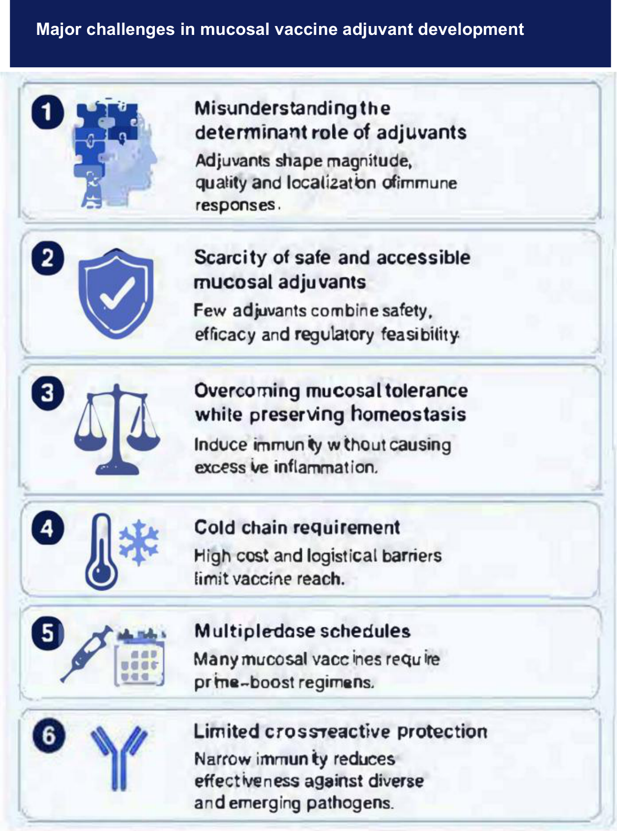
Figure 2.
Sources of mucosal adjuvant activity and innovative vaccination strategies. Exogenous adjuvants are added to the formulation, self-adjuvanting components are intrinsic to the delivery platform, and endogenous danger signals are released after vaccination. Pattern-recognition receptors on dendritic cells, macrophages, and epithelial cells sense these signals, promoting antigen presentation, cytokine production, and immune polarization. Innovative approaches include Bacillus subtilis OG101 spores displaying antigen on the spore surface, cochleates as multilamellar antigen-delivery particles rich in pathogen-associated molecular patterns, and SinTimVaS, a single-time vaccination strategy combining parenteral and mucosal priming. DAMPs, damage-associated molecular patterns; NLRs, NOD-like receptors; OMVs, outer-membrane vesicles; PAMPs, pathogen-associated molecular patterns; PRRs, pattern-recognition receptors; RLRs, RIG-I-like receptors; TLRs, Toll-like receptors; CLR, C-type lectin receptors; MPLA, monophosphoryl lipid A.
Figure 2.
Sources of mucosal adjuvant activity and innovative vaccination strategies. Exogenous adjuvants are added to the formulation, self-adjuvanting components are intrinsic to the delivery platform, and endogenous danger signals are released after vaccination. Pattern-recognition receptors on dendritic cells, macrophages, and epithelial cells sense these signals, promoting antigen presentation, cytokine production, and immune polarization. Innovative approaches include Bacillus subtilis OG101 spores displaying antigen on the spore surface, cochleates as multilamellar antigen-delivery particles rich in pathogen-associated molecular patterns, and SinTimVaS, a single-time vaccination strategy combining parenteral and mucosal priming. DAMPs, damage-associated molecular patterns; NLRs, NOD-like receptors; OMVs, outer-membrane vesicles; PAMPs, pathogen-associated molecular patterns; PRRs, pattern-recognition receptors; RLRs, RIG-I-like receptors; TLRs, Toll-like receptors; CLR, C-type lectin receptors; MPLA, monophosphoryl lipid A.
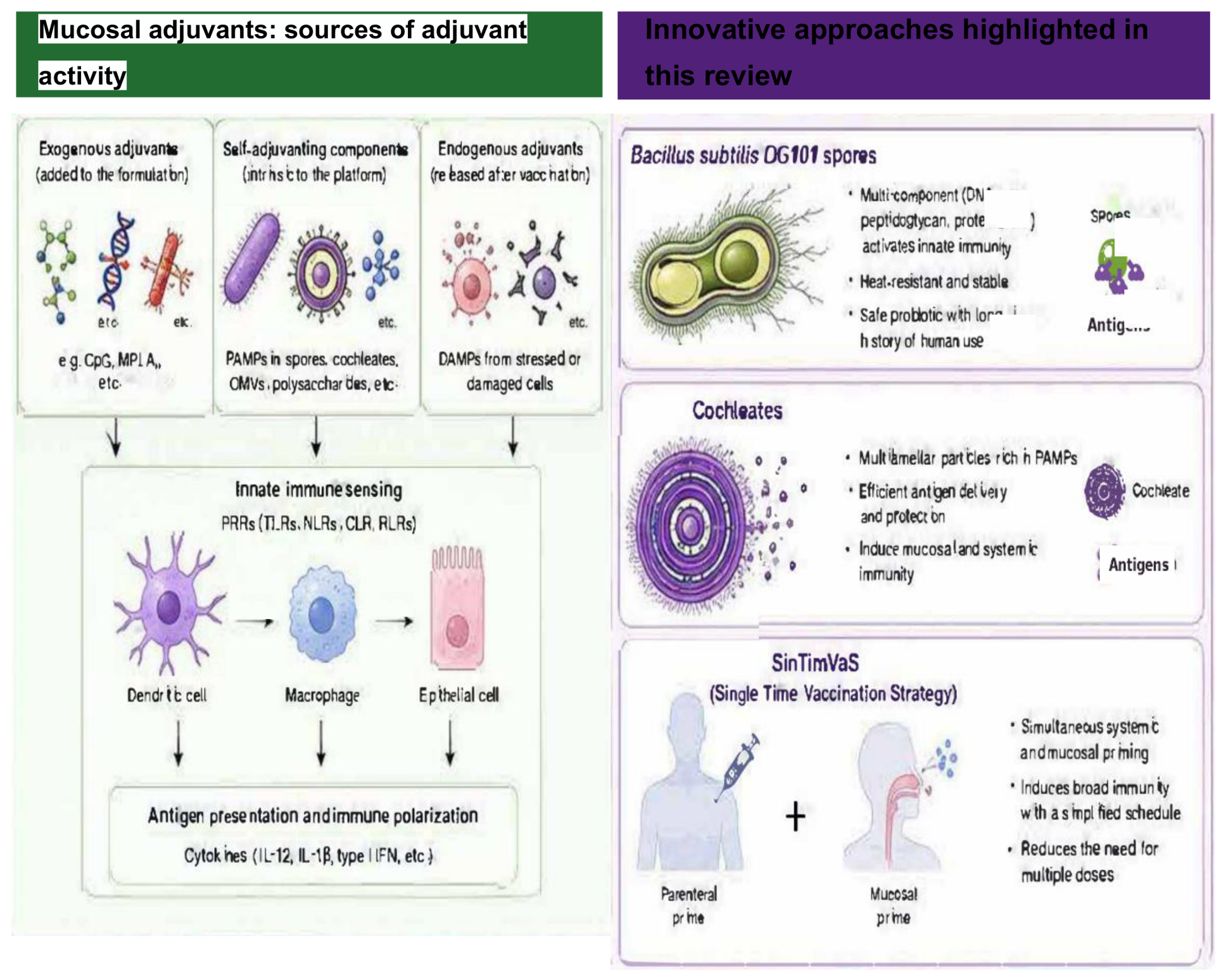
Figure 3.
Immune programming by mucosal adjuvants. Mucosal adjuvants promote innate immune activation, Th1/Tc1 polarization, B-cell help and class switching, mucosal secretory IgA and systemic IgG production, tissue-resident T-cell and B-cell memory, and broader cross-reactive protection. The sequence summarizes immune responses linking innate activation to durable mucosal and systemic immunity. AID, activation-induced cytidine deaminase; B-RM, memory B cells; IFN-γ, interferon gamma; IL-12, interleukin 12; sIgA, secretory IgA; T-RM, tissue-resident memory T cells; Th1, type 1 helper T cells; Tc1, type 1 cytotoxic T cells.
Figure 3.
Immune programming by mucosal adjuvants. Mucosal adjuvants promote innate immune activation, Th1/Tc1 polarization, B-cell help and class switching, mucosal secretory IgA and systemic IgG production, tissue-resident T-cell and B-cell memory, and broader cross-reactive protection. The sequence summarizes immune responses linking innate activation to durable mucosal and systemic immunity. AID, activation-induced cytidine deaminase; B-RM, memory B cells; IFN-γ, interferon gamma; IL-12, interleukin 12; sIgA, secretory IgA; T-RM, tissue-resident memory T cells; Th1, type 1 helper T cells; Tc1, type 1 cytotoxic T cells.
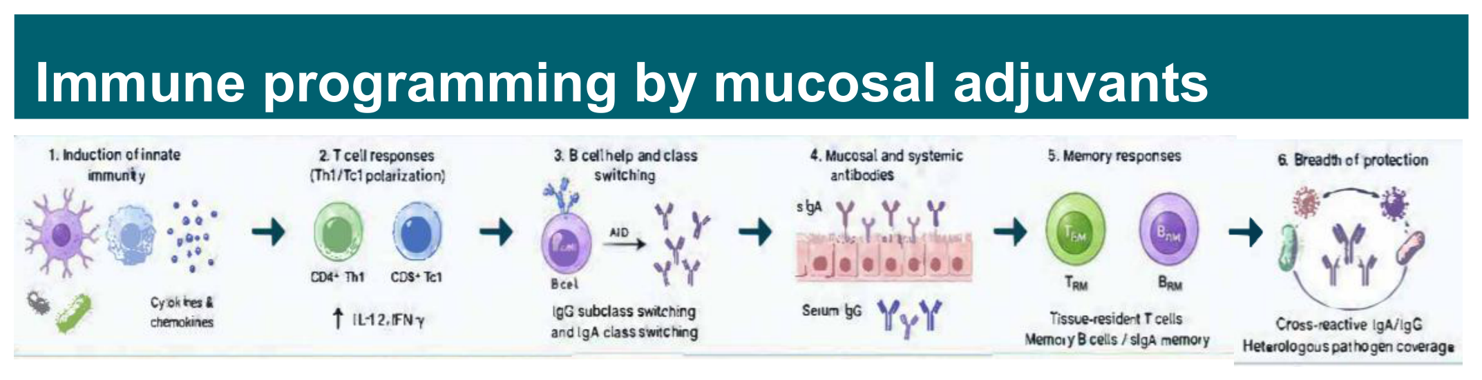
Figure 4.
Translational and practical advantages of mucosal vaccination approaches. Potential advantages include thermostability, simplified vaccination schedules, compatibility with nasal, oral, sublingual, vaginal, and pulmonary administration, broader access in low-resource settings, and stronger, broader, and longer-lasting protection involving secretory IgA, memory T and B cells, and cross-reactive immunity. sIgA, secretory IgA.
Figure 4.
Translational and practical advantages of mucosal vaccination approaches. Potential advantages include thermostability, simplified vaccination schedules, compatibility with nasal, oral, sublingual, vaginal, and pulmonary administration, broader access in low-resource settings, and stronger, broader, and longer-lasting protection involving secretory IgA, memory T and B cells, and cross-reactive immunity. sIgA, secretory IgA.
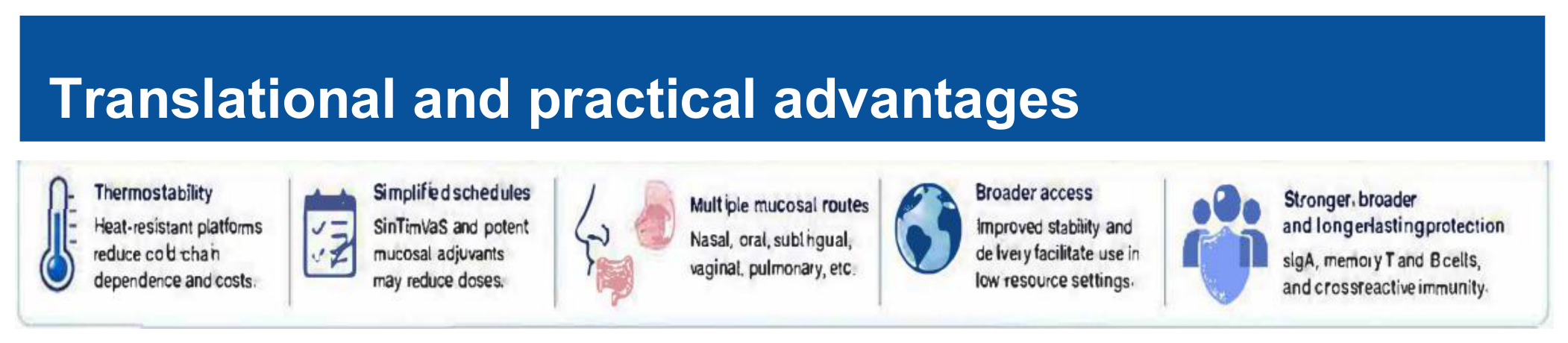
Table 1.
Licensed mucosal human vaccines References. Food and Drug Administration. Vaccines Licensed for Use in the United States. Available online: https://www.fda.gov/vaccines-blood-biologics/vaccines/vaccines-licensed-use-united-states (accessed on 19 August 2026); World Health Organization. Prequalified Vaccines. Available online: https://extranet.who.int/prequal/vaccines/prequalified-vaccines (accessed on 19 August 2026); Griesenauer RH, Kinch MS. An overview of FDA-approved vaccines & their innovators. Expert Rev. Vaccines 2017, 16 (12), 1253–1266; Tscherne A, Krammer F. A review of currently licensed mucosal COVID-19 vaccines. Vaccine 2025, 61, 127356; Cassimos DC, Effraimidou E, Medic S et al. Vaccination Programs for Adults in Europe, 2019. Vaccines 2020, 8, 34. https://doi.org/10.3390/vaccines8010034; Pattyn J, Del Riccio M, Bechini A, et al. The Adult Immunization Board (AIB): A new platform to provide multidisciplinary guidelines for the implementation and optimization of adult immunization in Europe. Vaccine 2024, 42, 1-3. https://doi.org/10.1016/j.vaccine.2023.11.060; Gräf DD, Westphal L, Kimmelman J, Hallgreen CE. Populations Addressed in Vaccines Approved via the European Medicines Agency. Clin. Pharmacol. Ther. 2025, 118, 459–469. doi:10.1002/cpt.3694; and Pezzoli L. Oral Cholera Vaccine Working Group of the Global Task Force on Cholera Control. Global oral cholera vaccine use, 2013-2018. Vaccine 2020, 38 Suppl 1, A132–A140..
Table 1.
Licensed mucosal human vaccines References. Food and Drug Administration. Vaccines Licensed for Use in the United States. Available online: https://www.fda.gov/vaccines-blood-biologics/vaccines/vaccines-licensed-use-united-states (accessed on 19 August 2026); World Health Organization. Prequalified Vaccines. Available online: https://extranet.who.int/prequal/vaccines/prequalified-vaccines (accessed on 19 August 2026); Griesenauer RH, Kinch MS. An overview of FDA-approved vaccines & their innovators. Expert Rev. Vaccines 2017, 16 (12), 1253–1266; Tscherne A, Krammer F. A review of currently licensed mucosal COVID-19 vaccines. Vaccine 2025, 61, 127356; Cassimos DC, Effraimidou E, Medic S et al. Vaccination Programs for Adults in Europe, 2019. Vaccines 2020, 8, 34. https://doi.org/10.3390/vaccines8010034; Pattyn J, Del Riccio M, Bechini A, et al. The Adult Immunization Board (AIB): A new platform to provide multidisciplinary guidelines for the implementation and optimization of adult immunization in Europe. Vaccine 2024, 42, 1-3. https://doi.org/10.1016/j.vaccine.2023.11.060; Gräf DD, Westphal L, Kimmelman J, Hallgreen CE. Populations Addressed in Vaccines Approved via the European Medicines Agency. Clin. Pharmacol. Ther. 2025, 118, 459–469. doi:10.1002/cpt.3694; and Pezzoli L. Oral Cholera Vaccine Working Group of the Global Task Force on Cholera Control. Global oral cholera vaccine use, 2013-2018. Vaccine 2020, 38 Suppl 1, A132–A140..
| Company | Vaccine (s) | Target | Adjuvants (Exogenous) | Route | Status | |
|---|---|---|---|---|---|---|
| MedImmune-acquired by AstraZeneca | FluMist. Live attenuated virus |
Influenza virus subtypes A and type B (quadrivalent) | None | Intranasal, spray into the nose | FDA Approved | |
| Serum Institute of India | NASOVAC Live attenuated (Human) Freeze-Dried | Influenza virus HiN1. Propagated in Embryonated hen eggs (SPF) | None | Intranasal vial plus ampoule | WHO prequalified | |
| Nasovac. Live attenuated virus | Influenza virus subtypes A and type B (trivalent, version S or quadrivalent version S4) | None | Intranasal, spray into the nose | WHO prequalified | ||
| AstraZeneca | Fluenz, Live attenuated virus | Influenza virus two subtypes A-H1N1 and A-H3N2 and one type B virus (Victoria lineage), in accordance with annual flu season | None | Intranasal, spray into the nose | EMA Approved | |
| Pandemic LAIV. Live attenuated virus | Influenza H5N1 | None | Intranasal, spray into the nose | WHO prequalified | ||
| GSK Biologics, Belgium | Rotarix (Presentation-plastic tube)/ Rotarix (Presentation-applicator). Live attenuated virus | Rotavirus gastroenteritis caused by G1 and non-G1 serotypes | None | Oral | WHO prequalified | |
| Merck & Co., Inc., USA | RotaTeq (Presentation-Plastic tube) . Live attenuated virus | Pentavalent rotavirus vaccine to protect against G1, G2, G3, G4, and G9 serotypes | None | Oral | FDA approved and WHO prequalified | |
| Bharat Biotech, India | ROTAVAC (Presentation-Vial/ ROTAVAC-5D (Presentation-Vial). Live attenuated virus | Severe rotavirus gastroenteritis. Monovalent and pentavalent (5D) | None | Oral | WHO prequalified | |
| Serum Institute of India | ROTASIIL (Presentation- Two vial set)/ ROTASIIL-Liquid (Presentation-Vial-plastic tube)/ ROTASIIL-Thermo(Presentation-Two vial set). Live attenuated virus | Pentavalent rotavirus vaccine, strains from human-bovine rearrangements with the G1, G2, G3, G4 and G9 serotypes | None | Oral | WHO prequalified | |
| Bavarian Nordic Inc, USA | Vivotif. Live attenuated bacteria | Salmonella typhi bacteria (specifically the Ty21a strain) | None | Oral | FDA approved and WHO prequalified | |
| Bharat Biotech International Limited | BIOPOLIO B1/3 | Polioviruses Bivalent Types 1 and 3 | None | Oral | WHO prequalified | |
| Bharat Biotech International Limited | BIOPOLIO | Polioviruses Trivalent | None | Oral | WHO prequalified | |
| Panacea Biotec Ltd. | OPV. Live attenuated virus | PoliovIruses Bivalent Types 1 and 3 | None | Oral | WHO prequalified | |
| PT Bio Farma (Persero) | Bivalent OPV Type 1 and 3 Poliomyelitis Vaccine/Polio Vaccine - Novel Oral (nOPV) Type 2 | OPV Bivalent Types 1 and 3 | None | Oral | WHO prequalified | |
| Haffkine Bio Pharmaceutical Corporation Ltd | Bivalent type 1&3 Oral Poliomyelitis vaccine, IP (bOPV1&3)/Polioviral vaccine | OPV Bivalent Types 1 and 3/ Trivalent | None | Oral | WHO prequalified | |
| GlaxoSmithKline Biologicals SA | Polio Sabin Mono T1/Polio Sabin Mono Three/Polio Sabin Mono Two/Polio Sabin One and Three (different formulations) | OPV Monovalent Type 1/Monovalent type 3/Monovalent type 2/Bivalent types 1 and 3 | None | Oral | WHO prequalified | |
| Biological E. Limited | Poliomyelitis Vaccine - Novel Oral (nOPV) Type 2 (Biological E. bulk | Novel Oral (nOPV) Type 2 | None | Oral | WHO prequalified | |
| Beijing Institute of Biological Products Co., Ltd. | Poliomyelitis Vaccine (live, oral attenuated, human Diploid Cell), type 1 and 3 | OPV Bivalent Types 1 and 3 | None | Oral | WHO prequalified | |
| Serum Institute of India Pvt. Ltd. | Poliomyelitis Vaccine (Oral) Bivalent Type 1 and 3 (Bilthoven Biologicals B.V. Bulk)/Poliomyelitis Vaccine (Oral), Bivalent types 1 and 3 (PT Bio Farma Bulk) | OPV Bivalent Types 1 and 3 | None | Oral | WHO prequalified | |
| Bavarian Nordic Inc, USA | Vaxchora, Live attenuated bacteria | Vibrio cholerae serogroup O1 bacteria | None | Oral | FDA approved | |
| EuBiologics Co., Ltd. | Euvichol killed whole-cell vaccine | V. cholerae O1 and O139 bivalent inactivated vaccine. V. cholerae O1 Inaba Cairo 48 (Heat) V. cholerae O1 Inaba Phil 6973 El Tor (Formalin) V. cholerae O1 Ogawa Cairo 50 (Formalin) V. cholerae O1 Ogawa Cairo 50(Heat) V. cholerae O139 4260B (Formalin) (Presentation-Vial) |
None | Oral | WHO prequalified | |
| EuBiologics Co., Ltd. | Euvichol-Plus killed whole-cell vaccine | V. cholerae O1 and O139 bivalent inactivated vaccine V. cholerae O1 Inaba Cairo 48 (Heat) V. cholerae O1 Inaba Phil 6973 El Tor (Formalin) V. choleraeO1 Ogawa Cairo 50 (Formalin) V. cholerae O1 Ogawa Cairo 50(Heat) V. cholerae O139 4260B (Formalin) (Presentation-Plastic Tube) |
None | Oral | WHO prequalified | |
| EuBiologics Co., Ltd. | Euvichol-S killed whole-cell vaccine | V. cholerae O1 Inaba Phil 6973 El Tor (Formalin) V. cholerae O1 Ogawa Cairo 50 (Formalin) (Simplified formulation- Presentation-plastic tube) |
None | Oral | WHO prequalified | |
| GCBC Vaccines Pvt. Ltd, India/ Euvichol (EuBiologics Co., Ltd, Republic of Korea | Shanchol Inactivated bacteria | Inactivated Vibrio cholerae O1 (classical & El Tor biotypes, Inaba & Ogawa serotypes) and O139 strain | None | Oral | WHO prequalified | |
| Barr Laboratories, Inc, USA | Adenovirus 4 & 7 vaccine.Live unattenuated virus | Febrile acute respiratory disease (ARD) caused by adenovirus types 4 and 7 | None | Oral | WHO prequalified | |
| Partnership between Washington University in St. Louis (USA), and Bharat Biotech International Limited (India) | iNCOVACC (BBV154) non-replicating chimpanzee adenovirus-vectored vaccine (ChAd36) encoding a prefusion-stabilized S protein with two proline substitutions in S2 | COVID-19 | None | Intranasal | Approved in India | |
| CanSino Biologics, China | Ad5-nCoV-IH/ Convidecia Air®. Replication- deficient hAd5 expressing full-length S in its native form | COVID-19 | None | Intranasal. Nebulizer. Aerogen’s proprietary aerosol drug delivery technology | Approved in China | |
| Gamaleya Research Institute of Epidemiology and Microbiology, Health Ministry of the Russian Federation | Salnavac Sputnik . Adenovirus viral vector platform | COVID-19 | None | Intranasal (nasal Spray) | Approved in Russia | |
| Razi Vaccine and Serum Research Institute | RAZI-COV PARS. Recombinant spike protein | COVID-19 | RAS-01. Razi adjuvant system-01 novel plant-derived oil-in-water emulsion adjuvant system | Intranasal | Approved in India | |
| Beijing Wantai Biological Pharmacy Enterprise | dNS1-RBD, Pneucolin (live-attenuated influenza virus vector-expressing RBD of the S protein) | COVID-19 | None | Intranasal | Approved in China for emergency use |
Disclaimer/Publisher’s Note: The statements, opinions and data contained in all publications are solely those of the individual author(s) and contributor(s) and not of MDPI and/or the editor(s). MDPI and/or the editor(s) disclaim responsibility for any injury to people or property resulting from any ideas, methods, instructions or products referred to in the content. |
© 2026 by the authors. Licensee MDPI, Basel, Switzerland. This article is an open access article distributed under the terms and conditions of the Creative Commons Attribution (CC BY) license.
Copyright: This open access article is published under a Creative Commons CC BY 4.0 license, which permit the free download, distribution, and reuse, provided that the author and preprint are cited in any reuse.