

Submitted:
28 October 2026
Posted:
30 October 2026
You are already at the latest version
Abstract
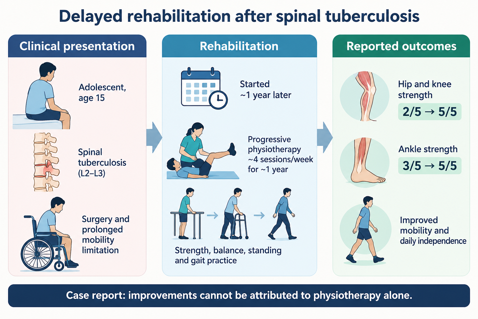
Spinal tuberculosis can cause vertebral destruction, neurological impairment, prolonged immobility and major loss of independence. We describe a 15-year-old male whose retained case documentation reported spinal tuberculosis involving the L2-L3 vertebral levels, followed by spinal surgery with internal fixation. Early rehabilitation was delayed and a short initial course of physiotherapy was discontinued. Approximately one year later, he presented to a specialist rehabilitation service with pain, restricted joint motion, marked lower-limb weakness, impaired bed mobility, poor balance and inability to walk independently. He then completed a progressive physiotherapy programme approximately four times weekly for about one year. Management advanced from symptom control, stretching, isometric exercise, bed-mobility practice and supported standing to isotonic strengthening, postural control, balance and coordination training, stepping and gait re-education. Lower-limb manual muscle testing improved from grade 2/5 to 5/5 at the hips and knees and from 3/5 to 5/5 at the ankles. Selected recorded functional items also showed marked improvement in self-care, transfers and locomotion. At later follow-up, the patient reported independent daily activities and no perceived residual disability. Because original imaging, operative, medication and standardized neurological records could not be retrieved, the exact neurological classification and contribution of each treatment component cannot be established. This case illustrates the potential for substantial functional recovery despite delayed rehabilitation and the importance of prospective standardized documentation.
Keywords:
spinal tuberculosis
; Pott disease
; rehabilitation
; physiotherapy
; gait training
; adolescent
Key Clinical Message
Delayed rehabilitation after spinal tuberculosis can still be followed by meaningful functional recovery. This case supports progressive, goal-oriented strengthening, balance, weight-bearing and gait training, while underscoring the need to document neurological classification and standardized outcomes early because missing source records limit later interpretation.
1. Introduction
Spinal tuberculosis (Pott disease) is an extrapulmonary manifestation of tuberculosis that can affect vertebral structures and lead to pain, deformity, neurological deficit, prolonged immobility and reduced functional independence. Functional outcomes are influenced by the severity and duration of neurological impairment, spinal deformity, timing of treatment and access to rehabilitation.1,2
Rehabilitation is an important component of multidisciplinary management after the medical and surgical stabilization of spinal tuberculosis. Recent literature describes individualized programmes that may include range-of-motion exercise, progressive strengthening, postural and balance training, weight-bearing, gait practice and appropriate assistive devices.1 However, detailed reports of prolonged rehabilitation after a substantial delay remain limited, particularly when the main outcome is recovery of day-to-day function.
This case report describes an adolescent with severe mobility limitation after surgically managed spinal tuberculosis who entered a structured rehabilitation programme approximately one year after the initial illness. The report focuses on the rehabilitation progression, functional changes, barriers to participation and lessons arising from incomplete retrospective clinical documentation.
2. Case Presentation
2.1. Patient Information and Initial Presentation
A 15-year-old male student developed sudden back pain that worsened over the following days and was accompanied by low-grade fever and night sweats. According to the retained case documentation, his ability to stand deteriorated rapidly and his family subsequently observed that he could no longer move his lower limbs during transfer for hospital assessment.
The original case record states that radiography suggested spinal tuberculosis at the L2-L3 vertebral levels and that magnetic resonance imaging supported the diagnosis. The original radiology report and images are no longer available to the authors. The retained documentation also does not contain microbiological or histopathological confirmation, inflammatory markers, or the original antitubercular medication record.
Spinal surgery with internal fixation was performed the following day. The exact operative procedure, levels of decompression or fusion, and postoperative precautions could not be verified because the operative and discharge records were unavailable at the time of manuscript preparation. The patient was discharged after seven days of postoperative observation with medical treatment and advice to mobilize and obtain physiotherapy.
2.2. Rehabilitation History Before Specialist Assessment
Formal rehabilitation was delayed during the early postoperative period because of wound discomfort, fear of movement and family concern. A physiotherapist was later consulted and provided a brief course of passive stretching and lower-limb strengthening, but this treatment stopped after approximately three weeks. Thereafter, the patient remained largely inactive and became increasingly dependent for mobility.
Approximately one year after the initial illness, he attended a specialist rehabilitation service in Bangladesh. Assessment was completed over several sessions because of multiple post-surgical functional problems. He required full caregiver assistance for rolling and maximum assistance for moving from lying to sitting. He could maintain sitting but used a slouched posture. Independent standing and walking were not possible.
2.3. Clinical Findings at Rehabilitation Presentation
Documented problems included pain around both elbows, hips and knees; reduced range of motion at the shoulders, elbows, hips and knees; reduced muscle power in the upper and lower limbs; impaired dynamic sitting balance; impaired static and dynamic standing balance; and absent independent gait. Muscle tone was recorded as normal. Light touch and pin-prick sensation were recorded as intact, proprioception as good, and selective movement as fair. Lower-limb coordination was also recorded as fair.
The source manuscript labelled the patient as having ‘incomplete tetraplegia, ASIA-C’. Because the original International Standards for Neurological Classification of Spinal Cord Injury (ISNCSCI) worksheet and sacral examination findings could not be retrieved, this classification cannot be independently verified and is not reproduced as a definitive diagnosis in this report. Current ISNCSCI standards require a standardized neurological examination to determine neurological level and AIS grade.5
2.4. Clinical Timeline

Table 1.
Clinical timeline from retained case documentation.
| Approximate time | Clinical event |
| Initial illness | Back pain followed by low-grade fever, night sweats and rapid loss of standing/lower-limb movement. |
| Hospital assessment | Radiography and MRI were reported in the retained case record to support spinal tuberculosis involving L2-L3. |
| Following day | Spinal surgery with internal fixation; seven days of postoperative observation. |
| First postoperative weeks | Rehabilitation delayed; later a short course of stretching and resisted lower-limb exercise was undertaken and then discontinued. |
| ~1 year after initial illness | Presentation to specialist rehabilitation with severe mobility dependence, joint restriction, weakness, impaired balance and absent independent gait. |
| Following ~1 year | Structured physiotherapy approximately four sessions/week with progressive mobility, strengthening, balance, weight-bearing and gait training. |
| Later follow-up | Independent daily activities were reported; the patient later reported a healthy lifestyle without perceived residual disability. |
2.5. Therapeutic Intervention
The principal rehabilitation goals were to reduce pain and joint restriction, improve bed mobility and postural control, increase muscle strength, restore tolerance to weight-bearing, improve balance and re-establish gait. The patient and caregiver were educated about the functional problems, expected rehabilitation course, the importance of regular participation and the need for gradual progression.
Treatment was delivered approximately four times per week for about one year. Early sessions emphasized symptom management, sustained stretching, isometric exercise, bed-mobility practice, selective movement, bridging and supported standing. The programme then progressed to isotonic strengthening, reaching tasks, ball activities, four-point and kneeling positions, postural control, supported sit-to-stand, stepping, balance-board activities, half-squatting, eccentric exercise and gait re-education. Support was gradually reduced as strength and balance improved. A Taylor brace was used during part of the gait rehabilitation.
Pain after exercise, fear of falling and caregiver concern initially limited participation and home practice. Rehabilitation intensity and assistance were advanced according to tolerance and functional change.
Table 2.
Approximate phases of the rehabilitation programme.
| Phase | Main goals | Key interventions |
| Weeks 1-4 | Pain, range of motion, bed mobility, early stability | Massage/manual symptom management, sustained stretching, isometric exercise, selective movement, bridging, supported standing. |
| Months 2-3 | Dynamic balance, postural control, pelvic/lower-limb strength | Reaching and ball tasks, four-point and half-kneeling, isotonic hip exercise, supported standing, resisted lower-limb strengthening. |
| Months 4-6 | Trunk control, transfers, progressive lower-limb strength | Trunk mobility/control, kneeling activities, sit-to-stand with support, supported standing, progressive hip/knee/ankle strengthening. |
| Months 7-9 | Balance, joint stability, gait initiation | Stepping, balance board, half-squatting, eccentric exercise, parallel-bar gait training, static and dynamic stretching. |
| Final months | Functional independence, coordination and gait progression | Reduced support during squatting/stepping, dynamic balance, selective movement and supervised gait training. |
2.6. Follow-Up and Outcomes
Reassessment documented improvement in selective movement, lower-limb coordination, joint range of motion, muscle strength and functional independence. Manual muscle testing of bilateral hip flexors, extensors, adductors and abductors improved from grade 2/5 to 5/5. Knee flexors and extensors improved from 2/5 to 5/5, and ankle flexors and extensors from 3/5 to 5/5. Upper-limb elbow, wrist and finger muscle groups, which had been recorded as grade 4/5, improved to 5/5.
Range of motion was recorded qualitatively rather than in degrees. Shoulder motion progressed from minimal loss to full range. Elbow active motion improved from major loss to minimal loss and passive motion to full range. Hip and knee motion also improved substantially, with full or near-full range documented at final reassessment.
The retained case document contained 23 item-level functional ratings scored from 1 to 7. Their labels correspond to selected domains of the Functional Independence Measure plus Functional Assessment Measure (FIM+FAM), which is a 30-item instrument.9 Because the original standardized assessment form was unavailable and seven items were not retained in the case document, no official FIM or FIM+FAM total score was calculated. The available scores are reported only as descriptive evidence of change.
Table 3.
Descriptive summary of retained functional item ratings.
| Recorded domain | Items retained | Pre-rehabilitation | Post-rehabilitation |
| Self-care | 7 | 29/49 | 49/49 |
| Mobility/transfers | 4 | 7/28 | 26/28 |
| Locomotion | 3 | 3/21 | 16/21 |
| Psychosocial adjustment | 4 | 21/28 | 27/28 |
| Cognitive function | 5 | 35/35 | 35/35 |
Table note: These are descriptive sums of only the item-level scores retained in the source case document. They are not validated FIM or FIM+FAM total scores and should not be interpreted as such.
Functionally, the patient progressed from full caregiver dependence for basic bed mobility and absent independent gait to supported standing, stepping, parallel-bar walking and later supervised gait. By the end of rehabilitation, he was reported to perform activities of daily living independently. At later contact, he reported living a healthy lifestyle without perceived residual disability. This later status was self-reported and was not confirmed by a standardized face-to-face examination.
3. Discussion
This case demonstrates substantial improvement in strength, mobility and functional independence during prolonged rehabilitation that began after a major delay following surgically managed spinal tuberculosis. The main teaching point is not that physiotherapy alone caused recovery, but that delayed presentation to rehabilitation should not automatically be viewed as a barrier to a structured, progressive functional programme.
Spinal tuberculosis may lead to neurological deficit and long-term functional limitation, and rehabilitation outcomes depend on both disease-related and treatment-related factors.2 A recent review of rehabilitation management in spinal tuberculosis describes range-of-motion exercise, strengthening, postural correction, balance training, walking practice and assistive devices as components of individualized rehabilitation.1 The progression used in this case broadly reflects those principles.
Task-specific mobility and gait practice may also be relevant when neurological deficits are incomplete. Locomotor training studies in incomplete spinal cord injury have shown that appropriately dosed stepping practice can improve aspects of walking performance, although those findings cannot be directly generalized to spinal tuberculosis.7 The gradual progression from supported standing to stepping and gait training in this case was clinically consistent with that rehabilitation rationale.
Fear of falling was a meaningful barrier during the early rehabilitation period. In ambulatory individuals with spinal cord injury, fear of falling has been associated with recurrent and injurious falls.6 Although the present patient was initially non-ambulatory, this evidence supports the clinical importance of addressing confidence and graded exposure to standing and walking tasks during recovery.
3.1. Strengths and Limitations
A strength of the case is the long rehabilitation follow-up and the retained serial functional data showing change across strength, mobility and independence. The rehabilitation progression is also described in enough detail to illustrate how support and task complexity were advanced over time.
Several limitations substantially affect interpretation. First, the original MRI report, radiographs, operative report, microbiological or histopathological confirmation, antitubercular regimen and detailed discharge records could not be retrieved. The diagnosis and treatment chronology are therefore reported from the retained case documentation rather than reverified from primary source records. Second, the original ISNCSCI worksheet was unavailable, so the neurological level and AIS grade cannot be confirmed. The earlier description of ‘incomplete tetraplegia, ASIA-C’ is therefore not treated as a definitive classification. Third, the functional score table contained only 23 item-level ratings corresponding to selected FIM+FAM domains; reporting these as a complete FIM score would be inappropriate. Fourth, joint range of motion was documented qualitatively rather than with goniometric values. Finally, surgery, antitubercular treatment, spontaneous neurological recovery, prolonged time since onset and rehabilitation may all have contributed to the observed outcome, so causal attribution to physiotherapy alone is not possible.
These limitations also provide a practical lesson for similar cases: neurological classification, imaging findings, surgical details, medication regimens and standardized functional outcomes should be documented prospectively and retained, particularly when a rare or complex rehabilitation course may later be reported.
4. Conclusion
Meaningful functional recovery was documented during a prolonged, progressive rehabilitation programme in an adolescent who began specialist rehabilitation approximately one year after surgically managed spinal tuberculosis. Delayed rehabilitation should not preclude goal-oriented mobility, strengthening, balance and gait training, but accurate prospective neurological and outcome documentation is essential for interpreting recovery and communicating clinical lessons.
Author Contributions
Md. Shohag Rana: Conceptualization, Methodology, Investigation, Supervision, Writing - original draft, Writing - review & editing. Md. Nesaruddin: Methodology, Investigation, Data curation, Validation, Writing - review & editing. Farhin Islam Peya: Investigation, Data curation, Validation, Writing - review & editing. Fyzul Kabir: Investigation, Data curation, Formal analysis, Writing - review & editing. Shahnur Zinia Parvin: Data curation, Formal analysis, Validation, Writing - review & editing. Suprantha Sutra Dhar: Methodology, Validation, Supervision, Writing - review & editing. Md. Asadul Islam: Investigation, Resources, Data curation, Writing - review & editing. Polok Halder: Data curation, Formal analysis, Visualization, Project administration, Writing - original draft, Writing - review & editing.
Funding
The authors received no specific funding for this work.
Ethics Approval
Ethical approval was granted by the Institutional Review Board of the Institute of Physiotherapy, Rehabilitation & Research (IPRR), the academic institute of the Bangladesh Physiotherapy Association (BPA), reference BPA-IPRR/IRB/16/03/2022/1652, approval letter dated 12 June 2022. The IRB reviewed and approved the case proposal and the participant information sheet and consent form and approved the research to be conducted in its presented form. The approval letter states that the committee operates in accordance with the Declaration of Helsinki, ICH-GCP and applicable local requirements.
Data Availability Statement
The clinical information supporting this case report is contained within the article. Additional patient-level records are not publicly available because of privacy and confidentiality considerations.
Informed Consent for Publication
Written informed consent for publication of this case report was obtained from the patient.
Acknowledgments
The authors thank the staff of the Centre for the Rehabilitation of the Paralysed (CRP) and the patient for their assistance and cooperation.
Conflicts of Interest
The authors declare no conflicts of interest.
Use of AI-Assisted Tools
OpenAI ChatGPT was used to assist with language refinement, organization, and formatting of submission materials. The clinical information and reported observations are based on author-provided case documentation, not AI-generated patient data. The authors remain responsible for reviewing and verifying the submitted manuscript and its scientific content.
References
- Dhouibi, J.; Kalai, A.; Chaabeni, A.; Aissa, A.; Ben Salah Frih, Z.; Jellad, A. Rehabilitation management of patients with spinal tuberculosis (Review). Med. Int. 2024, 4(3), 28. [Google Scholar] [CrossRef] [PubMed]
- Yong, L.N.; Ahmedy, F.; Yin, K.N.; Engkasan, J.P. Functional outcomes in spinal tuberculosis: a review of the literature. Asian Spine J. 2021, 15(3), 381–391. [Google Scholar] [CrossRef] [PubMed]
- Zaoui, A.; Kanoun, S.; Boughamoura, H.; et al. Patients with complicated Pott’s disease: management in a rehabilitation department and functional prognosis. Ann. Phys. Rehabil. Med. 2012, 55(3), 190–200. [Google Scholar] [CrossRef] [PubMed]
- Selfe, J.; Mbada, C.; Kaka, B.; et al. Red flags for spinal pain in patients diagnosed with spinal infection in Nigeria: a 10-year medical records review. Musculoskelet. Sci. Pract. 2022, 60, 102571. [Google Scholar] [CrossRef] [PubMed]
- Rupp, R.; Biering-Sorensen, F.; Burns, S.P.; et al. International Standards for Neurological Classification of Spinal Cord Injury: Revised 2019. Top. Spinal Cord. Inj. Rehabil. 2021, 27(2), 1–22. [Google Scholar] [CrossRef] [PubMed]
- Jorgensen, V.; Butler Forslund, E.; Opheim, A.; et al. Falls and fear of falling predict future falls and related injuries in ambulatory individuals with spinal cord injury: a longitudinal observational study. J. Physiother. 2017, 63(2), 108–113. [Google Scholar] [CrossRef] [PubMed]
- Leech, K.A.; Kinnaird, C.R.; Holleran, C.L.; Kahn, J.; Hornby, T.G. Effects of locomotor exercise intensity on gait performance in individuals with incomplete spinal cord injury. Phys. Ther. 2016, 96(12), 1919–1929. [Google Scholar] [CrossRef] [PubMed]
- Yu, P.; Zhang, W.; Liu, Y.; Sheng, C.; So, K.F.; Zhou, L.; Zhu, H. The effects and potential mechanisms of locomotor training on improvements of functional recovery after spinal cord injury. Int. Rev. Neurobiol. 2019, 147, 199–217. [Google Scholar] [CrossRef] [PubMed]
- Hall, K.M.; Hamilton, B.B.; Gordon, W.A.; Zasler, N.D. Characteristics and comparisons of functional assessment indices: Disability Rating Scale, Functional Independence Measure, and Functional Assessment Measure. J. Head Trauma Rehabil. 1993, 8(2), 60–74. [Google Scholar] [CrossRef]
- Turgut, M. Spinal tuberculosis (Pott’s disease): its clinical presentation, surgical management, and outcome. A survey study on 694 patients. Neurosurg. Rev. 2001, 24(1), 8–13. [Google Scholar] [CrossRef] [PubMed]
Disclaimer/Publisher’s Note: The statements, opinions and data contained in all publications are solely those of the individual author(s) and contributor(s) and not of MDPI and/or the editor(s). MDPI and/or the editor(s) disclaim responsibility for any injury to people or property resulting from any ideas, methods, instructions or products referred to in the content. |
© 2026 by the authors. Licensee MDPI, Basel, Switzerland. This article is an open access article distributed under the terms and conditions of the Creative Commons Attribution (CC BY) license (http://creativecommons.org/licenses/by/4.0/).
Copyright: This open access article is published under a Creative Commons CC BY 4.0 license, which permit the free download, distribution, and reuse, provided that the author and preprint are cited in any reuse.