

Submitted:
28 October 2026
Posted:
29 October 2026
You are already at the latest version
Abstract
Protein glycosylation alterations in cancer have been recognized for their potential as a diagnostic tool. This review aims to explore the structural diversity and functional significance of glycans, as well as the relationship between aberrant glycosylation and cancer. Currently used glycomic biomarkers and their translational features in selected types of cancer are described in detail. The potential of liquid biopsy glycomics as an oncological tool is discussed, presenting different analytical strategies for glycan analysis, including total serum N-glycan analysis, O-glycosylation, and proteoglycan analysis. These strategies utilize chromatographic methods and high-resolution mass spectrometry, offering high precision and useful information. Other approaches include in situ fluorescence imaging, as well as lectin- and antibody-based methods for the detection of specific glycoforms, which have gained attention for their relative simplicity in use and clinical interpretation. With the rapid development of novel technologies for glycan detection and characterization, as well as bioinformatics tools and databases, it is expected that exploring the molecular mechanisms of glycosylation in cancer will become more accessible in everyday clinical implementation. Integrating knowledge of altered glycosylation patterns, proteomics, genomics, and metabolomics (and other omics approaches) with clinical data has potential to contribute to a more comprehensive biomolecular characterization of malignant diseases.
Keywords:
cancer
; extracellular vesicles
; glycans
; glycosylation
; omics
1. Introduction
Over a decade ago, the United States National Academy recognized the direct role of glycans “in the pathophysiology of every major disease”[1] and cancer is no exception. Because glycans are both structurally diverse and biologically informative, cancer-associated glycosylation signatures have emerged as promising biomarkers with translational relevance in selected tumor types and clinical settings. However, despite the fact that aberrant protein glycosylation in cancer has attracted the interest of the scientific and clinical community since the 1950s [2,3] they are yet to reach their full clinical potential. In recent years, the potential of extracellular vesicle glycomics as an oncological tool has also led to new perspectives of glycomics in oncological diagnostics and treatment. This manuscript aims to provide a comprehensive overview of protein glycosylation alterations in cancer and their potential as a diagnostic tool in oncology.
2. Glycan Biosynthetic Pathways
Glycosylation is an enzymatic process that forms glycoconjugates, in which glycans (carbohydrate chains) are covalently attached to various biomolecules, including proteins, lipids, or RNA [4,5]. Unlike DNA, RNA, and protein biosynthesis, which follow a template, glycan biosynthesis results from complex interactions between the activity of glycosylation enzymes and the availability of the corresponding monosaccharide donors and acceptors [6]. Among the major protein-bound glycan types in eukaryotic cells are N-linked glycans (N-glycans), O-linked glycans (O-glycans) and glycosaminoglycans (GAGs) [4]. N-glycosylation is initiated in the endoplasmic reticulum (ER) where the universal N-glycan precursor is attached to the side chain of an asparagine (Asn) residue within the Asn-X-Serine (Ser)/threonine (Thr) structural motif, with X being any amino acid but proline [7]. Further structural processing in the ER and Golgi complex diverts the nascent N-glycan into one of the following glycan types depending on the processing pathway that is taken: pauci-mannosidic (less than five Man residues), oligo-mannosidic (five or more Man residues), complex (in which both the 3- and 6-arms of the core are elongated with antennae, each starting with N-acetylglucosamine (GlcNAc)), and hybrid (with Man residues attached on the 6-arm of the core and one or two antennae elongating the 3-arm). Their common structural core consists of two N-acetylglucosamines (GlcNAc) and a mannose (Man), which is further extended by two Man residues at the 3 and 6 positions. Antennary galactosylation and sialylation, fucosylation (core/antennary), or the presence of the bisecting GlcNAc on the central Man residue within the core additionally contribute to the structural complexity [7]. O-glycosylation takes place in the Golgi complex. Usually, the process starts with the enzymatic attachment of N-acetylgalactosamine (GalNAc) to the hydroxyl group of a Ser or Thr residue. After further extension into one of four core structures and additional structural upgrades, the nascent glycan can become a mature linear or branched O-glycan [8]. Glycan branching exponentially increases the number of isomers, thus explaining the enormous number of structural variants that can be assembled from a small number of monosaccharide building units [6]. GAGs are long, linear, polysaccharides built from repeating disaccharide units. The main Mammalian classes include heparan sulfate, chondroitin sulfate, dermatan sulfate, and keratan sulfate [9]. Proteins decorated with GAG chains are known as proteoglycans and they are found at the cell surface, in secretory granules, and throughout pericellular and extracellular matrices [10]. Through their sulfation patterns, chain length, and presentation on proteoglycan core proteins, GAGs contribute to extracellular matrix organization and regulate interactions with growth factors, cytokines, chemokines, adhesion molecules, and other proteins [9,11].
3. Biological Role of Glycoproteins
Glycoproteins comprise more than 85% of secreted proteins [12] and, given their widespread presence and complexity, glycans play a wide range of biological roles. Regardless of whether they directly impact glycoprotein structure and function, interact with numerous glycan-binding proteins (GBPs), or use both mechanisms, glycans are indispensable in the physiology of multicellular organisms [4]. They participate in cellular activation and signaling, embryonic development, organogenesis, differentiation, regulatory mechanisms, cell–cell and cell–matrix interactions. In addition, numerous structural functions (like tissue elasticity, interstitial fluid homeostasis, or epithelial barrier integrity) depend on glycans. Furthermore, glycans are essential in molecular host-microorganism interactions, self-recognition, and immune mechanisms [13]. Besides, glycans mediate and regulate important physiological processes in all organ systems. For example, in the mouse respiratory system, O-glycosylated mucins create protective barriers in the airways, while loss of core fucose from N-glycans impairs lung tissue homeostasis, leading to emphysema-like changes driven by dysregulated matrix metalloproteinase expression. Alterations in O-glycome composition influence insulin effects, while glucose transporters’ functionalities in diabetic patients depend on N-glycans’ changes. Sialic acid residues are necessary for glomerular filtration, while in the nervous system, they can modulate neuronal plasticity and post-injury regeneration potential [14].
The composition of the glycome (the set of glycans present on a protein, in a cell, tissue, or entire organism) is dynamic and reflects the influence of genetic, epigenetic, and environmental factors [15]. Thus, it comes as no surprise that considerable changes in the glycome occur in cancer, a condition marked by extensive alterations in gene and protein expression, cell morphology, and signaling. Glycosylation patterns also might vary depending on sex/hormone status, thus influencing cancer susceptibility, progression, and responses to anticancer therapies, a notion that has been highlighted as an important part of stratified analyses in future cancer research studies [16].
4. Cancer-Associated Protein Glycosylation Patterns: Mechanisms and Implications
Significant glycosylation changes occur in the tumor microenvironment during carcinogenesis and disease progression, involving cancer, stromal, and immune cells [6]. These changes are not random and contribute significantly to malignant transformation, tumor growth, invasion, and the spread of metastases [17,18]. Furthermore, their effects outweigh those of expression changes at the level of individual proteins because disruption of a cell’s glycosylation machinery inevitably impacts numerous glycoproteins [19]. Many cancer-associated glycosylation patterns are described as oncofetal because they resemble those appearing during fetal development [20].
Multiple, interrelated molecular mechanisms underline cancer-associated protein glycosylation changes. These include alterations in the expression, activity, and localization of glycosylation enzymes within the Golgi apparatus [19], which can be brought on by genetic mutations, epigenetic regulation, non-coding RNAs, or ontogenetic mechanisms [21]. In addition, disturbances in cancer cell metabolism, influencing substrate and cofactor availability, or protein-acceptor structural modifications, can also modify the tumor’s glycosylation pattern [19].
A prominent alteration in tumor N-glycosylation is increased branching, driven by elevated expression of N-acetylglucosaminyltransferase IV (MGAT4) and MGAT5 [22,23]. Fucosyltransferase VIII (FUT8), which mediates core fucosylation [24], as well as FUT6 and FUT7, which are required for antennary fucosylation [18], are also more active in tumors, leading to an increase in fucosylation. Increased bisecting GlcNAc, another feature of cancer-associated N-glycosylation [25,26], arises from upregulated expression of MGAT3[17]. Regarding O-glycosylation, one of the most prominent features of cancers is the increased expression of mucins carrying truncated O-glycans [17,27]. Besides mucin overexpression, the damaged polarization of malignant epithelial cells contributes to their relocation from the apical to the basal membrane and flux into the bloodstream or extracellular fluid. Alongside these changes, cancer cells produce high amounts of truncated O-linked glycans (O-glycans), attached to mucins and other glycoproteins [28]. Increased sialylation is a cancer-associated alteration common to N- and O-linked glycans caused by the overexpression of various sialyltransferases [29]. Particular attention among the alterations associated with hypersialylation should be devoted to the high levels of sialyl Lewis antigens, which are ligands for selectins, the adhesion molecules on endothelial cells. Their binding can facilitate the extravasation of cancer cells floating in blood or lymphatic circulation and colonization of secondary sites [30]. Furthermore, de novo–synthesized glycan structures unique to cancer may emerge as the disease progresses [21].
Protein glycosylation changes are integral to the mechanisms of carcinogenesis and cancer progression [31]. They promote tumor cell migration, invasiveness, metabolic reprogramming, angiogenesis, and metastatic potential [17]. Recent findings highlight the regulation of tumor progression via the crosstalk between N- and O-glycosylation and ferroptosis, a unique, iron-dependent mechanism of cell death [32,33]. Aberrant protein glycosylation also contributes to the chronic inflammation accompanying cancer development and progression, which has unveiled a novel research focus; the glycome-mediated interplay between cellular senescence and carcinogenesis [31]. In the context of all the mechanisms mentioned, it’s essential to emphasize the fundamental impact of altered protein glycosylation on the communication between cancer cells as well as their communication with both the microenvironment and immune cells, which ultimately enables them to evade the immune system [31].
Besides aberrant glycan structures on glycoproteins, cancer-associated immunosuppression mechanisms involve three GBP families: sialic acid-binding immunoglobulin-type lectins (Siglecs), galectins, and C-type lectin receptors [34]. Increased sialylation and consequent binding to Siglecs can resemble a “self” recognition signal, which hinders the antitumor immune activity of T cells, natural killer cells, macrophages, dendritic cells, and neutrophils [34,35]. Galectins, represent a lectin family that recognizes galactose-containing glycans and aberrant signaling through these receptors contributes to cancer-associated immunosuppression via several mechanisms targeting distinct immune cell populations [34,36]. These mechanisms affect T-cells through elevation of T-cell receptor activation thresholds, enhancement of regulatory T-cell function, and induction of effector T-cell apoptosis [36]. They also influence antigen-presenting cells, promoting phenotypic transitions in dendritic cells and macrophages toward tolerogenic and immunosuppressive profiles [37]. C-type lectins are receptors primarily present on antigen-presenting cells, such as macrophages and dendritic cells, and they recognize high-mannose N-glycans, fucosylated glycans, or GalNAc residues [34,38]. Their interactions with glycan structures overexpressed in cancer cells, such as highly branched or fucosylated N-glycans, or truncated O-glycans, suppress effector T-cells and trigger IL10-mediated immunotolerance [6,38].
Schematic representation of the glycome composition on healthy and cancer cells is given in Figure 1.
5. Diagnostic and Therapeutic Potential of Glycans in Cancer
Cancer-associated glycosylation alterations vary among different malignancies, although several common molecular features are shared across tumor types. An overview of the major glycosylation changes discussed in this section is presented in Figure 2.
Knowledge about aberrant protein glycosylation in cancer settings has direct clinical implications. Numerous glycoproteins and specific glycan epitopes, such as carbohydrate antigen 19-9 (CA19-9, also known as sialyl Lewis A), CA125, carcinoembryonic antigen (CEA), prostate-specific antigen (PSA), or alpha-fetoprotein (AFP), are already routinely used as tumor markers to assess disease progression or recurrence [21]. The implementation of state-of-the-art analytical approaches for glycans and glycoproteins, such as ultrahigh-performance liquid chromatography, capillary electrophoresis, matrix-assisted laser desorption/ionization (MALDI)-mass spectrometry (MS), and liquid chromatography coupled to high-resolution MS [39], has enabled large-scale glycomic studies that have identified novel glycan biomarker candidates [18,40,41]. In a therapeutic context, cancer-associated alterations in protein glycosylation represent targets to be explored in strategies employing monoclonal antibodies, chimeric antigen receptor expressing T-cells, and vaccines, while glycan-engineering approaches can advance next-generation cancer immunotherapies [5,6] . Some examples and strategies are presented in Table 1.
5.1. Lung Cancer
In the setting of lung cancer (LC), a number of studies have explored the potential of N-glycans and O-glycans as biomarkers [60]. Regarding N-glycans, Alvarez et al. revealed that serum N-glycan profiles differ between patients with lung adenocarcinoma and healthy controls, with N-glycans in LC patients being more highly fucosylated, sialylated and/or branched [61]. In their 2019 study, Qin et al. found that LC patient serum was enriched with agalactosylated fucosylated glycans, high-manose glycans and highly sialylated glycans [62]. O-glycosylation changes, although less studied, also have diagnostic relevance. Cancer cells often display truncated O-GalNAc glycans, notably the Tn and sialyl-Tn (sTn) antigens, due to reduced glycosyltransferase activity [53]. In LC, T and Tn antigens occur more frequently in lung adenocarcinoma (AC) than in squamous cell carcinoma (SqCC) and elevated T-antigen levels in advanced AC stages correlate with poor prognosis [63]. These antigens could therefore aid in differentiating non-small cell lung cancer (NSCLC) subtypes [54]. Proteoglycans show tumor-specific remodeling of glycosaminoglycans (GAGs), especially chondroitin sulfates (CSs). Lung tumor tissue exhibits a higher abundance of CS disaccharides, with increased abundance of the monosulfated D0a4 and D0a6 forms, and a higher CS 6-O/4-O sulfation ratio in AC compared to other histologies. Proteomic studies have identified several glycoproteins including haptoglobin, alpha 2 -HS glycoprotein and alpha-1-acid glycoprotein, whose expression is altered in NSCLC patient serum, tissue and extracellular vesicles [64].
From a therapeutic perspective, tumor-associated carbohydrate antigens (TACAs), like Lewis-Y, have been targeted with monoclonal antibodies and explored as vaccine candidates, though clinical efficacy has been limited [6]. Glycosylation also changes dynamically during anti-programmed cell death protein 1 (PD-1)/programmed death-ligand 1 (PD-L1) immunotherapy in responders, specific N-glycopeptides such as IGHG4_N177 (H3N4) show significant increases during treatment, while non-responders display minimal changes [65]. Targeting glycan-binding proteins offers another avenue – galectin-1 inhibition can suppress angiogenesis and enhance T-cell cytotoxicity, while galectin-3 antagonists (GB1211) in combination with the PD-L1 blocking atezolizumab are in clinical trials for treatment of NSCLC [6].
5.2. Gastrointestinal Cancer
5.2.1. Non-Metastatic Setting
Glycomics provides valuable diagnostic and therapeutic insights across all gastrointestinal (GI) tumors through the identification of cancer-associated glycan alterations on proteins [66]. Among clinically used examples is AFP-L3, a fucosylated glycoform of alpha-fetoprotein. Although a meta-analysis of early hepatocellular carcinoma reported moderate diagnostic performance (AUC = 0.755), AFP-L3% showed high specificity (92%) but low sensitivity (34%), suggesting that it is more suitable as a complementary marker than as a stand-alone diagnostic test [51]. Glycosylation changes may help predict risk of progression to cancer, as shown in patients with inflammatory bowel disease where branched N-glycans in T cells correlate with progression to colitis-associated cancer [67]. In gastric cancer, site-specific IgG N-glycosylation alterations reflect early tumor development [68]. In addition, recent studies have explored the potential of glycan expression patterns of exosomes as biomarkers for the colorectal cancer (CRC) disease monitoring [69].
Swainsonine, for example, showed antitumor activity by reducing the resistance of cancer cells to 5-flourouracil (5-FU) and, in combination with cisplatin or irinotecan, enhanced cytotoxicity in the HCT-116 human colon cell line [70]. However, all these applications have not been validated in randomized clinical trials.
5.2.2. Metastatic Setting
Glycans, especially sialylated ones, play a crucial role in mediating cell–cell and cell–matrix interactions, which are integral for tumor invasion and metastasis, particularly through epithelial–mesenchymal transition (EMT), a process that enables tumor cells to invade surrounding tissues and disseminate to distant sites [43]. Previous studies have shown that alterations in N-glycosylation, particularly the increased formation of β1,6-GlcNAc-branched N-glycans catalyzed by N-acetylglucosaminyltransferase V (MGAT5), are associated with tumor progression [71]. In metastatic colorectal cancer (mCRC), elevated MGAT5 expression has been linked to poor prognosis, with bioinformatic analyses and in vitro studies demonstrating its role in promoting tumor progression and EMT [71]. In addition to alterations in N-glycosylation, increased sialylation, dysregulated sialyltransferase activity, and the expression of cancer-associated sialoglycans are also recognized as important contributors to metastatic progression [49].
Furthermore, altered sialylation contributes to tumor progression through multiple mechanisms. Increased α2-3 sialylation in gastric carcinoma has been shown to promote the activation of receptor tyrosine kinases (RTKs), including MET (c-Met) and RON (MST1R), thereby enhancing oncogenic signaling [46]. In addition, terminal α2-6 sialylation of N-glycans can modulate E-cadherin function, weakening cell–cell adhesion and promoting tumor cell invasion and metastasis [46]. Sialyl Lewis epitopes facilitate metastatic dissemination through interactions with endothelial and platelet selectins, promoting tumor cell adhesion to the vascular endothelium, survival in the circulation, and colonization of distant organs; notably, increased expression of sialyl Lewis x (sLex) has been associated with advanced disease, metastatic spread, and poorer patient survival in colorectal cancer, highlighting its potential as a prognostic biomarker [72,73].
Another important aspect of glycosylation in mCRC is its impact on receptor signaling, particularly the function of membrane-bound receptors such as the epidermal growth factor receptor (EGFR). This has important therapeutic implications, as EGFR-targeting monoclonal antibodies, including cetuximab and panitumumab, are key treatments used in the management of mCRC, especially in patients with RAS wild-type tumors [74]. Altered glycosylation patterns, including increased sialylation and MGAT5-mediated formation of β1,6-GlcNAc-branched N-glycans, can enhance EGFR stability at the cell surface by reducing receptor endocytosis, thereby promoting sustained signaling and increased oncogenic activity [42]. These and other glycosylation-dependent changes may contribute to reduced sensitivity or resistance to anti-EGFR therapies by deregulation of EGFR-dependent signaling pathways [75,76].
Overall, glycans have strong potential as biomarkers and therapeutic targets in metastatic gastrointestinal cancers, but further research and standardization are needed for their implementation in the clinic.
5.2.3. Pancreatic Cancer
Glycosylation changes in pancreatic cancer cells are closely connected to their proliferation, survival, and interaction with the microenvironment [77]. N-glycosylation changes specific to transformed pancreatic cells provide valuable information on their location in the tumor and metastatic potential [78]. Earlier work demonstrated that the relative abundance of highly-branched sialylated N-glycans is significantly up-regulated on Panc-1 and MIA PaCa-2 pancreatic cancer cells compared to that of normal pancreas cells [79].
Common glycosylation changes in pancreatic ductal adenocarcinoma include dysregulation of O-GlcNAc, increased sialylation and fucosylation, aberrantly branched O-glycan structures [77]. The O-GlcNAc modification regulates the localization and activity of metabolic enzymes, histones, and transcriptional factors [80]. Truncated O-linked glycans (Tn antigens) and abnormal mucins are frequently present in pancreatic cancer. CA19-9, a mucin-associated glycan epitope, is used to monitor therapeutic outcomes in pancreatic cancer, although its utility for screening and early detection is limited [81].
It has been reported that the presence of extracellular N-glycans negatively correlates with chimeric antigen receptor T-cell (CAR T) therapy outcomes when applied in pancreatic adenocarcinoma [82].
5.3. Breast Cancer and Gynecological Malignancies
Various approaches have been explored for the detection of aberrant glycosylation patterns in breast and gynecological malignancies. A study using a lectin-based capturing assay reported that serum N-glycolylneuraminic acid (Neu5Gc) levels can distinguish patients with breast cancer from cancer-free individuals with both very high specificity and sensitivity [83]. Another approach involved glycoprofiling of clinically approved glycoprotein biomarkers; for example, glycoforms of CA125 (STn-CA125, ST-CA125) and CA15-3 (STn-CA15-3) demonstrated higher specificity in distinguishing benign ovarian neoplasms from epithelial ovarian cancer when compared to the commercial CA125 assay [84]. Similarly, combining fucosylated haptoglobin with CA125 showed improved performance in distinguishing early-stage ovarian cancer from benign disease compared to CA125 alone [85]. An additional interesting approach, explored by Dhar et al., involves using an artificial intelligence model based on serum glycopeptide profiles to distinguish between patients with benign pelvic masses and epithelial ovarian cancer, as well as to differentiate between early and late stages of the disease [52].
Several therapeutic strategies targeting glycosylation modifications have also been researched as treatment options for breast and ovarian cancers. These include various antibody‒drug conjugates (ADCs) targeting glycan structures on MUC1, MUC16, and MUC13[86] as well as approaches that involve inhibition of sialyltransferases since hypersialylation contributes to immune evasion [87]. Some examples of immunotherapy include anti-MUC1/anti-erbB-2 bispecific antibodies and TnMUC1 CAR T-cells against breast cancer, as well as various MUC1 peptide-based adjuvant subunit vaccines and MUC1-based dendritic cell vaccines [50] .
5.4. Melanoma
Differential expression of certain glycoconjugates and glycan epitopes is being exploited in the quest to diagnose and categorize melanoma. For example, chondroitin sulfate proteoglycan 4 (CSPG4) is expressed in more than 70% of melanomas, while its expression in normal tissues is low, except in certain cell populations [88]. Leveraging this differential expression, a CSPG4-based assay was developed to separate CSPG4-positive exosomes, considered to originate predominantly from melanoma cells, from the CSPG4-negative fraction. This method could therefore be used to isolate melanoma-derived exosomes, enabling their subsequent characterization for potential diagnostic applications [89].
Lectins have also shown potential as tools for cell discrimination between healthy and malignant skin cell types. One study reported that two lectins, Dolichos biflorus agglutinin (DBA) and Maackia amurensis lectin (MAL), could distinguish between normal skin cells (melanocytes and keratinocytes) and melanoma cells. DBA, which recognizes GalNAc, is preferentially bound to keratinocytes and melanocytes, whereas MAL, which recognizes sialic acids, binds preferentially to melanoma cells. When either lectin was used to coat a surface, this differential binding enabled the selective capture of its corresponding cell population under low-shear flow conditions. The findings of this study were based on in vitro experiments, and further investigation is needed before this method can be considered for clinical implementation [90].
PD-L1 glycosylation is observed in melanoma but varies between individual tumors, which may contribute to variable PD-L1 detection by immunohistochemistry [91]. As this is not a melanoma-specific event, numerous non-melanoma studies evaluated the usefulness of a preparative deglycosylation step in mitigating this potential problem, revealing that it improved detection in some settings, but was detrimental in other settings, which currently leaves its potential for clinical utility undetermined [92,93,94,95]. Whether deglycosylation improves PD-L1 detection in melanoma remains, to our knowledge, untested.
Exploiting glycan-related features in melanoma may prove to be useful in the clinical settings, but current evidence from randomized trials is missing.
5.5. Hematological Malignancies
Lymphomas are broadly classified into Hodgkin lymphoma, which includes classical Hodgkin lymphoma (cHL) and nodular lymphocyte-predominant Hodgkin lymphoma (NLPHL), and non-Hodgkin lymphoma (NHL). Immunohistochemistry is routinely used in clinical settings to distinguish cHL from NLPHL and NHL, where CD15 is one of the recommended diagnostic markers. CD15 (also known as Lewis X) is a glycan epitope (3-fucosyl-N-acetyl-lactosamine) attached to protein and lipid carriers that are displayed on the cell surface of various malignant and non-malignant cells. Neoplastic cHL cells are positive for CD15 in approximately 75-80% of cases, though specificity is only moderate because other cell types (such as tumor infiltrating immune cells, NHL and NLPHL) can occasionally be CD15 positive [96,97,98] .
CD138, a transmembrane heparan sulfate proteoglycan [99], is another marker that is clinically relevant in the context of hematological malignancies. It is typically expressed by neoplastic cells in multiple myeloma [100], and its presence is utilized in the diagnostic assessment of this disease [101,102].
These established applications demonstrate the value of cell-surface glycan epitopes and glycoconjugates as targets in the clinical diagnosis of hematological malignancies.
6. The Extracellular Vesicle-Derived Glycome as a Cancer Biomarker
Extracellular vesicles (EVs) are a heterogeneous population of membranous nano-sized vesicles secreted by cells into extracellular space. Based on the mechanism of biogenesis, EVs can be classified into exosomes, microvesicles and apoptotic bodies [103]. EVs carry a diverse cargo of molecules, such as proteins, nucleic acid, lipids and metabolites, allowing them to act as messengers between cells and their microenvironment [104]. Due to these important signaling properties, EVs play a role in cancer biology and participate in tumorigenesis, tumor progression, drug resistance and metastasis.
Glycomic analysis of EVs, including exosomes and microvesicles, has emerged as a powerful tool in oncology research and biomarker identification. Techniques such as lectin microarrays and mass spectrometry (e.g., Matrix-Assisted Laser Desorption/Ionization Time-of-Flight Mass Spectrometry (MALDI-TOF-MS), Liquid Chromatography-Electrospray Ionization-Tandem Mass Spectrometry (LC-ESI-MS/MS), and ion mobility mass spectrometry) enable comprehensive glycomic profiling of low-yielding EV samples [69,105]. Because EVs reflect the glycosylation pattern of their parent cell, tumor-derived EVs exhibit cancer cell-type-specific glycan modifications implicated in tumor growth, metastasis, and immune modulation [106]. Notably, these glycan motifs direct EV uptake, biodistribution, and immunoevasive interactions [106]. Clinically relevant is the presence of specific glycosylation profiles in tumor EVs highlighting the potential of EV-based glycomics in cancer detection, especially due to the relatively non-invasive nature of diagnostic procedures used for EV analysis, like liquid biopsies. New advances, such as multiplex glycan biosensors (e.g., EV-GLYPH), combined with surface-enhanced Raman spectroscopy facilitate rapid cancer EV glyco-signature classification [107]. Overall, EV glycomic profiling has vast potential for precision oncology applications, especially early cancer detection.
Despite this potential, EV glycomics is particularly sensitive to pre-analytical and methodological variability. Sample type, storage conditions, EV isolation or enrichment strategy, co-isolation of soluble glycoproteins, lipoproteins and protein aggregates, as well as differences in EV subpopulation composition may substantially influence downstream glycomic readouts. Therefore, EV glycomic data should be interpreted together with standard EV characterization parameters, including particle concentration, protein content, EV-enriched markers, negative markers and purity indicators [108]. Harmonization of EV isolation, characterization and glycan analysis workflows will be essential before EV-associated glycosylation signatures can be reliably translated into routine clinical diagnostics.
Current research efforts of our consortium within the EXPAND-EV1 project [109,110,111,112,113], aim to advance EV-based liquid biopsy approaches and EV-supported diagnostic and therapeutic strategies. Using breast, hepatocellular and lung cancer as clinically relevant models, EVs are being explored as biomarker-enriched sources for downstream glycomic and multi-omics analyses. Such approaches may support earlier diagnosis and improved patient stratification by monitoring treatment response in the long term, integration of EV glycomics with clinical data and other omics layers may contribute to more personalized oncology workflows and improved patient outcomes.
7. Clinical Application
Vast literature data demonstrates the importance of aberrant glycosylation in cancer, highlighting its potential as a diagnostic marker and therapeutic target. In the future, the potential of glycomic analysis for diagnostic and prognostic/predictive purposes is expected to be assessed based on the results of ongoing, or recently completed, clinical trials (Table 2).
Most of the studies are initiated in academic centers, but there are also those that originate from the private sector. The studies will contribute real-world data on large cohorts of patients with cancer, enabling more precise integration of glycomics analysis with clinical data important for future diagnostic application.
8. Methodological Advances and Current Limitations
Different analytical strategies for glycan analysis have been used in research thus far, including analysis of total serum N-glycans, O-glycans, and proteoglycans, each offering a distinct perspective on the underlying biology. Glycopeptide-based strategies simultaneously provide information on both glycoprotein content and corresponding glycosylation. In addition, specific intact glycoproteins can be detected, and their attached glycans further analyzed. These strategies utilize various chromatography methods and high-resolution MS, offering high precision and a variety of information [41]. Other approaches include in situ fluorescence imaging using either lectin- or antibody-based methods for the detection of specific glycoforms. Such methods have gained attention for their relative simplicity in use and interpretation – an advantage in routine clinical analysis [120].
With the rapid development of novel technologies for glycan detection and characterization, as well as bioinformatics tools and databases such as GlycoWorkbench, UniCarb-DB, GlycoDigest, GlycoBase, and UniCarbKB [121], it is becoming easier to explore the molecular mechanisms of glycosylation in cancer. Integrating knowledge of altered glycosylation patterns, proteomics, genomics, metabolomics (and other omics approaches) with clinical data will allow for a more comprehensive biomolecular characterization of disease and, consequently, the development of more precise diagnostic and prognostic tools.
Although cancer glycomics has generated a substantial number of promising diagnostic, prognostic and predictive biomarker candidates, several challenges still limit its routine clinical implementation. Glycan structures are highly complex, frequently isomeric and strongly dependent on biological context, sample type and analytical platform. In addition, pre-analytical variability, lack of harmonized sample processing protocols, differences between mass spectrometry-, chromatography-, lectin- and antibody-based approaches, and limited availability of reference materials complicate inter-study comparison and clinical validation [18,39,40,41,120].
Future progress will likely depend on standardized workflows, multicenter validation studies and integration of glycomic data with proteomics, genomics, transcriptomics, metabolomics and clinical parameters. In this context, artificial intelligence and machine learning approaches may support the identification of clinically informative glycosylation signatures and improve patient classification. EV-associated glycomics is particularly promising for liquid biopsy applications, but its translation will require careful control of EV isolation, purity and characterization. Finally, the growing field of glyco-immunology may open new therapeutic opportunities through targeting glycan-mediated immune checkpoints, glycan-binding proteins and cancer-specific glycoforms [6,18,31,41,105,106].
9. Conclusions
Cancer-associated alterations in protein glycosylation represent a biologically meaningful and clinically promising layer of tumor molecular complexity. Glycomic and glycoproteomic approaches have already contributed to the identification of diagnostic, prognostic and predictive biomarker candidates across multiple cancer types. The integration of glycomics with EV research, multi-omics technologies, and clinical datasets may further improve non-invasive cancer diagnostics, patient stratification and therapy monitoring. However, successful clinical translation will depend on methodological standardization, robust validation and careful interpretation of glycosylation signatures within specific biological and clinical contexts.
Author Contributions
Conceptualization, A.B., M.V., T.S.K., M.P. and M.C.; methodology, A.B., M.V., T.S.K., T.C., L.L., M.P. and M.C.; software, M.V., T.S.K., T.C., L.L. and M.C.; investigation, A.B., M.V., T.S.K., T.C., L.L., V.S., A.S., M.M., A.Dj., A.D., M.P. and M.C.; resources, T.S.K., F.K., G.L., M.P. and M.C; data curation, A.B., M.V., T.S.K., T.C., L.L., V.S., A.S., M.M., A.Dj., A.D., S.B., J.M., F.K., G.L., M.P. and M.C.; writing—original draft preparation, A.B., M.V., T.S.K., T.C., L.L., V.S., A.S., M.M., A.Dj., A.D., M.P. and M.C; writing—review and editing, A.B., M.V., T.S.K., T.C., L.L., V.S., A.S., M.M., A.Dj., A.D., S.B., J.M., F.K., G.L., M.P. and M.C; supervision, T.S.K., G.L., M.P. and M.C.; project administration, A.B., M.V., T.S.K., F.K., G.L., M.P. and M.C.; funding acquisition, G.L., M.P. and M.C. All authors have read and agreed to the published version of the manuscript.
Funding
This study was funded by Horizon Europe Project EXPAND-EV (European Commission, Agreement No. 101182851).
Institutional Review Board Statement
Not applicable.
Informed Consent Statement
Not applicable.
Data Availability Statement
Not applicable.
Acknowledgments
The study was supported by the Science Fund of the Republic of Serbia, Grant PRISMA No. 4747, Project title: Advancing REversible immunocapture toward SCALablE EV purification—RESCALE-EV and the Ministry of Science, Technological Development and Innovation of the Republic of Serbia (Agreement No. 451-03-33/2026-03/ 200043 and 451-03-33/2026-03/ 200168).
Conflicts of Interest
GL is the co-founder of GlycanAge Ltd. and the founder and owner of Genos Ltd., a private research organization that specializes in high- throughput glycomic analysis and has several patents in this field. T.S.K., A.B., and F.K. are/were employees of Genos Ltd. at the time of writing. All other authors declare no competing interests.
Abbreviations
The following abbreviations are used in this manuscript:
| N-glycans | N-linked glycans |
| O-glycans | O-linked glycans |
| GAGs | Glycosaminoglycans |
| ER | Endoplasmic reticulum |
| Asn | Asparagine |
| Ser | Serine |
| Thr | Threonine |
| GlcNAc | N-acetylglucosamine |
| Man | Mannose |
| GalNAc | N-acetylgalactosamine |
| GBPs | Glycan-binding proteins |
| MGAT | N-acetylglucosaminyltransferase |
| FUT | Fucosyltransferase |
| Fuc | Fucose |
| Neu5Ac | N-acetylneuraminic acid |
| CS | Chondroitin sulfate |
| Tn | Tn antigen |
| sTn | Sialyl-Tn antigen |
| TA-MUC1 | Tumor-associated mucin-1 |
| IgG | Immunoglobulin G |
| AFP-L3 | Alpha-fetoprotein-L3 |
| HCC | Hepatocellular carcinoma |
| 2-DG | 2-deoxy-D-glucose |
| MGAT5 | N-acetylglucosaminyltransferase V |
| β1,6-GlcNAc | β1,6-linked N-acetylglucosamine |
| sLeX | Sialyl Lewis X |
| sLeA | Sialyl Lewis A |
| STn-CA125 | Sialyl-Tn cancer antigen 125 |
| ST-CA125 | Sialylated cancer antigen 125 |
| STn-CA15-3 | Sialyl-Tn cancer antigen 15-3 |
| CSPG4 | Chondroitin sulfate proteoglycan 4 |
| MAL | Maackia amurensis lectin |
| PD-L1 | Programmed death-ligand 1 |
| RS cells | Reed–Sternberg cells |
| CEA | Carcinoembryonic antigen |
| PSA | Prostate-specific antigen |
| AFP | Alpha-fetoprotein |
| MALDI | Matrix-assisted laser desorption/ionization |
| MS | Mass spectrometry |
| SqCC | Squamous cell carcinoma |
| NSCLC | Non-small cell lung cancer |
| PD-1 | Programmed cell death protein 1 |
| PD-L1 | Programmed death-ligand 1 |
| CRC | Colorectal cancer |
| 5-FU | 5-flourouracil |
| EMT | Epithelial–mesenchymal transition |
| cHL | classical Hodgkin lymphoma |
| NLPHL | Nodular lymphocyte-predominant Hodgkin lymphoma |
| NHL | non-Hodgkin lymphoma |
| EVs | Extracellular vesicles |
References
- Lauc, G.; Pezer, M.; Rudan, I.; Campbell, H. Mechanisms of Disease: The Human N-Glycome. Biochim. Et. Biophys. Acta (BBA) -General. Subj. 2016, 1860, 1574–1582. [Google Scholar] [CrossRef] [PubMed]
- Baldwin, R.W.; Harries, H.J. Serum Protein and Glycoprotein Changes During Growth of Experimental Tumours in the Rat. Br. J. Cancer 1958, 12, 99–107. [Google Scholar] [CrossRef] [PubMed]
- Weimer, H.E.; Quinn, F.A.; Redlich-Moshin, J.; Nishihara, H. Effects of Tumor Growth on Serum Glycoprotein Concentrations in the Rat. JNCI J. Natl. Cancer Inst. 1957, 19, 409–417. [Google Scholar] [CrossRef] [PubMed]
- Varki, A.; Kornfeld, S. Historical Background and Overview. In Essentials of Glycobiology [Internet]; Cold Spring Harbor Laboratory Press: Cold Spring Harbor (NY), 2022; ISBN 9781621824213. [Google Scholar]
- Hu, J.; Huynh, D.T.; Boyce, M. Sugar Highs: Recent Notable Breakthroughs in Glycobiology. Biochemistry 2024, 63, 2937–2947. [Google Scholar] [CrossRef] [PubMed]
- Pinho, S.S.; Macauley, M.S.; Läubli, H. Tumor Glyco-Immunology, Glyco-Immune Checkpoints and Immunotherapy. J. Immunother. Cancer 2025, 13, e012391. [Google Scholar] [CrossRef] [PubMed]
- Stanley, P.; Moremen, K.W.; Lewis, N.E.; Taniguchi, N.; Aebi, M. N-Glycans. In Essentials of Glycobiology [Internet]; Cold Spring Harbor Laboratory Press: Cold Spring Harbor (NY), 2022; ISBN 9781621824213. [Google Scholar]
- Brockhausen, I.; Wandall, H.H.; Ten Hagen, K.G.; Stanley, P. O-GalNAc Glycans. In Essentials of Glycobiology [Internet]; Cold Spring Harbor Laboratory Press: Cold Spring Harbor (NY), 2022; ISBN 9781621824213. [Google Scholar]
- Perez, S.; Makshakova, O.; Angulo, J.; Bedini, E.; Bisio, A.; de Paz, J.L.; Fadda, E.; Guerrini, M.; Hricovini, M.; Hricovini, M.; et al. Glycosaminoglycans: What Remains To Be Deciphered? JACS Au 2023, 3, 628–656. [Google Scholar] [CrossRef] [PubMed]
- Iozzo, R. V.; Schaefer, L. Proteoglycan Form and Function: A Comprehensive Nomenclature of Proteoglycans. Matrix Biol. 2015, 42, 11–55. [Google Scholar] [CrossRef] [PubMed]
- Shi, D.; Sheng, A.; Chi, L. Glycosaminoglycan-Protein Interactions and Their Roles in Human Disease. Front. Mol. Biosci. 2021, 8, 639666. [Google Scholar] [CrossRef] [PubMed]
- Schjoldager, K.T.; Narimatsu, Y.; Joshi, H.J.; Clausen, H. Global View of Human Protein Glycosylation Pathways and Functions. Nat. Rev. Mol. Cell Biol. 2020, 21, 729–749. [Google Scholar] [CrossRef] [PubMed]
- Varki, A. Biological Roles of Glycans. Glycobiology 2017, 27, 3–49. [Google Scholar] [CrossRef] [PubMed]
- Sackstein, R.; Stowell, S.R.; Hoffmeister, K.M.; Freeze, H.H.; Varki, A. Glycans in Systemic Physiology. In Essentials of Glycobiology [Internet]; Cold Spring Harbor Laboratory Press: Cold Spring Harbor (NY), 2022; ISBN 9781621824213. [Google Scholar]
- Klarić, T.S.; Lauc, G. The Dynamic Brain N-Glycome. Glycoconj. J. 2022, 39, 443–471. [Google Scholar] [CrossRef] [PubMed]
- Grahovac, J. The Importance of Sex as a Biological Variable in Cancer Research. Oncol. Insights 2023, No. 1, 8–15. [Google Scholar] [CrossRef]
- Bellis, S.L.; Reis, C.A.; Varki, A.; Kannagi, R.; Stanley, P. Glycosylation Changes in Cancer. In Essentials of Glycobiology [Internet]; Cold Spring Harbor Laboratory Press: Cold Spring Harbor (NY), 2022; ISBN 9781621824213. [Google Scholar]
- He, K.; Baniasad, M.; Kwon, H.; Caval, T.; Xu, G.; Lebrilla, C.; Hommes, D.W.; Bertozzi, C. Decoding the Glycoproteome: A New Frontier for Biomarker Discovery in Cancer. J. Hematol. Oncol. 2024, 17, 12. [Google Scholar] [CrossRef] [PubMed]
- Silva, M.L.S. Capitalizing Glycomic Changes for Improved Biomarker-Based Cancer Diagnostics. Explor. Target. Antitumor Ther. 2023, 366–495. [Google Scholar] [CrossRef] [PubMed]
- Reily, C.; Stewart, T.J.; Renfrow, M.B.; Novak, J. Publisher Correction: Glycosylation in Health and Disease. Nat. Rev. Nephrol. 2025, 21, 216–216. [Google Scholar] [CrossRef] [PubMed]
- Qusairy, Z.; Rada, M. Glycosylation in Cancer: Mechanisms, Diagnostic Markers, and Therapeutic Applications. Mol. Cell. Biochem. 2025, 480, 4941–4959. [Google Scholar] [CrossRef] [PubMed]
- Murata, K.; Miyoshi, E.; Kameyama, M.; Ishikawa, O.; Kabuto, T.; Sasaki, Y.; Hiratsuka, M.; Ohigashi, H.; Ishiguro, S.; Ito, S.; et al. Expression of N-Acetylglucosaminyltransferase V in Colorectal Cancer Correlates with Metastasis and Poor Prognosis. Clin. Cancer Res. 2000, 6, 1772–1777. [Google Scholar] [PubMed]
- Ide, Y.; Miyoshi, E.; Nakagawa, T.; Gu, J.; Tanemura, M.; Nishida, T.; Ito, T.; Yamamoto, H.; Kozutsumi, Y.; Taniguchi, N. Aberrant Expression of N-Acetylglucosaminyltransferase-IVa and IVb (GnT-IVa and b) in Pancreatic Cancer. Biochem. Biophys. Res. Commun. 2006, 341, 478–482. [Google Scholar] [CrossRef] [PubMed]
- Yang, Q.; Zhang, R.; Cai, H.; Wang, L.-X. Revisiting the Substrate Specificity of Mammalian A1,6-Fucosyltransferase Reveals That It Catalyzes Core Fucosylation of N-Glycans Lacking A1,3-Arm GlcNAc. J. Biol. Chem. 2017, 292, 14796–14803. [Google Scholar] [CrossRef] [PubMed]
- Anugraham, M.; Jacob, F.; Nixdorf, S.; Everest-Dass, A.V.; Heinzelmann-Schwarz, V.; Packer, N.H. Specific Glycosylation of Membrane Proteins in Epithelial Ovarian Cancer Cell Lines: Glycan Structures Reflect Gene Expression and DNA Methylation Status. Mol. Cell. Proteom. 2014, 13, 2213–2232. [Google Scholar] [CrossRef] [PubMed]
- Kohler, R.S.; Anugraham, M.; López, M.N.; Xiao, C.; Schoetzau, A.; Hettich, T.; Schlotterbeck, G.; Fedier, A.; Jacob, F.; Heinzelmann-Schwarz, V. Epigenetic Activation of MGAT3 and Corresponding Bisecting GlcNAc Shortens the Survival of Cancer Patients. Oncotarget 2016, 7, 51674–51686. [Google Scholar] [CrossRef] [PubMed]
- Coelho, R.; Marcos-Silva, L.; Mendes, N.; Pereira, D.; Brito, C.; Jacob, F.; Steentoft, C.; Mandel, U.; Clausen, H.; David, L.; et al. Mucins and Truncated O-Glycans Unveil Phenotypic Discrepancies between Serous Ovarian Cancer Cell Lines and Primary Tumours. Int. J. Mol. Sci. 2018, 19, 2045. [Google Scholar] [CrossRef] [PubMed]
- Rømer, T.B.; Aasted, M.K.M.; Dabelsteen, S.; Groen, A.; Schnabel, J.; Tan, E.; Pedersen, J.W.; Haue, A.D.; Wandall, H.H. Mapping of Truncated O-Glycans in Cancers of Epithelial and Non-Epithelial Origin. Br. J. Cancer 2021, 125, 1239–1250. [Google Scholar] [CrossRef] [PubMed]
- Wu, X.; Zhao, J.; Ruan, Y.; Sun, L.; Xu, C.; Jiang, H. Sialyltransferase ST3GAL1 Promotes Cell Migration, Invasion, and TGF-Β1-Induced EMT and Confers Paclitaxel Resistance in Ovarian Cancer. Cell Death Dis. 2018, 9, 1102. [Google Scholar] [CrossRef] [PubMed]
- Munkley, J. Aberrant Sialylation in Cancer: Therapeutic Opportunities. Cancers 2022, 14, 4248. [Google Scholar] [CrossRef] [PubMed]
- Xu, X.; Peng, Q.; Jiang, X.; Tan, S.; Yang, W.; Han, Y.; Oyang, L.; Lin, J.; Shen, M.; Wang, J.; et al. Altered Glycosylation in Cancer: Molecular Functions and Therapeutic Potential. Cancer Commun. 2024, 44, 1316–1336. [Google Scholar] [CrossRef] [PubMed]
- Ma, H.; Chen, X.; Mo, S.; Zhang, Y.; Mao, X.; Chen, J.; Liu, Y.; Tong, W.-M.; Lu, Z.; Yu, S.; et al. Targeting N-Glycosylation of 4F2hc Mediated by Glycosyltransferase B3GNT3 Sensitizes Ferroptosis of Pancreatic Ductal Adenocarcinoma. Cell Death Differ. 2023, 30, 1988–2004. [Google Scholar] [CrossRef] [PubMed]
- Yu, F.; Zhang, Q.; Liu, H.; Liu, J.; Yang, S.; Luo, X.; Liu, W.; Zheng, H.; Liu, Q.; Cui, Y.; et al. Dynamic O-GlcNAcylation Coordinates Ferritinophagy and Mitophagy to Activate Ferroptosis. Cell Discov. 2022, 8, 40. [Google Scholar] [CrossRef] [PubMed]
- RodrÍguez, E.; Schetters, S.T.T.; van Kooyk, Y. The Tumour Glyco-Code as a Novel Immune Checkpoint for Immunotherapy. Nat. Rev. Immunol. 2018, 18, 204–211. [Google Scholar] [CrossRef] [PubMed]
- Stanczak, M.A.; Siddiqui, S.S.; Trefny, M.P.; Thommen, D.S.; Boligan, K.F.; von Gunten, S.; Tzankov, A.; Tietze, L.; Lardinois, D.; Heinzelmann-Schwarz, V.; et al. Self-Associated Molecular Patterns Mediate Cancer Immune Evasion by Engaging Siglecs on T Cells. J. Clin. Invest. 2018, 128, 4912–4923. [Google Scholar] [CrossRef] [PubMed]
- Rubinstein, N.; Alvarez, M.; Zwirner, N.W.; Toscano, M.A.; Ilarregui, J.M.; Bravo, A.; Mordoh, J.; Fainboim, L.; Podhajcer, O.L.; Rabinovich, G.A. Targeted Inhibition of Galectin-1 Gene Expression in Tumor Cells Results in Heightened T Cell-Mediated Rejection; A Potential Mechanism of Tumor-Immune Privilege. Cancer Cell 2004, 5, 241–251. [Google Scholar] [CrossRef] [PubMed]
- Ilarregui, J.M.; Croci, D.O.; Bianco, G.A.; Toscano, M.A.; Salatino, M.; Vermeulen, M.E.; Geffner, J.R.; Rabinovich, G.A. Tolerogenic Signals Delivered by Dendritic Cells to T Cells through a Galectin-1-Driven Immunoregulatory Circuit Involving Interleukin 27 and Interleukin 10. Nat. Immunol. 2009, 10, 981–991. [Google Scholar] [CrossRef] [PubMed]
- da Costa, V.; van Vliet, S.J.; Carasi, P.; Frigerio, S.; García, P.A.; Croci, D.O.; Festari, M.F.; Costa, M.; Landeira, M.; Rodríguez-Zraquia, S.A.; et al. The Tn Antigen Promotes Lung Tumor Growth by Fostering Immunosuppression and Angiogenesis via Interaction with Macrophage Galactose-Type Lectin 2 (MGL2). Cancer Lett. 2021, 518, 72–81. [Google Scholar] [CrossRef] [PubMed]
- de Haan, N.; Pučić-Baković, M.; Novokmet, M.; Falck, D.; Lageveen-Kammeijer, G.; Razdorov, G.; Vučković, F.; Trbojević-Akmačić, I.; Gornik, O.; Hanić, M.; et al. Developments and Perspectives in High-Throughput Protein Glycomics: Enabling the Analysis of Thousands of Samples. Glycobiology 2022, 32, 651–663. [Google Scholar] [CrossRef] [PubMed]
- Wang, M.; Zhu, J.; Lubman, D.M.; Gao, C. Aberrant Glycosylation and Cancer Biomarker Discovery: A Promising and Thorny Journey. Clin. Chem. Lab. Med. (CCLM) 2019, 57, 407–416. [Google Scholar] [CrossRef] [PubMed]
- van der Burgt, Y.; Wuhrer, M. The Role of Clinical Glyco(Proteo)Mics in Precision Medicine. Mol. Cell. Proteom. 2023, 22, 100565. [Google Scholar] [CrossRef] [PubMed]
- de-Souza-Ferreira, M.; Ferreira, É.E.; de-Freitas-Junior, J.C.M. Aberrant N-Glycosylation in Cancer: MGAT5 and Β1,6-GlcNAc Branched N-Glycans as Critical Regulators of Tumor Development and Progression. Cell. Oncol. 2023, 46, 481–501. [Google Scholar] [CrossRef] [PubMed]
- Pinho, S.S.; Oliveira, P.; Cabral, J.; Carvalho, S.; Huntsman, D.; Gärtner, F.; Seruca, R.; Reis, C.A.; Oliveira, C. Loss and Recovery of Mgat3 and GnT-III Mediated E-Cadherin N-Glycosylation Is a Mechanism Involved in Epithelial-Mesenchymal-Epithelial Transitions. PLoS ONE 2012, 7, e33191. [Google Scholar] [CrossRef] [PubMed]
- Rodrigues, E.; Macauley, M.S. Hypersialylation in Cancer: Modulation of Inflammation and Therapeutic Opportunities. Cancers 2018, 10. [Google Scholar] [CrossRef] [PubMed]
- Hugonnet, M.; Singh, P.; Haas, Q.; von Gunten, S. The Distinct Roles of Sialyltransferases in Cancer Biology and Onco-Immunology. Front. Immunol. 2021, 12. [Google Scholar] [CrossRef] [PubMed]
- Munkley, J.; Scott, E. Targeting Aberrant Sialylation to Treat Cancer. Medicines 2019, 6. [Google Scholar] [CrossRef] [PubMed]
- Mereiter, S.; Balmaña, M.; Gomes, J.; Magalhães, A.; Reis, C.A. Glycomic Approaches for the Discovery of Targets in Gastrointestinal Cancer. Front. Oncol. 2016, 6. [Google Scholar] [CrossRef] [PubMed]
- Ballehaninna, U.K.; Chamberlain, R.S. The Clinical Utility of Serum CA 19-9 in the Diagnosis, Prognosis and Management of Pancreatic Adenocarcinoma: An Evidence Based Appraisal. J. Gastrointest. Oncol. 2012, 3, 105–119. [Google Scholar] [CrossRef] [PubMed]
- Josic, D.; Martinovic, T.; Pavelic, K. Glycosylation and Metastases. Electrophoresis 2019, 40, 140–150. [Google Scholar] [CrossRef] [PubMed]
- Li, Z.; Yang, D.; Guo, T.; Lin, M. Advances in MUC1-Mediated Breast Cancer Immunotherapy. Biomolecules 2022, 12. [Google Scholar] [CrossRef] [PubMed]
- Zhou, J.-M.; Wang, T.; Zhang, K.-H. AFP-L3 for the Diagnosis of Early Hepatocellular Carcinoma: A Meta-Analysis. Medicine 2021, 100, e27673. [Google Scholar] [CrossRef] [PubMed]
- Dhar, C.; Ramachandran, P.; Xu, G.; Pickering, C.; Čaval, T.; Wong, M.; Rice, R.; Zhou, B.; Srinivasan, A.; Aiyetan, P.; et al. Diagnosing and Staging Epithelial Ovarian Cancer by Serum Glycoproteomic Profiling. Br. J. Cancer 2024, 130, 1716–1724. [Google Scholar] [CrossRef] [PubMed]
- Doud, E.H.; Yeh, E.S. Mass Spectrometry-Based Glycoproteomic Workflows for Cancer Biomarker Discovery. Technol. Cancer Res. Treat. 2023, 22, 15330338221148812. [Google Scholar] [CrossRef] [PubMed]
- López-Ferrer, A.; Barranco, C.; de Bolós, C. Differences in the O-Glycosylation Patterns Between Lung Squamous Cell Carcinoma and Adenocarcinoma. Am. J. Clin. Pathol. 2002, 118, 749–755. [Google Scholar] [CrossRef] [PubMed]
- Qorri, B.; Harless, W.; Szewczuk, M.R. Novel Molecular Mechanism of Aspirin and Celecoxib Targeting Mammalian Neuraminidase-1 Impedes Epidermal Growth Factor Receptor Signaling Axis and Induces Apoptosis in Pancreatic Cancer Cells. Drug Des. Devel. Ther. 2020, 14, 4149–4167. [Google Scholar] [CrossRef] [PubMed]
- Pires, J.; Rochat, E.; Bagdonaite, I.; Dabelsteen, S.; Wandall, H.H. Cancer-Associated Alterations in O-GalNAc Glycosylation. Glycobiology 2026, 36. [Google Scholar] [CrossRef] [PubMed]
- Rosenglick, N.; Valentin, G.; Marineni, K.; Neto, E.P.; Andreana, P.R. Conjugate Vaccines Targeting Tumor-Associated Carbohydrate Antigens. Vaccines 2026, 14, 287. [Google Scholar] [CrossRef] [PubMed]
- Meier, E.P.W.; Laustsen, A.H. Advances in Antibody-Based Strategies for Targeting Cancer-Associated Glycopeptide Antigens. Drug Discov. Today 2025, 30, 104507. [Google Scholar] [CrossRef] [PubMed]
- Li, C.-W.; Lim, S.-O.; Xia, W.; Lee, H.-H.; Chan, L.-C.; Kuo, C.-W.; Khoo, K.-H.; Chang, S.-S.; Cha, J.-H.; Kim, T.; et al. Glycosylation and Stabilization of Programmed Death Ligand-1 Suppresses T-Cell Activity. Nat. Commun. 2016, 7, 12632. [Google Scholar] [CrossRef] [PubMed]
- Ruhaak, L.R.; Stroble, C.; Dai, J.; Barnett, M.; Taguchi, A.; Goodman, G.E.; Miyamoto, S.; Gandara, D.; Feng, Z.; Lebrilla, C.B.; et al. Serum Glycans as Risk Markers for Non–Small Cell Lung Cancer. Cancer Prev. Res. 2016, 9, 317–323. [Google Scholar] [CrossRef] [PubMed]
- Alvarez, M.R.S.; Zhou, Q.; Tena, J.; Lebrilla, C.B.; Completo, G.C.; Heralde, F.M.; Cabanatan, M.; Barzaga, M.T.; Tan-Liu, N.; Ladrera, G.I.; et al. N-Glycan and Glycopeptide Serum Biomarkers in Philippine Lung Cancer Patients Identified Using Liquid Chromatography-Tandem Mass Spectrometry. ACS Omega 2022, 7, 40230–40240. [Google Scholar] [CrossRef] [PubMed]
- Qin, W.; Zhang, Z.; Qin, R.; Han, J.; Zhao, R.; Gu, Y.; Pan, Y.; Gu, J.; Ren, S. Providing Bionic Glycome as Internal Standards by Glycan Reducing and Isotope Labeling for Reliable and Simple Quantitation of N-Glycome Based on MALDI- MS. Anal. Chim. Acta 2019, 1081, 112–119. [Google Scholar] [CrossRef] [PubMed]
- Liang, Y.; Han, P.; Wang, T.; Ren, H.; Gao, L.; Shi, P.; Zhang, S.; Yang, A.; Li, Z.; Chen, M. Stage-Associated Differences in the Serum N- and O-Glycan Profiles of Patients with Non-Small Cell Lung Cancer. Clin. Proteom. 2019, 16, 20. [Google Scholar] [CrossRef] [PubMed]
- Balbisi, M.; Sugár, S.; Turiák, L. Protein Glycosylation in Lung Cancer from a Mass Spectrometry Perspective. Mass Spectrom. Rev. 2024. [Google Scholar] [CrossRef] [PubMed]
- Cao, X.; Hu, Z.; Sheng, X.; Sun, Z.; Yang, L.; Shu, H.; Liu, X.; Yan, G.; Zhang, L.; Liu, C.; et al. Glyco-Signatures in Patients with Advanced Lung Cancer during Anti-PD-1/PD-L1 Immunotherapy. Acta Biochim. Biophys. Sin. . 2024, 56, 1099–1107. [Google Scholar] [CrossRef] [PubMed]
- de Freitas Junior, J.C.M.; Morgado-Díaz, J.A. The Role of N-Glycans in Colorectal Cancer Progression: Potential Biomarkers and Therapeutic Applications. Oncotarget 2016, 7, 19395–19413. [Google Scholar] [CrossRef] [PubMed]
- Leite-Gomes, E.; Silva, M.C.; Dias, A.M.; Fernandes, Â.; Faria, G.; Nogueira, R.; Santos-Pereira, B.; Fernandes-Mendes, H.; Azevedo, C.M.; Raposo, J.; et al. T-Cell Branched Glycosylation as a Mediator of Colitis-Associated Colorectal Cancer Progression: A Potential New Risk Biomarker in Inflammatory Bowel Disease. J. Crohns Colitis 2025, 19. [Google Scholar] [CrossRef] [PubMed]
- Xu, T.; Huang, J.; Lin, J.; Liu, Y.; Wang, Y.; Shen, W.; He, J.; Chen, S.; Zhu, X.; Que, Y.; et al. Site-Specific Immunoglobulin G N-Glycosylation Is Associated with Gastric Cancer Progression. BMC Cancer 2025, 25, 217. [Google Scholar] [CrossRef] [PubMed]
- Sun, X.; Chen, B.; Shan, Y.; Jian, M.; Wang, Z. Lectin Microarray Based Glycan Profiling of Exosomes for Dynamic Monitoring of Colorectal Cancer Progression. Anal. Chim. Acta 2024, 1316, 342819. [Google Scholar] [CrossRef] [PubMed]
- Hamaguchi, J.; Nakagawa, H.; Takahashi, M.; Kudo, T.; Kamiyama, N.; Sun, B.; Oshima, T.; Sato, Y.; Deguchi, K.; Todo, S.; et al. Swainsonine Reduces 5-Fluorouracil Tolerance in the Multistage Resistance of Colorectal Cancer Cell Lines. Mol. Cancer 2007, 6, 58. [Google Scholar] [CrossRef] [PubMed]
- Zhan, Y.; Huang, C.; Wang, R.; Xiao, X.; Xu, X.; Gao, C. N-Acetylglucosaminyltransferase V Drives Colorectal Cancer Metastasis by Facilitating ZO-1 Ubiquitination and Degradation. Cancer Cell Int. 2024, 24, 366. [Google Scholar] [CrossRef] [PubMed]
- Nakamori, S.; Kameyama, M.; Imaoka, S.; Furukawa, H.; Ishikawa, O.; Sasaki, Y.; Kabuto, T.; Iwanaga, T.; Matsushita, Y.; Irimura, T. Increased Expression of Sialyl Lewisx Antigen Correlates with Poor Survival in Patients with Colorectal Carcinoma: Clinicopathological and Immunohistochemical Study. Cancer Res. 1993, 53, 3632–3637. [Google Scholar] [PubMed]
- Habeeb, I.F.; Alao, T.E.; Delgado, D.; Buffone, A. When a Negative (Charge) Is Not a Positive: Sialylation and Its Role in Cancer Mechanics and Progression. Front. Oncol. 2024, 14, 1487306. [Google Scholar] [CrossRef] [PubMed]
- Pietrantonio, F.; Cremolini, C.; Petrelli, F.; Di Bartolomeo, M.; Loupakis, F.; Maggi, C.; Antoniotti, C.; de Braud, F.; Falcone, A.; Iacovelli, R. First-Line Anti-EGFR Monoclonal Antibodies in PanRAS Wild-Type Metastatic Colorectal Cancer: A Systematic Review and Meta-Analysis. Crit. Rev. Oncol. Hematol. 2015, 96, 156–166. [Google Scholar] [CrossRef] [PubMed]
- Rodrigues, J.G.; Duarte, H.O.; Gomes, C.; Balmaña, M.; Martins, Á.M.; Hensbergen, P.J.; de Ru, A.H.; Lima, J.; Albergaria, A.; van Veelen, P.A.; et al. Terminal A2,6-Sialylation of Epidermal Growth Factor Receptor Modulates Antibody Therapy Response of Colorectal Cancer Cells. Cell. Oncol. 2021, 44, 835–850. [Google Scholar] [CrossRef] [PubMed]
- Lam, D.; Arroyo, B.; Liberchuk, A.N.; Wolfe, A.L. Effects of N361 Glycosylation on Epidermal Growth Factor Receptor Biological Function. bioRxiv 2024. [Google Scholar] [CrossRef] [PubMed]
- Lumibao, J.C.; Tremblay, J.R.; Hsu, J.; Engle, D.D. Altered Glycosylation in Pancreatic Cancer and Beyond. J. Exp. Med. 2022, 219. [Google Scholar] [CrossRef] [PubMed]
- Pan, S.; Brentnall, T.A.; Chen, R. Glycoproteins and Glycoproteomics in Pancreatic Cancer. World J. Gastroenterol. 2016, 22, 9288–9299. [Google Scholar] [CrossRef] [PubMed]
- Park, H.-M.; Hwang, M.P.; Kim, Y.-W.; Kim, K.-J.; Jin, J.M.; Kim, Y.H.; Yang, Y.-H.; Lee, K.H.; Kim, Y.-G. Mass Spectrometry-Based N-Linked Glycomic Profiling as a Means for Tracking Pancreatic Cancer Metastasis. Carbohydr. Res. 2015, 413, 5–11. [Google Scholar] [CrossRef] [PubMed]
- Nagel, A.K.; Ball, L.E. Intracellular Protein O-GlcNAc Modification Integrates Nutrient Status with Transcriptional and Metabolic Regulation. Adv. Cancer Res. 2015, 126, 137–166. [Google Scholar] [CrossRef] [PubMed]
- Kaur, S.; Kumar, S.; Momi, N.; Sasson, A.R.; Batra, S.K. Mucins in Pancreatic Cancer and Its Microenvironment. Nat. Rev. Gastroenterol. Hepatol. 2013, 10, 607–620. [Google Scholar] [CrossRef] [PubMed]
- Greco, B.; Malacarne, V.; De Girardi, F.; Scotti, G.M.; Manfredi, F.; Angelino, E.; Sirini, C.; Camisa, B.; Falcone, L.; Moresco, M.A.; et al. Disrupting N-Glycan Expression on Tumor Cells Boosts Chimeric Antigen Receptor T Cell Efficacy against Solid Malignancies. Sci. Transl. Med. 2022, 14. [Google Scholar] [CrossRef] [PubMed]
- Shewell, L.K.; Day, C.J.; Kutasovic, J.R.; Abrahams, J.L.; Wang, J.; Poole, J.; Niland, C.; Ferguson, K.; Saunus, J.M.; Lakhani, S.R.; et al. N-Glycolylneuraminic Acid Serum Biomarker Levels Are Elevated in Breast Cancer Patients at All Stages of Disease. BMC Cancer 2022, 22, 334. [Google Scholar] [CrossRef] [PubMed]
- Chen, K.; Gentry-Maharaj, A.; Burnell, M.; Steentoft, C.; Marcos-Silva, L.; Mandel, U.; Jacobs, I.; Dawnay, A.; Menon, U.; Blixt, O. Microarray Glycoprofiling of CA125 Improves Differential Diagnosis of Ovarian Cancer. J. Proteome Res. 2013, 12, 1408–1418. [Google Scholar] [CrossRef] [PubMed]
- Wu, J.; Zhu, J.; Yin, H.; Buckanovich, R.J.; Lubman, D.M. Analysis of Glycan Variation on Glycoproteins from Serum by the Reverse Lectin-Based ELISA Assay. J. Proteome Res. 2014, 13, 2197–2204. [Google Scholar] [CrossRef] [PubMed]
- Malik, S.; Sikander, M.; Bell, N.; Zubieta, D.; Bell, M.C.; Yallapu, M.M.; Chauhan, S.C. Emerging Role of Mucins in Antibody Drug Conjugates for Ovarian Cancer Therapy. J. Ovarian Res. 2024, 17, 161. [Google Scholar] [CrossRef] [PubMed]
- Al Saoud, R.; Hamrouni, A.; Idris, A.; Mousa, W.K.; Abu Izneid, T. Recent Advances in the Development of Sialyltransferase Inhibitors to Control Cancer Metastasis: A Comprehensive Review. Biomed. Pharmacother. 2023, 165, 115091. [Google Scholar] [CrossRef] [PubMed]
- Ilieva, K.M.; Cheung, A.; Mele, S.; Chiaruttini, G.; Crescioli, S.; Griffin, M.; Nakamura, M.; Spicer, J.F.; Tsoka, S.; Lacy, K.E.; et al. Chondroitin Sulfate Proteoglycan 4 and Its Potential As an Antibody Immunotherapy Target across Different Tumor Types. Front. Immunol. 2018, 8. [Google Scholar] [CrossRef] [PubMed]
- Sharma, P.; Ludwig, S.; Muller, L.; Hong, C.S.; Kirkwood, J.M.; Ferrone, S.; Whiteside, T.L. Immunoaffinity-Based Isolation of Melanoma Cell-Derived Exosomes from Plasma of Patients with Melanoma. J. Extracell. Vesicles 2018, 7, 1435138. [Google Scholar] [CrossRef] [PubMed]
- Szydlak, R.; Luty, M.; Prot, V.E.; Øvreeide, I.H.; Zemła, J.; Stokke, B.T.; Lekka, M. Detecting Normal and Cancer Skin Cells via Glycosylation and Adhesion Signatures: A Path to Enhanced Microfluidic Phenotyping. Biosens. Bioelectron. 2024, 258, 116337. [Google Scholar] [CrossRef] [PubMed]
- Morales-Betanzos, C.A.; Lee, H.; Gonzalez Ericsson, P.I.; Balko, J.M.; Johnson, D.B.; Zimmerman, L.J.; Liebler, D.C. Quantitative Mass Spectrometry Analysis of PD-L1 Protein Expression, N-Glycosylation and Expression Stoichiometry with PD-1 and PD-L2 in Human Melanoma. Mol. Cell. Proteom. 2017, 16, 1705–1717. [Google Scholar] [CrossRef] [PubMed]
- Lee, H.H.; Wang, Y.N.; Xia, W.; Chen, C.H.; Rau, K.M.; Ye, L.; Wei, Y.; Chou, C.K.; Wang, S.C.; Yan, M.; et al. Removal of N-Linked Glycosylation Enhances PD-L1 Detection and Predicts Anti-PD-1/PD-L1 Therapeutic Efficacy. Cancer Cell 2019, 36, 168–178.e4. [Google Scholar] [CrossRef] [PubMed]
- Mei, J.; Xu, J.; Yang, X.; Gu, D.; Zhou, W.; Wang, H.; Liu, C. A Comparability Study of Natural and Deglycosylated PD-L1 Levels in Lung Cancer: Evidence from Immunohistochemical Analysis. Mol. Cancer 2021, 20, 12. [Google Scholar] [CrossRef] [PubMed]
- Dressler, F.F.; Dabadghao, D.S.; Klapper, L.; Perner, S.; Idel, C.; Ribbat-Idel, J. Deglycosylation of Pathological Specimens Alters Performance of Diagnostic PDL1 Antibodies. Virchows Arch. 2022, 481, 443–451. [Google Scholar] [CrossRef] [PubMed]
- Lawson, N.L.; Scorer, P.W.; Williams, G.H.; Vandenberghe, M.E.; Ratcliffe, M.J.; Barker, C. Impact of Decalcification, Cold Ischemia, and Deglycosylation on Performance of Programmed Cell Death Ligand-1 Antibodies With Different Binding Epitopes: Comparison of 7 Clones. Mod. Pathol. 2023, 36. [Google Scholar] [CrossRef] [PubMed]
- Falini, B.; Martino, G.; Lazzi, S. A Comparison of the International Consensus and 5th World Health Organization Classifications of Mature B-Cell Lymphomas. Leukemia 2022, 37, 18–34. [Google Scholar] [CrossRef] [PubMed]
- Szlasa, W.; Wilk, K.; Knecht-Gurwin, K.; Gurwin, A.; Froń, A.; Sauer, N.; Krajewski, W.; Saczko, J.; Szydełko, T.; Kulbacka, J.; et al. Prognostic and Therapeutic Role of CD15 and CD15s in Cancer. Cancers 2022, 14, 2203. [Google Scholar] [CrossRef] [PubMed]
- Medeiros, J.L.; Miranda, R.N. Lymphocyte-Rich Classic Hodgkin Lymphoma. In Diagnostic Pathology: Lymph Nodes and Extranodal Lymphomas; Elsevier, 2018; pp. 266–273. [Google Scholar]
- Palaiologou, M.; Delladetsima, I.; Tiniakos, D. CD138 (Syndecan-1) Expression in Health and Disease. Histol. Histopathol. 2014, 29, 177–189. [Google Scholar] [CrossRef] [PubMed]
- O’Connell, F.P.; Pinkus, J.L.; Pinkus, G.S. CD138 (Syndecan-1), a Plasma Cell Marker. Am. J. Clin. Pathol. 2004, 121, 254–263. [Google Scholar] [CrossRef]
- Medeiros, L.J.; Chadburn, A.; Natkunam, Y.; Naresh, K.N. Fifth Edition of the World Health Classification of Tumors of the Hematopoietic and Lymphoid Tissues: B-Cell Neoplasms. Mod. Pathol. 2024, 37, 100441. [Google Scholar] [CrossRef] [PubMed]
- Caers, J.; Garderet, L.; Kortüm, K.M.; O’Dwyer, M.E.; van de Donk, N.W.C.J.; Binder, M.; Dold, S.M.; Gay, F.; Corre, J.; Beguin, Y.; et al. European Myeloma Network Recommendations on Tools for the Diagnosis and Monitoring of Multiple Myeloma: What to Use and When. Haematologica 2018, 103, 1772–1784. [Google Scholar] [CrossRef] [PubMed]
- Latifkar, A.; Hur, Y.H.; Sanchez, J.C.; Cerione, R.A.; Antonyak, M.A. New Insights into Extracellular Vesicle Biogenesis and Function. J. Cell Sci. 2019, 132. [Google Scholar] [CrossRef] [PubMed]
- Kalluri, R.; LeBleu, V.S. The Biology, Function, and Biomedical Applications of Exosomes. Science 2020, 367. [Google Scholar] [CrossRef] [PubMed]
- Li, Y.; Wang, J.; Chen, W.; Lu, H.; Zhang, Y. Comprehensive Review of MS-based Studies on N-glycoproteome and N-glycome of Extracellular Vesicles. Proteomics 2024, 24. [Google Scholar] [CrossRef] [PubMed]
- Martins, Á.M.; Ramos, C.C.; Freitas, D.; Reis, C.A. Glycosylation of Cancer Extracellular Vesicles: Capture Strategies, Functional Roles and Potential Clinical Applications. Cells 2021, 10, 109. [Google Scholar] [CrossRef] [PubMed]
- Zhou, Q.; Niu, X.; Zhang, Z.; O’Byrne, K.; Kulasinghe, A.; Fielding, D.; Möller, A.; Wuethrich, A.; Lobb, R.J.; Trau, M. Glycan Profiling in Small Extracellular Vesicles with a SERS Microfluidic Biosensor Identifies Early Malignant Development in Lung Cancer. Adv. Sci. 2024, 11. [Google Scholar] [CrossRef] [PubMed]
- Welsh, J.A.; Goberdhan, D.C.I.; O’Driscoll, L.; Buzas, E.I.; Blenkiron, C.; Bussolati, B.; Cai, H.; Di Vizio, D.; Driedonks, T.A.P.; Erdbrügger, U.; et al. Minimal Information for Studies of Extracellular Vesicles (MISEV2023): From Basic to Advanced Approaches. J. Extracell. Vesicles 2024, 13, e12404. [Google Scholar] [CrossRef] [PubMed]
- Terzić, J.; Filipović, L.; Mitić, N.; Stevanović, S.; Krstić, J.; de Marco, A.; Courraud, J.; Popović, M. Application of VHH-Immobilized Cryogel-Based Immunoaffinity Chromatography for Isolation of Extracellular Vesicles. Molecules 2025, 30, 4337. [Google Scholar] [CrossRef] [PubMed]
- Tursunović, M.; Filipović, L.; Mitić, N.; Stevanović, S.; Spasojević Savković, M.; de Marco, A.; Popović, M. Implementation of a Novel Nanobody Panel for the Efficient Capture of Extracellular Vesicles from Human Plasma. Molecules 2025, 30, 3677. [Google Scholar] [CrossRef] [PubMed]
- Vukovic, M.; Filipovic, L.; Petrovic, N.; Zecevic, A.; Kosanovic, M.; Tanic, M.; Jankovic, R.; Stanojkovic, T.; Popovic, M.; Korac, A.; et al. Isolation of Extracellular Vesicles from Pleural Effusion of Patients with Lung Cancer for Downstream Application in the Clinical Setting. medRxiv 2025. [Google Scholar] [CrossRef]
- Cato, T.; Vukovic, M.; Cavic, M. Liquid biopsy application in cancer diagnostics and scientific research. Hem. Pregl. 2025, 66, 65–77. [Google Scholar]
- Popovic, M. Unlocking the Diagnostic Potential of Extracellular Vesicles in Cancer. Oncol. Insights 2025, 3, 13–21. [Google Scholar] [CrossRef]
- Hashimoto, T.; Shibuki, T.; Imai-Sumida, M.; Fujisawa, T.; Nakamura, Y.; Bando, H.; Yamashita, R.; Makiyama, A.; Kizuka, Y.; Matsuhashi, N.; et al. 164TiP MONSTAR-GLYCO: A Multi-Institutional Prospective Study Harnessing Glycomics and Multi-Omics on the j-Glyconet and SCRUM-MONSTR Platform. ESMO Open 2024, 9, 103926. [Google Scholar] [CrossRef]
- Study Details | NCT05866783 | Serum Glycomics as Prognostic and Diagnostic Biomarkers of Disease Recurrence in Liver Transplant Recipients With Hepatocellular Carcinoma | ClinicalTrials.Gov. Available online: https://clinicaltrials.gov/study/NCT05866783 (accessed on 5 May 2026).
- Study Details | NCT03878550 | Case-Control Study of the GlycotestTM HCC Panel Vs AFP for the Detection of Early-Stage Hepatocellular Carcinoma | ClinicalTrials.Gov. Available online: https://clinicaltrials.gov/study/NCT03878550 (accessed on 5 May 2026).
- Study Details | NCT03837327 | Clinical Validation of the InterVenn Ovarian CAncer Liquid Biopsy | ClinicalTrials.Gov. Available online: https://clinicaltrials.gov/study/NCT03837327 (accessed on 5 May 2026).
- Study Details | NCT04393376 | PSA Glycomics Assay for Early Detection of Prostate Cancer | ClinicalTrials.Gov. Available online: https://clinicaltrials.gov/study/NCT04393376 (accessed on 5 May 2026).
- Study Details | NCT05539079 | A Prospective Long-Term Observational Study in Patients With Monoclonal Gammopathy of Undetermined Significance | ClinicalTrials.Gov. Available online: https://clinicaltrials.gov/study/NCT05539079 (accessed on 5 May 2026).
- Guo, Y.; Jia, W.; Yang, J.; Zhan, X. Cancer Glycomics Offers Potential Biomarkers and Therapeutic Targets in the Framework of 3P Medicine. Front. Endocrinol. . 2022, 13, 970489. [Google Scholar] [CrossRef]
- Li, X.; Xu, Z.; Hong, X.; Zhang, Y.; Zou, X. Databases and Bioinformatic Tools for Glycobiology and Glycoproteomics. Int. J. Mol. Sci. 2020, 21, 6727. [Google Scholar] [CrossRef] [PubMed]
Figure 1.
Schematic representation of N- and O-glycan profiles in healthy and cancer cells. GlcNAc (N-acetylglucosamine), GalNAc (N-acetylgalactosamine), GlcN (glucosamine), Man (mannose), Gal (galactose), Neu5Ac (N-acetylneuraminic acid), Fuc (fucose). Created in BioRender. Cato, T. (2026) https://BioRender.com/b04htkc.
Figure 1.
Schematic representation of N- and O-glycan profiles in healthy and cancer cells. GlcNAc (N-acetylglucosamine), GalNAc (N-acetylgalactosamine), GlcN (glucosamine), Man (mannose), Gal (galactose), Neu5Ac (N-acetylneuraminic acid), Fuc (fucose). Created in BioRender. Cato, T. (2026) https://BioRender.com/b04htkc.
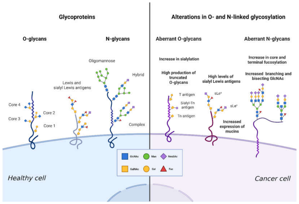
Figure 2.
Schematic overview of the major glycosylation alterations identified in different human malignancies. The central panel illustrates a cancer cell with representative changes in the glycocalyx, including altered N-glycosylation, O-glycosylation, and proteoglycan composition. The surrounding panels summarize cancer-specific glycosylation features reported in lung cancer, gastrointestinal cancers, pancreatic cancer, breast cancer and gynecological malignancies, melanoma, and hematological malignancies. GlcNAc, N-acetylglucosamine; Neu5Ac, N-acetylneuraminic acid; GAGs, glycosaminoglycans; CS, chondroitin sulfate; Tn, Tn antigen; sTn, sialyl-Tn antigen; TA-MUC1, tumor-associated mucin-1; IgG, immunoglobulin G; AFP-L3, alpha-fetoprotein-L3; HCC, hepatocellular carcinoma; 2-DG, 2-deoxy-D-glucose; MGAT5, N-acetylglucosaminyltransferase V; β1,6-GlcNAc, β1,6-linked N-acetylglucosamine; sLeX, sialyl Lewis X; sLeA, sialyl Lewis A; Neu5Gc, N-glycolylneuraminic acid; STn-CA125, sialyl-Tn cancer antigen 125; ST-CA125, sialylated cancer antigen 125; STn-CA15-3, sialyl-Tn cancer antigen 15-3; CSPG4, chondroitin sulfate proteoglycan 4; MAL, Maackia amurensis lectin; PD-L1, programmed death-ligand 1; RS cells, Reed–Sternberg cells. Created in BioRender. Vukovic, M. (2026) https://BioRender.com/6mge8ne.
Figure 2.
Schematic overview of the major glycosylation alterations identified in different human malignancies. The central panel illustrates a cancer cell with representative changes in the glycocalyx, including altered N-glycosylation, O-glycosylation, and proteoglycan composition. The surrounding panels summarize cancer-specific glycosylation features reported in lung cancer, gastrointestinal cancers, pancreatic cancer, breast cancer and gynecological malignancies, melanoma, and hematological malignancies. GlcNAc, N-acetylglucosamine; Neu5Ac, N-acetylneuraminic acid; GAGs, glycosaminoglycans; CS, chondroitin sulfate; Tn, Tn antigen; sTn, sialyl-Tn antigen; TA-MUC1, tumor-associated mucin-1; IgG, immunoglobulin G; AFP-L3, alpha-fetoprotein-L3; HCC, hepatocellular carcinoma; 2-DG, 2-deoxy-D-glucose; MGAT5, N-acetylglucosaminyltransferase V; β1,6-GlcNAc, β1,6-linked N-acetylglucosamine; sLeX, sialyl Lewis X; sLeA, sialyl Lewis A; Neu5Gc, N-glycolylneuraminic acid; STn-CA125, sialyl-Tn cancer antigen 125; ST-CA125, sialylated cancer antigen 125; STn-CA15-3, sialyl-Tn cancer antigen 15-3; CSPG4, chondroitin sulfate proteoglycan 4; MAL, Maackia amurensis lectin; PD-L1, programmed death-ligand 1; RS cells, Reed–Sternberg cells. Created in BioRender. Vukovic, M. (2026) https://BioRender.com/6mge8ne.
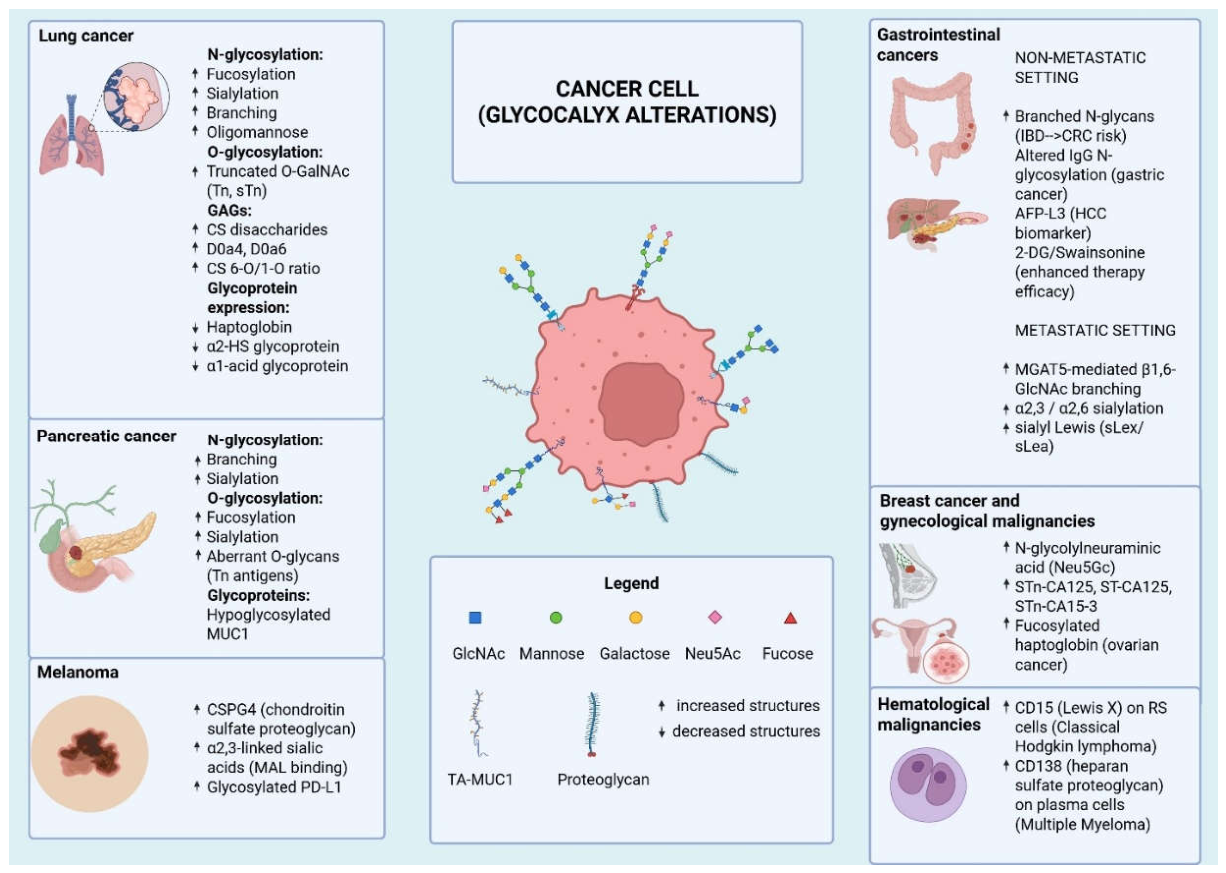

Table 1.
Major cancer-associated alterations in protein glycosylation and their clinical relevance.
| Glycosylation alteration | Main molecular basis | Functional consequence | Clinical relevance |
| Increased N-glycan branching | Upregulation of MGAT4/MGAT5 [17,18] | Enhanced growth signalling, receptor stability and invasion [42,43] | Therapy resistance and biomarker discovery [18,42,44] |
| Increased sialylation | Overexpression of sialyltransferases [17,45] | Immune evasion, metastasis and selectin-mediated adhesion [30,42,44,46,47,48,49] | Prognostic markers and glyco-immune checkpoint targeting [6,50] |
| Increased fucosylation | Altered FUT8, FUT6 and FUT7 activity [17,18] | Altered adhesion, migration and receptor signalling [18,47] | AFP-L3, CA19-9 and other glycoform-based biomarkers [48,51,52] |
| Truncated O-glycans | Incomplete O-GalNAc glycan extension [8,17,53] | Exposure of Tn/sTn antigens and altered mucin biology [53,54,55] | Diagnostic, prognostic and vaccine/ADC targets [56,57,58] |
| Altered glycan–lectin interactions | Engagement of Siglecs, galectins and C-type lectins [6] | Suppression of antitumor immune responses [6,31] | Immunotherapy modulation and novel therapeutic targets [6,31,59] |
Table 2.
An overview of ongoing or recently completed clinical trials evaluating usability of glycomic analysis for diagnostic and/or prognostic/predictive purposes.
Table 2.
An overview of ongoing or recently completed clinical trials evaluating usability of glycomic analysis for diagnostic and/or prognostic/predictive purposes.
| Study name | Year | Country source | Description | Responsible entity |
| MONSTAR-GLYCO [114] | 2024 | Japan | Predictive potential of glycomic analysis for patients with solid tumors | Academic/Investigators |
| Serum Glycomics as Prognostic and Diagnostic Biomarkers of Disease Recurrence in Liver Transplant Recipients with Hepatocellular Carcinoma (GLITCA) [115] | 2023 | Belgium | Predictive potential of glycomic markers for HCC recurrence after liver transplantation | Academic/Investigators |
| Case-Control Study of the Glycotest™ HCC Panel Vs AFP for the Detection of Early-stage Hepatocellular Carcinoma [116] |
2019 | USA, Israel | Diagnostic potential of glycomic test to differentiate between patients with early-stage HCC from those without HCC in the at-risk population | Glycotest, Inc. |
| Clinical Validation of the InterVenn Ovarian CAncer Liquid Biopsy (VOCAL) [117] |
2019-2024 | USA, Australia, Malaysia, Philippines | Diagnostic accuracy of circulating glycoproteins to differentiate between benign and malignant pelvic masses | Venn Biosciences Corporation |
| PSA Glycomics Assay for Early Detection of Prostate Cancer [118] |
2020- | the Netherlands | Diagnostic potential of glycoforms of PSA in plasma and urine to enable early detection of prostate cancer | Academic/Investigators |
| A Prospective Long-term Observational Study in Patients with Monoclonal Gammopathy of Undetermined Significance (SECURE) [119] | 2023- | United Kingdom | Diagnostic potential of serum sphingosine-1-phosphate as a biomarker of progression from monoclonal gammopathy of undetermined significance to multiple myeloma | Academic/Investigators |
| 1 |
Disclaimer/Publisher’s Note: The statements, opinions and data contained in all publications are solely those of the individual author(s) and contributor(s) and not of MDPI and/or the editor(s). MDPI and/or the editor(s) disclaim responsibility for any injury to people or property resulting from any ideas, methods, instructions or products referred to in the content. |
© 2026 by the authors. Licensee MDPI, Basel, Switzerland. This article is an open access article distributed under the terms and conditions of the Creative Commons Attribution (CC BY) license (http://creativecommons.org/licenses/by/4.0/).
Copyright: This open access article is published under a Creative Commons CC BY 4.0 license, which permit the free download, distribution, and reuse, provided that the author and preprint are cited in any reuse.