

Submitted:
26 October 2026
Posted:
28 October 2026
You are already at the latest version
Abstract
Traumatic injuries cause morbidity and mortality in Mediterranean loggerhead sea turtles (Caretta caretta). This retrospective study evaluated 157 turtles at the University of Bari Sea Turtle Clinic from 2015 to 2022; these represented 7.74% of 2028 turtles evaluated. Lesions were classified by site, severity, and chronicity. All turtles underwent radiography; multidetector computed tomography (MDCT) was performed in selected cases. Each wound was swabbed for aerobic bacterial culture and antimicrobial susceptibility testing. Lesions involved a single site in 115 turtles (73.2%) and multiple sites in 42 (26.8%); the carapace, flippers, and skull were most frequently affected. MDCT characterized cranial and skeletal injuries and residual flipper vascularization. Cultures yielded 121 bacterial isolates, of which 76 (63%) exhibited a multidrug-resistant phenotype. Overall, 137 turtles (87.3%) were released, 17 (10.8%) died, and 3 (1.9%) were euthanized. In single-site cases, lesion severity was associated with outcome (p = 0.045), whereas anatomical site was not. In multiple-site cases, neither severity nor site was associated with outcome; associations with hook and fishing-line ingestion were exploratory. These findings indicate that outcome may reflect the combined effects of lesion severity and concurrent clinical conditions and support the use of advanced imaging, microbiological investigation, and targeted therapeutic management in sea turtle rehabilitation.
Keywords:
loggerhead sea turtle
; Caretta caretta
; traumatic injury
; multidetector computed tomography
; antimicrobial resistance
; rehabilitation
; clinical outcome
1. Introduction
Caretta caretta, commonly known as the loggerhead sea turtle, is the most abundant sea turtle species in the Mediterranean Sea, which represents an important foraging area and a nesting region of global conservation significance [1,2]. The species is currently classified as Vulnerable at the global level on the IUCN Red List, whereas the Mediterranean subpopulation is classified as Least Concern [1]. Despite this comparatively favourable regional assessment, anthropogenic activities remain major sources of injury and mortality throughout the species’ range [2,3]. The main threats include incidental capture in fishing gear, vessel strikes, entanglement or ingestion of marine debris and fishing hooks and lines [3,4]. Among these threats, traumatic injuries associated with fisheries interactions and vessel collisions are important causes of morbidity and mortality in stranded and rescued sea turtles in the Mediterranean [2]. Increasing maritime traffic has been associated with elevated exposure of loggerhead turtles to vessel strikes in high boat-density areas [5]. Cranial trauma has also been reported as a potentially important and likely underestimated threat in specific Mediterranean sub-regions [6]. Similarly, entanglement in fishing gear or other anthropogenic debris can cause severe appendicular injuries, including deep soft tissue lacerations, fractures, bone loss, ischemic necrosis, and spontaneous or surgically required amputation [7]. Radiography is widely used as a first-line imaging modality for skeletal trauma in chelonians; however, its two-dimensional nature and superimposition of anatomical structures may hinder the identification and characterization of complex or deep-seated lesions [8]. Computed tomography overcomes these limitations by providing cross-sectional and three-dimensional evaluation without anatomical superimposition [9,10]. In severely traumatized sea turtles, MDCT is particularly valuable for assessing cranial injuries, as it can reveal lesions not reliably detected on plain radiographs, including pneumocephalus, cerebral lacerations, and displacement of the ocular globe [11]. Color Doppler ultrasonography, supplemented by contrast-enhanced MDCT in selected cases, can also assess residual vascularization in severely entangled flippers and support clinical decisions between conservative management and amputation [7]. Surgical curettage followed by the daily application of a plant-derived topical dressing has been reported as a useful management approach for severe traumatic head wounds, with favorable wound healing and clinical outcomes [11,12].
Although previous studies have investigated specific forms of traumatic injury in sea turtles—including cranial trauma [6,11], flipper entanglement [7], an integrated clinical characterization of traumatic lesions across anatomical sites, incorporating severity, chronicity, and site-specific outcome, remains limited. Therefore, the aim of the present study was to describe and classify traumatic lesions in a large cohort of loggerhead sea turtles from Adriatic and Ionian Seas, according to anatomical site, severity, and chronicity, and to identify clinical and lesion-related factors associated with outcome.
2. Materials and Methods
2.1. Study Population
This retrospective observational study included loggerhead sea turtles (Caretta caretta) referred to the Sea Turtle Clinic (STC) of the Department of Veterinary Medicine, University of Bari, between May 2015 and August 2022 because of one or more traumatic lesions. Turtles had been found stranded along the coast or drifting at sea, or had been incidentally captured in fishing gear, and were transferred to the STC by the Adriatic and Ionian Sea Turtle Rescue Centres.
Inclusion criteria were (1) confirmed traumatic injury involving at least one anatomical site; (2) availability of complete clinical records, including lesion classification and outcome; and (3) availability of follow-up information until release or death. Turtles with insufficient documentation to classify the lesions or determine the outcome were excluded.
2.2. Clinical Evaluation and Lesion Classification
The circumstances of rescue were recorded for each turtle. All animals underwent a complete physical examination, a specific assessment of each traumatic lesion, and haematological and biochemical analysis. The main morphometric parameters — curved carapace length (CCL), curved carapace width (CCW), and body weight — were recorded, along with the Body Condition Score (BCS) on a 1–5 scale and internal body temperature was measured using a cloacal probe.
Lesions were classified as either acute, characterized by active bleeding, serous exudation, well-defined wound margins, and absence of necrosis, or chronic, characterized by fibrinonecrotic exudate, necrotic tissue, and blunted margins. Based on severity, a score from 1 (mild) to 3 (severe) was assigned to the lesion at each of five anatomical regions: head, carapace and plastron, neck, and flippers.
Head [11]: (1) mild — superficial wound of limited extent, linear fracture or mild bone depression, no exposure of underlying soft tissue; (2) moderate — intermediate extent, non-linear fracture with non-penetrating bone fragments, minimal soft tissue exposure; (3) severe — fracture with penetrating bone fragments, potential exposure of the cerebral parenchyma and hemorrhage.
Carapace and plastron: given their anatomical continuity and shared traumatic mechanism, carapace and plastron lesions were graded on a single combined scale: (1) mild — superficial scute fracture or crack, limited extent, no bone exposure, no coelomic involvement; (2) moderate — fracture through the full scute/shell thickness, exposed but non-displaced bone, no coelomic exposure; (3) severe — multi-fragmentary or displaced fracture with tissue loss, exposure of the coelomic cavity or organs.
Neck and flippers: Even their shared entanglement-related mechanism, a comparable three-grade framework was applied to both sites, though scored independently for each: (1) mild — superficial entanglement groove or constriction mark, no bone or deep tissue involvement; (2) moderate — deep groove with partial-thickness soft tissue involvement (neck) or incomplete diaphyseal fracture (flippers), without vascular, tracheal, or complete bone compromise; (3) severe — full-thickness constriction with vascular, tracheal, or esophageal compromise (neck), or complete diaphyseal fracture, bone loss, or limb loss (flippers).
In turtles presenting multiple concurrent lesions at different anatomical sites, an overall severity grade was assigned corresponding to the most severe individual lesion.
2.3. Neurological Examination
Turtles with cranial trauma underwent a neurological examination both in and out of the water. General activity and mentation were assessed together with the ability to open the rhamphotheca, bilateral palpebral and corneal reflexes, menace response, nociceptive response to stimulation of the nares, neck-withdrawal response, cloacal reflex, and withdrawal responses of each front and rear flipper. Buoyancy was classified as normal or abnormal. Abnormalities were further characterized as symmetric or asymmetric, and the ability to dive was recorded. Mentation was classified into three grades [11,13]:
1.Alert: appropriate response to external stimuli.
2.Depressed: reduced but appropriate responsiveness to external stimuli.
3.Lethargic/comatose: minimal or absent responsiveness to external stimuli, with possible loss of consciousness.
2.4. Diagnostic Imaging
All turtles underwent a total-body radiographic study in the dorso-ventral (DV), latero-lateral (LL), and cranio-caudal (CrCd) projections. In turtles with cranial trauma, additional DV and LL radiographs of the head were obtained. In cases of moderate or severe trauma, multi-detector computed tomography (MDCT) was performed using a 16-slice scanner (Somatom Emotion, Siemens, Forchheim, Germany), following general anesthesia induced with intravenous propofol at a dose of 5 mg/kg (Propovet®, Zoetis, Rome, Italy). Scanning and reconstruction technical parameters were as follows: 110 kVp, 180 mAs, slice thickness 1 mm, pitch 0.8, rotation time 0.6 s, reconstruction interval 0.5 mm, and a standard bone acquisition algorithm. Three-dimensional multiplanar reconstructions (3D MPR), maximum intensity projections (MIP), and volume-rendered images were obtained using dedicated software (OsiriX DICOM viewer®, Pixmeo, Bernex, Switzerland). To reduce visual stimulation and stress during handling and diagnostic procedures, an elastic bandage was placed around the head prior to the examination, covering the eyes and tympanic scutes while leaving the nares free.
2.5. Surgical Treatment
Surgical curettage was performed under general anesthesia. Premedication was provided with tramadol (Altadol, Formevet®, Milan, Italy; 5 mg/kg) and, after 20 minutes, induction was achieved with propofol (Propovet®, Zoetis, Rome, Italy; 3–7.0 mg/kg); both drugs were administered into the external jugular vein. The turtles were subsequently intubated. In turtles with head trauma, anesthesia was maintained for the duration of the procedure with 2–3% sevoflurane in oxygen (SevoFlo®, Ecuphar, Greifswald, Germany). In all other turtles, anesthesia was maintained with 3% isoflurane in oxygen (IsoFlo®, Zoetis, Rome, Italy). Anesthesia was monitored by electrocardiography. A catheter was placed in the cephalic vein, through which a 1:1 mixture of Lactated Ringer’s solution and 0.9% NaCl was infused at a rate of 2 mL/kg/h. The wound was cleansed with sterile physiological saline (0.9% NaCl), and the surrounding skin was aseptically prepared with povidone-iodine scrub. Surgical curettage of the lesion was then performed using a No. 21 scalpel blade and a Volkmann curette, until complete removal of necrotic and fibrotic tissue and exposure of viable tissue was achieved. At the end of the procedure, swabs were collected from the lesion for microbiological culture and antimicrobial susceptibility testing. A plant-derived preparation (One Primary Wound Dressing®, Phytoceutical AG, Endospin Italia, Rome, Italy) was applied to the wound surface as primary dressing, renewed daily for the first month and subsequently every other day until the end of treatment.
In turtles unable to feed voluntarily, an esophago-gastric tube was surgically placed through a temporary right ventrolateral cervical stoma, to ensure adequate nutritional support during hospitalization [14].
2.6. Microbiological Analysis and Antimicrobial Susceptibility Test
Swabs were cultured on Columbia blood agar (CBA), MacConkey agar (MCK), Mannitol salt agar (MSA), and Tryptic Soy broth (TSB) (Liofilchem, Teramo, Italy), incubated aerobically at 35 °C for 24–48 h. Presumptive identification was based on Gram staining; isolates were then identified biochemically according to growth pattern — API 20E/API 20NE (Biomérieux, France) for MCK isolates (oxidase-tested beforehand) and API STAPH System (Biomérieux, France) for MSA isolates. Isolates not identified by biochemical testing underwent 16S rRNA gene amplification and sequencing: PCR products (~300 bp) were purified (QIAquick PCR Purification Kit, Qiagen, Germantown, MD, USA) and sequenced by MiSeq NGS (Illumina, San Diego, CA, USA), with sequences compared against the GenBank database (accessed 27 July 2020).
Antimicrobial susceptibility was determined by the Kirby–Bauer disk diffusion method on Mueller–Hinton agar (Liofilchem, Teramo, Italy), with clinical breakpoints per CLSI (2018) guidelines. Isolates were tested against 12 antibiotics selected according to standardized therapeutic protocols for turtles: ampicillin (10 μg), amoxicillin/clavulanic acid (30 μg), cephalexin (30 μg), cefotaxime (30 μg), cefuroxime (30 μg), ciprofloxacin (5 μg), danofloxacin (10 μg), enrofloxacin (15 μg), moxifloxacin (5 μg), norfloxacin (5 μg), doxycycline (30 μg), and gentamicin (30 μg). Plates were incubated at 37 °C for 24 h, and inhibition zone diameters were measured to classify strains as susceptible (S) or resistant (R). Escherichia coli ATCC 25922 was used as quality control.
2.7. Medical Therapy and Post-Operative Management
Empiric treatment with enrofloxacin (Baytril® 5%; 5 mg/kg IM every 24 h), diluted 1:4 with 0.9% NaCl, was initiated in severely traumatized or markedly debilitated turtles and in those with concomitant conditions, including entanglement injuries, gastrointestinal hooks or fishing lines, and pulmonary disease. The antimicrobial treatment was modified, when indicated, according to the results of antimicrobial susceptibility testing. Post-operative pain management was achieved with tramadol (Altadol, 5 mg/kg IM, every 48 hours) for 3–7 days following surgical debridement. Dexamethasone (Dexadreson®; 0.2 mg/kg IM every 48 h) was administered to turtles with severe cranial trauma and cerebral edema documented by computed tomography. Treatment was continued or discontinued based on serial clinical reassessments and the individual response of each turtle. During the first 24 hours after surgery, turtles were maintained on foam padding and covered with damp cloths in shallow lukewarm water. From the second postoperative day onward, they were housed in tanks with saltwater at 36–37‰ salinity — corresponding to the values of the Adriatic and Ionian Seas, respectively — in a temperature-controlled room at approximately 23–25 °C, until clinical improvement was observed. Animals with mild neurological signs, able to independently lift their head out of the water, were housed in deep tanks; turtles with moderate-to-severe neurological signs or respiratory difficulty were supported on foam padding in shallow-water tanks, with the water level maintained below the nares. with a water level kept below the level of the nares. Serial neurological examinations were performed throughout hospitalization to monitor neurological function and clinical recovery.
2.8. Outcome
Final outcome was classified as favourable or unfavourable. A favourable outcome was defined as successful rehabilitation and release into the sea. An unfavourable outcome was defined as death during hospitalization or euthanasia performed because of irreversible injuries incompatible with survival in the wild; death and euthanasia were also recorded separately. For euthanasia, turtles were deeply anaesthetized as described in Section 2.5, and death was then induced by intravenous administration of potassium chloride (1–2 mmol/kg), in accordance with the AVMA Guidelines for the Euthanasia of Animals [21]. Death was confirmed by absence of cardiac activity on Doppler ultrasonography and absence of reflexes. Length of hospitalization was calculated as the number of days elapsed between the date of admission and the date of release, death, or euthanasia.
2.9. Statistical Analysis
Continuous variables (CCL, CCW, body weight, and length of hospitalization) are reported as median and interquartile range (IQR), given their skewed distributions. For comparisons of hospitalization length, the assumptions of the corresponding parametric procedures (one-way ANOVA across severity grades and independent-samples t-test between outcomes) were checked on model residuals, using the Shapiro–Wilk test and Q–Q plots for normality and Levene’s test for homogeneity of variance. Residuals were markedly right-skewed and variances were unequal across groups (both p < 0.001), including after log-transformation; therefore, the Kruskal–Wallis test (with Dunn’s post-hoc comparisons, Benjamini–Hochberg corrected) and the Mann–Whitney U test were used. Associations between categorical variables were assessed with Fisher’s exact test (Fisher–Freeman–Halton extension for tables larger than 2×2), and the correlation between severity and neurological grade in turtles with head trauma with Spearman’s ρ; neither procedure assumes normality. Turtles with a single lesion and those with multiple concurrent lesions were analyzed separately. Within each subgroup, outcome (favourable vs. unfavourable) was tested against severity grade (three categories, and severe vs. mild/moderate combined) and against each anatomical site; when neither was associated with outcome, ingested hook and fishing line were tested as potential co-causes of death. In the full cohort, outcome was tested against acute/chronic classification and neurological grade. All tests were two-tailed, with significance set at p < 0.05. Analyses were performed using SPSS (version 27.0.1.0; IBM Corp., Armonk, NY, USA).
3. Results
3.1. Study Population and Incidence
During the study period, 2028 loggerhead sea turtles (Caretta caretta) were clinically evaluated at the STC. Of these 157 presented traumatic lesions that met the inclusion criteria, representing 7.74% of all turtles evaluated (95% CI: 6.62–8.99%). Regarding the circumstances of rescue, 61 turtles (38.9%) were incidentally captured during trawling activities, 55 (35.0%) were found stranded along the coast, and 41 (26.1%) were found drifting at sea. Median curved carapace length (CCL) was 59.0 cm (range: 22.0–81.0), median curved carapace width (CCW) was 54.0 cm (range: 18.0–73.0), and median body weight was 23.6 kg (range: 1.1–60.8). Based on body size, the cohort comprised 82 juveniles (52.2%), 38 adults (24.2%), and 37 subadults (23.6%). Cloacal temperature ranged from 16 °C to 23 °C.
Among the 157 turtles, 115 (73.2%) presented a single traumatic lesion, whereas 42 (26.8%) had concurrent lesions involving multiple anatomical sites (Figure 1). Among the 115 turtles with a single traumatic injury, 33 presented an injury to the carapace (28.7%), 32 to the head (27.8%), 31 to the flippers (27.0%), 12 to the neck (10.4%), and 7 to the plastron (6.1%). Among the 42 turtles with multiple anatomical sites affected, the flippers were involved in 25 turtles (59.5%), the carapace in 24 (57.1%), the plastron in 15 (35.7%), the neck in 15 (35.7%), and the head in 14 (33.3%). Because individual turtles could have lesions involving more than one site, these categories were not mutually exclusive.
Overall, 18.5% of lesions were classified as mild, 40.8% as moderate, and 40.8% as severe. Regarding chronicity, 43 lesions (27.4%) were classified as acute and 114 (72.6%) as chronic. Severity and chronicity of lesions varied by anatomical site among the 115 turtles with a single traumatic lesion (Table 1).
3.2. Neurological Findings
Among the 46 turtles with head trauma, 32 (69.6%) were classified as alert, 9 (19.6%) as depressed, and 5 (10.9%) as lethargic/comatose, according to the neurological grade. An unfavourable outcome occurred in 4/32 alert turtles (12.5%), 1/9 depressed turtles (11.1%), and 2/5 lethargic or comatose turtles (40.0%). In 12 turtles, cranial trauma involved one or more structures potentially affecting sensory or feeding function. The affected structures included the frontal bone in 10 turtles, the parietal bone in 5, the orbital/periorbital region in 4, the maxillary bones in 3, the nasal bones in 2, the zygomatic bone in 1 and the turbinates in 1. Rhamphothecal involvement was observed in 4 turtles. During evaluation in water, buoyancy abnormalities were observed in 18/46 turtles (39.1%). These abnormalities were symmetric in 11 turtles (23.9%) and asymmetric in 7 (15.2%).
3.3. Radiographic and Computed Tomographic Findings
Radiographic examination showed multi-fragmentary or depressed head fractures in all 46 head-traumatized turtles (Figure 2). Fractures associated with varying degrees of bone loss were identified in 15/56 turtles with flipper entanglement injuries (8 right humerus, 4 left humerus, 1 right femur, and 2 left femur). In one turtle, a fracture-associated osteomyelitis involving the radius and ulna was observed. Ingested fishing hooks were identified radiographically within the gastrointestinal tract of 19 turtles. Radiographic findings consistent with severe gas embolism were observed in one turtle [15]. Among the 57 turtles with carapace injuries, 54 underwent MDCT examination. Pulmonary changes characterized by a reticular pattern and considered consistent with mild pneumonia were identified in 15/54 turtles.
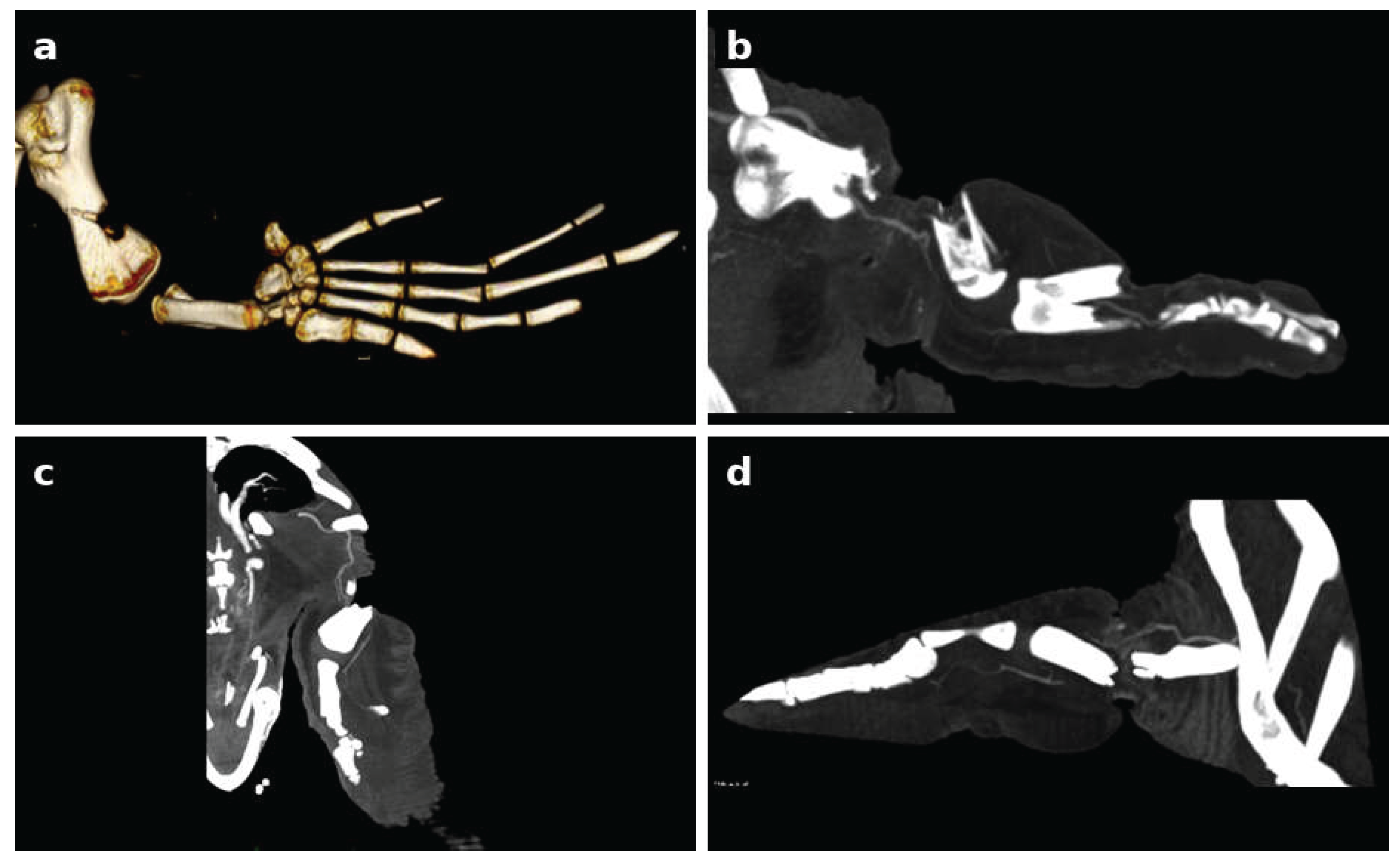
Out of 46 sea turtles with head trauma, 40 underwent total head and body CT examination (Figure 3,4). In severe cases, CT images revealed depressed, multi-fragmentary fractures of the parietal, postorbital, prefrontal, frontal, orbital, nasal, and squamosal bones. In 8 turtles, bone fragments were displaced inward toward the cranial cavity, with suspected disruption of the dura mater. Associated intracranial findings included pneumocephalus, cerebral laceration, and cerebral swelling. In 10 cases, the ocular globe was shifted downward due to pressure from the surrounding tissue, together with cerebral contusion and compression of the salt gland. In turtles with moderate cranial trauma, the parietal bone fragments showed deep depression, whereas the postorbital fragments were superficially depressed. Contrast-enhanced MDCT was performed in 51 turtles with moderate or severe flipper entanglement injuries to assess residual limb vascularization and the extent of skeletal damage (Figure 5). The entangled front flippers showed variable degrees of bone damage of the middle humeral diaphysis, ranging from incomplete fractures to multi-fragmentary, complete fractures, or even complete loss of the diaphysis. Rear-flipper injuries were characterized by complete fractures and segmental bone loss involving the mid-femoral diaphysis.
Figure 3.
Computed tomography findings in a turtle with severe head trauma. (a) Three-dimensional volume-rendered reconstruction showing a multy-fragmentary head fracture. (b) Axial CT slice at the level of the vertex showing the same head defect.
Figure 3.
Computed tomography findings in a turtle with severe head trauma. (a) Three-dimensional volume-rendered reconstruction showing a multy-fragmentary head fracture. (b) Axial CT slice at the level of the vertex showing the same head defect.
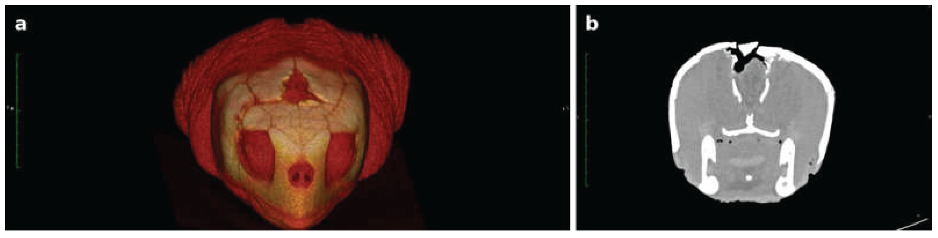
Figure 4.
Computed tomography findings in a second turtle with severe skull trauma. (a) Three-dimensional reconstruction showing multiple, extensive fracture lines across the skull roof. (b–d) Axial CT slices at different levels showing the corresponding skull defects.
Figure 4.
Computed tomography findings in a second turtle with severe skull trauma. (a) Three-dimensional reconstruction showing multiple, extensive fracture lines across the skull roof. (b–d) Axial CT slices at different levels showing the corresponding skull defects.
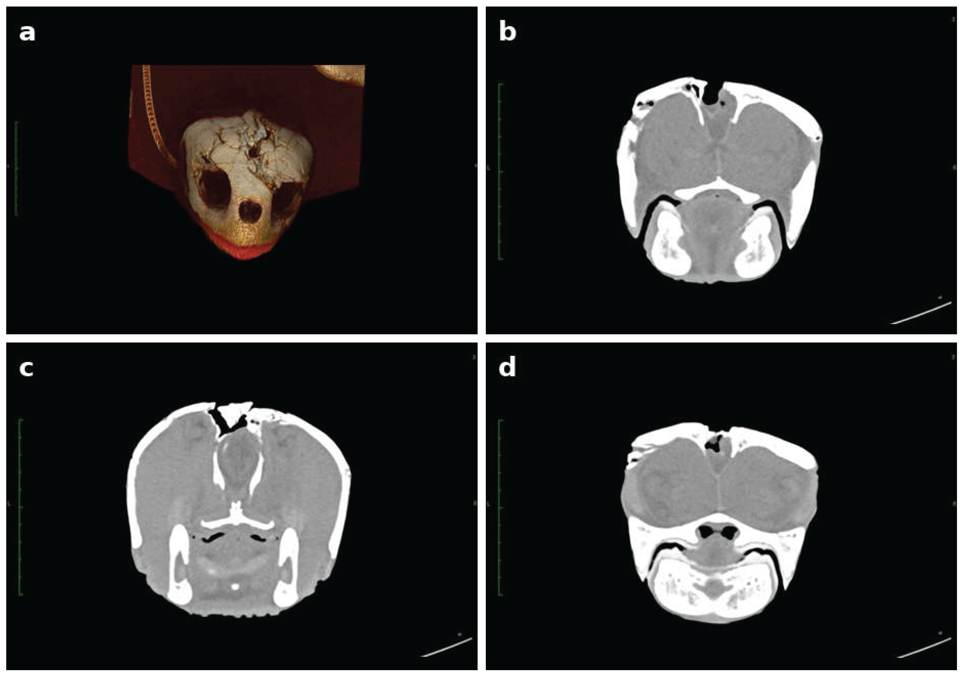
Figure 5.
Three-dimensional CT reconstructions of a severely entangled flipper. (a) Lateral view showing complete fracture of the humeral diaphysis. (b) Detail of the multi-fragmentary fracture pattern. (c) Alternative lateral view of the fracture. (d) Longitudinal slice showing associated digit fracture.
Figure 5.
Three-dimensional CT reconstructions of a severely entangled flipper. (a) Lateral view showing complete fracture of the humeral diaphysis. (b) Detail of the multi-fragmentary fracture pattern. (c) Alternative lateral view of the fracture. (d) Longitudinal slice showing associated digit fracture.
3.4. Microbiological Findings and Antimicrobial Resistance
Among all samples collected, a total of 121 bacterial strains were identified, belonging to 9 different genera. Eight were Gram-negative, and 1 Gram-positive. Out of 157 samples, 36 exhibites no growth even after 48 h of incubation. Pure, young colonies were biochemically tested: 43 were identified with the API 20NE System, 76 with the API 20E System, and the Gram-positive isolates were identified with the API STAPH System. Collectively, 94 isolates were identified with identity scores higher than 98%, and a further 27 isolates were confirmed by 16S rRNA PCR amplification and sequencing. The isolates belonged to the following genera: Pseudomonas spp. (37/121, 30.6%), Citrobacter spp. (28/121, 23.1%), Vibrio spp. (20/121, 16.5%), Aeromonas spp. (16/121, 13.2%), Acinetobacter spp. (10/121, 8.3%), Proteus spp. (4/121, 3.3%), Enterobacter spp. (2/121, 1.7%), Klebsiella spp. (2/121, 1.7%), and Staphylococcus spp. (2/121, 1.7%). Resistance to at least one antibiotic class was found in all isolates collected. Bacterial genus distribution across anatomical sites is summarized in Table 2.
The isolates displayed the following resistance rates: ampicillin, 104/121 (86%); amoxicillin/clavulanic acid, 76/121 (63%); cephalexin, 76/121 (63%); cefotaxime, 33/121 (27%); cefuroxime, 62/121 (51%); ciprofloxacin, 21/121 (17%); danofloxacin, 49/121 (40%); enrofloxacin, 27/121 (22%); moxifloxacin, 38/121 (31%); norfloxacin, 24/121 (20%); doxycycline, 32/121 (26%); and gentamicin, 16/121 (13%). Overall, 76 isolates (63%) displayed a multidrug-resistant (MDR) phenotype, defined as resistance to at least three antibiotic classes [16].
Notably, 94% of Pseudomonas spp. isolates, 73% of Vibrio spp. isolates, and 60% of Acinetobacter spp. isolates were resistant to beta-lactams. The Staphylococcus spp. isolates were resistant to 100% of the beta-lactams and aminoglycosides tested, while remaining fully sensitive to fluoroquinolones and tetracyclines. Enterobacter spp. was 100% resistant to beta-lactams and cephalosporins. Citrobacter spp. and Aeromonas spp. showed average resistance levels close to 50% across nearly all antibiotic classes tested. Proteus spp. was sensitive to all tetracyclines and aminoglycosides tested, and Klebsiella spp. was the only genus sensitive to all antibiotic classes tested.
3.5. Outcome and Prognostic Factors
Twenty-nine of the 157 turtles (18.5%) required esophagostomy tube placement due to inability to feed spontaneously. Surgical amputation was performed in 3 turtles with severe flipper entanglement. Systemic antibiotic therapy with enrofloxacin was initiated in 32/64 severely traumatized turtles (50.0% of severe cases); however, as the Citrobacter freundii isolates from 8 of these 32 turtles (25.0%) were found to be resistent to enrofloxacin, empirical therapy was replaced with cefotaxime based on antimicrobial susceptibility testing results. Dexamethasone was administered in 8/46 turtles with head trauma and CT evidence of cerebral edema or other severe intracranial abnormalities, including pneumocephalus and cerebral laceration.
Overall, 137/157 turtles (87.3%) had a favorable outcome and were successfully rehabilitated and released, while 20/157 (12.7%) had an unfavorable outcome. Of these, 17 turtles (10.8%) died during hospitalization and 3 (1.9%) were euthanized because their injuries were considered irreversible with survival. Two turtles were euthanized due to irreversible traumatic damage: one presented an avulsion fracture of the left caudal carapace and plastron, with exposure of muscle and coelomic organs and paralysis of the left rear flipper; the other presented severe bilateral flipper entanglement with bilateral humeral fracture and an intestinal hook. One turtle was euthanized because of severe intestinal lacerations caused by an ingested fishing line, with concurrent hook ingestion and severe carapace and plastron fracture.
Among the 115 turtles with lesions confined to a single anatomical site, lesion severity was significantly associated with outcome (Fisher’s exact test, p = 0.045): the proportion of unfavorable outcomes was 4.0% (1/25) in mild, 5.9% (3/51) in moderate, and 20.5% (8/39) in severe lesions. No individual anatomical site was significantly associated with outcome (Fisher’s exact test, p = 0.905). In the multiple-lesion turtles, neither severity grade—assigned to the most severe individual lesion— (three-way comparison, p = 0.613; collapsed comparison, p = 0.439) nor the presence of a lesion at any individual anatomical site (head: OR = 0.61, p = 0.697; carapace: OR = 0.70, p = 0.706; plastron: OR = 0.54, p = 0.689; neck: OR = 2.09, p = 0.425; flippers: OR = 1.17, p = 1.000) was significantly associated with outcome. The presence of an ingested hook was significantly associated with an unfavorable outcome (p = 0.040), as was the presence of an ingested fishing line (p = 0.033).
Length of hospitalization differed significantly according to injury severity across the full cohort (Kruskal-Wallis test, p < 0.001), with median hospitalization of 10 days (grade 1), 23 days (grade 2), and 39.5 days (grade 3); Dunn’s post-hoc test with Benjamini-Hochberg correction confirmed significant differences between all three grades (grade 1 vs. 2, p < 0.001; grade 1 vs. 3, p < 0.001; grade 2 vs. 3, p < 0.001). Turtles with an unfavorable outcome had a significantly shorter hospitalization than those with a favorable outcome (median 13.5 vs. 26 days; Mann-Whitney U test, p < 0.001). No significant association was found between acute/chronic classification and outcome (Fisher’s exact test, p = 1.000). Among the 46 turtles with head trauma, no significant association was found between neurological grade and outcome (Fisher’s exact test, p = 0.277), nor between lesion severity and neurological grade (Spearman’s ρ = 0.18, p = 0.242).
4. Discussion
This retrospective study characterized traumatic lesions in a large cohort of loggerhead sea turtles (Caretta caretta) from the Adriatic and Ionian Seas admitted to the STC, examining the relationships between lesion distribution, severity, chronicity, and clinical outcome. To our knowledge, this represents one of the largest cohorts of traumatized Caretta caretta described to date in which severity and chronicity were systematically graded across all anatomical sites, rather than being restricted to a single anatomical region as in most previous studies [6,7,11]. Traumatic lesions were identified in 7.74% of all turtles evaluated during the study period. This proportion indicates that anthropogenic trauma remains one of the leading causes of morbidity in stranded and rescued sea turtles along the Italian Adriatic and Ionian coasts, consistent with previous reports from the same geographic area [2,5,6].The predominance of carapace and flipper lesions in our cohort is compatible with two major anthropogenic mechanisms repeatedly associated with trauma in this species in the Mediterranean: vessel collisions, responsible for blunt carapace trauma, and entanglement in fishing gear, responsible for appendicular injuries [4,5,7]. Notably, more than one-quarter of turtles (26.8%) presented concurrent lesions at multiple anatomical sites, most frequently a combination of neck and flipper entrapment, or carapace and plastron fractures. These combinations may have resulted from either a single traumatic event or multiple events occurring at different times; the retrospective nature of the study did not allow the sequence or precise mechanism of injury to be determined.
The high proportion of chronic lesions observed across all anatomical sites (72.6% overall, ranging from 67.9% in flipper lesions to 77.8% in neck lesions) is consistent with previous findings [11] and likely reflects delayed detection at sea or a prolonged interval between the traumatic event and rescue. In our cohort lesion chronicity was not a prognostic factor for outcome (Fisher’s exact test, p = 1.000). However, chronicity represents only an indirect estimate of the time elapsed since trauma; therefore, this result should not be interpreted as evidence that delayed rescue has no effect on outcome.
The added diagnostic value of MDCT over conventional radiography observed in this study is consistent with previous reports [11]. In turtles with cranial trauma. MDCT identified pneumocephalus and cerebral lacerations in eight turtles and ventral displacement of the ocular globe with compression of the salt gland in ten. These findings had potential therapeutic relevance and contributed to clinical management and surgical planning; however, their independent prognostic value was not formally evaluated. Similarly, in turtles with severe flipper entanglement, MDCT allowed detailed characterization of humeral and femoral bone damage, ranging from incomplete fractures to complete diaphyseal loss. Contrast-enhanced MDCT also permitted assessment of residual limb vascularization, providing information that supported the choice between conservative management and amputation, consistent with the approach previously described [7].
The wound-management protocol adopted in this study—surgical curettage followed by daily application of a plant-derived topical preparation (One Primary Wound Dressing®)—is based on an approach previously described as effective in the management of severe wounds in this species, even in the presence of substantial tissue infection [12]. The therapeutic rationale for this dressing lies in its antimicrobial activity, attributed to the fatty acid content of the formulation, combined with the semi-occlusive, moisture-balanced environment it creates over the wound surface, both properties capable of promoting healing while limiting the need for systemic antimicrobial therapy [17,18]. However, because this study did not include an untreated or alternatively treated control group, the independent effect of the dressing on wound healing, survival, or the requirement for systemic antimicrobial therapy could not be determined.
This consideration is particularly relevant given the high prevalence of multidrug-resistant isolates observed in the present study and previously reported in the same population [19,20]. No molecules are registered for sea turtles, and an extra label use is needed. Moreover, drugs classified as medically important for human medicine, such as fluroquinolones or third and fourth class cephalosporins are effective against MDR bacteria.
Reducing reliance on systemic antibiotics through effective topical wound management may therefore represent an important strategy, not only for individual patient care, but also for limiting selective pressure toward antimicrobial resistance in a population increasingly recognized as a potential reservoir and sentinel of environmental AMR, in a One Health approach [19]. When systemic therapy was needed, enrofloxacin was started empirically and then adjusted according to susceptibility results: enrofloxacin-resistant Citrobacter freundii prompted the switch to cefotaxime in 8 turtles, consistent with fluoroquinolone resistance reported in wound isolates from Mediterranean loggerheads [20]. Targeted systemic antimicrobial therapy—reserved for severely traumatized or debilitated turtles and guided by antimicrobial susceptibility testing, as adopted in the present protocol—combined with effective topical wound management, appears to represent the most balanced approach to both individual clinical outcome and antimicrobial stewardship in sea turtle rehabilitation.
Regarding in-hospital management, in debilitated turtles unable to feed independently, nutritional support through esophago-gastric tube placement represented an important component of the rehabilitation protocol, contributing to an improved clinical outcome during prolonged hospitalization [14].
Among turtles with lesions confined to a single anatomical site, outcome was significantly associated with lesion severity (p = 0.045), with mortality increasing progressively from 4.0% in mild cases to 20.5% in severe cases. Anatomical site was not significantly associated with outcome (p = 0.905). These findings suggest that, among turtles with single-site lesions, lesion severity may be more closely associated with outcome than anatomical location. In turtles with multiple concurrent lesions, neither the severity of the most severe lesion nor the site involved was found to be a predictor of outcome. This finding suggests that, in these cases, other factors—potentially including complications specific to combined lesions—may play a more relevant role and would require more detailed, lesion-level characterization in future studies conducted on larger cohorts of polytraumatized patients. Consistent with this observation, the absence of an association between severity/site and outcome in the multiple-lesion subgroup led us to investigate concurrent clinical findings not directly attributable to the anatomical trauma: the presence of an ingested hook was significantly associated with an unfavourable outcome (OR = 9.60, p = 0.040), as was the presence of an ingested fishing line (100.0% vs. 15.0%; p = 0.033)—although the latter finding should be interpreted with caution, given the small sample size. Analysis of individual cases with an unfavorable outcome generally reflected the association between severity and outcome observed in the single-site subgroup, while also revealing informative exceptions. Among the 12 turtles with an unfavorable outcome and lesions confined to a single anatomical site, all unfavorable outcomes involving the head or carapace occurred in turtles with severe lesions. This descriptive finding is consistent with greater clinical risk associated with severe injuries at these sites but does not independently confirm a prognostic effect.
In contrast, unfavorable outcomes involving the flippers occurred predominantly in animals with mild or moderate, rather than severe, lesions. Two of these outcomes occurred in association with concurrent conditions not represented by the local lesion-severity grade: severe gas embolism in one turtle and ingestion of a hook with severe involvement of the cervical esophagus and a fishing line protruding from the rhamphotheca in another. In the third case, no concurrent condition was documented, and the death remains an observation not fully explained by the variables collected in this study. Among the 8 turtles with multiple lesions that died or were euthanized, 6 (75%) presented ingestion of a hook and/or fishing line, often in association with severe flipper or neck entanglement, or extensive shell fractures. This pattern reinforces the observation, discussed above, that in turtles with multiple traumatic lesions, foreign body ingestion represents a relevant prognostic factor. In the remaining cases, death or euthanasia was associated with irreversible tissue damage including exposure of coelomic organs, flipper paralysis or extensive shell fracture.
Overall, this descriptive review suggests that unfavorable outcomes occurred in at least three clinical patterns: (1) severe, direct anatomical damage, predominantly involving the head and carapace, well captured by the severity classification adopted; (2) concurrent conditions, mainly foreign body ingestion and gas embolism, which may determine outcome independently of local traumatic lesion severity, particularly in flipper lesions and multi-site cases; and (3) a limited number of cases in which none of the variables collected fully explains the outcome, highlighting the limitations of a classification approach based solely on the external anatomical lesion.
Studies from other regions indicate similar predominant mechanisms, although direct comparison is limited by differences in case definitions and catchment areas. Entanglement in fishing gear or plastics was the leading cause of admission in the Canary Islands (50.81%), with hook ingestion accounting for 11.88% [22], and in Tenerife (42.8%), where vessel collisions accounted for only 4.3% [23]; in the Croatian Adriatic, dead turtles with visible injuries had mainly boat collisions or fishing hooks and lines [24]. As in our cohort, interactions with fishing gear are therefore prominent, whereas traumatic lesions carried the worst outcomes: the highest euthanasia and unassisted mortality in the Canary series (18.67% and 30.67%) [22], and the lowest release rate (63.2%) and longest median rehabilitation (37 days) in Tenerife [23]. These authors attribute the poor prognosis of vessel strikes to extensive carapace and plastron fractures with damage to internal organs, whereas entanglement, even with flipper amputation, was associated with high release rates [22,23]. Consistently with this, hospitalization in our cohort lengthened with lesion severity (median 10, 23 and 39.5 days for mild, moderate and severe lesions). Unlike those series, which classify cases by cause of admission, we graded severity within each anatomical site, and severity rather than site was associated with outcome.
The proportion of turtles successfully rehabilitated and released was 87.3%, comparable to that previously reported for hospitalized sea turtles with traumatic injuries [11]. Notably, turtles that died or were euthanized had a significantly shorter hospitalization than those released (median 13.5 vs. 26 days; p < 0.001). Together with the longer hospitalization of more severe lesions, this pattern suggests that death occurred predominantly during the early phase of clinical management: turtles that survive the initial critical period appear to have a relatively favourable prognosis, even in the presence of severe lesions, and the first days of hospitalization represent the period of greatest clinical risk.
Our data refer exclusively to turtles found alive and referred to the STC. As a university veterinary clinic that receives animals transferred by the regional rescue centres, the STC by definition sees only live turtles; turtles that died at sea, sank, were carried away from the coast, were never detected, or died before transfer are absent. Much information is lost before animals reach a clinic: in Tenerife, the cause of admission remained unknown in 63.9% of turtles that arrived already dead [23], and on the Croatian side of the Adriatic most stranded turtles were too decomposed for the cause of death to be ascertained [24]. Strandings represent only a fraction of at-sea mortality: the probability that a carcass reaches the shore is low and highly variable, as observed along the Pacific coast of Mexico [25], and depends on season, winds and distance from the coast; moreover, as found in the northern Gulf of Mexico [26], only a minority of the carcasses that reach the shore are actually reported. The most severe or rapidly fatal lesions, for example extensive head trauma with exposure of brain tissue, or entanglement leading to drowning, are those least likely to be survived until discovery and are therefore probably under-represented, whereas injuries compatible with prolonged survival are probably over-represented. The high incidence of chronic lesions (72.6%) is consistent with this “survivor effect”. Our results should be interpreted in this light and should not be taken as estimates of the frequency, severity or lethality of traumatic injuries in the entire loggerhead sea turtle (Caretta caretta) population in Italian waters.
This study has several limitations that should be considered when interpreting the results. Its retrospective, single-center design limits the generalizability of the findings and did not permit standardized prospective collection of clinical, imaging, and follow-up data as previously noted for similar cohorts [11]. In turtles with lesions involving multiple anatomical sites, use of the most severe lesion as the animal-level severity grade may have resulted in loss of information regarding the number, combination, and cumulative extent of concurrent lesions. Chronicity was based on lesion appearance and may not accurately represent the time elapsed since trauma.
The study also lacked control groups for evaluating the independent effects of MDCT, surgical treatment, topical wound management, antimicrobial therapy, and nutritional support.
5. Conclusions
This study provides a characterization of traumatic injuries in a large cohort of loggerhead sea turtles (Caretta caretta) from the Adriatic and Ionian Seas. In turtles with lesions confined to a single anatomical site, greater lesion severity was associated with an unfavorable outcome. In turtles with multiple anatomical sites affected, no significant association between outcome and either lesion severity or anatomical site was detected, whereas exploratory findings suggest that concurrent hook or fishing-line ingestion may contribute to clinical outcome. MDCT provided detailed diagnostic information and supported clinical and surgical decision-making, particularly in turtles with cranial trauma and severe flipper entanglement injuries. The high proportion of multidrug-resistant isolates underscores the importance of bacterial culture, antimicrobial susceptibility testing, prudent antimicrobial use, and appropriate wound management in sea turtle rehabilitation. Overall, the findings suggest that clinical outcome may reflect the combined effects of anatomical injury severity and concurrent conditions. Larger prospective, multicenter studies incorporating standardized lesion classification and multivariable analyses are needed to identify independent prognostic factors and refine outcome assessment in this species.
Supplementary Materials
The following supporting information is provided: Table S1: Individual data of the 157 loggerhead sea turtles, with unique codes (CC-001 to CC-157) and references to previous publications.
Author Contributions
Conceptualization, DF and IL; methodology, DF, MLC and ADB.; software, AG; validation, DF, IL and SP; formal analysis, SP, A.G and IL.; investigation, ADB, DF, CV, SC, DB, MLC.; data curation IL, MLC, SP, PS , EDE and MS.; writing—original draft preparation, DF and IL; writing—review and editing, DF, EDE and IL.; visualization, MS, CV.; supervision, DF and ADB.; All authors have read and agreed to the published version of the manuscript.
Funding
This research received no external funding.
Institutional Review Board Statement
The study was approved by the Ethics Committee of the Department of Veterinary Medicine, University of Bari (Authorization No. 4/19).
Data Availability Statement
The data presented in this study are available from the corresponding author upon reasonable request. The individual data underlying the clinical and statistical analyses are provided in Supplementary Table S1.
Conflicts of Interest
The authors declare no conflicts of interest.
References
- Casale, P.; Tucker, A.D. Caretta caretta (Amended Version of 2015 Assessment). IUCN Red. List Threat. Species 2017. [Google Scholar] [CrossRef]
- Casale, P.; Broderick, A.; Camiñas, J.; Cardona, L.; Carreras, C.; Demetropoulos, A.; Fuller, W.; Godley, B.; Hochscheid, S.; Kaska, Y.; Lazar, B.; Margaritoulis, D.; Panagopoulou, A.; Rees, A.; Tomás, J.; Türkozan, O. Mediterranean Sea Turtles: Current Knowledge and Priorities for Conservation and Research. Endang. Species. Res. 2018, 36, 229–267. [Google Scholar] [CrossRef]
- Lutcavage, M.E.; Plotkin, P.; Witherington, B.; Lutz, P.L. Human Impacts on Sea Turtle Survival. In The Biology of Sea Turtles, Vol. I; Lutz, P.L., Musick, J.A., Eds.; CRC Press: Boca Raton, FL, USA, 1997; pp. 387–409. [Google Scholar]
- Duncan, E.; Botterell, Z.; Broderick, A.; Galloway, T.; Lindeque, P.; Nuno, A.; Godley, B. A Global Review of Marine Turtle Entanglement in Anthropogenic Debris: A Baseline for Further Action. Endang. Species. Res. 2017, 34, 431–448. [Google Scholar] [CrossRef]
- Pasanisi, E.; Chimienti, M.; Blasi, M.F.; Maffucci, F.; Hochscheid, S. Ocean Highways in the Western Mediterranean: Which Are the Areas with Increased Exposure to Maritime Traffic for Loggerhead Turtles? Front. Mar. Sci. 2022, 9, 924532. [Google Scholar] [CrossRef]
- Ottone, E.; Pisto, A.; Cirelli, G.; Catucci, F.; Aquaro, V.; Colucci, A.; Tragni, N.; Ciccarelli, S.; Maglietta, R.; Fanizza, C.; Carlucci, R. Head Injuries in Loggerheads (Caretta caretta): New Threat in the Gulf of Taranto?; Naples, Italy, 2020; pp. 149–154.
- Franchini, D.; Valastro, C.; Ciccarelli, S.; Ricciardi, M.; Lenoci, D.; Corrente, M.; Di Bello, A. Assessment Of Residual Vascularization Of The Limb As A Prognostic Factor To Avoid Sea Turtle Flipper Amputation. J. Wildl. Dis. 2020, 56, 145. [Google Scholar] [CrossRef] [PubMed]
- Gumpenberger, M.; Henninger, W. The Use of Computed Tomography in Avian and Reptile Medicine. Semin. Avian Exot. Pet. Med. 2001, 10, 174–180. [Google Scholar] [CrossRef]
- Arencibia, A.; Rivero, M.A.; De Miguel, I.; Contreras, S.; Cabrero, A.; Orós, J. Computed Tomographic Anatomy of the Head of the Loggerhead Sea Turtle (Caretta caretta). Res. Vet. Sci. 2006, 81, 165–169. [Google Scholar] [CrossRef] [PubMed]
- Jones, M.E.H. The Head and Neck Anatomy of Sea Turtles. PLoS ONE 2012, 7, e47852. [Google Scholar] [CrossRef] [PubMed]
- Franchini, D.; Paci, S.; Ciccarelli, S.; Valastro, C.; Salvemini, P.; Di Bello, A. Clinical Findings, Management, Imaging, and Outcomes in Sea Turtles with Traumatic Head Injuries: A Retrospective Study of 29 Caretta caretta. Animals 2022, 13, 152. [Google Scholar] [CrossRef] [PubMed]
- Franchini, D.; Cavaliere, L.; Valastro, C.; Carnevali, F.; Van Der Esch, A.; Lai, O.; Di Bello, A. Management of Severe Head Injury with Brain Exposure in Three Loggerhead Sea Turtles Caretta caretta. Dis. Aquat. Org. 2016, 119, 145–152. [Google Scholar] [CrossRef] [PubMed]
- Chrisman, C.L.; Walsh, M.; Meeks, J.C.; Zurawka, H.; LaRock, R.; Herbst, L.; Schumacher, J. Neurologic Examination of Sea Turtles. J. Am. Vet. Med. Assoc. 1997, 211, 1043–1047. [Google Scholar] [CrossRef]
- Di Bello, A.; Franchini, D. Esophago-Gastric Tube in Debilitated Sea Turtle Rehabilitation: 195 Loggerheads Caretta caretta, Southern Italy. Vet. Sci. 2024, 11, 35. [Google Scholar] [CrossRef] [PubMed]
- Franchini, D.; Valastro, C.; Ciccarelli, S.; Trerotoli, P.; Paci, S.; Caprio, F.; Salvemini, P.; Lucchetti, A.; Di Bello, A. Analysis of Risk Factors Associated with Gas Embolism and Evaluation of Predictors of Mortality in 482 Loggerhead Sea Turtles. Sci. Rep. 2021, 11, 22693. [Google Scholar] [CrossRef] [PubMed]
- Magiorakos, A.P.; Srinivasan, A.; Carey, R.B.; Carmeli, Y.; Falagas, M.E.; Giske, C.G.; Harbarth, S.; Hindler, J.F.; Kahlmeter, G.; Olsson-Liljequist, B. Multidrug-Resistant, Extensively Drug-Resistant and Pandrug-Resistant Bacteria: An International Expert Proposal for Interim Standard Definitions for Acquired Resistance. Clin. Microbiol. Infect. 2012, 18, 268–281. [Google Scholar] [CrossRef]
- Läuchli, S.; Hafner, J.; Wehrmann, C.; French, L.E.; Hunziker, T. Post-Surgical Scalp Wounds with Exposed Bone Treated with a Plant-Derived Wound Therapeutic. J. Wound Care 2012, 21, 228–233. [Google Scholar] [CrossRef] [PubMed]
- Carnevali, F.; Argentieri, M.; Ippedico, G.; Minniti, C.A.; Amodio, L.; Mellano, L.; van der Esch, S.A. Managing Horse Wounds Either Presenting or Not with Exuberant Granulation Tissue Using an Innovative Wound Dressing: A Retrospective Non-Controlled Study. J. Anim. Vet. Sci. 2014, 1, 6–16. [Google Scholar]
- Trotta, A.; Marinaro, M.; Sposato, A.; Galgano, M.; Ciccarelli, S.; Paci, S.; Corrente, M. Antimicrobial Resistance in Loggerhead Sea Turtles (Caretta caretta): A Comparison between Clinical and Commensal Bacterial Isolates. Animals 2021, 11, 2435. [Google Scholar] [CrossRef] [PubMed]
- Trotta, A.; Cirilli, M.; Marinaro, M.; Bosak, S.; Diakoudi, G.; Ciccarelli, S.; Paci, S.; Buonavoglia, D.; Corrente, M. Detection of Multi-Drug Resistance and AmpC β-Lactamase/Extended-Spectrum β-Lactamase Genes in Bacterial Isolates of Loggerhead Sea Turtles (Caretta caretta) from the Mediterranean Sea. Mar. Pollut. Bull. 2021, 164, 112015. [Google Scholar] [CrossRef] [PubMed]
- AVMA Panel on Euthanasia. AVMA Guidelines for the Euthanasia of Animals: 2020 Edition; American Veterinary Medical Association: Schaumburg, IL, USA, 2020. [Google Scholar]
- Orós, J.; Montesdeoca, N.; Camacho, M.; Arencibia, A.; Calabuig, P. Causes of Stranding and Mortality, and Final Disposition of Loggerhead Sea Turtles (Caretta caretta) Admitted to a Wildlife Rehabilitation Center in Gran Canaria Island, Spain (1998–2014): A Long-Term Retrospective Study. PLoS ONE 2016, 11, e0149398. [Google Scholar] [CrossRef] [PubMed]
- Trujillo-Camejo, F.; Montesdeoca, N.; Capelli, S.; Méndez-Corona, C.D.; Orós, J. Benchmark Indicators for Evaluating Sea Turtle Rehabilitation Outcomes at a Wildlife Rehabilitation Center on Tenerife Island, Spain (1982–2025). Front. Vet. Sci. 2026, 13, 1948954. [Google Scholar] [CrossRef]
- Hama, F.; Karaica, D.; Karaica, B.; Rodić, P.; Jelić, K.; Mahečić, I.; Jelić, D. Sea Turtle Strandings, Sightings and Accidental Catch along the Croatian Adriatic Coast. Mediterr. Mar. Sci. 2020, 21, 452–459. [Google Scholar] [CrossRef]
- Koch, V.; Peckham, H.; Mancini, A.; Eguchi, T. Estimating At-Sea Mortality of Marine Turtles from Stranding Frequencies and Drifter Experiments. PLoS ONE 2013, 8, e56776. [Google Scholar] [CrossRef] [PubMed]
- Cook, M.; Reneker, J.L.; Nero, R.W.; Stacy, B.A.; Hanisko, D.S.; Wang, Z. Use of Drift Studies to Understand Seasonal Variability in Sea Turtle Stranding Patterns in Mississippi. Front. Mar. Sci. 2021, 8, 659536. [Google Scholar] [CrossRef]
Figure 1.
Representative traumatic injuries in loggerhead sea turtles (Caretta caretta) (a) Acute head injury involving the left frontal, parietal, and postorbital bones, and the right parietal bone. (b) Chronic right front flipper entanglement lesion, with extensive soft tissue loss and exposure of the humerus. (c) Fracture of the right third and fourth carapacial scutes, with exposure of the underlying soft tissues.
Figure 1.
Representative traumatic injuries in loggerhead sea turtles (Caretta caretta) (a) Acute head injury involving the left frontal, parietal, and postorbital bones, and the right parietal bone. (b) Chronic right front flipper entanglement lesion, with extensive soft tissue loss and exposure of the humerus. (c) Fracture of the right third and fourth carapacial scutes, with exposure of the underlying soft tissues.
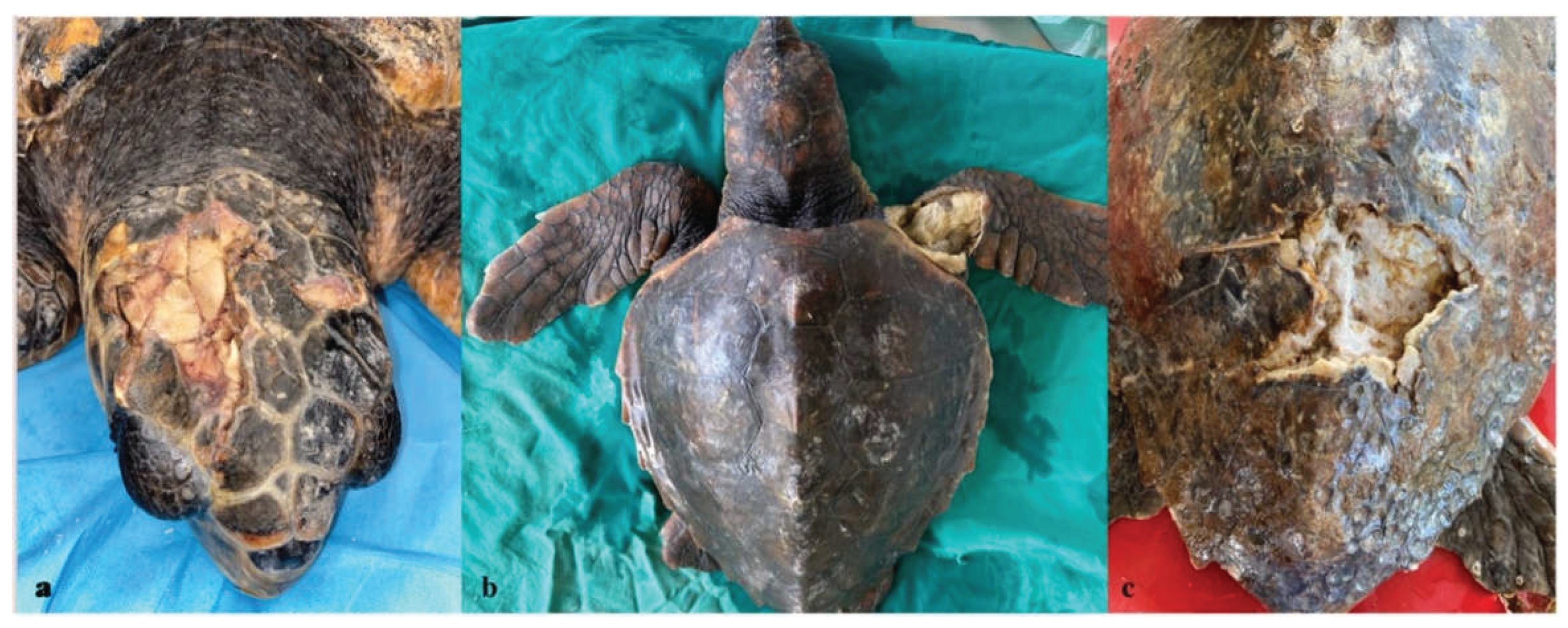
Figure 2.
Representative radiographic findings in loggerhead sea turtles (Caretta caretta) with traumatic injuries. (a) Latero-lateral view showing fractures of the parietal and postorbital bones of the head. (b) Latero-lateral view showing a caudal carapace fracture. (c) Dorso-ventral view showing a previous fracture of the right inframarginal region, with a concomitant left humeral fracture and marked flipper soft-tissue swelling of the flipper secondary to entanglement.
Figure 2.
Representative radiographic findings in loggerhead sea turtles (Caretta caretta) with traumatic injuries. (a) Latero-lateral view showing fractures of the parietal and postorbital bones of the head. (b) Latero-lateral view showing a caudal carapace fracture. (c) Dorso-ventral view showing a previous fracture of the right inframarginal region, with a concomitant left humeral fracture and marked flipper soft-tissue swelling of the flipper secondary to entanglement.
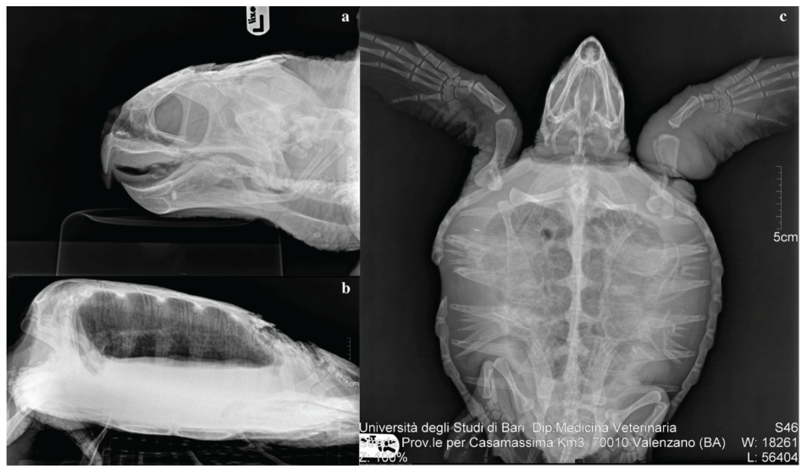

Table 1.
Severity and chronicity of traumatic lesions by anatomical site, among the 115 turtles with a single traumatic lesion.
Table 1.
Severity and chronicity of traumatic lesions by anatomical site, among the 115 turtles with a single traumatic lesion.
| Anatomical site | N | Mild n (%) | Moderate n (%) | Severe n (%) | Acute n (%) | Chronic n (%) |
|---|---|---|---|---|---|---|
| Head | 32 | 4 (12.5) | 11 (34.4) | 17 (53.1) | 8 (25.0) | 24 (75.0) |
| Carapace | 33 | 11 (33.3) | 15 (45.5) | 7 (21.2) | 5 (15.2) | 28 (84.8) |
| Plastron | 7 | 4 (57.1) | 2 (28.6) | 1 (14.3) | 2 (28.6) | 5 (71.4) |
| Neck | 12 | 3 (25.0) | 8 (66.7) | 1 (8.3) | 4 (33.3) | 8 (66.7) |
| Flipper | 31 | 3 (9.7) | 15 (48.4) | 13 (41.9) | 10 (32.3) | 21 (67.7) |
Table 2.
distribution of bacteria genera across anatomical sites.
| GENUS | HEAD | CARAPACE | PLASTRON | NECK | FLIPPER |
|---|---|---|---|---|---|
| Pseudomonas spp. | 12 | 18 | 2 | 5 | 0 |
| Citrobacter spp. | 6 | 3 | 0 | 0 | 19 |
| Vibrio spp. | 7 | 3 | 3 | 0 | 7 |
| Aeromonas spp. | 0 | 8 | 0 | 0 | 8 |
| Acinetobacter spp. | 2 | 4 | 0 | 2 | 2 |
| Proteus spp. | 0 | 4 | 0 | 0 | 0 |
| Enterobacter spp. | 0 | 2 | 0 | 0 | 0 |
| Klebsiella spp. | 0 | 2 | 0 | 0 | 0 |
| Staphylococcus spp. | 0 | 0 | 0 | 0 | 2 |
Disclaimer/Publisher’s Note: The statements, opinions and data contained in all publications are solely those of the individual author(s) and contributor(s) and not of MDPI and/or the editor(s). MDPI and/or the editor(s) disclaim responsibility for any injury to people or property resulting from any ideas, methods, instructions or products referred to in the content. |
© 2026 by the authors. Licensee MDPI, Basel, Switzerland. This article is an open access article distributed under the terms and conditions of the Creative Commons Attribution (CC BY) license (http://creativecommons.org/licenses/by/4.0/).
Copyright: This open access article is published under a Creative Commons CC BY 4.0 license, which permit the free download, distribution, and reuse, provided that the author and preprint are cited in any reuse.