

Submitted:
26 October 2026
Posted:
28 October 2026
You are already at the latest version
Abstract
Small leucine-rich proteoglycans (SLRPs) are an important group of molecules in the extracellular matrix (ECM) that are involved in regulating collagen assembly, tissue homeostasis, and cell signalling pathways. Among them, Decorin represents the proto-type member and plays a central role in maintaining ECM integrity, regulating fibrosis, and controlling tumor progression. This review examines the structural and functional biology of SLRPs, with a special focus on Decorin, highlighting its involvement in fibrotic diseases, degenerative disorders and the tumor microenvironment. Decorin acts as a multifunctional regulator through its interactions with TGF-β, various tyrosine kinase receptors, and multiple ECM components, carrying out anti-fibrotic, anti-angiogenic, anti-cancer, and tumour-suppressive functions. Additionally, factors such as the contribution of SLRPs to the desmoplastic response of tumors, their potential use as biomarkers for extracellular matrix remodelling, and the emerging therapeutic prospects based on administering recombinant Decorin, gene therapies, mimetic molecules, and strategies targeting the ECM. Overall, SLRPs, especially Decorin, stand out as key molecules in integrating structural and molecular signals and as promising diagnostic and therapeutic targets in fibrotic and neoplastic diseases.
Keywords:
small leucine-rich proteoglycans (SLRPs)
; Decorin
; extracellular matrix (ECM)
; desmoplasia
; fibrosis
; tumor microenvironment
; TGF-b signaling
; targeted therapy
1. Introduction
Small leucine-rich proteoglycans (SLRPs) are active components of the extracellular matrix (ECM) and are characterized by a small protein core with repeating leucine-rich structural motifs (LRRs), cysteine clusters, and covalently attached glycosaminoglycan (GAG) chains [34,40,41]. The family includes 18 grouped members in five categories, which differ based on chromosomal location, homology, and characteristics of the N- and C-terminal ends. Some of the most studied examples include Decorin and Biglycan (class I), Lumican and Fibromodulin (class II), Osteoglycin (class III), Tsukushi (class IV), and others [30,34,41]. Following their final structural modifications in the Golgi apparatus, SLRPs are secreted into the pericellular space, where whether bound to the extracellular matrix or in soluble forms, they interact with collagen fibers, with glycosaminoglycan (GAG) molecules, and also with membrane receptors, thus regulating the assembly and partly the organization of the extracellular matrix, cell–extracellular space interactions, and cell signaling [34,40,41]. Changes in the expression of specific SLRPs modify the morphology of collagen fibrils and the biomechanical properties of tissues, as well as normal functions like wound healing, corneal transparency, skin fragility, bone structure, having often a central role on non-neoplastic or neoplastic human diseases (see Table 1) [34,40,41].
Decorin, the archetypal member of class I, has only one N-terminal GAG chain, twelve LRRs and a C-terminal domain. Structurally, it can form dimers in crystals but is also active as a monomer in solution [43,57]. In addition to regulating fibrilogenesis, Decorin exerts tumor suppressor functions by modifying p21 and interacting with tyrosine kinase receptors such as EGFR, Met, VEGFR and IGF-1R [43,55,57]. In normal liver, decorin is localized around vascular structures and is increased in connective tissue septa as well as in the stroma of liver tumors and metastases. Its deficiency promotes hepatocarcinogenesis, while its excess suppresses it, suggesting a protective role in liver carcinogenesis [55]. In various epithelial carcinomas, Decorin expression in cancer cells is often reduced, while its increase attenuates premalignant functions through mechanisms involving autophagy, mitophagy, and immunomodulation, making it a candidate for therapeutic approaches based on molecules derived from the extracellular matrix [43,57]. Other SLRPs, such as Asporin and Biglycan, display more complex and extracellular environment-dependent roles. For example, while Decorin is generally considered a tumor suppressor, Biglycan tends to have pro- and anti-tumor effects, highlighting pro- and anti-tumor activities linked to the microenvironment [30,34,41]. The possibility of developing peptides or synthetic mimetic molecules derived from SLRPs that interfere with factors such as TGF-β and VEGF opens therapeutic prospects in fibrotic, ocular, renal and oncological diseases, with Decorin being a prototype molecule for targeting the extracellular matrix [20,40,55]. Data from knockout mouse models have elucidated some functional aspects, such as that the absence of Decorin is associated with alterations in collagen fiber homeostasis and deregulation of proliferative signals, including increased activation of tyrosine kinase receptors (PDGFRα, EGFR, RON, IGF-IR) and reduced p21 induction during experimental hepatocarcinogenesis, contributing to accelerated tumor growth [23].
2. Basic Biology of SLRPs
2.1. Structure and Molecular Features
SLRPs have a protein core with leucine-rich repeat regions (LRRs) that form an arched and curved tube with concave (rich in β-sheets and binding sites) and convex (helical) faces, delimited by terminal cysteine-rich motifs at the N- and C-termini, whose distinct arrangement is one of the criteria for their classification into distinct classes [14,20,49,57]. Each LRR (20-29 amino acids) has conserved motifs of the type LxxLxLxxNxL that alternate with other variable regions. The penultimate region of the structural motif at the C-terminus is relevant to conformation and function in many normal proteins [14,20,64]. The classes are distinguished by the number of LRRs, disulfide bond regions, and modifications: class I (e.g., Decorin, Biglycan, Asporin) typically has ~12 LRRs and O-linked GAGs (Decorin: 40 kDa, 12 LRRs, one Ser-linked GAG chain) with a concave/convex structure in a “horseshoe” shape [20,30,49,57,64]. Class II (e.g., Lumican, Fibromodulin, Keratocan) displays 10-12 LRRs, the conserved cysteine residues CX_3CXCX_9C, and sulfated tyrosine clusters at the N-terminus. Some members are proteoglycans (Lumican sulfate, keratan) or glycoproteins depending on the tissue [20,30,49]. Classes IV and V include non-canonical members with varying numbers of LRRs, distinct glycosidic patterns, and the absence of GAGs in many cases (e.g., Podocan, Podocan-like 1 with 17–20 LRRs and a highly acidic C-terminal domain) [20,30,49]. SLRPs are extensively modified (GAG-O-linked, N-glycosylation, and other glycosylations) affecting folding, secretion, and binding affinity. GAGs contribute to the assembly of the extracellular matrix (ECM) and the ability to bind cytochromes [14,20]. Preclinical studies show that local expression or targeted Decorin derivatives can reduce fibrosis and modify the effects of TGF-β (e.g., endothelial gene delivery or CAR-decorin in mouse models) and preserve retinal ganglion cells, although effective levels and dosages vary (with reported in vitro concentrations of over 1 μg/mL) [7,26,53]. In cancer, reduction of stromal Decorin has been associated with tumor tissue promotion and the facilitation of invasion [46]. Finally, post-transcriptional regulation of DCN involves microRNAs such as miR-181b, whose enhanced expression reduces Decorin in fibroproliferative environments, and blockade of this miRNA can partially restore DCN and limit differentiation into myofibroblasts, suggesting a potential therapeutic approach that should be further explored [29].
2.2. Classification and Evolutionary Context
The classification of SLRPs is based on structural and evolutionary criteria (chromosomal organization, protein homology, cysteine clusters at the N-terminus and regions at the C-terminus) and includes 17-18 members grouped into five classes (I-V), with conservation of the LRR motif and cysteine patterns at the N- and C-termini [20,30,41,49]. Classes I–III are canonical (proteoglycans with a protein core and one or more GAG chains), while classes IV–V are non-canonical, largely devoid of glycanization except for Chondroadherin, reflecting phenomena of gene duplication and functional divergence [20,30,41,49]. In summary: class I includes Decorin, Biglycan, Asporin, ECM2 and ECMX, characterized by the N-terminal consensus sequence CX_3 CXCX_6 C and conserved exonic and typical CS/DS glycanization [20,30,49]. Class II (Lumican, Fibromodulin, Keratocan, PRELP, Osteoadherin) consists of the CX_3 CXCX_9 C sequence, three exons with a large central exon and a predominance of keratan sulfate, with subgroups based on homology [20,30,49]. Class III (Opticin, Epiphycan, Osteoglycin) has fewer LRRs, a consensus CX_2 CXCX_6 C and a seven-exon organization [30,49]. Classes IV (Chondroadherin, Nyctalopin, Tsukushi) and V (Podocan, Podocan-like 1) are non-canonical branches with variations in the distance between the N-termini and in acidic or functionally distinct regions of the C-termini [20,30,49]. Chromosomal affinity suggests co-evolution: For example, ECM2 and Asporin are located side by side on chromosome 9, along with other members such as Osteoadherin and Osteoglycin, suggesting coordinated regulation in collagen-rich tissues [20,30]. Functionally, variations in SLRP expression and glycation modulate tissue and regeneration processes. Loss or modification of glycation can alter tissue homeostasis and regenerative responses [14,16]. Finally, in addition to their structural roles, SLRPs such as Decorin and Lumican affect signaling pathways associated with tumorigenesis: Decorin antagonizes tyrosine kinase receptors (EGFR, Met), binds to TGF-β limiting its activation and regulates angiogenesis, while alterations in Lumican have been associated with variable tumor outcomes, suggesting that changes in SLRP expression or glycation have relevant phenotypic consequences beyond the architecture of the EGFR [8]
3. Physiological Role of SLRPs
3.1. Organization and Homeostasis of the Extracellular Matrix (ECM)
The structural and functional balance of the extracellular matrix depends on the control of collagen assembly, fibril arrangement and their interactions with other macromolecular components, with SLRPs being central regulators of the initial stage of organization, subsequent fibril development and the overall homeostasis of the matrix (see Table 2) [14,20,34,52]. Decorin, Biglycan, and Lumican, along with Fibromodulin, regulate lateral growth, diameter, and interfibrillar spacing by binding to distinct sites on fibrils and working together to maintain tissue architecture [14,41]. This redundant and complementary relationship between members and classes is reflected in tissue fragility phenotypes, when they are absent [34,41]. Decorin and Biglycan share binding sites and regulatory functions in fibrilogenesis, often acting together in specialized tissues [14,34]. GAG chains and glycosylations of SLRPs influence interfibrillar spacing, hydration, and cytokine and growth factor sequestration, integrating protein and glucose components into ECM homeostasis [20,34]. In the kidney, these proteoglycans are mainly located in the tubular interstitial compartment where they regulate fibril assembly, cell-ECM interactions, and fibrotic processes. If released as soluble forms, they can activate receptors such as TLR2/TLR4 and modulate inflammatory responses [40,41]. In the desmoplastic tumor microenvironment, SLRP-mediated remodeling of the stromal matrix alters the physical and biological properties of the stromal layer, affecting tumor cell proliferation and migration. In many tumors, Decorin is stromal and its loss (sometimes through methylation) can promote tumor growth, while its reintroduction in vitro reduces tumor cell clusters formation [45,61].
3.2. Physiology of Dense and Soft Connective Tissues
In dense and loose connective tissues, SLRPs coordinate fiber architecture and mechanical properties in interaction with collagen and elastin, influencing fibrilogenesis, interfibrillar spacing and proteolytic protection [14,20,34]. In tendon, Decorin, Biglycan, Lumican and Fibromodulin coordinate fibril maturation and regulate their size and organization, with partially overlapping temporal and functional roles [14]. More specifically, in the cornea, a cluster of SLRPs with specific spatial and temporal distributions orchestrates stromal fibrilogenesis, hydration, and transparency. Loss of individual SLRPs leads to fibril disorganization and loss of transparency, with effects amplified by combined deletions [14,20]. The GAG chains of SLRPs (CS, DS, KS) regulate hydration, interfibrillar crosslinking, and force equilibration between fibrils. Alterations in the chains lead to defective fibrilogenesis and the mechanical properties of the ECM [14,50]. Specifically in muscle and adipose tissue, decorin as a myokine and other myokines and adipokines produced by skeletal muscle influence connective tissue composition, angiogenesis, and metabolism, contributing to homeostasis or dysfunction in pathological environments such as cachexia [15,33,36]. Changes in Decorin expression have also been linked to altered cardiac collagen fibril organization in models of muscle weakness, supporting a broader role for this proteoglycan in the mechanical homeostasis of muscle and cardiovascular connective tissues [60].
4. SLRPs in Non-Neoplastic Pathologies
4.1. Systemic and Organic Fibrotic Diseases
Systemic and organic fibrotic diseases are considered as paradigms in which interactions between ECM proteoglycans and profibrotic cytokines represent therapeutic targets (see Table 3). Decorin emerges as a key regulator for the kidney and liver [2,4,40,41]. In renal fibrosis, the deposition and increase of ECM leads to glomerulosclerosis and tubulointerstitial fibrosis with progression to renal failure, which is centrally mediated by TGF-β through Smad and non-Smad pathways [41]. Decorin, which is secreted by renal fibroblasts and increased in fibrotic areas, exhibits antifibrotic activity, as the absence of decorin appears to exacerbate fibrosis, while its overexpression or exogenous administration attenuates renal damage in experimental models [41,64]. The antifibrotic mechanisms include direct TGF-β binding, competition for TGF-βRI/II receptors, and modulation of pathways such as IGF-IR → PI3K/Akt/mTOR/p70S6K, consistent with a multifactorial action [40,41,64]. Even in hepatic fibrosis, activated stellate cells produce extracellular matrix (ECM) under the control of TGF-β1, and Decorin interferes with its bioactivity in vitro and in vivo, suggesting it as a strategic target against hepatic fibrosis [4,35]. Studies in liver injury models have also highlighted an increase in circulating soluble Biglycan that correlates with the degree of fibrosis, suggesting the application of Biglycan as a biomarker through ELISA methods specific for fragments produced by the metalloproteinases MMP-9 and MMP-12 [24]. Therapeutic approaches targeting local elevation of decorin have been tested with expression vectors and nanoparticles, showing a reduction in fibrotic and inflammatory markers, indicative of the potential to modulate peritoneal and pulmonary responses through local elevation of Decorin [2,10,63]. From an immunomodulatory perspective, Decorin and Biglycan may behave as danger molecules released upon cellular damage (DAMPs) activating TLR2/4 receptors and inflammasomes, integrating inflammatory and repair responses and complicating therapeutic strategies. This has prompted the development of peptides and SLRP mimetics that interact with TGF-β, VEGF and other mediators for the treatment of renal, hepatic, pulmonary and ocular fibrosis [20,30,40,63]. Finally, non-canonical members of the SLRP family, such as Tsukushi, bind to multiple extracellular ligands and modulate the FGF/MAPK, BMP and Wnt pathways, suggesting that the overall effect of SLRPs on fibrosis, results from the synergistic regulation of multiple pathways in addition to TGF-β and that therapeutic strategies must balance the control of fibrosis with the preservation of regenerative functions [17]. Beyond renal, hepatic, pulmonary, and ocular fibrosis, altered Decorin expression has also been implicated in endometriosis, where single-cell and experimental evidence suggests a relationship between stromal Decorin dysregulation and disease development with the exact pathogenetic mechanism that still needing to be elucidated [13].
4.2. Degenerative Bone and Joint Diseases
In the skeletal system, class I and II SLRPs regulate bone and cartilage homeostasis, and their alteration is associated with degenerative phenotypes associated with joint diseases. Decorin and Biglycan, regulate collagen fibrilogenesis and mineralization: their absence causes tissue fragility and reduced bone mass with synergistic effects in experimental double knockout models, partly due to reduced ability to bind TGF-β to the extracellular membrane and altered bone formation/resorption balance [44,71]. Class II SLRPs, including Lumican, also contribute to skeletal structure and mineralization. Lumican is secreted by mature osteoblasts and is associated with osteoblast maturation, while loss of some SLRPs negatively affects growth and mineral formation with partial compensation of other SLRPs [44,71].
In hyaline cartilage, Decorin, Biglycan, Keratocan and Asporin participate in fibrilogenesis and regulate TGF-β availability, interacting with regulators of mineralization and mechanical properties. In osteoarthritis, degradation of Aggrecan and collagen, activation of MMP/ADAMTS, as well as catabolic and inflammatory processes are observed. Pro-degenerative pathways such as TLR and Wnt promote protease production and disease progression. Pharmacological inhibition of the Wnt pathway has been shown in preclinical models to reduce cartilage degradation and promote regeneration, suggesting the therapeutic potential of regulating this pathway [44,59]. The interaction between SLRPs and TGF-β represents a critical point: Decorin, Biglycan, Asporin, and Fibromodulin bind to TGF-β and can be incorporated into collagen fibrils, regulating osteoprogenitor cell differentiation and bone formation. Biglycan also promotes osteoblastic differentiation through the activation of BMP-related signals and supports Wnt signaling, thereby promoting osteogenic signals. SLRPs collectively regulate fibril architecture, mineralization, and link TGF-β/BMP, Wnt pathways, and inflammatory responses, making them relevant targets for degenerative bone and joint diseases [44,59].
Finally, polymorphisms in genes involved in metabolism and inflammatory response can modify the susceptibility and progression of joint diseases, suggesting a link between systemic genetic profiles, circulating mediators and local tissue phenotypes that affect the anabolic/catabolic balance of the extracellular matrix (ECM) [44,59].
5. SLRPs in Cancer
5.1. Role in the Tumor Microenvironment and Tumorigenesis
In the neoplastic context (see Figure 1), the ECM provides mechanical support and a signaling platform that regulates angiogenesis, inflammation, and tumor progression. Increased tumor stiffness is associated with excessive deposition of extracellular matrix (ECM), fiber reorientation and crosslinking mediated by enzymes such as lysyl oxidase, and intense proteolytic remodeling by metalloproteases and cathepsins [6]. Proteoglycans and glycoproteins of the EGFR, including SLRPs, determine the mechanical and biochemical properties of the microenvironment, influencing adhesion, migration, and invasion through collagen organization and the presentation of growth factors and cytokines [6,30].
SLRPs such as Decorin and Biglycan can behave as DAMPs whose degradation releases fragments that activate TLR2 and TLR4 in immune cells, contributing to a circuit in which proteolytic remodeling and profibrotic cytokines perpetuate fibrosis and intratumoral inflammation [19,63]. The arrangement and density of collagen fibers also modulate T cell infiltration and activity, with implications for antitumor responses [63]. Decorin, which is mainly secreted by fibroblasts, endothelial cells, and myofibroblasts, possesses potent tumor suppressor properties and is a ligand for tyrosine kinase receptors (EGFR, IGF-1R, FGFR, Met) as well as TLR2/4 and TGF-β [3,30]. Its binding to EGFR activates the MAPK pathway and induces p21 with cell cycle arrest and reduced proliferation. In experimental models, Decorin also reduces the expression of oncoproteins such as HER-2 and inactivates Met, suppressing β-catenin signaling and limiting tumor dissemination with anti-angiogenic effects [3,59]. As an inhibitor of TGF-β, Decorin binds this cytokine to the extracellular membrane, reducing its bioavailability and attenuating pro-fibrotic and pro-oncogenic signaling [3,63]. TLR2/4 binding to Decorin on immune cells promotes programmed cell death 4 gene (PDCD4) production and miR-21 repression, contributing to an inflammatory microenvironment that may inhibit tumor growth. Increased stromal levels of Decorin have been associated with improved prognosis in various epithelial tumors [3,59]. Biglycan, although homologous to Decorin, often exhibits preneoplastic and immunosuppressive effects. Its soluble release and its fragments produced by MMPs (e.g., MMP-9, MMP-12) can also serve as biomarkers of desmoplastic remodeling in the tumor stroma [24,30,63].
5.2. SLRPs as Tumor Suppressors and Contextual Oncoproteins
The function of SLRPs in cancer is often interdependent on several factors. They can behave like tumor suppressors or oncoproteins depending on the tumor, the microenvironment and the signaling pathways involved. Decorin is the prototypical tumor suppressor among SLRPs as it binds to tyrosine kinase receptors such as EGFR, Met, IGF-1R, and FGFR, attenuates pro-proliferative signals, induces p21, cell cycle arrest, and apoptosis, and represses oncogenes such as β-catenin and Myc, translating EGFR signals into intracellular tumor suppressor molecular pathways [3,42]. Its LRR structure favors multivalent interactions with receptors and ligands, explaining its efficacy in several solid tumors [42]. Lumican also exhibits anti-tumor effects in specific contexts: its loss is associated with progression in certain tumors (e.g., osteosarcoma, melanoma) and acts through induction of p21, downregulation of cyclin D1, activation of pro-apoptotic pathways and modulation of integrins. However, its functions vary between tissues depending on the receptor conformation [30,42]. Despite structural homology to Decorin, Biglycan exhibits distinct interactions and functional effects [30,42]. Biglycan is primarily pro-neoplastic and pro-inflammatory, capable of suppressing immune responses and acting as a DAMP via TLR2/4. However, in humans (e.g., pancreatic cancer) it can exhibit inhibitory effects on proliferation associated with the induction of p27 and the reduction in proliferative markers, indicating tumor suppressive potential under specific conditions [30,42,63]. Asporin is an example of an oncoprotein with a high degree of involvement in tumorigenesis. In some cases (e.g., specific breast cancer subtypes) it antagonizes TGF-β1, reducing Epithelial-Mesenchymal transition (EMT) and proliferative capacity, while in other tumors (colon, gastric) it promotes progression and metastasis by interacting with Smads or EGFR/HER2 receptors and activating EMT-related MAPK/ERK pathways [30]. This demonstrates how LRR domains can mediate antagonistic interactions with TGF-β/Smads or EGFR receptors, reversing the functional role of the molecule [30].
In the tumor microenvironment, Decorin and Biglycan can act simultaneously as tumor suppressors and as proinflammatory regulators/DAMPs: TLR2/4 binding activates the production of TNF-α and IL-12p70, while Decorin/TGF-β complexes suppress anti-inflammatory mediators such as IL-10 via PDCD4. At the same time, Decorin binds TGF-β in the stroma, reducing its bioavailability and attenuating pro-fibrotic and pro-neoplastic functions, with a favorable prognostic effect in various epithelial tumors [3,43,63]. Variations in Decorin concentration and distribution also affect collagen fibril organization and tissue mechanical properties, contributing to its divergent effects on fibrosis and tumor progression in different histological contexts [14,43]. These findings highlight that SLRP itself can integrate tumor suppressor, immunomodulatory, and fibrotic programs, and that the outcome depends on tumor type, receptor profile, and tumorigenicity and the associated signaling pathways. Its clinical significance is reflected in the proposed use of Decorin and Biglycan levels in the bloodstream as biomarkers of inflammation and fibrosis and in the development of Decorin peptides derived from LRRs as therapeutic strategies targeting tyrosine kinase receptors [3,20,24,43]. A comparable stromal pattern has been reported in prostate cancer, where Decorin expression is reduced relative to the developing or non-neoplastic prostate stroma, supporting the concept that loss of stromal Decorin may accompany malignant progression [22]. Mechanistically, Decorin can induce autophagy in endothelial cells through Peg3, providing a direct link between extracellular Decorin signaling, endothelial homeostasis, and the autophagic program relevant to tumor angiogenesis [67].
6. SLRPs in Tumor Desmoplasia
6.1. Characteristics of ECM Desmoplasia
The ECM is a hierarchically organized network of collagens, glycoproteins, proteoglycans, and glycosaminoglycans whose composition and supramolecular architecture determine tissue mechanics, molecular transport, and cell signaling [79].
Desmoplastic ECM surrounding tumors results from profound reorganization of the interstitial extracellular matrix (ECM), dominated by fibrillar collagen (types I, III, V), Fibronectin, and proteoglycans such as hyaluronic acid and sulfated GAGs, which confer tensile and compressive strength and regulate hydration and force transmission [19,48,56,63]. Highly hydrophilic GAGs (hyaluronic acid >100 kDa; CS, DS, KS 20–60 kDa) produce hydrated and stressed regions that together with collagen form a dense and interconnected network in desmoplastic tumors, affecting cell migration, angiogenesis, and drug access [48,50,56]. Fibronectin in the provisional matrix serves as a scaffold for the assembly of collagen fibrils and the deposition of TGF-β and integrin complexes. Its mechanical expansion exposes cryptic sites that promote profibrotic phenotypes and EMT in stiff, anelastic microenvironments [63]. Increased density and stiffness of the matrix induce mechanical stress, integrin accumulation and aggregation, and activation of pathways such as FAK/Src, promoting actomyosin contraction, invasive migration, and tumor angiogenesis [19,63]. Stiffness and accumulation of extracellular matrix (ECM) compress vessels and increase interstitial pressure, reducing drug transport and making the desmoplastic extracellular matrix a therapeutic target [48,63].
Among the regulatory proteoglycans (see Table 4) , Decorin (class I SLRP) controls fibrogenesis and inhibits TGF-β by sequestering it in the ECM. Its loss causes aberrant fibrogenesis, increased fibroblast proliferation, and favors a microenvironment that allows tumor progression [19,34]. Decorin is also induced by pro-autophagic and nutritional stimuli, and its deficiency negatively affects autophagic events in tissues such as the heart, suggesting a functional role in regulating autophagy in the tumor microenvironment [21]. In the context of a tumor, the level and distribution of Decorin can therefore modify the balance between response, repair and malignant transformation [19,34]. Proteolytic degradation of collagen, fibronectin and proteoglycans releases DAMPs, including Decorin and Biglycan, which activate TLR2/4 on macrophages, dendritic cells and T lymphocytes, integrating inflammatory and fibrotic responses in the desmoplastic extracellular matrix [34,63]. Furthermore, the Variations in GAG modifications (sulfonation, epimerization) alter the charge, hydration, and binding of growth factors, contributing to functional matrix heterogeneity [50,56]. Finally, SLRPs regulate inflammation, angiogenesis, and fibroblast activity, influencing the transition between the repair and pre-invasive microenvironment [47].
6.2. Clinical Implications for Drug Response and Prognosis
The stiffness and density of the desmoplastic extracellular matrix negatively affect the penetration of drug agents, intratumoral perfusion, and induce hypoxia, contributing to chemotherapeutic resistance and affecting prognosis in fibrotic tumors such as aggressive breast and pancreatic cancer [48,63]. Colorectal tumors with a high tumor-stroma ratio (proportion of cancer cells and desmoplastic stroma) demonstrate better survival rates [18] Reducing stromal stiffness, achieved by minimizing cancer associated fibroblasts (CAFs) or reducing collagen, improved perfusion and facilitated drug delivery in preclinical models. Antifibrotic therapies (e.g., losartan, tranilast, hyaluronidase) have increased the response to chemotherapeutics in recent clinical trials (e.g., losartan + FOLFIRINOX in the pancreatic route) [63]. Targeting collagen cross-linking enzymes (LOX/LOXL2) and molecular mechanosensors such as FAK have shown preclinical efficacy, but mixed clinical results, suggesting the need to optimize therapeutic timing and combinations [48,63]. FAK inhibitors can normalize tumor immunity and increase sensitivity to chemotherapy and immunotherapy, but also activate compensatory pathways (e.g., STAT3), requiring targeted combination approaches [48,63].
SLRP proteoglycans play a dual role: Lumican has been exploited as a retention platform for cytokines (IL-2, IL-12), prolonging their intratumoral action and enhancing the effect in combination with checkpoint inhibitors [3,48]. Decorin acts as a pan-inhibitor of receptor tyrosine kinases and, due to its structural properties (repetitive structural motifs and leucine-rich sites for TGF-β), represents a stromal therapy strategy for both topical administration and gene therapy, with evidence of tumor suppressive function in in vivo models [3,28]. The ability of Decorin to form complexes with growth factors in the extracellular membrane (ECM) and to modulate signaling through receptors such as LRP-1 (e.g., via LRR6 sequences) offers opportunities to reduce local fibrosis and improve stromal permeability using mimicry peptides [9,28]. CAFs and ECM also influence immunotherapy: CXCL12 secretion by FAP-positive CAFs limits T-cell infiltration and response to anti-CTLA4/anti-PD-L1, but inhibition of this pathway restores T-cell access and improves therapeutic efficacy [19,63]. Since SLRPs such as Decorin and Biglycan can also act as DAMPs via TLR2/4 and modify collagen architecture, their conformation affects the balance between mechanical entrapment of T cells and immune activation, with relevant clinical implications [3,63]. Circulating or stromal levels of SLRPs (e.g., MMP-9/MMP-12 modified Biglycan; stromal Decorin) are proposed as biomarkers of fibrosis and prognosis and could guide the selection of antifibrotic and anticancer therapies [3,24,35]. Finaly, with cardiovascular studies, quantification of extracellular matrix parameters (volume and density of fibrotic components) through imaging and integrated molecular analysis could improve the prediction of tumor ischemia and therapeutic response [48,51] and the distinction between atherosclerotic plaque burden, anatomical stenosis, and functional ischemia illustrates why structural imaging findings should be integrated with functional measurements rather than interpreted as interchangeable endpoints [37].
7. Class I SLRPs and the Archetypal Decorin Model
7.1. Structure and Variants of Decorin
Decorin is the prototype of class I SLRPs, with a highly conserved protein core of approximately 36–40 kDa organized into four domains, a single O-linked glycosaminoglycan chain (chondroitin/dermatan sulfate-CS/DS) linked to a serine near the N-terminus, and a central leucine-rich region (LRR) that forms the arched, curved tubular architecture typical of the family [30,43,57,64]. GAG is a heterogeneous polymer of disaccharides containing D-glucuronic acid or L-iduronic acid. Its length and the degree of epimerization and sulfation sites vary between tissues, with potential implications for fibrilogenesis and cytokine binding [14,50,57]. The LRR domain consists of twelve repeats of 20–29 amino acids that create a tubular-like fold with a concave surface rich in β-sheets and a convex surface with helical elements. This organization has been confirmed by crystallographic structures depicting the molecule as a “horseshoe” (see Figure 2) [14,20,64]. LRRs contain Histidine-Cysteine clusters (HCS) motifs of the LxxLxLxxNxL type and variable segments (VS) that can form 3-10 α-helices, or other secondary elements, contributing to tubular curvature and binding diversity [14,20]. Functional studies have mapped specific activities to individual LRRs: for example, LRR V/VI binds to VEGFR2, LRR VII mediates binding to type I collagen on the concave surface, LRR XII interacts with cellular communication network 2/ connective tissue growth factor (CCN2/CTGF), while the penultimate C-terminal LRR (LRR XI) constitutes the terminal or LRRCE cysteine rich cap domain, critical for corneal-specific formation and function. Mutations in this region are associated with congenital corneal stromal dystrophy [20,64]. In the N- and C-terminal regions, Decorin contains cysteine-rich residues that form stabilizing disulfide bonds (LRRNT and LRRCT), which are essential for folding of the LRR domain and for regulating ligand binding [14,20,49]. Regarding post-translational modifications, the protein core contains N-glycosylation sites, and the protein can exist as a proteoglycan with a CS/DS chain or as a glycoprotein without GAGs. These glycosylations affect stability, secretion, and binding affinity, making Decorin a model for studying how glycosylation variants modify ECM function and its tumor suppressor activity [14,20,43,57]. Although dimers have been observed in some cases, under natural conditions, the biologically active form appears predominantly monomeric [20,43,64]. The phylogenetic identity of Decorin in class I, together with Biglycan, Asporin, and ECM2, is supported by a CX_3CXCX_6C signature at the N-terminus that forms two disulfide bridges and by a gene organization into eight exons with conserved junctions, confirming structural homology and a common group evolutionary cluster [30,49]. Decorin is richly glycosylated, and its modifications (N-glycosylation and O-linked GAG chains) are essential for proper folding, secretion and regulation of interactions with collagen, cytokines and tyrosine kinase receptors, with implications for tissue function and the molecule’s role in tumor suppression. Clinical data also suggest potential diagnostic and prognostic applications, although confirmation in larger cohorts is required [14,20,31,43,57].
7.2. Physiological Functions and Regulation of Fibrilogenesis
The physiological functions of Decorin in the regulation of collagen fibrilogenesis depend on its LRR architecture, the single GAG CS/DS chain, and the ability to selectively bind to fibril regions. Decorin binds to fibril collagens, particularly type I, through the central LRR 4-6 domain containing the SYIRIADTNIT sequence and may possess multiple binding domains that allow the simultaneous interaction of a monomer with multiple collagen molecules [14,20]. This multipotent action regulates the growth, diameter, and organization of lateral fibrils, and binding to procollagen suggests control from early stages of protofibril assembly [14,41]. During early growth, protofibrils deposited in the extracellular matrix mature into large-diameter fibrils through end-to-end and side-linkages and increased cross-linking mediated by lysyl oxidase. Decorin stabilizes protofibrils, and its absence in experimental knockout models results in increased fibril diameter, loose collagen networks, and altered tissue mechanics [14,34,41]. In vitro, Decorin controls lateral growth and diameter by binding to the “d” and “e” bands of tendon collagen through at least two regions along the tubular LRR [14,34]. The Decorin protein core protects fibrils from proteolytic degradation as its binding to fibril surfaces limits the access of collagenases and specific MMPs, contributing to structural homeostasis in tissues such as cornea, tendon, and bone [14,48]. The concave β-sheet region of Decorin is the main collagen-binding interface, while the CS/DS GAG extends into the interfibrillar space. Sulfurized GAGs regulate the hydration of ECM and connect neighboring fibrils, forming networks that transfer forces between fibrils and confer mechanical integrity to the tissue [14,20]. GAG-deficient Decorin mutants result in larger diameter fibrils in 3D cultures, and mice lacking dermatan sulfate epimerase 1 display alterations in skin collagen structure, indicating that the CS/DS chain is essential for hydration, lateral growth, and interfibrillar spacing [14,50]. Furthermore, Decorin and Biglycan, through their GAGs, inhibit hydroxyapatite (HAP)-induced crystal growth and regulate bone mineralization, linking the control of fibrilogenesis to skeletal matrix mineralization [14,52]. In the intermediate matrix, Decorin also interacts with collagen VI and Tenascin-X with collagen VI forming microfilaments that cross-link with collagen I in the “d” zone, where Decorin and Biglycan bind, allowing the formation of pericellular complexes with Matrilin-1 and Aggrecan. The GAG chain of Decorin also mediates the attachment of Tenascin-X to fibrils, contributing to three-dimensional organization and force transmission in the extracellular matrix [14,20,34,48]. Finally, the regulation of fibrogenesis by Decorin must be considered in conjunction with the dynamics of secretion, N-glycosylation and CS/DS chain modifications, which vary depending on tissue and development and can modify binding affinities and functions during matrix assembly and remodeling [14,20,34,41].
8. Future Perspectives and Clinical Applications
8.1. SLRPs as Biomarkers in Extracellular Matrix (ECM) Pathologies and Oncology
The use of Decorin and Biglycan as biomarkers emphasize their soluble presence in biological fluids and their role in tissue remodeling. In liver fibrosis, Biglycan fragments produced by metalloproteases (particularly MMP-9 and MMP-12) have been quantified by ELISA as markers of fibrosis [24]. For lung diseases, serum Decorin and cleaved Decorin fragments are proposed as markers of ECM degradation and fibrotic progression, measurable by ELISA and quantitative proteomics [56]. In oncology, proteomic studies show under expression of Decorin in colon cancer compared to normal tissue, with validation by Western blot and immunohistochemistry indicating it as a potential negative biomarker in colon carcinogenesis [31]. Similarly, high expression of Biglycan has been associated with poor prognosis in gastric adenocarcinoma and lung cancer, suggesting prognostic value in various tumors [56].
Systemic biomarkers may also complement ECM-derived markers: promoter variants in the adiponectin gene have been associated with susceptibility to non-small cell lung cancer in a Han Chinese population, although their relationship with SLRP remodeling remains to be clarified [32].
Circulating adipocytokines have likewise been investigated as biomarkers of colorectal neoplasia, with adiponectin proposed for colorectal adenoma and resistin and visfatin for colorectal cancer, indicating that ECM-related biomarkers may be strengthened by integration with systemic metabolic mediators [39].
8.2. Decorin as a Therapeutic Target and Delivery Strategies
The therapeutic use of Decorin requires formulations that protect the protein from degradation and allow its targeted release since recombinant Decorin is particularly suitable for accessible and immunologically privileged areas, such as the eye, with local or topical administrations capable of modifying fibrosis and inflammation [5,20]. To overcome its short half-life, systems based on biomaterials, hydrogels, scaffolds (e.g., hyaluronic acid or collagen-based) and microrods have been developed, which are used as reservoirs for controlled release or for the delivery of plasmids encoding DCN. Co-administration of pDCN with anti-inflammatory genes enhanced the repression of TGF-β1 and ECM genes compared to single plasmids [1,5]. Gene therapy with viral vectors (adenovirus, AAV) carrying DCN has shown efficacy in various models in reducing fibrosis and modulating TGF-β signaling [5,57]. Cellular approaches include modified cells capable of expressing Decorin in a regulated manner (so-called “smart cells”), offering potential anti-fibrotic feedback systems that have not yet been validated in vivo [5,12]. In oncology, oncolytic vectors expressing Decorin (e.g., elements coexpressing Ad-DCN and IL-12) combine oncolysis and tumor suppression by reducing tumor extracellular matrix, attenuating TGF-β signaling, and enhancing CD8+ lymphocyte activation, with potential benefits in viral penetration and treatment sensitivity [48,57,63,64].
8.3. SLRP Mimetics and Therapies Targeting ECM
The interest in SLRP mimetic molecules and strategies targeting ECM stems from the multifactorial role of proteoglycans such as Decorin, which is defined as the “guardian of ECM” for its antifibrotic, anti-inflammatory and antitumor activity through TGF-β1 antagonism and modification of pro-tumor pathways (see Table 5) [5,20]. Studies show that inclusion of SLRP in experimental collagen-rich ECM preparations reduces fibrilogenesis and compaction, but models lacking the GAG chain show that the protein core alone may not be sufficient because of the absence of GAGs that is associated with abnormal collagen maturation and mechanical damage, suggesting that effective mimetic molecules should also reproduce GAG-mediated interactions [11,16]. For targeting the extracellular matrix (ECM), guide peptides such as CendR (C-terminal R/KXXR/K motif), the CAR peptide that binds to heparan sulfate proteoglycans (HSPGs), and tCRK promote extravasation, tissue penetration, and delivery of therapeutic cargoes. The use of nanoparticles improves half-life and local delivery and allows for antifibrotic targeting of the skin when bound to proteins such as decorin [5,20,27]. In oncology, the ability of SLRPs to anchor factors to the stroma has been employed with Lumican to bind cytokines (e.g., IL-2, IL-12) to the tumor stroma periphery, increasing retention and therapeutic efficacy in combination with immunotherapy [48]. Genetic and cellular approaches include decorin-encoding plasmids (pDCN) delivered into collagen scaffolds and smart cells expressing decorin in response to TGF-β, aiming to replicate physiological regulations and remodeling of the extracellular matrix (ECM) in fibrosis and cancer [1,5,64]. Clinical perspectives also consider the use of Decorin and ECM-targeted mimetics in emerging fibrotic diseases, such as pulmonary fibrosis after COVID-19, with aerosol formulations or nanoparticles directed to the parenchyma, extending the SLRP paradigm to new therapeutic scenarios [2,5,63].
8.4. Emerging Directions in Translational Research for SLRPs
Translational research on SLRPs is mainly focused on Decorin, which is presented both as a therapeutic agent and as a delivery platform. Decorin is being studied for its anti-inflammatory, and antitumor properties, and a decorin-based preparation has been developed for retinal and macular diseases in combination with anti-VEGF and immunotherapies [5,20,56,63]. Gene and controlled delivery approaches include AAV-DCN vectors for tissue transfer and pDCN plasmids released from collagen scaffolds. In some models, coexpression of DCN with cytokines such as IL-10 enhanced the suppression of TGF-β1 and ECM genes compared to single treatments [1,5,20,57]. In oncology, Decorin has been introduced into oncolytic adenoviruses and IL-12-expressing constructs (e.g., Ad-DCN, Ad-ΔE1B-DCN, RdB/IL12/DCN) to enhance viral spread, reduce tumor extracellular matrix, and attenuate TGF-β-dependent immunosuppression, with documented antitumor effects [27,48,63,64]. Other SLRPs are used as cytokine platforms, for example, Lumican has been used to bind IL-2 and IL-12 to the tumor stroma, improving their retention and duration of action in combination with PD-L1 blockade, and LRR-derived peptides are under development to obtain smaller, pharmacokinetically favorable molecules [5,20,48,56]. Soluble SLRPs such as Decorin and Biglycan, as well as fragments produced by MMP-9 and MMP-12, are proposed as biomarkers of extracellular matrix remodeling and fibrosis. Incorporation of native ECM proteins with peptide carriers and viral or gene vectors opens up prospects for ECM-targeted therapies, even in emerging contexts such as post-COVID-19 pulmonary fibrosis [2,5,56,63]. Finaly, directed differentiation of induced pluripotent stem cells toward alveolar-like phenotypes has also been used as a platform for developmental and teratogenicity assessment, illustrating the growing role of advanced cell models in translational safety studies [38].
9. Final Conclusions
SLRP family members act as connecting links between the structural properties of the matrix and the modulation of extracellular signals. This is mainly due to the LRR architecture, the cysteine-rich conserved alleles at the C/N termini and the glycosidic modifications. Thus, they coordinate fibrilogenesis, interfibrillar hydration and growth factor sequestration, translating the characteristics of the SLRP into the regulation of proliferation, autophagy, angiogenesis and immune responses [14,20,57]. Decorin, a functional example, demonstrates how a single SLRP can simultaneously contribute to collagen fibril stabilization, tyrosine kinase receptor antagonism, and the restriction of active TGF-β, with effects that manifest in fibrotic and oncological contexts and are dependent on isoforms, glycosylation, and proteolysis in specific contexts [14,20,57]. Translationally, the evaluation of soluble forms and fragments of SLRPs offers potential as biomarkers of extracellular matrix (ECM) remodeling, while therapeutic strategies including recombinant Decorin, gene vectors, DCN-expressing oncolytics, hydrogels and homing peptides aim to improve targeting, stability and local delivery in fibrotic and desmoplastic tumors [5,56,63]. Addressing practical issues such as pharmacokinetics, immunogenicity, preservation of glycan interactions and standardization of assays to validate efficacy and safety remains critical. Controlled studies that incorporate molecular, functional and structural measurements of the target tissue are therefore essential to progress from proof of principle to specific clinical applications [1,20].
Author Contributions
Conceptualization, D.B.; Methodology, D.B. A.L, N.K, G.T; Resources, X.X.; Data Curation, D.B.; Writing – Original Draft Preparation, D.B, A.L.; Writing – Review & Editing, D.B, A.K, A.L, N.K, G.T.; Visualization, D.B, A.L, N.K, G.T.; Supervision, A.L, N.K, G.T.; Project Administration, A.L, N.K, G.T. All authors have read and agreed to the published version of the manuscript.
Funding
This research received no external funding.
Institutional Review Board Statement
Not applicable.
Informed Consent Statement
Not applicable.
Data Availability Statement
No new data were created or analyzed in this study. Data sharing is not applicable to this article.
Acknowledgments
During the preparation of this manuscript, the authors made limited use of Copilot Microsoft solely as a basic writing assistant for minor linguistic refinement and formatting adjustments and for generation of the drawings embedded in Figure 1 and Figure 2. All scientific content, interpretations, and conclusions remain entirely the responsibility of the authors. All authors consented to the acknowledgements.
Conflicts of Interest
The authors declare no conflicts of interest.
Abbreviations
The following abbreviations are used in this manuscript:
| AAV | Adeno-associated virus |
| ADAMTS | A disintegrin and metalloproteinase with thrombospondin motifs |
| AMPK | AMP-activated protein kinase |
| BMP | Bone morphogenetic protein |
| CAF | Cancer-associated fibroblast |
| CAR | Cysteine–alanine–arginine peptide |
| CCN2/CTGF | Cellular communication network factor 2/connective tissue growth factor |
| CS | Chondroitin sulfate |
| DAMP | Damage-associated molecular pattern |
| DCN | Decorin |
| DS | Dermatan sulfate |
| ECM | Extracellular matrix |
| EGFR | Epidermal growth factor receptor |
| ELISA | Enzyme-linked immunosorbent assay |
| EMT | Epithelial–mesenchymal transition |
| FAK | Focal adhesion kinase |
| FGF/FGFR | Fibroblast growth factor/fibroblast growth factor receptor |
| GAG | Glycosaminoglycan |
| HAP | Hydroxyapatite |
| HSPG | Heparan sulfate proteoglycan |
| IGF-1R | Insulin-like growth factor 1 receptor |
| IL | Interleukin |
| KS | Keratan sulfate |
| LOX/LOXL2 | Lysyl oxidase/lysyl oxidase-like 2 |
| LRP-1 | Low-density lipoprotein receptor-related protein 1 |
| LRR | Leucine-rich repeat |
| MAPK | Mitogen-activated protein kinase |
| MMP | Matrix metalloproteinase |
| PDCD4 | Programmed cell death protein 4 |
| PDGFR | Platelet-derived growth factor receptor |
| PRELP | Proline/arginine-rich end leucine-rich repeat protein |
| SLRP | Small leucine-rich proteoglycan |
| TGF-β | Transforming growth factor beta |
| TLR | Toll-like receptor |
| TNF-α | Tumor necrosis factor alpha |
| VEGF/VEGFR | Vascular endothelial growth factor/vascular endothelial growth factor receptor |
References
- Abbah, S.A.; et al. Co-transfection of decorin and interleukin-10 modulates pro-fibrotic extracellular matrix gene expression in human tenocyte culture. Sci. Rep. 2016, 6(1). [Google Scholar] [CrossRef]
- Allawadhi, P.; et al. Decorin as a possible strategy for the amelioration of COVID-19. Med. Hypotheses 2021, 152. [Google Scholar] [CrossRef]
- Appunni, S.; et al. Small leucine rich proteoglycans (decorin, biglycan and lumican) in cancer. Clin. Chim. Acta 2019, 491. [Google Scholar] [CrossRef]
- Baghy, K.; Iozzo, R.V.; Kovalszky, I. Decorin–TGFβ axis in hepatic fibrosis and cirrhosis. J. Histochem. Amp Cytochem. 2012, 60(4). [Google Scholar] [CrossRef]
- Baghy, K.; Szakadáti, H.; Kovalszky, I. Decorin the antifibrotic proteoglycan and its progression in therapy. Am. J. Physiol.-Cell Physiol. 2025, 328(6). [Google Scholar] [CrossRef]
- Balkwill, F.R.; Capasso, M.; Hagemann, T. The tumor microenvironment at a glance. J. Cell Sci. 2012, 125(23). [Google Scholar] [CrossRef]
- Begum, G.; et al. Altered decorin biology in proliferative vitreoretinopathy: A mechanistic and cohort study. Investig. Opthalmology Amp Vis. Sci. 2018, 59(12). [Google Scholar] [CrossRef]
- Bi, X.-L.; Yang, W. Biological functions of decorin in cancer. Chin. J. Cancer 2013, 32(5). [Google Scholar] [CrossRef]
- Cabello-Verrugio, C.; et al. The internal region leucine-rich repeat 6 of decorin interacts with low density lipoprotein receptor-related protein-1, modulates transforming growth factor (TGF)-β-dependent signaling, and inhibits TGF-β-dependent fibrotic response in skeletal muscles. J. Biol. Chem. 2012, 287(9). [Google Scholar] [CrossRef]
- Chaudhary, K.; et al. Nanotechnology and adeno-associated virus-based decorin gene therapy ameliorates peritoneal fibrosis. Am. J. Physiol.-Ren. Physiol. 2014, 307(7). [Google Scholar] [CrossRef]
- Chen, D.; et al. Distinct effects of different matrix proteoglycans on collagen fibrillogenesis and cell-mediated collagen reorganization. Sci. Rep. 2020, 10(1). [Google Scholar] [CrossRef]
- Chen, F.; et al. Cardioprotective effect of decorin in type 2 diabetes. Front. Endocrinol. 2020, 11. [Google Scholar] [CrossRef]
- Chen, G.; et al. Unraveling the significance of decorin in endometriosis development through single cell sequencing and experimental approaches. PLoS ONE 2026, 21(5). [Google Scholar] [CrossRef]
- Chen, S.; Birk, D.E. The regulatory roles of small leucine-rich proteoglycans in extracellular matrix assembly. FEBS J. 2013, 280(10). [Google Scholar] [CrossRef]
- Dalamaga, M. Interplay of adipokines and myokines in cancer pathophysiology: Emerging therapeutic implications. World J. Exp. Med. 2013, 3(3). [Google Scholar] [CrossRef]
- Daquinag, A.C.; et al. Glycosaminoglycan modification of decorin depends on MMP14 activity and regulates collagen assembly. Cells 2020, 9(12). [Google Scholar] [CrossRef]
- Dellett, M.; et al. Small leucine rich proteoglycan family regulates multiple signalling pathways in neural development and maintenance. Dev. Growth Amp Differ. 2012, 54(3). [Google Scholar] [CrossRef]
- Eriksen, A.C.; et al. Does heterogeneity matter in the estimation of tumour budding and tumour stroma ratio in colon cancer? Diagn. Pathol. 2018, 13(1). [Google Scholar] [CrossRef]
- Foster, D.S.; et al. The evolving relationship of wound healing and tumor stroma. JCI Insight 2018, 3(18). [Google Scholar] [CrossRef]
- Gesteira, T.F.; Verma, S.; Coulson- Thomas, V.J. Small leucine rich proteoglycans: Biology, function and their therapeutic potential in the ocular surface. Ocul. Surf. 2023, 29. [Google Scholar] [CrossRef]
- Gubbiotti, M.A.; et al. Decorin is an autophagy-inducible proteoglycan and is required for proper in vivo autophagy. Matrix Biol. 2015, 48. [Google Scholar] [CrossRef]
- Henke, A.; et al. Stromal expression of decorin, Semaphorin6D, SPARC, Sprouty1 and tsukushi in developing prostate and decreased levels of decorin in prostate cancer. PLoS ONE 2012, 7(8). [Google Scholar] [CrossRef]
- Horváth, Z.; et al. Decorin deficiency promotes hepatic carcinogenesis. Matrix Biol. 2014, 35. [Google Scholar] [CrossRef]
- Hsieh, L.T.-H.; et al. Soluble biglycan as a biomarker of inflammatory renal diseases. Int. J. Biochem. Amp Cell Biol. 2014, 54. [Google Scholar] [CrossRef]
- Järvinen, T.A.H.; Prince, S. Decorin: A growth factor antagonist for tumor growth inhibition; BioMed Research International, 2015. [Google Scholar]
- Järvinen, T.A.H.; Ruoslahti, E. Target-seeking antifibrotic compound enhances wound healing and suppresses scar formation in mice. Proc. Natl. Acad. Sci. 2010, 107(50). [Google Scholar] [CrossRef]
- Järvinen, T.A.H.; Ruoslahti, E. Generation of a multi-functional, target organ-specific, anti-fibrotic molecule by molecular engineering of the extracellular matrix protein, decorin. Br. J. Pharmacol. 2018, 176(1). [Google Scholar] [CrossRef]
- Kubo, E.; et al. Role of decorin in the lens and ocular diseases. Cells 2022, 12(1). [Google Scholar] [CrossRef]
- Kwan, P.; Ding, J.; Tredget, E.E. MicroRNA 181b regulates decorin production by dermal fibroblasts and may be a potential therapy for hypertrophic scar. PLoS ONE 2015, 10(4). [Google Scholar] [CrossRef]
- Lall, S.P.; et al. ASPORIN: A root of the matter in tumors and their host environment. Biochim. Et. Biophys. Acta (BBA) -Rev. Cancer 2024, 1879(1). [Google Scholar] [CrossRef]
- Li, G.; et al. Identifying DCN and HSPD1 as potential biomarkers in colon cancer using 2D-LC-MS/MS combined with iTRAQ technology. J. Cancer 2017, 8(3). [Google Scholar] [CrossRef]
- Li, Y.; et al. The association of adiponectin gene promoter variations with non-small cell lung cancer in a han chinese population. PLoS ONE 2015, 10(5). [Google Scholar] [CrossRef]
- Manole, E.; et al. Myokines as possible therapeutic targets in cancer cachexia. J. Immunol. Res. 2018. [Google Scholar] [CrossRef]
- Merline, R.; Schaefer, R.M.; Schaefer, L. The matricellular functions of small leucine-rich proteoglycans (SLRPs). J. Cell Commun. Signal. 2009, 3, 3–4. [Google Scholar] [CrossRef]
- Meyer, D.H.; et al. Biglycan and decorin gene expression in normal and fibrotic rat liver: Cellular localization and regulatory factors. Hepatology 1992, 16(1). [Google Scholar] [CrossRef]
- Miao, Y.; et al. Myokines related to leukocyte recruitment are downregulated in osteosarcoma. Int. J. Med. Sci. 2018, 15(9). [Google Scholar] [CrossRef]
- Min, J.K.; Chandrashekhar, Y. Atherosclerosis, stenosis, and ischemia. JACC Cardiovasc. Imaging 2018, 11(4). [Google Scholar] [CrossRef]
- Mitchell, A.; et al. Assessment of iPSC teratogenicity throughout directed differentiation toward an alveolar-like phenotype. Differentiation 2019, 105. [Google Scholar] [CrossRef]
- Nakajima, T.E.; et al. Adipocytokines as new promising markers of colorectal tumors: Adiponectin for colorectal adenoma, and resistin and visfatin for colorectal cancer. Cancer Sci. 2010, 101(5). [Google Scholar] [CrossRef]
- Nastase, M.V.; et al. Small leucine-rich proteoglycans in renal inflammation: Two sides of the coin. J. Histochem. Amp Cytochem. 2018, 66(4). [Google Scholar] [CrossRef]
- Nastase, M.V.; Iozzo, R.V.; Schaefer, L. Key roles for the small leucine-rich proteoglycans in renal and pulmonary pathophysiology. Biochim. Et. Biophys. Acta (BBA) -General. Subj. 2014, 1840(8). [Google Scholar] [CrossRef]
- Neill, T.; Schaefer, L.; Iozzo, R.V. Decorin. Am. J. Pathol. 2012, 181(2). [Google Scholar] [CrossRef]
- Neill, T.; Schaefer, L.; Iozzo, R.V. Decorin as a multivalent therapeutic agent against cancer. Adv. Drug Deliv. Rev. 2016, 97. [Google Scholar] [CrossRef]
- Nikitovic, D.; et al. The biology of small leucine-rich proteoglycans in bone pathophysiology. J. Biol. Chem. 2012, 287(41). [Google Scholar] [CrossRef]
- Nyman, M.C.; et al. Decorin in human colon cancer. J. Histochem. Amp Cytochem. 2015, 63(9). [Google Scholar] [CrossRef]
- ODA, G.; et al. Significance of stromal decorin expression during the progression of breast cancer. Oncol. Rep. 2012, 28(6). [Google Scholar] [CrossRef]
- Pang, X.; Dong, N.; Zheng, Z. Small leucine-rich proteoglycans in skin wound healing. Front. Pharmacol. 2020, 10. [Google Scholar] [CrossRef]
- Piersma, B.; Hayward, M.-K.; Weaver, V.M. Fibrosis and cancer: A strained relationship. Biochim. Et. Biophys. Acta (BBA) -Rev. Cancer 2020, 1873(2). [Google Scholar] [CrossRef]
- Pietraszek-Gremplewicz, K.; et al. Small leucine-rich proteoglycans and matrix metalloproteinase-14: Key partners? Matrix Biol. 2019, 75–76. [Google Scholar] [CrossRef]
- Pomin, V.; Mulloy, B. Glycosaminoglycans and proteoglycans. Pharmaceuticals 2018, 11(1). [Google Scholar] [CrossRef]
- Poole, J.J.A.; Mostaço-Guidolin, L.B. Optical microscopy and the extracellular matrix structure: A review. Cells 2021, 10(7). [Google Scholar] [CrossRef]
- Randilini, A.; Fujikawa, K.; Shibata, S. Expression, localization and synthesis of small leucine-rich proteoglycans in developing mouse molar tooth germ. Eur. J. Histochem. 2020, 64(1). [Google Scholar] [CrossRef]
- Rao, P.; et al. AAV-IKV mediated expression of decorin inhibits EMT and fibrosis in a murine model of glaucoma and AAV-IKV transduction in non-human primates. Sci. Rep. 2025, 15(1). [Google Scholar] [CrossRef]
- RAO, Y.; et al. Research progress on the role of decorin in the development of oral mucosal carcinogenesis. Oncol. Res. 2025, 33(3). [Google Scholar] [CrossRef]
- Reszegi, A.; et al. The protective role of decorin in hepatic metastasis of colorectal carcinoma. Biomolecules 2020, 10(8). [Google Scholar] [CrossRef]
- Ricard-Blum, S.; et al. A biological guide to glycosaminoglycans: Current perspectives and pending questions. FEBS J. 2024, 291(15). [Google Scholar] [CrossRef]
- Sainio, A.O.; Järveläinen, H.T. Decorin-mediated oncosuppression – a potential future adjuvant therapy for human epithelial cancers. Br. J. Pharmacol. 2018, 176(1). [Google Scholar] [CrossRef]
- Schaefer, L.; Iozzo, R.V. Biological functions of the small leucine-rich proteoglycans: From genetics to signal transduction. J. Biol. Chem. 2008, 283(31). [Google Scholar] [CrossRef]
- Theocharis, A.D.; Manou, D.; Karamanos, N.K. The extracellular matrix as a multitasking player in disease. FEBS J. 2019, 286(15). [Google Scholar] [CrossRef]
- Velleman, S.G.; Ely, D. Low score normal muscle weakness alters cardiac decorin expression: Implication for cardiac collagen fibril organization. Poult. Sci. 2001, 80. [Google Scholar] [CrossRef]
- Walker, C.; Mojares, E.; Del Río Hernández, A. Role of extracellular matrix in development and cancer progression. Int. J. Mol. Sci. 2018, 19(10). [Google Scholar] [CrossRef]
- Wegrowski, Y.; et al. Stimulation of sulphated glycosaminoglycan and decorin production in adult dermal fibroblasts by recombinant human interleukin-4. Biochem. J. 1995, 307(3). [Google Scholar] [CrossRef]
- Yamauchi, M.; et al. The fibrotic tumor stroma. J. Clin. Investig. 2018, 128(1). [Google Scholar] [CrossRef]
- Zhang, W.; et al. Decorin is a pivotal effector in the extracellular matrix and tumour microenvironment. Oncotarget 2018, 9(4). [Google Scholar] [CrossRef]
- Ameye, L.; Young, M.F. Mice deficient in small leucine-rich proteoglycans: novel in vivo models for osteoporosis, osteoarthritis, Ehlers-Danlos syndrome, muscular dystrophy, and corneal diseases. Glycobiology 2002, 12(9), 107R–116R. [Google Scholar] [CrossRef]
- Border, W.A.; et al. Natural inhibitor of transforming growth factor-β protects against scarring in experimental kidney disease. Nature 1992, 360(6402), 361–364. [Google Scholar] [CrossRef]
- Buraschi, S.; et al. Decorin causes autophagy in endothelial cells via Peg3. Proc. Natl. Acad. Sci. USA 2013, 110(28), E2582–E2591. [Google Scholar] [CrossRef]
- Goyal, A.; Neill, T.; Owens, R.T.; Schaefer, L.; Iozzo, R.V. Decorin activates AMPK, an energy sensor kinase, to induce autophagy in endothelial cells. Matrix Biol. 2014, 34, 46–54. [Google Scholar] [CrossRef]
- Yao, T.; Zhang, C.-G.; Gong, M.-T.; Zhang, M.; Wang, L.; Ding, W. Decorin-mediated inhibition of the migration of U87MG glioma cells involves activation of autophagy and suppression of TGF-β signaling. FEBS Open Bio 2016, 6(7), 707–719. [Google Scholar] [CrossRef]
- Iozzo, R.V.; Schaefer, L. Proteoglycan form and function: A comprehensive nomenclature of proteoglycans. Matrix Biol. 2015, 42, 11–55. [Google Scholar] [CrossRef]
- Iozzo, R.V.; Sanderson, R.D. Proteoglycans in cancer biology, tumour microenvironment and angiogenesis. J. Cell. Mol. Med. 2011, 15(5), 1013–1031. [Google Scholar] [CrossRef]
- Kalamajski, S.; Oldberg, Å. The role of small leucine-rich proteoglycans in collagen fibrillogenesis. Matrix Biol. 2010, 29(4), 248–253. [Google Scholar] [CrossRef]
- Kao, W.W.-Y.; Liu, C.-Y. Roles of lumican and keratocan on corneal transparency. Glycoconj. J. 2002, 19(4–5), 275–285. [Google Scholar] [CrossRef]
- Merline, R.; et al. Signaling by the matrix proteoglycan decorin controls inflammation and cancer through PDCD4 and microRNA-21. Sci. Signal. 2011, 4(199), ra75. [Google Scholar] [CrossRef]
- Giatagana, E.-M.; Berdiaki, A.; Tsatsakis, A.; Tzanakakis, G.N.; Nikitovic, D. Lumican in Carcinogenesis—Revisited. Biomolecules 2021, 11(9), 1319. [Google Scholar] [CrossRef]
- Reed, C.C.; Iozzo, R.V. The role of decorin in collagen fibrillogenesis and skin homeostasis. Glycoconj. J. 2002, 19(4–5), 249–255. [Google Scholar] [CrossRef]
- Schaefer, L.; et al. The matrix component biglycan is proinflammatory and signals through Toll-like receptors 4 and 2 in macrophages. J. Clin. Investig. 2005, 115(8), 2223–2233. [Google Scholar] [CrossRef]
- Tasheva, E.S.; et al. Mimecan/osteoglycin-deficient mice have collagen fibril abnormalities. Mol. Vis. 2002, 8, 407–415. [Google Scholar]
- Theocharis, A.D.; Skandalis, S.S.; Gialeli, C.; Karamanos, N.K. Extracellular matrix structure. Adv. Drug Deliv. Rev. 2015, 97, 4–27. [Google Scholar] [CrossRef]
- Nulali, J.; Zhan, M.; Zhang, K.; Tu, P.; Liu, Y.; Song, H. Osteoglycin: An ECM Factor Regulating Fibrosis and Tumorigenesis. Biomolecules 2022, 12(11), 1674. [Google Scholar] [CrossRef]
- Hutter, R.; Huang, L.; Speidl, W.S.; Giannarelli, C.; Trubin, P.; Bauriedel, G.; Klotman, M.E.; Fuster, V.; Badimon, J.J.; Klotman, P.E. Novel small leucine-rich repeat protein podocan is a negative regulator of migration and proliferation of smooth muscle cells, modulates neointima formation, and is expressed in human atheroma. Circulation 2013, 128(22), 2351–2363. [Google Scholar] [CrossRef]
Figure 1.
Figure Members of the SLRP family, like Decorin, Biglycan, Lumican, Fibromodulin, and Asporin, actively take part in organizing the extracellular matrix, collagen fibrilogenesis, and remodeling of the tumor’s desmoplastic extracellular matrix. Specifically, Decorin has anti-cancer effects by binding TGF-β, inhibiting tyrosine kinase receptors like EGFR, MET, and IGF1R, suppressing angiogenesis, and regulating the immune response. Additionally, SLRPs influence interactions between cancer cells, cancer-associated fibroblasts (CAFs), immune cells, and other ECM molecules, helping control tumor progression, invasion, fibrosis, and metastatic spread.
Figure 1.
Figure Members of the SLRP family, like Decorin, Biglycan, Lumican, Fibromodulin, and Asporin, actively take part in organizing the extracellular matrix, collagen fibrilogenesis, and remodeling of the tumor’s desmoplastic extracellular matrix. Specifically, Decorin has anti-cancer effects by binding TGF-β, inhibiting tyrosine kinase receptors like EGFR, MET, and IGF1R, suppressing angiogenesis, and regulating the immune response. Additionally, SLRPs influence interactions between cancer cells, cancer-associated fibroblasts (CAFs), immune cells, and other ECM molecules, helping control tumor progression, invasion, fibrosis, and metastatic spread.
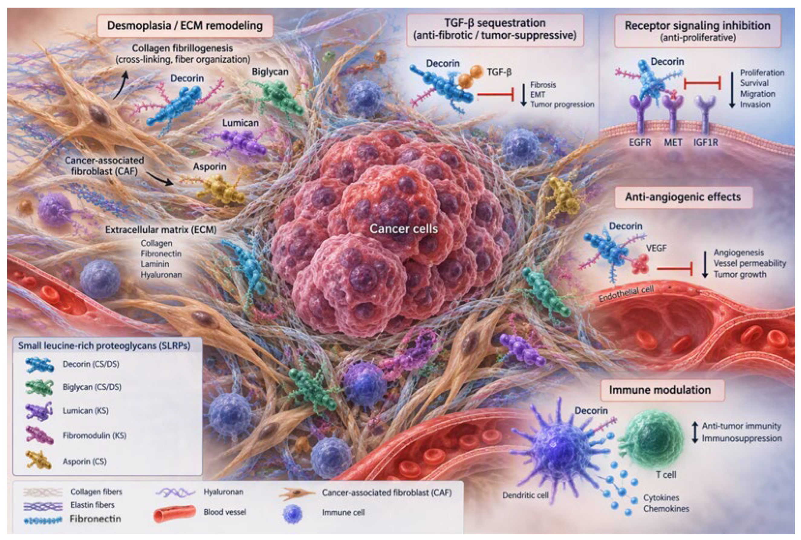
Figure 2.
A multifaced role molecule from organizing the extracellular matrix to regulating angiogenesis, autophagy, and cell growth, Decorin plays a key role in keeping tissues balanced. Its reduction or absence is linked to fibrosis, tumor progression, pathological angiogenesis, and issues with tissue repair.
Figure 2.
A multifaced role molecule from organizing the extracellular matrix to regulating angiogenesis, autophagy, and cell growth, Decorin plays a key role in keeping tissues balanced. Its reduction or absence is linked to fibrosis, tumor progression, pathological angiogenesis, and issues with tissue repair.
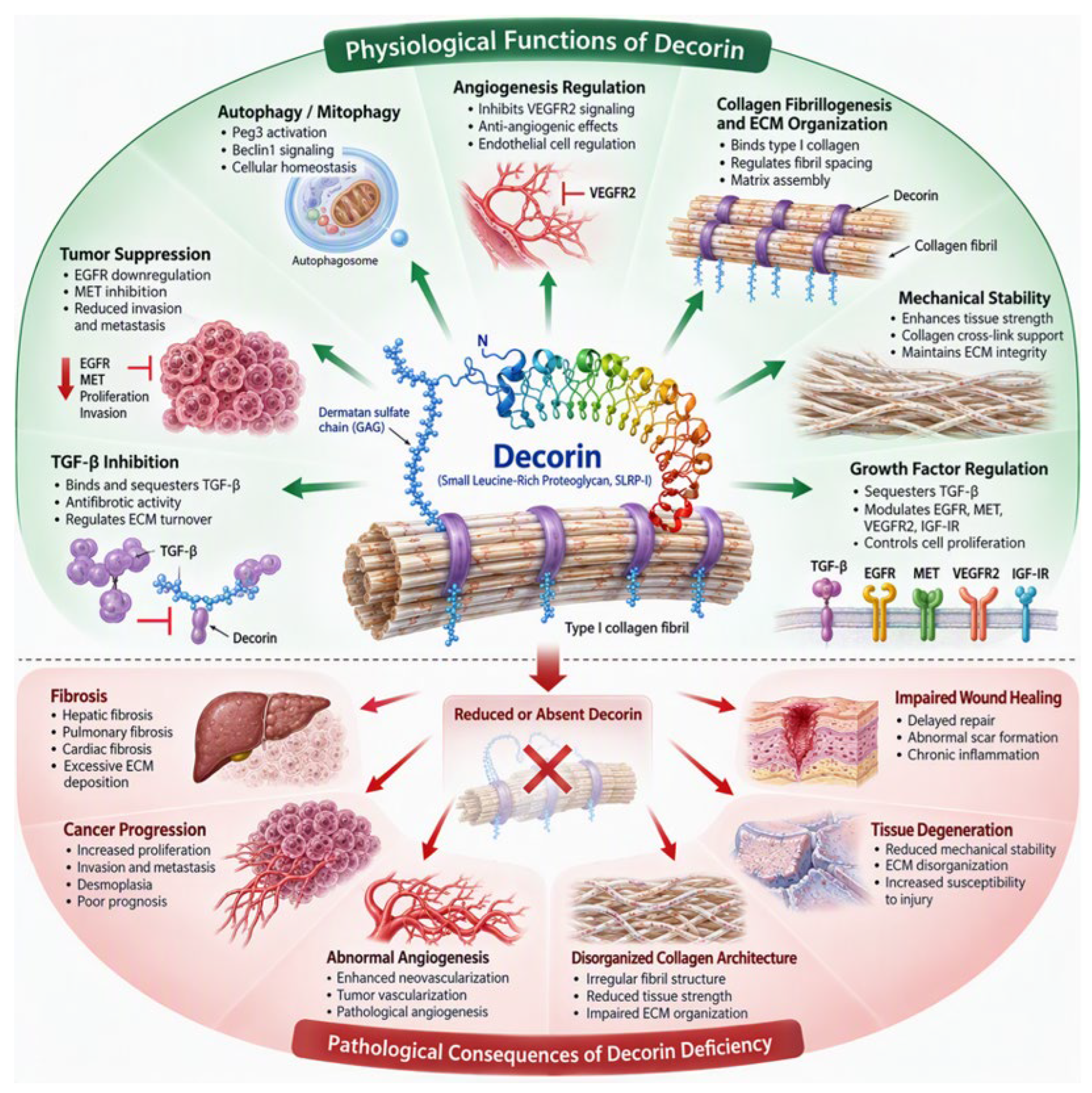

Table 1.
Basic SLRP classes.
| Class | Members | Function | References |
| Class Ι | Decorin, Biglycan, Asporin | ECM organization, growth factor regulation | [58,70] |
| Class ΙΙ | Fibromodulin, Lumican, Keratocan, PRELP | Collagen assembly regulation, tissue organization | [58,70] |
| Class ΙΙΙ | Osteoglycin, Epiphycan, Opticin | Skeletal and ocular tissue homeostasis | [58,70] |
| Class ΙV: | Chondroadherin, Tsukushi, Nyctalopin | Cell adhesion and growth signaling | [58,70] |
| Class V: | Podocan, PODNL1 - | Tissue remodeling and fibrosis | [70,81] |
Table 2.
Main structural and functional features of SLRPs.
| SLRPs | Structure | Main functions | References |
| Decorin | 12 LRR repeats, one dermatan sulfate chain | Collagen reorganization, tumor suppression, anti-fibrotic signaling | [43,76] |
| Biglycan | Two CS/DS keratan/dermatan keratan sulfate chains | Inflammation, innate immunity, ECM reorganization | [77] |
| Lumican | Keratan sulfate-rich proteoglycan | Collagen fiber formation, wound healing, cancer regulation | [58,75] |
| Fibromodulin | Keratan sulfate | LRP Tissue repair and collagen assembly | [72] |
| Keratocan | Corneal-specific proteoglycan | Corneal transparency | [73] |
| Osteoglycin | Small keratan sulfate proteoglycan | Bone formation and cardiovascular regulation | [78,80] |
Table 3.
SLRPs in non-neoplastic and neoplastic diseases.
| Disease | Main SLRP | Pathogenetic mechanism | References |
| Fibrosis | Decorin, Biglycan, Lumican | TGFb signaling and deposition in the ECM | [4,41,66] |
| Chronic inflammatory disease | Biglycan, Lumican | Activation of autoimmune mechanisms | [40,77] |
| Osteoarthritis | Decorin, Biglycan, Fibromodulin | Cartilage degeneration | [65] |
| Cardiovascular Diseases | Decorin, Osteoglycin | Vascular remodeling | [78,80] |
| Colorectal cancer | Decorin, Biglycan, Lumican | ECM transformation and microenvironment regulation | [31,45,75] |
| Breast cancer | Decorin, Lumican | Regulation of growth factors | [46,75] |
| Pancreatic cancer | Decorin. Biglycan | Tumor stroma rearrangement and invasion | [3,43] |
| Gliomas | Decorin Biglycan | Cell migration | [69] |
Table 4.
SLRPs and fibrosis.
| SLRPs | Pathogenesis | Desmoplastic effect | References |
| Decorin | TGF-β binding and receptor modification | Potent anti-fibrotic | [4,62,66] |
| Biglycan | Signaling of inflammatory pathways such as TLR2/TLR4 | Promotes desmoplasia | [77] |
| Lumican | Control of fibroblasts | Environment-dependent | [75] |
| Fibromodulin | Regulation of collagen fibril maturation | Stabilization of ECM | [72] |
| Osteoglycin | Regulation of ECM | Potential regulatory role | [78,80] |
| Podocan | Regulation of mesenchymal cells | Tissue-dependent anti-desmoplastic | [81] |
Table 5.
Decorin: biological function and potential therapeutic target.
| Function | Molecular mechanism | Potential therapeutic target | References |
| Anti-fibrosis | Neutralization of TGF-b | Liver, Kidney, Lung, Cardiac fibrosis | [4,66] |
| Antineoplastic role | Reduction of EGFR,MET,IGF-R suppression | Cancer therapeutics | [25,43,54,71] |
| Anti-angiogenesis | VEGFA suppression and receptor interference | Control of neoplastic neovascularization | [42,43] |
| ECM organization | Control of collagen fibrillogenesis | Tissue Regeneration | [76] |
| Anti-inflammatory | Cytokine modulation | Chronic inflammatory diseases | [74] |
| Autophagy | VEGFR2/PEG3 signaling | Novel cancer therapies | [68] |
| Biomarker | Altered expression in tumors and fibrosis | Prognostic and predictive marker | [31,43,75] |
| Gene therapy | Genetically recombinant Decorin and delivery with genetic vectors | Precision medicine | [10,43,53,64] |
Disclaimer/Publisher’s Note: The statements, opinions and data contained in all publications are solely those of the individual author(s) and contributor(s) and not of MDPI and/or the editor(s). MDPI and/or the editor(s) disclaim responsibility for any injury to people or property resulting from any ideas, methods, instructions or products referred to in the content. |
© 2026 by the authors. Licensee MDPI, Basel, Switzerland. This article is an open access article distributed under the terms and conditions of the Creative Commons Attribution (CC BY) license (http://creativecommons.org/licenses/by/4.0/).
Copyright: This open access article is published under a Creative Commons CC BY 4.0 license, which permit the free download, distribution, and reuse, provided that the author and preprint are cited in any reuse.