Preprint
Article

This version is not peer-reviewed.

Aminoalkyl-Substituted 2-Hetarylimidazophenazines: Synthesis, DNA Binding and Cytotoxicity

Submitted:

26 October 2026

Posted:

27 October 2026

You are already at the latest version

Abstract

A series of ethylamino- and propylamino-substituted 2-(pyridin-2-yl)imidazophenazines bearing various tertiary amine moieties, as well as a series of 2-hetarylimidazophenazines with N,N-diethylethan-1-amine moiety, were synthesized via the reaction of phenazine-2,3-diamine with heterocyclic aldehydes followed by alkylation with chloro-substituted alkylamines. In a study on HCT116, K562 and Kelly cells compounds containing N-methylindole, 2-chloroquinoline, and N-phenylpyrrole moieties exhibited the highest cytotoxicity. The values of binding constants with G-quadruplexes tel22, c-Myc for the leader compounds, determined by the method of fluorescence spectroscopy, were within the order of 105-106 M-1.

Keywords: 
;  ;  ;  ;  

1. Introduction

DNA is widely considered a target in anticancer therapy; however, the action of organic molecules on the DNA double helix is often non-selective, leading to the development of side effects. Targeting DNA structures other than the double helix, so called non-canonical structures, can increase the selectivity of anticancer therapy [1]. Among the non-canonical structures, the most studied are G-quadruplexes (G4), the quartets of guanine bases arranged in layers. The guanine-rich regions that form G-quadruplexes have been found in the promoter regions of oncogenes and telomeres. The formation of G4 leads to the inhibition of gene expression and telomerase activity, and ultimately to cell death [2,3].
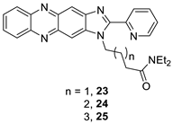
Polyaromatic derivatives, such as anthraquinones and phenazines, can be used as G4 ligands. While anthraquinone-based G4 ligands have been explored extensively [4,5,6,7], the phenazine-derived G4 binders are few, being represented mainly by coordination compounds based on dipyridophenazines [8,9,10]. In our previous studies, we have shown that imidazophenazine derivatives exhibited an efficient binding to G4 and high selectivity to G4 over double stranded DNA [11]. However, our amidoalkyl-substituted 2-(pyridin-2-yl)imidazophenazines were poorly soluble in DMSO, therefore, their use as biologically compatible compounds was limited. To solve the solubility problem and to increase the efficiency of binding to G4, we herein introduced the polymethylene linkers with terminal tertiary amine fragments into imidazophenazines instead of a linker with a terminal amide group. Furthermore, the heteroaromatic substituent at the position 2 of the synthesized imidazophenazines varied to identify the derivatives that enhance ligand binding efficiency and G4 preference. The new series of aminoalkyl-substituted 2-hetarylimidazophenazines was tested for binding to various DNA structures and antitumor potency. All tested substances demonstrated moderate cytotoxicity against HCT116, K562 and Kelly cell lines.

2. Materials and Methods

2.1. General

All solvents used were purified and dried using standard techniques. All starting reagents were purchased from commercial sources (Sigma-Aldrich, ABCR, AKSci, Burlington, VT, USA). Compounds: 1-(3-nitrophenyl)-1H-pyrrole-2-carbaldehyde [12], 4-(3,5-diphenyl-4,5-dihydro-1H-pyrazol-1-yl)benzaldehyde [13], 1 [11] and 2 [11] were synthesized according to previously described methods. Unless otherwise stated, all reactions were monitored by TLC analysis using DCM/methanol 20:1 mixture as eluent and silica plates with a fluorescent indicator (254 nm) and visualized with a UV lamp.
1H and 13C NMR spectra were recorded on a Bruker Avance-400 (Bruker Biospin, Karlsruhe, Germany) spectrometer (400 MHz for 1H, 101 MHz for 13C). All chemical shifts (δ) are reported in parts per million (ppm) with 1H and 13C NMR referenced to solvent signals [1H NMR: CDCl3 (7.27), DMSO-d6 (2.50); 13C NMR: CDCl3 (77.16), DMSO-d6 (39.52)]. Coupling constants (J) are reported in Hertz (Hz).
High-resolution mass spectra (HRMS) were recorded on an Orbitrap Elite mass spectrometer (Thermo Fisher Scientific, Waltham, Massachusetts, USA) using electrospray ionization (ESI). Solutions with concentrations of 0.1–9 μg/mL (in 1% formic acid in acetonitrile) were injected directly into the ion source using a syringe pump (5 μL/min). The spray voltage was ±3.5 kV, and the capillary temperature was 275 ºC. Mass spectra were recorded on an Orbitrap analyzer (Thermo Fisher Scientific, Waltham, Massachusetts, USA) with 480000 resolution (1 microscan). The maximum injection time was 900 ms, averaging over 9 spectra; the mass range 90–2000 Da. For internal calibration, the signals of DMSO and diisooctyl phthalate (m/z 157.03515 and 413.26623) in the positive mode and the signal of dodecyl sulfate (m/z 265.14790) in the negative mode were used.

2.2. Biological Testing

Fluorescence spectra were recorded on a Cary Eclipse scanning spectrofluorimeter (Varian, Palo Alto, California, USA) at 20 °C using a quartz cuvette. Sample fluorescence was recorded in the wavelength range of 500–750 nm with excitation at 390 nm. Measurements were performed for the ligand diluted to a concentration of 5 μM in an aqueous buffer solution (100 mM KCl, 10 mM Na phosphate buffer pH 7.5). G4 DNA structures were prepared by rapid annealing of DNA oligonucleotides (100 μM) in the buffer solution. DNA oligonucleotides with sequences c-Myc: d(TGAGGGTGGGTAGGGTGGGTAA; tel22: d(AGGGTTAGGGTTAGGGTTAGGG) were used. Based on the dependence of fluorescence on the ligand concentration, the maximum saturation value r was calculated:
c B = c 0 I − I F I B − I F ;   c F = c 0 − c B ;   r = c B c D N A
where CB is the concentration of the bound ligand, IF is the fluorescence intensity of the free ligand, IB is the fluorescence intensity of the DNA-ligand complex, C0 is the initial ligand concentration, and CF is the concentration of the free ligand. Based on the obtained data, a binding isotherm was constructed in Scatchard coordinates (r/CF vs. r) and approximated by a linear relationship.
The HCT116 human colon carcinoma, K562 chronic myelogenous leukemia and Kelly neuroblastoma cell lines were propagated in Dulbecco modified Eagle’s medium supplemented with 10% fetal bovine serum (Biolot, Saint-Petersburg), 2 mM L-glutamine, 100 U/ml penicillin, and 100 μg/ml streptomycin (PanEco, Moscow, Russia) at 37°C, 5% CO2 in a humidified atmosphere. Cells in the logarithmic phase were used in the experiments. For cytotoxicity tests, cells (5000 per well) were seeded into 96-well plates (Nunc, USA). Tested compounds were reconstituted in culture medium from 10 mM stock solutions in DMSO immediately before experiments. Final concentrations of compounds were 0.1-50 μM, each concentration in triplicate. Cells containing DMSO vehicle (<0.1 %) served as a control. Cells were incubated for 70 h at 37 °C, 5% CO2. Two h prior to the completion of incubation, 10 μl of aqueous MTT solution (5 mg/ml, PanEco, Moscow) were added to the wells for an additional incubation. The culture medium was removed, cells were resuspended in 200 μl DMSO, and the optical density of solutions was measured on a Multiscan FC plate spectrophotometer (Thermo Scientific, Waltham, MA) at 570 nm. The percentages of cell survival were calculated as the ratio of mean optical density in drug-containing wells to that in vehicle-treated cells (100%).

2.3. Synthesis

General procedure for synthesis of compounds 3-11. Diaminophenazine 1 (0.21 g, 1.0 mmol) was dissolved in 10 ml of DMF, than aldehyde (1.0 mmol) and p-toluenesulfonic acid (36 mg, 0.2 mmol) were added. The mixture was stirred at reflux for 48 hours. After completion of the reaction, 50 ml of water was added to the reaction mixture, the resulting precipitate was filtered off, washed with water, and dried. The resulting precipitate was recrystallized from ethanol.
3-(1H-Imidazo[4,5-b]phenazin-2-yl)pyridin-2-amine (3). From 0.21 g of diaminophenazine 1 (1.0 mmol), 0.12 g of 2-aminonicotinaldehyde (1.0 mmol) and 36 mg of p-toluenesulfonic acid (0.2 mmol) in 10 mL of DMF 0.19 g of compound 3 was obtained as an orange solid (62% yield). 1H NMR (400 MHz, DMSO-d6) δ 13.44 (s, 1H), 8.47 (d, J = 7.64 Hz, 2H), 8.41 (br.s, 2H), 8.16–8.28 (m, 4H), 7.84–7.91 (m, 2H), 6.84–6.92 (m, 1H). HRMS (ESI) m/z: [M+H]+ calculated for [C18H12N6+H]+ 313.1157; found 313.1199.
2-(2-Chloroquinolin-3-yl)-1H-imidazo[4,5-b]phenazine (4). From 0.21 g of diaminophenazine 1 (1.0 mmol), 0.19 g of 2-chloroquinoline-3-carbaldehyde (1.0 mmol) and 36 mg of p-toluenesulfonic acid (0.2 mmol) in 10 mL of DMF 0.24 g of compound 4 was obtained as an orange solid (62% yield). 1H NMR (400 MHz, DMSO-d6) δ 13.45 (br.s, 1H), 9.21 (s, 1H), 8.19–8.31 (m, 4H), 8.11 (d, J = 8.44 Hz, 1H), 7.99 (t, J = 7.70 Hz, 1H), 7.86–7.96 (m, 3H), 7.80 (t, J = 7.86 Hz, 1H). HRMS (ESI) m/z: [M+H]+ calculated for [C22H12ClN5+H]+ 382.0859; found 382.0859.
2-(1-Methyl-1H-pyrrol-2-yl)-1H-imidazo[4,5-b]phenazine (5). From 0.21 g of diaminophenazine 1 (1.0 mmol), 0.11 g of 1-methyl-1H-pyrrole-2-carbaldehyde (1.0 mmol) and 36 mg of p-toluenesulfonic acid (0.2 mmol) in 10 mL of DMF 0.18 g of compound 5 was obtained as an orange solid (59% yield). 1H NMR (400 MHz, DMSO-d6) δ 12.92 (s, 1H), 8.31 (s, 1H), 8.14–8.26 (m, 2H), 8.07 (s, 1H), 7.78–7.93 (m, 2H), 7.17–7.31 (m, 2H), 6.29 (t, J = 2.84 Hz, 1H), 4.21 (s, 3H). HRMS (ESI) m/z: [M+H]+ calculated for [C18H13N5+H]+ 300.1249; found 300.1249.
2-(1-(3-Nitrophenyl)-1H-pyrrol-2-yl)-1H-imidazo[4,5-b]phenazine (6). From 0.21 g of diaminophenazine 1 (1.0 mmol), 0.22 g of 1-(3-nitrophenyl)-1H-pyrrole-2-carbaldehyde (1.0 mmol) and 36 mg of p-toluenesulfonic acid (0.2 mmol) in 10 mL of DMF 0.15 g of compound 6 was obtained as an orange solid (37% yield). 1H NMR (400 MHz, DMSO-d6) δ 13.08 (s, 1H), 8.34 (s, 1H), 8.31–8.34 (m, 1H), 8.14–8.21 (m, 2H), 8.11 (s, 1H), 8.05 (s, 1H), 7.94–7.97 (m, 1H), 7.81–7.87 (m, 2H), 7.78 (t, J = 8.28 Hz, 1H), 7.52–7.56 (m, 1H), 7.41–7.45 (m, 1H), 6.57–6.63 (m, 1H). HRMS (ESI) m/z: [M+H]+ calculated for [C23H14N6O2+H]+ 407.1256; found 407.1256.
2-(4-(3,5-Diphenyl-4,5-dihydro-1H-pyrazol-1-yl)phenyl)-1H-imidazo[4,5-b]phenazine (7). From 0.21 g of diaminophenazine 1 (1.0 mmol), 0.33 g of 4-(3,5-diphenyl-4,5-dihydro-1H-pyrazol-1-yl)benzaldehyde (1.0 mmol) and 36 mg of p-toluenesulfonic acid (0.2 mmol) in 10 mL of DMF 0.31 g of compound 7 was obtained as an orange solid (59% yield). 1H NMR (400 MHz, DMSO-d6) δ 13.07 (s, 1H), 8.32 (s, 1H), 8.13–8.25 (m, 4H), 8.11 (s, 1H), 7.79–7.91 (m, 4H), 7.25–7.53 (m, 9H), 7.22 (d, J = 8.80 Hz, 2H), 5.73 (dd, J = 12.35, 5.14 Hz, 1H), 4.02 (dd, J = 17.45, 12.26 Hz, 1H). HRMS (ESI) m/z: [M+H]+ calculated for [C34H24N6+H]+ 517.2141; found 517.2134.
2-(1-Methyl-1H-indol-3-yl)-1H-imidazo[4,5-b]phenazine (8). From 0.21 g of diaminophenazine 1 (1.0 mmol), 0.16 g of 1-methyl-1H-indole-3-carbaldehyde (1.0 mmol) and 36 mg of p-toluenesulfonic acid (0.2 mmol) in 10 mL of DMF 0.29 g of compound 8 was obtained as an orange solid (83% yield). 1H NMR (400 MHz, DMSO-d6) δ 13.06 (br.s, 1H), 8.59–8.68 (m, 1H), 8.42 (s, 1H), 8.31 (s, 1H), 8.13–8.24 (m, 2H), 8.09 (s, 1H), 7.79–7.87 (m, 2H), 7.58–7.65 (m, 1H), 7.30–7.39 (m, 2H), 3.96 (s, 3H). HRMS (ESI) m/z: [M+H]+ calculated for [C22H15N5+H]+ 350.1406; found 350.1405; [M+Na]+ calculated for [C22H15N5+Na]+ 372.1225; found 372.1224.
2-(1H-Imidazol-2-yl)-1H-imidazo[4,5-b]phenazine (9). From 0.21 g of diaminophenazine 1 (1.0 mmol), 0.10 g of 1H-imidazole-2-carbaldehyde (1.0 mmol) and 36 mg of p-toluenesulfonic acid (0.2 mmol) in 10 mL of DMF 0.17 g of compound 9 was obtained as an orange solid (59% yield). 1H NMR (400 MHz, DMSO-d6) δ 13.79 (br.s, 1H), 13.55 (s, 1H), 8.24–8.53 (m, 2H), 8.12–8.24 (m, 2H), 7.83–7.94 (m, 2H), 7.31–7.60 (m, 2H). HRMS (ESI) m/z: [M+H]+ calculated for [C16H10N6+H]+ 287.1045; found 287.1043; [M+Na]+ calculated for [C16H10N6+Na]+ 309.0865; found 309.0861.
2-(1H-Imidazol-4-yl)-1H-imidazo[4,5-b]phenazine (10). From 0.21 g of diaminophenazine 1 (1.0 mmol), 0.10 g of 1H-imidazole-4-carbaldehyde (1.0 mmol) and 36 mg of p-toluenesulfonic acid (0.2 mmol) in 10 mL of DMF 0.15 g of compound 10 was obtained as an orange solid (51% yield). 1H NMR (400 MHz, DMSO-d6) δ 13.05 (br.s, 1H), 12.83 (br.s, 1H), 8.24–8.41 (m, 1H), 8.08–8.24 (m, 4H), 8.01 (s, 1H), 7.79–7.90 (m, 2H). HRMS (ESI) m/z: [M+Na]+ calculated for [C16H10N6+Na]+ 309.0865; found 309.0862.
2-(1H-Indol-3-yl)-1H-imidazo[4,5-b]phenazine (11). From 0.21 g of diaminophenazine 1 (1.0 mmol), 0.15 g of 1H-indole-3-carbaldehyde (1.0 mmol) and 36 mg of p-toluenesulfonic acid (0.2 mmol) in 10 mL of DMF 0.26 g of compound 11 was obtained as an orange solid (77% yield). 1H NMR (400 MHz, DMSO-d6) δ 12.10 (br.s, 1H), 8.56–8.65 (m, 1H), 8.47 (d, J = 2.63 Hz, 1H), 8.15–8.30 (m, 4H), 7.81–7.89 (m, 2H), 7.53–7.61 (m, 1H), 7.23–7.36 (m, 2H). HRMS (ESI) m/z: [M+H]+ calculated for [C21H13N5+H]+ 336.1249; found 336.1248.
General procedure for synthesis of compounds 12a-c, 13a-c. Sodium hydride (60% suspension in oil, 44 mg, 1.1 mmol) was added to a suspension of imidazophenazine 2 (300 mg, 1 mmol) in 20 mL of dioxane. The mixture was stirred for 30 min, then chloro-substituted amine (1.2 mmol) was added. The mixture was stirred at reflux for 48 h. Upon completion of the reaction, the solvent was evaporated under reduced pressure, the residue was suspended in saturated NaCl solution, and extracted with dichloromethane (4×100 mL). The organic fractions were combined and dried over anhydrous sodium sulfate, after which the solvent was evaporated under reduced pressure. The resulting residue was separated by column chromatography on silica gel, (eluent CH2Cl2, CH3OH 20:1).
N,N-Diethyl-2-(2-(pyridin-2-yl)-1H-imidazo[4,5-b]phenazin-1-yl)ethan-1-amine (12a). From 44 mg of sodium hydride (60% suspension in oil, 1.1 mmol), 300 mg of imidazophenazine 2 (1 mmol) and 160 mg of 2-chloro-N,N-diethylethan-1-amine (1.2 mmol) in 20 mL of dioxane 40 mg of compound 12a was obtained as an yellow solid (10% yield). 1H NMR (400 MHz, CDCl3) δ 8.77 (d, J = 4.83 Hz, 1H), 8.74 (s, 1H), 8.58 (d, J = 8.01 Hz, 1H), 8.25– 8.14 (m, 3H), 7.90 (td, J = 7.8, 1.8 Hz, 1H), 7.79 – 7.70 (m, 2H), 7.42 (dd, J = 7.6, 4.8, 1H), 5.05 (t, J = 7.27, 2H), 3.02 (t, J = 7.34, 2H), 2.58 (q, J = 7.1 Hz, 4H), 0.93 (t, J = 7.1 Hz, 6H). 13C NMR (101 MHz, CDCl3) δ 157.03, 149.18, 148.58, 146.52, 142.44, 142.24, 141.23, 140.29, 140.08, 136.67, 129.50, 129.14, 129.06, 128.77, 125.42, 124.64, 116.96, 105.24, 76.96, 76.64, 76.32, 54.27, 46.90 (2C), 44.68, 10.70, 10.67. HRMS (ESI) m/z: [M+H]+ calculated for [C24H24N6+H]+ 397.2135; found 397.2131.
2-(Pyridin-2-yl)-1-(2-(pyrrolidin-1-yl)ethyl)-1H-imidazophenazine (12b). From 44 mg of sodium hydride (60% suspension in oil, 1.1 mmol), 300 mg of imidazophenazine 2 (1 mmol) and 160 mg of 1-(2-chloroethyl)pyrrolidine (1.2 mmol) in 20 mL of dioxane 330 mg of compound 12b was obtained as an yellow solid (85% yield). 1H NMR (400 MHz, CDCl3) δ 8.80 (d, J = 4.7 Hz, 1H), 8.74 (s, 1H), 8.56 (d, J = 7.95 Hz, 1H), 8.30 (s, 1H), 8.28–8.20 (m, 2H), 7.95 (td, J = 7.8, 1.8 Hz, 1H), 7.85–7.77 (m, 2H), 7.48 (dd, J = 7.6, 4.8, 1H), 5.12 (t, J = 7.76 Hz, 2H), 3.09 (t, J = 7.70 Hz, 2H), 2.67-2.58 (m, 4H), 1.79-1.68 (m, 4H). 13C NMR (101 MHz, CDCl3) δ 157.03, 149.18, 148.58, 146.52, 142.44, 142.24, 141.23, 140.29, 140.08, 136.67, 129.50, 129.14, 129.06, 128.77, 125.42, 124.64, 116.96, 105.24, 76.96, 76.64, 76.32, 54.27, 54.33 (2C), 44.68, 23.18 (2C). HRMS (ESI) m/z: [M+H]+ calculated for [C24H22N6+H]+ 395.1979; found 395.1977.
2-(Pyridin-2-yl)-1-(2-(piperidin-1-yl)ethyl)-1H-imidazo[4,5-b]phenazine (12c). From 44 mg of sodium hydride (60% suspension in oil, 1.1 mmol), 300 mg of imidazophenazine 2 (1 mmol) and 177 mg of 1-(2-chloroethyl)piperidine (1.2 mmol) in 20 mL of dioxane 190 mg of compound 12c was obtained as an yellow solid (46% yield). 1H NMR (400 MHz, CDCl3) δ 8.72 (d, J = 4.71 Hz, 1H), 8.66 (s, 1H), 8.52 (d, J = 7.95 Hz, 1H), 8.23 (s, 1H), 8.22–8.10 (m, 2H), 7.90 (td, J = 7.8, 1.8 Hz, 1H), 7.82–7.70 (m, 2H), 7.42 (dd, J = 7.6, 4.8, 1H), 5.09 (t, J = 7.4 Hz, 2H), 3.02–2.85 (m, 2H), 2.64–2.42 (m, 4H), 1.60–1.45 (m, 4H), 1.45–1.32 (m, 2H). 13C NMR (101 MHz, CDCl3) δ 157.03, 149.18, 148.58, 146.52, 142.44, 142.24, 141.23, 140.29, 140.08, 136.67, 129.50, 129.14, 129.06, 128.77, 125.42, 124.64, 116.96, 105.24, 54.05 (2C), 31.51, 29.28 (2C), 28.95, 22.28, 13.72. HRMS (ESI) m/z: [M+H]+ calculated for [C25H24N6+H]+ 409.2135; found 409.2138.
N,N-Diethyl-3-(2-(pyridin-2-yl)-1H-imidazo[4,5-b]phenazin-1-yl)propan-1-amine (13a). From 44 mg of sodium hydride (60% suspension in oil, 1.1 mmol), 300 mg of imidazophenazine 2 (1 mmol) and 180 mg of 3-chloro-N,N-diethylpropan-1-amine (1.2 mmol) in 20 mL of dioxane 49 mg of compound 13a was obtained as an yellow solid (12% yield). 1H NMR (400 MHz, CDCl3) δ 8.78 (d, J = 4.22 Hz, 1H), 8.73 (s, 1H), 8.58 (d, J = 7.95 Hz, 1H), 8.29 (s, 1H), 8.21–8.28 (m, 2H), 7.94 (td, J = 7.78, 1.74 Hz, 1H), 7.76–7.85 (m, 2H), 7.47 (dd, J = 6.69, 4.98 Hz, 1H), 4.98 (t, J = 7.82 Hz, 2H), 2.65 (t, J = 7.06 Hz, 2H), 2.57 (q, J = 7.03 Hz, 4H), 2.14–2.29 (m, 2H), 1.03 (t, J = 7.15 Hz, 6H). 13C NMR (101 MHz, CDCl3) δ 157.06, 149.10, 148.62, 146.52, 142.47, 142.14, 141.23, 140.30, 140.07, 136.68, 129.56, 129.15, 129.11, 128.79, 125.49, 124.58, 116.83, 105.24, 54.49, 46.42 (2C), 42.45, 26.14, 11.65 (2C). HRMS (ESI) m/z: [M+H]+ calculated for [C25H26N6+H]+ 411.2292; found 411.2300.
2-(Pyridin-2-yl)-1-(3-(pyrrolidin-1-yl)propyl)-1H-imidazo[2,5-b]phenazine (13b). From 44 mg of sodium hydride (60% suspension in oil, 1.1 mmol), 300 mg of imidazophenazine 2 (1 mmol) and 178 mg of 1-(3-chloropropyl)pyrrolidine (1.2 mmol) in 20 mL of dioxane 49 mg of compound 13b was obtained as an yellow solid (28% yield). 1H NMR (400 MHz, CDCl3) δ (d, J = 4.71 Hz, 1H), 8.64 (s, 1H), 8.59 (s, 1H), 8.52 (d, J = 7.95 Hz, 1H), 8.19–8.26 (m, 2H), 8.11 (td, J = 7.81, 1.68 Hz, 1H), 7.85–7.92 (m, 2H), 7.67 (dd, J = 7.58, 4.83 Hz, 1H), 5.03 (t, J = 7.18 Hz, 2H), 3.20–3.48 (m, 4H), 2.34–2.46 (m, 4H), 1.86–1.92 (m, 4H). 13C NMR (101 MHz, CDCl3) δ 156.97, 149.10, 148.48, 146.45, 142.49, 142.16, 141.33, 137.89, 137.61, 136.57, 129.17, 129.01, 128.90, 128.60, 125.56, 124.42, 116.73, 104.75, 54.99, 53.81 (2C), 40.69, 25.24 (2C), 19.61. HRMS (ESI) m/z: [M+H]+ calculated for [C25H24N6+H]+ 409.2141; found 409.2142.
2-(Pyridin-2-yl)-1-(3-(piperidin-1-yl)propyl)-1H-imidazo[2,5-b]phenazine (13c). From 44 mg of sodium hydride (60% suspension in oil, 1.1 mmol), 300 mg of imidazophenazine 2 (1 mmol) and 194 mg of 1-(3-chloropropyl)piperidine (1.2 mmol) in 20 mL of dioxane 80 mg of compound 13c was obtained as an yellow solid (19% yield). 1H NMR (400 MHz, CDCl3) δ 8.81 (d, J = 3.48 Hz, 1H), 8.72 (s, 1H), 8.58 (d, J = 7.76 Hz, 1H), 8.17–8.30 (m, 3H), 7.95 (t, J = 7.86 Hz, 1H), 7.77–7.84 (m, 2H), 7.49 (dd, J = 7.12, 4.86 Hz, 1H), 5.03 (t, J = 7.26 Hz, 2H), 2.42–2.53 (m, 2H), 2.32–2.42 (m, 4H), 2.19–2.29 (m, 2H), 1.53–1.62 (m, 6H). 13C NMR (101 MHz, CDCl3) δ 157.05, 149.18, 148.73, 146.52, 142.45, 142.21, 141.23, 140.35, 139.98, 136.82, 129.49, 129.21, 128.98, 128.67, 125.45, 124.43, 116.80, 105.29, 53.76, 53.03 (2C), 42.10, 28.95 (2C), 26.27, 22.71. HRMS (ESI) m/z: [M+H]+ calculated for [C26H26N6+H]+ 423.2292; found 423.2295.
General procedure for synthesis of compounds 14-19. Sodium hydride (60% suspension in oil, 44 mg, 1.1 mmol) was added to a suspension of imidazophenazine (1 mmol) in 20 mL of dioxane. The mixture was stirred for 30 min, then 2-chloro-N,N-diethylethan-1-amine (160 mg, 1.2 mmol) was added. The mixture was stirred at reflux for 48 h. Upon completion of the reaction, the solvent was evaporated under reduced pressure, the residue was suspended in saturated NaCl solution and extracted with dichloromethane (4×100 mL). The organic fractions were combined and dried over anhydrous sodium sulfate, after which the solvent was evaporated under reduced pressure. The resulting residue was separated by column chromatography on silica gel (eluent CH2Cl2, CH3OH 20:1).
3-(1-(2-(Diethylamino)ethyl)-1H-imidazo[2,5-b]phenazin-2-yl)pyridin-2-amine (14). From 44 mg of sodium hydride (60% suspension in oil, 1.1 mmol), 312 mg of 3-(1H-imidazo[4,5-b]phenazin-2-yl)pyridin-2-amine 3 (1 mmol) and 160 mg of 2-chloro-N,N-diethylethan-1-amine (1.2 mmol) in 20 mL of dioxane 276 mg of compound 14 was obtained as an yellow solid (67% yield). 1H NMR (400 MHz, CDCl3) δ 8.58 (s, 1H), 8.21–8.28 (m, 2H), 8.15–8.20 (m, 1H), 8.13 (s, 1H), 8.04 (dd, J = 7.61, 1.74 Hz, 1H), 7.73–7.82 (m, 2H), 6.78 (dd, J = 7.58, 4.89 Hz, 1H), 6.21 (br s, 2H), 4.37 (t, J = 6.42 Hz, 2H), 2.88 (t, J = 6.39 Hz, 2H), 2.41 (q, J = 7.09 Hz, 4H), 0.79 (t, J = 7.12 Hz, 6H). 13C NMR (101 MHz, CDCl3) δ 159.08, 152.74, 150.30, 143.01, 142.19, 142.06, 140.96, 140.49, 139.62, 136.68, 129.93, 129.79, 129.52, 129.29, 116.83, 114.68, 108.37, 104.51, 51.03, 47.51 (2C), 40.63, 10.26 (2C). HRMS (ESI) m/z: [M+Na]+ calculated for [C24H25N7+Na]+ 434.2069; found 434.2076; [2M+Na]+ calculated for [C48H50N14+Na]+ 845.4241; found 845.4249.
2-(2-(2-Chloroquinolin-3-yl)-1H-imidazo[4,5-b]phenazin-1-yl)-N,N-diethylethan-1-amine (15). From 44 mg of sodium hydride (60% suspension in oil, 1.1 mmol), 382 mg of 2-(2-chloroquinolin-3-yl)-1H-imidazo[4,5-b]phenazine 4 (1 mmol) and 160 mg of 2-chloro-N,N-diethylethan-1-amine (1.2 mmol) in 20 mL of dioxane 170 mg of compound 15 was obtained as an yellow solid (35% yield). 1H NMR (400 MHz, CDCl3) δ 8.79 (s, 1H), 8.60 (s, 1H), 8.22–8.30 (m, 3H), 8.16 (d, J = 8.44 Hz, 1H), 7.94 (d, J = 8.13 Hz, 1H), 7.90 (td, J = 7.79, 1.34 Hz, 1H), 7.79–7.86 (m, 3H), 7.69 (t, J = 8.07 Hz, 1H), 4.31 (t, J = 6.33 Hz, 2H), 2.84 (t, J = 6.33 Hz, 2H), 2.26 (q, J = 7.09 Hz, 4H), 0.62 (t, J = 7.12 Hz, 6H). 13C NMR (101 MHz, CDCl3) δ 149.37, 149.18, 148.15, 143.35, 142.79, 142.27, 141.46, 140.98, 140.67, 136.39, 132.20, 130.15, 129.56, 129.36, 129.34, 129.02, 128.27, 126.49, 126.44, 122.29, 119.03, 105.24, 50.72, 48.01 (2C), 40.63, 10.70 (2C). HRMS (ESI) m/z: [M+Na]+ calculated for [C28H25ClN6+Na]+ 503.1727; found 503.1737.
N,N-Diethyl-2-(2-(1-methyl-1H-pyrrol-2-yl)-1H-imidazo[4,5-b]phenazin-1-yl)ethan-1-amine (16). From 44 mg of sodium hydride (60% suspension in oil, 1.1 mmol), 300 mg of 2-(1-methyl-1H-pyrrol-2-yl)-1H-imidazo[4,5-b]phenazine 5 (1 mmol) and 160 mg of 2-chloro-N,N-diethylethan-1-amine (1.2 mmol) in 20 mL of dioxane 260 mg of compound 16 was obtained as an yellow solid (65% yield). 1H NMR (400 MHz, CDCl3) δ 8.56 (s, 1H), 8.16–8.31 (m, 2H), 8.10 (s, 1H), 7.68–7.85 (m, 2H), 6.92–6.99 (m, 1H), 6.80–6.89 (m, 1H), 6.24–6.38 (m, 1H), 4.52 (t, J = 6.57 Hz, 2H), 4.07 (s, 3H), 2.91 (t, J = 6.54 Hz, 2H), 2.50 (q, J = 6.77 Hz, 4H), 0.86 (t, J = 6.94 Hz, 6H). 13C NMR (101 MHz, CDCl3) δ 143.25, 142.55, 142.39, 142.26, 140.07, 139.73, 136.54, 129.65, 129.46, 128.97, 128.86, 128.74, 125.12, 118.10, 115.56, 111.39, 102.59, 51.12, 48.01 (2C), 41.48, 35.28, 10.26 (2C). HRMS (ESI) m/z: [M+H]+ calculated for [C24H26N7+H]+ 399.2297; found 399.2302; [M+Na]+ calculated for [C24H26N7+Na]+ 421.2117; found 421.2122; [2M+Na]+ calculated for [C48H52N14+Na]+ 819.4336; found 819.4347.
N,N-Diethyl-2-(2-(1-(3-nitrophenyl)-1H-pyrrol-2-yl)-1H-imidazo[4,5-b]phenazin-1-yl)ethan-1-amine (17). From 44 mg of sodium hydride (60% suspension in oil, 1.1 mmol), 400 mg of 2-(1-(3-nitrophenyl)-1H-pyrrol-2-yl)-1H-imidazo[4,5-b]phenazine 6 (1 mmol) and 160 mg of 2-chloro-N,N-diethylethan-1-amine (1.2 mmol) in 20 mL of dioxane 230 mg of compound 17 was obtained as an yellow solid (45% yield). 1H NMR (400 MHz, CDCl3) δ 8.40 (s, 1H), 8.30 (t, J = 2.11 Hz, 1H), 8.19–8.26 (m, 2H), 8.13–8.18 (m, 2H), 7.75–7.83 (m, 2H), 7.72 (dd, J = 8.04, 2.08 Hz, 1H), 7.43 (t, J = 8.13 Hz, 1H), 7.25–7.26 (m, 1H), 7.01 (dd, J = 3.79, 1.53 Hz, 1H), 6.59 (dd, J = 3.76, 2.90 Hz, 1H), 4.60 (t, J = 6.14 Hz, 2H), 2.93 (t, J = 6.14 Hz, 2H), 2.46 (q, J = 7.11 Hz, 4H), 0.73 (t, J = 7.09 Hz, 6H). 13C NMR (101 MHz, CDCl3) δ 147.47, 144.56, 142.39, 142.20, 141.67, 139.45, 138.59, 138.02, 136.32, 130.40, 129.62, 129.11, 129.01, 128.92, 128.75, 127.63, 124.35, 121.37, 118.74, 116.80, 116.37, 113.34, 102.44, 49.81, 46.84 (2C), 41.67, 10.95 (2C). HRMS (ESI) m/z: [M+H]+ calculated for [C29H27N7O2+H]+ 506.2304; found 506.2309; [M+Na]+ calculated for [C29H27N7O2+Na]+ 528.2124; found 528.2129; [2M+Na]+ calculated for [C58H54N14O4+Na]+ 1033.4350; found 1033.4377.
2-(2-(4-(3,5-Diphenyl-4,5-dihydro-1H-pyrazol-1-yl)phenyl)-1H-imidazo[4,5-b]phenazin-1-yl)-N,N-diethylethan-1-amine (18). From 22 mg of sodium hydride (60% suspension in oil, 0.55 mmol), 260 mg of 2-(4-(3,5-diphenyl-4,5-dihydro-1H-pyrazol-1-yl)phenyl)-1H-imidazo[4,5-b]phenazine 7 (0.5 mmol) and 80 mg of 2-chloro-N,N-diethylethan-1-amine (0.6 mmol) in 10 mL of dioxane 190 mg of compound 18 was obtained as an red solid (64% yield). ). 1H NMR (400 MHz, CDCl3) δ 8.59 (s, 1H), 8.19–8.30 (m, 4H), 8.13 (s, 1H), 7.73–7.83 (m, 4H), 7.29–7.43 (m, 8H), 7.19 (d, J = 8.80 Hz, 2H), 5.41 (dd, J = 12.26, 5.96 Hz, 1H), 4.44 (t, J = 7.03 Hz, 2H), 3.91 (dd, J = 17.21, 12.26 Hz, 1H), 3.23 (dd, J = 17.21, 5.96 Hz, 1H), 2.90 (t, J = 7.27 Hz, 2H), 2.48 (q, J = 6.93 Hz, 4H), 0.86 (t, J = 7.12 Hz, 6H). 13C NMR (101 MHz, CDCl3) δ 152.61, 152.41, 146.63, 142.80, 142.69, 142.23, 140.15, 140.12, 139.26, 135.34, 132.27, 130.96 (2C), 129.29, 129.26, 129.25, 128.86, 128.63, 128.17, 128.03, 127.97, 127.45, 126.32, 120.40, 116.57, 114.74 (2C), 103.27, 63.35, 50.54, 47.08 (2C), 41.82, 41.47, 10.62 (2C). HRMS (ESI) m/z: [M+H]+ calculated for [C40H37N7+H]+ 616.3189; found 616.3200; [M+Na]+ calculated for [C40H37N7+Na]+ 638.3008; found 638.3020.
N,N-Diethyl-2-(2-(1-methyl-1H-indol-3-yl)-1H-imidazo[4,5-b]phenazin-1-yl)ethan-1-amine (19). From 44 mg of sodium hydride (60% suspension in oil, 1.1 mmol), 350 mg of 2-(1-methyl-1H-indol-3-yl)-1H-imidazo[4,5-b]phenazine 8 (1 mmol) and 160 mg of 2-chloro-N,N-diethylethan-1-amine (1.2 mmol) in 20 mL of dioxane 190 mg of compound 19 was obtained as an yellow solid (43% yield). 1H NMR (400 MHz, CDCl3) δ 8.64 (s, 1H), 8.48–8.55 (m, 1H), 8.26–8.32 (m, 1H), 8.21–8.26 (m, 1H), 8.12 (s, 1H), 8.08 (s, 1H), 7.75–7.84 (m, 2H), 7.42–7.47 (m, 1H), 7.36–7.41 (m, 2H), 4.59 (t, J = 6.66 Hz, 2H), 3.97 (s, 3H), 3.01 (t, J = 6.94 Hz, 2H), 2.59 (q, J = 6.95 Hz, 4H), 0.98 (t, J = 7.09 Hz, 6H). 13C NMR (101 MHz, CDCl3) δ 157.28, 148.31, 141.97, 141.87, 141.10, 140.57, 140.26, 136.74, 131.04, 129.03, 128.93, 128.64, 128.58, 126.85, 123.02, 121.90, 121.54, 114.88, 109.20, 103.63, 51.10, 47.34 (2C), 44.37, 33.09, 11.44 (2C). HRMS (ESI) m/z: [M+H]+ calculated for [C28H28N6+H]+ 449.2454; found 449.2461; [M+Na]+ calculated for [C28H28N6+Na]+ 471.2273; found 471.2281.
General procedure for synthesis of compounds 20-22. Sodium hydride (60% suspension in oil, 88 mg, 2.2 mmol) was added to a suspension of imidazophenazine (1 mmol) in 20 mL of dioxane. The mixture was stirred for 30 min, then 2-chloro-N,N-diethylethan-1-amine (320 mg, 2.4 mmol) was added. The mixture was stirred at reflux for 48 h. After completion of the reaction, the solvent was evaporated under reduced pressure, the residue was suspended in a saturated NaCl solution and extracted with dichloromethane (4×100 mL). The organic fractions were combined and dried over anhydrous sodium sulfate, after which the solvent was evaporated under reduced pressure. The resulting residue was separated by column chromatography on silica gel (eluent CH2Cl2, CH3OH 20:1).
2-(2-(1-(2-(Diethylamino)ethyl)-1H-imidazo[4,5-b]phenazin-2-yl)-1H-imidazol-1-yl)-N,N-diethylethan-1-amine (20). From 88 mg of sodium hydride (60% suspension in oil, 2.2 mmol), 290 mg of 2-(1H-imidazol-2-yl)-1H-imidazo[4,5-b]phenazine 9 (1 mmol) and 320 mg of 2-chloro-N,N-diethylethan-1-amine (2.4 mmol) in 20 mL of dioxane 200 mg of compound 20 was obtained as an orange amorphous residue (42% yield). 1H NMR (400 MHz, CDCl3) δ 8.60 (s, 1H), 8.20-8.33 (m, 3H), 7.76–7.86 (m, 2H), 7.29 (s, 2H), 5.07 (t, J = 7.03 Hz, 2H), 4.88 (t, J = 6.97 Hz, 2H), 2.92–3.07 (m, 4H), 2.65–2.74 (m, 4H), 2.57–2.66 (m, 4H), 1.07 (t, J = 6.82 Hz, 6H), 0.94 (t, J = 6.85 Hz, 6H). 13C NMR (101 MHz, CDCl3) δ 150.40, 146.92, 142.32, 142.10, 140.39, 140.26, 140.14, 136.23, 129.38, 129.14, 129.01, 128.75, 124.35, 116.11, 104.87, 53.05, 50.86, 46.91 (2C), 46.89 (2C), 46.28, 43.57, 11.45 (2C), 11.30 (2C). HRMS (ESI) m/z: [M+H]+ calculated for [C28H36N8+H]+ 485.3141; found 485.3151; [M+Na]+ calculated for [C28H36N8+Na]+ 507.2962; found 507.2962.
2-(4-(1-(2-(Diethylamino)ethyl)-1H-imidazo[4,5-b]phenazin-2-yl)-1H-imidazol-1-yl)-N,N-diethylethan-1-amine (21). From 88 mg of sodium hydride (60% suspension in oil, 2.2 mmol), 290 mg of 2-(1H-imidazol-4-yl)-1H-imidazo[4,5-b]phenazine 10 (1 mmol) and 320 mg of 2-chloro-N,N-diethylethan-1-amine (2.4 mmol) in 20 mL of dioxane 130 mg of compound 21 was obtained as an orange amorphous residue (27% yield). 1H NMR (400 MHz, CDCl3) δ 8.59 (s, 1H), 8.22–8.30 (m, 2H), 8.21 (s, 1H), 7.89 (s, 1H), 7.83 (s, 1H), 7.79–7.83 (m, 2H), 4.67 (t, J = 6.72 Hz, 2H), 4.55 (t, J = 6.88 Hz, 2H), 2.98 (t, J = 6.94 Hz, 2H), 2.91 (t, J = 6.60 Hz, 2H), 2.50–2.66 (m, 8H), 0.97–0.90 (m, 12H). 13C NMR (101 MHz, CDCl3) δ 146.57, 142.37, 142.33, 141.93, 140.45, 140.19, 140.02, 136.69, 135.12, 129.78, 129.69, 128.11, 127.90, 124.07, 116.41, 102.24, 51.72, 50.12, 46.97 (2C), 46.53 (2C), 44.23, 42.07, 10.94 (2C), 10.27 (2C). HRMS (ESI) m/z: [M+H]+ calculated for [C28H36N8+H]+ 485.3141; found 485.3151; [M+Na]+ calculated for [C28H36N8+Na]+ 507.2962; found 507.2964.
2-(3-(1-(2-(Diethylamino)ethyl)-1H-imidazo[4,5-b]phenazin-2-yl)-1H-indol-1-yl)-N,N-diethylethan-1-amine (22). From 88 mg of sodium hydride (60% suspension in oil, 2.2 mmol), 335 mg of 2-(1H-indol-3-yl)-1H-imidazo[4,5-b]phenazine 11 (1 mmol) and 320 mg of 2-chloro-N,N-diethylethan-1-amine (2.4 mmol) in 20 mL of dioxane 290 mg of compound 22 was obtained as an orange amorphous residue (55% yield). 1H NMR (400 MHz, CDCl3) δ 8.63 (s, 1H), 8.53 (d, J = 5.26, 1H), 8.21–8.31 (m, 2H), 8.13 (s, 1H), 8.12 (s, 1H), 7.75–7.84 (m, 2H), 7.43–7.50 (m, 1H), 7.33–7.41 (m, 3H), 4.59 (t, J = 7.06 Hz, 2H), 4.32 (t, J = 6.94 Hz, 2H), 3.01 (t, J = 7.06 Hz, 2H), 2.90 (t, J = 6.88 Hz, 2H), 2.53–2.66 (m, 8H), 0.93-1.04 (m, 12H). 13C NMR (101 MHz, CDCl3) δ 147.83, 142.28, 142.14, 141.64, 139.90, 139.27, 138.31, 136.64, 131.47, 130.12, 130.06, 128.42, 128.36, 125.55, 124.23, 121.30, 120.82, 115.53, 109.94, 106.57, 100.92, 51.24, 49.43, 47.42 (2C), 46.89 (2C), 43.22, 41.25, 11.09 (2C), 10.62 (2C). HRMS (ESI) m/z: [M+H]+ calculated for [C33H39N7+H]+ 534.3345; found 534.3340.

3. Results and Discussion

3.1. Synthesis

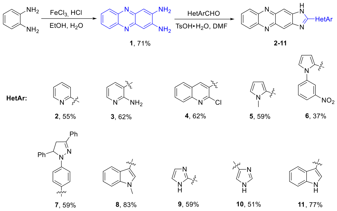
To obtain the target aminoalkyl-substituted 2-hetarylimidazophenazines 12–22, a synthetic scheme was developed starting from available o-phenylenediamine, hetaryl carbaldehydes, and tertiary ω-haloalkylamines. Initially, 2-hetaryl-substituted imidazophenazines 2–11 were obtained by condensation of phenazine-2,3-diamine 1, synthesized by oxidation of o-phenylenediamine with iron(III) chloride [11] with heterocyclic aldehydes in DMF in the presence of p-toluenesulfonic acid (50–80% yields, Figure 1). The resulting compounds 2–11 were precipitated from the solution by diluting the reaction mixture with water, filtered off and recrystallized from ethanol. The obtained substances were poorly soluble in organic solvents, complicating 13C NMR spectroscopy.
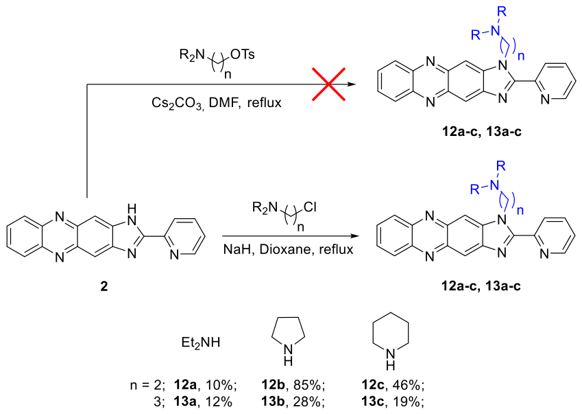
Alkylation of imidazophenazine 2 was carried out to introduce the aminoalkyl linkers. Initial attempts to alkylate 2 with tosylated amino alcohols in DMF in the presence of cesium carbonate (Figure 2) were unsuccessful. Varying the bases (K2CO3, Cs2CO3, NaH) and their amounts, as well as solvents (MeCN, DMF, DMSO, dioxane) did not lead to target compounds. Substitution of the tosyl group with the imidazophenazine fragment probably does not occur due to steric hindrance; we proposed to use a reagent with a smaller leaving group, that is, the chlorine atom, for alkylation. Indeed, 2-chloro-N,N-diethylethan-1-amine as a model alkylating reagent was successful when the reaction was carried out in the NaH/dioxane system. Using chloro-substituted ethyl- and propylamines as alkylating reagents, a series of 2-pyridineimidazophenazines with aminoalkyl substituents 12a-c, 13a-c were synthesized (Figure 2). Low yields of the products were associated with difficulties of purification by column chromatography.
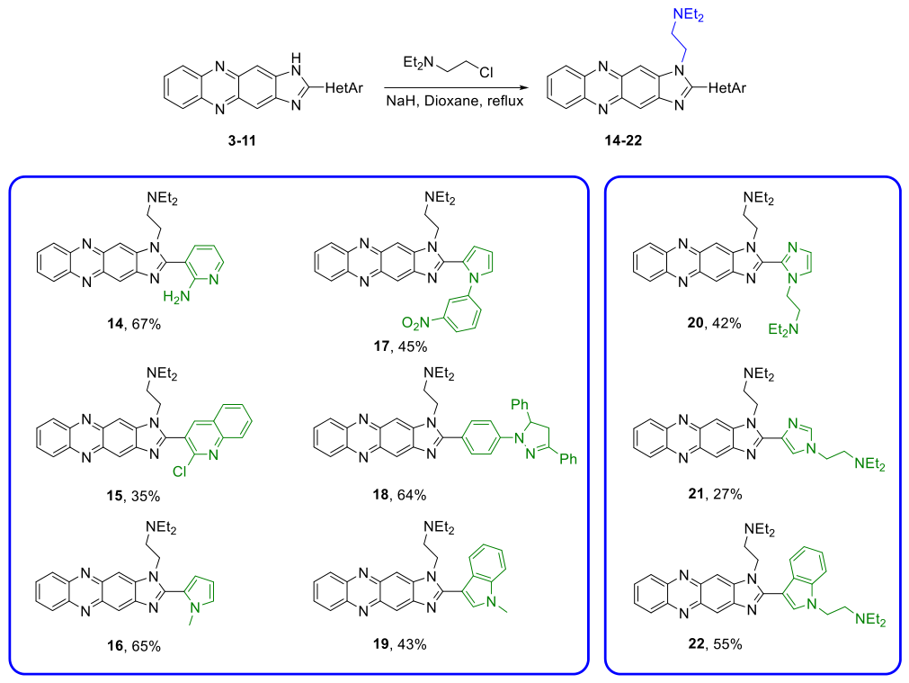
Alkylation of imidazophenazines 3–11 was carried out using 2-chloro-N,N-diethylethan-1-amine under optimized conditions, yielding alkyl-substituted imidazophenazines 14–22 (Figure 3). For compound 14, alkylation occurred at the imidazophenazine ring rather than at the NH2-group of the pyridine ring, as evidenced by the absence of a singlet of the imidazophenazine ring NH proton and retention of a broadened singlet of the NH2 group in the 1H NMR spectrum. In the case of phenazines 9–11, alkylation occurred not only at the nitrogen atom of the imidazophenazine moiety but also at the nitrogen atom of the heterocyclic substituent, yielding dialkylated products 20–22. Formation of dialkylated products 20-22 was evidenced by the absence of both singlets of NH protons of the imidazophenazine ring and the heterocycle at position 2 of the imidazophenazine ring (imidazole or indole), which were observed in the 11-13 ppm region of 1H NMR spectrum, as well as the presence of two triplets at 5.0 ppm, corresponding to the CH2-groups at N atoms of the imidazophenazine ring and the imidazole or indole rings. In the 13C NMR spectra of 20-22 in the 5-70 ppm region, the number of signals from carbon atoms was twice bigger than in 14-19.

3.2. Biological Studies

The cytotoxicity of compounds 12-22 in comparison with 23-25 [11] was studied on the HCT116 human colon carcinoma cell line (Table 1). The cytotoxic potency was moderate. It is worth noting that increasing the linker length elevated the cytotoxicity (e.g.: 12a, mean IC50 = 28 μM; 13a, IC50 = 6 μM). Compounds with pyrrolidine and piperidine fragments in the linkers (12b,c; 13b,c) had a lower toxicity (12b,c: IC50 = 17 μM, >50 μM, respectively; 13b,c: IC50 = 10 μM and 15 μM, respectively) compared to 12a, 13a carrying a diethylamine fragment (IC50 = 28 μM and 6 μM, respectively) Moreover, the cytotoxicity of imidazophenazines 12a, 13a was comparable to that of 23-25 with a terminal diethylamide group. Among the compounds with heterocyclic substituents in the 2-position, the most potent were those with two aromatic rings (N-phenylpyrrole derivative 17, IC50 = 9 μM; 2-chloroquinoline derivative 15, IC50 = 8 μM; N-methylindole derivative 19, IC50 = 4 μM).
Following the initial screening, a number of lead compounds were selected and evaluated for cytotoxicity against the K562 myeloid leukemia and Kelly neuroblastoma cell lines (Table 2). Compounds 17 and 19 demonstrated a micromolar cytotoxicity against all tested cell lines. Compound 12a exhibited marked cytotoxicity against the K562 cell line (IC50 = 8 µM), whereas its homolog 13a was inert to K562 cells but was effective against the HCT116 cell line (mean IC50 = 6 µM); compound 15 also demonstrated high efficacy against the HCT116 cell line. These results indicated that both the substituent at the 2-position of the imidazole ring and the length of the aminoalkyl linker significantly influence the cytotoxicity and cell type preference of hetarylimidazophenazines.
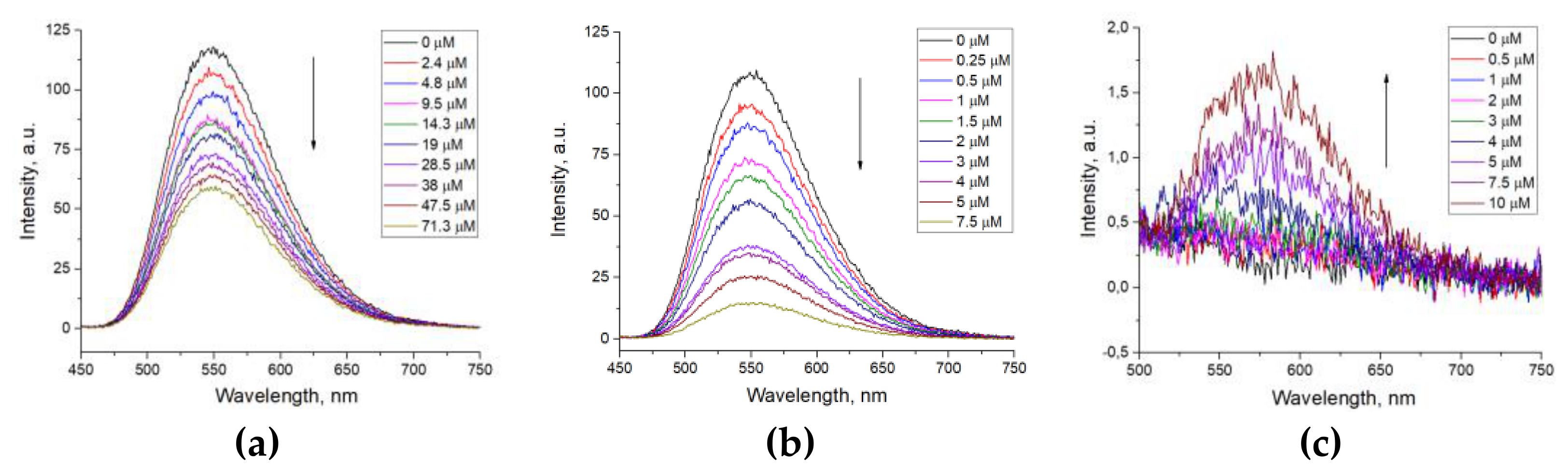
To determine the binding efficiency of the obtained compounds to various DNA structures, the binding constants Kb of ligands 12a, 13a, 15, 17 and 19 to DNA (double-stranded and G4 tel22, c-Myc) were determined with fluorescence spectrometry of ligand solutions in aqueous buffer at various DNA concentrations. Fluorescence quenching was observed upon the addition of both double-stranded and G-quadruplex DNA to a solution of compound 12a, with the addition of double-stranded DNA resulting in weaker fluorescence quenching than the addition of G4 (Figure 4a,b). In the case of compound 13a, ligand fluorescence was significantly lower than that of compound 12a and previously studied compounds 23–25. When titrating a solution of compound 13a with DNA, fluorescence quenching was observed only with the addition of c-Myc G-quadruplex. For compounds 15 and 17, weak quenching of ligand fluorescence was observed upon the addition of dsDNA and the tel22 G-quadruplex, whereas a more pronounced decrease of fluorescence intensity was detectable upon titration with the c-Myc G-quadruplex. In the case of 19, its fluorescence in an aqueous solution was not observed due to dissipation of the ligand excitation energy by vibrations of O-H bonds of water molecules. However, upon addition of a c-Myc solution, the appearance of a ligand fluorescence band was observed (Figure 4c), which may be due to the replacement of water molecules in the environment of the compound 19 molecule by the DNA molecule, on which the dissipation of excitation energy is less than on water molecules. A similar phenomenon has been observed with G-quadruplex ligands [14,15,16]. It is worth noting that the addition of double stranded DNA or tel22 did not change the fluorescence spectra of 19, indicating the absence of binding to these DNA structures.
Based on the decreased fluorescence intensity (Figure S30), the concentrations of bound and free ligands were calculated, and binding constants Kb with G4 and dsDNA were estimated. The calculated Kb values for 12a, 13a, 15, 17, 19 were approximately 105-106 M-1 (Table 3), which is comparable with the literature data for 23-25 and other G4 ligands [17]. It is worth noting that 15, 17 and 19 exhibited weak binding to tel22 G4 and dsDNA but a higher binding efficiency to c-Myc G4, indicating selectivity not only to G4 structures over dsDNA but also to gene-specific quadruplexes. Some ligands specific to the particular types of quadruplexes—specifically those targeting c-Myc— have been described previously in the literature [1,17]. According to published data, these compounds exhibit binding constants in the range of 106–107 M-1. Based on the information available on various G4-binding ligands, it has not been possible to identify the specific structural features that explain selectivity for c-Myc, including for compounds from this paper. The solubility of alkylamino-substituted imidazophenazines 12a-c, 13a-c in DMSO was higher (~ 15-17 mg/ml DMSO) than that of amidoalkyl-substituted derivatives 23-25 (~ 7-10 mg/ml DMSO), which facilitates further modification and biological studies.
Most tested compounds exhibited a weak binding to double-stranded DNA. It is worth noting that the obtained Kb values with G4 were comparable for different compounds, while their cytotoxicity varied significantly (e.g., mean IC50 for 12a = 28 μM vs 4 μM for 19). Differential cytotoxicity despite similar binding efficiencies to G4 suggests either binding to other DNA regions not tested in these experiments or interactions of ligands with other cellular targets. Thus, the obtained results indicate that synthesised compounds can be considered promising DNA ligands with a preference for G-quadruplexes over dsDNA. G4 binding may be one of the mechanisms underlying their cytotoxicity, but the absence of a correlation between binding constants and IC₅₀ values suggests that other cellular targets contribute as well; their identification is of interest for the further development of this scaffold.

4. Conclusions

Thus, in this study, we firstly developed the syntheses of novel aminoalkyl-substituted imidazophenazine derivatives containing various nitrogen heterocycles. The synthesized compounds exhibited moderate cytotoxicity toward HCT116, K562 and Kelly cell lines and selective binding to G4 DNAs, primarily G4 c-Myc. The resulting compounds are characterized by ease of synthesis and high solubility in DMSO, while maintaining cytotoxicity, selectivity, and binding efficiency to G4 comparable to those in the literature.

Supplementary Materials

The following supporting information can be downloaded at: Preprints.org (1H NMR spectra, HRMS of compounds 3-11; 1H, 13C NMR spectra, HRMS of compounds 12-22, fluorescence spectra of compounds 12a, 13a, 15, 17, 19 in the presence of DNA).

Author Contributions

Conceptualization, M.S.A., E.S.B., E.K.B.; methodology, M.S.A.; validation, E.S.B., E.K.B.; formal analysis, M.S.A., D.N.K., A.A.S.; investigation, M.S.A., M.I.V., P.S.B., D.N.K., E.S.I., A.V.G., L.D.P.; resources, E.K.B.; data curation, D.N.K., A.A.S., E.K.B.; writing—original draft preparation, M.S.A.; writing—review and editing, D.N.K., A.A.S., E.K.B; visualization, M.S.A., E.S.B.; supervision, E.K.B.; project administration, E.K.B.; funding acquisition, E.K.B. All authors have read and agreed to the published version of the manuscript.

Funding

The study was conducted under the state assignment of the Lomonosov Moscow State University, project № AAAA-A21-121012290046-4.

Data Availability Statement

The raw data supporting the conclusions of this article will be made available by the authors on request.

Conflicts of Interest

The authors declare no conflicts of interest.

References

  1. Abramovich, M.S.; Zaikina, P. V.; Barskaya, E.S.; Beloglazkina, E.K. Synthesis and Biological Properties of Small Molecules — Ligands of Non-Canonical DNA and RNA Structures. Russ. Chem. Rev. 2024, 93, RCR5134. [Google Scholar] [CrossRef]
  2. Roy, S.; Pramanik, P.; Bhattacharya, S. Exploring the Role of G-Quadruplex DNA, and Their Structural Polymorphism, in Targeting Small Molecules for the Design of Anticancer Therapeutics: Progress, Challenges, and Future Directions. Biochimie 2025, 234, 120–145. [Google Scholar] [CrossRef] [PubMed]
  3. Roxo, C.; Pasternak, A. Switching off Cancer – An Overview of G-Quadruplex and I-Motif Functional Role in Oncogene Expression. Bioorg. Med. Chem. Lett. 2025, 116, 130038. [Google Scholar] [CrossRef] [PubMed]
  4. Roy, S.; Ali, A.; Kamra, M.; Muniyappa, K.; Bhattacharya, S. Specific Stabilization of Promoter G-Quadruplex DNA by 2,6-Disubstituted Amidoanthracene-9,10-Dione Based Dimeric Distamycin Analogues and Their Selective Cancer Cell Cytotoxicity. Eur. J. Med. Chem. 2020, 195, 112202. [Google Scholar] [CrossRef] [PubMed]
  5. Zagotto, G.; Ricci, A.; Vasquez, E.; Sandoli, A.; Benedetti, S.; Palumbo, M.; Sissi, C. Tuning G-Quadruplex vs Double-Stranded DNA Recognition in Regioisomeric Lysyl-Peptidyl-Anthraquinone Conjugates. Bioconjug. Chem. 2011, 22, 2126–2135. [Google Scholar] [CrossRef] [PubMed]
  6. Andreeva, D. V.; Vedekhina, T.S.; Gostev, A.S.; Dezhenkova, L.G.; Volodina, Y.L.; Markova, A.A.; Nguyen, M.T.; Ivanova, O.M.; Dolgusheva, V.; Varizhuk, A.M.; et al. Thiadiazole-, Selenadiazole- and Triazole-Fused Anthraquinones as G-Quadruplex Targeting Anticancer Compounds. Eur. J. Med. Chem. 2024, 268, 116222. [Google Scholar] [CrossRef] [PubMed]
  7. Fukuda, H.; Zou, T.; Fujii, S.; Sato, S.; Wakahara, D.; Higashi, S.; Tseng, T.Y.; Chang, T.C.; Yada, N.; Matsuo, K.; et al. Cyclic Anthraquinone Derivatives, Unique G-Quadruplex Binders, Selectively Induce Cancer Cell Apoptosis and Inhibit Tumor Growth. PNAS Nexus 2023, 2, 1–10. [Google Scholar] [CrossRef] [PubMed]
  8. Holden, L.; Gkika, K.S.; Burke, C.S.; Long, C.; Keyes, T.E. Selective, Disruptive Luminescent Ru(II) Polypyridyl Probes of G-Quadruplex. Inorg. Chem. 2023, 62, 2213–2227. [Google Scholar] [CrossRef] [PubMed]
  9. Liu, X.; Ma, L.; Hu, X.; Chen, Y.; Zhang, S. Tuning the Selectivity of Ruthenium-Based Luminescent G-Quadruplex Probe by Triphenylamine Groups. Appl. Organomet. Chem. 2026, 40. [Google Scholar] [CrossRef]
  10. Wang, X.-D.; Lin, J.-H.; Hu, M.-H. Discovery of a Tribenzophenazine Analog for Binding to the KRAS mRNA G-Quadruplex Structures in the Cisplatin-Resistant Non–small Cell Lung Cancer. J. Biol. Chem. 2025, 301, 108164. [Google Scholar] [CrossRef] [PubMed]
  11. Abramovich, M.S.; Bymbygydenov, P.S.; Kalyuzhny, D.N.; Zyk, N. V.; Beloglazkina, E.K.; Barskaya, E.S. Synthesis of Amidoalkyl-Substituted 2-(Pyridin-2-yl)imidazo[4,5-B]phenazines as Potential Ligands of G-Quadruplexes. Russ. J. Org. Chem. 2026, 62, 99. [Google Scholar] [CrossRef]
  12. Pina, M. del K.; Budylin, V.A.; Rodrigues, M.; Terent’ev, P.B.; Bundel, Y.G. Synthesis and Spectral Data of 1-Aryl-2-Formylpyrroles. Khimiya Geterotsiklicheskikh Soedin. 1989, 180–184. [Google Scholar]
  13. Kutulya, L.A.; Shevchenko, A.F.; Surov, Y.N. Structure of Formylation Products of 1,3,5-Triphenyl- and 1,5-Diphenyl-3-Styryl-2-Pyrazolines. Khimiya Geterotsiklicheskikh Soedin. 1975, 250–253. [Google Scholar]
  14. Xie, X.; Reznichenko, O.; Chaput, L.; Martin, P.; Teulade-Fichou, M.; Granzhan, A. Topology-Selective, Fluorescent “Light-Up” Probes for G-Quadruplex DNA Based on Photoinduced Electron Transfer. Chem. – A Eur. J. 2018, 24, 12638–12651. [Google Scholar] [CrossRef] [PubMed]
  15. Turaev, A. V.; Tsvetkov, V.B.; Tankevich, M. V.; Smirnov, I.P.; Aralov, A. V.; Pozmogova, G.E.; Varizhuk, A.M. Benzothiazole-Based Cyanines as Fluorescent “light-Up” Probes for Duplex and Quadruplex DNA. Biochimie 2019, 162, 216–228. [Google Scholar] [CrossRef] [PubMed]
  16. Fujita, H.; Watatani, N.; Sasaki, S.; Okabe, S.; Seimiya, H.; Hirokawa, T.; Tera, M.; Ma, Y.; Nagasawa, K. Synthesis of a Light-Up Probe with a Trioxazole Skeleton for Tracking G-Quadruplex Dynamic Behavior. Anal. Chem. 2025, 97, 16482–16490. [Google Scholar] [CrossRef] [PubMed]
  17. Andreeva, D. V.; Tikhomirov, A.S.; Shchekotikhin, A.E. Ligands of G-Quadruplex Nucleic Acids. Russ. Chem. Rev. 2021, 90, 1–38. [Google Scholar] [CrossRef]
Figure 1. Synthesis of 2-hetarylimidazophenazines 2-11.
Preprints 240465 g001
Figure 2. Synthesis of aminoalkyl-substituted 2-(pyridine-2-yl)imidazophenazines 12a-c, 13a-c.
Preprints 240465 g002
Figure 3. Synthesis of aminoalkyl-substituted 2-hetarylimidazophenazines 14-22.
Preprints 240465 g003
Figure 4. Fluorescence spectra: a) compound 12a in the presence of dsDNA, b) compound 12a in the presence of G4 c-Myc, c) compound 19 in the presence of G4 c-Myc.
Preprints 240465 g004
Table 1. Cytotoxicity of new compounds 12-22 and previously described 23-25 against HCT116 cell line.
Compound IC50, μM Compound IC50, μM Preprints 240465 i001
23 17.1±2.3 15 8.6±2.5
24 9.4±2.2 16 16.4±3.5
25 6.3±1.7 17 9.5±2.5
12a 28.8±3.8 18 >50
12b 17.4±3.7 19 4.7±2.0
12c >50 20 10.4±2.4
13a 6.1±2.0 21 15.3±2.2
13b 10.4±3.4 22 17.4±3.2
13c 15.0±3.1
Table 2. Cytotoxicity of lead compounds against leukemia and neuroblastoma cell lines.
Compound K562 Kelly
12a 8.2±2.0 17.3±3.0
13a >50 10.1±2.1
15 15.3±2.2 31.4±3.2
17 6.4±1.1 9.4±2.1
19 1.0±0.7 2.1±0.8
Table 3. Binding constants Kb×106 M-1 of 12a, 13a, 15, 17, 19 with DNA structures.
12a 13a 15 17 19
dsDNA 0.09±0.01 - 0.10±0.03 0.20±0.01 -
tel22 1.01±0.03 0.12±0.02 0.05±0.01 0.003±0.001 -
c-Myc 0.79±0.04 0.20±0.01 0.25±0.02 0.68±0.05 0.68±0.03
Disclaimer/Publisher’s Note: The statements, opinions and data contained in all publications are solely those of the individual author(s) and contributor(s) and not of MDPI and/or the editor(s). MDPI and/or the editor(s) disclaim responsibility for any injury to people or property resulting from any ideas, methods, instructions or products referred to in the content.
Copyright: This open access article is published under a Creative Commons CC BY 4.0 license, which permit the free download, distribution, and reuse, provided that the author and preprint are cited in any reuse.