

Submitted:
23 October 2026
Posted:
26 October 2026
You are already at the latest version
Abstract
Chronic stress, circadian disruption, sedentary behaviour, industrialised diets and disturbances in the gut microbiome have created an evolutionary mismatch between ancestral physiology and the modern environment. When midlife women enter the menopausal transition, cumulative exposure to lifestyle and environmental factors may exacerbate the hormonal changes associated with menopause. As a result, midlife women can present with a mixture of vasomotor, somatic, urogenital, psychological and cognitive symptoms that are often poorly diagnosed and treated in isolation. Our previous review described how evolutionary mismatch in Generation X (Gen X) women disrupts perimenopausal physiology through converging endocrine, psychosocial stress and immunometabolic mechanisms, amplifying symptoms and cardiometabolic risk. This included a detailed discussion of the endocrine changes that underpin the menopausal transition, how chronic psychosocial stress overwhelms whole-body stress response systems, and the impact of converging immunometabolism mechanisms such as chronic inflammation and insulin resistance on whole-body energy homeostasis. In the current review, we extend this model by proposing a Gen X clinical phenotype and an evolutionary realignment framework that translates it into an integrated approach to clinical management. The proposed framework addresses circadian alignment, metabolic flexibility, nutritional and lifestyle factors, allostatic load, environmental exposures, cerebral and mitochondrial health, musculoskeletal function, gut–hormone–microbiome signalling and menopausal hormone therapy, offering practitioners a systems-based approach to midlife care that addresses underlying physiological drivers rather than isolated symptoms.
Keywords:
evolutionary mismatch
; generation X women
; gen X
; metabolic dysfunction
; metabolic inflexibility
; insulin resistance
; mitochondrial dysfunction
; allostatic load
; cognitive dysfunction
1. Introduction
Midlife women increasingly present with a mixture of problems that extend beyond the classical vasomotor symptoms of hot flushes and night sweats. These include persistent fatigue, weight gain, cognitive changes, reduced motivation and declining resilience [1,2,3,4]. Although these symptoms are commonly attributed to menopause or ageing, reproductive hormone decline alone does not fully account for their breadth or severity [5,6,7]. Despite normal or borderline pathology results, many women experience progressive deterioration in metabolic health, body composition and overall wellbeing that is difficult to explain within existing hormone-focused models [6,7,8,9]. Symptom- and/or hormone-based treatment approaches may provide partial relief but often fail to address the underlying drivers of this broader clinical presentation.
Generation X (Gen X) women, born between 1965 and 1980 (aged 46–61 years), represent the first cohort to reach midlife following lifelong exposure to modern lifestyle and environmental conditions [2],10–12). This is occurring alongside a rapidly escalating metabolic burden. In the United States, the prevalence of overweight and obesity in women aged 40–64 years has increased from approximately 49% in 1990 to over 74% in 2021, with projections suggesting more than 80% will be affected by 2050 [13]). Comparable patterns are evident in Australia, where approximately 80% of women in the Gen X cohort have an increased or substantially increased metabolic risk based on waist circumference [14]). These trends highlight the scale and urgency of metabolic dysfunction emerging during the midlife transition, reflecting disruption across interconnected metabolic, neuroendocrine and immune networks [15,16].
The midlife changes observed in many Gen X women can be better understood within the context of evolutionary mismatch, whereby human biology, adapted to conditions of intermittent food availability, high physical activity, stable circadian rhythms and intermittent stress, is increasingly discordant with the modern environment [7,17]. Contemporary lifestyles are characterised by continuous access to energy-dense foods, reduced physical activity, circadian disruption and sustained psychosocial demands, alongside increased exposure to environmental pollutants and endocrine-disrupting chemicals (EDC) [8,18]. These exposures, alongside industrialised dietary patterns, are associated with progressive reductions in gut microbiome diversity, further compounding metabolic and immune dysregulation [19,20]. The aim of the current review is to extend our evolutionary mismatch model by proposing a Gen X clinical phenotype and translating this model into an evolutionary realignment framework that can be implemented in clinical practice (Figure 1).
2. The Proposed Generation X Clinical Phenotype
Building on the evolutionary mismatch model described in our previous review [21]), the current paper considers how disruption of whole-body energy homeostasis may be expressed clinically in Gen X women. Homeostasis depends on coordinated signalling between central neuroendocrine networks and peripheral metabolic tissues to regulate appetite, substrate utilisation, energy expenditure, stress responses and anabolic function according to physiological demand [22,23]. Persistent insulin resistance (IR), inflammatory signalling, stress-axis activation and circadian disruption may reduce the flexibility and fidelity of these regulatory systems, impairing the capacity to maintain metabolic balance despite adequate energy availability [23,24,25]. Within an evolutionary framework, this may favour adaptive responses that conserve energy while increasing the drive to acquire it [26,27]. During the menopausal transition, fluctuating and declining ovarian hormone levels may further challenge this homeostatic capacity [28,29].
2.1. Adaptive Energy Conservation Under Perceived Scarcity
We propose that IR, chronic inflammation, stress-axis activation, circadian disruption and fluctuating ovarian hormones may together be interpreted centrally as reduced effective energy availability despite caloric abundance. Hypothalamic inflammation can induce central leptin and insulin resistance, blunting the cues which arcuate and ventromedial neurons use to gauge long-term energy sufficiency, while glucocorticoid excess further shifts regulatory set points towards energy conservation [22],23,30). Kisspeptin neurons are involved in coupling reproductive function to energy status and are suppressed by cortisol, inflammation and impaired leptin signalling. In addition, declining 17β-oestradiol (E2) may amplify this suppression during the menopausal transition [28],31,32). Reduced kisspeptin tone may in turn disinhibit arcuate agouti-related peptide/neuropeptide Y (AgRP/NPY) neurons, heightening hunger salience and weakening satiety [33,34]. In parallel, declining E2 alters dopaminergic tone within prefrontal and striatal pathways, which may reduce motivation for non-immediate goals while increasing responsiveness to palatable food cues [35,36].
Energy expenditure may be similarly downregulated. Altered hypothalamic–pituitary–thyroid dynamics and reduced tissue-level triiodothyronine (T3) action may lower thermogenesis and mitochondrial energy production. This ‘thyroid economy’ reflects a deliberate down-shifting of effective thyroid hormone action to reduce metabolic rate and conserve energy, and is a hallmark feature of adaptive thermogenesis [37,38]. Somatopause, with declining growth hormone (GH) and insulin-like growth factor-1 (IGF-1) signalling, further reduces anabolic capacity and lean mass, lowering resting energy expenditure and metabolic flexibility [39,40].
Together, these adaptations may result in a scarcity-adapted set point in which energy acquisition and conservation are co-activated despite caloric abundance. Adiposity and IR further impair hypothalamic signalling, and reduced lean mass lowers energy availability, creating a self-reinforcing cycle that may contribute to fatigue, weight-loss resistance and progressive metabolic decline. This model is proposed as one contributor to the Gen X phenotype rather than a complete explanation, and several of its components, particularly kisspeptin–AgRP interactions and thyroid economy, derive largely from experimental and caloric-restriction studies. Nonetheless, it provides a mechanistic basis for the clinical features described below.
2.2. Metabolic Dysregulation, Central Adiposity and Lean Mass Decline
A central feature of the proposed Gen X clinical phenotype is metabolic disturbance accompanied by visceral and ectopic fat deposition and central adiposity [41,42]. Visceral adipose tissue is metabolically active and strongly associated with IR, chronic inflammation and impaired metabolic flexibility [4,43], contributing to dysregulated glucose and lipid metabolism that may occur even when body mass index (BMI) is normal [44,45]. These changes frequently occur alongside progressive loss of lean muscle mass and function. Declining ovarian hormone levels, together with IR and chronic inflammation, may impair muscle protein synthesis and contribute to gradual loss of metabolically active tissue [40,46]. Because skeletal muscle is a major site of insulin-mediated glucose disposal, reductions in lean mass further compound IR, metabolic dysfunction and reduced physical capacity [18,47], reinforcing the adaptive cycle described in Section 2.1. Where increasing adiposity and declining muscle mass and function coexist, this pattern may progress towards sarcopenic obesity [48]). These changes can occur gradually and may be overlooked during midlife, yet they have important implications for long-term health, physical independence and quality of life [49]).
2.3. Neuroendocrine Symptom Cluster
In addition to measurable changes in body composition and metabolic function, many Gen X women experience a constellation of symptoms reflecting disruption of neuroendocrine and energy-regulation systems. This cluster commonly includes persistent fatigue, cognitive difficulties often described as “brain fog”, reduced concentration and focus and difficulty with memory retrieval, dysregulated appetite and cravings, and changes in mood and motivation [3,15].
These symptoms reflect the convergent effects of the neuroendocrine and metabolic disturbances described in our previous review [21]). Fatigue is associated with impaired mitochondrial function, reduced metabolic flexibility and disrupted circadian regulation; cognitive changes with altered cerebral energy metabolism and neuroinflammatory signalling; dysregulated appetite with disruption of hypothalamic energy sensing and reward pathways in the context of IR and modern food environments; and mood and motivational changes with altered neuromodulatory tone and stress-axis dysregulation [21]).
Cognitive symptoms warrant particular attention. Oestrogen supports cerebral glucose metabolism, mitochondrial bioenergetics and synaptic function, and neuroimaging studies have demonstrated reduced cerebral glucose metabolism across the menopausal transition, alongside changes in brain E2-receptor density that are associated with memory performance and menopausal symptoms [50,51]. These changes may contribute to the memory retrieval difficulties reported most frequently during perimenopause [52]). Although such symptoms are often transient, declining E2 signalling may also mark a window of vulnerability for later cognitive decline, particularly where metabolic dysfunction persists [50,53].
Collectively, these symptoms are best understood as clinical expressions of integrated system-level dysfunction rather than isolated hormonal effects, reflecting the cumulative impact of evolutionary mismatch across metabolic, neuroendocrine and behavioural domains.
2.4. Increased Long-Term Chronic Disease Risk
These metabolic, endocrine, and neurophysiological changes may have important implications for long-term health. The convergence of central adiposity, IR, declining muscle mass and neuroendocrine dysregulation during midlife may create an environment that increases susceptibility to several chronic diseases later in life. These include type 2 diabetes mellitus (T2DM) [54]), metabolic dysfunction associated steatotic liver disease (MASLD) [55]), cardiovascular disease (CVD) [56]), osteoporosis [57]) and dementia [50,53].
Women demonstrate a well-established longevity advantage relative to men across most contemporary human populations, a pattern attributed to interacting biological, behavioural and reproductive factors [58,59]. However, the convergence of metabolic, endocrine and environmental pressures described in this review raises the possibility that the health trajectories of Gen X women may be increasingly shaped by conditions that were largely absent during earlier periods of human evolutionary history. Whether these interacting influences may alter the long-standing female longevity advantage in future generations remains an important area for further investigation.
3. Evolutionary Realignment Framework
The mechanisms described in preceding sections indicate that evolutionary mismatch influences metabolic, endocrine and neurological health through disruption of core regulatory systems, including circadian rhythms, energy metabolism, stress physiology and gut microbiome composition [20]). Because the adaptive cycle described in Section 2.1 is sustained across regulatory networks, interventions targeting isolated pathways or symptoms are unlikely to be effective. Translating the proposed phenotype into clinical practice therefore requires interventions that target upstream regulatory processes. The proposed evolutionary realignment framework comprises eight intervention domains addressing circadian regulation, metabolic flexibility, stress physiology, environmental exposures, cerebral and mitochondrial function, musculoskeletal health, gut–hormone signalling and, where clinically appropriate, ovarian hormone signalling through menopausal hormone therapy (MHT), as summarised in Table 1.
Although these domains are relevant to midlife women generally, the Gen X cohort framing shapes clinical priorities within the described intervention domain. Many Gen X women entered the workforce before childcare and paid parental leave were widely available and have spent much of their adult lives combining paid employment with substantial household and caregiving responsibilities, including care of children and, increasingly in midlife, ageing parents [21],60,61). This cumulative role strain positions allostatic load and stress regulation as a central rather than adjunctive treatment target. Similarly, lifelong exposure to industrialised food environments, artificial light and EDCs, means that dietary pattern, circadian behaviour and exposure history warrant systematic assessment [12],20,21). The framework therefore emphasises identifying the mismatch exposures that have been most sustained for each woman, and prioritising the corresponding domains, rather than applying all interventions uniformly.
3.1. Circadian Realignment and Chrononutrition
The menopausal transition may increase vulnerability to circadian dysregulation, with declining ovarian hormones associated with reduced circadian alignment, altered sleep–wake regulation and changes in peripheral clock function [62,63,64]. Menopause-related sleep changes are associated with increased cardiometabolic risk, while circadian rhythmicity has been proposed as a potential biomarker of healthy ageing in women [65,66].
Circadian synchronisation is therefore a foundational strategy for addressing midlife metabolic and neuroendocrine dysfunction. Circadian rhythms regulate glucose metabolism, hormonal signalling, sleep architecture and energy balance, and are strongly influenced by meal timing [67,68]. Sleep, feeding and activity evolved as coordinated systems entrained by natural light–dark cycles, physical activity and food availability [69,70]. Modern exposure to artificial light and evening screen use, delayed sleep, prolonged sedentary periods, extended eating windows and late-night food intake can create temporal mismatch between behaviour and endogenous circadian physiology [71,72,73].
Chrononutrition concerns food intake timing relative to endogenous circadian rhythms, typically favouring earlier daytime energy consumption [67,74]. Meal timing reflects an evolutionarily conserved feeding–fasting cycle and provides a temporal signal for gastrointestinal physiology and gut microbial rhythms [74,75,76]. Metabolic responses to food vary across the biological day, with glucose tolerance and insulin sensitivity generally more favourable during the early active phase [77,78]. Later eating and circadian misalignment are associated with impaired glucose regulation and adverse metabolic outcomes [77,78]. Sleep and circadian disruption may also alter gut microbial composition, with reductions in Faecalibacterium and Lactobacillus and increases in Clostridium reported across some studies, with potential implications for inflammation and metabolic health [79]). Insufficient sleep may further increase energy intake and alter appetite and reward-related responses [80]).
A systematic review of 41 randomised controlled trials (RCTs) found that time-restricted eating (TRE) reduced body weight, BMI, fat mass, waist circumference, systolic blood pressure, fasting glucose, fasting insulin and triglycerides compared with usual diets [81]). Early TRE (eTRE) was superior to late TRE for body weight, waist circumference and fasting insulin. By confining intake to an earlier daytime window, eTRE may better align feeding with circadian metabolic rhythms while extending periods of lower insulin exposure and greater reliance on stored energy substrates, potentially supporting insulin sensitivity and glycaemic regulation [81,82].
Evidence in women is also emerging. Peters et al. reported improved sleep efficiency and reduced sleep fragmentation during eTRE in 31 women, 26 of whom were postmenopausal[83]). Jóźwiak et al. found that 16:8 TRE combined with exercise reduced psychological and somatic menopausal symptoms and improved physical and psychosocial quality-of-life measures more than exercise alone [84]). These findings support TRE and earlier, circadian-aligned eating as potentially useful strategies during the menopausal transition, although longer-term trials specifically examining eTRE in peri- and postmenopausal women are needed.
Circadian alignment can also be supported through regular sleep–wake timing, early natural light exposure, daytime physical activity and reduced evening exposure to bright and blue-enriched light [85]). Greater sleep regularity has been associated with lower mortality risk and may predict mortality more strongly than sleep duration [86]). Sleep hygiene practices may be particularly relevant during the menopausal transition, when vasomotor symptoms and changing ovarian hormone levels can further disrupt sleep continuity and circadian rhythmicity [66]).
3.2. Fuel Utilisation and Metabolic Flexibility
Metabolic flexibility describes the capacity to shift appropriately between glucose and fatty-acid oxidation according to nutrient availability and physiological demand [87,88]. A recent review identified perimenopause as a period of metabolic vulnerability characterised by reduced fat oxidation and impaired metabolic flexibility [89]). Declining E2 may also contribute to increasing visceral adiposity and altered lipid metabolism [30,41], while higher follicle-stimulating hormone (FSH) has been associated with lower postprandial fat oxidation [90]). In metabolically inflexible states, increased free fatty-acid availability coupled with reduced oxidative capacity can promote ectopic lipid accumulation, including hepatic steatosis, further impairing insulin signalling and mitochondrial function [88,91]. Insulin resistance further impairs fuel switching by reducing insulin-mediated glucose uptake into insulin-sensitive tissues, while increasing adipose lipolysis and free fatty-acid delivery to the liver, disrupting normal transitions between glucose and fat oxidation [30,88].
Dietary strategies should therefore target insulin sensitivity and glycaemic regulation while supporting appropriate substrate utilisation. Priorities include minimally processed, fibre-rich foods, adequate protein, high-quality complex carbohydrates in place of refined carbohydrates, and limiting excessive saturated-fat intake [87,92]. Mediterranean-style dietary patterns have been associated with lower indices of hepatic steatosis in women with overweight and obesity and improved body composition and cardiometabolic outcomes in midlife women [93,94]. A recent review further identified polyphenol-rich dietary patterns as a promising strategy for mitigating menopause-associated cardiometabolic risk through effects on glucose and lipid metabolism, blood pressure, inflammation and gut microbiota [95]).
Consistent meal structure and avoidance of routine breakfast skipping may further support insulin sensitivity and glycaemic regulation [96,97]. Exercise provides an important complementary stimulus for improving insulin sensitivity, mitochondrial function and substrate utilisation and is addressed further in Section 3.6. Despite increasing evidence for these individual approaches, direct dietary intervention studies measuring changes in metabolic flexibility across the menopausal transition remain limited, representing an important area for further research.
3.3. Allostatic Load and Stress Regulation
Allostatic load reflects the cumulative physiological burden generated by repeated or prolonged adaptation to stress across neuroendocrine, autonomic, immune, inflammatory and metabolic systems. During the menopausal transition, stress regulation may be challenged by interacting hormonal and behavioural changes, including fluctuating ovarian hormone levels and sleep disturbance, while age-related declines in dehydroepiandrosterone sulphate (DHEAS) may further reduce stress-adaptive capacity [3,98]. Importantly, menopause-associated sleep fragmentation can disrupt cortisol dynamics independently of E2 decline, identifying sleep as a modifiable contributor to hypothalamic-pituitary-adrenal (HPA)-axis dysregulation [99]). Chronic stress and altered HPA-axis feedback have also been associated with epigenetic changes in stress- and immune-related pathways that may promote inflammatory signalling and contribute to cumulative allostatic burden [100]). In Gen X women, occupational demands, caregiving responsibilities, role strain and persistent time pressure may further compound this physiological load [60,61].
Lifestyle and behavioural interventions should therefore prioritise reduction of modifiable stressors and restoration of adaptive capacity through psychological support, setting behavioural boundaries, adequate recovery, social connection, physical activity and sleep optimisation [101,102,103]. Menopause-specific studies provide support for the use of mindfulness-based interventions in clinical practice. A meta-analysis of 13 studies involving 1,138 women found a significant reduction in perceived stress (SMD −0.84, 95% CI −1.64 to −0.05), although effects on anxiety and depression were less consistent [104]). More recent evidence similarly supports the use of mindfulness, cognitive behavioural therapy and other mind-body interventions for stress, sleep and psychological symptoms during menopause [102,103].
Complementary therapies may also be considered within an individualised integrative approach, including selected herbal medicines and nutritional interventions according to symptom presentation, patient preference and clinical context [102,103]. For example, Withania somnifera has demonstrated menopause-specific benefits, with a recent RCT reporting improvements in perceived stress, menopausal symptoms and quality of life over eight weeks, alongside modest increases in E2 and progesterone and reductions in FSH and luteinising hormone, relative to placebo [105]). Overall, complementary therapies appear generally safe, with few serious adverse events reported, although further high-quality research is required [103]). These approaches should complement, rather than substitute for, addressing upstream psychosocial and lifestyle drivers of chronic stress.
3.4. Exposome and Environmental Load Reduction
The exposome encompasses cumulative environmental exposures across the life course and how these interact with an individual’s biological susceptibility. Environmental chemicals relevant to midlife women include compounds such as phthalates, bisphenols and parabens, alongside persistent pollutants such as perfluoroalkyl and polyfluoroalkyl substances, organochlorines, some heavy metals and air pollution [106]). Recent menopause-specific reviews associate these exposures with metabolic dysfunction and a range of menopausal symptoms, including adiposity, sleep disturbance, vasomotor symptoms, and cognitive and psychological symptoms [107,108]. Emerging evidence further suggests that the menopausal transition may represent a period of increased vulnerability to the effects of these exposures [108]). For Gen X women, this life-course perspective directs attention to both cumulative and ongoing exposure, particularly to persistent compounds that may bioaccumulate and remain in the body for prolonged periods.
Clinical management should emphasise pragmatic reduction of avoidable exposure rather than complete avoidance. A structured environmental assessment can identify major sources from food and food packaging, plastics, fragranced personal and household products, indoor dust and air, water and occupational exposures. Once identified, targeted individualised advice and practical recommendations can be made [106,109]. Preliminary intervention studies indicate that dietary modification and substitution of household and personal products can reduce measured concentrations of several EDC, although evidence related to menopausal health outcomes has yet to be established [109]). Personalised exposure assessment may further improve environmental-health literacy and behavioural change, with reductions in urinary phthalate exposure demonstrated following targeted recommendations [110]).
3.5. Cerebral Bioenergetics and Neuroprotection
The menopausal transition is increasingly recognised as a period of neuroendocrine and metabolic adaptation within the female brain. Neuroimaging studies have demonstrated menopause-related alterations in cerebral glucose metabolism, brain structure and E2-receptor density, supporting the importance of preserving neurological function across midlife [50,51].
Clinical strategies should therefore prioritise preservation of cerebral metabolic and vascular function while reducing factors that contribute to neuroinflammation and cognitive vulnerability. The circadian, metabolic and stress-regulation strategies outlined above may contribute synergistically through improved sleep, insulin sensitivity, vascular health and mitochondrial function. Dietary patterns emphasising minimally processed plant foods and a diversity of polyphenol-rich foods may provide additional neuroprotective support [95],111,112).
Specific polyphenol- and phytoestrogen-rich compounds may provide additional benefits. In postmenopausal women, long-term resveratrol supplementation improved cognition and cerebrovascular responsiveness alongside improvements in insulin sensitivity [113]). Cocoa and other flavanol-rich foods have also demonstrated favourable effects on selected cognitive outcomes [114,115]. Phytoestrogens, including soy isoflavones, are of particular interest during the menopausal transition, with preclinical evidence suggesting potential neuroprotective effects through E2 receptor signalling, antioxidant pathways, synaptic plasticity and mitochondrial energy metabolism [116,117]. Many phytoestrogens, including genistein and daidzein, exhibit preferential affinity for E2 receptor beta (ERβ), which is highly expressed in brain regions involved in memory and cognition. ERβ signalling has been implicated in synaptic plasticity, neurogenesis, mitochondrial function and neuroprotection, providing a plausible mechanistic basis for tissue-selective neurocognitive effects [118,119]. A meta-analysis of RCTs suggests modest benefits of soy isoflavones for overall cognition and memory, although effects were not evident across all cognitive domains [120]). Individual responses may also be influenced by differences in phytoestrogen metabolism and gut microbial conversion to bioactive metabolites [116]).
3.6. Musculoskeletal and Mitochondrial Function
Mitochondrial function is central to cellular energy production, metabolic flexibility and physiological resilience and may become increasingly vulnerable across the menopausal transition through the interacting effects of ageing, declining ovarian hormones , IR , inflammation and oxidative stress [121,122]. E2 influences mitochondrial biogenesis, respiration and redox regulation across multiple tissues, while human postmenopausal data suggest alterations in skeletal-muscle mitochondrial content and respiratory capacity [123,124]. Maintaining mitochondrial capacity therefore represents a broader therapeutic target than preservation of skeletal muscle alone.
Exercise is a potent stimulus for musculoskeletal and mitochondrial adaptation. Progressive resistance training supports muscle mass and strength while stimulating mitochondrial biogenesis and respiratory adaptation, with aerobic activity providing complementary benefits for oxidative capacity and mitochondrial turnover [125,126]. Individualised programmes incorporating resistance training two to three times weekly, regular aerobic activity and, where appropriate, weight-bearing or impact exercise can support muscle, mitochondrial and bone health [127,128].
Adequate energy, protein and micronutrient availability supports ATP production, training adaptation and musculoskeletal maintenance. Protein intakes of approximately 1.0–1.2 g/kg/day are commonly recommended for healthy older adults, although menopause-specific intervention evidence remains limited [129]), with protein distributed across the day and obtained from diverse high-quality sources, including plant and/or animal proteins. In a prospective study of 38,283 women followed across the menopausal transition, dietary patterns rich in legumes, nuts, fruits, vegetables and whole grains were associated with less weight gain and lower obesity risk, with the plant-forward Planetary Health Diet showing the strongest inverse association with weight gain [130]).
Adequate calcium, vitamin D and magnesium are also important for bone, muscle and cellular energy metabolism [131,132], while polyphenol-rich foods may support mitochondrial quality control through biogenesis, autophagy and redox regulation pathways [133,134]. Phytoestrogens may also support skeletal health, with recent evidence indicating modest improvements in lumbar spine, femoral neck and total-hip bone mineral density in peri- and postmenopausal women [135]).
3.7. Gut–Hormone–Microbiome Integration
The gut microbiome represents an important interface between diet, metabolic regulation and hormone signalling during the menopausal transition. Menopause has been associated with alterations in microbial composition and function, with potential consequences for oestrogen metabolism, inflammation, intestinal barrier integrity and cardiometabolic health [136,137].
Two microbiome-mediated pathways are particularly relevant to systemic E2 physiology. The estrobolome includes specific microbial species with β-glucuronidase activity that deconjugate oestrogens within the intestine, facilitating enterohepatic reabsorption [138,139]. Separately, gut microbes metabolise dietary phytoestrogens into biologically active metabolites. For example, the soy isoflavone daidzein can be converted to equol, while plant lignans are metabolised to enterodiol and enterolactone [117,139]. These pathways provide a rationale for supporting microbiome function during the menopausal transition, as microbial activity may influence the availability of endogenous oestrogens and the generation of bioactive dietary phytoestrogen metabolites, potentially supporting oestrogen-responsive signalling as ovarian E2 declines [140]). As the capacity for these microbial transformations varies between individuals, differences in microbiome composition and function may contribute to variation in response to phytoestrogen-rich foods [139,141].
Dietary fibre provides an additional link between microbiome and metabolic health. Fermentation of microbiota-accessible carbohydrates produces short-chain fatty acids (SCFA), particularly butyrate, acetate and propionate, which contribute to intestinal barrier integrity, inflammatory regulation, appetite signalling and glucose and lipid metabolism [142]). SCFAs may also influence appetite regulation through modulation of enteroendocrine hormones, including glucagon-like peptide-1 (GLP-1) and peptide YY (PYY), vagal signalling and central appetite pathways [143,144,145]. The gut-brain axis integrates enteroendocrine, vagal, immune and central neural signals involved in homeostatic and hedonic regulation of eating behaviour [145]). Evidence from inflammatory gastrointestinal disorders further suggests that intestinal inflammation can alter enteroendocrine and gut-brain signalling, with downstream effects on appetite regulation [146]).
Clinically, emphasis on increasing the diversity of fibre-rich whole plant foods, including foods rich in polyphenols and phytoestrogens may support microbial diversity, intestinal barrier integrity and increase the production of beneficial microbial metabolites such as SCFAs [147,148,149]. Responses to fibre vary according to factors including baseline microbiome composition and habitual diet, supporting an individualised approach rather than a uniform prescription [150]). Fibre intake may therefore be increased gradually according to gastrointestinal tolerance, particularly in individuals with low habitual intake.
3.8. The Role of Menopausal Hormone Therapy
Lifestyle medicine offers a foundational evidence-based framework for menopausal care and forms the central focus of our proposed clinical management pathway [101]). These approaches are emerging as important factors in empowering women to embrace menopause, improve symptoms and optimise wellbeing during the menopausal transition and beyond [151]). Lifestyle and nutritional strategies may also influence several hormone-responsive pathways, including insulin sensitivity, adiposity-related sex-steroid bioavailability, circadian regulation and gut-mediated oestrogen metabolism, and remain relevant irrespective of whether MHT is used [101,140]. Nevertheless, MHT remains an important therapeutic option for menopausal symptom management and the prevention of menopause-associated bone loss and fracture and may contribute to reducing future cardiometabolic risk when initiated in appropriately selected women early in the menopausal transition. Where MHT is considered, treatment should be individualised according to symptom burden, age and timing since menopause, contraindications, comorbidities and patient preference [152]).
MHT is an effective intervention for vasomotor symptoms, sleep disturbance, and genitourinary syndrome of menopause, and its physiological relevance extends beyond symptom relief [153]). In the hypothalamus, fluctuating E2 destabilizes KNDy neuron activity, narrows the thermoneutral zone, amplifies sympathetic output and contributes to menopausal vasomotor symptoms [154,155]. MHT with body-identical transdermal E2 restores E2-receptor mediated modulation of these circuits and helps re-establish thermoregulatory stability [156]). At the cellular level, E2 enhances mitochondrial biogenesis, improves oxidative phosphorylation efficiency, and increases antioxidant capacity, thereby counteracting the midlife decline in ATP production and redox balance [157]). Loss of E2 also promotes microglial activation and nuclear factor kappa B (NFκB)–driven cytokine release, while impairing inflammasome regulation [158]). MHT attenuates these neuroinflammatory processes and supports synaptic resilience via PI3K–Akt and ERK signalling. Systemically, E2 maintains metabolic flexibility by facilitating insulin signalling, promoting GLUT4 mediated glucose uptake, supporting mitochondrial β-oxidation, and modulating AMPK–mTOR nutrient sensing pathways [159]).
Progesterone has broad physiologic and molecular actions that are sleep-promoting, anxiolytic, mood stabilising, anti-inflammatory and metabolically beneficial. Micronised progesterone and its metabolites (e.g., allopregnanolone) act through nuclear PR-A/PR-B transcriptional regulation, membrane-bound progesterone receptors, GABA-A neurosteroid modulation, anti-inflammatory NF-κB suppression, and metabolic signalling, including insulin mediated and endothelial nitric oxide pathways [160]). Clinically, micronised progesterone may also support sleep, with randomised trial evidence demonstrating improvements in selected objective and self-reported sleep outcomes, predominantly in postmenopausal women [161]).
In summary, these mechanistic effects position MHT as a physiological stabiliser during the menopausal transition, with benefits shaped by timing, formulation and individual cardiometabolic risk, within a broader framework of evidence-based lifestyle and nutritional management.
3.9. Applying the evolutionary realignment framework in clinical practice
Implementation of these strategies may benefit from multidisciplinary care models integrating clinical medicine, nutrition, lifestyle interventions, and psychosocial support [162]). Recognising midlife as a critical window for metabolic and neuroendocrine intervention may allow earlier identification of risk and more targeted prevention strategies [101]). At a health systems level, approaches that address lifestyle, environmental exposures, and behavioural factors may contribute to reducing the long-term burden of chronic disease and improving population-level metabolic health outcomes [163]).
In Gen X women, the proposed phenotype can be identified through a combination of routine clinical measures. Waist circumference provides a useful marker of visceral adiposity and cardiometabolic risk beyond BMI [164]); fasting insulin and glucose, surrogate markers of IR such as triglyceride/HDL ratio or HOMA-IR index, HbA1c, lipid profile and elevated liver enzymes may indicate IR, dyslipidaemia or hepatic steatosis; and grip strength can screen for declining muscle strength [165]). Validated instruments such as the Menopause Rating Scale can identify somatic, psychological and urogenital symptom burden [166]), while a structured history of sleep and meal timing, physical activity, psychosocial load and environmental exposures can identify the mismatch domains most relevant to each woman. The cognitive component may be assessed using the Everyday Memory Questionnaire–Revised, which has been evaluated in an Australian community sample of 417 women aged 40–60 years. The questionnaire retrieval subscale identified elevated memory retrieval difficulties during perimenopause and has been proposed as a measure of menopause-related ‘brain fog’ [52,167].
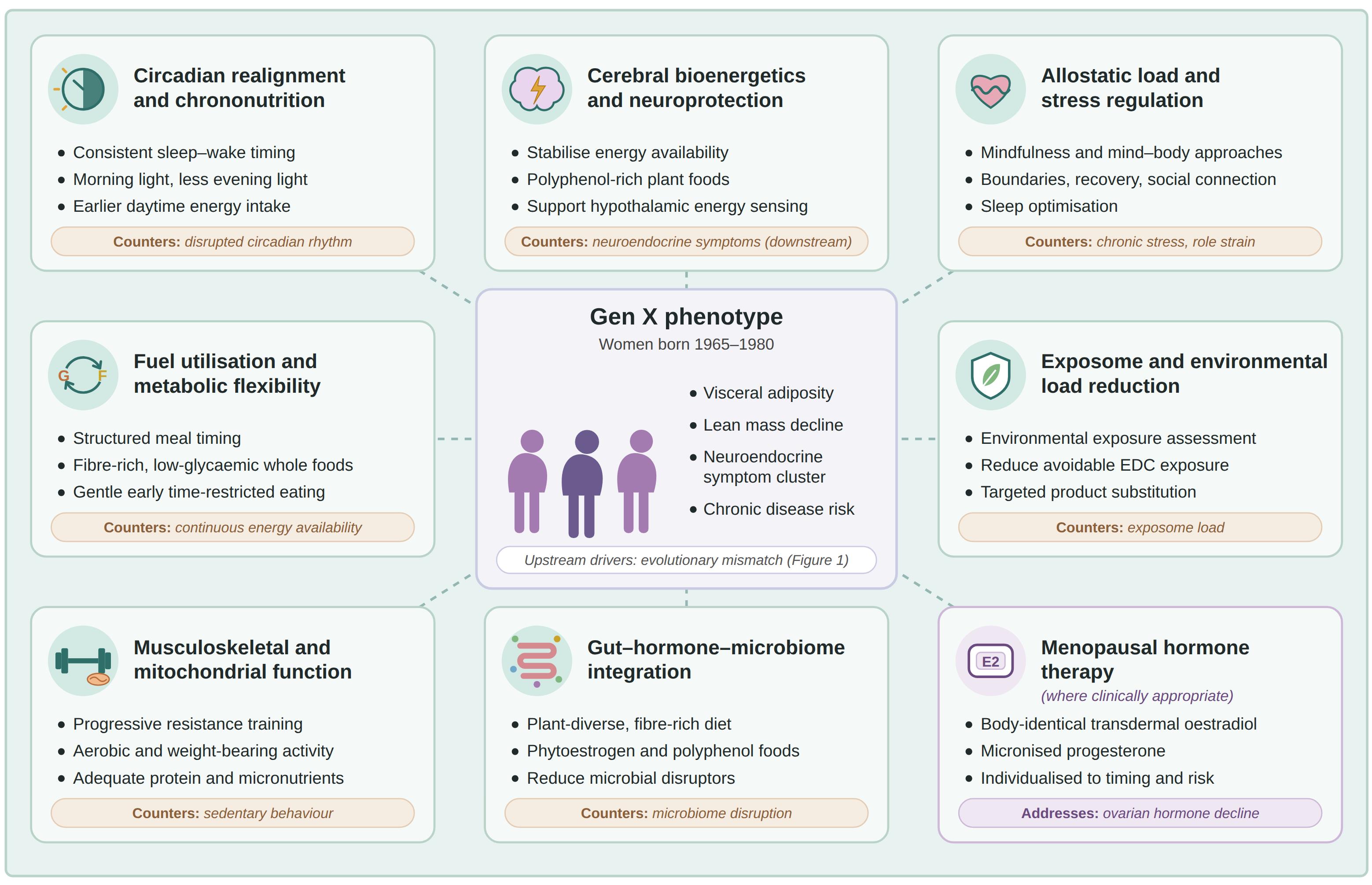
The evolutionary realignment framework is summarised in Figure 2. The proposed Gen X clinical phenotype is positioned centrally, surrounded by the eight intervention domains, each paired with the evolutionary mismatch factor it addresses, with MHT included where clinically appropriate. Rather than operating as discrete treatments, the domains are intended to act together to re-establish the conditions under which human physiology evolved, including predictable light–dark cycles, intermittent energy intake, regular physical activity and adaptive rather than chronic stress, supporting a realigned trajectory for metabolic, neuroendocrine and long-term health.
3.10. Limitations
This narrative review has several limitations. It did not follow a systematic search strategy or formal quality appraisal, and evidence was selected to inform a conceptual framework rather than to estimate intervention effects. The proposed Gen X phenotype is derived from convergent mechanistic, observational and cohort evidence and has yet to be validated prospectively. Some of the intervention evidence comes from general or postmenopausal populations and draws predominantly on high-income Westernised settings. The combined effects of the domains have not been tested.
4. Conclusions
This review extends our evolutionary mismatch model by translating it into a clinically focused framework for Gen X women during the menopausal transition. We propose that the interaction between cumulative mismatch exposures, cohort-specific psychosocial pressures and decliningovarian hormone production may contribute to a recognisable clinical phenotype characterised by metabolic dysregulation, altered body composition, vasomotor symptoms and neuroendocrine features including fatigue, cognitive changes and dysregulated appetite.
Management should therefore extend beyond treatment of individual symptoms to consider the interacting physiological systems contributing to this presentation. The proposed evolutionary realignment framework integrates circadian, metabolic, nutritional, psychosocial, environmental, musculoskeletal, mitochondrial and gut–hormone strategies, together with MHT where clinically appropriate. It is not intended as a prescriptive protocol or to imply a uniform phenotype across all Gen X women, but as a practical and testable framework for individualised midlife care. Prospective studies are required to validate the proposed phenotype and determine whether interventions targeting the identified pathways improve clinical and long-term health outcomes in Gen X women.
Author Contributions
Conceptualization, V.H., J.P. and C.L.O.; methodology, V.H., J.P. and C.L.O.; writing—original draft preparation, V.H., J.P. and C.L.O.; writing—review and editing, V.H., J.P. and C.L.O.; supervision, J.P. All authors have read and agreed to the published version of the manuscript.
Funding
This research received no external funding.
Institutional Review Board Statement
Not applicable.
Informed Consent Statement
Not applicable.
Data Availability Statement
Not applicable.
Acknowledgments
During the preparation of this manuscript, generative artificial intelligence tools (Microsoft Copilot; Microsoft 2025; GPT-5; Claude) were used in a limited capacity to assist with language refinement and manuscript organization. All scientific content, conceptual development, interpretation of evidence, and final editorial decisions were made by the authors, who take full responsibility for the integrity and accuracy of the work.
Conflicts of Interest
The authors declare no conflicts of interest.
Abbreviations
| AgRP | Agouti-Related Peptide |
| AMPK | 5′ Adenosine Monophosphate-Activated Protein Kinase |
| ANS | Autonomic Nervous System |
| ATP | Adenosine Triphosphate |
| BMI | Body Mass Index |
| CSI | Chronic Systemic Inflammation |
| CVD | Cardiovascular Disease |
| DHEA | Dehydroepiandrosterone |
| DHEAS | Dehydroepiandrosterone Sulphate |
| E2 | 17β-Oestradiol |
| EDC | Endocrine-Disrupting Chemical |
| ERK | Extracellular Signal-Regulated Kinase |
| ERβ | Oestrogen Receptor Beta |
| eTRE | Early Time-Restricted Eating |
| FSH | Follicle-Stimulating Hormone |
| GABA | Gamma-Aminobutyric Acid |
| Gen X | Generation X |
| GH | Growth Hormone |
| GLP-1 | Glucagon-Like Peptide-1 |
| GLUT4 | Glucose Transporter Type 4 |
| HbA1c | Glycated Haemoglobin |
| HPA | Hypothalamic–Pituitary–Adrenal |
| IGF-1 | Insulin-Like Growth Factor-1 |
| IR | Insulin Resistance |
| KNDy | Kisspeptin, Neurokinin B and Dynorphin |
| MASLD | Metabolic Dysfunction-Associated Steatotic Liver Disease |
| MHT | Menopausal Hormone Therapy |
| mTOR | Mechanistic Target of Rapamycin |
| NF-κB | Nuclear Factor Kappa B |
| NPY | Neuropeptide Y |
| PYY | Peptide YY |
| SCFA | Short-Chain Fatty Acid |
| SCN | Suprachiasmatic Nucleus |
| T2DM | Type 2 Diabetes Mellitus |
| T3 | Triiodothyronine |
References
- Rangel-Zuñiga, O.A.; Cruz-Teno, C.; Haro, C.; Quintana-Navarro, G.M.; Camara-Martos, F.; Perez-Martinez, P.; et al. Differential menopause- versus aging-induced changes in oxidative stress and circadian rhythm gene markers. Mech. Ageing Dev. 2017, 164, 41–8. [Google Scholar] [CrossRef]
- Logan, N.E.; Gaudreau, J.; Owens, B.; Oaks, B.M.; Ward-Ritacco, C.L.; Sabik, N.J. Heightened menopausal symptoms mediate the associations between stress and depressive symptoms among midlife women. Menopause 2025, 32, 956–65. [Google Scholar] [CrossRef] [PubMed]
- Taylor-Swanson, L.; Wong, A.E.; Pincus, D.; Butner, J.E.; Hahn-Holbrook, J.; Koithan, M.; et al. The dynamics of stress and fatigue across menopause: attractors, coupling, and resilience. Menopause 2018, 25, 380. [Google Scholar] [CrossRef]
- Simoncig-Netjasov, A.; Vujović, S.; Ivović, M.; Tančić-Gajić, M.; Marina, L.; Barać, M. Relationships between obesity, lipids and fasting glucose in the menopause. Srp. Arh. Za Celok. Lek. 2013, 141, 41–7. [Google Scholar] [CrossRef]
- Kivimäki, M.; Bartolomucci, A.; Kawachi, I. The multiple roles of life stress in metabolic disorders. Nat. Rev. Endocrinol. 2023, 19, 10–27. [Google Scholar] [CrossRef]
- Baker, C.; Benayoun, B.A. Menopause Is More Than Just Loss of Fertility. Public Policy Aging Rep. 2023, 33, 113–9. [Google Scholar] [CrossRef]
- Moore, A.M.; Fine, E.J. Menopause from an integrative historical and evolutionary perspective. jevohealth 2018, 3. [Google Scholar] [CrossRef]
- Froehle, A.W. Postmenopausal health and disease from the perspective of evolutionary medicine. Anthropol. Aging 2013, 34, 61–86. [Google Scholar] [CrossRef]
- Jeong, H.G.; Park, H. Metabolic Disorders in Menopause. Metabolites 2022, 12, 954. [Google Scholar] [CrossRef]
- Balon, R. An Explanation of Generations and Generational Changes. Acad. Psychiatry 2024, 48, 280–2. [Google Scholar] [CrossRef]
- “(Having?) Doing it All”: Narrative Exploration of Self-Care and Well-being for Generation X Women at Midlife. 2020 Sep 23.
- Hernandez, H.; Santamaria-Garcia, H.; Moguilner, S.; Farina, F.R.; Legaz, A.; Prado, P.; et al. The exposome of healthy and accelerated aging across 40 countries. Nat. Med. 2025, 31, 3089–100. [Google Scholar] [CrossRef]
- Bhagavathula, A.S.; Aldhaleei, W.A.; Abegaz, T.M. Trends and projections of overweight and obesity among midlife women in the United States, 1990–2050: A sub-study of the Global Burden of Disease 2021. Womens Health (Lond Engl) 2026, 22, 17455057261430197. [Google Scholar] [CrossRef]
- AIHW. Overweight and obesity [Internet]. AIHW, Australian Government. 2024. Available online: https://www.aihw.gov.au/reports/overweight-obesity/overweight-and-obesity.
- Genazzani AD, Giannini A, Napolitano A. From Menopause to Aging: Endocrine and Neuroendocrine Biological Changes. In: Birkhaeuser M, Genazzani AR, editors. Pre-Menopause, Menopause and Beyond: Volume 5: Frontiers in Gynecological Endocrinology. Cham: Springer International Publishing; 2018. p. 17– 33. [CrossRef]
- Priyanka, H.P.; Nair, R.S. Neuroimmunomodulation by estrogen in health and disease. In AIMS Neurosci; 2020; Volume 7, pp. 401–17. [Google Scholar] [CrossRef] [PubMed] [PubMed Central]
- Johnstone, R.A.; Cant, M.A. Evolution of menopause. Curr. Biol. 2019, 29, R112–5. [Google Scholar] [CrossRef] [PubMed]
- Assaf, S.; Park, J.; Chowdhry, N.; Ganapuram, M.; Mattathil, S.; Alakeel, R.; et al. Unraveling the Evolutionary Diet Mismatch and Its Contribution to the Deterioration of Body Composition. Metabolites 2024, 14[7]. [Google Scholar] [CrossRef]
- Parker, J.; O’Brien, C.; Hawrelak, J.; Gersh, F.L. Polycystic Ovary Syndrome: An Evolutionary Adaptation to Lifestyle and the Environment. In Int J Environ Res Public Health; 2022; Volume 19. [Google Scholar] [CrossRef] [PubMed] [PubMed Central]
- Longman, D.P.; Shaw, C.N. Homo sapiens, industrialisation and the environmental mismatch hypothesis. Biol. Rev. 2025. [Google Scholar] [CrossRef] [PubMed]
- Hitch, V.; O’Brien, C.; Parker, J. Evolutionary Mismatch in Generation X Women During the Menopausal Transition: An Integrated Model of Hormonal, Metabolic and Cognitive Dysfunction. 2026, Vol. 45 45, 72–87. [Google Scholar]
- Myers, M.G.; Affinati, A.H.; Richardson, N.; Schwartz, M.W. Central nervous system regulation of organismal energy and glucose homeostasis. Nat. Metab. 2021, 3, 737–50. [Google Scholar] [CrossRef]
- Timper, K.; Brüning, J.C. Hypothalamic circuits regulating appetite and energy homeostasis: pathways to obesity. Dis. Model Mech. 2017, 10, 679–89. [Google Scholar] [CrossRef]
- Bhusal, A.; Rahman, M.H.; Suk, K. Hypothalamic inflammation in metabolic disorders and aging. Cell Mol. Life Sci. 2021, 79, 32. [Google Scholar] [CrossRef]
- Dulloo, A.G.; Schutz, Y. Adaptive Thermogenesis in Resistance to Obesity Therapies: Issues in Quantifying Thrifty Energy Expenditure Phenotypes in Humans. Curr. Obes. Rep. 2015, 4, 230–40. [Google Scholar] [CrossRef]
- Jones, A.; Pruessner, J.C.; McMillan, M.R.; Jones, R.W.; Kowalik, G.T.; Steeden, J.A.; et al. Physiological adaptations to chronic stress in healthy humans – why might the sexes have evolved different energy utilisation strategies? In J Physiol; 2016; Volume 594, pp. 4297–307. [Google Scholar] [CrossRef] [PubMed] [PubMed Central]
- Gallardo, N.; Artigas-Jerónimo, S.; Mazuecos, L.; Andrés, A. Neuroendocrine control of glucose homeostasis: integrative mechanisms from the hypothalamus to the brainstem. Front Endocrinol. 2025, 16. [Google Scholar] [CrossRef]
- De Jesus, A.N.; Henry, B.A. The role of oestrogen in determining sexual dimorphism in energy balance. J. Physiol. 2023, 601, 435–49. [Google Scholar] [CrossRef]
- Goodpaster, B.H.; Sparks, L.M. Metabolic Flexibility in Health and Disease. Cell Metab. 2017, 25, 1027–36. [Google Scholar] [CrossRef] [PubMed]
- Ko, S.H.; Jung, Y. Energy Metabolism Changes and Dysregulated Lipid Metabolism in Postmenopausal Women. Nutrients 2021, 13[12]. [Google Scholar] [CrossRef]
- Huang, Y.; Liu, Q.; Huang, G.; Wen, J.; Chen, G. Hypothalamic Kisspeptin Neurons Regulates Energy Metabolism and Reproduction Under Chronic Stress. Front Endocrinol. 2022, 13. [Google Scholar] [CrossRef]
- Barabás, K.; Szabó-Meleg, E.; Ábrahám, I.M. Effect of Inflammation on Female Gonadotropin-Releasing Hormone (GnRH) Neurons: Mechanisms and Consequences. Int. J. Mol. Sci. 2020, 21[2]. [Google Scholar] [CrossRef]
- Faour, M.; Mesto, N.; Martin, C.; Luquet, S. Emerging role of AgRP neurons as integrators of metabolic, sensory and environmental cues in the control of energy homeostasis. Rev. Endocr. Metab. Disord. 2025. [Google Scholar] [CrossRef]
- Deem, J.D.; Faber, C.L.; Morton, G.J. AgRP neurons: Regulators of feeding, energy expenditure, and behavior. FEBS J. 2022, 289, 2362–81. [Google Scholar] [CrossRef]
- Kim, D.I.; Park, Y.M. Effects of Menopause on Physical Activity and Dopamine Signaling in Women. In Iran J Public Health; 2022; Volume 51, pp. 2372–4. [Google Scholar] [CrossRef] [PubMed] [PubMed Central]
- Cameron, J.D.; Chaput, J.P.; Sjödin, A.M.; Goldfield, G.S. Brain on Fire: Incentive Salience, Hedonic Hot Spots, Dopamine, Obesity, and Other Hunger Games. Annu. Rev. Nutr. 2017, 37((Volume 37), 2017):183–205. [Google Scholar] [CrossRef]
- Dulloo, A.G. Peripheral thyroid hormone deiodination: Entry points to elucidate mechanisms of metabolic adaptation during weight regain. Obesity 2023, 31, 1179–82. [Google Scholar] [CrossRef]
- Theodorakis, N.; Kreouzi, M.; Pappas, A.; Nikolaou, M. Beyond Calories: Individual Metabolic and Hormonal Adaptations Driving Variability in Weight Management—A State-of-the-Art Narrative Review. Int. J. Mol. Sci. 2024, 25[24]. [Google Scholar] [CrossRef]
- Milman, S.; Huffman, D.M.; Barzilai, N. The Somatotropic Axis in Human Aging: Framework for the Current State of Knowledge and Future Research. In Cell Metab; 2016; Volume 23, pp. 980–9. [Google Scholar] [CrossRef] [PubMed] [PubMed Central]
- Ajdžanovic, V.Z.; Trifunovic, S.; Miljic, D.; Šošic-Jurjevic, B.; Filipovic, B.; Miler, M.; et al. Somatopause, weaknesses of the therapeutic approaches and the cautious optimism based on experimental ageing studies with soy isoflavones. In EXCLI J; PubMed; 2018; Volume 17, pp. 279–301. [Google Scholar] [CrossRef] [PubMed] [PubMed Central]
- Oliveira, P.J.; Carvalho, R.A.; Portincasa, P.; Bonfrate, L.; Sardao, V.A. Fatty Acid Oxidation and Cardiovascular Risk during Menopause: A Mitochondrial Connection? J. Lipids 2012, 2012, 365798. [Google Scholar] [CrossRef]
- Gumpeny, N.; Gumpeny, L.; Gumpeny, S.R. Metabolic syndrome in menopause. Explor Endocr. Metab. Dis. 2025, 2, 101440. [Google Scholar] [CrossRef]
- Viana, DP da C; Câmara, L.C.; Palau, R.B. Menopause as a Critical Turning Point in Lipedema: The Estrogen Receptor Imbalance, Intracrine Estrogen, and Adipose Tissue Dysfunction Model. Int. J. Mol. Sci. 2025, 26[15]. [Google Scholar] [CrossRef]
- Pedamallu, H.; Aghabazaz, Z.; Lancki, N.; Rodriguez, L.A.; Siddique, J.; Moorthy, M.; et al. Prevalence and Trends in Cardiovascular Risk Factors Among Middle-Aged South Asian Adults Compared With Other Racial and Ethnic Groups in the United States: A Longitudinal Analysis of 2 Cohort Studies. J. Am. Heart Assoc. 2026, 15, e41221. [Google Scholar] [CrossRef]
- Kim, M.H.; Kim, H.N.; Choi, W.S. The association between subclinical inflammation and abnormal glucose and lipid metabolisms in normal-weight Korean individuals. Nutr. Metab. Cardiovasc. Dis. 2018, 28, 1106–13. [Google Scholar] [CrossRef]
- Vigil, P.; Meléndez, J.; Petkovic, G.; Del Río, J.P. The importance of estradiol for body weight regulation in women. Front Endocrinol. 2022, 13. [Google Scholar] [CrossRef]
- Balakrishnan, R.; Thurmond, D.C. Mechanisms by Which Skeletal Muscle Myokines Ameliorate Insulin Resistance. Int. J. Mol. Sci. 2022, 23[9]. [Google Scholar] [CrossRef]
- Donini, L.M.; Busetto, L.; Bischoff, S.C.; Cederholm, T.; Ballesteros-Pomar, M.D.; Batsis, J.A.; et al. Definition and diagnostic criteria for sarcopenic obesity: ESPEN and EASO consensus statement. Clin. Nutr. 2022, 41, 990–1000. [Google Scholar] [CrossRef] [PubMed]
- Beaudart, C.; Demonceau, C.; Reginster, J.Y.; Locquet, M.; Cesari, M.; Cruz Jentoft, A.J.; et al. Sarcopenia and health-related quality of life: A systematic review and meta-analysis. J. Cachexia Sarcopenia Muscle 2023, 14, 1228–43. [Google Scholar] [CrossRef]
- Mosconi, L.; Berti, V.; Guyara-Quinn, C.; McHugh, P.; Petrongolo, G.; Osorio, R.S.; et al. Perimenopause and emergence of an Alzheimer’s bioenergetic phenotype in brain and periphery. PLoS ONE 2017, 12, e0185926. [Google Scholar] [CrossRef]
- Mosconi, L.; Nerattini, M.; Matthews, D.C.; Jett, S.; Andy, C.; Williams, S.; et al. In vivo brain estrogen receptor density by neuroendocrine aging and relationships with cognition and symptomatology. Sci. Rep. 2024, 14, 12680. [Google Scholar] [CrossRef]
- Zhu, C.; Thomas, E.H.; Li, Q.; Arunogiri, S.; Thomas, N.; Gurvich, C. Evaluation of the Everyday Memory Questionnaire-Revised in a menopausal population: understanding the brain fog during menopause. Menopause 2023, 30, 1147. [Google Scholar] [CrossRef]
- Mervosh, N.; Devi, G. Estrogen, menopause, and Alzheimer’s disease: understanding the link to cognitive decline in women. Front Mol. Biosci. 2025, 12. [Google Scholar] [CrossRef]
- Bansal, S.; Balagopalan, J.P.; Dani, S.; Zargar, A.H.; Bhattacharyya, A.; Deka, N.; et al. Diabetes and Cardiovascular Risk in Menopausal Women: An Emerging Concern. Eur. J. Cardiovasc. Med. 2025, 15, 246–57. [Google Scholar] [CrossRef]
- Venetsanaki, V.; Polyzos, S.A. Menopause and Non-Alcoholic Fatty Liver Disease: A Review Focusing on Therapeutic Perspectives. Curr. Vasc. Pharmacol. 2019, 17, 546–55. [Google Scholar] [CrossRef]
- Fasero, M.; Coronado, P.J. Cardiovascular Disease Risk in Women with Menopause. J. Clin. Med. 2025, 14, 3663. [Google Scholar] [CrossRef]
- de Villiers, T.J. Bone health and menopause: Osteoporosis prevention and treatment. Best Pract. Res. Clin. Endocrinol. Metab.;Menopause-Diagn. Ther. Strateg. 2024, 38, 101782. [Google Scholar] [CrossRef]
- Austad, S.N.; Fischer, K.E. Sex Differences in Lifespan. In Cell Metab; 2016; Volume 23, pp. 1022–33. [Google Scholar] [CrossRef] [PubMed] [PubMed Central]
- Brooks, R.C.; Garratt, M.G. Life history evolution, reproduction, and the origins of sex-dependent aging and longevity. Ann. N Y Acad. Sci. 2017, 1389, 92–107. [Google Scholar] [CrossRef] [PubMed]
- Ciciolla, L.; Luthar, S.S. Invisible Household Labor and Ramifications for Adjustment: Mothers as Captains of Households. In Sex Roles; 2019; Volume 81[7–8], pp. 467–86. [Google Scholar] [CrossRef] [PubMed] [PubMed Central]
- Aviv, E.; Waizman, Y.; Kim, E.; Liu, J.; Rodsky, E.; Saxbe, D. Cognitive household labor: gender disparities and consequences for maternal mental health and wellbeing. Arch. Womens Ment. Health 2025, 28, 5–14. [Google Scholar] [CrossRef]
- Barrea, L.; Vetrani, C.; Altieri, B.; Verde, L.; Savastano, S.; Colao, A.; et al. The Importance of Being a ‘Lark’ in Post-Menopausal Women with Obesity: A Ploy to Prevent Type 2 Diabetes Mellitus? Nutrients 2021, 13, 3762. [Google Scholar] [CrossRef]
- Pérez-Medina-Carballo, R.; Kosmadopoulos, A.; Boudreau, P.; Robert, M.; Walker, C.D.; Boivin, D.B. The circadian variation of sleep and alertness of postmenopausal women. In Sleep; 2023; Volume 46, p. zsac272. [Google Scholar] [CrossRef] [PubMed] [PubMed Central]
- Hou, S.Y.; Chiu, C.J.; Shea, J.L.; Wang, C.L.; Tang, H.H.; Kuo, P.C.; et al. Role of age, menopausal status, and symptoms in midlife women: Examination of sleep patterns and rest-activity circadian rhythms. Sleep Med. 2024, 113, 172–9. [Google Scholar] [CrossRef] [PubMed]
- Malhan, D.; Yalçin, M.; Liedtke, S.; Grötsch, R.; Enzmann, C.; Rau, M.; et al. A prospective study to investigate circadian rhythms as health indicator in women’s aging. npj Womens Health 2025, 3, 18. [Google Scholar] [CrossRef]
- Sparks, J.R.; Wang, X. Menopause-Related Changes in Sleep and the Associations with Cardiometabolic Health: A Narrative Review. Healthcare 2025, 13, 2085. [Google Scholar] [CrossRef]
- Drăgoi, C.M.; Nicolae, A.C.; Ungurianu, A.; Margină, D.M.; Grădinaru, D.; Dumitrescu, I.B. Circadian Rhythms, Chrononutrition, Physical Training, and Redox Homeostasis—Molecular Mechanisms in Human Health. Cells 2024, 13, 138. [Google Scholar] [CrossRef]
- BaHammam, A.S.; Pirzada, A. Timing Matters: The Interplay between Early Mealtime, Circadian Rhythms, Gene Expression, Circadian Hormones, and Metabolism—A Narrative Review. Clocks Sleep 2023, 5, 507–35. [Google Scholar] [CrossRef]
- Bragazzi, N.L.; Ceylan, H.İ.; Rosi, A.; Scazzina, F.; de Giorgio, A.; Dergaa, I.; et al. The Co-Evolution of Sleep and Diet: Toward an Emerging Framework of Evolutionary Chrononutrition in Circadian–Metabolic Health. Nutrients 2026, 18, 1947. [Google Scholar] [CrossRef]
- García-Cobarro, C.; Ignez Soares, L.; Kutsenko, Y.; Tomas Loba, A. Circadian system and aging: where both times interact. Front. Aging 2025, 6, 1646794. [Google Scholar] [CrossRef]
- Lea, A.J.; Clark, A.G.; Dahl, A.W.; Devinsky, O.; Garcia, A.R.; Golden, C.D.; et al. Applying an evolutionary mismatch framework to understand disease susceptibility. PLoS Biol. 2023, 21, e3002311. [Google Scholar]
- Cordain, L.; Eaton, S.B.; Sebastian, A.; Mann, N.; Lindeberg, S.; Watkins, B.A.; et al. Origins and evolution of the Western diet: health implications for the 21st century. Am. J. Clin. Nutr. 2005, 81, 341–54. [Google Scholar] [CrossRef] [PubMed]
- Vahlhaus, J.; Peters, B.; Hornemann, S.; Ost, A.C.; Kruse, M.; Busjahn, A.; et al. Later eating timing in relation to an individual internal clock is associated with lower insulin sensitivity and affected by genetic factors. 2025, 116. [Google Scholar] [CrossRef] [PubMed]
- Franzago, M.; Alessandrelli, E.; Notarangelo, S.; Stuppia, L.; Vitacolonna, E. Chrono-Nutrition: Circadian Rhythm and Personalized Nutrition. Int. J. Mol. Sci. 2023, 24, 2571. [Google Scholar] [CrossRef]
- McHill, A.W.; Butler, M.P. Eating Around the Clock: Circadian Rhythms of Eating and Metabolism. Annu. Rev. Nutr. 2024, 44((Volume 44), 2024):25–50. [Google Scholar] [CrossRef]
- de Oliveira Melo, N.C.; Cuevas-Sierra, A.; Souto, V.F.; Martínez, J.A. Biological Rhythms, Chrono-Nutrition, and Gut Microbiota: Epigenomics Insights for Precision Nutrition and Metabolic Health. Biomolecules 2024, 14, 559. [Google Scholar] [CrossRef]
- Reytor-González, C.; Simancas-Racines, D.; Román-Galeano, N.M.; Annunziata, G.; Galasso, M.; Zambrano-Villacres, R.; et al. Chrononutrition and Energy Balance: How Meal Timing and Circadian Rhythms Shape Weight Regulation and Metabolic Health. Nutrients 2025, 17, 2135. [Google Scholar] [CrossRef]
- Ahluwalia, M.K. Chrononutrition—When We Eat Is of the Essence in Tackling Obesity. Nutrients 2022, 14, 5080. [Google Scholar] [CrossRef]
- Maki, K.A.; Barb, J.J.; Alkhatib, J.; Butera, G.; Wallen, G.R. From alpha diversity to zeitgebers: Bacterial gut microbiome associations with sleep and circadian disruption (Part I). Neurosci. Biobehav. Rev. 2026, 184, 106578. [Google Scholar] [CrossRef]
- Tan, S.Y.; Tjahjono, I.E.; Feyesa, I.; Hettiarachchi, J.; Kim, N.; Pennycuff, R.; et al. Does Sleep Curtailment Stimulate Appetite, Motivation to Eat and Dietary Intake in Humans? A Narrative Synthesis of Evidence and Considerations for Future Studies. J. Sleep Res. 2026, 35, e70358. [Google Scholar] [CrossRef]
- Marjot, T.; Tomlinson, J.W.; Hodson, L.; Ray, D.W. Timing of energy intake and the therapeutic potential of intermittent fasting and time-restricted eating in NAFLD. Gut 2023, 72, 1607–19. [Google Scholar] [CrossRef]
- Jamshed, H.; Steger, F.L.; Bryan, D.R.; Richman, J.S.; Warriner, A.H.; Hanick, C.J.; et al. Effectiveness of Early Time-Restricted Eating for Weight Loss, Fat Loss, and Cardiometabolic Health in Adults With Obesity: A Randomized Clinical Trial. JAMA Intern Med. 2022, 182, 953. [Google Scholar] [CrossRef]
- Peters, B.; Jokisch, J.; Schwarz, J.; Schuppelius, B.; Pfeiffer, A.F.H.; Michalsen, A.; et al. Early but not late time-restricted eating improves an actigraphy-estimated sleep quality in women with overweight or obesity: secondary analysis of the crossover ChronoFast trial. Front Nutr. 2026, 13. [Google Scholar] [CrossRef]
- Jóźwiak, B.; Szulc, A.; Laudańska-Krzemińska, I. Time-Restricted Eating Combined with Exercise Reduces Menopausal Symptoms and Improves Quality of Life More than Exercise Alone in Menopausal Women: A Quasi-Randomized Controlled Trial. In Nutrients; 2025; Volume 17. [Google Scholar] [CrossRef] [PubMed] [PubMed Central]
- Kramer, A.; Lange, T.; Spies, C.; Finger, A.M.; Berg, D.; Oster, H. Foundations of circadian medicine. PLoS Biol. 2022, 20, e3001567. [Google Scholar] [CrossRef]
- Windred, D.P.; Burns, A.C.; Lane, J.M.; Saxena, R.; Rutter, M.K.; Cain, S.W.; et al. Sleep regularity is a stronger predictor of mortality risk than sleep duration: A prospective cohort study. Sleep 2024, 47, zsad253. [Google Scholar] [CrossRef]
- Silva, T.R.; Oppermann, K.; Reis, F.M.; Spritzer, P.M. Nutrition in Menopausal Women: A Narrative Review. Nutrients 2021, 13, 2149. [Google Scholar] [CrossRef]
- Shoemaker, M.E.; Gillen, Z.M.; Fukuda, D.H.; Cramer, J.T. Metabolic Flexibility and Inflexibility: Pathology Underlying Metabolism Dysfunction. J. Clin. Med. 2023, 12[13]. [Google Scholar] [CrossRef]
- Lobato, V.A.; Minari, T.P.; Pisani, L.P. Perimenopause and metabolic vulnerability: hormones, body composition and lifestyle changes. Climacteric 2026, 0, 1–16. [Google Scholar] [CrossRef] [PubMed]
- Marcantei, C.; Metz, L.; Ramos, I.; Pereira, B.; Dutheil, F.; De Saint Vincent, S.; et al. Energy metabolism in resting and postprandial states might not be influenced by menopausal stage in women. Nutr. Metab. Cardiovasc. Dis. 2026, 36, 104637. [Google Scholar] [CrossRef]
- Bo, T.; Gao, L.; Yao, Z.; Shao, S.; Wang, X.; Proud, C.G.; et al. Hepatic selective insulin resistance at the intersection of insulin signaling and metabolic dysfunction-associated steatotic liver disease. Cell Metab. 2024, 36, 947–68. [Google Scholar] [CrossRef] [PubMed]
- Barnard, N.D.; Kahleova, H.; Holtz, D.N.; Znayenko-Miller, T.; Sutton, M.; Holubkov, R.; et al. A dietary intervention for vasomotor symptoms of menopause: a randomized, controlled trial. In Menopause; 2023; Volume 30, pp. 80–7. [Google Scholar] [CrossRef] [PubMed] [PubMed Central]
- Leone, A.; Bertoli, S.; Bedogni, G.; Vignati, L.; Pellizzari, M.; Battezzati, A. Association between Mediterranean Diet and Fatty Liver in Women with Overweight and Obesity. Nutrients 2022, 14, 3771. [Google Scholar] [CrossRef]
- Gonçalves, C.; Moreira, H.; Santos, R. Systematic review of mediterranean diet interventions in menopausal women. In AIMS Public Health; 2024; Volume 11, pp. 110–29. [Google Scholar] [CrossRef] [PubMed] [PubMed Central]
- Sánchez-Martínez, L.; González-Barrio, R.; Periago, M.J. The Potential Role of Dietary (Poly)phenols in Cardiometabolic Risk During Menopause: A Narrative Review. In Nutrients; 2026; Volume 18. [Google Scholar] [CrossRef] [PubMed] [PubMed Central]
- Minari, T.P.; Pisani, L.P. Skipping breakfast and its wide-ranging health consequences: A systematic review from multiple metabolic disruptions to socioeconomic factors. Nutr. Res. 2025, 141, 34–45. [Google Scholar] [CrossRef]
- Martínez, C.F.; Stern, D.; Cortés-Valencia, A.; Ortiz-Panozo, E.; Mattei, J.; Campos, H.; et al. The association between breakfast frequency and diabetes incidence in middle-aged women: Results from the MTC study. Nutr. Metab. Cardiovasc. Dis. 2024, 34, 2360–8. [Google Scholar] [CrossRef]
- Erceg, N.; Micic, M.; Forouzan, E.; Knezevic, N.N. The Role of Cortisol and Dehydroepiandrosterone in Obesity, Pain, and Aging. In Diseases; 2025; Volume 13. [Google Scholar] [CrossRef] [PubMed] [PubMed Central]
- Cohn, A.Y.; Grant, L.K.; Nathan, M.D.; Wiley, A.; Abramson, M.; Harder, J.A.; et al. Effects of Sleep Fragmentation and Estradiol Decline on Cortisol in a Human Experimental Model of Menopause. J. Clin. Endocrinol. Metab. 2023, 108, e1347–57. [Google Scholar] [CrossRef]
- Palma-Gudiel, H.; Prather, A.A.; Lin, J.; Oxendine, J.D.; Guintivano, J.; Xia, K.; et al. HPA axis regulation and epigenetic programming of immune-related genes in chronically stressed and non-stressed mid-life women. Brain Behav. Immun. 2021, 92, 49–56. [Google Scholar] [CrossRef]
- Anekwe, C.V.; Cano, A.; Mulligan, J.; Ang, S.B.; Johnson, C.N.; Panay, N.; et al. The role of lifestyle medicine in menopausal health: a review of non-pharmacologic interventions. Climacteric 2025, 28, 478–96. [Google Scholar] [CrossRef] [PubMed]
- Kennard, A.; Saunders, K.T. Integrative approaches to perimenopause. Am. J. Med. 2026, 139, 522–9. [Google Scholar] [CrossRef]
- Maunder, A.; Mardon, A.K.; Rao, V.; Torkel, S.; Metri, N.J.; Liu, J.; et al. Complementary therapies for management of menopausal symptoms: a systematic review to inform the update of the International Menopause Society recommendations on women’s midlife health. Climacteric 2026, 29, 165–209. [Google Scholar] [CrossRef] [PubMed]
- Liu, H.; Cai, K.; Wang, J.; Zhang, H. The effects of mindfulness-based interventions on anxiety, depression, stress, and mindfulness in menopausal women: A systematic review and meta-analysis. Front Public Health 2023, 10. [Google Scholar] [CrossRef]
- Vani, I.; Muralidhar, G.; Rao, B.S. A prospective, randomized, double-blind, placebo-controlled study on efficacy and safety of Ashwagandha root extract (Withania somnifera) for managing menopausal symptoms in women. In Front Reprod Health; 2026; Volume 7. [Google Scholar] [CrossRef] [PubMed] [PubMed Central]
- Gherman-Lencu, C.C.; Alexescu, T.G.; Mureșanu, C.; Gerdanovics, C.A.; Milaciu, M.V.; Iancu, D.M. Implications of Endocrine-Disrupting Chemicals for Human Health and Effective Methods for Prevention and Reduction. Toxics 2026, 14, 515. [Google Scholar] [CrossRef]
- Thomas, E.C.; Perttula, K.; Joseph, G.; Yardeni, T.; DeBolt, C.A.; Goshen, A.; et al. Impact of Environmental Exposures on Menopausal Symptoms – A Scoping Review. Curr. Epidemiol. Rep. 2025, 12, 16. [Google Scholar] [CrossRef]
- Guetterman, H.M.; Flaws, J.A.; Strakovsky, R.S. Metabolism-disrupting compounds and metabolic health during the menopausal transition: a narrative review. Int. J. Obes. 2026, 50, 1426–42. [Google Scholar] [CrossRef]
- Park, J.; Lee, H.; Lee, S.; Lee, H. Interventions on Reducing Exposure to Endocrine Disrupting Chemicals in Human Health Care Context: A Scoping Review. Risk Manag. Healthc. Policy 2022, 15, 779–91. [Google Scholar] [CrossRef] [PubMed]
- Rochester, J.R.; Kwiatkowski, C.F.; Lathrop, M.K.; Neveux, I.; Daza, E.J.; Grzymski, J.; et al. Reducing Exposures to Endocrine Disruptors (REED) study, a personalized at-home intervention program to reduce exposure to endocrine disrupting chemicals among a child-bearing age cohort: study protocol for a randomized controlled trial. Trials 2024, 25, 793. [Google Scholar] [CrossRef]
- Jennings, A.; Thompson, A.S.; Tresserra-Rimbau, A.; O’Neill, J.K.; Hill, C.; Bondonno, N.P.; et al. Flavonoid-Rich Foods, Dementia Risk, and Interactions With Genetic Risk, Hypertension, and Depression. In JAMA Netw Open; 2024; Volume 7, p. e2434136. [Google Scholar] [CrossRef] [PubMed] [PubMed Central]
- Valls-Pedret, C.; Sala-Vila, A.; Serra-Mir, M.; Corella, D.; de la Torre, R.; Martínez-González, M.Á.; et al. Mediterranean Diet and Age-Related Cognitive Decline: A Randomized Clinical Trial. JAMA Intern Med. 2015, 175, 1094–103. [Google Scholar] [CrossRef]
- Thaung Zaw, J.J.; Howe, P.R.; Wong, R.H. Long-term effects of resveratrol on cognition, cerebrovascular function and cardio-metabolic markers in postmenopausal women: A 24-month randomised, double-blind, placebo-controlled, crossover study. Clin. Nutr. 2021, 40, 820–9. [Google Scholar] [CrossRef] [PubMed]
- Garcia-Yu, I.A.; Garcia-Ortiz, L.; Gomez-Marcos, M.A.; Rodriguez-Sanchez, E.; Mora-Simon, S.; Maderuelo-Fernandez, J.A.; et al. Effects of cocoa-rich chocolate on cognitive performance in postmenopausal women. A randomised clinical trial. Nutr. Neurosci. 2022, 25, 1147–58. [Google Scholar] [CrossRef] [PubMed]
- Cheng, N.; Bell, L.; Lamport, D.J.; Williams, C.M. Dietary Flavonoids and Human Cognition: A Meta-Analysis. In Mol Nutr Food Res; 2022; Volume 66, p. 2100976. [Google Scholar] [CrossRef] [PubMed] [PubMed Central]
- Li, Y.; Huang, F.; Qian, X.; Liu, C.; Yao, Y.; Wang, Z.; et al. Exploring the anti-aging potential of phytoestrogens: focus on molecular mechanisms and menopausal symptom modulation. Front Nutr. 2025, 12. [Google Scholar] [CrossRef]
- Sekikawa, A.; Wharton, W.; Butts, B.; Veliky, C.V.; Garfein, J.; Li, J.; et al. Potential Protective Mechanisms of S-equol, a Metabolite of Soy Isoflavone by the Gut Microbiome, on Cognitive Decline and Dementia. Int. J. Mol. Sci. 2022, 23, 11921. [Google Scholar] [CrossRef]
- Harahap, I.A.; Weber, D.; Grune, T. Non-hormonal strategies for menopausal health: the role of phytoestrogens. Crit. Rev. Food Sci. Nutr. 2026, 0, 1–23. [Google Scholar] [CrossRef] [PubMed]
- Vargas, K.G.; Milic, J.; Zaciragic, A.; Wen, K.X.; Jaspers, L.; Nano, J.; et al. The functions of estrogen receptor beta in the female brain: A systematic review. Maturitas 2016, 93, 41–57. [Google Scholar] [CrossRef] [PubMed]
- Till, L.; Lamport, D.; Williams, C.M. Systematic review and meta-analysis of isoflavone interventions on cognitive function. Neurosci. Biobehav Rev. 2026, 183, 106533. [Google Scholar] [CrossRef] [PubMed]
- Blümel, J.E.; Chedraui, P.; Vallejo, M.S. Do menopausal symptoms signal early biological aging? Mitochondrial, endocrine and clinical insights. Climacteric 2026, 29, 358–63. [Google Scholar] [CrossRef] [PubMed]
- Casanova, A.; Wevers, A.; Navarro-Ledesma, S.; Pruimboom, L. Mitochondria: It is all about energy. Front Physiol. 2023, 14. [Google Scholar] [CrossRef]
- Pellegrino, A.; Tiidus, P.M.; Vandenboom, R. Mechanisms of Estrogen Influence on Skeletal Muscle: Mass, Regeneration, and Mitochondrial Function. Sports Med. 2022, 52, 2853–69. [Google Scholar] [CrossRef] [PubMed]
- Kleis-Olsen, A.S.; Farlov, J.E.; Petersen, E.A.; Schmücker, M.; Flensted-Jensen, M.; Blom, I.; et al. Metabolic flexibility in postmenopausal women: Hormone replacement therapy is associated with higher mitochondrial content, respiratory capacity, and lower total fat mass. Acta Physiol. 2024, 240, e14117. [Google Scholar] [CrossRef]
- Groennebaek, T.; Vissing, K. Impact of Resistance Training on Skeletal Muscle Mitochondrial Biogenesis, Content, and Function. Front Physiol. 2017, 8. [Google Scholar] [CrossRef]
- Nguyen, B.; Owen, K.B.; Luo, M.; Brown, W.; Mielke, G.I.; Clare, P.J.; et al. Physical activity across mid-life and mortality outcomes in Australian women: A target trial emulation using a prospective cohort. PLoS Med. 2026, 23, e1004976. [Google Scholar] [CrossRef]
- Izquierdo, M.; Merchant, R.A.; Morley, J.E.; Anker, S.D.; Aprahamian, I.; Arai, H.; et al. International Exercise Recommendations in Older Adults (ICFSR): Expert Consensus Guidelines. J. Nutr. Health Aging 2021, 25, 824–53. [Google Scholar] [CrossRef]
- Isenmann, E.; Kaluza, D.; Havers, T.; Elbeshausen, A.; Geisler, S.; Hofmann, K.; et al. Resistance training alters body composition in middle-aged women depending on menopause - A 20-week control trial. BMC Women’s Health 2023, 23, 526. [Google Scholar] [CrossRef]
- Black, K.E.; Matkin-Hussey, P. The Impact of Protein in Post-Menopausal Women on Muscle Mass and Strength: A Narrative Review. Physiologia 2024, 4, 266–85. [Google Scholar] [CrossRef]
- Xia, T.; Haslam, D.E.; Eliassen, A.H.; Manson, J.E.; Sun, Q.; Willett, W.C.; et al. Optimal Dietary Patterns for Lower Weight Gain and Risk of Obesity Surrounding Menopause. JAMA Netw. Open 2026, 9, e2613102. [Google Scholar] [CrossRef]
- Wong, P.; Chen, W.; Ewald, D.; Girgis, C.; Rawlin, M.; Tsingos, J.; et al. 2024 Royal Australian College of General Practitioners and Healthy Bones Australia guideline for osteoporosis management and fracture prevention in postmenopausal women and men over 50 years of age. Med. J. Aust. 2025, 222, 472–80. [Google Scholar] [CrossRef]
- Castiglioni, S.; Mazur, A.; Maier, J.A. The central role of magnesium in skeletal muscle: from myogenesis to performance. Magnes. Res. 2024, 37, 1–11. [Google Scholar] [CrossRef] [PubMed]
- Deledda, A.; Giordano, E.; Velluzzi, F.; Flore, G.; Franceschelli, S.; Speranza, L.; et al. Mitochondrial Aging and Senolytic Natural Products with Protective Potential. In Int J Mol Sci.; 2022; Volume 23. [Google Scholar] [CrossRef] [PubMed] [PubMed Central]
- Gu, Y.; Han, J. Autophagy and polyphenol intervention strategy in aging. Trends Food Sci. Technol. 2023, 132, 1–10. [Google Scholar] [CrossRef]
- Zhuge, L.; Chen, L.; Pan, W. Effects of Isoflavone Interventions on Bone Metabolism in Perimenopausal and Postmenopausal Women: An Umbrella Review of Meta-Analyses of Randomized Controlled Trials. Endocr. Pract. 2025, 31, 226–35. [Google Scholar] [CrossRef] [PubMed]
- Chaudhary, R.; Bansal, N.; Sharma, S.; Rohilla, M.; Chauhan, S.; Gupta, S.; et al. Mechanistic pathways of estrogen mitigating postmenopausal gut dysbiosis. Mol. Biol. Rep. 2025, 53, 45. [Google Scholar] [CrossRef]
- Maffei, V.J.; Bertoni, A.G.; Wood, A.C.; Rotter, J.I.; Crago, O.; Chen, Y.D.I.; et al. Menopause factors and alterations in gut microbiota and insulin homeostasis: A cross-sectional analysis of the microbiome and insulin longitudinal evaluation study (MILES). Diabetes Obes. Metab. 2025, 27, 5444–54. [Google Scholar] [CrossRef]
- Peters, B.A.; Lin, J.; Qi, Q.; Usyk, M.; Isasi, C.R.; Mossavar-Rahmani, Y.; et al. Menopause Is Associated with an Altered Gut Microbiome and Estrobolome, with Implications for Adverse Cardiometabolic Risk in the Hispanic Community Health Study/Study of Latinos. mSystems 2022, 7, e00273-22. [Google Scholar] [CrossRef]
- Kumari, N.; Kumari, R.; Dua, A.; Singh, M.; Kumar, R.; Singh, P.; et al. From Gut to Hormones: Unraveling the Role of Gut Microbiota in (Phyto)Estrogen Modulation in Health and Disease. Mol. Nutr. Food Res. 2024, 68, 2300688. [Google Scholar] [CrossRef]
- Lim, M.J.S.; Parlindungan, E.; See, E.; Gan, C.H.; Yap, R.; Yong, G.J.M. Diet, the Gut Microbiome, and Estrogen Physiology: A Review in Menopausal Health and Interventions. In Nutrients; 2026; Volume 18. [Google Scholar] [CrossRef] [PubMed] [PubMed Central]
- Yoshikata, R.; Myint, K.Z.; Ohta, H. Relationship between equol producer status and metabolic parameters in 743 Japanese women: equol producer status is associated with antiatherosclerotic conditions in women around menopause and early postmenopause. Menopause 2017, 24, 216–24. [Google Scholar] [CrossRef]
- Deehan, E.C.; Mocanu, V.; Madsen, K.L. Effects of dietary fibre on metabolic health and obesity. Nat. Rev. Gastroenterol. Hepatol. 2024, 21, 301–18. [Google Scholar] [CrossRef]
- Krishna, K.V.; Malviya, S.; Bhattacharyya, D.; Malaviya, A. Gut–Brain Axis: Role in Hunger and Satiety. In Probiotic Research in Therapeutics; Chopra, K., Bishnoi, M., Kondepudi, K.K., Eds.; Springer Nature: Singapore, 2022; Volume 5, pp. 1–27. [Google Scholar] [CrossRef]
- Byrne, C.S.; Chambers, E.S.; Morrison, D.J.; Frost, G. The role of short chain fatty acids in appetite regulation and energy homeostasis. Int. J. Obes. 2015, 39, 1331–8. [Google Scholar] [CrossRef]
- Fasano, A. The Physiology of Hunger. N. Engl. J. Med. 2025, 392, 372–81. [Google Scholar] [CrossRef]
- Moran, G.W.; Thapaliya, G. The Gut–Brain Axis and Its Role in Controlling Eating Behavior in Intestinal Inflammation. Nutrients 2021, 13, 981. [Google Scholar] [CrossRef]
- Cheng, J.; Liu, Q.; Qiu, Z.; Pan, M.; Song, J.; Zhao, Y.; et al. Dietary management improves sleep quality in patients with metabolic syndrome: the mediating roles of metabolic, inflammatory, and oxidative stress changes. Front Nutr. 2025, 12. [Google Scholar] [CrossRef]
- Besong, C.; Philippeaux, S.; Bham, A.; Gustinvil, N.; Castine, A.; Varrassi, G.; et al. Managing Menopause: The Evolving Role of Estrogens, Selective Serotonin Reuptake Inhibitors, and Phytoestrogens in Balancing Hormonal Fluctuations. Cureus 2024. [Google Scholar] [CrossRef]
- Edwards, C.A.; Havlik, J.; Cong, W.; Mullen, W.; Preston, T.; Morrison, D.J.; et al. Polyphenols and health: Interactions between fibre, plant polyphenols and the gut microbiota. Nutr. Bull. 2017, 42, 356–60. [Google Scholar] [CrossRef]
- Kok, C.R.; Rose, D.; Hutkins, R. Predicting Personalized Responses to Dietary Fiber Interventions: Opportunities for Modulation of the Gut Microbiome to Improve Health. Annu. Rev. Food Sci. Technol. 2023, 14, 157–82. [Google Scholar] [CrossRef]
- Hickey, M.; LaCroix, A.Z.; Doust, J.; Mishra, G.D.; Sivakami, M.; Garlick, D.; et al. An empowerment model for managing menopause. Lancet 2024, 403[10430]), 947–57. [Google Scholar] [CrossRef] [PubMed]
- Lumsden, M.A.; Dekkers, O.M.; Faubion, S.S.; Lindén Hirschberg, A.; Jayasena, C.N.; Lambrinoudaki, I.; et al. European Society of Endocrinology clinical practice guideline for evaluation and management of menopause and the perimenopause. Eur. J. Endocrinol. 2025, 193, G49–81. [Google Scholar] [CrossRef]
- Cho, L.; Kaunitz, A.M.; Faubion, S.S.; Hayes, S.N.; Lau, E.S.; Pristera, N.; et al. Rethinking Menopausal Hormone Therapy: For Whom, What, When, and How Long? In Circulation; 2023; Volume 147, pp. 597–610. [Google Scholar] [CrossRef] [PubMed] [PubMed Central]
- Torres, E.; Wall, E.G.; Navarro, V.M. Neurokinin receptor antagonists for vasomotor symptoms: from KNDy neurons to clinical translation. Nat. Rev. Endocrinol. 2026. [Google Scholar] [CrossRef] [PubMed]
- Fidecicchi, T.; Giannini, A.; Chedraui, P.; Luisi, S.; Battipaglia, C.; Genazzani, A.R.; et al. Neuroendocrine mechanisms of mood disorders during menopause transition: A narrative review and future perspectives. Maturitas 2024, 188, 108087. [Google Scholar] [CrossRef]
- Rance, N.E.; Dacks, P.A.; Mittelman-Smith, M.A.; Romanovsky, A.A.; Krajewski-Hall, S.J. Modulation of body temperature and LH secretion by hypothalamic KNDy (kisspeptin, neurokinin B and dynorphin) neurons: A novel hypothesis on the mechanism of hot flushes. Front. Neuroendocrinol. 2013, 34, 211–27. [Google Scholar] [CrossRef]
- Kemper, M.F.; Stirone, C.; Krause, D.N.; Duckles, S.P.; Procaccio, V. Genomic and non-genomic regulation of PGC1 isoforms by estrogen to increase cerebral vascular mitochondrial biogenesis and reactive oxygen species protection. In Eur J Pharmacol; 2014; Volume 723, pp. 322–9. [Google Scholar] [CrossRef] [PubMed] [PubMed Central]
- Lu, J.; Xian, T.J.; Li, C.J.; Wang, Y. The estrogen-brain interface in neuroinflammation: a multidimensional mechanistic insight. In Front Aging Neurosci.; 2025; Volume 17. [Google Scholar] [CrossRef] [PubMed] [PubMed Central]
- Gupte, A.A.; Pownall, H.J.; Hamilton, D.J. Estrogen: An Emerging Regulator of Insulin Action and Mitochondrial Function. J. Diabetes Res. 2015, 2015, 916585. [Google Scholar] [CrossRef]
- Fedotcheva, T.A.; Shimanovsky, N.L. Neurosteroids Progesterone and Dehydroepiandrosterone: Molecular Mechanisms of Action in Neuroprotection and Neuroinflammation. In Pharmaceuticals (Basel); 2025; Volume 18. [Google Scholar] [CrossRef] [PubMed] [PubMed Central]
- Nolan, B.J.; Liang, B.; Cheung, A.S. Efficacy of Micronized Progesterone for Sleep: A Systematic Review and Meta-analysis of Randomized Controlled Trial Data. J. Clin. Endocrinol. Metab. 2021, 106, 942–51. [Google Scholar] [CrossRef] [PubMed]
- Kushner, R.F.; Sorensen, K.W. Lifestyle medicine: the future of chronic disease management. Curr. Opin. Endocrinol. Diabetes Obes. 2013, 20, 389. [Google Scholar] [CrossRef]
- Lianov, L.S.; Adamson, K.; Kelly, J.H.; Matthews, S.; Palma, M.; Rea, B.L. Lifestyle Medicine Core Competencies: 2022 Update. Am. J. Lifestyle Med. 2022, 16, 734–9. [Google Scholar] [CrossRef]
- Ross, R.; Neeland, I.J.; Yamashita, S.; Shai, I.; Seidell, J.; Magni, P.; et al. Waist circumference as a vital sign in clinical practice: a Consensus Statement from the IAS and ICCR Working Group on Visceral Obesity. Nat. Rev. Endocrinol. 2020, 16, 177–89. [Google Scholar] [CrossRef]
- Sayer, A.A.; Cruz-Jentoft, A. Sarcopenia definition, diagnosis and treatment: consensus is growing. In Age Ageing; 2022; Volume 51, p. afac220. [Google Scholar] [CrossRef] [PubMed] [PubMed Central]
- Heinemann, K.; Ruebig, A.; Potthoff, P.; Schneider, H.P.; Strelow, F.; Heinemann, L.A.; et al. The Menopause Rating Scale (MRS) scale: A methodological review. Health Qual. Life Outcomes 2004, 2, 45. [Google Scholar] [CrossRef]
- Zhu, C.; Thomas, E.H.X.; Li, Q.; Arunogiri, S.; Gurvich, C. Cut-off point development for the Everyday Memory Questionnaire – Revised in perimenopausal women. Climacteric 2025, 28, 51–60. [Google Scholar] [CrossRef] [PubMed]
Figure 1.
Evolutionary mismatch model of Gen X menopausal transition. Abbreviations: Gen X = Generation X. Adapted from Hitch et al [21]).
Figure 1.
Evolutionary mismatch model of Gen X menopausal transition. Abbreviations: Gen X = Generation X. Adapted from Hitch et al [21]).
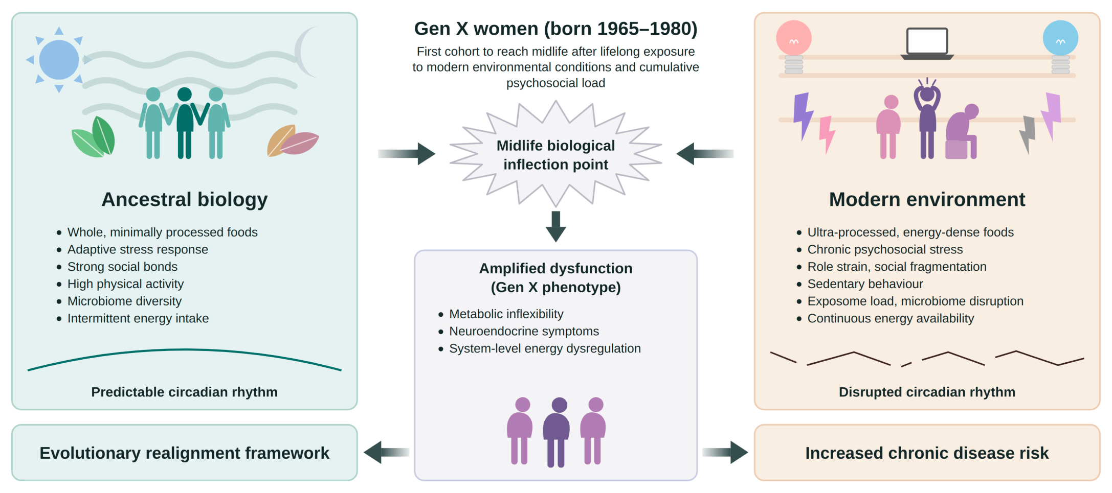
Figure 2.
Evolutionary realignment framework: preventative and therapeutic intervention domains for Gen X women during the menopausal transition. Each domain is paired with the evolutionary mismatch factor it counters, with MHT included where clinically appropriate. Abbreviations: E2 = 17β-oestradiol; EDC = endocrine-disrupting chemicals; Gen X = Generation X; MHT=Menopausal hormone therapy.
Figure 2.
Evolutionary realignment framework: preventative and therapeutic intervention domains for Gen X women during the menopausal transition. Each domain is paired with the evolutionary mismatch factor it counters, with MHT included where clinically appropriate. Abbreviations: E2 = 17β-oestradiol; EDC = endocrine-disrupting chemicals; Gen X = Generation X; MHT=Menopausal hormone therapy.
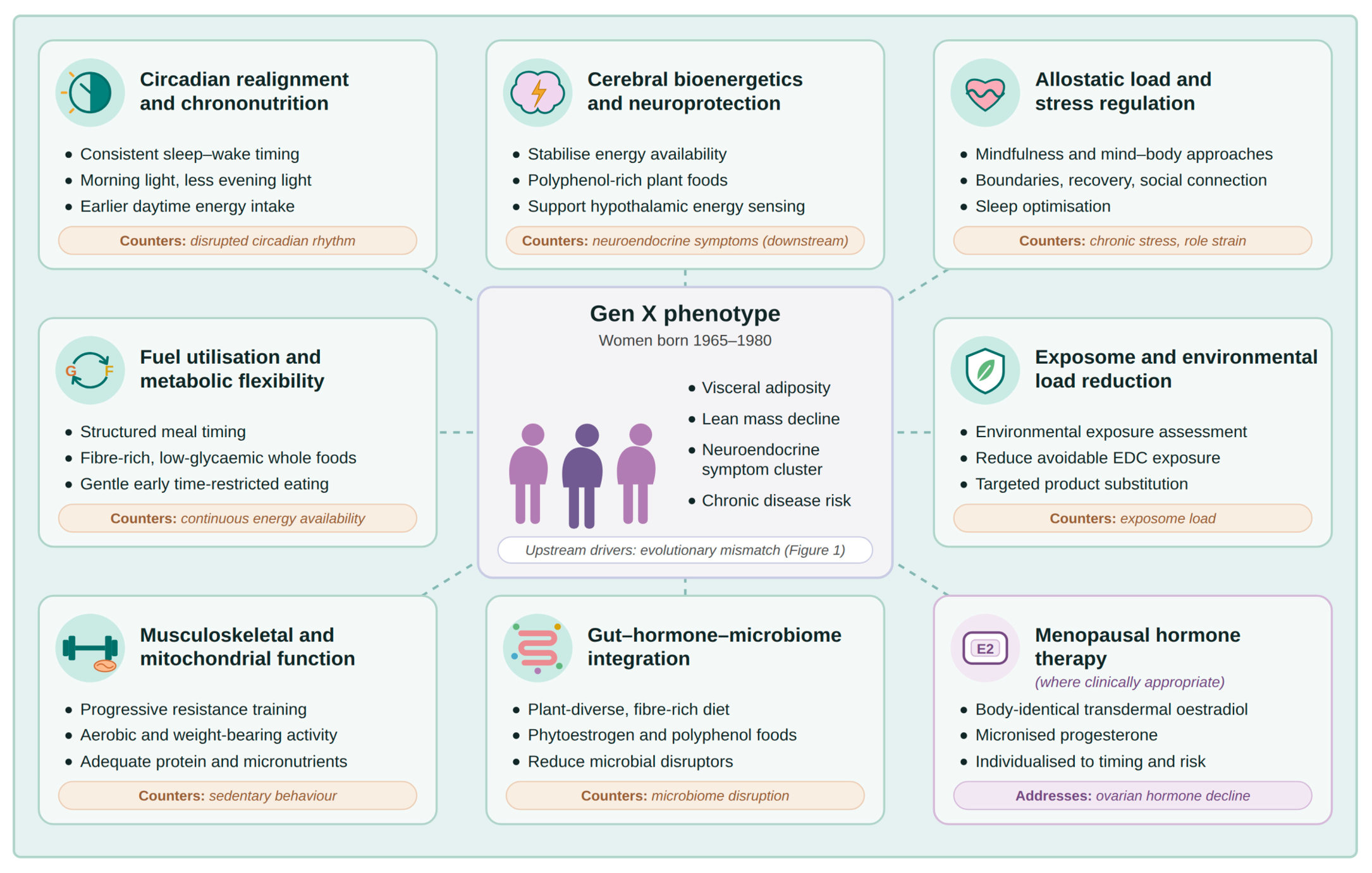

Table 1.
Evolutionary realignment framework for Gen X women: preventative and therapeutic intervention domains and physiological targets.
Table 1.
Evolutionary realignment framework for Gen X women: preventative and therapeutic intervention domains and physiological targets.
| Intervention domain | Key interventions | Primary physiological targets | Key references |
|---|---|---|---|
| Circadian realignment and chrononutrition | Consistent sleep–wake timing, morning light exposure, reduction of evening light exposure, alignment of feeding and activity with circadian rhythms, earlier daytime energy intake | SCN circadian entrainment, hypothalamic–pituitary rhythm regulation, cortisol–melatonin cycling, metabolic timing | 62–64, 67, 68, 74, 77, 78, 81–86 |
| Fuel utilisation and metabolic flexibility | Structured meal timing, fibre-rich low-glycaemic dietary patterns to promote metabolic balance and support substrate switching, gentle early time-restricted eating where appropriate | Glucose–fat oxidation balance, insulin sensitivity and IR, hepatic fuel processing, cellular metabolic flexibility | 81, 82, 87–89, 91–97 |
| Allostatic load and stress regulation | Stress-modulation strategies, psychosocial support, behavioural boundaries, adequate recovery, social connection and sleep optimisation | HPA-axis regulation, ANS balance, inflammatory signalling and CSI, neuroendocrine stability | 60, 61, 98–104 |
| Exposome and environmental load reduction | Environmental exposure assessment, reduction of avoidable EDC and pollutant exposure, targeted product substitution | Endocrine signalling, metabolic regulation, inflammatory signalling, neuroendocrine function | 12, 106–110 |
| Cerebral bioenergetics and neuroprotection | Stabilisation of energy availability, dietary patterns associated with reduced neuroinflammation, strategies supporting hypothalamic energy sensing and neuroendocrine signalling | Hypothalamic regulation of total body energy balance, cerebral energy metabolism, neuroinflammation, dopaminergic signalling, appetite and energy perception pathways | 22–24, 33–36, 50, 51, 95, 111, 112 |
| Musculoskeletal and mitochondrial function | Progressive resistance training, physical activity promoting mitochondrial biogenesis, adequate protein and micronutrient intake | Skeletal muscle mass, mitochondrial function, thermogenesis, serum glucose disposal | 47, 121–129, 131, 132 |
| Gut–hormone–microbiome integration | Plant-diverse, fibre-rich dietary patterns, phytoestrogen exposure, reduction of dietary and environmental disruptors of microbial ecosystems and gut barrier | Microbial diversity, estrobolome activity, gut barrier function, gut enteroendocrine cell function, gut–brain–endocrine signalling | 19, 20, 136–145, 149, 150 |
| Menopausal hormone therapy (where clinically appropriate) | Body-identical transdermal E2, micronised progesterone, low-dose testosterone in selected women; individualised by timing, formulation and risk | KNDy neuron activity and thermoregulation, mitochondrial bioenergetics, neuroinflammation, insulin signalling and metabolic flexibility, sleep, bone and cognition | 124, 152–161 |
Abbreviations: ANS = autonomic nervous system; CSI = chronic systemic inflammation; E2 = 17β-oestradiol; EDC = endocrine-disrupting chemicals; Gen X = Generation X; HPA = hypothalamic–pituitary–adrenal; IR = insulin resistance; KNDy = kisspeptin, neurokinin B and dynorphin; SCN = suprachiasmatic nucleus.
Disclaimer/Publisher’s Note: The statements, opinions and data contained in all publications are solely those of the individual author(s) and contributor(s) and not of MDPI and/or the editor(s). MDPI and/or the editor(s) disclaim responsibility for any injury to people or property resulting from any ideas, methods, instructions or products referred to in the content. |
© 2026 by the authors. Licensee MDPI, Basel, Switzerland. This article is an open access article distributed under the terms and conditions of the Creative Commons Attribution (CC BY) license.
Copyright: This open access article is published under a Creative Commons CC BY 4.0 license, which permit the free download, distribution, and reuse, provided that the author and preprint are cited in any reuse.