

Submitted:
22 October 2026
Posted:
23 October 2026
You are already at the latest version
Preprints on COVID-19 and SARS-CoV-2
Abstract
Background: Post-COVID-19 syndrome (PCS) is associated with persistent pulmonary, autonomic, and hemodynamic impairments. However, the acute effects of spinal manipulation on these systems in adults with PCS remain unclear. Purpose: To investigate the acute effects of combined cervical and thoracic spinal manipulation on pulmonary function, heart rate variability, and blood pressure in adults with PCS. Methods: This exploratory, hypothesis-generating study included 10 adults with PCS randomized to three sequences of spinal manipulation, sham manipulation, and control using a Latin-square crossover design with one-week washouts. Spirometry (FVC, FEV1, and FEV1/FVC) was assessed immediately after each intervention. Blood pressure and heart rate variability indices (RMSSD, SDNN, pNN50, LF, HF, and LF/HF ratio) were recorded at rest and 15, 30, 45, and 60 minutes post-intervention. Friedman/Wilcoxon tests and baseline-corrected sensitivity analyses were performed. Results: No significant differences were observed among conditions for FVC, FEV1, FEV1/FVC (all p>0.70), or the area under the curve of blood pressure, RMSSD, pNN50, HF, and LF/HF ratio (all p>0.27). Differences in SDNN and LF did not survive Bonferroni-corrected comparisons and disappeared after baseline correction, indicating between-session baseline variability rather than intervention effects. Conclusion: A single session of combined cervical and thoracic spinal manipulation did not produce consistent acute improvements in pulmonary function, blood pressure, or heart rate variability in adults with PCS. These physiological variables should not be considered primary indicators of response to an isolated manipulation session.
Keywords:
post-acute COVID-19 syndrome
; manipulation spinal
; heart rate
; blood pressure
; respiratory function tests
; cross-over studies
1. Introduction
The coronavirus belongs to the Coronaviridae family and is characterized by its crown-like appearance under electron microscopy, which inspired its name [1]. This viral group includes strains responsible for a spectrum of human respiratory illnesses, ranging from mild upper respiratory tract infections, such as the common cold, to severe diseases including Severe Acute Respiratory Syndrome (SARS-CoV-1), Middle East Respiratory Syndrome (MERS-CoV), and Coronavirus Disease 2019 (COVID-19), caused by SARS-CoV-2 [1,2]. According to the Brazilian Ministry of Health’s Secretariat for Health and Environmental Surveillance, Brazil continues to record new COVID-19 cases and deaths on an ongoing basis [3], consistent with global surveillance data indicating that SARS-CoV-2 transmission remains active worldwide [4], underscoring the continued relevance of surveillance and vaccination strategies in mitigating disease transmission and severity.
Among the pulmonary sequelae commonly observed after the acute phase of infection, post-COVID-19 syndrome (PCS) stands out as a condition capable of inducing transient or persistent impairments in respiratory function. From a biomechanical perspective, effective breathing depends on the coordinated activation of respiratory musculature, pulmonary elasticity, and thoracic compliance [5]. Within this clinical profile, an increased respiratory workload is commonly observed, leading to muscle fatigue and dyspnea, alterations in pulmonary ventilation, and disruptions in thoracic mechanics that hinder diaphragmatic excursion and further compromise respiratory efficiency [6].
Pulmonary disease is also associated with a range of biodynamic alterations, particularly affecting cellular behavior within lung tissue, which may contribute to disease progression [7]. These structural disruptions can alter the biomechanical properties of the alveolar wall, potentially resulting in localized deformation and accelerating the pathophysiological cascade [7]. Consequently, pulmonary impairment is expected to influence musculoskeletal biomechanics, including respiratory muscle performance, joint mobility, and breathing patterns, dysfunctions that may collectively reduce exercise capacity, elevate fall risk, and contribute to broader physical limitations in affected individuals [8].
A range of therapeutic strategies has been proposed for managing this clinical profile, including active interventions, such as physical exercise, and passive modalities, such as spinal manipulation. Kovanur Sampath et al. [9] suggested that mechanical stimuli delivered through manual techniques may contribute to the restoration of joint function and influence broader physiological responses. Previous studies have reported improvements in pulmonary function after spinal manipulation in individuals recovering from stroke [10] and in patients with chronic obstructive pulmonary disease [11]. Spinal manipulation has also been associated with reductions in post-exercise systolic blood pressure and changes in autonomic modulation [12]. However, no published study has determined whether spinal manipulation produces specific acute respiratory, autonomic, or hemodynamic effects in adults with PCS beyond those attributable to contextual factors, therapist contact, or repeated-measurement variability. Addressing this gap is clinically relevant because persistent respiratory dysfunction, autonomic dysregulation, and abnormal cardiovascular responses may contribute to dyspnea, exercise intolerance, and reduced functional capacity after COVID-19.
We hypothesized that combined cervical and thoracic spinal manipulation would acutely improve pulmonary function, favorably modify heart rate variability, and reduce blood pressure compared with both sham manipulation and a non-intervention control condition. If this hypothesis is supported, the findings would provide preliminary mechanistic evidence for spinal manipulation as a potential adjunct to multidisciplinary rehabilitation in adults with PCS. This approach may be particularly relevant when thoracic mobility, respiratory mechanics, or autonomic regulation are impaired. Conversely, the absence of meaningful between-condition effects would argue against the routine use of spinal manipulation for these outcomes and help refine rehabilitation strategies for this population. Therefore, the present study investigated the acute effects of combined cervical and thoracic spinal manipulation on pulmonary function, heart rate variability, and blood pressure in adults with PCS using a randomized, double-blind, sham- and control-controlled crossover design.
2. Materials and Methods
2.1. Study Design
This was an exploratory and hypothesis-generating study performed in two-center, with three-arm, randomized, double-blind crossover clinical trial design involving 10 adults clinically diagnosed with PCS. Their baseline characteristics were as follows: age, 40.0 ± 9.2 years; body mass, 79.8 ± 16.2 kg; height, 167.0 ± 7.0 cm; body mass index, 28.5 ± 5.6 kg/m2; and self-reported current weekly physical activity at study enrollment, 271.1 ± 150.1 min/week. The trial was reported in accordance with the CONSORT 2025 statement [13] and the SPIRIT 2025 guidance [14]. Ten eligible participants were enrolled and completed all three experimental conditions: spinal manipulation, sham manipulation, and non-intervention control. Assessments and interventions were conducted at the [blinded for reviewer]. To minimize circadian variation in autonomic measures, all sessions were scheduled in the afternoon, and ambient temperature and relative humidity were maintained at 22–24 °C and 60–70%, respectively. An overview of the crossover study design is presented in Figure 1.
2.2. Registry
This trial was approved by the Research Ethics Committee of [blinded for reviewer], according to the guidelines of Law No. 14.874/24 of the National Health Council in accordance with the Helsinki Declaration for research in humans, and prospectively registered in the Brazilian Registry of Clinical Trials [blinded for reviewer]; WHO Universal Trial Number: [blinded for reviewer]).
2.3. Participants
Participants were recruited through invitations disseminated via social media and public announcements at the [blinded for reviewer], targeting adults with PCS. Eligible participants were aged 19 to 44 years and were free from functional limitations other than those attributable to PCS. Exclusion criteria were any compromise of the vertebral (basilar) or subclavian artery contraindicating cervical spinal manipulation, assessed through the screening procedures described under interventions, as well as pregnancy or breastfeeding. Participation was entirely voluntary, and all participants provided written informed consent before enrollment.
Between October and November of 2025, 77 individuals were assessed for eligibility. Of those individuals, 67 were excluded before randomization: 40 did not respond to contact, 11 declined to participate, 7 did not meet the inclusion criteria, and 9 attended an initial screening visit but did not return for randomization. The remaining 10 participants were randomized to one of three sequences of the spine manipulation, sham, and control conditions using a Latin-square design. A one-week washout period separated consecutive sessions (Figure 2). Data collection took place between November 2025 and June 2026. All 10 randomized participants completed the three experimental conditions and were included in the final analysis, with no losses to follow-up after randomization.
The sample size required for the present clinical trial was estimated using data from the first three participants enrolled in the internal pilot trial, which was used exclusively to estimate the variance parameters for the sample size calculation. The mean difference and standard deviation of the difference between the spinal manipulation and sham conditions (the primary comparison of interest, which isolates the specific effect of spinal manipulation from placebo/expectation effects) and between the spinal manipulation and control conditions (the secondary comparison) were used to estimate the sample size required for each outcome. The calculation was performed using G*Power’s procedure for the t-test for dependent means (matched pairs), assuming 80% statistical power and a two-tailed significance level of 5% and determined that 17 participants would be adequate.
2.4. Randomization
As this was a three-condition, three-period crossover trial, each participant was randomly allocated to one of the possible sequences of the spine manipulation, sham, and control conditions. A one-week washout period separated consecutive sessions to minimize carryover effects. An independent researcher not involved in participant assessment or intervention delivery prepared the consecutively numbered, opaque, sealed envelopes that concealed sequence allocation. Envelopes were opened only after participants had signed the informed consent form and completed the baseline evaluations for that session.
Blinding was maintained through a two-examiner protocol. Examiner 1 performed all pre- and post-intervention outcome assessments while remaining blinded to the condition assigned for that session. After the baseline assessments, Examiner 1 left the testing area; Examiner 2, a licensed physical therapist certified in chiropractic and osteopathic manual techniques, then entered and delivered the allocated intervention according to the randomized sequence, remaining blinded to all outcome measures throughout the study. Once the intervention was completed, Examiner 2 left the room and Examiner 1 returned to conduct the post-intervention assessments, following the same standardized procedures used at baseline. Immediately after the post-intervention assessments of each session, participants were asked to report which intervention they believed they had received to check the effectiveness of the blinding procedure.
2.5. Criteria for Discontinuing Allocated Interventions
The study will be terminated if any serious adverse events arise, including significant disability, hospitalization, life-threatening conditions, or death, that render continuation of the trial unsafe for participants, regardless of whether these events are directly associated with the intervention or control/sham conditions.
2.6. Ancillary and Post-Trial Care
Participants who experience lasting harm as a consequence of their involvement in the study will receive ancillary and post-trial care, such as necessary medical treatment for immediate adverse effects related to trial procedures, at no expense. Furthermore, all participants will be ensured access to standard physiotherapy care following their completion of the trial.
2.7. Interventions
All interventions were delivered by a licensed physical therapist (Examiner 2) certified in chiropractic and osteopathic manual techniques, following the Template for Intervention Description and Replication (TIDieR) checklist [15].
Spine manipulation: Prior to the manipulation session, cervical vascular and neurological screening was performed. This included a vertebrobasilar positional tolerance test, together with clinical history, vascular risk-factor screening, blood pressure measurement, and neurological examination. The positional test was not interpreted as a stand-alone measure of vertebral artery integrity or as evidence that cervical manipulation was risk-free, given its limited diagnostic accuracy [16,17]. Participants then underwent Adson’s test, a provocative maneuver commonly used in the clinical assessment of suspected thoracic outlet syndrome. The test was performed with the participant seated, the examined upper limb positioned in slight abduction and extension, and the head rotated toward the tested side while the radial pulse and symptom response were monitored 18. Because of its limited diagnostic specificity, Adson’s test was interpreted only as part of the broader clinical screening process and not as a stand-alone indicator of vascular integrity (Figure 3).
A positive response to either test (e.g., headache, nausea, nystagmus, or a marked reduction in radial pulse) led to exclusion from the manipulation condition; no participant met these exclusion criteria. Upper cervical spinal manipulation was delivered using the Toggle Recoil technique, as previously described by Monteiro et al. [19], a high-velocity, low-amplitude thrust applied between C3 and C5 with the participant side-lying, without rotational force, through rapid triceps-driven elbow extension (toggle) followed by immediate withdrawal (recoil). Thoracic spinal manipulation was performed with the participant lying on their back with their arms crossed over their chest. The clinician applied a high-velocity, low-amplitude anterior-inferior thrust to the T3–T7 region using core-driven force to promote segmental cavitation.
Sham: The sham condition replicated the positioning and contact points of the true manipulation without delivering a thrust. For the cervical component, the clinician placed the pisiform contact between C3 and C5 while the participant was lying on their side but did not perform the toggle-recoil maneuver. For the thoracic component, the clinician applied gentle static pressure over T5–T8 for approximately 3 to 5 seconds while the participant was lying face down. There was no high-velocity thrust or joint cavitation.
Control: Participants in the control condition remained in a resting supine position on the treatment table and underwent only the physiological assessments (spirometry, heart rate variability, and blood pressure), with no manual intervention.
2.8. Outcome Measures
Pulmonary function was assessed using spirometry in accordance with the recommendations of the Brazilian Thoracic Association [20] and ATS/ERS Task Force [21]. A calibrated MINISPIR spirometer (MIR – Medical International Research, Rome, Italy) with single-use MED300 mouthpieces was used for the assessment [22]. Participants were seated and wore a nasal clip during testing. Three technically acceptable and reproducible maneuvers were performed per session, and the highest value was retained for analysis. Forced vital capacity (FVC), forced expiratory volume in the first second (FEV1), the FEV1/FVC ratio, and the Tiffeneau index (calculated as FEV1/FVC) were recorded once, immediately after each intervention.
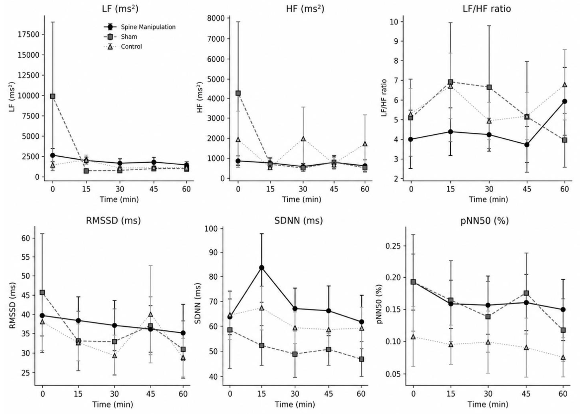
Heart rate variability (HRV) was recorded while the participants were lying on their backs in a quiet, temperature-controlled room. A Polar H10 [23] chest sensor (Polar Electro Oy, Kempele, Finland) was positioned over the lower third of the sternum and connected via Bluetooth to the Elite HRV mobile application [24]. The raw R-R interval data were exported and processed using Kubios software (version 2.0; Kuopio, Finland). Time-domain indices (the root mean square of successive differences (RMSSDms), the standard deviation of NN intervals (SDNNms), and the percentage of successive NN intervals differing by more than 50 milliseconds (pNN50%) and frequency-domain indices derived by fast Fourier transform (low frequency power (LFms2), high frequency power (HFms2), and the LF/HF ratio were extracted at rest (baseline) and 15, 30, 45, and 60 minutes after each intervention.
Systolic and diastolic blood pressure were measured on the left arm while the participants were in a supine position using a fully automated oscillometric monitor (OMRON HEM-7156T; OMRON Healthcare Co., Ltd., Kyoto, Japan). Before each use, the monitor underwent automatic calibration according to the manufacturer’s instructions, and blood pressure measurements were performed in accordance with the American Heart Association guidelines [25]. Baseline measurements were taken after a 15-minute passive rest period upon arrival. Subsequent measurements were obtained at 15, 30, 45, and 60 minutes after each intervention, in parallel with the HRV assessment schedule.
2.9. Data Management
The original data will be digitized as image files by a trained research assistant and subsequently reviewed by a second team member. All digital records will be securely stored on a password-protected computer. To ensure data integrity, a double-entry verification process will be implemented using automated validation procedures within a Microsoft Excel spreadsheet (Microsoft Corporation), conducted remotely by an independent assessor. Any discrepancies identified during this process will be resolved through direct comparison with the original source documents.
2.10. Confidentiality
Each participant will be assigned a unique identification code to preserve anonymity throughout the study. The confidentiality of all data collected at every stage of the trial will be strictly maintained in accordance with ethical research standards.
2.11. Data Analysis
Given the small sample size (n = 10) and the repeated within-subject design, change scores were calculated for each outcome in each experimental condition as the post-intervention value minus the corresponding pre-intervention value (Δ = post − pre). The normality of the change scores was assessed using the Shapiro–Wilk test. Within-condition pre–post differences were examined using paired-samples tests, whereas between-condition differences were evaluated by comparing the change scores across the spinal manipulation, sham manipulation, and non-intervention control conditions. Comparisons among the three conditions (spine manipulation, sham, and control) were performed using the Friedman test.
Effect sizes were calculated to complement null-hypothesis significance testing and to characterize the magnitude of the observed responses. Kendall’s W was reported as the omnibus effect-size estimate for Friedman tests. For Wilcoxon signed-rank comparisons, effect sizes were calculated as r=Z/√N. Pre-to-post change scores were calculated separately for each experimental condition, and the magnitude of within-condition changes and between-condition differences in change scores was examined. Median changes, interquartile ranges, and 95% confidence intervals for paired differences were reported whenever applicable. Clinical relevance was evaluated by comparing the observed changes with established minimal clinically important differences, minimal detectable changes, or accepted clinical thresholds when such values were available.
Spirometric indices (forced vital capacity [FVC], forced expiratory volume in the first second [FEV1], FEV1/FVC ratio, and the Tiffeneau index were recorded once, immediately after each intervention, and compared directly among the three conditions. Systolic and diastolic blood pressure and heart rate variability indices (RMSSD, SDNN, pNN50, LF, HF, and LF/HF ratio) were recorded at rest and 15, 30, 45, and 60 minutes post-intervention. For each participant and condition, the response over time was summarized by the area under the curve (AUC, trapezoidal rule). AUC captured both the magnitude and the persistence of the hemodynamic and autonomic responses over the hour following the intervention. AUC values were compared among conditions using the same approach (Friedman/Wilcoxon).
As this was a crossover design, the equivalence of the baseline values (rest and pre-intervention) across the three conditions was verified before interpreting the effects of the intervention. This was done as a means of screening for possible carryover or period effects arising from insufficient washout. When a variable showed a significant difference among conditions in the raw AUC, a sensitivity analysis was conducted by recalculating the AUC on baseline-corrected values (delta = value − rest), isolating the response attributable to the intervention from individual baseline variation between sessions.
To assess the potential influence of period or carryover effects inherent to the crossover design, pre-intervention baseline values were compared across the three experimental conditions for systolic and diastolic blood pressure and all heart rate variability indices, including RMSSDms, SDNNms, pNN50%, HFms2 power, LF ms2 power, and the LF/HF ratio. These comparisons were performed using the Friedman test. When a significant omnibus difference was identified, pairwise comparisons were conducted using Wilcoxon signed-rank tests with Bonferroni adjustment for multiple comparisons. Baseline equivalence across conditions was interpreted as supporting the absence of a relevant carryover effect. Spirometric variables were not included in this analysis because they were assessed only once, immediately after each intervention, and no corresponding pre-intervention values were available.
All tests were two-tailed, with a significance level of 5%, and analyses were performed in Python 3.10. The anonymized dataset, data dictionary, and analysis scripts were deposited in an open repository before submission: [https://doi.org/10.17605/OSF.IO/BMGUP]. No subgroup analysis is planned for this study.
3. Results
3.1. Pulmonary Function
No statistically significant differences were observed among the spine manipulation, sham, and control conditions for FVC (χ2(2)=0.60; p=0.741), FEV1 (χ2(2)=0.05; p=0.975), or the FEV1/FVC ratio, equivalent to the Tiffeneau index (χ2(2) =0.20; p=0.905) (Table 1). Effect sizes (Kendall’s W) were close to zero in all cases (W ≤ 0.03), suggesting no immediate response of the combined cervical and thoracic adjustment on pulmonary function in this sample.
3.2. Hemodynamic and Autonomic Response
There were no significant differences among conditions in the AUC of systolic blood pressure (χ2(2) = 2.60; p = 0.273), diastolic blood pressure (χ2(2) = 1.40; p = 0.497), RMSSDms (χ2(2)=0.80; p=0.670), pNN50% (χ2(2)=2.51; p=0.285), HFms2 (χ2(2)=1.40; p=0.497), or LF/HF ratio (χ2(2)=1.40; p=0.497) (Figure 4 and Figure 5); the same pattern of no difference was observed at each individual time point (rest, 15, 30, 45, and 60 minutes).
Two variables showed a significant difference in the raw AUC: SDNNms (χ2(2)=7.20; p=0.027; W=0.36) and LFms2 (χ2(2)=6.20; p=0.045; W=0.31). In both cases, however, no pairwise comparison survived Bonferroni correction (SDNN: spine manipulation vs. sham, adjusted p=0.252; spine manipulation vs. control, adjusted p=0.697; sham vs. control, adjusted p=0.580; LFms2: spine manipulation vs. sham, adjusted p=0.252; spine manipulation vs. control, adjusted p=0.826; sham vs. control, adjusted p=0.826). Given that baseline LFms2 already differed significantly among conditions prior to any intervention, a sensitivity analysis was conducted by recalculating the AUC on individually baseline-corrected values (delta = value − rest). After this correction, the difference among conditions was no longer significant for either SDNNms (χ2(2)=0.60; p=0.741; W=0.03) or LFms2 (χ2(2)=0.60; p=0.741; W=0.03), indicating that the raw findings mainly reflected baseline variation between sessions rather than a specific, immediate response of the intervention on global autonomic or sympathetic modulation.
4. Discussion
This randomized, double-blind, sham- and control-controlled crossover trial examined the effects of a single session of combined cervical and thoracic spinal manipulation on pulmonary function, blood pressure, and heart rate variability in adults with PCS. The results showed that this manipulation did not produce statistically significant acute changes relative to sham manipulation or no intervention. Effect sizes for all outcomes were small (Kendall’s W ≤ 0.07), with two exceptions, SDNNms and LFms2. An apparent effect in the unadjusted analysis did not survive either pairwise comparison with Bonferroni correction or the baseline-corrected sensitivity analysis. This indicates that the effect reflected pre-existing variability between sessions rather than a genuine response to the intervention.
Spinal manipulation has been hypothesized to influence autonomic regulation through stimulation of mechanoreceptors located in paraspinal joints and myofascial tissues. Afferent input arising from these structures may interact with somatosympathetic and somatovisceral reflex pathways and, theoretically, influence supraspinal autonomic centers involved in cardiovascular regulation. Nevertheless, the available clinical evidence does not consistently support a meaningful autonomic effect. A systematic review and meta-analysis found low-quality evidence that spinal manipulation did not substantially alter heart rate variability, blood pressure, catecholamine concentrations, or other autonomic outcomes when compared with sham or control interventions, although isolated effects on selected HRV indices have occasionally been reported [9]. Individual trials have similarly produced inconsistent findings across anatomical regions and clinical populations [26,27,28]. Despite this generally inconclusive evidence, the present investigation addressed a clinically distinct population. Individuals with post-COVID-19 syndrome may exhibit persistent autonomic dysregulation, reduced heart rate variability, sympathetic predominance, dyspnea, exercise intolerance, and impaired cardiorespiratory function. These features provided a specific rationale for examining whether a mechanosensory intervention could elicit an acute physiological response that might differ from that observed in healthy adults or in patients with primarily musculoskeletal disorders [29,30]. In the present study, spinal manipulation did not produce consistent acute improvements in HRV, blood pressure, or pulmonary function compared with sham manipulation or non-intervention control. These findings are consistent with the broader evidence base and with our previous studies showing no meaningful acute modulation of sympathovagal balance or blood pressure after upper cervical manipulation or manual massage [19], as well as inconsistent post-exercise blood pressure responses following manual therapies [12].
Another consideration for this population is that PCS is associated with persistent autonomic imbalance. This imbalance is characterized by sympathetic overactivation and reduced parasympathetic responsiveness. It can be detected months after acute infection. Proposed mechanisms include direct or indirect viral injury to central autonomic structures, dysregulated autoantibodies against adrenergic and vasoregulatory receptors, and persistent low-grade inflammation [31]. If the autonomic dysregulation underlying PCS is predominantly driven by these systemic immunological and neurohormonal processes rather than by segmental musculoskeletal dysfunction, then a single mechanically delivered afferent stimulus, such as spinal manipulation, would likely be insufficient to produce a measurable acute shift in autonomic balance, despite its theoretical capacity to modulate somatosympathetic reflexes in other populations. This interpretation is also compatible with our finding that baseline LF values varied between sessions before any intervention was delivered. Rather than reflecting an inadequate washout period, this variability may itself be a manifestation of the labile, day-to-day autonomic fluctuations described in PCS [31].
The absence of an acute effect on spirometric indices likewise contrasts with previous reports of improved pulmonary function following spinal manipulation in individuals recovering from stroke or with chronic obstructive pulmonary disease (COPD), as noted in the Introduction section. Unlike the fixed airflow obstruction of COPD or the spasticity-related chest wall restriction that can follow stroke, dyspnea in PCS has increasingly been linked to a distinct pattern of respiratory muscle weakness and neuromuscular disturbance. This pattern is termed complex ventilatory dysfunction and ocurrs in the presence of preserved total lung capacity and relatively normal spirometric ratios [32]. Since FVC, FEV1, and the FEV1/FVC ratio primarily characterize ventilatory volume and airflow limitation and are not direct measures of respiratory muscle strength, these outcomes may have been insufficiently sensitive to detect the type of respiratory impairment present in some individuals with PCS. Previous evidence indicates that respiratory muscle dysfunction may occur in persistently symptomatic patients despite the absence of pronounced abnormalities on conventional pulmonary function testing [21,33]. This is true irrespective of whether thoracic manipulation produced any transient effect on rib cage mechanics or diaphragmatic excursion. Previous positive findings in stroke patients and those with COPD also largely derive from multiple-session protocols delivered over several weeks. In contrast, the present trial evaluated the acute response to a single intervention, and spirometry was performed once immediately after each intervention. Cumulative, dose-dependent, or delayed effects, if present, would not have been captured by this design.
Clinically, the findings do not support the use of a single session of cervical and thoracic spinal manipulation as an isolated intervention intended to generate immediate autonomic, hemodynamic, or pulmonary benefits in adults with PCS. However, because the sample was small and the effect estimates were imprecise, the results should be interpreted as preliminary rather than as definitive evidence of ineffectiveness. The findings instead suggest that any potential clinical benefit of spinal manipulation in PCS is unlikely to be explained by an immediate generalized change in the physiological outcomes assessed. Future adequately powered trials should examine repeated treatment exposure, longer follow-up periods, functional capacity, dyspnea, fatigue, orthostatic intolerance, symptom burden, and individual responder profiles.
4.1. Limitations
This trial has several limitations that should be considered when interpreting the findings. First, the relatively small sample size (n = 10) reflected the practical challenges encountered in recruiting eligible individuals with PCS and maintaining adherence throughout a crossover protocol requiring participation in three separate experimental sessions. These difficulties should not be interpreted as indicating a low population burden of PCS in Brazil. According to the Brazilian Ministry of Health, approximately 55 million individuals had experienced at least one laboratory-confirmed or medically diagnosed episode of COVID-19 by the first quarter of 2023. Among them, an estimated 13.808.479 individuals reported symptoms that persisted or developed more than 30 days after the acute infection, corresponding to 25.1% of those with confirmed COVID-19 and an estimated national prevalence of 6.522.8 cases per 100.000 inhabitants1. In the state of Rio de Janeiro, where the present study was conducted, approximately 884.999 individuals were estimated to have experienced a post-COVID condition, corresponding to 5.141.2 cases per 100.000 inhabitants [34]. Nevertheless, national primary care records documented only 180.581 encounters coded as post-COVID condition between September 2021 and December 2024, including 40.427 encounters in 2024 [34]. This marked discrepancy between population-based estimates and routinely recorded healthcare encounters suggests substantial underrecognition, undercoding, heterogeneous clinical presentation, and possible barriers to healthcare access, all of which may restrict the pool of identifiable and contactable participants for clinical research. Therefore, despite the substantial epidemiological burden of PCS, recruiting individuals who met the study criteria, remained clinically stable, and were able to complete all crossover visits was challenging. The resulting sample may have reduced the precision of the estimates and the ability to detect small between-condition differences. Second, only the acute, single-session response to spinal manipulation was evaluated. Whether repeated sessions produce cumulative effects on pulmonary function, blood pressure, or heart rate variability remains unknown. Third, participants were asked to report which intervention they believed they had received to check the effectiveness of the blinding. However, these data were not incorporated into the present analysis, so residual unblinding, if present, cannot be ruled out as a source of bias.
4.2. Practical Applicability
From a practical perspective, the present findings indicate that a single session of combined cervical and thoracic spinal manipulation should not be expected to produce immediate changes in pulmonary function, blood pressure, or heart rate variability in adults with post-COVID-19 syndrome. Therefore, clinicians should not use acute variations in these physiological measures as indicators of treatment response following a single spinal manipulation session. The sham manipulation and non-intervention conditions were appropriate comparators for the mechanistic purpose of this study because they helped distinguish the specific effects of spinal manipulation from contextual influences, resting effects, and short-term physiological variability. Active rehabilitation strategies, such as respiratory muscle training, individualized exercise, and symptom-directed physiotherapy, were not included as comparators because they generally require repeated treatment exposure and address a different clinical question. Because pain, disability, musculoskeletal symptoms, functional capacity, and perceived well-being were not assessed, the present findings do not establish whether spinal manipulation may provide benefits in these domains. Its potential role as an adjunct to comprehensive rehabilitation therefore remains uncertain and should be examined in future trials using patient-centered outcomes, repeated treatment sessions, and longer follow-up periods. Future confirmatory studies should also define a single clinically meaningful primary outcome based on patient and clinician priorities.
5. Conclusion
This randomized, double-blind, sham- and control-controlled crossover trial examined the acute effects of a single session of combined cervical and thoracic spinal manipulation on pulmonary function, blood pressure, and heart rate variability in adults with PCS. There were no statistically significant differences compared with sham manipulation or control. Apparent differences in two heart rate variability indices were due to baseline variability rather than a genuine intervention effect. These findings do not support using a single manipulation session to acutely modulate cardiopulmonary or autonomic function in this population. They should be confirmed in adequately powered trials with a pre-specified primary outcome and potentially repeated intervention sessions.
Supplementary Materials
The following supporting information can be downloaded at the website of this paper posted on Preprints.org.
Author Contributions
Conceptualization, F.P.S, A.G.deM., J.C.deO.M.C., B.P.F., L.M.A., I.R.T.deJ., and E.R.M.; methodology, F.P.S, A.G.deM., J.C.deO.M.C., B.P.F., L.M.A., I.R.T.deJ., and E.R.M.; validation, F.P.S, A.G.deM., J.C.deO.M.C., B.P.F., L.M.A., I.R.T.deJ., and E.R.M. formal analysis, F.P.S., L.S.P., P.H.M., A.G.deM., J.C.deO.M.C., B.P.F., L.M.A., L.F.daF.R., J.G.S., B.C.B., A.J.L., I.R.T.deJ., and E.R.M., investigation, F.P.S, A.G.deM., J.C.deO.M.C., B.P.F., L.M.A., I.R.T.deJ., and E.R.M., resources, F.P.S., L.S.P., P.H.M., A.G.deM., J.C.deO.M.C., B.P.F., L.M.A., L.F.daF.R., J.G.S., B.C.B., A.J.L., I.R.T.deJ., and E.R.M., data curation, F.P.S., L.S.P., P.H.M., A.G.deM., J.C.deO.M.C., B.P.F., L.M.A., L.F.daF.R., J.G.S., B.C.B., A.J.L., I.R.T.deJ., and E.R.M., writing—original draft preparation, F.P.S., L.S.P., P.H.M., A.G.deM., J.C.deO.M.C., B.P.F., L.M.A., L.F.daF.R., J.G.S., B.C.B., A.J.L., I.R.T.deJ., and E.R.M., writing—review and editing, F.P.S., L.S.P., P.H.M., A.G.deM., J.C.deO.M.C., B.P.F., L.M.A., L.F.daF.R., J.G.S., B.C.B., A.J.L., I.R.T.deJ., and E.R.M., visualization, F.P.S., L.S.P., P.H.M., A.G.deM., J.C.deO.M.C., B.P.F., L.M.A., L.F.daF.R., J.G.S., B.C.B., A.J.L., I.R.T.deJ., and E.R.M., supervision, I.R.T.d.J., and E.R.M. All authors have read and agreed to the published version of the manuscript.
Funding
This research received no external funding.
Institutional Review Board Statement
The study was approved by the Research Ethics Committee of the Augusto Motta University Center – UNISUAM (IRB No. 7.584.897), according to the guidelines of Law No. 14.874/24 of the National Health Council in accordance with the Helsinki Declaration for research in humans and prospectively registered in the Brazilian Registry of Clinical Trials (RBR-4j8p3wq; WHO Universal Trial Number: U1111-1323-2361).
Informed Consent Statement
Informed consent will be obtained from all subjects involved in the study.
Data Availability Statement
The Data was availability and can be downloaded at: https://doi.org/10.17605/OSF.IO/BMGUP.
Conflicts of Interest
The authors declare no conflicts of interest.
Abbreviations
The following abbreviations are used in this manuscript:
| ATS | American Thoracic Society |
| AUC | Area Under the Curve |
| BP | Blood Pressure |
| CONSORT | Consolidated Standards of Reporting Trials |
| COPD | Chronic Obstructive Pulmonary Disease |
| COVID-19 | Coronavirus Disease 2019 |
| ERS | European Respiratory Society |
| FEV1 | Forced Expiratory Volume in the First Second |
| FEV1/FVC | Ratio of Forced Expiratory Volume in the First Second to Forced Vital Capacity |
| HFnu | High Frequency |
| FVC | Forced Vital Capacity |
| HF | High Frequency |
| HRV | Heart Rate Variability |
| IRB | Institutional Review Board |
| LF | Low Frequency |
| LF/HF | Low-Frequency to High-Frequency Ratio |
| MERS-CoV | Middle East Respiratory Syndrome Coronavirus |
| RMSS | Root Mean Square of Successive Differences |
| NN | Normal-to-Normal Intervals |
| OSF | Open Science Framework |
| PCS | Post-COVID-19 Syndrome |
| SDNN | Standard Deviation of Normal-to-Normal Intervals |
| pNN50 | Percentage of Successive NN Intervals Differing by More Than 50 ms |
| RMSSD | Root Mean Square of Successive Differences |
| SARS-CoV-1 | Severe Acute Respiratory Syndrome Coronavirus 1 |
| SARS-CoV-2 | Severe Acute Respiratory Syndrome Coronavirus 2 |
| SDNN | Standard Deviation of Normal-to-Normal Intervals |
| SEM | Standard Error of the Mean |
| SPIRIT | Standard protocol Items: Recommendations for Interventional Trials |
| TIDIeR | Template for Intervention Description and Replication |
| WHO | World Health Organization |
References
- Abdelrahman, Z.; Li, M.; Wang, X. Comparative review of SARS-CoV-2, SARS-CoV, MERS-CoV, and influenza A respiratory viruses. Front. Immunol. 2020, 11, 552909. [Google Scholar] [CrossRef] [PubMed]
- V’Kovski, P.; Kratzel, A.; Steiner, S.; Stalder, H.; Thiel, V. Coronavirus biology and replication: Implications for SARS-CoV-2. Nat. Rev. Microbiol. 2021, 19, 155–170. [Google Scholar] [CrossRef] [PubMed]
- Brasil. Ministério da Saúde. Secretaria de Vigilância em Saúde e Ambiente. Cenário Epidemiológico da COVID-19, Influenza e Outros Vírus Respiratórios no Brasil: Semana Epidemiológica 1 a SE 52 de 2024; Ministério da Saúde: Brasília, DF, Brazil, 2025; Available online: https://www.gov.br/saude/pt-br/centrais-de-conteudo/publicacoes/boletins/epidemiologicos/covid-19/2025/cenario-epidemiologico-da-covid-no-brasil-semana-epidemiologica-1-a-se-52-de-2024 (accessed on 7 July 2026).
- World Health Organization. WHO COVID-19 Dashboard. Available online: https://data.who.int/dashboards/covid19/summary (accessed on 7 July 2026).
- Patel, N.; Chong, K.; Baydur, A. Methods and applications in respiratory physiology: Respiratory mechanics, drive and muscle function in neuromuscular and chest wall disorders. Front. Physiol. 2022, 13, 838414. [Google Scholar] [CrossRef] [PubMed]
- Kocjan, J.; Rydel, M.; Szczegielniak, J.; Bogacz, K.; Adamek, M. Diaphragm muscle atrophy contributes to low physical capacity in COVID-19 survivors. Life 2024, 14, 1117. [Google Scholar] [CrossRef] [PubMed]
- He, A.; He, L.; Chen, T.; Li, X.; Cao, C. Biomechanical properties and cellular responses in pulmonary fibrosis. Bioengineering 2024, 11, 747. [Google Scholar] [CrossRef] [PubMed]
- Mou, K.; Chan, S.M.H.; Vlahos, R. Musculoskeletal crosstalk in chronic obstructive pulmonary disease and comorbidities: Emerging roles and therapeutic potentials. Pharmacol. Ther. 2024, 257, 108635. [Google Scholar] [CrossRef] [PubMed]
- Kovanur Sampath, K.; Tumilty, S.; Wooten, L.; Belcher, S.; Farrell, G.; Spontelli Gisselman, A. Effectiveness of spinal manipulation in influencing the autonomic nervous system: A systematic review and meta-analysis. J. Man. Manip. Ther. 2024, 32, 10–27. [Google Scholar] [CrossRef] [PubMed]
- Joo, S.; Lee, Y.; Song, C.H. Immediate effects of thoracic spinal manipulation on pulmonary function in stroke patients: A preliminary study. J. Manip. Physiol. Ther. 2018, 41, 602–608. [Google Scholar] [CrossRef] [PubMed]
- Dougherty, P.E.; Engel, R.M.; Vemulpad, S.; Burke, J. Spinal manipulative therapy for elderly patients with chronic obstructive pulmonary disease: A case series. J. Manip. Physiol. Ther. 2011, 34, 413–417. [Google Scholar] [CrossRef] [PubMed]
- Monteiro, E.R.; Pescatello, L.S.; Winchester, J.B.; Corrêa Neto, V.G.; Brown, A.F.; Budde, H.; Marchetti, P.H.; Silva, J.G.; Vianna, J.M.; Novaes, J.S. Effects of manual therapies and resistance exercise on postexercise hypotension in women with normal blood pressure. J. Strength Cond. Res. 2022, 36, 948–954. [Google Scholar] [CrossRef] [PubMed]
- Hopewell, S.; Chan, A.W.; Collins, G.S.; Hróbjartsson, A.; Moher, D.; Schulz, K.F.; Tunn, R.; Aggarwal, R.; Berkwits, M.; Berlin, J.A.; et al. CONSORT 2025 statement: Updated guideline for reporting randomised trials. BMJ 2025, 389, e081123. [Google Scholar] [CrossRef] [PubMed]
- Chan, A.W.; Boutron, I.; Hopewell, S.; Moher, D.; Schulz, K.F.; Collins, G.S.; Tunn, R.; Aggarwal, R.; Berkwits, M.; Berlin, J.A.; et al. SPIRIT 2025 statement: Updated guideline for protocols of randomised trials. BMJ 2025, 389, e081477. [Google Scholar] [CrossRef] [PubMed]
- Hoffmann, T.C.; Glasziou, P.P.; Boutron, I.; Milne, R.; Perera, R.; Moher, D.; Altman, D.G.; Barbour, V.; Macdonald, H.; Johnston, M.; et al. Better reporting of interventions: Template for Intervention Description and Replication (TIDieR) checklist and guide. BMJ 2014, 348, g1687. [Google Scholar] [CrossRef] [PubMed]
- Hutting, N.; Verhagen, A.P.; Vijverman, V.; Keesenberg, M.D.M.; Dixon, G.; Scholten-Peeters, G.G.M. Diagnostic accuracy of premanipulative vertebrobasilar insufficiency tests: A systematic review. Man. Ther. 2013, 18, 177–182. [Google Scholar] [CrossRef] [PubMed]
- Rushton, A.; Carlesso, L.C.; Flynn, T.; Hing, W.A.; Rubinstein, S.M.; Vogel, S.; Kerry, R. International framework for examination of the cervical region for potential of vascular pathologies of the neck prior to musculoskeletal intervention: International IFOMPT Cervical Framework. J. Orthop. Sports Phys. Ther. 2023, 53, 7–22. [Google Scholar] [CrossRef] [PubMed]
- Jones, M.R.; Prabhakar, A.; Viswanath, O.; Urits, I.; Green, J.B.; Kendrick, J.B.; Brunk, A.J.; Eng, M.R.; Orhurhu, V.; Cornett, E.M.; et al. Thoracic outlet syndrome: A comprehensive review of pathophysiology, diagnosis, and treatment. Pain Ther. 2019, 8, 5–18. [Google Scholar] [CrossRef] [PubMed]
- Monteiro, E.R.; Pescatello, L.S.; Garcia, G.H.; Meirelles, A.G.; Oliveira, F.; Souza, R.C.; Nogueira, L.A.C.; Lopes, A.J.; Moreira-Gonçalves, D. Upper cervical manipulation and manual massage do not modulate sympatho-vagal balance or blood pressure in women: A randomized, placebo-controlled clinical trial. Healthcare 2025, 13, 2554. [Google Scholar] [CrossRef] [PubMed]
- Albuquerque, A.L.P.; Berton, D.C.; Campos, E.V.M.F.A.S.; Queiroga-Júnior, F.J.P.; Santana, A.N.C.; Wong, B.M.S.; Batista, D.R.; Melo, F.X.; Didier-Neto, F.M.F.; Coutinho, H.M.; et al. Novas recomendações de espirometria da Sociedade Brasileira de Pneumologia e Tisiologia—Atualização 2024. J. Bras. Pneumol. 2024, 50, e20240169. [Google Scholar] [CrossRef] [PubMed]
- Graham, B.L.; Steenbruggen, I.; Miller, M.R.; Barjaktarevic, I.Z.; Cooper, B.G.; Hall, G.L.; Hallstrand, T.S.; Kaminsky, D.A.; McCarthy, K.; McCormack, M.C.; et al. Standardization of spirometry 2019 update: An official American Thoracic Society and European Respiratory Society technical statement. Am. J. Respir. Crit. Care Med. 2019, 200, e70–e88. [Google Scholar] [CrossRef] [PubMed]
- Mukharesh, L.; Ryan, M.; Hayden, L.P.; Dahlberg, S.E.; Gaffin, J.M. Comparison of pneumotachometer and portable digital turbine spirometry for field-based assessment: An Air Quality, Environment, and Respiratory Outcomes in Bronchopulmonary Dysplasia study. Pediatr. Allergy Immunol. Pulmonol. 2023, 36, 115–118. [Google Scholar] [CrossRef] [PubMed]
- Schaffarczyk, M.; Rogers, B.; Reer, R.; Gronwald, T. Validity of the Polar H10 sensor for heart rate variability analysis during resting state and incremental exercise in recreational men and women. Sensors 2022, 22, 6536. [Google Scholar] [CrossRef] [PubMed]
- Perrotta, A.S.; Jeklin, A.T.; Hives, B.A.; Meanwell, L.E.; Warburton, D.E.R. Validity of the Elite HRV smartphone application for examining heart rate variability in a field-based setting. J. Strength Cond. Res. 2017, 31, 2296–2302. [Google Scholar] [CrossRef] [PubMed]
- Muntner, P.; Shimbo, D.; Carey, R.M.; Charleston, J.B.; Gaillard, T.; Misra, S.; Myers, M.G.; Ogedegbe, G.; Schwartz, J.E.; Townsend, R.R.; et al. Measurement of blood pressure in humans: A scientific statement from the American Heart Association. Hypertension 2019, 73, e35–e66. [Google Scholar] [CrossRef] [PubMed]
- Younes, M.; Nowakowski, K.; Didier-Laurent, B.; Gombert, M.; Cottin, F. Effect of spinal manipulative treatment on cardiovascular autonomic control in patients with acute low back pain. Chiropr. Man. Ther. 2017, 25, 33. [Google Scholar] [CrossRef] [PubMed]
- Galaasen Bakken, A.; Axén, I.; Eklund, A.; O’Neill, S. The effect of spinal manipulative therapy on heart rate variability and pain in patients with chronic neck pain: A randomized controlled trial. Trials 2019, 20, 590. [Google Scholar] [CrossRef] [PubMed]
- Carnevali, L.; Lombardi, L.; Fornari, M.; Sgoifo, A. Exploring the effects of osteopathic manipulative treatment on autonomic function through the lens of heart rate variability. Front. Neurosci. 2020, 14, 579365. [Google Scholar] [CrossRef] [PubMed]
- Rinaldi, L.; Rigo, S.; Pani, M.; Bisoglio, A.; Khalaf, K.; Minonzio, M.; Shiffer, D.; Romeo, M.A.; Verzeletti, P.; Ciccarelli, M.; et al. Long-COVID autonomic syndrome in working age and work ability impairment. Sci. Rep. 2024, 14, 11835. [Google Scholar] [CrossRef] [PubMed]
- Santos-de-Araújo, A.D.; Bassi-Dibai, D.; Marinho, R.S.; Dourado, I.M.; de Almeida, L.V.; dos Santos, S.S.; Phillips, S.A.; Borghi-Silva, A. Impact of COVID-19 on heart rate variability in post-COVID individuals compared to a control group. Sci. Rep. 2024, 14, 31099. [Google Scholar] [CrossRef] [PubMed]
- Mooren, F.C.; Böckelmann, I.; Waranski, M.; Kotewitsch, M.; Teschler, M.; Schäfer, H.; Schmitz, B. Autonomic dysregulation in long-term patients suffering from post-COVID-19 syndrome assessed by heart rate variability. Sci. Rep. 2023, 13, 15814. [Google Scholar] [CrossRef]
- Steinbeis, F.; Kedor, C.; Meyer, H.J.; Thibeault, C.; Mittermaier, M.; Knape, P.; Ahrens, K.; Rotter, G.; Temmesfeld-Wollbrück, B.; Sander, L.E.; et al. A new phenotype of patients with post-COVID-19 condition is characterised by a pattern of complex ventilatory dysfunction, neuromuscular disturbance and fatigue symptoms. ERJ Open Res. 2024, 10, 01027–2023. [Google Scholar] [CrossRef] [PubMed]
- Hennigs, J.K.; Huwe, M.; Hennigs, A.; Oqueka, T.; Simon, M.; Harbaum, L.; Körbelin, J.; Schmiedel, S.; Schulze Zur Wiesch, J.; Addo, M.M.; et al. Respiratory muscle dysfunction in long-COVID patients. Infection 2022, 50, 1391–1397. [Google Scholar] [CrossRef] [PubMed]
- Brazil. Ministry of Health. Secretariat of Health and Environmental Surveillance. Monitoring Post-COVID Conditions in Brazil. Epidemiol. Bull. 2025, 56, 1–18. Available online: https://www.gov.br/saude/pt-br/centrais-de-conteudo/publicacoes/boletins/epidemiologicos/edicoes/2025/boletim-epidemiologico-volume-56-no-7-21-de-mai.pdf/view.
Figure 1.
Study design.
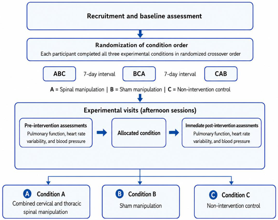
Figure 2.
CONSORT 2025 participant flow diagram.
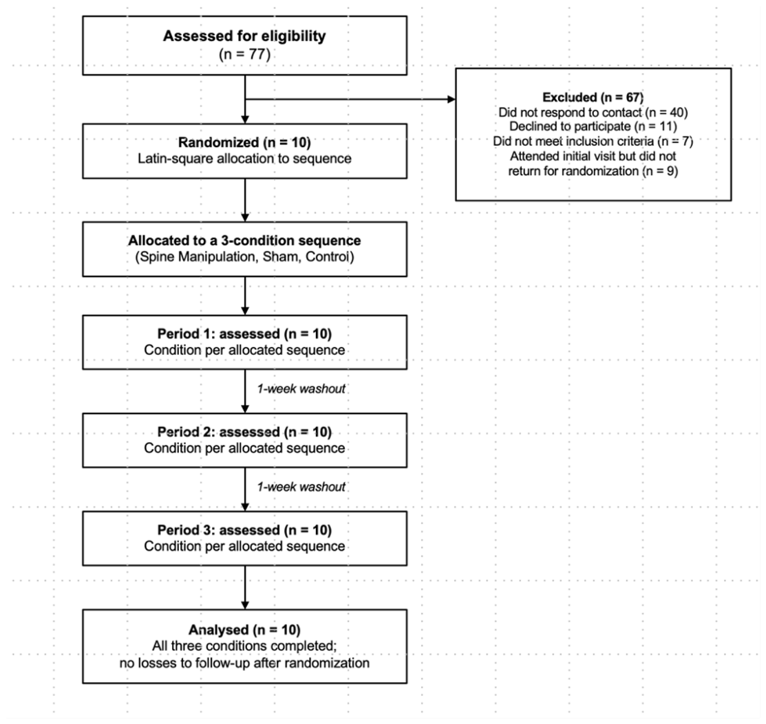
Figure 3.
Pre-manipulative cervical vascular and neurovascular screening procedures. (A) Cervical positional tolerance test used as part of the pre-manipulative cervical vascular screening. (B) Adson’s test used as part of the broader clinical screening for possible thoracic outlet involvement. The procedures were illustrated based on the methodological descriptions provided by Hutting et al. [16], Rushton et al. [17], and Jones et al. [18]. The illustrations were generated using artificial intelligence for didactic and scientific communication purposes.
Figure 3.
Pre-manipulative cervical vascular and neurovascular screening procedures. (A) Cervical positional tolerance test used as part of the pre-manipulative cervical vascular screening. (B) Adson’s test used as part of the broader clinical screening for possible thoracic outlet involvement. The procedures were illustrated based on the methodological descriptions provided by Hutting et al. [16], Rushton et al. [17], and Jones et al. [18]. The illustrations were generated using artificial intelligence for didactic and scientific communication purposes.
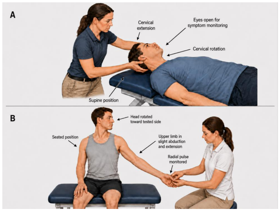
Figure 4.
Trajectory of systolic and diastolic blood pressure over 60 minutes following each intervention condition (n=10). Data are mean ± standard error of the mean (SEM) at rest (baseline) and at 15-, 30-, 45-, and 60-minutes post-intervention. No significant differences among conditions were observed in the area under the curve or at any individual time point (Friedman test, all p>0.27).
Figure 4.
Trajectory of systolic and diastolic blood pressure over 60 minutes following each intervention condition (n=10). Data are mean ± standard error of the mean (SEM) at rest (baseline) and at 15-, 30-, 45-, and 60-minutes post-intervention. No significant differences among conditions were observed in the area under the curve or at any individual time point (Friedman test, all p>0.27).
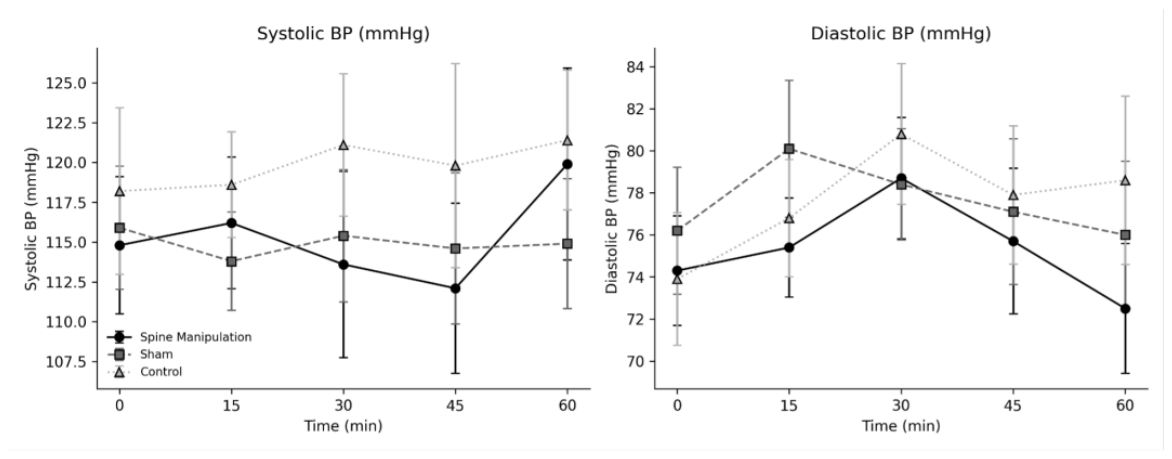
Figure 5.
Trajectory of time-domain and frequency-domain heart rate variability indices over 60 minutes (n=10). Data are mean ± SEM. RMSSDms: root mean square of successive differences; SDNNms: standard deviation of NN intervals; pNN50%: percentage of successive NN intervals differing by more than 50 ms; LFms2: low-frequency power; HF ms2: high-frequency power. The area under the curve for SDNNms and LFms2 differed significantly among conditions in the unadjusted analysis (Friedman p=0.027 and p=0.045, respectively), an effect that did not persist after baseline correction or pairwise comparison (see Results).
Figure 5.
Trajectory of time-domain and frequency-domain heart rate variability indices over 60 minutes (n=10). Data are mean ± SEM. RMSSDms: root mean square of successive differences; SDNNms: standard deviation of NN intervals; pNN50%: percentage of successive NN intervals differing by more than 50 ms; LFms2: low-frequency power; HF ms2: high-frequency power. The area under the curve for SDNNms and LFms2 differed significantly among conditions in the unadjusted analysis (Friedman p=0.027 and p=0.045, respectively), an effect that did not persist after baseline correction or pairwise comparison (see Results).
Disclaimer/Publisher’s Note: The statements, opinions and data contained in all publications are solely those of the individual author(s) and contributor(s) and not of MDPI and/or the editor(s). MDPI and/or the editor(s) disclaim responsibility for any injury to people or property resulting from any ideas, methods, instructions or products referred to in the content. |
© 2026 by the authors. Licensee MDPI, Basel, Switzerland. This article is an open access article distributed under the terms and conditions of the Creative Commons Attribution (CC BY) license.
Copyright: This open access article is published under a Creative Commons CC BY 4.0 license, which permit the free download, distribution, and reuse, provided that the author and preprint are cited in any reuse.