

Submitted:
21 October 2026
Posted:
22 October 2026
You are already at the latest version
Abstract
Background: We previously reported a familial balanced translocation t(8;20)(p12;q11.23) segregating with early-onset, non-progressive, mild cerebellar ataxia. The association between the translocation and the phenotype remains elusive, even though the chromosome 20 breakpoint disrupts the DLGAP4 gene. Objectives: In this project, we aim to revisit the breakpoint region on chromosome 8 for any genes, transcripts, or mechanisms affected by the rearrangement and assess their potential contribution to the cerebellar ataxia phenotype. Results: The chromosome 8 breakpoint disrupts a previously uncharacterized conjoined transcript, PURG-TEX15, expressed in the adult human brain, particularly in Purkinje cell dendrites in the cerebellum, which are important for motor coordination. PURG-TEX15 encodes a stable protein localized to the nucleus and cytoplasm, where it presents as distinct, dense perinuclear foci. In addition, the translocation generates a fusion transcript, PURG::DLGAP4, encoding a stable protein with slightly altered properties compared with wild-type PURG. Methods: We assessed the molecular consequences of the rearrangement on the disrupted novel transcript, PURG-TEX15, at the bioinformatic, RNA, and protein level using material from human and mouse brain. Conclusions: We identify PURG-TEX15, a previously unrecognized protein-coding conjoined transcript expressed in Purkinje cells, disrupted by a balanced translocation, t(8;20), associated with familial cerebellar ataxia. The translocation alters multiple neuronal transcripts through gene disruption and fusion transcript formation. The breakpoints span PURG-TEX15 and the brain-specific isoform of DLGAP4, generating a potentially functional PURG::DLGAP4 fusion protein. This supports a model in which PURG-TEX15 disruption plays a central role in cerebellar ataxia pathogenesis, either alone or in combination with the PURG::DLGAP4 fusion protein and/or DLGAP4 truncation. More broadly, this work highlights the biological significance of chimeric transcripts in the nervous system and implicates their dysregulation as an underrecognized mechanism in neurodevelopmental and neurological disorders. The study also shows the complexity of genome reorganization by balanced structural variants.
Keywords:
PURG
; PURG-TEX15
; DLGAP4
; early onset
; non-progressive
; mild cerebellar ataxia
; chimeric transcript
; conjoined gene
; fusion gene
; translocation
; t(8
; 20)
# Authors contributed equally to the study
Introduction
Conjoined genes produce transcripts derived from two or more neighboring genes on the same chromosome, often through transcriptional read-through followed by cis-splicing between adjacent genes (cis-SAGe) [1,2,3]. Chimeric transcripts are present in multiple healthy tissues, including the brain, and contribute to transcript and protein diversity by regulating expression of their parental genes, functioning as long non-coding RNAs, or encoding alternative protein isoforms with distinct biological functions [3,4,5,6,7,8]. Unlike conjoined genes, fusion genes originate in chromosomal rearrangements instead of a transcriptional readthrough [9,10]. In this study, we have characterized two novel chimeric transcripts, formed by the two different mechanisms mentioned above.
We have previously reported a constitutional balanced translocation, t(8;20)(p12;q11.23), segregating with early-onset, non-progressive, mild cerebellar ataxia across five generations, where the clinical features included impaired motor coordination, clumsiness, tremor, nystagmus, and an ataxic gait [11,12]. The chromosome 20 breakpoint (chr20: 36307203, hg38) disrupts DLGAP4 (Disks Large-Associated Protein 4), which encodes a postsynaptic scaffold protein important for cortical development and neuronal migration in mice. So far, DLGAP4 has no direct monogenic disease association, except for a single variant in a cohort of cortical abnormalities. [13,14].
The chromosome 8 breakpoint (chr8: 30923997, hg18) is in the intergenic region between PURG (Purine Rich Element Binding Protein G) and TEX15 (Testis Expressed 15) [12,15] PURG belongs to the conserved PUR family of nucleic-acid-binding proteins, which are involved in transcriptional regulation, double-strand break repair, and mRNA transport for local translation [16,17,18,19,20]. No established disease associations have been reported for PURG so far. Yet, its paralog PURA is associated with PURA syndrome, a severe neurodevelopmental disorder characterized by intellectual disability, epilepsy, hypotonia, and developmental delay [16,21] suggesting an important role for PUR-family proteins in neuronal development.
As the translocation breakpoint lies in the intergenic region between PURG and TEX15, the PURG-encoding transcripts are predicted to remain intact. However, disruption of an unannotated readthrough transcript or gene element could plausibly contribute to the underlying phenotype [22].
In this study, we have investigated the chromosome 8 breakpoint of the t(8;20)(p12;q11.23) in detail. We identified and characterized a previously unrecognized, brain-associated PURG-TEX15 conjoined transcript generated by cis-splicing between the adjacent PURG and TEX15 genes. We provide evidence that disruption of this transcript, together with generation of a translocation-specific new PURG variant, PURG::DLGAP4, plays a central role in the mechanism underlying the mild cerebellar ataxia observed in this family.
Results
A novel chimeric RNA transcript joining PURG and TEX15 is found in human cells and tissue.
To see if there could be any gene elements in the vicinity of the chromosome 8 breakpoint, which was in a gene-void area, we analyzed publicly available RNA-seq datasets in Gene Expression Omnibus (GEO; https://www.ncbi.nlm.nih.gov/geo/) [23] generated from human brain tissues within the intergenic region between the PURG and TEX15 genes. We observed reads that span the intergenic region in the GSE63513 and GSE62098 datasets [24] from postmortem human amygdala and corpus callosum, respectively (Figure 1A). Exon 1 of PURG was joined to the second untranslated exon of TEX15 to form a novel conjoined gene, PURG-TEX15 (Figure 1). The splicing donor site of this new PURG-TEX15 transcript is in the first coding exon of the PURG gene and is at the same location as the splicing donor site of the PURG-B isoform (Figure 2A).
To verify the existence of the PURG-TEX15 transcript, we amplified fragments spanning the PURG and TEX15 junction by RT-PCR from cDNA derived from human brain tissue and a human embryonic stem cell line differentiated into neurons (IsoHD-30Q-NGN2), and analyzed the PURG-TEX15 fragments by Sanger sequencing. This revealed that PURG-TEX15 starts in the 5’ untranslated region of PURG. Additionally, we amplified 1867 bp of cDNA and observed that some transcripts were terminated after exon 7, while others extended to exon 8 of TEX15. We could not detect any PURG-TEX15 transcripts containing the entire TEX15 gene. (Figure 1A, B & 2A).
A new isoform of PURG, translated from the PURG-TEX15 transcript, is expressed as a 38 KD protein
To investigate whether the new PURG-TEX15 transcripts encode a stable protein, we translated the sequences in silico using the EMBOSS tool (https://www.ebi.ac.uk/jdispatcher/emboss). Translation revealed an open reading frame encoding a new putative PURG isoform, which we refer to as PURG-TEX15. PURG-TEX15 chimeric protein is predicted to be 298 aa long, of which the first 288 aa are homologous to the known PURG isoforms PURG-A and PURG-B. Notably, the last 10 aa are encoded in the second exon of TEX15, which is normally not translated (Figure 2A and Figure 3A & C).
PURG-A is a 347-amino-acid protein with a theoretically calculated molecular weight of 39.5 kDa, and PURG-TEX15 is calculated to have a molecular weight of 32.6 kDa using the ExPASy Compute pi/mW tool (http://web.expasy.org/compute_pi/). To test whether PURG-TEX15 can be expressed as a stable protein, HEK293 cells were transfected with an expression vector carrying cDNA encoding PURG-TEX15 or PURG-A in the correct and opposite orientations. We harvested the transfected cells after 48 hours and analyzed the lysates by Western blotting using an antibody raised against a 49-amino-acid peptide sequence at the N-terminal end common to all known PURG isoforms (Figure 3B).
A PURG immunoreactive band of approximately 38 kDa was detected in the cell lysates of cells transfected with an expression vector carrying a cDNA encoding PURG-TEX15 in the correct orientation, whereas no band could be detected at this position in lysates from cells transfected with an expression vector with the same insert in the opposite orientation (Figure 3B). However, a range of other bands were observed, indicating that HEK293 cells express other PURG-related proteins detected by the antibody. Likewise, we detected a specific PURG immunoreactive band at 40 kDa in lysates from cells transfected with PURG-A cDNA inserted in the correct orientation, whereas no band was detected with the opposite orientation (Figure 3B).
In conclusion, PURG-TEX15 is expressed as a stable protein migrating like a 38 kDa protein and is therefore distinct from PURG-A, which migrates like a 40 kDa protein. The small difference between the calculated and the experimental molecular weights of PURG-TEX15 and PURG-A may be due to post-translational modifications. In addition, our results show that the PURG antibody reacts to both PURG-A and PURG-TEX15, as well as an endogenous protein of unknown identity (Figure 3B).
The t(8:20) breakpoint creates a new open reading frame that can be expressed as a stable protein
The translocation (8;20) creates a new gene rearrangement on derivative 8 where PURG-TEX15 exon 1 and DLGAP4 exon 2 – 12 are joined (Figure 2B). Derivative 8 retains intact PURG-A, PURG-B, DLGAP4 transcript variant B, and DLGAP4 transcript variant C genes. On derivative 20, the first untranslated exon of DLGAP4 is juxtaposed to the TEX15 gene, which is left totally intact (Figure 2B). PURG, TEX15, and PURG-TEX15 are all expressed from the minus strand on chromosome 8, and DLGAP4 is expressed from the plus strand on chromosome 20. On derivative 8, however, DLGAP4 is located on the minus strand in the same orientation as PURG (Figure 2C).
We investigated whether the PURG::DLGAP4 fusion gene on derivative 8 would lead to an expressed fusion protein. We used the EMBOSS translate tool to identify open reading frames in the putative fusion transcript composed of parts of the PURG and DLGAP4 transcripts. This showed that the PURG::DLGAP4 transcript contains an open reading frame encoding a fusion protein, which we will refer to as PURG::DLGAP4 (Figure 2C). The PURG::DLGAP4 protein is predicted to be translated from the start codon in the first exon of PURG through exon 2 and into exon 3 of DLGAP4, with a predicted length of 378 amino acids corresponding to a theoretical molecular weight of 42 kDa (Figure 3A & C). A cDNA sequence encoding this theoretical protein was inserted into an expression vector in the correct and opposite orientations and transiently expressed in HEK293 cells. (Figure 3B). A PURG::DLGAP4-specific band (46 kDa) was detected on the Western blot (Figure 3B) only when the insert was in the correct orientation. This suggests that the PURG::DLGAP4 fusion protein can be expressed as a stable protein, with a molecular weight distinct from PURG-A and PURG-TEX15.
PURG-TEX15 can be detected in the human brain
As no specific antibodies against PURG-TEX15 are available, expression of PURG-TEX15 was assessed at the mRNA level using quantitative PCR (qPCR) and in situ hybridization. To examine PURG-TEX15 tissue specificity, we performed qPCR and reverse transcription PCR (RT-PCR) using cDNA from 13 different human tissues.
We found PURG-TEX15 in the whole adult brain, substantia nigra, cerebellum, parietal lobe, and testis samples (Figure 4A). We did not detect PURG-TEX15 in the whole fetal brain, hippocampus, thymus, liver, colorectal, fetal lung, or spleen. In conclusion, we detected PURG-TEX15 mRNA only in specific regions of the human brain and in the testis (Figure 4A), suggesting that the PURG-TEX15 protein is also expressed in these tissues.
For in situ hybridization experiments, we used a probe spanning the junction between the two parental genes at the exon boundary between the first and second exons of PURG-TEX15 transcripts (Figure 2A, Table S2). We detected PURG-TEX15 expression in the human adult cerebellum (Figure 4C). In the cerebellum, PURG-TEX15 transcripts were mainly in Purkinje cells, including their dendrites in the molecular layer, and appeared as small, condensed bodies attached to the perinuclear region of granular neurons (Figure 4C). We also tested whether we could detect PURG-TEX15 transcripts in human fetal cerebellum. We could not detect PURG-TEX15 in human fetal cerebellum by in situ hybridization or qPCR, even though there was PURG expression (Figure 4A & B).
Overall, RNA levels of PURG-A, PURG-B, and TEX15 were higher than those of PURG-TEX15 in the tissues we examined. The PURG-TEX15 transcript shows a high degree of brain tissue specificity, in contrast to PURG-A, PURG-B, and TEX15, which are present in nearly every tissue we tested (Figure 4A).
PURG and TEX15, the parental genes of PURG-TEX15, are neuronal proteins.
Chimeric transcripts may function in the fine-tuning of their parental genes’ expression [26]. To further characterize PURG and TEX15 in the brain, we examined their localization by immunocytochemistry. The PURG antibody recognizes the N-terminus of all PURG variants except PURG-TEX15, which is not expressed in mice. In primary mouse hippocampal neurons, Purg was expressed in the soma, in the dendrites, and axons (Figure 5A). Co-staining with the postsynaptic marker PSD-95 showed no evidence of synaptic enrichment (Figure 5B). In contrast, Purg localized along the tubulin-positive neuronal network, indicating a transport function along the neuronal processes (Figure 5A). Both Purg and Tex15 are expressed in the Purkinje cells in the mouse cerebellum (Figure 5B, Supplementary Figure S1).
Recombinantly expressed PURG-TEX15, PURG::DLGAP4, and PURG-A proteins are localized in the cytosol and nucleus
Because the PURG antibody detects recombinantly expressed PURG-TEX15, PURG::DLGAP4, and PURG-A proteins equally, we generated tagged versions of these proteins to distinguish them. To investigate the subcellular localization of PURG-TEX15, PURG::DLGAP4, and PURG-A, we produced expression constructs with N-terminal tags. A His-Tag was added to PURG-TEX15 and PURG::DLGAP4, whereas an HA-tag was added to PURG-A at the cDNA level. Subsequently, the subcellular localization of PURG-TEX15 and PURG::DLGAP4 was examined, and the steady-state protein levels and intracellular distribution of the tagged and untagged proteins were compared to determine whether the N-terminal tags affect protein stability or induce proteasomal degradation.
To do so, we separated transfected HEK293 cells expressing PURG-A, PURG-TEX15, and PURG::DLGAP4 into nuclear and cytosolic fractions. Western blot analyses using the anti-PURG antibody and tag-specific antibodies showed that all isoforms were present in both the nucleus and the cytosol, in different ratios. PURG isoform A was detected mostly in the cytosol, with very small, faint, punctate signals in the nucleus. PURG-TEX15 and PURG::DLGAP4 were distributed more evenly between the cytosol and the nucleus (Figure 6A). Since these proteins are synthesized in the cytoplasm, the results indicate that PURG-A, PURG-TEX15, and the variant PURG::DLGAP4 are being shuttled to the nucleus. The chimeric proteins PURG-TEX15 and PURG::DLGAP4 form dense bodies in the cytoplasm that are attached to the outer layer of the nucleus (Figure 6B).
PURG-TEX15 and PURG::DLGAP4 dimers are less stable compared to PURG-A and PURG-B dimers
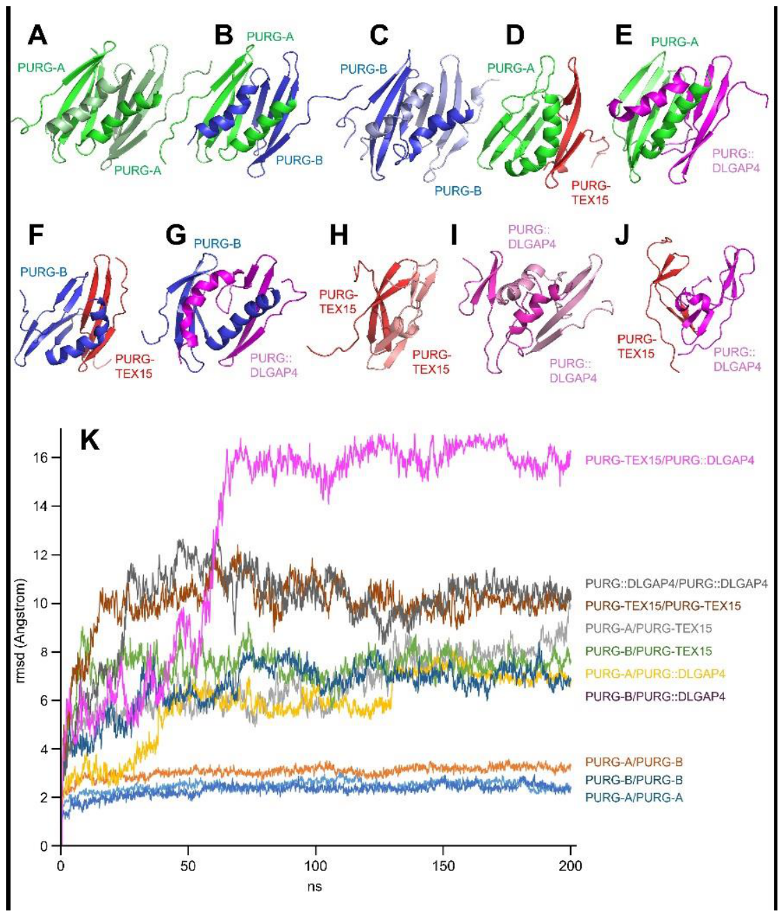
To investigate the impact of chimeric isoforms on PURG dimerization, homology models of the PUR repeat III dimerization domains were generated for PURG-A, PURG-B, and the chimeric isoforms PURG-TEX15 and PURG::DLGAP4 using the dimeric fragment of human PURA as a template (Protein Data Bank ID: 8CHW) [27]. The models were subjected to molecular dynamics (MD) simulations along 200 ns trajectories, and structural stability changes were monitored by root mean square deviation (RMSD) analysis (Figure 7).
Dimers composed solely of the isoforms (PURG-A/PURG-A, PURG-A/PURG-B, and PURG-B/PURG-B) remained highly stable throughout the simulations, with RMSD values ranging from 2.0 to 3.0 Angstroms (Figure 7A-C, K, respectively). In contrast, heterodimers between the wild-type forms (PURG-A or PURG-B) and the chimeric forms (PURG-TEX15 or PURG::DLGAP4) were more unstructured, with RMSD values of approximately 7.0 to 8.0 Angstroms (Figure 2K).
The simulation of dimers containing only the chimeric variants showed even greater disorganization after 200 ns of MD (Figure 7, panels 7H, 7I, and 7J). The RMSD values for these trajectories range from approximately 10.0 to 11.0 Angstroms for homodimers (Figure 2K). The PURG-TEX15/PURG::DLGAP4 heterodimer is particularly unstable, with an RMSD value exceeding 15.0 Angstroms (Figure 2K). These results suggest that both chimeric variants destabilize PURG dimerization. PURG-TEX15 may have a completely different function than PURG-A/B, whereas PURG::DLGAP4 may cause incorrect dimerization and impaired protein function in cells expressing this isoform.
Discussion
This study aimed to identify a potential disease gene or a mechanism for early-onset, mild cerebellar ataxia phenotype segregating with a balanced translocation t(8;20). Previous studies have reported strong linkage between the translocation and the disease. We have also shown that the breakpoint on derivative 20 disrupts the promoter region of the brain-specific DLGAP4 transcript [11,12]. Here, we focused on the chromosome 8 breakpoint to elucidate any disrupted genes or mechanisms due to the breakpoint to unravel the molecular mechanism underlying the phenotype. We identified a brain-specific conjoined gene, PURG-TEX15, spanning the breakpoint on chromosome 8p, and a fusion gene, PURG::DLGAP4, formed by the translocation. Both chimeric genes can encode a stable protein (Figure 2). This study provides new insights into the molecular complexity and plausible consequences of a balanced translocation and a chimeric transcript potentially contributing to neuronal development, maturation, and pathogenesis in cerebellar ataxia.
Chimeric transcripts, escaping nonsense-mediated decay, are expressed in normal tissues in a cell- and stage-specific dynamic manner during development, especially in the brain, where there are already 1400 identified conjoined genes at the RNA level [28,29,30,31,32]. Here, we found a chimeric gene, PURG-TEX15, with distinct expression in specific brain regions (Figure 4), indicating a specific functional role for PURG-TEX15 in the central nervous system.
The PURG-TEX15 transcript is composed of the second-to-last exon of PURG and the second exon of TEX15 (Figure 2A), consistent with the known structure of conjoined genes [1,32,33]. Although PURG and TEX15 are relatively far apart, being localized in the same topologically associating domain may promote their interaction and conjoining; yet other mechanisms underlying the formation and regulation of PURG-TEX15 cannot be excluded [2,34]. Given that some chimeric transcripts encode proteins with functions distinct from the parental proteins [35,36,37,38], we investigated whether PURG-TEX15 and PURG::DLGAP4 could produce stable proteins. We showed that PURG-TEX15 encodes a novel protein with high homology to other annotated PURG isoforms, and that the chimeric transcript can be translated into a stable PURG-TEX15 protein in human cells. PURG::DLGAP4, which is specific to this case, also produced a stable protein in the HEK cell model (Figure 3).
Before this study, PURG-TEX15 had not been identified in humans, but an annotated PURG isoform in Macaca fascicularis (UniProt accession number: A0A2K5VC47 [39] has a C-terminal sequence highly similar to PURG-TEX15. This suggests that this protein is also expressed in Macaca brain. Although TEX15 and PURG are evolutionarily conserved, no PURG-TEX15 transcript or protein with a similar C-terminal sequence has been reported in non-primate mammals, and Purg-Tex15 could not be amplified in mice. This is not unexpected, as both genes reside within a primate-specific evolutionary inverted region [40].
The differences in expression patterns between PURG and PURG-TEX15 (Figure 4) may point to a function more independent of the parental genes. In the present study, we investigated the connection between PURG-TEX15 and brain function and whether the translocation t(8:20) that disrupted the conjoined gene could contribute to the ataxia phenotype. We showed that the PURG-TEX15 transcript is expressed in specific parts of the human adult cerebellum (Figure 4C), whereas the human fetal cerebellum does not express PURG-TEX15 (Figure 4B). In the adult cerebellum, it is highly expressed and is likely transported along the dendrites of Purkinje cells. Interestingly, we observed PURG-TEX15 as single dense structures attached to the perinuclear region of granule neurons or glial cells of the cerebellum (Figure 4C). They could be mature RNA leaving the nucleus but if these mark single contact points between different cerebellar cell types, then a mechanistic RNA activity needs to be elucidated [41]. PURG-TEX15 is predominantly expressed in the adult brain, suggesting a more specific role in maturing neurons rather than early brain development. Disruption of PURG-TEX15 may affect the age of onset of mild cerebellar ataxia in translocation carriers and supports the possibility that it causes mild symptoms by impairing cerebellar neuron function after differentiation. [42].
Chimeric genes can increase phenotypic diversity in several ways [2,35,43]. One possible mechanism is the regulation of the expression or function of the parental genes [5,44]. Disrupting PURG-TEX15 might dysregulate PURG or TEX15. Examination of Tex15 and Purg showed that both genes are expressed in the adult mouse cerebellum (Figure 5 & S1). TEX15 is implicated in human reproduction, with mutations linking TEX15 to male infertility [45,46,47,48]. Here, we have shown that TEX15 is expressed in different brain areas, including the cerebellum (Figure 4A & S1). Because there is no direct evidence for TEX15's role in neurodevelopment or neurological disease, and because the TEX15 portion of the PURG-TEX15 protein involves only an untranslated exon of TEX15, the significance of TEX15 for the ataxia phenotype remains speculative.
The biological role of PURG remains poorly understood because no direct functional studies or disease-associated variants have been reported so far. PURG has a high sequence homology with its paralog PURA [17]. Pura knockout mice exhibit severe neurodevelopmental abnormalities [49,50]. In humans, PURA mutations cause PURA syndrome characterized by severe neurodevelopmental phenotypes and movement disorders [51,52]. PURA is active in hippocampal dendrites where it is involved in mRNA transport and translation [53]. Our results show that Purg is also expressed in mouse hippocampal neurons. Localization of the protein along the beta-tubulin-positive neuronal processes but not on the punctate structure of Psd-95 suggests that Purg is not a core protein of the postsynaptic density (Figure 5A). The distribution suggests a role in neurite growth and stability, and in maintaining neuronal architecture. With these findings, we raise the possibility that PURG, like PURA, can bind nucleotides and proteins [17] and can also have a role in mRNA trafficking, local translation, and synapse formation in dendrites and axons.
In addition to its neuronal expression, we found that when recombinantly expressed in HEK cells, PURG proteins localized predominantly to the cytoplasm and formed bright, dense, round perinuclear aggregates in a portion of the cells (Figure 6B). The nature and function of these structures are yet to be known [54,55].
The main difference between PURG-A and PURG-TEX15 is the truncation of the third PUR domain repeat (PURIII) in PURG-TEX15 (Figure 3C). As PURIII is critical for homodimerization in PURA and highly conserved among PUR family members, it is likely to play a similar role in PURG [17,27,56]. Consistent with this, our computational analyses predicted reduced dimerization of PURG-TEX15 compared with other PURG variants (Figure 7), suggesting a different function. If PURG-TEX15 has a distinct function, as our results suggest, disruption of the PURG-TEX15 conjoined gene and a subsequent decrease in protein expression could directly affect the phenotype. Therefore, disruption of the PURG-TEX15 conjoined gene and reduced protein expression may contribute to the ataxia phenotype, although its biological significance remains to be determined.
Identifying the PURG-TEX15 transcript led us to investigate new genetic arrangements created by the t(8:20) translocation, and we found that the PURG::DLGAP4 fusion gene can express the PURG::DLGAP4 fusion protein. Recombinant expression in HEK cells showed that the PURG::DLGAP4 protein is stably expressed and has a higher molecular weight than other PURG protein variants. Fusion proteins are shown to cause severe disease morphologies in patients with breakpoints before [57,58,59]. PURG::DLGAP4 may also be expressed in certain brain regions, replacing PURG-TEX15. Although its expression in t(8:20) patients has not been demonstrated, PURG::DLGAP4 protein could also be pathogenic and contribute to the cerebellar ataxia phenotype observed in t(8:20) carriers. Computational simulations predict that PURG::DLGAP4 is the least stable among the PURG complexes and has the potential to alter dimerization and protein function. PURG::DLGAP4/PURG-TEX15 heterodimer is predicted to be particularly unstable in the cells expressing both fusion genes.
In summary, we have identified two brain-specific and potentially protein-coding chimeric genes, PURG-TEX15 and PURG::DLGAP4, through studying the disrupted region of the genome at the breakpoints of a balanced familial translocation t(8;20). We suggest that disruption of PURG-TEX15, along with the formation of a translocation-specific fusion protein, may directly affect or modify PURG- and/or TEX15-dependent molecular pathways in neurons and lead to mild, early-onset, non-progressive cerebellar ataxia in carriers of t(8;20). This study also introduces a new layer of complexity in PURG gene regulation in the brain. Further investigation of PURG isoforms, and whether PURG-TEX15 is involved in neurodevelopment, will be essential to elucidate the extent to which disruption of PURG-TEX15 contributes to or explains the mild cerebellar ataxia phenotype.
Materials and Methods
Cells
Mouse hippocampal neurons were prepared from E18 mouse embryos of either sex (C57/BL6, Charles River) as published elsewhere [60]. Animal use was in accordance with the guidelines of the Danish Veterinary and Food Administration and the Ministry
of Food, Agriculture and Fisheries of Denmark, and the procedure was approved by the Department of Experimental Medicine at the University of Copenhagen. The neurons were cultured in Neurobasal medium supplemented with B27 (ThermoFisher, cat no. 17504044), 10 IU/ml penicillin-streptomycin, and 5 mM Glutamax (ThermoFisher, cat no. 35050061) and kept in a heat chamber at 37°C with 5% CO2.
HEK293 cells (ATCC #CRL-1573) were grown in Dulbecco's Modified Eagle's Medium supplemented with 2 mM L-glutamine, 10% foetal bovine serum, 100 U/ml penicillin and 100 µg/ml streptomycin (growth medium) at 37°C in a humidified atmosphere of 5% CO2. One day before transfection, cells were seeded into 6-well cell culture plates (600,000 cells per well), with or without coverslips, and grown to 80% confluence. They were later used for transfection and recombinant expression.
Quantitative PCR and Sanger Sequencing
RNA from different tissues was purchased from different vendors: Research Genetics, Ambion, Stratagene, BioChain Institute, and Clontech, or isolated from IsoHD-30Q-NGN2
(WAe009-A-1P) healthy human embryonic stem cell line that can be differentiated into neurons [61].
CDNA was prepared with Superscript II Reverse Transcriptase (Invitrogen, cat. no. 18064014). We used the cDNA to quantify gene expression with Brilliant III UltraFast SYBR Green Master Mix (Agilent Technologies, cat. no. 600882). The samples were run in duplicates on a 7500 fast real-time PCR system (ThermoFischer) with 7500 fast system v.1.3.1 software, and the qPCR data were normalized with the ΔCt method against three housekeeping genes, human: GAPDH, M-RIP and POLR2F, mouse: Gapdh, Psmd4 and Hprt (Table S1). Touchdown PCR was performed using DNA polymerase (VWR, cat no. 5101600) on cDNA. The PCR was run on a 2720 thermal cycler PCR machine (Applied Biosystems). To analyze the PCR products, they were run on an agarose gel (SeaKem, cat. no. 50004) and visualized on a GeneGenius (Syngene, India) using GeneSnap v.7.09 software. See supplementary table S1 for primer sequences. We sequenced PCR products by Sanger sequencing. First, we performed a forward and reverse PCR using Big Dye mastermix (Applied Biosystems, cat no. 4337449) with the PCR primers in Table S1, followed by sequencing on 3130xl genetic analyser (Applied Biosystems) with genetic analyser data collection software v.3.0. The sequences were analysed using Unipro UGENE software v.1.26.
Public RNA-sequencing datasets
Publicly available RNA-seq GEO datasets: GSE63513, GSE62098 and ENCODE: ENCFF730CJE, ENCFF883NIZ, ENCFF325NGX and ENCFF463ZNL [24] were extracted from the Gene Expression Omnibus (https://www.ncbi.nlm.nih.gov/gds). Sequences were analysed with HISAT2 v2.0.5 and RNA-seq data were visualized with SAMtools v1.3. IGV v.2.3.86.
Recombinant expression of PURG isoforms
DNA fragments encoding different PURG transcripts were amplified by PCR from cDNA derived from human differentiated neuron mRNA (PURG-A and PURG-TEX15) or synthesized de novo by Eurofins Genomics (PURG::DLGAP4, PURG-A-HA, PURG-TEX15-His, PURG::DLGAP4-His). Each gene strand was then ligated into a pcDNA™3.1+ mammalian expression vector (Invitrogen™, Thermo Fisher Scientific).
For recombinant expression, the expression vectors containing an appropriate insert were transiently transfected into HEK293 cells by lipofection with Metafectene® (Biontex) according to the manufacturer's protocols. The transfected cells were harvested after 48 hours. To determine steady-state levels of the recombinant proteins, cells were washed in phosphate-buffered saline (PBS) and lysed in PBS containing 1% Triton X-100 and 0.5% deoxycholate (lysis buffer). Lysates were centrifuged at 20 000×gat 4°C for 20 min, and the supernatants were transferred to a new tube.
To obtain nuclear and cytosolic fractions, cells were diluted in 100 µl PBS, lysed, and fractionated using the NuCLEAR™ Extraction Kit (Sigma-Aldrich®) according to the manufacturer's protocol, with one modification to the nuclear fraction. Before the final centrifugation and collection of the nuclear fraction, the samples were sonicated (2 × 5 s, 30%) to dissociate DNA-binding proteins from DNA.
SDS-PAGE and Western blot
Samples were prepared by addition of loading buffer (to final concentrations of 1 mM Tris–HCl pH 6.8, 0.1% SDS, 1% glycerol, 0.1 mM EDTA, 0.005% bromophenol blue) and, for reducing conditions, 0.2 M dithiothreitol (DTT), followed by incubation at 98°C for 5 min. Proteins were separated on 10% Tris-glycine SDS polyacrylamide gels and transferred to Immobilon-P PVDF membranes (Millipore). The blots were blocked with 10% non-fat dry milk in PBS containing 0.1% Tween-20 (PBS-T) for 1 h at room temperature. Membranes were probed with primary antibodies in 1% non-fat dry milk in PBS-T at 4°C overnight, followed by 3 × 5 min washes in PBS-T and 1 h incubation with appropriate secondary antibodies in 1% non-fat dry milk in PBS-T at room temperature. After 3 × 5 min washes in PBS-T, the signal was developed using the WesternBright™ Sirius Western blotting detection kit (cat. no. 12043, advansta) according to the protocol provided by the manufacturer and visualised with a Fuji LAS-1000 camera and Intelligent DarkBox II (FujiFilm Sweden AB), using the program LAS1000 Lite v1.5.)
In situ hybridizations, Immunocytochemistry, immunohistochemistry
In situ hybridization and immunocytochemistry assays were performed on human fetal and adult tissue sections, mouse hippocampal neurons, or mouse brain sections.
Human brain tissue samples, including paraffin-embedded sections from brains from a 19-week post-conception fetus and adult cerebellum, were acquired from the Human Embryonic/Fetal Biobank, Institute of Cellular and Molecular Medicine, Faculty of Health and Medical Sciences, University of Copenhagen, Denmark, described in detail in [62] or bought commercially from Capital Biosciences, Rockville, Maryland. Mouse brain sections were obtained from adult BALB/c mice.
Mouse hippocampal neurons were fixed with 2 % paraformaldehyde for 2 minutes, followed by 10 minutes with ice-cold (-20°C) methanol. To block nonspecific binding, the neurons were treated with PBS containing 0.2% fish skin gelatine and 0.1% Triton X-100. Neurons were incubated for 1 hour with primary antibodies. Subsequently, the neurons were stained for 45 minutes with AlexaFluor®-coupled secondary antibodies (ThermoFisher).
HEK293 cells were seeded onto polylysine-coated glass coverslips 24 hours prior to transfection. Transiently transfected HEK cells were fixed with 4 % paraformaldehyde after 48 hours. Immunocytochemistry was performed according to standard protocols as described above.
In situ hybridizations were conducted on human adult and fetal cerebellum sections as described elsewhere [63] using the following 5´- and 3 ′-FAM-conjugated oligonucleotide probes.
PURG probe: 5’-ATCGTCTCCACCTCTTCGTTCTTCTATGTCTCCTTCGCCA-3’,
PURG-TEX15 probe: 5’-TGAAAACTGATAAGGGAACATCTTCAGGAAAATTCCA-3,
TEX15 probe: 5’-CAGAAAATTGGCTTTGAGAGCACCGACGCATCAGGCACA-3’.
Image acquisition
Images of hippocampal neurons and PURA-transfected HEK cells were captured using a laser scanning confocal microscope (LSM710, Zeiss) equipped with a 63x, 1.4 NA, oil-immersion objective. The pinhole was 1 Airy unit, and the pixel format was 1024x1024. Images of in situ hybridization and immunohistochemistry experiments were acquired either using a Macroview microscope (MVX10, Olympus) equipped with F-view black&white, colour-view cameras with Cell^P software or an Olympus IX81 inverted microscope equipped with an Andor iXon EMCCD camera and Xcellence Software. All images were processed in Fiji (Fiji_Is_Just_ImageJ) image software.
Structural modeling and molecular dynamics of PURG dimerization domain
To investigate the effect of chimeric isoforms on PURG dimerization, we generated models of the dimerization domains (PUR repeat III fragment) for both the wild-type isoforms PURG-A and PURG-B and the chimeric isoforms PURG-TEX15 and PURG::DLGAP4. The 3D structures of human homo- or heterodimers of wild-type and chimeric isoforms of PURG dimerization domains (PUR repeat III) were generated using the crystal structure of the corresponding dimeric fragment of human PURA (Protein Data Bank id: 8CHW; [27] as a template). The structure corresponding to the amino acid sequence of the PUR repeat III fragment (Figure 7A) of the wild-type PURG-A and PURG-B isoforms (Figure 7B), as well as the chimeric isoforms PURG-TEX15 and PURG::DLGAP4 (Figure 7B), was modeled using homology modeling techniques with the SWISS-MODEL server (https://swissmodel.expasy.org). The quality of the models was assessed using standard methods for protein structure evaluation (QMEANDisCo; [64]. In total, we generated 10 constructs, corresponding to all possible combinations among the four variants: PURG-A/PURG-A, PURG-A/PURG-B, PURG-B/PURG-B, PURG-A/PURG-TEX15, PURG-A/PURG::DLGAP4, PURG-B/PURG-TEX15, PURG-B/PURG::DLGAP4, PURG-TEX15/PURG-TEX15, PURG-TEX15/PURG::DLGAP4, and PURG::DLGAP4/PURG::DLGAP4.
Once modeled, the coordinates corresponding to the wild-type and chimeric isoforms of PURG dimerization domains were subjected to 200 ns of unrestricted MD simulation using the Amber18 package (https://ambermd.org; University of California-San Francisco, CA), as previously described [65]. In brief, the models were first solvated with a periodic octahedral pre-equilibrated solvent box using the LEaP module of Amber18, with 12 Å as the shortest distance between any atom in the protein subdomain and the periodic box boundaries. MD simulations were conducted employing the Particle Mesh Ewald (PME) method for non-bonded interactions with a cutoff distance of 8 Å. The temperature was regulated through the Langevin thermostat, maintaining a fixed temperature of 297 K with a collision frequency of 1 ps. Hydrogen bond constraints were implemented using the SHAKE algorithm, enabling a simulation timestep of 2 fs. To uphold NPT conditions (constant number of particles, pressure, and temperature), the pressure coupling was managed by a Monte Carlo Barostat set to 297 K and 1 bar. The initial model structures underwent 10000 cycles of energy minimization, followed by a 1 ns restrained equilibration phase, smoothly raising the temperature to 297 K, after which restraints were gradually removed over 10 ns. Subsequently, each system was subjected to a 200 ns long free MD production phase. Trajectories were analyzed using cpptraj [66] and VMD [67]. Figures were generated with PyMOL (https://pymol.org).
Data analysis
For in silico translation, we used the EMBOSS transeq command-line software v.6.6. We generated the PURG protein alignment using Clustal Omega Multiple Sequence Alignment by EMBL-EBI (https://www.ebi.ac.uk/jdispatcher/msa/clustalo; [25]) with sequences from PURG-A (NP_037489.1), PURG-B (NP_001310241.1), PURG-TEX15, and PURG::DLGAP4. The TAD structure around the TEX15 and PURG genes in neuronal cells was identified using the 3D Genome Browser 2.0 (https://3dgenome.fsm.northwestern.edu/) [68].
Figure design
Author Contributions
AS conceptualized the project. MM, AHR, and AS wrote the original draft. AHR, MM, SC, MBL, PF-V, and IM-A performed experiments; JMH provided clinical information; PG-P, HBR, and LKV contributed with ideas, supervised students in this project, and reviewed and edited the manuscript. All authors reviewed the final manuscript and agreed to submit a submission to Genes.
Funding
The project is supported by Lundbeck Foundation (Vort nr. R67-A6431) and Læge Sofus Carl Emil Friis og hustru Olga Doris Foundation (AS). This work was supported partially by the Spanish government grant PID2024-161968OB-I00 (MICIU/AEI/10.13039/501100011033/ FEDER, EU) (PG-P).
Data Availability Statement
The sequence is available at European Nucleotide Archive ENA under project identifier PRJEB100969 (ERP182393).
Acknowledgments
We thank technicians Annemette Friis Hansen, Ha Nguyen, Linda Boje Dalsgaard, Lillian Egeberg, Pernille Froh, Rabap Nima, and Stine M. Christensen for their excellent technical support. Finally, we acknowledge the support provided by the members of the Core Facility for Integrated Bioimaging at the University of Copenhagen. The computational support of the "Centro de Computación Científica CCC-UAM" is gratefully recognized.
Conflicts of Interest
There are no conflicts of interest.
References
- Akiva, P.; Toporik, A.; Edelheit, S.; Peretz, Y.; Diber, A.; Shemesh, R.; Novik, A.; Sorek, R. Transcription-Mediated Gene Fusion in the Human Genome. Genome Res. 2006, 16, 30–36. [Google Scholar] [CrossRef] [PubMed]
- Chwalenia, K.; Facemire, L.; Li, H. Chimeric RNAs in Cancer and Normal Physiology. Wiley Interdiscip. Rev. RNA 2017, 8. [Google Scholar] [CrossRef] [PubMed]
- Zhuo, J.-S.; Jing, X.-Y.; Du, X.; Yang, X.-Q. Generation of Chimeric RNAs by Cis-Splicing of Adjacent Genes (Cis-SAGe) in Mammals. Yi Chuan 2018, 40, 145–154. [Google Scholar] [CrossRef] [PubMed]
- Jividen, K.; Li, H. Chimeric RNAs Generated by Intergenic Splicing in Normal and Cancer Cells. Genes Chromosom. Cancer 2014, 53, 963–971. [Google Scholar] [CrossRef] [PubMed]
- Dhungel, B.P.; Monteuuis, G.; Giardina, C.; Tabar, M.S.; Feng, Y.; Metierre, C.; Ho, S.; Nagarajah, R.; Fontaine, A.R.M.; Shah, J.S.; et al. The Fusion of CLEC12A and MIR223HG Arises from a Trans-Splicing Event in Normal and Transformed Human Cells. Int. J. Mol. Sci. 2021, 22. [Google Scholar] [CrossRef] [PubMed]
- Honda, S.; Hatamura, M.; Kunimoto, Y.; Ikeda, S.; Minami, N. Chimeric PRMT6 Protein Produced by an Endogenous Retrovirus Promoter Regulates Cell Fate Decision in Mouse Preimplantation Embryos†. Biol. Reprod. 2024, 110, 698–710. [Google Scholar] [CrossRef] [PubMed]
- Qin, F.; Zhang, Y.; Liu, J.; Li, H. SLC45A3-ELK4 Functions as a Long Non-Coding Chimeric RNA. Cancer Lett. 2017, 404, 53–61. [Google Scholar] [CrossRef] [PubMed]
- Wu, C.S.; Yu, C.Y.; Chuang, C.Y.; Hsiao, M.; Kao, C.F.; Kuo, H.C.; Chuang, T.J. Integrative Transcriptome Sequencing Identifies Trans-Splicing Events with Important Roles in Human Embryonic Stem Cell Pluripotency. Genome Res. 2014, 24, 25–36. [Google Scholar] [CrossRef] [PubMed]
- Backx, L.; Seuntjens, E.; Devriendt, K.; Vermeesch, J.; Van Esch, H. A Balanced Translocation t(6;14)(Q25.3;Q13.2) Leading to Reciprocal Fusion Transcripts in a Patient with Intellectual Disability and Agenesis of Corpus Callosum. Cytogenet. Genome Res. 2011, 132, 135–143. [Google Scholar] [CrossRef] [PubMed]
- Glenfield, C.; Innan, H. Gene Duplication and Gene Fusion Are Important Drivers of Tumourigenesis during Cancer Evolution. Genes 2021, 12. [Google Scholar] [CrossRef] [PubMed]
- Hertz, J.M.; Sivertsen, B.; Silahtaroglu, A.; Bugge, M.; Kalscheuer, V.; Weber, A.; Wirth, J.; Ropers, H.H.; Tommerup, N.; Tümer, Z. Early Onset, Non-Progressive, Mild Cerebellar Ataxia Co-Segregating with a Familial Balanced Translocation t(8;20)(P22;Q13). J. Med. Genet. 2004, 41. [Google Scholar] [CrossRef] [PubMed]
- Minocherhomji, S.; Hansen, C.; Kim, H.G.; Mang, Y.; Bak, M.; Guldberg, P.; Papadopoulos, N.; Eiberg, H.; Doh, G.D.; Møllgård, K.; et al. Epigenetic Remodelling and Dysregulation of DLGAP4 Is Linked with Early-Onset Cerebellar Ataxia. Hum. Mol. Genet. 2014, 23, 6163–6176. [Google Scholar] [CrossRef] [PubMed]
- Romero, D.M.; Poirier, K.; Belvindrah, R.; Moutkine, I.; Houllier, A.; LeMoing, A.G.; Petit, F.; Boland, A.; Collins, S.C.; Soiza-Reilly, M.; et al. Novel Role of the Synaptic Scaffold Protein Dlgap4 in Ventricular Surface Integrity and Neuronal Migration during Cortical Development. Nat. Commun. 2022, 13. [Google Scholar] [CrossRef] [PubMed]
- Wang, T.; Bai, Y.; Zheng, X.; Liu, X.; Xing, S.; Wang, L.; Wang, H.; Feng, G.; Li, C. Sapap4 Deficiency Leads to Postsynaptic Defects and Abnormal Behaviors Relevant to Hyperkinetic Neuropsychiatric Disorder in Mice. Cereb. Cortex 2023, 33, 1104–1118. [Google Scholar] [CrossRef] [PubMed]
- Minocherhomji, S.; Seemann, S.; Mang, Y.; El-Schich, Z.; Bak, M.; Hansen, C.; Papadopoulos, N.; Josefsen, K.; Nielsen, H.; Gorodkin, J.; et al. Sequence and Expression Analysis of Gaps in Human Chromosome 20. Nucleic Acids Res. 2012, 40, 6660–6672. [Google Scholar] [CrossRef] [PubMed]
- Daniel, D.C.; Johnson, E.M. PURA, the Gene Encoding Pur-Alpha, Member of an Ancient Nucleic Acid-Binding Protein Family with Mammalian Neurological Functions. Gene 2018, 643, 133–143. [Google Scholar] [CrossRef] [PubMed]
- Johnson, E.M.; Daniel, D.C.; Gordon, J. The Pur Protein Family: Genetic and Structural Features in Development and Disease. J. Cell. Physiol. 2013, 228, 930–937. [Google Scholar] [CrossRef] [PubMed]
- Kaminski, R.; Darbinyan, A.; Merabova, N.; Deshmane, S.L.; White, M.K.; Khalili, K. Protective Role of Purα to Cisplatin. Cancer Biol. Ther. 2008, 7, 1926–1935. [Google Scholar] [CrossRef] [PubMed]
- Wang, H.; Wang, M.; Reiss, K.; Darbinian-Sarkissian, N.; Johnson, E.M.; Iliakis, G.; Amini, S.; Khalili, K.; Rappaport, J. Evidence for the Involvement of Puralpha in Response to DNA Replication Stress. Cancer Biol. Ther. 2007, 6, 596–602. [Google Scholar] [CrossRef] [PubMed]
- Wang, H.; White, M.K.; Kaminski, R.; Darbinian, N.; Amini, S.; Johnson, E.M.; Khalili, K.; Rappaport, J. Role of Puralpha in the Modulation of Homologous Recombination-Directed DNA Repair by HIV-1 Tat. Anticancer Res. 2008, 28, 1441–1447. [Google Scholar] [PubMed]
- Molitor, L.; Bacher, S.; Burczyk, S.; Niessing, D. The Molecular Function of PURA and Its Implications in Neurological Diseases. Front. Genet. 2021, 12. [Google Scholar] [CrossRef] [PubMed]
- Mellone, S.; Spano, A.; Vurchio, D.; Borgonovi, G.; Ugonotti, A.; Paglino, G.; Bianco, A.; Ronzani, S.; Sciancalepore, M.; Prodam, F.; et al. Integrating Genome and Transcriptome Analysis to Decipher Balanced Structural Variants in Unsolved Cases of Neurodevelopmental Disorders. Front. Genet. 2025, 16. [Google Scholar] [CrossRef] [PubMed]
- Clough, E.; Barrett, T.; Wilhite, S.E.; Ledoux, P.; Evangelista, C.; Kim, I.F.; Tomashevsky, M.; Marshall, K.A.; Phillippy, K.H.; Sherman, P.M.; et al. NCBI GEO: Archive for Gene Expression and Epigenomics Data Sets: 23-Year Update. Nucleic Acids Res. 2023, 52, D138. [Google Scholar] [CrossRef] [PubMed]
- Li, J.; Shi, M.; Ma, Z.; Zhao, S.; Euskirchen, G.; Ziskin, J.; Urban, A.; Hallmayer, J.; Snyder, M. Integrated Systems Analysis Reveals a Molecular Network Underlying Autism Spectrum Disorders. Mol. Syst. Biol. 2014, 10. [Google Scholar] [CrossRef] [PubMed]
- Madeira, F.; Madhusoodanan, N.; Lee, J.; Eusebi, A.; Niewielska, A.; Tivey, A.R.N.; Lopez, R.; Butcher, S. The EMBL-EBI Job Dispatcher Sequence Analysis Tools Framework in 2024. Nucleic Acids Res. 2024, 52, W521–W525. [Google Scholar] [CrossRef] [PubMed]
- Chitale, G.G.; Kulkarni, S.R.; Bapat, S.A. Chimerism: A Whole New Perspective in Gene Regulation. Biochim. Biophys. Acta Gen. Subj. 2025, 1869. [Google Scholar] [CrossRef] [PubMed]
- Proske, M.; Janowski, R.; Bacher, S.; Kang, H.S.; Monecke, T.; Koehler, T.; Hutten, S.; Tretter, J.; Crois, A.; Molitor, L.; et al. PURA Syndrome-Causing Mutations Impair PUR-Domain Integrity and Affect P-Body Association. Elife 2024, 13. [Google Scholar] [CrossRef] [PubMed]
- Prakash, T.; Sharma, V.K.; Adati, N.; Ozawa, R.; Takahashi, T.; Druker, B.J.; Michalak, P. Expression of Conjoined Genes: Another Mechanism for Gene Regulation in Eukaryotes. PLoS ONE 2010, 5, e13284. [Google Scholar] [CrossRef] [PubMed]
- Singh, S.; Qin, F.; Kumar, S.; Elfman, J.; Lin, E.; Pham, L.-P.; Yang, A.; Li, H. The Landscape of Chimeric RNAs in Non-Diseased Tissues and Cells. Nucleic Acids Res. 2020, 48, 1764–1778. [Google Scholar] [CrossRef] [PubMed]
- Moses, C.; Vanini, A.; Bocchi, M.C.; Wagner, L.; Van Loenen, I.; Latour, B.L.; Kasri, N.N.; Jacobs, F.M.J. Evolutionary Hotspots of Structural Variation Drive Inter-Individual Differences in the Expression of Fusion Transcripts in the Human Brain. Nucleic Acids Res. 2025, 53. [Google Scholar] [CrossRef] [PubMed]
- Ou, M.Y.; Xiao, Q.; Ju, X.C.; Zeng, P.M.; Huang, J.; Sheng, A.L.; Luo, Z.G. The CTNNBIP1-CLSTN1 Fusion Transcript Regulates Human Neocortical Development. Cell Rep. 2021, 35. [Google Scholar] [CrossRef] [PubMed]
- Tang, Y.; Ma, S.; Wang, X.; Xing, Q.; Huang, T.; Liu, H.; Li, Q.; Zhang, Y.; Zhang, K.; Yao, M.; et al. Identification of Chimeric RNAs in Human Infant Brains and Their Implications in Neural Differentiation. Int. J. Biochem. Cell Biol. 2019, 111, 19–26. [Google Scholar] [CrossRef] [PubMed]
- Kim, H.-P.; Cho, G.-A.; Han, S.-W.; Shin, J.-Y.; Jeong, E.-G.; Song, S.-H.; Lee, W.-C.; Lee, K.-H.; Bang, D.; Seo, J.-S.; et al. Novel Fusion Transcripts in Human Gastric Cancer Revealed by Transcriptome Analysis. Oncogene 2014, 33, 5434–5441. [Google Scholar] [CrossRef] [PubMed]
- Sridhar, A.; More, A.S.; Jadhav, A.R.; Patil, K.; Mavlankar, A.; Dixit, V.M.; Bapat, S.A. Pattern Recognition in the Landscape of Seemingly Random Chimeric Transcripts. Comput. Struct. Biotechnol. J. 2023, 21, 5153–5164. [Google Scholar] [CrossRef] [PubMed]
- Babiceanu, M.; Qin, F.; Xie, Z.; Jia, Y.; Lopez, K.; Janus, N.; Facemire, L.; Kumar, S.; Pang, Y.; Qi, Y.; et al. Recurrent Chimeric Fusion RNAs in Non-Cancer Tissues and Cells. Nucleic Acids Res. 2016, 44, 2859. [Google Scholar] [CrossRef] [PubMed]
- Li, H.; Wang, J.; Mor, G.; Sklar, J. A Neoplastic Gene Fusion Mimics Trans-Splicing of RNAs in Normal Human Cells. Science (1979) 2008, 321, 1357–1361. [Google Scholar] [CrossRef]
- Schmitt-Ney, M.; Camussi, G. The PAX3-FOXO1 Fusion Protein Present in Rhabdomyosarcoma Interferes with Normal FOXO Activity and the TGF-β Pathway. PLoS ONE 2015, 10, e0121474. [Google Scholar] [CrossRef] [PubMed]
- Venezia, O.; Kane, H.; Du, G.; Coughlan, H.D.; Johanson, T.M.; Wang, H.; Tilstam, P.; Kazer, S.W.; Gunner, G.; Hoagland, D.A.; et al. Functional Chimeric MRNAs Encode Proteins in Mammalian Immunity. Nature 2026, 1, 1–10. [Google Scholar] [CrossRef] [PubMed]
- Bateman, A.; Martin, M.J.; Orchard, S.; Magrane, M.; Adesina, A.; Ahmad, S.; Bowler-Barnett, E.H.; Bye-A-Jee, H.; Carpentier, D.; Denny, P.; et al. UniProt: The Universal Protein Knowledgebase in 2025. Nucleic Acids Res. 2025, 53, D609–D617. [Google Scholar] [CrossRef] [PubMed]
- Pébusque, M.J.; Coulier, F.; Birnbaum, D.; Pontarotti, P. Ancient Large-Scale Genome Duplications: Phylogenetic and Linkage Analyses Shed Light on Chordate Genome Evolution. Mol. Biol. Evol. 1998, 15, 1145–1159. [Google Scholar] [CrossRef] [PubMed]
- Kim, J.; Augustine, G.J. Molecular Layer Interneurons: Key Elements of Cerebellar Network Computation and Behavior. Neuroscience 2021, 462, 22–35. [Google Scholar] [CrossRef] [PubMed]
- Robinson, K.J.; Watchon, M.; Laird, A.S. Aberrant Cerebellar Circuitry in the Spinocerebellar Ataxias. Front. Neurosci. 2020, 14, 707. [Google Scholar] [CrossRef] [PubMed]
- Elfman, J.; Li, H. Chimeric RNA in Cancer and Stem Cell Differentiation. Stem Cells Int. 2018. [Google Scholar] [CrossRef] [PubMed]
- Choi, E.S.; Lee, H.; Lee, C.H.; Goh, S.H. Overexpression of KLHL23 Protein from Read-through Transcription of PHOSPHO2-KLHL23 in Gastric Cancer Increases Cell Proliferation. FEBS Open Bio 2016, 6, 1155–1164. [Google Scholar] [CrossRef] [PubMed]
- Colombo, R.; Pontoglio, A.; Bini, M. Two Novel TEX15 Mutations in a Family with Nonobstructive Azoospermia. Gynecol. Obstet. Invest. 2017, 82, 283–286. [Google Scholar] [CrossRef] [PubMed]
- Okutman, O.; Muller, J.; Baert, Y.; Serdarogullari, M.; Gultomruk, M.; Piton, A.; Rombaut, C.; Benkhalifa, M.; Teletin, M.; Skory, V.; et al. Exome Sequencing Reveals a Nonsense Mutation in TEX15 Causing Spermatogenic Failure in a Turkish Family. Hum. Mol. Genet. 2015, 24, 5581–5588. [Google Scholar] [CrossRef] [PubMed]
- Ruan, J.; He, X.-J.; Du, W.-D.; Chen, G.; Zhou, Y.; Xu, S.; Zuo, X.-B.; Fang, L.-B.; Cao, Y.-X.; Zhang, X.-J. Genetic Variants in TEX15 Gene Conferred Susceptibility to Spermatogenic Failure in the Chinese Han Population. Reprod. Sci. 2012, 19, 1190–1196. [Google Scholar] [CrossRef] [PubMed]
- Zhang, H.G.; Wang, R.X.; Li, L.L.; Sun, W.T.; Zhang, H.Y.; Liu, R.Z. Male Carriers of Balanced Reciprocal Translocations in Northeast China: Sperm Count, Reproductive Performance, and Genetic Counseling. Genet. Mol. Res. 2015, 14, 18792–18798. [Google Scholar] [CrossRef] [PubMed]
- Khalili, K.; Del Valle, L.; Muralidharan, V.; Gault, W.J.; Darbinian, N.; Otte, J.; Meier, E.; Johnson, E.M.; Daniel, D.C.; Kinoshita, Y.; et al. Purα Is Essential for Postnatal Brain Development and Developmentally Coupled Cellular Proliferation As Revealed by Genetic Inactivation in the Mouse. Mol. Cell. Biol. 2003, 23, 6857. [Google Scholar] [CrossRef] [PubMed]
- Barbe, M.F.; Krueger, J.J.; Loomis, R.; Otte, J.; Gordon, J. Memory Deficits, Gait Ataxia and Neuronal Loss in the Hippocampus and Cerebellum in Mice That Are Heterozygous for Pur-Alpha. Neuroscience 2016, 337, 177. [Google Scholar] [CrossRef] [PubMed]
- Reijnders, M.R.F.; Janowski, R.; Alvi, M.; Self, J.E.; van Essen, T.J.; Vreeburg, M.; Rouhl, R.P.W.; Stevens, S.J.C.; Stegmann, A.P.A.; Schieving, J.; et al. PURA Syndrome: Clinical Delineation and Genotype-Phenotype Study in 32 Individuals with Review of Published Literature. J. Med. Genet. 2018, 55, 104–113. [Google Scholar] [CrossRef] [PubMed]
- Afshar, S.; Ahmed, W.; Çelik, E.Ö.; Younis, S.S.; Campbell, C.; Potluri, Y.K.R.; Kaur, J.; Meda, D.S.; Manjari, K.K.; Khachikian, A.; et al. PURA-Related Neurodevelopmental Disorder: A Comprehensive Clinical Review of Genetics, Phenotype, and Emerging Therapeutic Strategies. Cureus 2026, 18, e105181. [Google Scholar] [CrossRef] [PubMed]
- Johnson, E.M.; Kinoshita, Y.; Weinreb, D.B.; Wortman, M.J.; Simon, R.; Khalili, K.; Winckler, B.; Gordon, J. Role of Purα in Targeting MRNA to Sites of Translation in Hippocampal Neuronal Dendrites. J. Neurosci. Res. 2006, 83, 929–943. [Google Scholar] [CrossRef] [PubMed]
- Formicola, N.; Vijayakumar, J.; Besse, F. Neuronal Ribonucleoprotein Granules: Dynamic Sensors of Localized Signals. Traffic 2019, 20, 639–649. [Google Scholar] [CrossRef] [PubMed]
- Bauer, K.E.; de Queiroz, B.R.; Kiebler, M.A.; Besse, F. RNA Granules in Neuronal Plasticity and Disease. Trends Neurosci. 2023, 46, 525–538. [Google Scholar] [CrossRef] [PubMed]
- Graebsch, A.; Roche, S.; Niessing, D. X-Ray Structure of Pur-α Reveals a Whirly-like Fold and an Unusual Nucleic-Acid Binding Surface. Proc. Natl. Acad. Sci. U. S. A. 2009, 106, 18521–18526. [Google Scholar] [CrossRef] [PubMed]
- Greco, A.; Fusetti, L.; Miranda, C.; Villa, R.; Zanotti, S.; Pagliardini, S.; Pierotti, M.A. Role of the TFG N-Terminus and Coiled-Coil Domain in the Transforming Activity of the Thyroid TRK-T3 Oncogene. Oncogene 1998, 16, 809–816. [Google Scholar] [CrossRef] [PubMed]
- Greco, A.; Mariani, C.; Miranda, C.; Lupas, A.; Pagliardini, S.; Pomati, M.; Pierotti, M.A. The DNA Rearrangement That Generates the TRK-T3 Oncogene Involves a Novel Gene on Chromosome 3 Whose Product Has a Potential Coiled-Coil Domain. Mol. Cell. Biol. 1995, 15, 6118–6127. [Google Scholar] [CrossRef] [PubMed]
- Roccato, E.; Miranda, C.; Ranzi, V.; Gishizki, M.; Pierotti, M.A.; Greco, A. Biological Activity of the Thyroid TRK-T3 Oncogene Requires Signalling through Shc. Br. J. Cancer 2002, 87, 645–653. [Google Scholar] [CrossRef] [PubMed]
- Kaech, S.; Banker, G. Culturing Hippocampal Neurons. Nat. Protoc. 2006, 1, 2406–2415. [Google Scholar] [CrossRef] [PubMed]
- Villegas, L.D.; Chandrasekaran, A.; Andersen, S.A.F.; Nørremølle, A.; Schmid, B.; Pouladi, M.A.; Freude, K. Generation of Three Isogenic Gene-Edited Huntington’s Disease Human Embryonic Stem Cell Lines with DOX-Inducible NGN2 Expression Cassette in the AAVS1 Safe Locus. Stem Cell Res. 2024, 77. [Google Scholar] [CrossRef] [PubMed]
- Holst, C.B.; Brøchner, C.B.; Vitting-Seerup, K.; Møllgård, K. Astrogliogenesis in Human Fetal Brain: Complex Spatiotemporal Immunoreactivity Patterns of GFAP, S100, AQP4 and YKL-40. J. Anat. 2019, 235, 590. [Google Scholar] [CrossRef] [PubMed]
- Silahtaroglu, A.N.; Nolting, D.; Dyrskjøt, L.; Berezikov, E.; Møller, M.; Tommerup, N.; Kauppinen, S. Detection of MicroRNAs in Frozen Tissue Sections by Fluorescence in Situ Hybridization Using Locked Nucleic Acid Probes and Tyramide Signal Amplification. Nat. Protoc. 2007, 2, 2520–2528. [Google Scholar] [CrossRef] [PubMed]
- Studer, G.; Rempfer, C.; Waterhouse, A.M.; Gumienny, R.; Haas, J.; Schwede, T. QMEANDisCo—Distance Constraints Applied on Model Quality Estimation. Bioinformatics 2020, 36, 1765–1771. [Google Scholar] [CrossRef] [PubMed]
- Ros-Pardo, D.; Gómez-Puertas, P.; Marcos-Alcalde, Í. STAG2: Computational Analysis of Missense Variants Involved in Disease. Int. J. Mol. Sci. 2024, 25, 1280. [Google Scholar] [CrossRef]
- Roe, D.R.; Cheatham, T.E. PTRAJ and CPPTRAJ: Software for Processing and Analysis of Molecular Dynamics Trajectory Data. J. Chem. Theory Comput. 2013, 9, 3084–3095. [Google Scholar] [CrossRef] [PubMed]
- Humphrey, W.; Dalke, A.; Schulten, K. VMD: Visual Molecular Dynamics. J. Mol. Graph. 1996, 14, 33–38. [Google Scholar] [CrossRef] [PubMed]
- Yu, S.; Fu, Y.; Wong, J.H.Y.; Wang, J.; Zhao, H.; Zhao, J.; Yue, F. The 3D Genome Browser 2.0: An Enhanced Online Platform for Visualizing and Analyzing 3D Genome Architecture. Nucleic Acids Res. 2026, 54, D48–D54. [Google Scholar] [CrossRef] [PubMed]
Figure 1.
Identification of the PURG-TEX15 conjoined gene. A) The two top panels display RNA-seq data from the human adult amygdala and corpus callosum (GSE63513 and GSE62098) (Gene Expression Omnibus, https://www.ncbi.nlm.nih.gov/gds) [24], respectively. In both datasets, the RNA-seq reads span the PURG gene, the intergenic region, and extend into the TEX15 gene. The third panel shows Sanger sequencing results for PURG-TEX15 from the human brain, along with the locations of PURG-A, -B, and TEX15 (Genome Browser; https://genome.ucsc.edu/cgi-bin/hgGateway). The t(8:20) breakpoint is located in the intronic region of PURG-TEX15, disrupting the transcript. Panel 3 also shows the location of the far-most primers for Sanger sequencing. B) Sashimi plot of RNA-seq reads from the PURG gene through the TEX15 gene from human adult amygdala (red) and corpus callosum (blue). The first exon of PURG-TEX15 skips the second exon of PURG-B and the first exon of the new TEX15.
Figure 1.
Identification of the PURG-TEX15 conjoined gene. A) The two top panels display RNA-seq data from the human adult amygdala and corpus callosum (GSE63513 and GSE62098) (Gene Expression Omnibus, https://www.ncbi.nlm.nih.gov/gds) [24], respectively. In both datasets, the RNA-seq reads span the PURG gene, the intergenic region, and extend into the TEX15 gene. The third panel shows Sanger sequencing results for PURG-TEX15 from the human brain, along with the locations of PURG-A, -B, and TEX15 (Genome Browser; https://genome.ucsc.edu/cgi-bin/hgGateway). The t(8:20) breakpoint is located in the intronic region of PURG-TEX15, disrupting the transcript. Panel 3 also shows the location of the far-most primers for Sanger sequencing. B) Sashimi plot of RNA-seq reads from the PURG gene through the TEX15 gene from human adult amygdala (red) and corpus callosum (blue). The first exon of PURG-TEX15 skips the second exon of PURG-B and the first exon of the new TEX15.
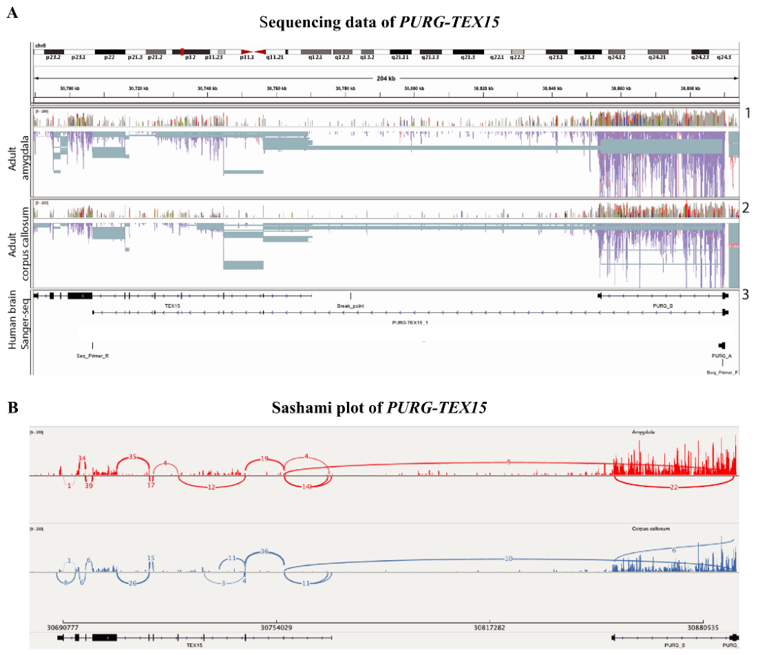
Figure 2.
Structural model of PURG-TEX15 and PURG::DLGAP4. A) Schematic representation of the genomic region of PURG and TEX15 and the conjoined PURG-TEX15 gene. The cis-splicing between adjacent genes (cis-SAGE) uses canonical splicing sites in the first exon of PURG and the second exon of TEX15. The distance between the two splicing sites is ~133 kb, and the distance between the two genes is ~82 kb. The PURG-TEX15 transcript contains an open reading frame encoding a new PURG protein variant with 298 amino acids. B) Schematic overview of the genomic regions around the breakpoints of the aforementioned t(8;20) translocation. On the left, the regions in a wild-type genome are depicted, including the intact PURG and TEX15 locus on chromosome 8 and the intact DLGAP4 gene on chromosome 20. On the right, the rearranged translocation-specific regions of the genome are depicted, including a new PURG and DLGAP4 locus on chromosome derivative 8 and a new DLGAP4 and TEX15 locus on chromosome derivative 20. C) Schematic representation of the translocation-specific genomic region of PURG and DLGAP4 and the PURG::DLGAP4 gene. The cis-splicing between adjacent genes (cis-SAGE) is predicted to use canonical splicing sites in the first exon of PURG and the second exon of DLGAP4. The PURG::DLGAP4 transcript contains an open reading frame encoding a new PURG protein variant with 378 amino acids.
Figure 2.
Structural model of PURG-TEX15 and PURG::DLGAP4. A) Schematic representation of the genomic region of PURG and TEX15 and the conjoined PURG-TEX15 gene. The cis-splicing between adjacent genes (cis-SAGE) uses canonical splicing sites in the first exon of PURG and the second exon of TEX15. The distance between the two splicing sites is ~133 kb, and the distance between the two genes is ~82 kb. The PURG-TEX15 transcript contains an open reading frame encoding a new PURG protein variant with 298 amino acids. B) Schematic overview of the genomic regions around the breakpoints of the aforementioned t(8;20) translocation. On the left, the regions in a wild-type genome are depicted, including the intact PURG and TEX15 locus on chromosome 8 and the intact DLGAP4 gene on chromosome 20. On the right, the rearranged translocation-specific regions of the genome are depicted, including a new PURG and DLGAP4 locus on chromosome derivative 8 and a new DLGAP4 and TEX15 locus on chromosome derivative 20. C) Schematic representation of the translocation-specific genomic region of PURG and DLGAP4 and the PURG::DLGAP4 gene. The cis-splicing between adjacent genes (cis-SAGE) is predicted to use canonical splicing sites in the first exon of PURG and the second exon of DLGAP4. The PURG::DLGAP4 transcript contains an open reading frame encoding a new PURG protein variant with 378 amino acids.
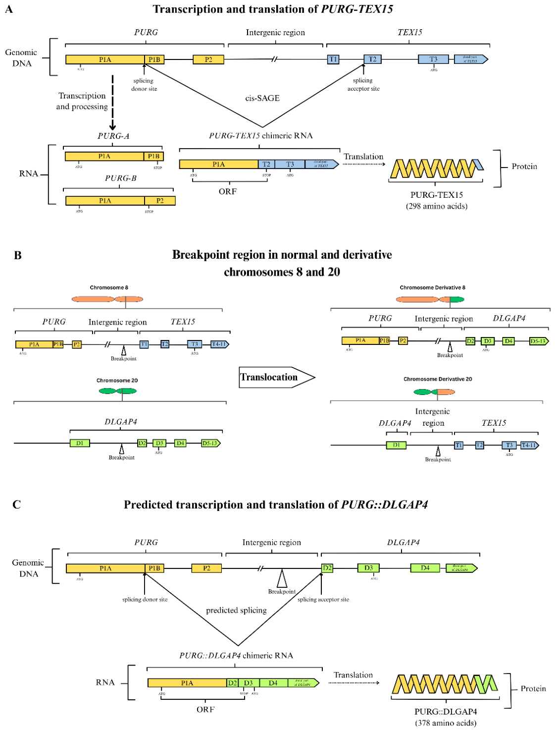
Figure 3.
PURG protein alignments, expression, and homology. A) Clustal Omega alignment [25] of PURG-A, PURB-B, PURG-TEX15, and PURG::DLGAP4 proteins shows that their sequences are mostly homologous but vary at the C-terminal end. B) PURG-A, PURG-TEX15, and PURG::DLGAP4 recombinant expression leads to the detection of proteins with distinct sizes of ca. 40, 38, and 46 kDa, respectively, on a Western blot. HEK293 cells were transfected with PURG-A cDNA (lane 1), PURG-TEX15 cDNA (lane 3), and PURG-DLGAP4 synthetic DNA (lane 5), respectively. The same DNA fragments in an inverted orientation were used as a negative control (lanes 2, 4 and 6). We used a primary polyclonal antibody against a peptide homologous to a PURG sequence, as indicated on the left. Primary antibodies against H3K27 and GAPDH were used as controls. Stars show bands due to endogenous proteins of unknown identity. C) Structural model of different PUR proteins. All PUR proteins contain 3 repeats of the conserved PUR domain. The 3rd repeat is truncated in PURG proteins with alternative splicing, including PURG-B, PURG-TEX15 and PURG::DLGAP4. The antibody used in the Western blot above binds to a sequence upstream of the first repeat of the PUR domain in all known PURG proteins.
Figure 3.
PURG protein alignments, expression, and homology. A) Clustal Omega alignment [25] of PURG-A, PURB-B, PURG-TEX15, and PURG::DLGAP4 proteins shows that their sequences are mostly homologous but vary at the C-terminal end. B) PURG-A, PURG-TEX15, and PURG::DLGAP4 recombinant expression leads to the detection of proteins with distinct sizes of ca. 40, 38, and 46 kDa, respectively, on a Western blot. HEK293 cells were transfected with PURG-A cDNA (lane 1), PURG-TEX15 cDNA (lane 3), and PURG-DLGAP4 synthetic DNA (lane 5), respectively. The same DNA fragments in an inverted orientation were used as a negative control (lanes 2, 4 and 6). We used a primary polyclonal antibody against a peptide homologous to a PURG sequence, as indicated on the left. Primary antibodies against H3K27 and GAPDH were used as controls. Stars show bands due to endogenous proteins of unknown identity. C) Structural model of different PUR proteins. All PUR proteins contain 3 repeats of the conserved PUR domain. The 3rd repeat is truncated in PURG proteins with alternative splicing, including PURG-B, PURG-TEX15 and PURG::DLGAP4. The antibody used in the Western blot above binds to a sequence upstream of the first repeat of the PUR domain in all known PURG proteins.
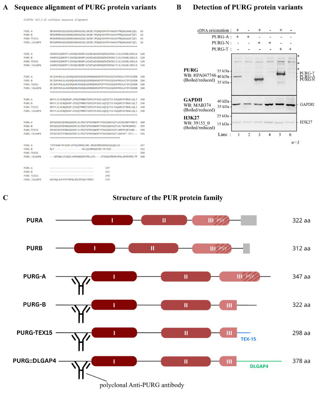
Figure 4.
Expression of PURG-TEX15 in the human brain. A) Bar plot of TEX15, PURG-A, PURG-B, and PURG-TEX15 qPCR data from a panel of human tissues. B) Images from in situ hybridization of PURG and PURG-TEX15 in human fetal cerebellum (19-week fetus), respectively. PURG-TEX15 is not expressed in the human fetal brain, including the cerebellum, whereas PURG is expressed in neuroepithelial progenitor cells in the ventricular zone of the developing cerebellar cortex; scale bars: 1mm. C) In situ hybridization of PURG-TEX15 (red) in human adult cerebellum. Note that PURG-TEX15 is expressed in (Top) large axon terminals as well as in Purkinje cells; (Bottom left) dendrite of a Purkinje cell. Granular cells (blue) do not express PURG-TEX15 (Bottom right), yet small signals were attached to each nucleus (indicated by grey arrows), which could represent contact points between Purkinje cells and interneurons, which are also visible in the top-right picture. GL: granular layer, ML: molecular layer, PL: Purkinje cell layer, and PC: Purkinje cell. Scale bar: 100 µm.
Figure 4.
Expression of PURG-TEX15 in the human brain. A) Bar plot of TEX15, PURG-A, PURG-B, and PURG-TEX15 qPCR data from a panel of human tissues. B) Images from in situ hybridization of PURG and PURG-TEX15 in human fetal cerebellum (19-week fetus), respectively. PURG-TEX15 is not expressed in the human fetal brain, including the cerebellum, whereas PURG is expressed in neuroepithelial progenitor cells in the ventricular zone of the developing cerebellar cortex; scale bars: 1mm. C) In situ hybridization of PURG-TEX15 (red) in human adult cerebellum. Note that PURG-TEX15 is expressed in (Top) large axon terminals as well as in Purkinje cells; (Bottom left) dendrite of a Purkinje cell. Granular cells (blue) do not express PURG-TEX15 (Bottom right), yet small signals were attached to each nucleus (indicated by grey arrows), which could represent contact points between Purkinje cells and interneurons, which are also visible in the top-right picture. GL: granular layer, ML: molecular layer, PL: Purkinje cell layer, and PC: Purkinje cell. Scale bar: 100 µm.
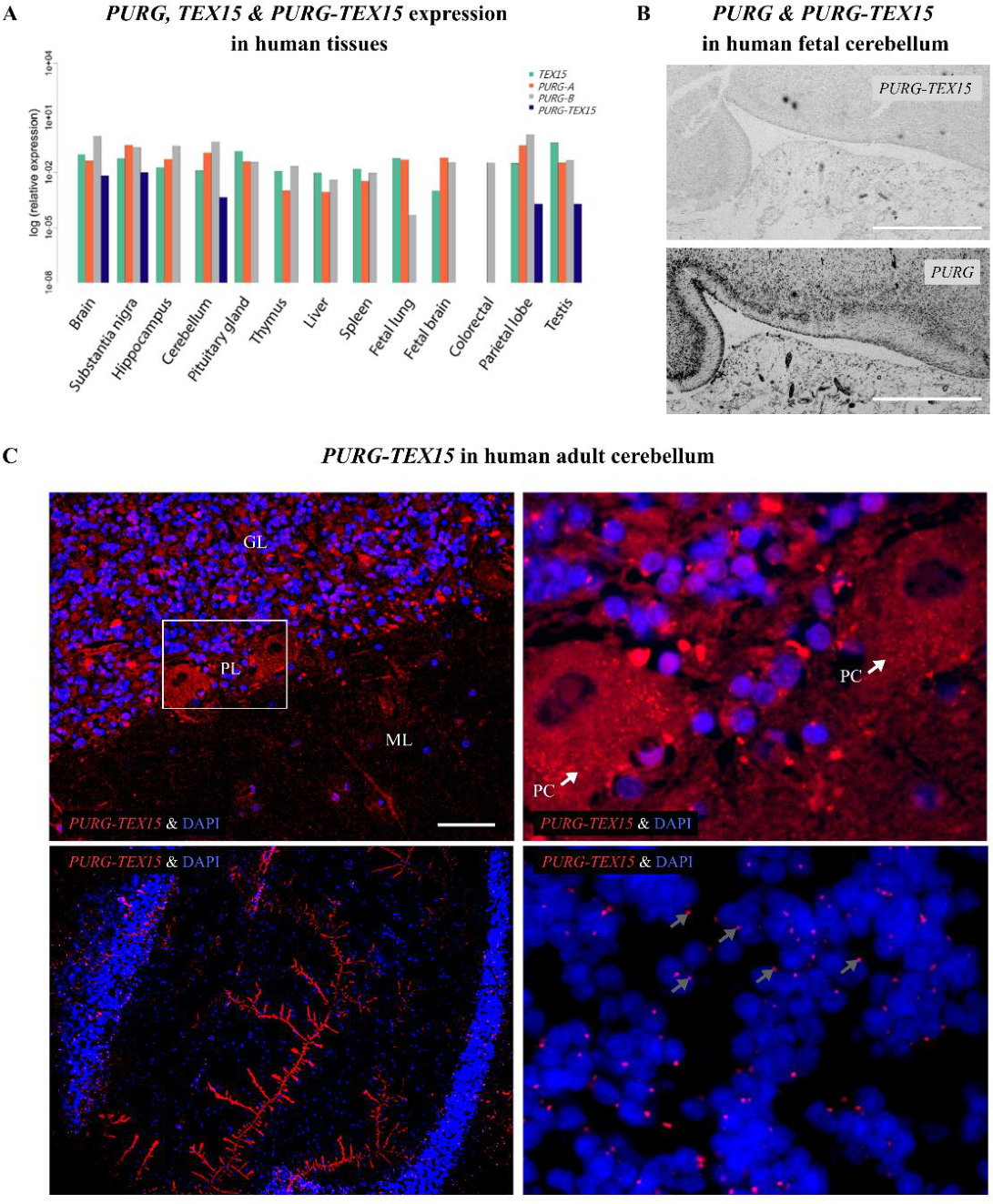
Figure 5.
Expression of Purg in the mouse brain. A) Mouse cultured hippocampal neurons co-immunolabelled for Purg and PSD-95 (top) or Purg and tubulin (bottom). Purg is expressed in the soma, axons, and dendrites, suggesting a role in transport. The white boxes highlight the zoomed regions displayed to the right. Purg is expressed along tubulin-positive neurites with no significant enrichment in the postsynaptic density, as indicated by the absence of colocalization with PSD-95. Scale bars: 15 µm. B) Immunohistochemistry in an adult mouse cerebellum with an antibody against Purg (green) showing expression in the Purkinje cell layer. GL: granular layer; ML: molecular layer; PC: Purkinje cell. Scale bar: 800 µm.
Figure 5.
Expression of Purg in the mouse brain. A) Mouse cultured hippocampal neurons co-immunolabelled for Purg and PSD-95 (top) or Purg and tubulin (bottom). Purg is expressed in the soma, axons, and dendrites, suggesting a role in transport. The white boxes highlight the zoomed regions displayed to the right. Purg is expressed along tubulin-positive neurites with no significant enrichment in the postsynaptic density, as indicated by the absence of colocalization with PSD-95. Scale bars: 15 µm. B) Immunohistochemistry in an adult mouse cerebellum with an antibody against Purg (green) showing expression in the Purkinje cell layer. GL: granular layer; ML: molecular layer; PC: Purkinje cell. Scale bar: 800 µm.
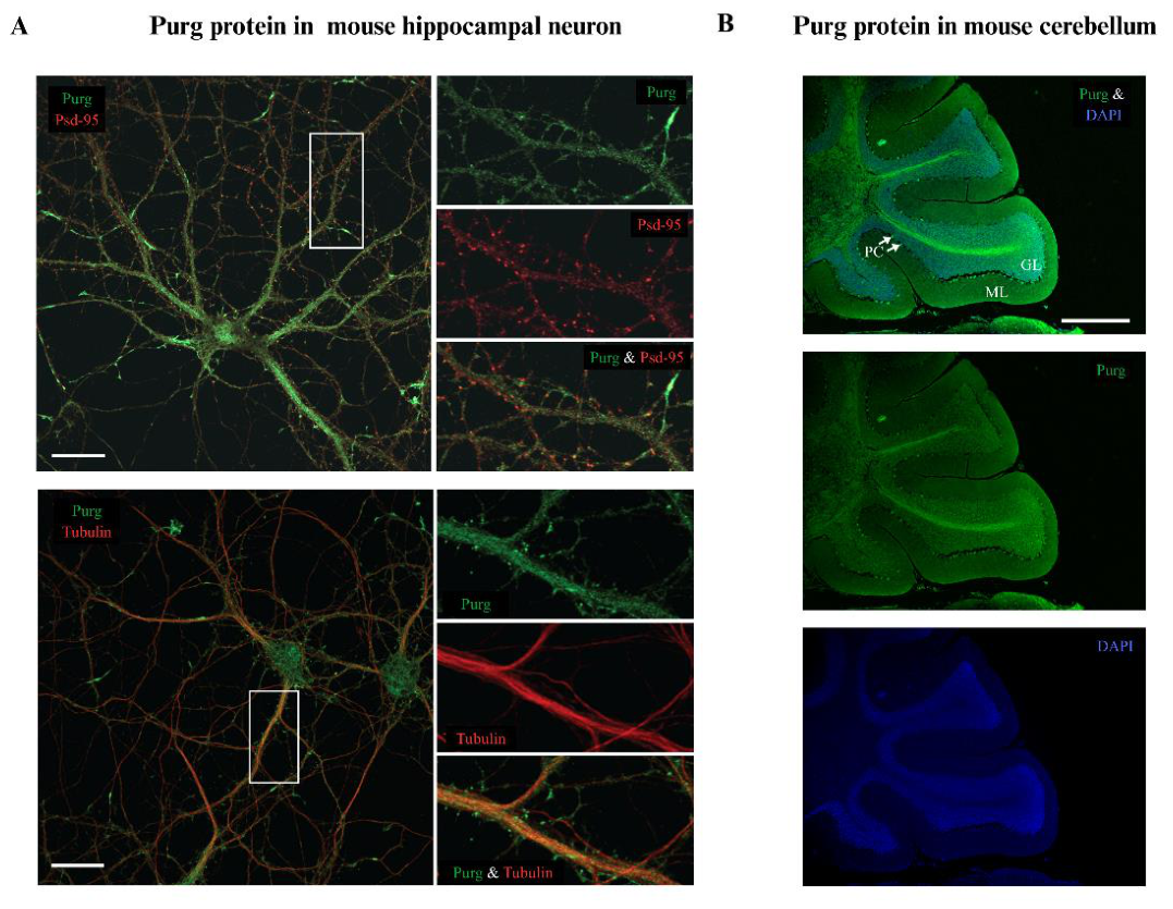
Figure 6.
Subcellular localization of PURG-A, PURG-TEX15, and PURG::DLGAP4. A) PURG-A, PURG-TEX15, and PURG-DLGAP4 recombinant expression leads to the detection of proteins in the cytosol and the nucleus on a Western blot. Recombinant PURG-A (WB1) was predominantly detected in the cytosol (lanes 2 & 4). Recombinant PURG-TEX15 (WB2) was detected in the cytosol (lanes 2 & 4) and nucleus (lanes 1 & 3) with similar intensity. Recombinant PURG::DLGAP4 (WB3) was detected in the cytosol (lanes 4 & 6) and the nucleus (lanes 3 & 5) with similar intensities. Tagged versions of the proteins followed the same distribution pattern as the untagged versions. HEK293 cells were transfected with tagged and untagged PURG-A DNA, PURG-TEX15 DNA, and PURG-DLGAP4 DNA, respectively. The PURG-A cDNA in inverted orientation and a mock transfection were used as negative controls. Primary antibodies against a peptide homologous to a PURG sequence, an HA-tag, and a His-tag were used as indicated on the left. Primary antibodies against Histone 3 and Vinculin were used as nuclear and cytosolic markers, respectively. The figure shows a representative experiment out of three. B) PURG-TEX15 (top left), PURG-A (top & center right), and PURG::DLGAP4 (center left) recombinant expression leads to the detection of the proteins in the cytosol (indicated by the grey arrows; center left), mostly as dense signals attached to the perinuclear membrane, as well as dim dotted signals in the nuclei (indicated by the white arrows; center right). Although these structures mostly accumulated in HEK293 cells transfected with PURG-TEX15 and PURG::DLGAP4, they are also visible in PURGA-transfected cells (top & center right). The bottom left shows how the transfected HEK293 cells look, and the bottom right shows the negative control: cells transfected with PURG-A in an inverted orientation.
Figure 6.
Subcellular localization of PURG-A, PURG-TEX15, and PURG::DLGAP4. A) PURG-A, PURG-TEX15, and PURG-DLGAP4 recombinant expression leads to the detection of proteins in the cytosol and the nucleus on a Western blot. Recombinant PURG-A (WB1) was predominantly detected in the cytosol (lanes 2 & 4). Recombinant PURG-TEX15 (WB2) was detected in the cytosol (lanes 2 & 4) and nucleus (lanes 1 & 3) with similar intensity. Recombinant PURG::DLGAP4 (WB3) was detected in the cytosol (lanes 4 & 6) and the nucleus (lanes 3 & 5) with similar intensities. Tagged versions of the proteins followed the same distribution pattern as the untagged versions. HEK293 cells were transfected with tagged and untagged PURG-A DNA, PURG-TEX15 DNA, and PURG-DLGAP4 DNA, respectively. The PURG-A cDNA in inverted orientation and a mock transfection were used as negative controls. Primary antibodies against a peptide homologous to a PURG sequence, an HA-tag, and a His-tag were used as indicated on the left. Primary antibodies against Histone 3 and Vinculin were used as nuclear and cytosolic markers, respectively. The figure shows a representative experiment out of three. B) PURG-TEX15 (top left), PURG-A (top & center right), and PURG::DLGAP4 (center left) recombinant expression leads to the detection of the proteins in the cytosol (indicated by the grey arrows; center left), mostly as dense signals attached to the perinuclear membrane, as well as dim dotted signals in the nuclei (indicated by the white arrows; center right). Although these structures mostly accumulated in HEK293 cells transfected with PURG-TEX15 and PURG::DLGAP4, they are also visible in PURGA-transfected cells (top & center right). The bottom left shows how the transfected HEK293 cells look, and the bottom right shows the negative control: cells transfected with PURG-A in an inverted orientation.
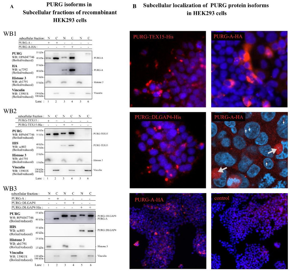
Figure 7.
Structure of PURG dimerization domain dimers after 200 ns of molecular dynamics simulation. Panels A through J show the structure of the dimer formed by two PUR repeat III fragments after the 200 ns molecular dynamics simulation, in the various possible combinations: A: PURG-A / PURG-A, B: PURG-A / PURG-B, C: PURG-B / PURG-B, D: PURG-A / PURG-TEX15, E: PURG-A / PURG::DLGAP4, F: PURG-B / PURG-TEX15, G: PURG-B / PURG::DLGAP4, H: PURG-TEX15 / PURG-TEX15, I: PURG-TEX15 / PURG::DLGAP4, J: PURG::DLGAP4 / PURG::DLGAP4. The stability difference between dimers containing only the wild-type isoforms (panels A, B, and C) is noticeable compared with the other combinations (panels D through J). K. RMSD values (root mean square deviation, in Angstroms) for the structures of the different dimers measured throughout the 200 ns molecular dynamics simulation. As in the panels above, the difference in stability between the wild-type isoform dimers (RMSD values around 2.0–3.0 Angstroms) and the other dimers (RMSD values greater than 6 Angstroms in all cases) is noticeable.
Figure 7.
Structure of PURG dimerization domain dimers after 200 ns of molecular dynamics simulation. Panels A through J show the structure of the dimer formed by two PUR repeat III fragments after the 200 ns molecular dynamics simulation, in the various possible combinations: A: PURG-A / PURG-A, B: PURG-A / PURG-B, C: PURG-B / PURG-B, D: PURG-A / PURG-TEX15, E: PURG-A / PURG::DLGAP4, F: PURG-B / PURG-TEX15, G: PURG-B / PURG::DLGAP4, H: PURG-TEX15 / PURG-TEX15, I: PURG-TEX15 / PURG::DLGAP4, J: PURG::DLGAP4 / PURG::DLGAP4. The stability difference between dimers containing only the wild-type isoforms (panels A, B, and C) is noticeable compared with the other combinations (panels D through J). K. RMSD values (root mean square deviation, in Angstroms) for the structures of the different dimers measured throughout the 200 ns molecular dynamics simulation. As in the panels above, the difference in stability between the wild-type isoform dimers (RMSD values around 2.0–3.0 Angstroms) and the other dimers (RMSD values greater than 6 Angstroms in all cases) is noticeable.
Disclaimer/Publisher’s Note: The statements, opinions and data contained in all publications are solely those of the individual author(s) and contributor(s) and not of MDPI and/or the editor(s). MDPI and/or the editor(s) disclaim responsibility for any injury to people or property resulting from any ideas, methods, instructions or products referred to in the content. |
© 2026 by the authors. Licensee MDPI, Basel, Switzerland. This article is an open access article distributed under the terms and conditions of the Creative Commons Attribution (CC BY) license (http://creativecommons.org/licenses/by/4.0/).
Copyright: This open access article is published under a Creative Commons CC BY 4.0 license, which permit the free download, distribution, and reuse, provided that the author and preprint are cited in any reuse.