

Submitted:
21 October 2026
Posted:
22 October 2026
You are already at the latest version
Abstract
Biomechanical analysis of human movement supports rehabilitation decisions requiring objective and reproducible measurements. However, motion capture technologies are commonly evaluated as separate technological families. This critical review compares marker-based optoelectronic, markerless, inertial, and hybrid systems in terms of accuracy, clinical validity, reliability, and implementation feasibility, and proposes methodological criteria for future validation studies. Scopus, Web of Science, PubMed, and IEEE Xplore were searched, supplemented by forward and backward citation searching from eight seed studies. The selection process was reported using a PRISMA 2020 flow diagram. No protocol was registered, and no formal risk-of-bias assessment was conducted. Eligible studies involved human participants in clinical, rehabilitation, or functional assessment settings and reported quantitative performance metrics. Evidence was synthesized by technological family and application scenario. Ninety-six documents were included. Marker-based systems provided the highest accuracy for joint kinematics, whereas markerless and inertial systems showed comparable validity for spatiotemporal variables and global movement characterization. However, errors in specific joint angles remained above clinically desirable thresholds. Calibration procedures and assessment duration substantially influenced performance. No technology was universally superior. Therefore, selection should be made by variable, task and population, using explicit validation criteria.
Keywords:
motion capture
; rehabilitation
; biomechanics
; markerless
; inertial sensors
; optoelectronic systems
; calibration
1. Introduction
In rehabilitation, movement assessment supports specific clinical decisions by establishing the patient’s functional baseline, quantifying progress across sessions, identifying compensatory strategies that either mask an underlying deficit or place excessive demands on other anatomical structures, and informing decisions regarding discharge or return to activity. Qualified clinical observation remains indispensable, but it is limited when these decisions require comparing measurements taken weeks or months apart, or when the magnitude of change is small relative to the variability of the observer itself [1,2]. Biomechanics, understood as the study of the movement of living beings through the principles of mechanics [3,4,5,6], provides at this point a quantitative framework that replaces appreciation with reproducible metrics.
Motion capture technologies are the instruments through which this framework becomes operational, as they allow spatiotemporal parameters, joint kinematics and movement patterns to be quantified. Marker-based optoelectronic systems, considered the reference standard, describe human movement with high spatial and temporal accuracy in controlled environments [7,8,9]. However, their integration into routine clinical care remains hindered by persistent barriers, including acquisition and maintenance costs, sensitivity to occlusion, the need for dedicated space, lengthy preparation and processing times, and reliance on specially trained personnel [10]. These constraints have driven the adoption of wireless inertial sensors [11,12], of markerless platforms based on computer vision and deep learning [13,14], and of hybrid fusion strategies that combine several sources to compensate for the limitations of each [15,16]. Markerless options now range from multi-camera commercial systems to open-source applications that estimate kinematics and dynamics from two or more smartphones [17], and consumer devices such as virtual-reality headsets have also been explored for upper-limb tracking in people with stroke and for postural sway [18,19].
The recent scientific output on these technologies is abundant, but it has been organized mostly in compartments. A first group of reviews addresses a single technological family. Inertial systems have been reviewed systematically for upper-limb kinematic analysis in clinical applications [11], for the estimation of kinetic variables with wearable devices, for the conversion of inertial data into upper-limb joint angles, for their concurrent validity in upper-limb range of motion and for rehabilitation applications [20-23]. The International Society of Biomechanics has also issued recommendations for defining, estimating and reporting joint kinematics with inertial sensors [24]. Markerless methods have reviews centered on human pose estimation, on the limitations of their application to clinical gait biomechanics, on clinical measurement in rehabilitation [14,25,26], on their comparison with marker-based systems in adult populations with pathology, on the accuracy, validity and reliability of three-dimensional markerless capture and on the reliability and validity of lower-limb and trunk kinematics [27-29]. Finally, the fusion of electromyographic and inertial signals has a specific review oriented to upper-limb pattern recognition [15].
A second group is organized by application domain rather than by technology. It includes the reviews on instrumented gait assessment in clinical practice, on balance assessment with force platforms and on lower-limb telerehabilitation [2,9,30,31]. These reviews offer a transversal view of their domain, but in them the comparison between technologies is subordinated to the clinical objective and is rarely addressed with homogeneous criteria.
A third set is formed by the validations of specific platforms against a reference system. Markerless systems have been compared with optical systems in walking at different speeds, in multiplanar movements such as squats, jumps and side-steps, in postural control and in a wide set of functional movements [32-35], and smartphone-based solutions have been validated in healthy and pathological gait [36]. Within this set, the commercial Theia3D system has been examined for inter-session repeatability, for day-to-day reliability, in healthy older adults and in pediatric patients with diverse diagnoses [37-40], while a deep-learning markerless system has been assessed for spatiotemporal gait parameters [41]. OpenCap has been studied for repeatability and minimal detectable change, inter-trial variability, the Timed Up and Go test and athletes after anterior cruciate ligament reconstruction [42-45], and single-smartphone approaches have been tested in rehabilitation tasks, with convolutional neural networks and in people with limb loss [46-48]. On the inertial side, the OpenSense toolbox [49] has been compared with marker-based capture in activities of daily living [50] and in young adults, older adults and older adults with knee osteoarthritis [51], and inertial models have been validated against optical references for the upper limb [52,53], the trunk and shoulders [54,55], and treadmill gait, with specific attention to sensor-to-segment calibration [56,57]. These studies provide the most precise error and agreement values available, but each remains circumscribed to one device, one protocol, one task and, frequently, a small sample of healthy people.
Several recent studies have started to enter clinical populations. Markerless, inertial and marker-based systems have been evaluated in people with stroke, cerebral palsy, multiple sclerosis, Parkinson's disease, cervical spinal cord injury, knee osteoarthritis, childhood toe-walking, limb loss and total hip arthroplasty [18,52,58-69]. Their results are not homogeneous, as the performance of markerless methods can be lower in patients than in controls and depends on the direction of walking and the distance relative to the camera [44,66]. Furthermore, Theia3D yielded lower Gait Deviation Index values than marker-based capture in children with cerebral palsy [61].
Five limitations of this configuration of the literature are addressed in the present work. The first is the absence of a transversal comparison between technological families on one common set of criteria. The available reviews show how one technology behaves against its reference standard, but not how all of them behave against the same clinical decision.
The second is the dissociation between technical accuracy and operational feasibility. Preparation time, calibration demands, required training or the possibility of home use condition the adoption of a technology as much as its angular error, and yet they are analyzed separately or mentioned only qualitatively [2,31].
The third limitation concerns the heterogeneity of the reported metrics. Different measures are used concurrently, including correlation coefficients, intraclass correlation coefficients, absolute and root-mean-square errors and Bland–Altman agreement analyses [32,33,70], applied to different variables, joints and planes. This heterogeneity hinders direct comparison between studies and invites erroneous interpretations, such as equating a high correlation with adequate agreement.
The fourth is the predominance of healthy participants. Most of the available evidence comes from samples without pathology, whereas studies in clinical populations, including people with stroke [71,72,73] or with other conditions that alter movement execution, such as obesity [74], remain insufficiently represented. This limits the generalization of the results, because the compensations and movement variability observed in these populations can degrade system performance.
The fifth is the scant attention to longitudinal and out-of-laboratory use. Between-session repeatability and temporal stability, which are decisive attributes for monitoring a rehabilitation process, receive much less treatment than accuracy in an isolated measurement [75,76]. The few studies that address them examine two sessions [38], test–retest agreement [42,69] or the number of trials needed for stable results [59].
The aim of this critical review is to analyze the state of the art of motion capture technologies applied to human rehabilitation, considering their accuracy, clinical validity, reliability and implementation feasibility, and to identify methodological criteria that guide the design of future experimental validation studies. To this end, five review questions are formulated in Section 2, which successively articulate the characterization of the technologies, their comparative performance, the conditioning factors of their implementation, the criteria for their selection and the requirements of future validation studies.
The distinctive contribution of this work is twofold. First, it offers a comparison between technological families built on common criteria that integrate metrological performance and operational feasibility in one framework. Second, it translates the knowledge gaps identified in the literature into methodological considerations for the design of validation studies, at a time when the authors' group has started the integration and calibration of an optoelectronic system and an inertial system with that purpose [77,78].
The article is organized as follows. Section 2 describes the methodology of search, selection, extraction and synthesis of the literature. Section 3 presents the results, based on the characterization of the technological families, their transversal comparison and the application scenarios in rehabilitation. Section 4 discusses emerging trends, the limitations of the available evidence and the implications for the design of future validation studies. Section 5 collects the conclusions.
2. Materials and Methods
2.1. Review Design and Research Questions
This study was conducted as a critical literature review supported by a structured literature search. Although the document identification and selection procedure is described in an explicit and reproducible manner, not all methodological elements associated with a systematic review were implemented or reported, including prospective protocol registration, independent duplicate screening, and quantitative risk-of-bias assessment. Accordingly, the evidence is synthesized through an interpretative approach focused on cross-sectional comparisons across technological families rather than on the statistical integration of published findings. The search and selection process was documented using the PRISMA 2020 flow diagram [79], adapted to incorporate an additional identification pathway based on citation searching (Figure 1), thereby improving the transparency, traceability, and reproducibility of the study selection process. The review was organized around five questions:
- What motion capture technologies are currently used in human rehabilitation, and for which biomechanical variables?
- What levels of accuracy, validity and reliability are reported for each of them against the systems taken as reference?
- What factors condition their implementation in clinical, community and home environments?
- What criteria should guide the selection of a technology according to the task, the population and the clinical purpose?
- What methodological elements should be incorporated into future experimental validation studies?
2.2. Information Sources and Search Strategy
The search was carried out in four bibliographic databases: Scopus, Web of Science, PubMed and IEEE Xplore. Google Scholar was used only as a complement, for tracking the citations of already included works and for locating full texts not accessible from the main databases. No systematic queries were run in it, so its results are not part of the count of identified records.
The combinations of terms were built on two conceptual axes: the capture technology and its clinical or biomechanical field of application. The resulting search strings are presented in Table 1 in their generic form, while the database-specific adaptations to the syntax and search fields are detailed in Table 2.
The searches were run between December 2025 and February 2026, and the last update of the strategy was made on 10 February 2026. In each database the following restrictions were applied, to the extent that its filtering system allowed (see Table 2): publication interval 2020–2025, English or Spanish language, engineering subject area and document type article, with preference for open-access documents. Works outside that interval were intentionally admitted when they ground the principles of stereophotogrammetry and the definition of anatomical reference systems [7,8], the biomechanics textbooks used as a conceptual framework [4,5,6] and the methodological antecedents needed to interpret sensor fusion techniques [16].
2.3. Eligibility Criteria
Studies were included when they simultaneously met four eligibility criteria. They involved human participants, were conducted in a clinical, rehabilitation or functional assessment context, explicitly employed one or more motion capture technologies, and reported quantitative metrics of performance, validity or reliability against a reference system or between sessions. Eight causes of exclusion were established and applied in the order given in Table 3.
2.4. Study Selection
The initial database search identified 4268 records (Scopus, 1231; Web of Science, 1243; PubMed, 976; IEEE Xplore, 818). Given the volume of results, the first 100 records of each database, in the relevance order provided by the database itself, were retained, giving a total of 400 records. After removing 43 duplicates, 357 records were screened by title and abstract, of which 214 were discarded. The remaining 143 were assessed in full text and 102 were excluded by applying the criteria of Section 2.3, so that 41 documents from the databases were incorporated into the review.
To broaden the evidence beyond the 100-record limit per database, the search was complemented with forward and backward citation searching in Scopus (see Table 4). Eight studies already identified in the database search, selected by the authors as representative of the main technological families, acted as seed studies [10,33,34,36,72,80,81,82]. For each seed, the list of citing documents (forward) and the list of its references (backward) were exported, giving 511 records (268 forward and 243 backward). After pooling the lists and removing 34 duplicates, 477 unique records remained. Ten records were already part of the manuscript, 143 were published outside the 2020–2025 interval (127 before 2020 and 16 in 2026) and 77 were not articles or reviews (conference papers, book chapters, editorials, data papers, letters or books), so 247 records were screened by title and abstract against the same criteria of Section 2.3. Of these, 43 were included after full-text reading, 64 were held in reserve as eligible but redundant or doubtful, and 140 were excluded. The citation searching was carried out in Scopus on 8 October 2026. Records published in 2026 were excluded in order to keep the same interval (2020–2025) as the database searches. The seed studies were selected by the authors for their relevance to validating markerless and inertial systems against marker-based references, which is the configuration of the authors' laboratory (Qualisys optoelectronic system and Xsens IMUs).
In addition, 10 documents identified through earlier citation tracking, among which are the seminal works and the textbooks that predate the search interval, and 2 documents from other sources were included, the technical documentation of the manufacturer of the reference optoelectronic system [77] and the previous communication of the authors describing the implementation of the laboratory [78]. The final set comprised 96 documents. With the addition of the PRISMA 2020 statement [79], which was cited as a methodological reference, the reference list contained 97 entries. The complete process is represented in Figure 1.
2.5. Data Extraction
Thirteen fields were extracted from each included study and recorded in a matrix prepared for this purpose. These fields comprised authors and year, type of population (healthy people or with pathology), sample size, clinical condition, when applicable, body region assessed, functional task analyzed, technology evaluated, system taken as reference, biomechanical variables considered; validity or reliability metrics reported, advantages indicated by the authors, recognized limitations, and application scenario. Data extraction was performed jointly by the three authors using a predefined extraction form. All authors reviewed every included study and discrepancies were resolved through discussion until consensus was reached.
This matrix is the basis of the comparative tables presented in Section 3 and allows each statement of the synthesis to be traced back to the study that supports it.
2.6. Assessment of the Evidence
No formal risk-of-bias assessment tool was applied. Instead, each study was characterized by four descriptors that directly condition the interpretation of its results. These descriptors comprised the nature of the sample (healthy people versus clinical population), the sample size, the existence of an independent reference system and the explicit description of the calibration procedure. These descriptors are used throughout the analysis to modulate the scope of the conclusions, so that results from a single protocol, a single task or a small sample are presented as such and not as general properties of the technology.
2.7. Synthesis
The synthesis was organized in three successive levels. In the first, studies were grouped by technological family (marker-based optoelectronic systems, markerless systems, inertial sensors, complementary sensors and hybrid approaches), and each family was characterized in terms of measurable variables, reported performance and operational requirements. In the second level, a transversal comparison between families was made on common criteria. These criteria included accuracy, intra- and inter-session reliability, preparation time, portability, calibration demands, sensitivity to occlusions and drift, relative cost and feasibility of use outside the laboratory. In the third, the findings were reorganized by application scenario, in order to derive technology selection criteria and, from the gaps identified, methodological considerations for the design of future validation studies.
Given the heterogeneity of protocols, biomechanical models, tasks and metrics reported in the included studies, no form of quantitative aggregation of results was considered appropriate. Numerical values are always presented together with the task, joint, population and reference system with which they were obtained.
2.8. Use of Generative Artificial Intelligence
Generative artificial intelligence (GenAI) was used in this work for specific supporting tasks. More specifically, an AI assistant (Claude Sonnet 5.5, Anthropic) was used to (i) review manuscript drafts prepared by the authors and assess their alignment with the journal’s required structure; (ii) consolidate and de-duplicate the Scopus exports obtained through citation searching and prepare a preliminary compilation of titles and abstracts for subsequent review by the authors; and (iii) generate preliminary versions of the flow diagram, which were subsequently reviewed and revised by the authors. The research direction, the design of the search strategy, the database searches, the exports for citation searching, the screening and the final selection of studies were carried out by the authors, who take full responsibility for the content of the article. No GenAI tool is listed as an author.
3. Results
3.1. Study Selection and Overview of the Evidence
The selection process is summarized in Figure 1. The database search yielded 41 studies and the other methods contributed 55 documents, so that the review comprises 96 documents. The technologies found in rehabilitation can be organized into five families, whose hierarchical classification is represented in Figure 2. Table 5, Table 6 and Table 7 gather the characteristics of the studies analyzed in the database search for each family, including population, sample size, task, reference system and reported metrics, and Table 8 summarizes the additional studies identified through citation searching.
The additional studies identified through citation searching (Table 8) shift the balance of the evidence in two directions. Most of them concern markerless systems (22 of 43), which confirms the pace at which this family is being validated, and a considerable share includes participants with pathology, such as stroke, cerebral palsy, multiple sclerosis, Parkinson's disease, spinal cord injury, knee osteoarthritis or limb loss. Eight are reviews, which complement the narrower reviews cited in the Introduction. Counts by family taken from the Excel of candidates; verify after full-text reading.
3.2. Characterization of the Motion Capture Technologies
3.2.1. Marker-Based Optoelectronic Systems
Systems based on reflective markers and infrared cameras constitute the methodological reference of the field [7,8,9]. Their operating principle, the stereophotogrammetric reconstruction of the position of each marker from its simultaneous visibility by at least two cameras, provides joint angles, ranges of motion and trajectories with high spatial and temporal resolution and, combined with force platforms, allows joint moments and powers to be estimated.
Their value in rehabilitation is seen in studies that quantify variables inaccessible to clinical observation. The characterization of patellofemoral loading across thirty-five weight-bearing exercises, which documented peaks between 0.6 and 8.2 times body weight depending on the exercise, made it possible to order those exercises into loading levels applicable to therapeutic planning [83]. Results of this nature could not be inferred from global kinematics. Marker-based capture is also the reference used to follow clinical populations: in eight patients with total hip arthroplasty, the test–retest standard error of measurement of three-dimensional joint angles ranged from 0.7° to 4.2°, below 5° in all cases [69], and in people with stroke and without disability performing a drinking task, the number of trials required to reach stable kinematics has been quantified [59].
The idealization of the reference standard should nevertheless be avoided. Marker-based systems themselves incorporate known sources of error: the approximation of joint centers from skin markers shows deviations of the order of 9 to 19 mm, and rotations of 2.2° to 5.5° [25]. To this are added the operational limitations repeatedly pointed out: acquisition and maintenance costs, the need for dedicated space, long preparation and processing times, sensitivity to occlusions and dependence on specifically trained personnel [10]. These restrictions explain why their use remains circumscribed to reference studies and to the validation of other technologies.
3.2.2. Markerless Systems
Markerless systems estimate body kinematics directly from video recordings using pose estimation algorithms based on deep learning [13,14]. Their appeal lies in the elimination of participant instrumentation time and in the possibility of using conventional cameras or smartphones; applications of this nature have been described in patients included in rehabilitation programs, still at the proof-of-concept stage [84].
The documented performance depends markedly on the variable considered. For spatiotemporal parameters, agreement with the reference systems is very high: intraclass correlation coefficients equal to or greater than 0.95 and correlations above 0.96 have been described in gait at different speeds [32], as well as the absence of significant differences in straight-line gait [10]. For joint kinematics, errors lie in a wider interval. A scoping review of the clinical field places the spatial error between 10 and 60 mm depending on the system architecture, and the angular error between 2.6° and 13.2° depending on the plane of movement [25]. Later validations confirm that pattern: root-mean-square errors of 5.9° or lower at the ankle and knee, but of 6.7° to 15.9° at the hip, with marked degradation in fast movements such as running [35].
Evaluation in scenarios close to clinical application introduces an additional nuance. A study that assessed four gait patterns (physiological, crouch, circumduction and equinus) obtained an overall root-mean-square error of 5.8° and peak errors of 11.3°, with significant differences between patterns and higher values in crouch and circumduction gait; its authors expressly conclude that these values remain above the clinically desirable threshold of two to five degrees [36].
The studies identified through citation searching add evidence in clinical populations, although with mixed results. In eight people with stroke, video from a single smartphone analyzed with a dedicated algorithm correlated with an optical reference with correlations of at least 0.71 to 0.79 and root-mean-square errors up to 4.6° [58], and in fifteen people with stroke a differentiable biomechanics approach reached the performance already summarized in Table 5 [72]. In children with cerebral palsy, an RGB-D camera reported mean absolute errors of 2% to 3.1% in spatiotemporal parameters [60], while Theia3D produced a Gait Deviation Index 6.9 points lower than marker-based capture [61]; in 36 paediatric patients with diverse diagnoses, Theia3D kinematics were compared with Vicon by joint, with a subanalysis in children using orthoses and walking aids [40]. In 53 patients with knee osteoarthritis and 30 controls, OpenCap showed a mean root-mean-square error of 6.1° and an intraclass correlation of 0.67, worse in patients and when walking toward the camera [66], whereas in athletes 12 to 18 months after anterior cruciate ligament reconstruction the mean absolute error was 3.85° and the correlation in the sagittal plane exceeded 0.94 [45]. In people with limb loss, a two-dimensional video application deviated from Qualisys by 3.7° to 5.4° in range of motion [48]. In older adults, Theia3D showed strong correlations with marker-based capture [39], and in 35 adults including young, older and people with Parkinson's disease a commercial multi-camera system had joint-center errors below 28 mm, with better agreement in the sagittal plane [63]. Multiple sclerosis has also been studied with markerless video, with differences by disability level but without a reference system [62]. In cervical spinal cord injury, MediaPipe and AlphaPose showed high repeatability and sensitivity to the shoulder–elbow compensation [64].
Repeatability studies in healthy people complete the picture. Theia3D showed root-mean-square differences of 0.96° to 3.71° between two days [38] and OpenCap 2.8° to 3.3° with a mean minimal detectable change of 6.0° in gait [42], while inter-trial variability was lower with marker-based capture than with OpenCap in simulated gait patterns [43] and averaged 2.5° with a multi-camera markerless system [37]. For spatiotemporal parameters, a deep-learning markerless system agreed with marker-based capture in nine variables [41], and OpenCap provided good spatiotemporal accuracy but worse kinematics with distance from the camera in the Timed Up and Go test [44].
3.2.3. Inertial Sensor-Based Systems
Inertial sensors record accelerations and angular velocities and allow the orientation of body segments to be estimated without restriction of the measurement volume, which has made them the most adopted alternative for assessment outside the laboratory [11,12,75].
Their performance depends critically on two factors. The first is the sensor-to-segment calibration procedure. A functional calibration method based on the estimation of joint axes from free elbow movement provided, in twenty healthy participants, median correlations of 0.977 to 0.987 and root-mean-square errors of 5° to 8°, significantly better than those obtained with a static posture calibration [80]. The contrast between a correlation of 0.98 and an error of 8° illustrates why both metrics should be reported together, an issue taken up in Section 3.3.1. A comparison of calibration approaches between Xsens and Vicon in eleven healthy participants reported root-mean-square errors below 5° at the hip and knee with the integrated method [57].
The second factor is the nature of the population assessed. The fixation of the sensors and their relation with the underlying segment vary with the body characteristics of the participant, with quantifiable consequences: in the assessment of upper-limb movements in people with obesity, correlations of the main shoulder rotation fell to values close to 0.40 and the root-mean-square errors reached 34°, compared with 26° in the normal-weight group, which led the authors to conclude that the observed bias limits the clinical applicability of the system in that population [74].
The incorporation of constrained biomechanical models has substantially improved the behavior of these systems. An open workflow that combines orientation fusion with inverse kinematics on a constrained model obtained root-mean-square differences of 3° to 6° in all angles except hip rotation, with practically no drift over ten minutes [49]. Along the same line, developments aimed at reducing the number of sensors have shown that comparable accuracy to configurations of four or more units can be achieved with only two, when physical constraints are incorporated into the learning model [71].
Finally, the availability of open implementations has lowered the economic barrier. An open-source wearable system, with an approximate cost of one hundred dollars plus twenty per instrumented segment, reached root-mean-square errors of 4.4° in five lower-limb angles during gait and of 5.6° in eight upper-limb angles during a Fugl-Meyer task, although the value per joint ranges from 2.5° to 10° in the upper limb [81]. The authors themselves set excellent performance at less than 2° and acceptable performance at less than 5°, so that their upper-limb result lies at the limit of their own criterion. In parallel, algorithmic developments aimed at estimating joint orientation in real time without recourse to the measurement of the magnetic field reduce the sensitivity of the system to magnetic disturbances in the environment, a recurrent limitation in the use of these devices outside the laboratory [85].
The studies identified through citation searching refine this picture. In fifteen people with mild to moderate stroke, five inertial sensors agreed with optical capture in a drinking task, with limits of agreement below the minimal clinically important difference in 12 of 15 measures [52]. OpenSense, evaluated in daily-living activities, showed mean absolute deviations and root-mean-square errors of 4.8° and 6.6° in the lower limbs and 9.2° and 11.4° in the upper limbs [50], and in young adults, older adults and older adults with knee osteoarthritis it differed from marker-based capture in sagittal kinematics without a tool-by-group interaction [51]. In treadmill gait of fourteen young healthy adults an inertial model reached root-mean-square differences of 1.7° to 7.5° with good to excellent intraclass correlations [56], a single inertial sensor reached an accuracy of 92.4% in trunk rotation but 61% to 72% in flexion, extension and lateral bending [55], and in eight healthy young adults the Perception Neuron system showed systematic and random biases of 4.5° or less in most trunk and shoulder movements [54]. In a participant with a transfemoral amputation and an osseointegrated implant, the root-mean-square error was 2.35° on the amputated side and 3.59° on the intact side [68]. In twelve healthy young adults, an upper-limb model based on four inertial sensors has also been compared with a 16-camera Vicon system, with results that depend on task and plane [53].
3.2.4. Complementary Sensors: Electromyography and Force Platforms
Surface electromyography provides information on muscle activation patterns that kinematics cannot supply, and its combination with motion capture data allows neuromuscular behavior to be characterized during functional tasks. Recent developments in high-density electromyography have shown that it is possible to classify complex hand gestures with accuracies above 97% and to estimate joint angles continuously with coefficients of determination close to 0.88, from sleeves instrumented with up to one hundred and fifty electrodes [86].
Force platforms remain the reference instrument for the measurement of kinetic variables and for the assessment of postural balance [30]. The development of portable and lower-cost solutions has broadened their accessibility: alternative commercial systems have shown agreement with the reference embedded platforms in countermovement jump tasks, with intraclass correlation coefficients above 0.95 and no systematic bias [87], and prototypes based on new three-dimensional sensing technologies have reached correlations of 0.950 to 0.999 with root-mean-square errors between 1.17% and 5.36% [88].
The establishment of normative values in healthy people during activities of daily living is a necessary previous step to identify altered patterns later [89], and the electromyographic signal has also been incorporated as a control element in gait rehabilitation interventions, beyond its use as a measurement variable [90].
The application of these technologies in clinical populations is, however, at an early stage. The comparison of electromyographic systems in people with hemiparesis during functional tasks has been performed, as far as this review can tell, in very small samples [73].
3.2.5. Sensor Fusion and Hybrid Approaches
Sensor fusion responds to the observation that no technology alone covers the set of variables of interest. The documented strategies combine inertial sensors with electromyography [15,91], with pressure insoles [92] or with vision systems, and resort to state estimation filters and machine learning techniques to integrate the signals.
The performance of these configurations illustrates the principle of complementarity and its limits. A fully wearable system that combines inertial units and pressure insoles, evaluated in thirteen activities ranging from slow walking to running, reached correlations of 0.929 in lower-limb angles, 0.954 in vertical reaction force and 0.917 in ankle torque; however, knee torque was 0.612, and hip torque and mediolateral center of pressure were 0.21 and 0.47, respectively, values that the authors themselves consider not coincident with the reference system [92]. Fusion therefore broadens the repertoire of accessible variables, but does not guarantee homogeneous accuracy in all of them. In people with cervical spinal cord injury, the combination of kinematics with electromyography has been integrated with clinical tests for the assessment of bilateral upper-limb movements [65], and in the postural domain a virtual-reality headset correlated with a force platform in eight sway metrics, with r = 0.54 to 0.91 [19].
3.3. Transversal Comparison between Technologies
Table 9 compares the five families on a common set of metrological and operational criteria. The evidence analyzed does not allow universal superiority to be established: each technology presents a different profile of accuracy, reliability and feasibility, and the appropriate choice depends on the balance between those dimensions in each context.
3.3.1. Operational Definitions
The comparison requires specifying a set of terms that, in the literature and also in everyday language, are used interchangeably despite designating different properties.
Accuracy expresses the closeness between the measured value and the true value, and is quantified by the root-mean-square error or the mean absolute error. Precision expresses the dispersion of repeated measurements under identical conditions, regardless of their closeness to the true value. Concurrent validity designates the degree of agreement with a reference system measured simultaneously. Reliability designates the reproducibility of the measurement, and it is appropriate to distinguish intra-session from inter-session reliability, which is the one relevant for monitoring a rehabilitation process. Agreement expresses the absolute concordance between two systems, including bias and the limits within which differences can be expected, and is assessed with the absolute-agreement intraclass correlation coefficient and Bland–Altman analysis.
The distinction is substantive, not terminological. A high correlation indicates that two systems vary proportionally, not that their values coincide: a system that systematically overestimates by eight degrees can show a correlation close to unity. The case already cited, in which median correlations of 0.977 to 0.987 coexist with root-mean-square errors of 5° to 8° [80], makes this difference visible. For this reason, in what follows correlation figures are accompanied by error figures whenever the source reports them.
3.3.2. Accuracy and Concurrent Validity
Marker-based systems retain the best accuracy in joint kinematics and are the only resource available when a detailed estimation of joint loads is required in combination with force platforms [83].
Markerless systems reach a validity equivalent to the reference in spatiotemporal variables and in global kinematics [10,32], and approach it in the distal joints of the lower limb [35]. Their performance deteriorates in proximal joints, in non-sagittal planes and in fast movements [35,36]. A result especially relevant for upper-limb rehabilitation is that obtained by combining markerless capture with differentiable biomechanical modeling in fifteen people with stroke during a drinking task: median correlations above 0.95 and root-mean-square errors of 2° to 5° in joint angles, with between-trial biases of 1° to 3° [72]. Within the corpus analyzed, it is the markerless approach that comes closest to the performance of reference systems in a clinical population. The additional studies in patients (Table 8) are consistent with this nuance: errors of the order of 3° to 5° have been reported in some clinical groups [45,58], but values of 6° or more appear in patients with knee osteoarthritis [66].
Inertial systems present errors of the order of 3° to 8° under controlled conditions and with adequate calibration [49,80,81], with substantial deterioration when fixation conditions are compromised [74]. Their validity for gait spatiotemporal variables is well documented [75].
Considered together, these results allow a more precise conclusion than the usual one. Portable and markerless technologies have reached adequate performance for spatiotemporal variables and for the global description of movement, but in specific joint angles they remain above the threshold of two to five degrees that the community itself considers clinically desirable [36,81]. This distinction, between the variables for which equivalence has been demonstrated and those for which it has not, is more useful for the clinician than a global assessment of the technology.
3.3.3. Reliability and Temporal Stability
In rehabilitation, reproducibility between sessions conditions the possibility of attributing an observed change to the evolution of the patient and not to the instrument. It is therefore as relevant as accuracy in an isolated measurement, and yet it receives considerably less attention in the literature.
Inertial systems show intraclass correlation coefficients of 0.76 to 0.97 in gait and stairs, with isolated drops in specific variables: stride frequency in fast walking fell to 0.22 and shoulder extension during stair climbing to 0.42 [75]. Markerless systems present inter-session reliability that depends on the task and the joint, with values between 0.60 and 0.94 [33]. It should be noted that, in that study, the task with the best reliability at the initial contact instant was the side hop, which does not support the assumption that tasks outside the sagittal plane systematically degrade reproducibility. The studies identified through citation searching report minimal detectable changes of about 6° in gait for OpenCap [42] and standard errors of measurement of 0.91° to 3.25° between days for Theia3D [38], values that frame the size of the change that can be attributed to the patient.
Temporal stability deserves specific consideration, because the available results are coherent only when the conditions under which they were obtained are taken into account. An inertial workflow with a constrained biomechanical model showed practically no drift, between −0.14 and 0.17 degrees per minute, in ten-minute trials [49]. In contrast, a three-sensor configuration evaluated before and after forty-one minutes of continuous walking recorded significant changes in bias and in the limits of agreement, with more marked degradation at the knee than at the hip [76]. Both results are compatible and, read together, indicate that the temporal stability of inertial systems depends on the time horizon considered and on the use of models that restrict the solution. The values obtained in short recordings are therefore not extrapolable to prolonged follow-up, which is precisely the scenario of interest in rehabilitation.
3.3.4. Operational Feasibility
Preparation time is one of the most determining conditions for clinical adoption. Marker-based systems require the individual placement of markers and the calibration of the capture volume in each session, which makes them hardly practicable for routine assessment [2]. Markerless systems eliminate participant instrumentation. Inertial systems occupy an intermediate position: placement is faster than that of markers, but sensor-to-segment calibration is decisive for the quality of the result [80].
Cost has ceased to be a discriminating factor to the extent it was. Open implementations of inertial systems place the cost of the material at around one hundred dollars plus twenty per segment [81], and portable force platforms and low-cost prototypes with high agreement with the reference systems have appeared [87,88]. The barrier has shifted from the equipment toward the training of staff and the standardization of procedures.
3.4. Technology Selection According to the Rehabilitation Scenario
Table 10 synthesizes, for each use scenario, the recommended technology, the available alternatives, the requirements it imposes and its main limitations. Figure 3 represents the corresponding decision framework, organized according to the required variable, the necessary accuracy, the environment, the population and the time horizon of the assessment.
When the objective is the quantification of joint loads, there is no alternative equivalent to the combination of marker-based optical capture and force platforms [83], although markerless capture has shown high agreement in ankle and knee moments [35]. When the objective is the spatiotemporal characterization of gait or the longitudinal follow-up of a patient, inertial and markerless systems offer sufficient performance and much higher feasibility [10,32,75].
The assessment of the upper limb in a clinical context presents particular requirements. Inertial systems with documented functional calibration [80] and markerless approaches with biomechanical modeling [72] have shown promising results, but both require explicit attention to the calibration procedure and to the characteristics of the participant [74].
The profile of the patient is, in fact, a selection criterion in its own right. In people with neurological pathology, motor variability and the presence of compensatory strategies demand flexible tools that tolerate atypical movement patterns [71,72]. In populations whose body characteristics make sensor fixation difficult, the documented degradation of inertial performance advises caution or the prior verification of the accuracy that can be achieved [74]. In adults with knee osteoarthritis, and when the person walks toward or away from the camera, the reported markerless errors recommend verifying the setup before clinical use [44,66].
4. Discussion
4.1. Emerging Trends and Technologies
Recent literature points to three convergent lines of development. The first is the incorporation of biomechanical knowledge into the processing workflows. Systems that integrate constrained musculoskeletal models or physics-informed formulations have managed to reduce the number of sensors required without appreciable loss of accuracy [71] and to mitigate the drift characteristic of inertial integration [49]. The application of differentiable biomechanical modeling to markerless capture in patients with stroke illustrates the potential of this convergence [72]. The use of machine learning in movement analysis has indeed consolidated as a line of work with its own identity within biomechanics [96].
The second is the automation of processing stages that traditionally required operator intervention. Automatic marker labelling algorithms based on neural networks have reached accuracies of 99.6% when trained with measured data and 92.8% with simulated trajectories, improvable to 98.8% through transfer learning [82]. These tools reduce post-processing time and the variability associated with manual intervention. The open-source platforms that estimate dynamics from smartphone video [17] belong to the same line.
The third is the widening of the repertoire of sensors toward contactless solutions and standardized assessment environments. Virtual reality has been used to standardize therapeutic tasks and increase patient participation, with simultaneous recording of kinematics through inertial sensors [94], and headsets have been tested as tracking devices in stroke and in postural sway [18,19]. Millimeter-wave radar technologies have been explored for the objective measurement of muscle reflexes, with a mean relative error of 10.39% with respect to optical capture, although in an exploratory study of four subjects [95].
4.2. Limitations of the Evidence and Identified Gaps
The joint analysis of the studies gathered in Table 5, Table 6, Table 7 and Table 8 allows five limitations to be identified that condition the interpretation of the available evidence and delimit the needs of future research.
Size and nature of the samples. The evidence on accuracy comes mostly from small samples. Among the primary studies analyzed, the median number of participants is around fifteen, and several of the most cited figures come from works with five or fewer participants. These include the characterization of thoracic spine movement through optical flow is based on a single subject [93], the evaluation of multi-camera pose estimation on two volunteers [13] and the validation of the open-source wearable system on five people, of whom only one completed the running condition [81]. This does not invalidate the results, but it prevents estimating the variability between people and discourages treating those figures as general properties of the technology.
Predominance of healthy participants. Most of the evidence comes from people without pathology. Studies that incorporate real clinical populations are scarce and very recent [71,72,73], although the studies identified through citation searching enlarge this group [58,61,64,66]. It is significant that even the works oriented to pathological gait resort, at times, to healthy people who voluntarily reproduce altered patterns [36]. This is certainly a reasonable strategy from the experimental point of view, but one that does not replace validation in patients, in whom motor variability and compensations can affect system performance in a qualitatively different way [74].
Heterogeneity of protocols and metrics. The studies differ in sensor placement, biomechanical model, task assessed and reported metrics, which prevents direct comparison and aggregation of results. The description of the calibration procedure, which demonstrably conditions the performance achieved [80], is frequently insufficient to reproduce the study. This shortcoming is addressed specifically in Section 4.3.
Distance between declared and clinically sufficient accuracy. Several works reach high agreement and, at the same time, errors above the thresholds that the community itself establishes as desirable [36,80,81]. The coexistence of both results advises that conclusions on clinical applicability be formulated by variable and by task, and not globally.
Scarce attention to longitudinal use and real conditions. Temporal stability and reliability between sessions, which are the decisive attributes for monitoring a rehabilitation process, are documented much less frequently than instantaneous accuracy [49,75,76]. In video-based solutions and remote monitoring, questions of data privacy, integration into the clinical workflow and acceptance by patients and professionals are added [2,31,97].
4.3. Implications for the Design of Future Validation Studies
The gaps identified in the previous section converge on a common issue. Comparisons between motion capture technologies are hindered less by the absence of studies than by the heterogeneity of the conditions under which their results are obtained. Sensor placement, the biomechanical model used, the task assessed, the population and, in a very special way, the calibration procedure vary between works and are rarely described in the detail necessary to reproduce them. The findings analyzed allow a series of considerations to be derived, starting from that observation, for the design of the validation studies that will have to support the clinical adoption of these technologies.
4.3.1. Calibration and Synchronization Requirements in Hybrid Configurations
Calibration is the precondition of any comparison between systems and, at the same time, one of the least documented factors in the literature reviewed. Its relevance is twofold, as it defines the geometric frame in which movement is reconstructed and, when two technologies are compared, it determines whether the differences observed are attributable to the systems or to the procedure with which they were prepared. In inertial systems, the correspondence between the sensor and the body segment has been pointed out as a main source of error, to the point of motivating the development of specific functional calibration methods for the clinical environment [80]; works that evaluate populations with body characteristics that hinder sensor fixation document considerable degradations of performance [74].
In an optoelectronic system, calibration is articulated at two levels. The intrinsic parameters of each camera are established through the linearization performed by the manufacturer and are loaded automatically when the system is initialized, guaranteeing the correct projection of the points onto the sensor. The extrinsic parameters, which define the relative position and orientation of the cameras, are obtained in each session with the calibration kit. This kit consist of the L-shaped frame fixes the origin and orientation of the reference coordinate system, while the movement of the wand through the measurement space provides the points with which the geometry of the set is solved.
The quality of the result is assessed from the indicators that the control software itself reports. These indicators include the average residual error of each camera, the standard deviation of the measured wand length and the number of points recorded per camera. The technical documentation of the manufacturer establishes as a reference that the average residual lies between 0.5 and 1.5 mm depending on the measurement volume, that each camera records of the order of five hundred points and, above all, that the residuals are similar between cameras, given that an anomalous value in one of them signals a placement or configuration problem that the system average can mask [77].
This stage must be preceded by the individual preparation of each camera. The adjustment of the aperture, the focus, the exposure time (usually in the range of 100 to 300 microseconds) and the detection threshold determines that markers are identified with a defined shape, homogeneous size and stable detection. Once these parameters are fixed, they must be kept constant throughout the session, so that the differences observed between captures are not confounded with variations in the acquisition conditions.
Camera placement responds to the fundamental principle of stereophotogrammetric reconstruction. Each marker must be simultaneously visible by at least two cameras at every instant of the capture [7,8]. This requirement, simple to state, decisively conditions the experimental design, because the effectively usable volume does not coincide with the calibrated volume, but with the region in which that condition is met for all the markers of the model throughout the task. Its transient breach causes the trajectory interruptions that the literature identifies as the main operational limitation of this technology [10,25], and which motivate the development of automatic marker identification procedures [82].
Besides placement, four environmental conditions affect the achievable quality in a recognized way. These conditions include the physical stability of the supports, whose displacement invalidates the established geometry; ambient lighting and the presence of reflective surfaces, which generate spurious detections; the reflectivity and state of conservation of the markers; and the completeness of the sweep of the volume during calibration, which determines which regions of space are effectively resolved.
Integration with an inertial system adds two further requirements. The first is spatial alignment, which is necessary because the two systems produce orientations referred to different frames (the one defined by the L-shaped frame in the optical case, the one established by the postural calibration and the magnetic reference in the inertial case), so that the comparison requires expressing the variables in a common frame. The second requirement is temporal synchronization, necessary for the series recorded by both systems to be comparable sample by sample. Inadequate synchronization introduces temporal offsets that may appear as amplitude discrepancies, particularly during movements involving higher angular velocities.
A recommendation applicable to any validation study follows from the above, which is that the calibration report be part of what is published. Documenting the quality indicators obtained, the acquisition parameters used, the effective volume verified and the alignment and synchronization procedure between systems is a necessary condition for the results to be interpretable and comparable with those of other groups. The heterogeneity pointed out in Section 4.2 is explained to a large extent by the absence of this information in the published works.
4.3.2. Reference Configuration and Planned Experimental Progression
From the previous considerations, the authors have started the implementation of an experimental configuration oriented to the validation of portable technologies against optical reference systems. The optoelectronic system is composed of five Oqus 300+ cameras mounted on tripods and operated through the manufacturer's control software, with passive markers placed on anatomical landmarks. The inertial system consists of wireless inertial measurement units distributed over the relevant body segments [12]. In this configuration, the optical system performs the function of reference and the inertial system that of the technology under evaluation. The work carried out so far has concentrated on the implementation and calibration of the optical system and on the establishment of a replicable procedure for its setup [77,78].
The number of cameras available conditions the design and should be made explicit. With five cameras, the condition of simultaneous visibility by at least two of them restricts the effective volume and increases sensitivity to occlusions in tasks that involve pronounced changes of body orientation. This limitation does not invalidate the configuration, but it does determine which tasks can be addressed with guarantees and in what order. Accordingly, validation should begin with controlled movements within the best resolved region of the volume and empirically verifying the continuity of trajectories before progressing to tasks of greater complexity.
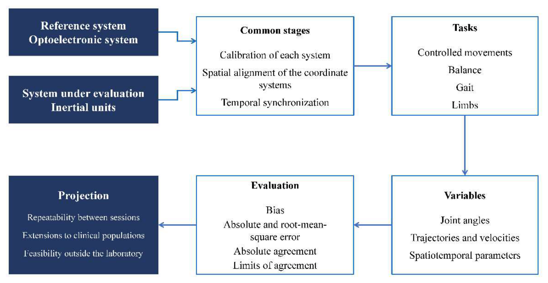
Consequently, the planned experimental progression contemplates three phases. In the first, controlled movements and functional tasks of low complexity will be used, in order to check the stability of the measurements and detect systematic discrepancies between both systems. In the second, tasks representative of functional assessment in rehabilitation will be incorporated (balance, gait and movements of the upper and lower limbs), selected so as to cover the scenarios gathered in Section 3.4. In the third, the extension to participants with neurological or musculoskeletal alterations is foreseen, given that, as pointed out, compensatory patterns and functional variability can affect the performance of the systems differently from what is observed in healthy people [71,72,74].
The comparison between systems must rely on metrics that report complementary aspects of agreement. These metrics should include mean bias, to detect systematic over- or underestimation; the mean absolute error or the root-mean-square error, to quantify the magnitude of the discrepancies; the absolute-agreement intraclass correlation coefficient, to assess the equivalence between measurements; and Bland–Altman agreement analysis, to examine whether the error varies with the magnitude of the movement. The choice of each metric must correspond to the nature of the variable assessed, avoiding the equating of correlation and agreement pointed out in Section 3.3.1.
Finally, the scope of this section should be specified. The present study does not report experimental results. Instead, this section presents a set of design criteria derived from the review, together with a description of the available system configuration and the implementation phase completed to date. The quantitative characterization of the performance of this configuration and the analysis of the acquired signals will be addressed in future studies.
Figure 4.
Conceptual scheme for the experimental validation of motion capture technologies.
4.4. Limitations of this Review
This review has limitations that should be acknowledged. First, it was conducted as a critical review based on a structured literature search rather than a systematic review. No formal risk-of-bias assessment tool was applied and no quantitative aggregation of results was performed, given the heterogeneity of protocols and metrics of the included studies. Instead, the quality of the evidence was characterized using a set of explicit study-level descriptors. Second, given the volume of results, the screening was limited to the first 100 records of each database ordered by relevance. Consequently, the possibility that relevant studies beyond this threshold were omitted cannot be excluded. Forward and backward citation searching mitigated this limitation but could not eliminate it entirely. Furthermore, the eight seed studies were selected by the authors based on their relevance to the technologies available in their laboratory. This selection may have influenced the citation network explored. The 43 studies added through citation searching were classified from titles and abstracts with the assistance of a generative AI tool. Final inclusion decisions were made by the authors, who also verified the extracted data against the full texts. Finally, the analysis has shown that a good part of the most widely disseminated accuracy figures comes from small samples of healthy people, which limits their generalization and constitutes, in itself, one of the results of this work.
5. Conclusions
This review has critically analyzed the state of the art of motion capture technologies applied to human rehabilitation and has derived from that synthesis a set of criteria for the design of future validation studies. Its distinctive contribution is twofold. First, it provides a transversal comparison between technological families on a set of common criteria that integrates metrological performance and operational feasibility. Second, it translates the identified gaps into concrete methodological requirements for experimental research.
The analysis confirms that no technology is universally superior, but it allows that statement to be made more precise than its usual formulation. The equivalence between technologies is demonstrated for certain variables and not for others. Specifically, markerless and inertial-sensor systems reach a validity comparable to that of reference systems in spatiotemporal parameters and in the global description of movement, whereas in specific joint angles, in proximal joints and in fast movements the errors remain above the two-to-five-degree interval that the literature establishes as clinically desirable. Technology selection must therefore be formulated by variable, task and population, and not as a global assessment of the system.
Two factors emerge as determinants of the achievable performance and receive less attention in the literature than they deserve. The first is the calibration procedure, whose influence on accuracy is documented and whose description is frequently insufficient to reproduce the studies. The second is the time horizon of the assessment. Specifically, the stability results obtained in short recordings are not extrapolable to prolonged follow-up, which is precisely the scenario of interest in rehabilitation.
Four recommendations for future research follow from the above. Validation studies should incorporate the calibration report among the published information, so that their results are interpretable and comparable. They should report jointly correlation and absolute error metrics, whose dissociation can convey an impression of accuracy that the data do not support. They should extend validation to real clinical populations, in whom motor variability and compensatory strategies can affect performance in a qualitatively different way. And they should extend the assessment of temporal stability to horizons compatible with the longitudinal follow-up of a rehabilitation process.
For the scientific and clinical community, the value of this work lies in offering a technology selection framework based on traceable evidence and in establishing the minimum requirements that validation studies should satisfy, a necessary condition for the transfer of these technologies to clinical practice to take place on solid foundations.
Author Contributions
Conceptualization, Y.B. and H.M.; Methodology, M.J.-V. and H.M.; Investigation, Y.B., M.J.-V. and H.M.; Data curation, Y.B.; Writing—original draft preparation, Y.B.; Writing—review and editing, M.J.-V. and H.M.; Supervision, H.M.; Project administration, H.M.; Funding acquisition, H.M. All authors have read and agreed to the published version of the manuscript.
Funding
This work was developed within the framework of the RETINA project, funded by the Secretaría Nacional de Ciencia, Tecnología e Investigación (SENACYT) of Panama, under merit contract No. PFID-FID-2021-131.
Institutional Review Board Statement
Not applicable.
Informed Consent Statement
Not applicable.
Data Availability Statement
The data supporting the findings of this study are available from publicly accessible sources. All relevant references have been cited in the manuscript.
Acknowledgments
Y.B. acknowledges the financial support provided by the IFARHU–SENACYT Scholarship Program for the pursuit of a Master of Science in Mechanical Engineering at the Universidad Tecnologica of Panama. H.M. acknowledges the support from the Sistema Nacional de Investigacion of Panama as a member of the SNI.
Conflicts of Interest
The authors declare no conflicts of interest.
Abbreviations
The following abbreviations are used in this manuscript:
| EMG | Electromyography |
| ICC | Intraclass correlation coefficient |
| IMU | Inertial measurement unit |
| LoA | Limits of agreement |
| MCID | Minimal clinically important difference |
| MDC | Minimal detectable change |
| PRISMA | Preferred Reporting Items for Systematic Reviews and Meta-Analyses |
| RMSE | Root-mean-square error |
| SEM | Standard error of measurement |
References
- Kulig, K.; Burnfield, J.M. The role of biomechanics in orthopedic and neurological rehabilitation. Acta Bioeng. Biomech. 2008, 10, 3–14. [Google Scholar]
- Hulleck, A.A.; Menoth Mohan, D.; Abdallah, N.; El Rich, M.; Khalaf, K. Present and future of gait assessment in clinical practice: Towards the application of novel trends and technologies. Front. Med. Technol. 2022, 4. [Google Scholar] [CrossRef]
- Lu, T.; Chang, C. Biomechanics of human movement and its clinical applications. Kaohsiung J. Med. Sci. 2012, 28. [Google Scholar] [CrossRef]
- Winter, D.A. Biomechanics and Motor Control of Human Movement, 4th ed.; Wiley: Hoboken, NJ, USA, 2009. [Google Scholar]
- Nigg, B.M.; Herzog, W. Biomechanics of the Musculo-Skeletal System, 3rd ed.; John Wiley & Sons: Hoboken, NJ, USA, 2007. [Google Scholar]
- Knudson, D. Introduction to Biomechanics of Human Movement. In Fundamentals of Biomechanics; Knudson, D., Ed.; Springer US: Boston, MA, USA, 2003; pp. 3–22. [Google Scholar] [CrossRef]
- Cappozzo, A.; Della Croce, U.; Leardini, A.; Chiari, L. Human movement analysis using stereophotogrammetry. Gait Posture 2005. [Google Scholar] [CrossRef]
- Cappozzo, A.; Catani, F.; Della Croce, U.; Leardini, A. Position and orientation in space of bones during movement: anatomical frame definition and determination. Clin. Biomech. 1995, 10, 171–178. [Google Scholar] [CrossRef]
- Alderink, G.; Õunpuu, S. Biomechanics of Human Motion and Its Clinical Applications: Instrumented Gait Analysis. Bioengineering 2025, 12, 1076. [Google Scholar] [CrossRef]
- Moro, M.; Marchesi, G.; Hesse, F.; Odone, F.; Casadio, M. Markerless vs. Marker-Based Gait Analysis: A Proof of Concept Study. Sensors 2022, 22, 2011. [Google Scholar] [CrossRef]
- Favata, A.; Gallart-Agut, R.; Pàmies-Vilà, R.; Torras, C.; Font-Llagunes, J.M. IMU-Based Systems for Upper-Limb Kinematic Analysis in Clinical Applications: A Systematic Review. IEEE Sens. J. 2024, 24, 28576–28594. [Google Scholar] [CrossRef]
- Roetenberg, D.; Luinge, H.; Slycke, P. Xsens MVN: Full 6DOF Human Motion Tracking Using Miniature Inertial Sensors. Xsens Motion Technol. BV Tech. Rep. 2009, 3. [Google Scholar]
- Nakano, N.; Sakura, T.; Ueda, K.; Omura, L.; Kimura, A.; Iino, Y.; Fukashiro, S.; Yoshioka, S. Evaluation of 3D Markerless Motion Capture Accuracy Using OpenPose With Multiple Video Cameras. Front. Sports Act. Living 2020, 2. [Google Scholar] [CrossRef]
- Ji, H.; Wang, L.; Zhang, Y.; Li, Z.; Wei, C. A Review of Human Pose Estimation Methods in Markerless Motion Capture. Comput.-Aided Des. Appl. 2023, 392–423. [Google Scholar] [CrossRef]
- Zhang, H.; Sid'El Moctar, S.M.; Boudaoud, S.; Rida, I. A comprehensive review of sEMG-IMU sensor fusion for upper limb movements pattern recognition. Inf. Fusion 2026, 125, 103422. [Google Scholar] [CrossRef]
- Atrsaei, A.; Salarieh, H.; Alasty, A. Human Arm Motion Tracking by Orientation-Based Fusion of Inertial Sensors and Kinect Using Unscented Kalman Filter. J. Biomech. Eng. 2016, 138, 091005. [Google Scholar] [CrossRef]
- Uhlrich, S.D.; Falisse, A.; Kidziński, Ł.; Muccini, J.; Ko, M.; Chaudhari, A.S.; Hicks, J.L.; Delp, S.L. OpenCap: Human movement dynamics from smartphone videos. PLoS Comput. Biol. 2023, 19, e1011462. [Google Scholar] [CrossRef]
- Casile, A.; Fregna, G.; Boarini, V.; Paoluzzi, C.; Manfredini, F.; Lamberti, N.; Baroni, A.; Straudi, S. Quantitative Comparison of Hand Kinematics Measured with a Markerless Commercial Head-Mounted Display and a Marker-Based Motion Capture System in Stroke Survivors. Sensors 2023, 23, 7906. [Google Scholar] [CrossRef]
- Saucier, D.; McDonald, K.; Mydlo, M.; Barber, R.; Wall, E.; Derby, H.; Reneker, J.C.; Chander, H.; Burch, R.F.; Weinstein, J.L. Validating a Wearable VR Headset for Postural Sway: Comparison with Force Plate COP Across Standardized Sensorimotor Tests. Electronics 2025, 14, 4156. [Google Scholar] [CrossRef]
- Lee, C.J.; Lee, J.K. Inertial Motion Capture-Based Wearable Systems for Estimation of Joint Kinetics: A Systematic Review. Sensors 2022, 22, 2507. [Google Scholar] [CrossRef]
- Fang, Z.; Woodford, S.; Senanayake, D.; Ackland, D. Conversion of Upper-Limb Inertial Measurement Unit Data to Joint Angles: A Systematic Review. Sensors 2023, 23, 6535. [Google Scholar] [CrossRef]
- Li, J.; Qiu, F.; Gan, L.; Chou, L.-S. Concurrent validity of inertial measurement units in range of motion measurements of upper extremity: A systematic review and meta-analysis. Wearable Technol. 2024, 5, e11. [Google Scholar] [CrossRef]
- Gu, C.; Lin, W.; He, X.; Zhang, L.; Zhang, M. IMU-based motion capture system for rehabilitation applications: A systematic review. Biomim. Intell. Robot. 2023, 3, 100097. [Google Scholar] [CrossRef]
- Cereatti, A.; Gurchiek, R.; Mündermann, A.; Fantozzi, S.; Horak, F.; Delp, S.; Aminian, K. ISB recommendations on the definition, estimation, and reporting of joint kinematics in human motion analysis applications using wearable inertial measurement technology. J. Biomech. 2024, 173, 112225. [Google Scholar] [CrossRef]
- Wade, L.; Needham, L.; McGuigan, P.; Bilzon, J. Applications and limitations of current markerless motion capture methods for clinical gait biomechanics. PeerJ 2022, 10, e12995. [Google Scholar] [CrossRef]
- Lam, W.W.T.; Tang, Y.M.; Fong, K.N.K. A systematic review of the applications of markerless motion capture (MMC) technology for clinical measurement in rehabilitation. J. NeuroEng. Rehabil. 2023, 20, 57. [Google Scholar] [CrossRef]
- Pardell, M.; Dolgoy, N.D.; Bernard, S.; Bayless, K.; Hirsche, R.; Dennett, L.; Tandon, P. Movement Outcomes Acquired via Markerless Motion Capture Systems Compared with Marker-Based Systems for Adult Patient Populations: A Scoping Review. Biomechanics 2024, 4, 618–632. [Google Scholar] [CrossRef]
- Scataglini, S.; Abts, E.; Van Bocxlaer, C.; Van den Bussche, M.; Meletani, S.; Truijen, S. Accuracy, Validity, and Reliability of Markerless Camera-Based 3D Motion Capture Systems versus Marker-Based 3D Motion Capture Systems in Gait Analysis: A Systematic Review and Meta-Analysis. Sensors 2024, 24, 3686. [Google Scholar] [CrossRef]
- Yoma, M.; Llurda-Almuzara, L.; Herrington, L.; Jones, R. Reliability and validity of lower extremity and trunk kinematics measured with markerless motion capture during sports-related and functional tasks: A systematic review. J. Sports Sci. 2025, 43, 1703–1730. [Google Scholar] [CrossRef]
- Chen, B.; Liu, P.; Xiao, F.; Liu, Z.; Wang, Y. Review of the Upright Balance Assessment Based on the Force Plate. Int. J. Environ. Res. Public Health 2021, 18, 2696. [Google Scholar] [CrossRef]
- Ettefagh, A.; Roshan Fekr, A. Technological advances in lower-limb tele-rehabilitation: A review of literature. J. Rehabil. Assist. Technol. Eng. 2024, 11, 20556683241259256. [Google Scholar] [CrossRef]
- de Borba, E.F.; Storniolo, J.L.; Cerfoglio, S.; Capodaglio, P.; Cimolin, V.; Peyré-Tartaruga, L.A.; Tartaruga, M.P.; Cavallari, P. Effect of Walking Speed on the Reliability of a Smartphone-Based Markerless Gait Analysis System. Sensors 2025, 25, 6474. [Google Scholar] [CrossRef]
- Lima, Y.L.; Collings, T.; Hall, M.; Bourne, M.N.; Diamond, L.E. Validity and reliability of trunk and lower-limb kinematics during squatting, hopping, jumping and side-stepping using OpenCap markerless motion capture application. J. Sports Sci. 2024, 42, 1847–1858. [Google Scholar] [CrossRef]
- Federolf, P.; Kühne, M.; Schiel, K.; Reimeir, E.; Debertin, D.; Calisti, M.; Mohr, M. Validation of markerless (Theia3D™) against marker-based (Vicon™) motion capture data of postural control movements analyzed through principal component analysis. J. Biomech. 2025, 189, 112831. [Google Scholar] [CrossRef]
- Song, K.; Hullfish, T.J.; Scattone Silva, R.; Silbernagel, K.G.; Baxter, J.R. Markerless motion capture estimates of lower extremity kinematics and kinetics are comparable to marker-based across 8 movements. J. Biomech. 2023, 157, 111751. [Google Scholar] [CrossRef]
- Horsak, B.; Eichmann, A.; Lauer, K.; Prock, K.; Krondorfer, P.; Siragy, T.; Dumphart, B. Concurrent validity of smartphone-based markerless motion capturing to quantify lower-limb joint kinematics in healthy and pathological gait. J. Biomech. 2023, 159, 111801. [Google Scholar] [CrossRef]
- Kanko, R.M.; Laende, E.; Selbie, W.S.; Deluzio, K.J. Inter-session repeatability of markerless motion capture gait kinematics. J. Biomech. 2021, 121, 110422. [Google Scholar] [CrossRef]
- Riazati, S.; McGuirk, T.E.; Perry, E.S.; Sihanath, W.B.; Patten, C. Absolute Reliability of Gait Parameters Acquired With Markerless Motion Capture in Living Domains. Front. Hum. Neurosci. 2022, 16, 867474. [Google Scholar] [CrossRef]
- Carvalho, A.; Vanrenterghem, J.; Cabral, S.; M. d'Assunção, A.; Carnide, F.; Veloso, A.P.; Moniz-Pereira, V. Construct validity of markerless three-dimensional gait biomechanics in healthy older adults. Gait Posture 2025, 120, 217–225. [Google Scholar] [CrossRef]
- Wren, T.A.L.; Isakov, P.; Rethlefsen, S.A. Comparison of kinematics between Theia markerless and conventional marker-based gait analysis in clinical patients. Gait Posture 2023, 104, 9–14. [Google Scholar] [CrossRef]
- Kanko, R.M.; Laende, E.K.; Strutzenberger, G.; Brown, M.; Selbie, W.S.; DePaul, V.; Scott, S.H.; Deluzio, K.J. Assessment of spatiotemporal gait parameters using a deep learning algorithm-based markerless motion capture system. J. Biomech. 2021, 122, 110414. [Google Scholar] [CrossRef]
- Horsak, B.; Kainz, H.; Dumphart, B. Repeatability and minimal detectable change including clothing effects for smartphone-based 3D markerless motion capture. J. Biomech. 2024, 175, 112281. [Google Scholar] [CrossRef]
- Horsak, B.; Prock, K.; Krondorfer, P.; Siragy, T.; Simonlehner, M.; Dumphart, B. Inter-trial variability is higher in 3D markerless compared to marker-based motion capture: Implications for data post-processing and analysis. J. Biomech. 2024, 166, 112049. [Google Scholar] [CrossRef]
- Martiš, P.; Košutzká, Z.; Kranzl, A. A Step Forward Understanding Directional Limitations in Markerless Smartphone-Based Gait Analysis: A Pilot Study. Sensors 2024, 24, 3091. [Google Scholar] [CrossRef]
- Turner, J.A.; Chaaban, C.R.; Padua, D.A. Validation of OpenCap: A low-cost markerless motion capture system for lower-extremity kinematics during return-to-sport tasks. J. Biomech. 2024, 171, 112200. [Google Scholar] [CrossRef]
- Lambricht, N.; Englebert, A.; Pitance, L.; Fisette, P.; Detrembleur, C. Quantifying performance and joint kinematics in functional tasks crucial for anterior cruciate ligament rehabilitation using smartphone video and pose detection. Knee 2025, 52, 171–178. [Google Scholar] [CrossRef]
- Ksoll, K.; Krätschmer, R.; Stöcker, F. Validity of a Convolutional Neural Network-Based, Markerless Pose Estimation System Compared to a Marker-Based 3D Motion Analysis System for Gait Assessment—A Pilot Study. Sensors 2025, 25, 6551. [Google Scholar] [CrossRef]
- Doerks, F.; Harms, F.; Schwarze, M.; Jakubowitz, E.; Welke, B. Gait assessment using a 2D video-based motion analysis app in healthy subjects and subjects with lower limb amputation – A pilot study. PLoS ONE 2025, 20, e0324499. [Google Scholar] [CrossRef]
- Al Borno, M.; O’Day, J.; Ibarra, V.; Dunne, J.; Seth, A.; Habib, A.; Ong, C.; Hicks, J.; Uhlrich, S.; Delp, S. OpenSense: An open-source toolbox for inertial-measurement-unit-based measurement of lower extremity kinematics over long durations. J. Neuroeng. Rehabil. 2022, 19, 22. [Google Scholar] [CrossRef]
- Uhlenberg, L.; Amft, O. Where to mount the IMU? Validation of joint angle kinematics and sensor selection for activities of daily living. Front. Comput. Sci. 2024, 6, 1347424. [Google Scholar] [CrossRef]
- Hafer, J.F.; Mihy, J.A.; Hunt, A.; Zernicke, R.F.; Johnson, R.T. Lower Extremity Inverse Kinematics Results Differ Between Inertial Measurement Unit- and Marker-Derived Gait Data. J. Appl. Biomech. 2023, 39, 133–142. [Google Scholar] [CrossRef]
- Unger, T.; de Sousa Ribeiro, R.; Mokni, M.; Weikert, T.; Pohl, J.; Schwarz, A.; Held, J.P.O.; Sauerzopf, L.; Kühnis, B.; Gavagnin, E.; Luft, A.R.; Gassert, R.; Lambercy, O.; Awai Easthope, C.; Schönhammer, J.G. Upper limb movement quality measures: comparing IMUs and optical motion capture in stroke patients performing a drinking task. Front. Digit. Health 2024, 6, 1359776. [Google Scholar] [CrossRef]
- Meng, L.; Chen, M.; Li, B.; He, F.; Xu, R.; Ming, D. An Inertial-Based Upper-Limb Motion Assessment Model: Performance Validation Across Various Motion Tasks. IEEE Sens. J. 2023, 23, 7168–7177. [Google Scholar] [CrossRef]
- Sers, R.; Forrester, S.; Moss, E.; Ward, S.; Ma, J.; Zecca, M. Validity of the Perception Neuron inertial motion capture system for upper body motion analysis. Measurement 2020, 149, 107024. [Google Scholar] [CrossRef]
- Dal Farra, F.; Cerfoglio, S.; Porta, M.; Pau, M.; Galli, M.; Lopomo, N.F.; Cimolin, V. Comparative Assessment of an IMU-Based Wearable Device and a Marker-Based Optoelectronic System in Trunk Motion Analysis: A Cross-Sectional Investigation. Appl. Sci. 2025, 15, 5931. [Google Scholar] [CrossRef]
- Bailey, C.A.; Uchida, T.K.; Nantel, J.; Graham, R.B. Validity and sensitivity of an inertial measurement unit-driven biomechanical model of motor variability for gait. Sensors 2021, 21, 7690. [Google Scholar] [CrossRef]
- Di Raimondo, G.; Vanwanseele, B.; van der Have, A.; Emmerzaal, J.; Willems, M.; Killen, B.A.; Jonkers, I. Inertial Sensor-to-Segment Calibration for Accurate 3D Joint Angle Calculation for Use in OpenSim. Sensors 2022, 22, 3259. [Google Scholar] [CrossRef]
- Barzyk, P.; Boden, A.-S.; Howaldt, J.; Stürner, J.; Zimmermann, P.; Seebacher, D.; Liepert, J.; Stein, M.; Gruber, M.; Schwenk, M. Steps to Facilitate the Use of Clinical Gait Analysis in Stroke Patients: The Validation of a Single 2D RGB Smartphone Video-Based System for Gait Analysis. Sensors 2024, 24, 7819. [Google Scholar] [CrossRef]
- Frykberg, G.E.; Grip, H.; Murphy, M.A. How many trials are needed in kinematic analysis of reach-to-grasp?—A study of the drinking task in persons with stroke and non-disabled controls. J. NeuroEng. Rehabil. 2021, 18, 101. [Google Scholar] [CrossRef]
- Balta, D.; Figari, G.; Paolini, G.; Pantzar-Castilla, E.; Riad, J.; Croce, U.D.; Cereatti, A. A Model-Based Markerless Protocol for Clinical Gait Analysis Based on a Single RGB-Depth Camera: Concurrent Validation on Patients With Cerebral Palsy. IEEE Access 2023, 11, 144377–144393. [Google Scholar] [CrossRef]
- Poomulna, J.; Knarr, B.A.; Dutt, V.; Kingston, D.C. Comparison of gait deviation index (GDI) and gait variability index (GVI) measured by marker-based and markerless motion capture systems in children with cerebral palsy (CP). Gait Posture 2025, 115, 7–13. [Google Scholar] [CrossRef]
- Moro, M.; Marchesi, G.; Cellerino, M.; Boffa, G.; Odone, F.; Inglese, M.; Casadio, M. Markerless Video-Based Gait Analysis in People With Multiple Sclerosis. IEEE Trans. Neural Syst. Rehabil. Eng. 2025, 33, 2743–2749. [Google Scholar] [CrossRef]
- Ripic, Z.; Nienhuis, M.; Signorile, J.F.; Best, T.M.; Jacobs, K.A.; Eltoukhy, M. A comparison of three-dimensional kinematics between markerless and marker-based motion capture in overground gait. J. Biomech. 2023, 159, 111793. [Google Scholar] [CrossRef]
- Lagomarsino, B.; Massone, A.; Odone, F.; Casadio, M.; Moro, M. Video-based markerless assessment of bilateral upper limb motor activity following cervical spinal cord injury. Comput. Biol. Med. 2025, 196, 110908. [Google Scholar] [CrossRef]
- Bellitto, A.; De Luca, A.; Gamba, S.; Losio, L.; Massone, A.; Casadio, M.; Pierella, C. Clinical, Kinematic and Muscle Assessment of Bilateral Coordinated Upper-Limb Movements Following Cervical Spinal Cord Injury. IEEE Trans. Neural Syst. Rehabil. Eng. 2023, 31, 3607–3618. [Google Scholar] [CrossRef]
- Wang, J.; Xu, W.; Wu, Z.; Zhang, H.; Wang, B.; Zhou, Z.; Wang, C.; Li, K.; Nie, Y. Evaluation of a smartphone-based markerless system to measure lower-limb kinematics in patients with knee osteoarthritis. J. Biomech. 2025, 181, 112529. [Google Scholar] [CrossRef]
- Kim, H.Y.; An, Y.S.; Oh, S.H.; Lee, H.C. Clinical Feasibility of a Markerless Gait Analysis System. Clin. Orthop. Surg. 2024, 16, 506–516. [Google Scholar] [CrossRef]
- Ahmed, K.; Taheri, S.; Weygers, I.; Ortiz-Catalan, M. Validation of IMU against optical reference and development of open-source pipeline: proof of concept case report in a participant with transfemoral amputation fitted with a Percutaneous Osseointegrated Implant. J. NeuroEng. Rehabil. 2024, 21, 128. [Google Scholar] [CrossRef]
- Langley, B.; Greaves, H.; Whelton, C.; Page, R.; Cramp, M.; Morrison, S.C.; Dey, P.; Board, T. Reliability of kinematic waveforms during gait analysis with total hip arthroplasty patients. J. Jt. Surg. Res. 2024, 2, 1–6. [Google Scholar] [CrossRef]
- Smith, J.; Parikh, D.; Tate, V.; Siddicky, S.F.; Hsiao, H.-Y. Validity of Valor Inertial Measurement Unit for Upper and Lower Extremity Joint Angles. Sensors 2024, 24, 5833. [Google Scholar] [CrossRef]
- Guo, Y.; Sekiguchi, Y.; Zeng, W.; Ebihara, S.; Owaki, D.; Hayashibe, M. Physics-Informed Learning Framework for Lower Limb Kinematic Prediction With Sparse Sensors and Its Application in Chronic Stroke. IEEE Trans. Neural Syst. Rehabil. Eng. 2025, 33, 2475–2486. [Google Scholar] [CrossRef]
- Unger, T.; Sal Moslehian, A.; Peiffer, J.D.; Ullrich, J.; Gassert, R.; Lambercy, O.; James Cotton, R.; Awai Easthope, C. Differentiable Biomechanics for Markerless Motion Capture in Upper Limb Stroke Rehabilitation: A Comparison With Optical Motion Capture. IEEE Trans. Med. Robot. Bionics 2025. [Google Scholar] [CrossRef]
- Pradon, D.; Tong, L.; Chalitsios, C.; Roche, N. Development of Surface EMG for Gait Analysis and Rehabilitation of Hemiparetic Patients. Sensors 2024, 24, 5954. [Google Scholar] [CrossRef]
- Cerfoglio, S.; Lopomo, N.F.; Capodaglio, P.; Scalona, E.; Monfrini, R.; Verme, F.; Galli, M.; Cimolin, V. Assessment of an IMU-Based Experimental Set-Up for Upper Limb Motion in Obese Subjects. Sensors 2023, 23, 9264. [Google Scholar] [CrossRef]
- Nilsson, S.; Ertzgaard, P.; Lundgren, M.; Grip, H. Test-Retest Reliability of Kinematic and Temporal Outcome Measures for Clinical Gait and Stair Walking Tests, Based on Wearable Inertial Sensors. Sensors 2022, 22, 1171. [Google Scholar] [CrossRef]
- Oliveira, N.; Park, J.; Barrance, P. Using Inertial Measurement Unit Sensor Single Axis Rotation Angles for Knee and Hip Flexion Angle Calculations During Gait. IEEE J. Transl. Eng. Health Med. 2023, 11, 80–86. [Google Scholar] [CrossRef]
- Qualisys, A.B. Calibration Results. Available online: https://docs.qualisys.com/qtm/content/running_the_system/calibration_results.htm (accessed on 2 December 2025).
- Batista, Y.; Jaén-Vargas, M.; Montes, H. Implementación y Calibración de un Sistema Optoelectrónico de Captura de Movimientos para el Análisis Biomecánico de Extremidades. In Proceedings of the Libro del XX Congreso Nacional de Ciencia y Tecnología, APANAC 2025; Panama City, Panama, Magallón Tejada, A., Talavera, M., Eds.; Asociación Panameña para el Avance de la Ciencia: Panama City, Panama, 30 September 2025; pp. 401–402. Available online: https://congreso.apanac.org.pa/wp-content/uploads/2025/10/libro_congreso_APANAC_2025.pdf (accessed on 5 December 2025).
- Page, M.J.; McKenzie, J.E.; Bossuyt, P.M.; Boutron, I.; Hoffmann, T.C.; Mulrow, C.D.; Shamseer, L.; Tetzlaff, J.M.; Akl, E.A.; Brennan, S.E.; et al. The PRISMA 2020 Statement: An Updated Guideline for Reporting Systematic Reviews. BMJ 2021, 372, n71. [Google Scholar] [CrossRef]
- McInnes, M.; Blana, D.; Starkey, A.; Chadwick, E.K. A Practical Sensor-to-Segment Calibration Method for Upper Limb Inertial Motion Capture in a Clinical Setting. IEEE J. Transl. Eng. Health Med. 2025, 13, 216–226. [Google Scholar] [CrossRef]
- Slade, P.; Habib, A.; Hicks, J.L.; Delp, S.L. An Open-Source and Wearable System for Measuring 3D Human Motion in Real-Time. IEEE Trans. Biomed. Eng. 2022, 69, 678–688. [Google Scholar] [CrossRef]
- Clouthier, A.L.; Ross, G.B.; Mavor, M.P.; Coll, I.; Boyle, A.; Graham, R.B. Development and Validation of a Deep Learning Algorithm and Open-Source Platform for the Automatic Labelling of Motion Capture Markers. IEEE Access 2021, 9, 36444–36454. [Google Scholar] [CrossRef]
- Song, K.; Scattone Silva, R.; Hullfish, T.J.; Silbernagel, K.G.; Baxter, J.R. Patellofemoral Joint Loading Progression Across 35 Weightbearing Rehabilitation Exercises and Activities of Daily Living. Am. J. Sports Med. 2023, 51, 2110–2119. [Google Scholar] [CrossRef]
- Cunha, B.; Maçães, J.; Amorim, I. Smartphone-Based Markerless Motion Capture for Accessible Rehabilitation: A Computer Vision Study. Sensors 2025, 25, 5428. [Google Scholar] [CrossRef]
- Majumder, S.; Deen, M.J. Wearable IMU-Based System for Real-Time Monitoring of Lower-Limb Joints. IEEE Sens. J. 2021, 21, 8267–8275. [Google Scholar] [CrossRef]
- Tacca, N.; Dunlap, C.; Donegan, S.P.; Hardin, J.O.; Meyers, E.; Darrow, M.J.; Colachis, S.; Gillman, A.; Friedenberg, D.A. Wearable high-density EMG sleeve for complex hand gesture classification and continuous joint angle estimation. Sci. Rep. 2024, 14, 18564. [Google Scholar] [CrossRef]
- Mao, C.; Li, M.; Li, X.; Li, Z.; Liu, T.; Wang, L.; Zhu, W.; Chen, L.; Sun, Y. The Validity of a Dual-Force Plate for Assessing Counter-Movement Jump Performance. Sensors 2024, 24, 5748. [Google Scholar] [CrossRef]
- Miller, J.D.; Cabarkapa, D.; Miller, A.J.; Frazer, L.L.; Templin, T.N.; Eliason, T.D.; Garretson, S.K.; Fry, A.C.; Berkland, C.J. Novel 3D Force Sensors for a Cost-Effective 3D Force Plate for Biomechanical Analysis. Sensors 2023, 23, 4437. [Google Scholar] [CrossRef]
- Santos, P.; Marquês, F.; Quintão, C.; Quaresma, C. Electromyographic and Kinematic Analysis of the Upper Limb During Drinking and Eating Using a Wearable Device Prototype. Sensors 2025, 25, 5227. [Google Scholar] [CrossRef]
- Tani, M.; Yamaguchi, T.; Honaga, K.; Takahashi, Y.; Kono, H.; Murakami, Y.; Isayama, R.; Haruyama, K.; Fujino, Y.; Matsuda, T.; Fukunaga, I.; Hori, M.; Tanuma, A.; Wada, F.; Hatori, K.; Fujiwara, T. Electromyography (EMG)-triggered transcutaneous spinal cord and hip stimulation for gait rehabilitation in persons with chronic stroke: a randomized, controlled trial. J. Neuroeng. Rehabil. 2025, 22, 154. [Google Scholar] [CrossRef]
- Hubaut, R.; Guichard, R.; Greenfield, J.; Blandeau, M. Validation of an Embedded Motion-Capture and EMG Setup for the Analysis of Musculoskeletal Disorder Risks during Manhole Cover Handling. Sensors 2022, 22, 436. [Google Scholar] [CrossRef]
- Wang, H.; Basu, A.; Durandau, G.; Sartori, M. A wearable real-time kinetic measurement sensor setup for human locomotion. Wearable Technol. 2023, 4, e11. [Google Scholar] [CrossRef]
- Issa Abdul-Kreem, L.; Abdul-Ameer, H.K. Optic Flow Estimation for Human Spine Movements Using a Motion Capture System. IEEE Access 2025, 13, 183480–183492. [Google Scholar] [CrossRef]
- Barclay, S.A.; Klausing, L.N.; Hill, T.M.; Kinney, A.L.; Reissman, T.; Reissman, M.E. Characterization of Upper Extremity Kinematics Using Virtual Reality Movement Tasks and Wearable IMU Technology. Sensors 2024, 24, 233. [Google Scholar] [CrossRef]
- Bresnahan, D.G.; Koziol, S.; Li, Y. Investigation of Patellar Deep Tendon Reflex Using Millimeter-Wave Radar and Motion Capture Technologies. IEEE Access 2024, 12, 9220–9228. [Google Scholar] [CrossRef]
- Stetter, B.J.; Stein, T. Machine Learning in Biomechanics: Enhancing Human Movement Analysis. In Artificial Intelligence in Sports, Movement, and Health; Dindorf, C., Bartaguiz, E., Gassmann, F., Fröhlich, M., Eds.; Springer Nature Switzerland: Cham, Switzerland, 2024; pp. 139–160. [Google Scholar] [CrossRef]
- Pang, T.Y.; Cheng, C.-T.; Feltham, F.; Rahman, A.; McCarney, L.; Rodriguez, C.Q. Feasibility of IMU-Based Wearable Sonification: Toward Personalized, Real-Time Gait Monitoring and Rehabilitation. Biosensors 2025, 15, 698. [Google Scholar] [CrossRef]
Figure 1.
PRISMA 2020 flow diagram of the search and selection process, with identification through databases and through other methods (citation searching and other sources).
Figure 1.
PRISMA 2020 flow diagram of the search and selection process, with identification through databases and through other methods (citation searching and other sources).
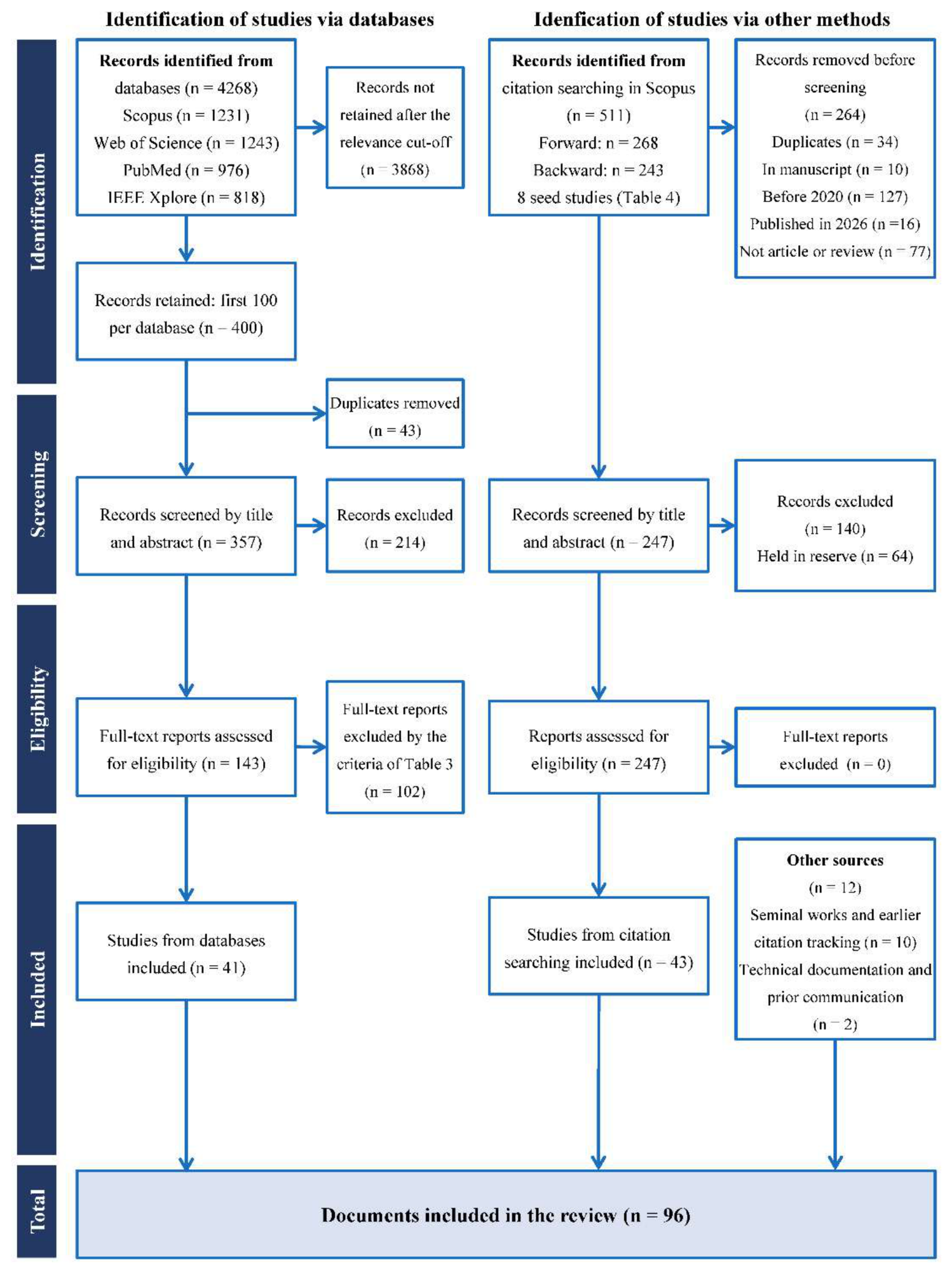
Figure 2.
Taxonomy of motion capture technologies applied to rehabilitation.
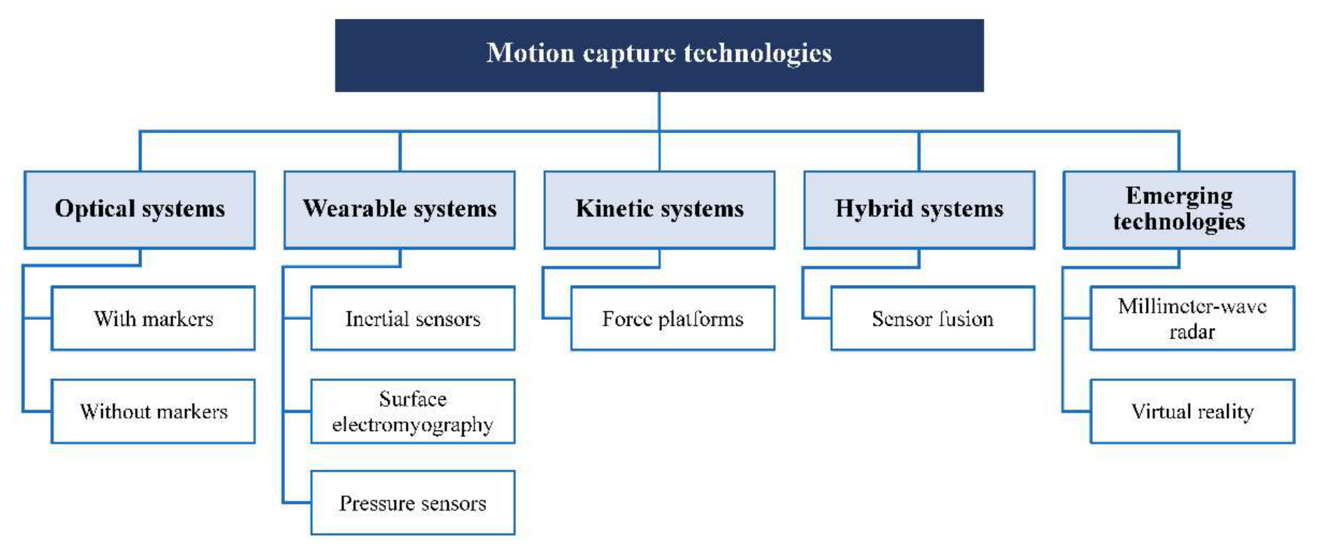
Figure 3.
Decision framework for technology selection according to the required variable and the application conditions.
Figure 3.
Decision framework for technology selection according to the required variable and the application conditions.
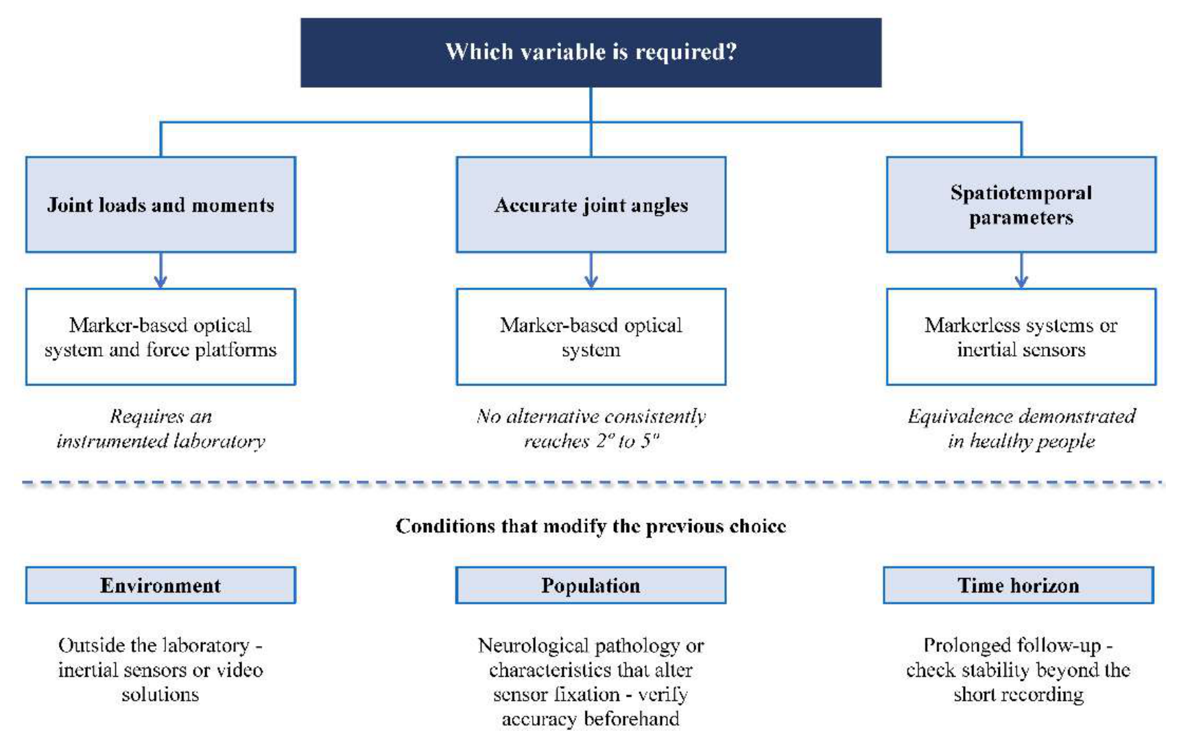

Table 1.
Search strings used in the databases.
| ID | Conceptual axes | Search string |
|---|---|---|
| S1 | Technology + application | ("motion capture" OR "motion analysis") AND (rehabilitation) |
| S2 | Markerless + biomechanics | (markerless OR "marker-less") AND biomechanics |
| S3 | Inertial sensors + rehabilitation or gait | ("inertial measurement unit" OR IMU) AND (rehabilitation OR "gait analysis") AND (kinematic* OR "joint angle") |
| S4 | Sensor fusion + human movement | ("sensor fusion" OR "data fusion") AND ("human movement" OR kinematics OR "gait analysis") AND (IMU) |
| S5 | Upper limb + clinical | ("upper limb" OR "upper extremity") AND kinematics AND (clinical OR patients) |
Table 2.
Databases, search fields, filters and number of records.
| Database | Search fields | Filters applied | Identified | Retained |
|---|---|---|---|---|
| Scopus | TITLE-ABS-KEY | 2020–2025; English or Spanish; engineering; article; open access preferred | 1231 | 100 |
| Web of Science | Topic (TS) | As above, within the options of the platform | 1243 | 100 |
| PubMed | Title/Abstract | As above, within the options of the platform | 976 | 100 |
| IEEE Xplore | Metadata or abstract | As above, within the options of the platform | 818 | 100 |
| Total | 4268 | 400 |
Note: Searches run between December 2025 and February 2026; last update 10 February 2026. Records were retained in the relevance order provided by each database.
Table 3.
Eligibility criteria.
| Code | Criterion |
|---|---|
| Inclusion | |
| I1 | Study conducted in human participants. |
| I2 | Clinical, rehabilitation or functional assessment context. |
| I3 | Explicit use of one or more motion capture technologies. |
| I4 | Quantitative metrics of performance, validity or reliability reported against a reference system or between sessions. |
| Exclusion (applied in this order) | |
| E1 | Non-human population. |
| E2 | No rehabilitation or functional assessment context. |
| E3 | Exclusively sports or performance studies. |
| E4 | No comparison or validation procedure. |
| E5 | No quantitative metrics. |
| E6 | Methodological description insufficient to interpret the results. |
| E7 | Duplicated population or results between publications of the same group. |
| E8 | Full text not accessible. |
Table 4.
Seed studies for forward and backward citation searching in Scopus.
| Seed study | Technology focus | Forward (cited by) | Backward (references) |
|---|---|---|---|
| Moro et al. 2022 [10] | Markerless vs. marker-based gait analysis | 88 | 36 |
| Lima et al. 2024 [33] | OpenCap, multiplanar movements | 7 | 32 |
| Federolf et al. 2025 [34] | Theia3D, postural control | 1 | 42 |
| Horsak et al. 2023 [36] | Smartphone-based markerless capture | 48 | 23 |
| Unger et al. 2025 [72] | Differentiable biomechanics, upper limb, stroke | 12* | 28 |
| McInnes et al. 2025 [80] | Sensor-to-segment calibration, inertial, upper limb | 4* | 23 |
| Slade et al. 2022 [81] | Open-source wearable inertial system | 97 | 43 |
| Clouthier et al. 2021 [82] | Deep learning labelling of markers | 11 | 16 |
| Total | 268 | 243 |
* Only records published in 2026 were exported for this seed.
Table 5.
Studies based on optoelectronic systems with and without markers.
| Ref. | Year | Population | n | Task | Technology evaluated | Reference | Main result |
|---|---|---|---|---|---|---|---|
| [13] | 2020 | Healthy (males, 22.0 y) | 2 | Gait, countermovement jump, throwing | Multi-camera OpenPose | Markers, 16 cameras | Mean absolute error: 47% of values < 20 mm and 80% < 30 mm, but 10% > 40 mm, owing to pose-tracking failures in 2D images |
| [32] | 2025 | Healthy (32.3 ± 8.3 y) | 15 | Gait at three speeds | OpenCap (smartphone) | Markers, 8 cameras | Spatiotemporal ICC ≥ 0.95, r ≥ 0.967; continuous angles RMSE < 2°; discrete ROM ICC 0.48–0.78; CoM RMSE < 6 mm |
| [33] | 2024 | Healthy (27.7 ± 4.1 y) | 19 | Single-leg squat, triple hop, countermovement jump, side hop, cutting | OpenCap | Markers | Lowest RMSE in the squat: mean 7.0°, range 2.9°–13.6°. Between-session ICC 0.60–0.94 depending on task and joint; side hop best at initial contact (0.70–0.94) |
| [83] | 2023 | Healthy | 20 | 35 weight-bearing exercises and activities of daily living | Marker-based optical + force plates | — | Peak patellofemoral load of 0.6 to 8.2 times body weight depending on the exercise |
| [93] | 2025 | Healthy (male, forties) | 1 | Flexion, lateral bending and rotation of the thoracic spine | 3D optical flow on capture data | Reference data | Median error 2.09°. The authors acknowledge that a single subject prevents estimating between-person variability |
| [10] | 2022 | Healthy (27 ± 2 y) | 16 | Gait, 6 m | Markerless, 3 RGB cameras | Optitrack, 8 cameras | MPJPE 23.65 mm; no spatiotemporal differences (p > 0.05) |
| [84] | 2025 | Patients in rehabilitation | 15 | Diagonal and rotation exercise | Smartphone + MediaPipe | Qualisys | Pilot study; no agreement reported |
| [72] | 2025 | People with stroke | 15 | Drinking task | Markerless + differentiable biomechanical model | Marker-based optical | Median r > 0.95; angle RMSE 2°–5°; 6 mm in trunk; between-trial bias 1°–3° |
| [34] | 2025 | Healthy | 13 | Quiet standing, eyes open and closed | Theia3D | Vicon | Principal movements 1 and 2: r = 0.998 and 0.990; 3 to 7: r = 0.814–0.928 |
| [35] | 2023 | Healthy | 10 | 8 movements | Markerless | Markers | Ankle and knee Rxy ≥ 0.877, RMSD ≤ 5.9°; hip RMSD 6.7°–15.9°, mainly in running |
| [36] | 2023 | Healthy simulating pathological gait | 21 | Physiological, crouch, circumduction and equinus gait | OpenCap | Markers | Overall RMSE 5.8°; peak error 11.3°; the authors conclude it remains above the clinically desirable threshold of 2°–5° |
| [82] | 2021 | None: measured and simulated data | — | Marker labelling | Deep neural network with recurrent layers | Commercial software | Labelling accuracy 99.6% with measured and 92.8% with simulated data, up to 98.8% with transfer learning |
| [25] | 2022 | Scoping review | — | Clinical gait | Markerless | Markers | Spatial error 10–60 mm depending on architecture; angular 2.6°–13.2° |
Note: ICC, intraclass correlation coefficient; RMSE, root-mean-square error; RMSD, root-mean-square deviation; ROM, range of motion; CoM, center of mass; MPJPE, mean per-joint position error.
Table 6.
Studies based on inertial sensors.
| Ref. | Year | Population | n | Task | Technology evaluated | Reference | Main result |
|---|---|---|---|---|---|---|---|
| [71] | 2025 | 6 healthy and 17 with chronic stroke | 23 | Gait | Temporal convolutional network with physical constraints, 2 IMUs | Configurations of 4 or more IMUs | Comparable accuracy with only two sensors |
| [70] | 2024 | Healthy (22.5 ± 3.6 y) | 20 | Shoulder, squats, jump, deadlift | Valor IMU, 10 sensors | Vicon, 12 cameras | RMSE 1.89°–16.62°; ICC3k 0.57–0.99 |
| [75] | 2022 | Healthy | 22 | Comfortable and fast gait, stairs | MoLab, 12 IMUs | — (test–retest, 3–6 days) | ICC 0.76–0.97 overall; stride frequency 0.22 and shoulder extension 0.42 |
| [80] | 2025 | Healthy | 20 | Free elbow movement, upper limb | Functional sensor-to-segment calibration, OpenSim workflow | Optical | Median correlation 0.977–0.987 and RMS 5°–8°; significantly better than static-posture calibration (p < 0.001) |
| [76] | 2023 | Healthy (20.7 ± 8.1 y) | 20 | Treadmill gait before and after 41 min | 3 Xsens MTw IMUs | Qualisys, 6 cameras | r² = 0.94–0.98; agreement degraded after the protocol, especially at the knee |
| [49] | 2022 | Healthy | 11 | Two 10-min trials: gait and varied sequence | OpenSense (IMU + inverse kinematics with constrained model) | Optical | RMS 3°–6° except hip rotation; r = 0.60–0.87; near-zero drift: −0.14 to 0.17 °/min |
| [74] | 2023 | 23 with obesity and 30 normal-weight | 53 | Upper-limb movements | Xsens Awinda, 17 IMUs | Vicon | r ≈ 0.40; RMSE 34° and 26°. The authors conclude that the bias limits clinical applicability |
| [81] | 2022 | Healthy young (25.0 ± 1.8 y) | 5 | Gait (3 min) and Fugl-Meyer task; only one subject completed the running condition | OpenSenseRT, open system | Optical | Mean RMSE 4.4° in lower limb and 5.6° in upper limb; by joint, upper limb ranges from 2.5° to 10°. Cost ≈ USD 100 + USD 20 per segment |
| [85] | 2021 | None: public dataset | — | Gait | Two-stage fusion algorithm | Public data and simulated acceleration | Algorithmic work; no participants and no repeatability assessment |
| [94] | 2024 | Healthy | 16 | Upper limb in virtual reality | Xsens | None | Characterization of angles; no validation |
| [20] | 2022 | Systematic review, 48 studies | — | Joint kinetics | Inertial capture | — | Two families of methods: inverse dynamics and machine learning |
Note: IMU, inertial measurement unit; RMS, root mean square.
Table 7.
Complementary sensors and hybrid approaches.
| Ref. | Year | Population | n | Task | Technology evaluated | Reference | Main result |
|---|---|---|---|---|---|---|---|
| [92] | 2023 | Healthy | 9 | 13 activities, from slow walking to running | IMU + pressure insoles | Optical + force plates | Angles r = 0.929; vertical force r = 0.954; ankle torque r = 0.917. Knee torque r = 0.612; hip torque r = 0.21 and mediolateral center of pressure r = 0.47, which the authors consider not coincident |
| [91] | 2022 | Healthy | 10 | Handling of 20 kg and 30 kg loads | 4 IMUs + EMG | Vicon, 13 cameras | Bias < 1°; limits of agreement −4.4° to 3.6° |
| [90] | 2025 | People with chronic stroke | 20 (17 completed) | Treadmill gait training | EMG-triggered spinal and hip stimulation | Randomized controlled trial | Improvement in 10 m walking time only within the experimental group; no significant between-group differences. Intervention study, not a technology validation |
| [86] | 2024 | Healthy | 10 | 37 hand gestures and grasps | High-density EMG sleeve, up to 150 electrodes | 18-sensor capture glove | Classification 97.3 ± 0.3%; continuous angle estimation R² = 0.884; median absolute error < 10° |
| [87] | 2024 | Healthy (22.8 ± 2.8 y, males) | 30 | Countermovement jump | Kunwei force platform | Embedded Kistler | ICC > 0.950; no fixed or proportional bias |
| [88] | 2023 | Healthy | 13 | Lunges and squats, with and without load | 3D force platform prototype | Standard platform | r = 0.950–0.999; RMSE 1.17%–5.36% |
| [73] | 2024 | People with hemiparesis | 3 | Two treadmill gaits and sit-to-stand | Low-cost commercial EMG system | Reference EMG system | Good to very good signal similarity over 883 cycles. Only three participants |
| [89] | 2025 | Healthy (17 women and 13 men, right-handed) | 30 | Drinking and eating | Wearable EMG and kinematics prototype | — | Establishment of normative values in healthy people; 32 recruited, 2 excluded |
| [95] | 2024 | Healthy | 4 | Patellar reflex | Millimeter-wave radar | Optical capture | Mean relative error 10.39% in six reflex features; four subjects, 60 trials |
| [30] | 2021 | Review | — | Postural balance | Force platforms | — | Recommendations on platform type, foot posture and choice of variables |
| [15] | 2026 | Review | — | Upper-limb pattern recognition | sEMG–IMU fusion | — | State of the art of multimodal fusion strategies |
Note: EMG, electromyography; sEMG, surface EMG.
Table 8.
Additional studies identified through citation searching.
| Ref. | Year | Technology | Population and design | Main result |
|---|---|---|---|---|
| [58] | 2024 | Markerless | 8 people with stroke; smartphone 2D video (SMARTGAIT) vs Vicon | Correlation ≥ 0.71–0.79 in kinematics; RMSE ≤ 4.6°; ICC good to excellent |
| [63] | 2023 | Markerless | 35 adults: young, older and with Parkinson’s disease; commercial multi-camera system vs markers | RMSE < 28 mm in joint centers; best agreement in the sagittal plane |
| [60] | 2023 | Markerless | 18 patients with cerebral palsy; one RGB-D camera vs marker-based protocol | Mean absolute error 2%–3.1% in spatiotemporal parameters |
| [40] | 2023 | Markerless | 36 paediatric patients with diverse diagnoses; Theia3D vs Vicon | RMSD of kinematics by joint; subanalysis with orthoses and aids |
| [66] | 2025 | Markerless | 53 patients with knee osteoarthritis and 30 controls; OpenCap vs markers | Mean RMSE 6.1°, ICC 0.67; worse in patients and when walking toward the camera |
| [48] | 2025 | Markerless | 10 healthy and 2 people with amputation; 2D video app (Orthelligent) vs Qualisys | Range-of-motion deviation 3.7°–5.4° |
| [67] | 2024 | Markerless | 14 patients; markerless system (DH Walk) vs optical marker-based system; 14 children with toe-walking | No significant differences in spatiotemporal parameters |
| [64] | 2025 | Markerless | 12 people with cervical spinal cord injury and 12 controls; MediaPipe and AlphaPose vs marker-based capture | High repeatability; sensitivity to shoulder–elbow compensation |
| [17] | 2023 | Markerless | OpenCap: open-source platform with two or more smartphones; validation against a reference | Kinematics and dynamics (moments, loads); reference work for the technology |
| [37] | 2021 | Markerless | 8 healthy adults, 3 sessions; multi-camera video | Mean inter-trial variability 2.5° |
| [38] | 2022 | Markerless | 21 participants aged 18–73 y; Theia3D, two days | RMSD 0.96°–3.71°; SEM 0.91°–3.25° (between-day reliability) |
| [42] | 2024 | Markerless | 20 healthy volunteers; OpenCap, test–retest with different clothing | RMSD 2.8°–3.3°; mean MDC 6.0° in gait |
| [39] | 2025 | Markerless | 30 healthy older adults; Theia3D vs markers | Strong correlations (Rxy ≥ 0.7); thresholds ≤ 5° and ≤ 10% |
| [44] | 2024 | Markerless | 10 participants; OpenCap vs Vicon during the Timed Up and Go | Good spatiotemporal accuracy; worse kinematics with distance from the camera |
| [45] | 2024 | Markerless | Athletes 12–18 months after ACL reconstruction; OpenCap vs optoelectronic | Mean error 3.85° (MAE) and 4.34° (RMSE); CMC > 0.94 in the sagittal plane |
| [46] | 2025 | Markerless | 12 healthy; smartphone video with OpenPifPaf in ACL rehabilitation tasks | Mean RMSE 2.8° at the knee |
| [43] | 2024 | Markerless | 18 healthy volunteers with 4 simulated gait patterns; OpenCap vs markers | Higher inter-trial variability without markers |
| [47] | 2025 | Markerless | 16 healthy adults; convolutional neural network (Orthelligent VISION) vs 3D with markers | CCC ≥ 0.624 sagittal and ≥ 0.805 frontal in most parameters |
| [41] | 2021 | Markerless | 30 and 25 healthy young adults; deep learning vs marker-based capture and gait mat | Agreement of 9 spatiotemporal parameters |
| [61] | 2025 | Markerless | Children with cerebral palsy; Theia3D vs marker-based capture; GDI and GVI indices | GDI 6.9 points lower with Theia3D than with markers (p < 0.05) |
| [18] | 2023 | Markerless | 14 people with stroke; commercial markerless VR headset vs marker-based capture in ecological upper-limb tasks | Quantitative comparison of hand kinematics |
| [62] | 2025 | Markerless | 25 people with multiple sclerosis and 25 matched controls; markerless video, normal and tandem gait | Spatiotemporal and kinematic differences by disability level (EDSS); no reference system, so context only |
| [69] | 2024 | Optoelectronic | 8 patients with total hip arthroplasty; 3D marker-based analysis, test–retest | SEM 0.7°–4.2°; all < 5° |
| [59] | 2021 | Optoelectronic | People without disability and people with stroke; drinking task; intra-session stability with optoelectronic capture | Number of trials needed to reach stability |
| [52] | 2024 | Inertial | 15 patients with mild to moderate stroke; 5 IMUs vs optical capture in a drinking task | 12 of 15 measures with limits of agreement below the MCID |
| [51] | 2023 | Inertial | Young adults, older adults and older adults with knee osteoarthritis; OpenSense vs markers | Differences in sagittal kinematics; no tool × group interaction |
| [50] | 2024 | Inertial | 19 participants; OpenSense in activities of daily living vs marker-based simulation | MAD/RMSE 4.8°/6.6° in lower limbs and 9.2°/11.4° in upper limbs |
| [56] | 2021 | Inertial | 14 healthy young adults; IMU model vs optoelectronic in treadmill gait | RMSD 1.7°–7.5°; ICC good to excellent in most |
| [57] | 2022 | Inertial | 11 healthy; sensor-to-segment calibration (Xsens vs Vicon) | RMSE < 5° at hip and knee with the integrated method |
| [55] | 2025 | Inertial | 27 healthy adults; one IMU vs optoelectronic in trunk range of motion | Accuracy 92.4% in rotation; 61%–72% in flexion, extension and lateral bending |
| [53] | 2023 | Inertial | 12 healthy young adults (21–25 y); upper-limb inertial model vs Vicon | Correlation, bias and RMSE depending on task and plane |
| [54] | 2020 | Inertial | 8 healthy young adults (20–25 y); Perception Neuron vs Vicon in trunk and shoulders | Systematic and random biases ≤ 4.5° in most movements |
| [68] | 2024 | Inertial | Case: person with transfemoral amputation and osseointegrated implant, and one participant without amputation | RMSE 2.35° (amputated side) and 3.59° (intact side) |
| [19] | 2025 | Hybrid | Virtual-reality headset vs force platform in postural sway; adults aged 50–75 y | Correlations r = 0.54–0.91 in 8 metrics |
| [65] | 2023 | Hybrid | 12 people with cervical spinal cord injury and 6 controls; kinematics combined with EMG | Instrumented assessment integrated with clinical tests |
| [29] | 2025 | Review | Systematic review: reliability and validity of markerless capture in lower limb and trunk, 53 studies | — |
| [26] | 2023 | Review | Systematic review: markerless technology in clinical measurement in rehabilitation | — |
| [27] | 2024 | Review | Scoping review: markerless vs marker-based systems in adult populations with pathology | — |
| [28] | 2024 | Review | Systematic review and meta-analysis: accuracy, validity and reliability of markerless 3D capture | — |
| [21] | 2023 | Review | Systematic review: conversion of IMU data to upper-limb joint angles | — |
| [22] | 2024 | Review | Systematic review and meta-analysis: concurrent validity of IMUs in upper-limb range of motion | — |
| [23] | 2023 | Review | Systematic review: inertial capture systems for rehabilitation | — |
| [24] | 2024 | Review | ISB recommendations for defining, estimating and reporting joint kinematics with inertial sensors | — |
Note: ACL, anterior cruciate ligament; CCC, concordance correlation coefficient; CMC, coefficient of multiple correlation; EDSS, Expanded Disability Status Scale; GDI, Gait Deviation Index; GVI, Gait Variability Index; MAD, mean absolute deviation; MAE, mean absolute error; MCID, minimal clinically important difference; MDC, minimal detectable change; SEM, standard error of measurement; VR, virtual reality.
Table 9.
Comparison of the technological families.
| Criterion | Marker-based optical | Markerless optical | Inertial sensors | Force platforms |
|---|---|---|---|---|
| Accuracy in joint kinematics | Reference of the field; own error of 9–19 mm in joint centers and 2.2°–5.5° in rotations [25] | RMSE 2°–5.9° at ankle and knee [35,72]; 5.8° overall in gait [36]; up to 15.9° at the hip [35] | RMS 3°–8° depending on calibration [49,80]; RMSE 4.4°–5.6° [81]; up to 34° in the population with obesity [74] | Not applicable |
| Spatiotemporal variables | Reference | ICC ≥ 0.95, equivalent to the reference standard [10,32] | Reliable in gait, with exceptions [75] | Not applicable |
| Joint kinetics | With force platforms, high resolution [83] | Moments Rxy ≥ 0.934 at ankle and knee [35] | Indirect estimation; hip torque r = 0.21 [92] | Reference; ICC > 0.95 between platforms [87,88] |
| Inter-session reliability | High under controlled conditions, difficult to reproduce | ICC 0.60–0.94, depending on task and joint [33] | ICC 0.76–0.97, with isolated drops [75] | Not documented in the corpus |
| Temporal stability | Not documented in the corpus | Not documented in the corpus | Near-zero drift in 10 min [49]; degradation after 41 min [76] | Not applicable |
| Preparation time | High: marker placement and calibration | Low | Intermediate: placement and sensor-to-segment calibration | Low |
| Calibration demand | High: volume calibration in each session | Low | High: sensor-to-segment calibration conditions the result [80] | Low |
| Sensitivity to occlusions | High: requires visibility by two cameras [7,8] | High: self-occlusion and lighting conditions [25] | None | None |
| Drift | None | None | Present; mitigable with a biomechanical model [49] | None |
| Relative cost | High | Low to medium | Low; USD 100 plus USD 20 per segment in open implementations [81] | Medium; low-cost prototypes emerging [88] |
| Staff required | Specialized | Reduced | Intermediate | Reduced |
| Use outside the laboratory | Not feasible | Feasible with lighting and framing limitations | Feasible | Feasible in portable versions |
Table 10.
Technology selection matrix by scenario.
| Scenario | Recommended technology | Alternative | Requirements | Main limitations |
|---|---|---|---|---|
| Gait analysis in the laboratory | Marker-based optical + force platforms | Multi-camera markerless | Calibrated volume, trained staff | Cost and preparation time |
| Gait spatiotemporal parameters | Markerless or inertial | Either of the two | Framing or sensor placement | Equivalence demonstrated only in healthy people [10,32] |
| Precision joint kinematics | Marker-based optical | — | Laboratory | No alternative consistently reaches the 2°–5° thresholds [36,81] |
| Estimation of joint loads | Optical + force platforms | Markerless + platforms | Kinetic instrumentation | Inertial torque estimation is unreliable at the hip [92] |
| Balance and postural control | Force platform | Markerless (validated in quiet standing) [34] | Instrumented surface | Markerless validation covers the dominant postural strategies, not all [34] |
| Upper limb in the clinic | Inertial with functional calibration [80] | Markerless with biomechanical model [72] | Documented calibration procedure | Degradation in populations whose body characteristics alter sensor fixation [74] |
| Longitudinal follow-up | Inertial | — | Stable protocol between sessions | Stability verified at 10 min [49]; degradation at 41 min [76] |
| Telerehabilitation and home | Smartphone markerless or inertial | — | Connectivity; patient instruction | Errors above the clinical threshold [36]; privacy in video solutions |
| Neuromuscular activity | Surface EMG, alone or fused | sEMG–IMU fusion [15] | Skin preparation | Evidence in clinical population very limited [73] |
| Validation of algorithms | Marker-based optical | — | Laboratory | — |
Disclaimer/Publisher’s Note: The statements, opinions and data contained in all publications are solely those of the individual author(s) and contributor(s) and not of MDPI and/or the editor(s). MDPI and/or the editor(s) disclaim responsibility for any injury to people or property resulting from any ideas, methods, instructions or products referred to in the content. |
© 2026 by the authors. Licensee MDPI, Basel, Switzerland. This article is an open access article distributed under the terms and conditions of the Creative Commons Attribution (CC BY) license (http://creativecommons.org/licenses/by/4.0/).
Copyright: This open access article is published under a Creative Commons CC BY 4.0 license, which permit the free download, distribution, and reuse, provided that the author and preprint are cited in any reuse.