

Submitted:
21 October 2026
Posted:
21 October 2026
You are already at the latest version
Abstract
Introduction: Full-endoscopic decompression has shown favorable outcomes for lumbar spinal stenosis (LSS) with Grade I degenerative lumbar spondylolisthesis (DLS); however, concerns remain regarding postoperative segmental instability and slip progression. This study evaluated the clinical and radiographic outcomes of sequential full-endoscopic unilateral laminotomy for bilateral decompression (FE-ULBD) followed by adjunct interspinous stabilization. Material and Methods: We retrospectively evaluated 47 patients with symptomatic single-level LSS and Grade I DLS who underwent sequential FE-ULBD followed by adjunct interspinous stabilization between January 2022 and April 2025. Clinical outcomes were assessed using visual analog scale (VAS) scores for leg and back pain, the Oswestry Disability Index (ODI), and the modified MacNab criteria. Radiographic outcomes included percent slip, mean disc height, dynamic angular motion, segmental translation, and segmental instability. Radiographic progression was defined as an increase in percent slip of more than 5 percentage points at 12 months. Results: VAS scores for leg and back pain and ODI scores improved significantly (all P < 0.001), and 42 patients (89.4%) achieved favorable modified MacNab outcomes. The percent slip and segmental translation showed no significant changes after 12 months. Segmental instability decreased from 36.2% to 23.4%, although the difference was not statistically significant (P = 0.146). Radiographic progression occurred in 4 patients (8.5%) and was not associated with worse 12-month clinical outcomes. Complications occurred in 6 patients (12.8%), and 4 patients (8.5%) underwent revision surgery. Conclusions: Sequential FE-ULBD, followed by adjunct interspinous stabilization, was associated with significant clinical improvement, and radiographic parameters were generally maintained over 1 year.
Keywords:
degenerative lumbar spondylolisthesis
; full-endoscopic spine surgery
; interspinous stabilization
1. Introduction
Degenerative lumbar spondylolisthesis (DLS) is a degenerative condition characterized by the anterior displacement of one vertebral body relative to the subjacent vertebra in the absence of a pars interarticularis defect. Degenerative changes in the intervertebral disc and facet joints are considered major components of the pathophysiology, and segmental instability may coexist in a subset of patients [1,2]. DLS is frequently associated with lumbar spinal stenosis (LSS), which can cause neurogenic claudication, radicular leg pain, and low back pain. Surgical treatment is generally considered when symptoms persist despite appropriate conservative management [3,4]. Whether decompression alone is sufficient or should be supplemented with instrumented fusion in patients with DLS and LSS has long been debated. Fusion can provide stability to an unstable motion segment and may reduce the risk of slip progression. However, compared with decompression alone, fusion is generally associated with a longer operative time, greater blood loss, longer hospitalization, and increased surgical morbidity. Conversely, recent evidence suggests that decompression alone may not be clinically inferior to decompression with fusion in patients with DLS. At the 5-year follow-up of the NORDSTEN-DS randomized trial, decompression alone was not inferior to decompression with instrumented fusion, with no significant difference in the frequency of subsequent surgeries at the index level [2]. Similarly, another comparative study found no clear differences between the two treatment strategies in quality of life, back and leg pain, or patient satisfaction at 3 years [5]. Against this background, increasing attention has been directed toward minimally invasive decompression techniques that preserve posterior stabilizing structures as much as possible. Endoscopic unilateral laminotomy for bilateral decompression (ULBD) has shown favorable clinical and radiographic outcomes in patients with low-grade DLS associated with LSS. Yoshikane et al., Choi et al., Mitha et al., Liang et al., and Bayram and Karataş reported significant improvements in pain and functional outcomes after full-endoscopic ULBD, while segmental stability was preserved in most patients [6,7,8,9,10]. Similar favorable outcomes and maintenance of postoperative stability have been reported after biportal endoscopic ULBD in patients with low-grade DLS [11]. Interspinous stabilization after decompression has been introduced as a strategy to provide limited supplemental stabilization while potentially reducing the surgical burden associated with instrumented fusion. However, the available evidence is mixed. Although favorable outcomes have been reported in some studies, recent comparative studies and systematic reviews have not demonstrated a clear additional clinical benefit of adding interspinous stabilization to decompression alone [12,13]. In contrast, decompression combined with spiked-plate-type interspinous fixation has been reported to have a lower surgical burden than fusion surgery while providing comparable short-term clinical and radiographic outcomes [14]. To our knowledge, no previous study has specifically evaluated sequential full-endoscopic ULBD followed by adjunct interspinous stabilization device (ISD) implantation in patients with Grade I DLS. Therefore, the present study aimed to evaluate the clinical and radiographic outcomes and complications at a minimum follow-up of 1-year following sequential full-endoscopic unilateral laminotomy for bilateral decompression (FE-ULBD) and adjunct ISD implantation in patients with single-level LSS associated with Grade I DLS and to explore factors associated with radiographic progression.
2. Materials and Methods
2.1. Study Design and Subjects
This single-center retrospective cohort study included patients diagnosed with symptomatic single-level LSS associated with Meyerding Grade I DLS who underwent sequential FE-ULBD followed by adjunct ISD implantation by a single surgeon at Chosun University Hospital between January 2022 and April 2025.
The inclusion criteria were as follows:
Meyerding Grade I DLS; lumbar spinal stenosis of Schizas grade B or higher on preoperative magnetic resonance imaging (MRI); radicular pain and/or neurogenic claudication concordant with the radiographic findings; persistent symptoms despite at least 12 weeks of conservative treatment; single-level FE-ULBD followed by adjunct ISD implantation; availability of preoperative MRI and standing plain and dynamic radiographs obtained preoperatively and at 1 year postoperatively; and a minimum clinical follow-up of 12 months.
The exclusion criteria were as follows:
isthmic spondylolisthesis; Meyerding Grade II or higher lumbar spondylolisthesis;
multilevel spondylolisthesis; previous surgery at the index level; spinal infection, tumor, or trauma; severe spinal deformity; and follow-up of less than 12 months. A total of 47 patients met the eligibility criteria and were included in the final analysis.
2.2. Surgical Technique: Sequential FE-ULBD Followed by Adjunct ISD Implantation
All procedures were performed with the patient in the prone position. Epidural anesthesia with sedation was administered to most patients (44/47, 93.6%) with continuous intraoperative monitoring. A longitudinal skin incision approximately 1 cm in length was made approximately 1–1.5 cm lateral to the midline on the ipsilateral side of the index level. After serial dilation, a working cannula was inserted and docked at the V-point, defined as the junction between the inferior margin of the cranial lamina and the superior margin of the caudal lamina. Soft tissue dissection and hemostasis were performed using a radiofrequency ablator. Bony drilling was initiated at the medial border of the ipsilateral facet joint and proceeded from the caudal to the cranial portion and from the deeper to the more superficial portion of the lamina. Drilling was extended toward the spinolaminar junction of the cranial and caudal laminae until the free margin of the deep layer of the ligamentum flavum (LF) was identified. Subsequently, the contralateral lamina and lateral recess were undercut until the free margin of the deep layer of the LF was adequately exposed. The LF was detached from the undersurface of the lamina using an endoscopic dissector and removed en bloc using Kerrison rongeurs and forceps. Both lateral recesses were inspected, and adequate decompression was confirmed by visualization of a freely mobile dural sac and traversing nerve roots. An epidural drain was placed and secured to the skin. For adjunct interspinous stabilization, the original incision was extended by approximately 1 cm cranially and caudally. Limited bilateral subperiosteal dissection was performed over the inferior portion of the cranial spinous process and the superior portion of the caudal spinous process. Interspinous fixation was then performed using the SOL™ Inter-Spinous Device System (Medyssey, 43176 Business Park Dr., Ste 107, Temecula, CA 92590, USA), consisting of two spiked titanium plates that engage the adjacent spinous processes. The fascia and skin were closed using absorbable sutures (Figure 1).
2.3. Patient Evaluation
2.3.1. Baseline Characteristics
Baseline characteristics included age, sex, body mass index (BMI), bone mineral density (BMD), osteoporosis status, and American Society of Anesthesiologists (ASA) physical status. Osteoporosis was defined as a T-score of ≤ −2.5 at either the lumbar spine or femoral neck. Preoperative comorbidities included diabetes mellitus and hypertension. The presence of facet cysts at the index level was assessed using preoperative MRI. Perioperative variables included the surgical level, operative time, type of anesthesia, and length of hospital stay. The follow-up duration was defined as the interval from the date of surgery to the most recent spine outpatient follow-up (Table 1).
2.3.2. Clinical Evaluation
Clinical outcomes were assessed using visual analog scale (VAS) scores for leg and back pain and the Oswestry Disability Index (ODI), which were measured preoperatively and at 1 week, 6 months, and 12 months postoperatively. The modified MacNab criteria were assessed at 12 months postoperatively, with outcomes categorized as excellent, good, fair, or poor. Outcomes rated as excellent or good were classified as favorable, whereas those rated as fair or poor were classified as unfavorable (Table 2).
2.3.3. Radiographic Evaluation
Radiographic evaluation was performed using standing plain and dynamic radiographs obtained preoperatively and at 12 months postoperatively. Static radiographic parameters included percent slip and mean disc height.
The percent slip was calculated as the horizontal displacement between the posterior margins of the upper and lower vertebral bodies divided by the anteroposterior length of the superior endplate of the lower vertebral body, multiplied by 100. Radiographic progression was defined as an absolute increase in percent slip of more than 5 percentage points at 12 months compared with the preoperative value [6,15]. The mean disc height was calculated as the average of the anterior and posterior disc heights measured at the index level on standing plain radiographs. The dynamic radiographic parameters included segmental translation and dynamic angular motion. Segmental translation was defined as the absolute difference in anterior vertebral displacement between the flexion and extension radiographs. Dynamic angular motion was defined as the absolute difference in the segmental Cobb angle between flexion and extension radiographs. Segmental instability was defined as segmental translation >4 mm or dynamic angular motion >10° [16,17] (Figure 2; Table 3).
2.3.4. Complications and Reoperations
Complications and reoperations were retrospectively assessed by reviewing medical records throughout the follow-up period. Complications included postoperative hematoma, surgical site infection, incidental dural tear, spinous process fracture, and lumbar kyphotic changes. The occurrences of reoperations, their indications, and revision procedures were also recorded. When more than one complication occurred in the same patient, each event was counted separately in the corresponding complication category (Table 4).
2.4. Statistical Analysis
Data were analyzed using R Statistical Software (version 4.2.2; R Foundation for Statistical Computing, Vienna, Austria). Continuous variables were presented as mean ± standard deviation or median with interquartile range, as appropriate, and categorical variables as numbers (%). Changes in visual analog scale scores for leg and back pain and the Oswestry Disability Index over time were analyzed using linear mixed-effects models, with time as a fixed effect and patient as a random effect. Preoperative and 1-year continuous radiographic parameters were compared using paired t-tests or Wilcoxon signed-rank tests, as appropriate, and changes in segmental instability were assessed using McNemar’s test. Comparisons between the radiographic progression and non-progression groups were performed using exact rank-permutation tests based on the Mann–Whitney test for continuous variables and Fisher’s exact test for categorical variables. Holm correction was applied for multiple comparisons of the continuous radiographic outcomes. Multivariate logistic regression was not performed because only four radiographic progression events occurred. All tests were two-sided, and statistical significance was defined as P < 0.05.
2.5. Ethical Considerations
This study was approved by the Institutional Review Board of Chosun University Hospital (approval number: 2026-04-017). The requirement for informed consent was waived by the Institutional Review Board because of the retrospective nature of the study. All data were handled in accordance with the ethical standards of the Declaration of Helsinki.
3. Results
3.1. Baseline and Perioperative Characteristics
A total of 47 patients were included in this study. The mean age at surgery was 69.3 ± 9.3 years (range, 46–87 years); 17 patients (36.2%) were male, and 30 (63.8%) were female. The mean BMI was 25.3 ± 2.5 kg/m². BMD data were available for 42 patients, with a mean T-score of −1.6 ± 1.1; 13 patients (31.0%) met the criteria for osteoporosis. Hypertension was present in 18 patients (38.3%), and diabetes mellitus in 16 (34.0%). According to the American Society of Anesthesiologists (ASA) physical status classification, 15 patients (31.9%) were classified as Class I, 22 (46.8%) as Class II, and 10 (21.3%) as Class III. Facet cysts were identified in 31 patients (66.0%). The most commonly treated level was L4–5 in 27 patients (57.4%), followed by L3–4 in 18 patients (38.3%), and L5–S1 in 2 patients (4.3%). The median operative time was 110 min (IQR, 100–120 min), and the median length of hospital stay was 10 days (IQR, 7–12 days). Epidural anesthesia with sedation was used in 44 patients (93.6%), whereas three patients (6.4%) underwent surgery under general anesthesia. The mean follow-up duration was 18.4 ± 6.3 months (Table 1).
3.2. Clinical Outcomes
The VAS scores for back and leg pain and the ODI scores improved significantly over time. The mean VAS score for leg pain decreased from 5.7 ± 1.7 preoperatively to 3.4 ± 1.4 at 1 week, 2.4 ± 0.9 at 6 months, and 2.1 ± 1.0 at 12 months postoperatively. The mean VAS score for back pain decreased from 4.5 ± 1.8 preoperatively to 4.0 ± 1.3 at 1 week, 2.5 ± 0.8 at 6 months, and 1.9 ± 1.0 at 12 months. The mean ODI score improved from 66.0 ± 14.9 preoperatively to 42.7 ± 17.1 at 1 week, 24.4 ± 10.5 at 6 months, and 19.5 ± 13.0 at 12 months. In the linear mixed-effects model, the VAS scores for leg pain, back pain, and ODI scores showed a significant overall effect of time (all P < 0.001). At 12 months, the modified MacNab outcomes were excellent in 29 patients (61.7%), good in 13 (27.7%), fair in 4 (8.5%), and poor in 1 (2.1%). Accordingly, 42 patients (89.4%) achieved favorable outcomes, defined as excellent or good (Table 2).
3.3. Radiographic Outcomes
The mean percent slip increased slightly from 13.6 ± 4.7% preoperatively to 14.3 ± 5.1% at 12 months postoperatively, but the change was not statistically significant (raw P = 0.189). Mean disc height increased from 7.6 ± 2.0 mm to 8.1 ± 2.4 mm (raw P = 0.033), whereas dynamic angular motion decreased from 6.7 ± 4.2° to 5.6 ± 3.6° (raw P = 0.021). However, neither change remained statistically significant after correcting for multiple comparisons (Holm-adjusted P = 0.098 and 0.085, respectively). Segmental translation changed from 1.5 ± 1.3 mm preoperatively to 1.4 ± 1.0 mm at 12 months, with no significant difference (raw P = 0.149). Segmental instability was present in 17 patients (36.2%) preoperatively and in 11 patients (23.4%) at 12 months postoperatively. Although the frequency of instability decreased numerically, the difference was not statistically significant (P = 0.146). Radiographic progression was observed in 4 patients (8.5%). After Holm correction across the four continuous radiographic parameters, no statistically significant preoperative-to-postoperative changes were observed (Table 3).
3.4. Complications and Reoperations
At least one complication occurred in 6 of the 47 patients (12.8%), and 4 patients (8.5%) underwent revision surgery. Postoperative hematoma occurred in 2 patients (4.3%), surgical site infection in 3 (6.4%), incidental dural tear in 3 (6.4%), and lumbar kyphotic change in 1 (2.1%). Some patients experienced more than one complication. Spinous process fractures were not observed. Reoperations consisted of hematoma evacuation in 2 patients, revision surgery for surgical site infection associated with a dural tear in 1 patient, and device removal followed by open transforaminal lumbar interbody fusion in 1 patient who developed lumbar kyphotic change (Table 4).
3.5. Comparison Between Progression and Non-Progression Groups
Four patients with radiographic progression were compared with 43 patients in the non-progression group. No statistically significant differences were identified between the two groups in terms of demographic, clinical, or radiographic variables. The median BMI was higher in the progression group than in the non-progression group [28.0 kg/m² (IQR, 27.0–28.1) vs. 25.0 kg/m² (IQR, 24.0–26.6)], although the difference did not reach statistical significance (P = 0.089).
Preoperative segmental instability was present in 3 patients (75.0%) in the progression group and in 14 patients (32.6%) in the non-progression group (P = 0.128). At 12 months, segmental instability was observed in 2 patients (50.0%) and 9 patients (20.9%), respectively (P = 0.229). The change in mean disc height from preoperative to 12 months was −0.8 mm (IQR, −1.1 to −0.2) in the progression group and +1.0 mm (IQR, −0.5 to +1.8) in the non-progression group, with no significant between-group difference (P = 0.113). At 12 months, VAS scores for leg pain, back pain, and ODI scores did not differ significantly between the progression and non-progression groups (P = 0.462, P = 0.514, and P = 0.817, respectively). Favorable modified MacNab outcomes were observed in 3 patients (75.0%) in the progression group and 39 patients (90.7%) in the non-progression group, with no significant difference between the groups (P = 0.372) (Table 5).
3.6. Representative Case
A 61-year-old man presented with low back pain (VAS score, 3) and bilateral radiating leg pain (VAS score, 6). Preoperative MRI revealed lumbar spinal stenosis at the L4–5 level, and standing plain radiographs demonstrated Meyerding Grade I degenerative lumbar spondylolisthesis. The patient underwent sequential FE-ULBD followed by adjunct ISD implantation at L4–5. Postoperative MRI confirmed adequate neural decompression. At the 12-month follow-up, the VAS scores for back and leg pain improved to 2 and 2, respectively, and follow-up standing plain and dynamic radiographs demonstrated maintained segmental alignment without radiographic progression (Figure 3).
4. Discussion
Degenerative lumbar spondylolisthesis (DLS) associated with lumbar spinal stenosis (LSS) has traditionally been treated with decompression and instrumented fusion, particularly when segmental instability is a concern [2,7]. However, accumulating evidence suggests that decompression alone can achieve clinical outcomes similar to those of decompression with fusion in certain patients. At the 5-year follow-up of the NORDSTEN-DS randomized trial, decompression alone was noninferior to decompression with instrumented fusion, with no significant difference in the rate of subsequent surgeries at the index level [2]. In parallel with this shift, lumbar decompression has evolved from conventional open surgery to minimally invasive techniques, and full-endoscopic ULBD has been increasingly applied to low-grade DLS because it enables neural decompression while preserving the posterior stabilizing structures and facet joints as much as possible. Nevertheless, even endoscopic decompression requires some degree of laminotomy and medial facetectomy to achieve adequate neural decompression, and excessive facet resection or disruption of the posterior ligamentous structures may lead to iatrogenic segmental instability [6]. However, routinely performing instrumented fusion in all patients with Grade I DLS solely because of this potential risk may expose patients to longer operative times, greater blood loss, implant-related complications, and a greater overall surgical burden. Against this background, we adopted sequential FE-ULBD followed by adjunct ISD implantation to achieve neural decompression while providing limited supplemental stabilization in selected patients in whom postoperative destabilization was a concern. In the present study, the VAS scores for leg and back pain and ODI scores improved significantly, and 42 patients (89.4%) achieved favorable modified MacNab outcomes at 12 months. Radiographically, percent slip increased slightly, whereas mean disc height increased, and dynamic angular motion, segmental translation, and the prevalence of segmental instability decreased numerically; however, none of the continuous radiographic parameters showed statistically significant changes after correction for multiple comparisons. Radiographic progression occurred in 4 patients (8.5%), and no significant demographic, clinical, or radiographic differences were identified between the progression and non-progression groups. Complications occurred in 6 patients (12.8%), and 4 patients (8.5%) required revision surgery. Overall, most patients achieved favorable clinical improvement without marked radiographic deterioration during the first postoperative year, although radiographic and safety findings require cautious interpretation.
Furthermore, favorable clinical outcomes following endoscopic ULBD alone have been reported in patients with LSS associated with Grade I DLS. Among studies of FE-ULBD alone, Bayram and Karataş reported significant improvement in 32 patients with radiographically stable Grade I DLS, with the VAS score for leg pain decreasing from 7.1 ± 1.2 preoperatively to 1.4 ± 0.8 at 12 months and the ODI improving from 64.3 to 18.5 (P < 0.001); 90.6% of patients achieved favorable modified MacNab outcomes [9]. In a multicenter study of 73 patients with low-grade DLS, Mitha et al. also reported significant improvements in VAS scores for back and leg pain and ODI at 3, 9, and 12 months after FE-ULBD alone [8]. Similarly, Choi et al. reported significant reductions in VAS scores for leg and back pain from 7.7 ± 1.2 and 7.2 ± 1.1 preoperatively to 2.1 ± 0.9 and 2.1 ± 0.8, respectively, at final follow-up in 43 patients with Grade I DLS who underwent FE-ULBD alone (P < 0.001) [7]. More recently, Liawrungrueang et al. compared biportal endoscopic ULBD in patients with and without DLS and found no significant differences in clinical outcomes between the two groups. In the DLS group, VAS scores for back and leg pain improved from 3.8 ± 2.4 and 6.4 ± 1.8 preoperatively to 1.9 ± 2.0 and 2.3 ± 2.0 at final follow-up, respectively, while ODI improved from 48.9 ± 15.7 to 23.1 ± 17.5 [11]. In the present study, VAS scores for leg and back pain decreased from 5.7 ± 1.7 and 4.5 ± 1.8 preoperatively to 2.1 ± 1.0 and 1.9 ± 1.0 at 12 months, respectively, while the ODI improved from 66.0 ± 14.9 to 19.5 ± 13.0 (P < 0.001). Although the final pain scores were similar in magnitude to those reported in previous FE-ULBD studies, early improvement in back pain was slower than that in leg pain. At 1 week, the VAS score for leg pain had decreased to 3.4 ± 1.4, whereas the VAS score for back pain remained at 4.0 ± 1.3. This slower early improvement in back pain may be related to the additional extension of the skin incision and the bilateral subperiosteal dissection required for adjunct ISD implantation after FE-ULBD. However, back pain continued to improve thereafter, and by 12 months, both back and leg pain scores had decreased to levels similar to those reported in the FE-ULBD-alone series [8,9]. These findings suggest that although adjunct ISD implantation requires additional surgical exposure compared with FE-ULBD alone, the associated early back pain may diminish over time, allowing favorable clinical outcomes to be achieved at 12 months in appropriately selected patients.
Previous studies have also evaluated radiographic outcomes after decompression, with or without interspinous stabilization, in patients with DLS. Jung et al. assessed radiographic changes after open decompression with interspinous fixation using parameters similar to those used in the present study, including Cobb angle difference (CAd), body translation (BT), body translation difference (BTd), and disc height. In the interspinous fixation group, CAd decreased from 8.0° preoperatively to 4.9° at 1 month and 2.6° at 12 months, while BTd decreased from 2.0 mm preoperatively to 1.1 mm at 1 month and 0.8 mm at 12 months [14]. Among FE-ULBD-alone studies, Bayram and Karataş evaluated a radiographically stable Grade I DLS cohort without preoperative instability using radiographic parameters similar to those used in the present study. Percent slip increased slightly from 13.4% to 13.8%, whereas translational motion decreased from 2.1 to 1.2 mm and angular motion from 6.1° to 5.3°; one patient (3.1%) developed new postoperative instability [9]. Mitha et al. included 73 patients with low-grade DLS, some with Grade II disease, and used the same instability thresholds: >4 mm translation or >10° angular change. Although detailed quantitative radiographic parameters were not reported, none of the patients demonstrated progression to a higher Meyerding grade after surgery [8]. Choi et al. used a study design more comparable to that of the present study as they included patients with preoperative instability, and percent slip, dynamic translation, angular motion, and radiographic progression were evaluated using similar definitions. Percent slip increased slightly from 10.75% to 11.17%, whereas translation decreased from 2.20 mm to 1.83 mm and angular motion decreased from 7.91° to 7.40°. Radiographic progression occurred in 3 patients (7.0%), and the prevalence of segmental instability decreased from 51.2% preoperatively to 27.9% at follow-up [7]. In the present study, percent slip increased slightly from 13.6% to 14.3% at 12 months, whereas segmental translation decreased from 1.5 to 1.4 mm and dynamic angular motion from 6.7° to 5.6°. The prevalence of segmental instability decreased from 36.2% to 23.4%, and radiographic progression was observed in four patients (8.5%). However, none of the continuous radiographic parameters showed statistically significant changes after correction for multiple comparisons. Compared with the open interspinous fixation study by Jung et al., the reduction in dynamic angular motion and translation was more pronounced in the open fixation cohort [14]. However, differences in surgical techniques, radiographic definitions, and patient selection preclude a direct comparison. The overall radiographic pattern in the present study was broadly consistent with previous FE-ULBD studies, in which slip-related and dynamic radiographic parameters generally remained stable after surgery [7,8,9]. In particular, the study by Choi et al., which included patients with preoperative instability and used radiographic definitions similar to those in the present study, reported a radiographic progression rate of 7.0%, slightly lower than the 8.5% observed in the present study [7]. Thus, the present findings do not demonstrate a radiographic advantage of adjunct ISD over FE-ULBD alone, but they suggest that sequential FE-ULBD with adjunct ISD implantation was associated with the maintenance of short-term radiographic stability without evident deterioration of segmental motion or slip. Whether adjunct ISD provides additional stabilization beyond that achieved with FE-ULBD alone requires direct comparative investigation.
Complications occurred in 6 patients (12.8%) in the present study, and 4 patients (8.5%) underwent revision surgery. In studies of FE-ULBD alone, Choi et al. reported complication and reoperation rates of 7.0% each. Among the three reoperations, two patients underwent endoscopic discectomy for recurrent disc herniation, whereas one required fusion because of worsening segmental instability [7]. Bayram and Karataş reported complication and reoperation rates of 6.3% and 3.1%, respectively; the single reoperation was performed for symptomatic postoperative instability and consisted of endoscopic fusion with posterior instrumentation [9]. Compared with the FE-ULBD-alone series, the overall complication and reoperation rates in the present study were somewhat higher. However, conversion to fusion was required in only 1 patient, who underwent device removal followed by open TLIF because of lumbar kyphotic changes. Thus, although adjunct ISD implantation involves an additional surgical step, conversion to fusion was observed in only 1 patient during short-term follow-up.
Interspinous process devices (IPDs) can be classified according to their design and intended function. Onggo et al. broadly categorized IPDs into interspinous distraction devices (IDDs) and interspinous stabilizers (ISSs). IDDs are designed to distract adjacent spinous processes and indirectly decompress neural elements, whereas ISSs are primarily intended to provide supplemental segmental stabilization after decompression [18]. Mikhail et al. also described the wide variety of interspinous device designs and functional concepts that have been developed [19]. In the present study, we used the SOL interspinous stabilization device, a fixation-type construct consisting of two spiked titanium plates that engage adjacent spinous processes, rather than a standalone spacer intended primarily for indirect decompression. Jung et al. evaluated a similar fixation concept and compared open decompression with interspinous fixation (DLISF) with posterior lumbar interbody fusion (PLIF) in patients with Grade I degenerative spondylolisthesis. In the DLISF group, a SPIRE interspinous fixation device, consisting of two spiked titanium plates, was used. At 12 months, VAS scores for back and leg pain improved to approximately 2.7–3.1, while Cobb angle difference and body translation difference also decreased after surgery. Only one patient (2.5%) required revision PLIF because of symptomatic progression of spondylolisthesis. In addition, DLISF was associated with a significantly shorter operative time and lower estimated blood loss than PLIF, whereas no significant differences in the 12-month clinical or radiographic outcomes were observed between the two groups [14]. Taken together with the present findings, these results suggest that adding an interspinous fixation device after decompression may provide limited supplemental stabilization in selected patients with low-grade DLS and LSS, although the incremental benefit of ISD implantation cannot be determined without a direct comparison with decompression alone.
However, the optimal indications for this procedure have not yet been established. Several studies of endoscopic decompression for Grade I DLS with stenosis have primarily focused on patients with radiographically stable disease. Bayram and Karataş excluded patients with preoperative dynamic translation >3 mm, whereas Hua et al. performed LE-ULBD in patients without facet joint diastasis or significant motion and recommended MI-TLIF when dynamic instability was suspected [9,20]. These studies suggest that decompression alone can provide favorable outcomes in patients with radiographically stable Grade I DLS. However, caution is warranted when considering interspinous stabilization as an alternative to fusion in patients with clear preoperative instability. In a long-term study of the Coflex interspinous device with a mean follow-up of approximately 14 years, Heo et al. reported a same-segment reoperation rate of 25%, with preoperative translational instability significantly associated with reoperation [21]. Similarly, Gazzeri et al., in a multicenter study of various interspinous process devices, emphasized that these devices should not be considered a substitute for fusion in the presence of major instability and suggested that poor bone density and inappropriate patient selection may contribute to complications [22]. In the present study, patients in the radiographic progression group had a higher BMI and a higher prevalence of preoperative segmental instability than those in the non-progression group (75.0% vs. 32.6% for preoperative instability), although these differences were not statistically significant. The 12-month clinical outcomes also did not differ significantly between the two groups, suggesting that mild radiographic progression was not necessarily accompanied by short-term clinical deterioration. Taken together, these findings suggest that potential candidates for this procedure may be patients with single-level Grade I DLS who do not have marked preoperative instability but in whom postoperative destabilization remains a concern. This procedure should not be regarded as a substitute for fusion; rather, it may be considered a strategy that combines facet-preserving FE-ULBD for adequate neural decompression with limited supplemental stabilization, potentially occupying an intermediate position between FE-ULBD alone and instrumented fusion.
This study has some limitations. First, it was a retrospective, single-center, single-surgeon study without a control group. Therefore, the additional clinical or radiographic benefits attributable to adjunct ISD implantation could not be determined. Second, the sample size was relatively small (47 patients), and radiographic progression occurred in only 4 patients, limiting the statistical power to reliably identify factors associated with progression. Third, the mean follow-up duration was 18.4 months, which was insufficient to assess long-term radiographic durability, device-related complications, and the risk of reoperation. Future studies should directly compare FE-ULBD combined with adjunct ISD implantation against FE-ULBD alone and instrumented fusion in larger cohorts with longer follow-up periods to better define the indications for this procedure and determine whether adjunct ISD implantation provides additional clinical or radiographic benefits.
5. Conclusions
Patients with Grade I DLS who underwent sequential FE-ULBD with adjunct ISD implantation showed significant improvements in clinical outcomes, while radiographic parameters remained generally stable during the 1-year follow-up period. This procedure may be considered a potential intermediate option between decompression alone and instrumented fusion in selected patients. However, long-term comparative studies are required to establish appropriate indications and determine whether adjunct ISD implantation provides additional clinical or radiographic benefits.
Author Contributions
J. H. S.: investigation, formal analysis, writing—original draft, resources, and funding acquisition. P.K.: investigation, formal analysis, writing—original draft, software, and resources. C.-I.J.: formal analysis, software, resources, writing—review and editing. C.-H.K.: methodology, data curation, validation, conceptualization, project administration, supervision, writing—review and editing. All authors have read and agreed to the published version of the manuscript.
Funding
This research was supported by the Global-Learning & Academic Research Institution for Master’s and Ph.D. Students and Postdoctoral Researchers (LAMP) Program through a National Research Foundation of Korea (NRF) grant funded by the Ministry of Education (No. RS-2023-00285353).
Institutional Review Board Statement
The study was conducted in accordance with the Declaration of Helsinki and approved by the Institutional Review Board of Chosun University Hospital. (approval date: April 29, 2026; approval number: 2026-04-017).
Informed Consent Statement
Patient consent was waived because the study involved a retrospective analysis of clinical cases, did not influence patient treatment or prognosis, and posed minimal risk to participants.
Data Availability Statement
All data obtained and analyzed in this clinical study are available from the corresponding author upon reasonable request.
Conflicts of Interest
The authors declare no conflict of interest.
Abbreviations
The following abbreviations are used in this manuscript:
| ASA | American Society of Anesthesiologists | |
| BMD | bone mineral density | |
| BMI | body mass index | |
| DLS | degenerative lumbar spondylolisthesis | |
| FE-ULBD | full-endoscopic unilateral laminotomy for bilateral decompression | |
| IDD | interspinous distraction device | |
| IPD | interspinous process device | |
| IQR | interquartile range | |
| ISD | interspinous stabilization device | |
| ISS | interspinous stabilizer | |
| LF | ligamentum flavum | |
| LSS | lumbar spinal stenosis | |
| MDH | mean disc height | |
| MI-TLIF | minimally invasive transforaminal lumbar interbody fusion | |
| MRI | magnetic resonance imaging | |
| ODI | Oswestry Disability Index | |
| PLIF | posterior lumbar interbody fusion | |
| SD | standard deviation | |
| TLIF | transforaminal lumbar interbody fusion | |
| VAS | visual analog scale | |
References
- Newman, P.H. Degenerative spondylolisthesis. Orthop. Clin. North Am. 1975, 6, 197–198. [Google Scholar] [CrossRef]
- Kgomotso, E.L.; Hellum, C.; Fagerland, M.W.; Solberg, T.; Brox, J.I.; Storheim, K.; Hermansen, E.; Franssen, E.; Weber, C.; Brisby, H.; et al. Decompression alone or with fusion for degenerative lumbar spondylolisthesis (Nordsten-DS): five year follow-up of a randomised, multicentre, non-inferiority trial. BMJ 2024, 386, e079771. [Google Scholar] [CrossRef] [PubMed]
- Matz, P.G.; Meagher, R.J.; Lamer, T.; Tontz, W.L., Jr.; Annaswamy, T.M.; Cassidy, R.C.; Cho, C.H.; Dougherty, P.; Easa, J.E.; Enix, D.E.; et al. Guideline summary review: An evidence-based clinical guideline for the diagnosis and treatment of degenerative lumbar spondylolisthesis. Spine J. 2016, 16, 439–448. [Google Scholar] [CrossRef] [PubMed]
- Katz, J.N.; Zimmerman, Z.E.; Mass, H.; Makhni, M.C. Diagnosis and Management of Lumbar Spinal Stenosis: A Review. Jama 2022, 327, 1688–1699. [Google Scholar] [CrossRef] [PubMed]
- Unterfrauner, I.; Muñoz Laguna, J.; Serra-Burriel, M.; Burgstaller, J.M.; Uçkay, I.; Farshad, M.; Hincapié, C.A. Fusion versus decompression alone for lumbar degenerative spondylolisthesis and spinal stenosis: a target trial emulation with index trial benchmarking. Eur. Spine J. 2024, 33, 4281–4291. [Google Scholar] [CrossRef] [PubMed]
- Yoshikane, K.; Kikuchi, K.; Okazaki, K. Lumbar Endoscopic Unilateral Laminotomy for Bilateral Decompression for Lumbar Spinal Stenosis Provides Comparable Clinical Outcomes in Patients with and without Degenerative Spondylolisthesis. World Neurosurg. 2021, 150, e361–e371. [Google Scholar] [CrossRef] [PubMed]
- Choi, S.-S.; Ahn, G.; Jang, I.-T.; Kim, H.S. Clinical and Radiological Outcomes of Full-Endoscopic Decompression for Lumbar Spinal Stenosis With Grade I Degenerative Spondylolisthesis: A Retrospective Study With a Minimum 1-Year Follow-up. Neurosurg. Pract. 2024, 5, e00078. [Google Scholar] [CrossRef] [PubMed]
- Mitha, R.; Mahan, M.A.; Patel, R.P.; Colan, J.A.; Leyendecker, J.; Zaki, M.M.; Harake, E.S.; Kathawate, V.; Kashlan, O.; Konakondla, S.; et al. Lumbar Endoscopic Unilateral Laminectomy for Bilateral Decompression in Degenerative Spondylolisthesis. World Neurosurg. 2024, 191, e644–e651. [Google Scholar] [CrossRef] [PubMed]
- Bayram, Y.; Karataş, M.E. Radiographic and clinical outcomes at 12 months following full endoscopic interlaminar decompression for grade 1 degenerative lumbar spondylolisthesis. Sci. Rep. 2025, 15, 44565. [Google Scholar] [CrossRef] [PubMed]
- Liang, Y.H.; Auron, M.; Roh, Y.H.; Liu, Y.T.; Kavishwar, R.A.; Setiawan, D.R.; Pedraza, M.; Boonsirikamchai, W.; Kim, J.H.; Kim, J.S. Stable low-grade degenerative spondylolisthesis does not adversely affect clinical outcome of full-endoscopic decompression in patients with spinal stenosis. Brain Spine 2026, 6, 106081. [Google Scholar] [CrossRef] [PubMed]
- Liawrungrueang, W.; Lee, H.-J.; Kim, S.B.; Park, S.-M.; Park, H.-J. Biportal Endoscopic Decompression for Degenerative Lumbar Spondylolisthesis With Stenosis. Neurospine 2025, 22, 556–565. [Google Scholar] [CrossRef] [PubMed]
- Cebulla, J.; Staub, L.P.; Barz, T. Additional dorsal interspinous stabilisation has no advantage after decompression of degenerative lumbar spinal stenosis. Brain Spine 2025, 5, 104166. [Google Scholar] [CrossRef] [PubMed]
- Pennington, Z.; Lakomkin, N.; Mikula, A.L.; Elsamadicy, A.A.; Astudillo Potes, M.; Fogelson, J.L.; Grossbach, A.J.; Elder, B.D. Decompression Alone Versus Interspinous/Interlaminar Device Placement for Degenerative Lumbar Pathologies: Systematic Review and Meta-Analysis. World Neurosurg. 2024, 185, 417–434.e413. [Google Scholar] [CrossRef] [PubMed]
- Jung, S.C.; Jung, J.H.; Hong, J.H.; Han, M.S.; Lee, S.S.; Lee, J.K. The efficacy and safety of decompression with interspinous fixation for lumbar spondylolisthesis when compared with posterior lumbar interbody fusion: A pilot study. Medicine 2024, 103, e38501. [Google Scholar] [CrossRef] [PubMed]
- Youn, M.S.; Shin, J.K.; Goh, T.S.; Son, S.M.; Lee, J.S. Endoscopic posterior decompression under local anesthesia for degenerative lumbar spinal stenosis. J. Neurosurg. Spine 2018, 29, 661–666. [Google Scholar] [CrossRef] [PubMed]
- Iguchi, T.; Kurihara, A.; Nakayama, J.; Sato, K.; Kurosaka, M.; Yamasaki, K. Minimum 10-year outcome of decompressive laminectomy for degenerative lumbar spinal stenosis. Spine 2000, 25, 1754–1759. [Google Scholar] [CrossRef] [PubMed]
- Slosar, P.J. Indications and outcomes of reconstructive surgery in chronic pain of spinal origin. Spine 2002, 27, 2555–2562; discussion 2563. [Google Scholar] [CrossRef] [PubMed]
- Onggo, J.R.; Nambiar, M.; Maingard, J.T.; Phan, K.; Marcia, S.; Manfrè, L.; Hirsch, J.A.; Chandra, R.V.; Buckland, A.J. The use of minimally invasive interspinous process devices for the treatment of lumbar canal stenosis: a narrative literature review. J. Spine Surg. 2021, 7, 394–412. [Google Scholar] [CrossRef] [PubMed]
- Mikhail, C.M.; Dowdell, J.E.; Hecht, A.C. Adult lumbar spondylolisthesis: Interspinous devices. Semin. Spine Surg. 2020, 32, 100806. [Google Scholar] [CrossRef]
- Hua, W.; Wang, B.; Ke, W.; Xiang, Q.; Wu, X.; Zhang, Y.; Li, S.; Yang, S.; Wu, Q.; Yang, C. Comparison of Clinical Outcomes Following Lumbar Endoscopic Unilateral Laminotomy Bilateral Decompression and Minimally Invasive Transforaminal Lumbar Interbody Fusion for One-Level Lumbar Spinal Stenosis With Degenerative Spondylolisthesis. Front. Surg. 2021, Volume 7, 2020. [Google Scholar] [CrossRef] [PubMed]
- Heo, J.; Baek, J.-H.; Kim, J.H.; Chang, J.C.; Park, H.-k.; Lee, S.C. Coflex Interspinous Stabilization with Decompression for Lumbar Spinal Stenosis: An Average 14-Year Follow-Up. J. Clin. Med. 2025, 14, 2856. [Google Scholar] [CrossRef] [PubMed]
- Gazzeri, R.; Galarza, M.; Neroni, M.; Fiore, C.; Faiola, A.; Puzzilli, F.; Callovini, G.; Alfieri, A. Failure rates and complications of interspinous process decompression devices: a European multicenter study. Neurosurg. Focus 2015, 39, E14. [Google Scholar] [CrossRef] [PubMed]
Figure 1.
Surgical procedure of sequential FE-ULBD followed by adjunct interspinous stabilization (a) Initial paramedian skin incision and placement of the working cannula.(b) Endoscopic view showing the adequately decompressed dural sac and traversing nerve root after removal of the ligamentum flavum.(c) After completion of FE-ULBD and placement of the epidural drain, the original incision was extended cranially and caudally, followed by limited bilateral subperiosteal dissection around the adjacent spinous processes.(d) Demonstration of interspinous device placement using a lumbar spine model.(e) Intraoperative appearance after insertion of the ISD and secure fixation of the device.(f) Final wound appearance after wound closure with the epidural drain maintained in place. Abbreviations: FE-ULBD, full-endoscopic unilateral laminotomy for bilateral decompression; ISD, interspinous stabilization device.
Figure 1.
Surgical procedure of sequential FE-ULBD followed by adjunct interspinous stabilization (a) Initial paramedian skin incision and placement of the working cannula.(b) Endoscopic view showing the adequately decompressed dural sac and traversing nerve root after removal of the ligamentum flavum.(c) After completion of FE-ULBD and placement of the epidural drain, the original incision was extended cranially and caudally, followed by limited bilateral subperiosteal dissection around the adjacent spinous processes.(d) Demonstration of interspinous device placement using a lumbar spine model.(e) Intraoperative appearance after insertion of the ISD and secure fixation of the device.(f) Final wound appearance after wound closure with the epidural drain maintained in place. Abbreviations: FE-ULBD, full-endoscopic unilateral laminotomy for bilateral decompression; ISD, interspinous stabilization device.
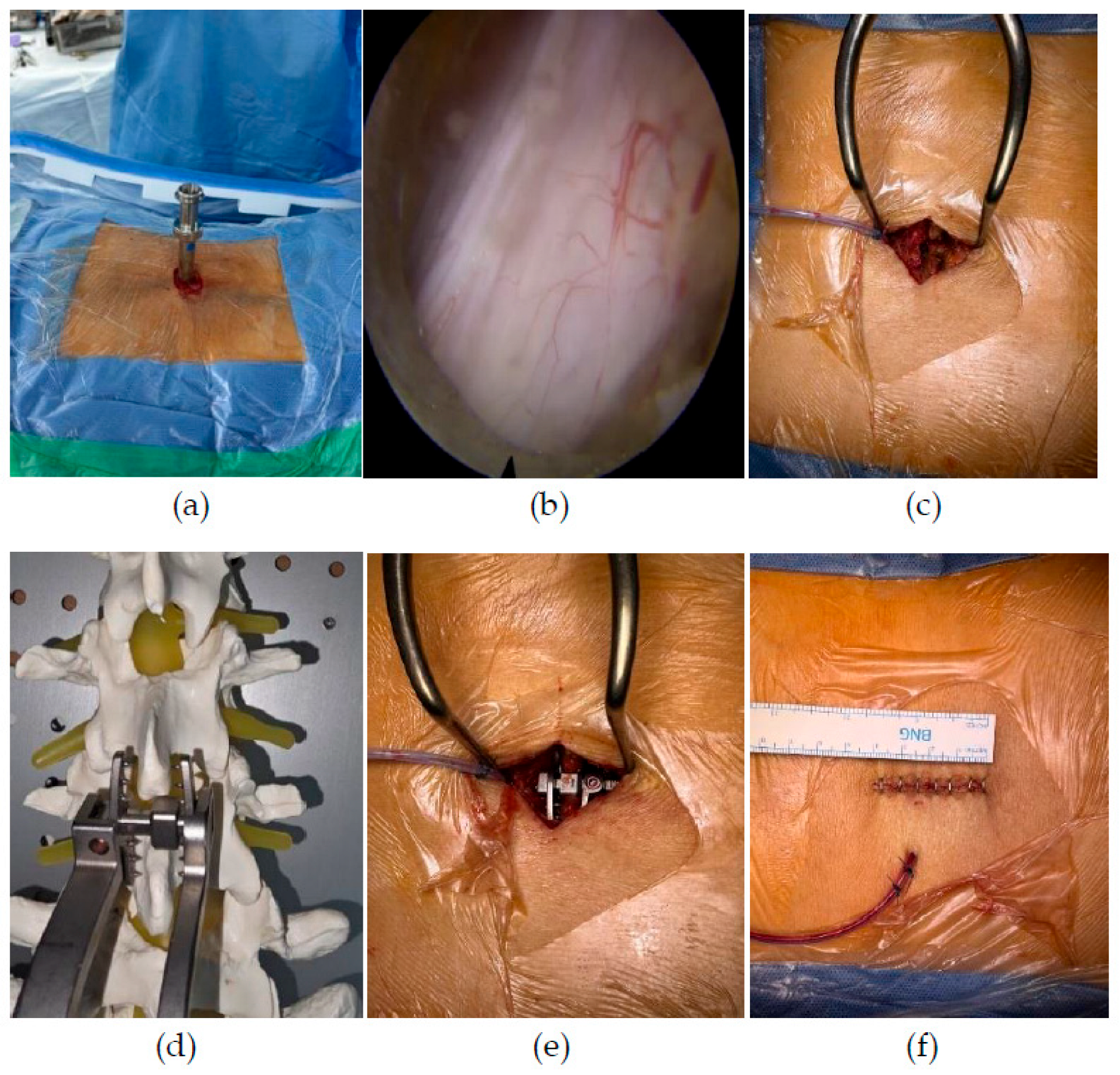
Figure 2.
Radiographic measurements (a) Percent slip and mean disc height (MDH) were measured on standing plain radiographs. The percent slip was calculated as the horizontal displacement between the posterior margins of the cranial and caudal vertebral bodies divided by the anteroposterior length of the superior endplate of the caudal vertebral body, multiplied by 100. MDH was calculated as the average of the anterior and posterior disc heights. (b,c) Segmental translation and dynamic angular motion were measured using dynamic standing radiographs. Segmental translation was defined as the absolute difference in vertebral displacement between flexion and extension. Dynamic angular motion was defined as the absolute difference in the segmental Cobb angle at the index level between the two positions.
Figure 2.
Radiographic measurements (a) Percent slip and mean disc height (MDH) were measured on standing plain radiographs. The percent slip was calculated as the horizontal displacement between the posterior margins of the cranial and caudal vertebral bodies divided by the anteroposterior length of the superior endplate of the caudal vertebral body, multiplied by 100. MDH was calculated as the average of the anterior and posterior disc heights. (b,c) Segmental translation and dynamic angular motion were measured using dynamic standing radiographs. Segmental translation was defined as the absolute difference in vertebral displacement between flexion and extension. Dynamic angular motion was defined as the absolute difference in the segmental Cobb angle at the index level between the two positions.
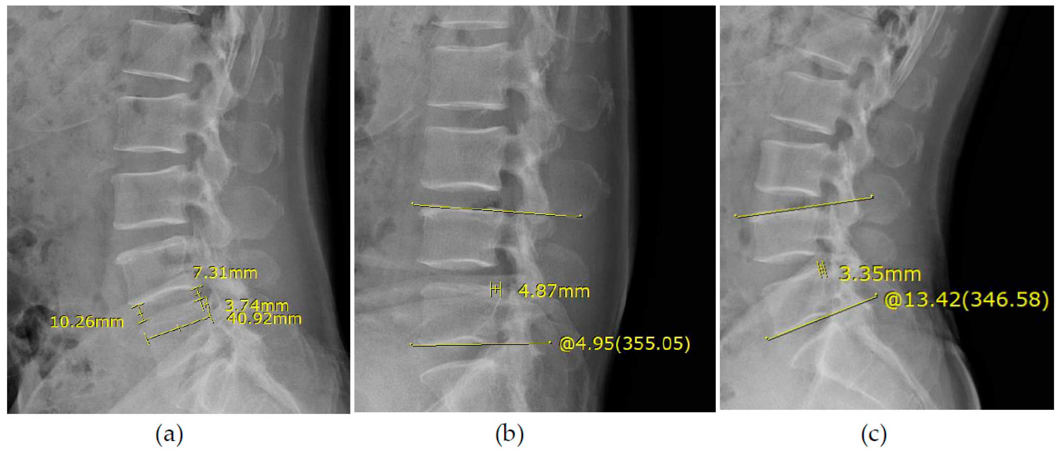
Figure 3.
Representative case (a) Preoperative standing plain radiograph showing a mean disc height of 8 mm and a percent slip of 9%. (b,c) Preoperative dynamic radiographs showing a segmental translation of 1.5 mm and a dynamic angular motion of 8°. (d) Preoperative axial T2-weighted magnetic resonance image showing severe lumbar spinal stenosis at L4–5. (e) Standing plain radiograph obtained at 12 months postoperatively showing a mean disc height of 9 mm and a percent slip of 8%. (f,g) Standing dynamic radiographs obtained at 12 months postoperatively showing a segmental translation of 0.3 mm and a dynamic angular motion of 8°. (h) Immediate postoperative axial T2-weighted magnetic resonance image confirming adequate neural decompression.
Figure 3.
Representative case (a) Preoperative standing plain radiograph showing a mean disc height of 8 mm and a percent slip of 9%. (b,c) Preoperative dynamic radiographs showing a segmental translation of 1.5 mm and a dynamic angular motion of 8°. (d) Preoperative axial T2-weighted magnetic resonance image showing severe lumbar spinal stenosis at L4–5. (e) Standing plain radiograph obtained at 12 months postoperatively showing a mean disc height of 9 mm and a percent slip of 8%. (f,g) Standing dynamic radiographs obtained at 12 months postoperatively showing a segmental translation of 0.3 mm and a dynamic angular motion of 8°. (h) Immediate postoperative axial T2-weighted magnetic resonance image confirming adequate neural decompression.
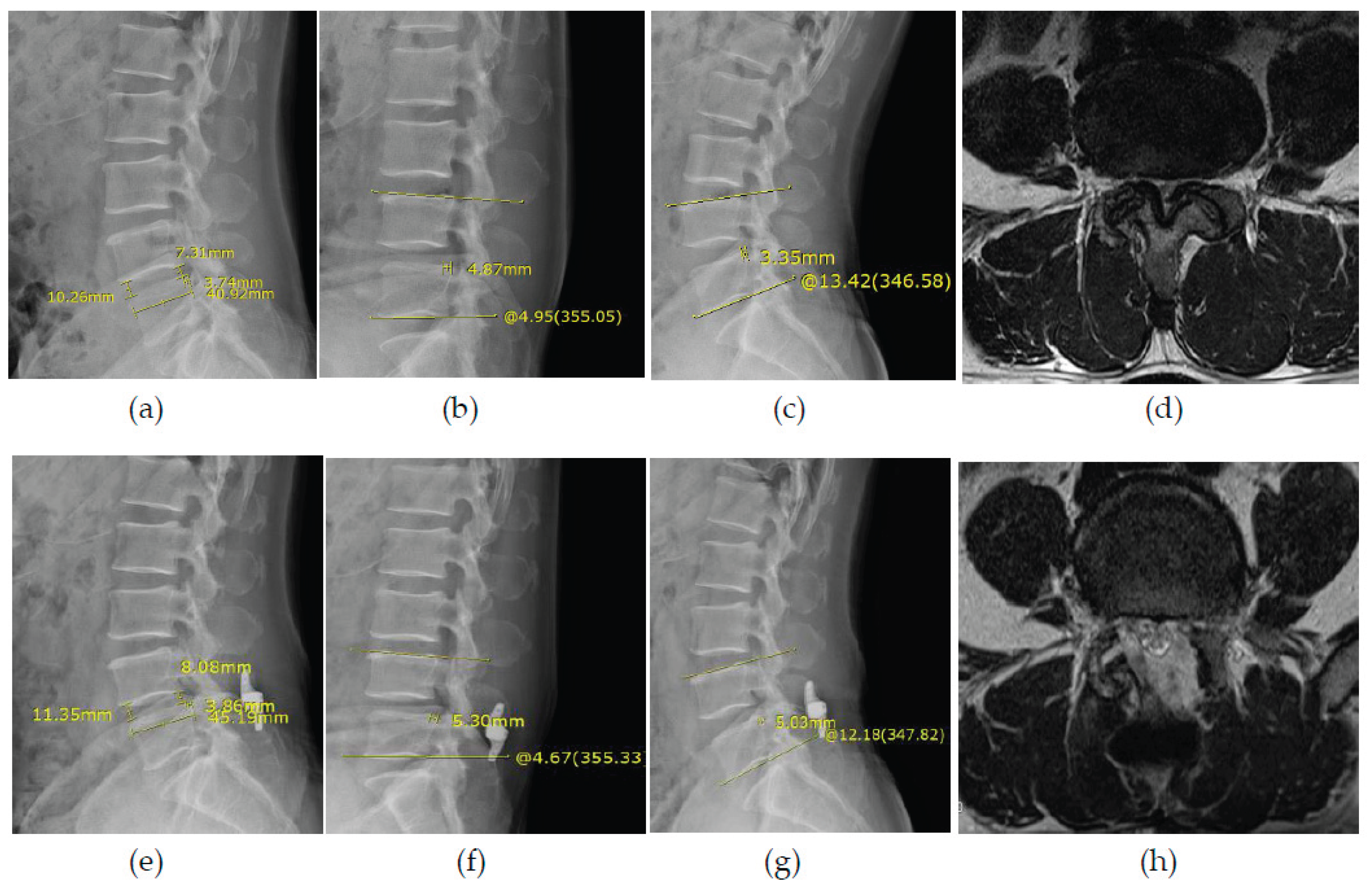

Table 1.
Baseline and Perioperative Characteristics.
| Variable | Value |
|---|---|
| Age (years) | 69.3 ± 9.3 |
| Sex, n (%) | |
| Male | 17 (36.2) |
| Female | 30 (63.8) |
| BMI (kg/m²) | 25.3 ± 2.5 |
| BMD T-score | −1.6 ± 1.1 (n = 42) |
| Osteoporosis, n/N (%) | 13/42 (31.0) |
| Facet cyst, n (%) | 31 (66.0) |
| Diabetes mellitus, n (%) | 16 (34.0) |
| Hypertension, n (%) | 18 (38.3) |
| ASA physical status, n (%) | |
| Class I | 15 (31.9) |
| Class II | 22 (46.8) |
| Class III | 10 (21.3) |
| Surgical level, n (%) | |
| L3–4 | 18 (38.3) |
| L4–5 | 27 (57.4) |
| L5–S1 | 2 (4.3) |
| Operation time (min) | 110 (100–120) |
| Hospital stay (days) | 10 (7–12) |
| Anesthesia type, n (%) | |
| Epidural | 44 (93.6) |
| General | 3 (6.4) |
| Follow-up duration (months) | 18.4 ± 6.3 |
Data are presented as mean ± SD, median (IQR), or n (%), unless otherwise indicated. BMD data were available for 42 patients, and the percentage of osteoporosis was calculated among the available cases. Abbreviations: ASA, American Society of Anesthesiologists; BMD, bone mineral density; BMI, body mass index; IQR, interquartile range; SD, standard deviation.
Table 2.
Clinical Outcomes.
| Variable | Preoperative | 1 Week | 6 Months | 12 Months | Overall P Value | ||
|---|---|---|---|---|---|---|---|
| VAS leg | 5.7 ± 1.7 | 3.4 ± 1.4 | 2.4 ± 0.9 | 2.1 ± 1.0 | <0.001 | ||
| VAS back | 4.5 ± 1.8 | 4.0 ± 1.3 | 2.5 ± 0.8 | 1.9 ± 1.0 | <0.001 | ||
| ODI | 66.0 ± 14.9 | 42.7 ± 17.1 | 24.4 ± 10.5 | 19.5 ± 13.0 | <0.001 | ||
| Modified MacNab criteria n (%) | |||||||
| Excellent | 29 (61.7) | ||||||
| Good | 13 (27.7) | ||||||
| Fair | 4 (8.5) | ||||||
| Poor | 1 (2.1) | ||||||
| Favorable (excellent + good) | 42 (89.4) | ||||||
Data are presented as mean ± SD or n (%), as appropriate. Overall P values for VAS leg pain, VAS back pain, and ODI were obtained using linear mixed-effects models, with time as a categorical fixed effect and patient as a random effect. The modified MacNab outcome was descriptively assessed at 12 months, and a favorable outcome was defined as excellent or good. Abbreviations: ODI, Oswestry Disability Index; SD, standard deviation; VAS, visual analog scale.
Table 3.
Radiographic Outcomes at 12 Months.
| Variable | Preoperative | 12 Months | Raw P Value | Holm-Adjusted P Value |
|---|---|---|---|---|
| Percent slip (%) | 13.6 ± 4.7 | 14.3 ± 5.1 | 0.189 | 0.298 |
| Mean disc height (mm) | 7.6 ± 2.0 | 8.1 ± 2.4 | 0.033 | 0.098 |
| Dynamic angular motion (°) | 6.7 ± 4.2 | 5.6 ± 3.6 | 0.021 | 0.085 |
| Segmental translation (mm) | 1.5 ± 1.3 | 1.4 ± 1.0 | 0.149 | 0.298 |
| Segmental instability, n (%) | 17 (36.2) | 11 (23.4) | 0.146 | — |
| Radiographic progression, n (%) | — | 4 (8.5) | — | — |
Continuous variables are presented as mean ± SD. Preoperative and 12-month continuous radiographic parameters were compared using paired t-tests or Wilcoxon signed-rank tests, as appropriate. Holm adjustment was applied across the four continuous radiographic comparisons. Segmental instability was compared using McNemar’s test and defined as translation >4 mm and/or angular motion >10°. Radiographic progression was defined as an absolute increase in percent slip of more than 5 percentage points from baseline to 12 months. Abbreviations: SD, standard deviation.
Table 4.
Complications and Reoperations.
| Event | n (%) | |
|---|---|---|
| Patients with ≥1 complication | 6 (12.8) | |
| Hematoma | 2 (4.3) | |
| Surgical-site infection | 3 (6.4) | |
| Dural tear | 3 (6.4) | |
| Spinous process fracture | 0 (0.0) | |
| Lumbar kyphotic change | 1 (2.1) | |
| Patients requiring reoperation | 4 (8.5) | |
| Reasons for reoperation, n | ||
| Hematoma | 2 | |
| Infection with dural tear | 1 | |
| Lumbar kyphotic change | 1 | |
Complication categories are not mutually exclusive; an individual patient may experience more than one complication. Reoperation counts represent the number of patients.
Table 5.
Exploratory Comparison Between Progression and Non-Progression Groups.
| Variable | Non-Progression (n = 43) | Progression (n = 4) | P Value |
|---|---|---|---|
| BMI (kg/m²) | 25.0 (24.0–26.6) | 28.0 (27.0–28.1) | 0.089 |
| Preoperative segmental instability, n (%) | 14 (32.6) | 3 (75.0) | 0.128 |
| Preoperative mean disc height (mm) | 7.5 (6.5–8.8) | 6.5 (5.9–6.9) | 0.194 |
| Segmental instability at 12 months, n (%) | 9 (20.9) | 2 (50.0) | 0.229 |
| Change in mean disc height (mm) | 1.0 (−0.5 to 1.8) | −0.8 (−1.1 to −0.2) | 0.113 |
| VAS leg at 12 months | 2.0 (1.0–3.0) | 2.0 (2.0–2.5) | 0.462 |
| VAS back at 12 months | 2.0 (1.0–2.0) | 2.0 (1.8–3.0) | 0.514 |
| ODI at 12 months | 15.0 (11.0–25.0) | 12.5 (10.8–27.2) | 0.817 |
| Favorable modified MacNab outcome, n (%) | 39 (90.7) | 3 (75.0) | 0.372 |
Continuous variables are presented as medians (IQR), and categorical variables are presented as n (%). Continuous variables were compared using exact rank-permutation tests based on the Mann–Whitney statistic, and categorical variables were compared using Fisher’s exact test. These comparisons were exploratory, and the P-values were not adjusted for multiple testing. Multivariable regression was not performed because only four radiographic progression events occurred. Radiographic progression was defined as an absolute increase in percent slip of more than 5 percentage points at 12 months. Segmental instability was defined as segmental translation >4 mm and/or dynamic angular motion >10°. Favorable modified MacNab outcomes were defined as either excellent or good. Abbreviations: BMI, body mass index; IQR, interquartile range; ODI, Oswestry Disability Index; VAS, visual analog scale.
Disclaimer/Publisher’s Note: The statements, opinions and data contained in all publications are solely those of the individual author(s) and contributor(s) and not of MDPI and/or the editor(s). MDPI and/or the editor(s) disclaim responsibility for any injury to people or property resulting from any ideas, methods, instructions or products referred to in the content. |
© 2026 by the authors. Licensee MDPI, Basel, Switzerland. This article is an open access article distributed under the terms and conditions of the Creative Commons Attribution (CC BY) license (http://creativecommons.org/licenses/by/4.0/).
Copyright: This open access article is published under a Creative Commons CC BY 4.0 license, which permit the free download, distribution, and reuse, provided that the author and preprint are cited in any reuse.