

Submitted:
21 October 2026
Posted:
21 October 2026
You are already at the latest version
Abstract
The oral and gastrointestinal microbiomes are increasingly recognized as interconnected ecological systems rather than isolated microbial communities. The oral cavity harbors diverse biofilm-associated microbial populations that are continuously introduced into the gastrointestinal tract through saliva, shed biofilms, and swallowed planktonic bacteria. Under homeostatic conditions, gastric acidity, bile, antimicrobial peptides, epithelial barriers, and resident gut microbiota limit the persistence of oral microbes. However, disruption of colonization resistance by inflammation, antibiotics, proton-pump inhibitors, immunosuppression, diet, or host susceptibility may allow oral-derived taxa to persist or engraft within the gut. This oral–gut microbiome axis is mediated through microbial migration, immune signaling, metabolite exchange, epithelial barrier disruption, and inflammatory feedback loops. Growing evidence links oral dysbiosis and periodontal inflammation with gastrointestinal disorders, including inflammatory bowel disease, irritable bowel syndrome, celiac disease, colitis, and colorectal cancer, as well as systemic diseases such as cardiovascular disease, metabolic disorders, neurodegenerative diseases, and autoimmune conditions. Among these, inflammatory bowel disease and colorectal cancer currently have the strongest mechanistic and translational support. Oral microbiome-based biomarkers, particularly from saliva and oral swabs, also show promise for non-invasive screening, disease monitoring, risk prediction, and personalized intervention. Nevertheless, many associations remain context-dependent, and causality is not fully established. Future progress will require longitudinal cohorts, standardized sampling pipelines, mechanistic validation, and integration of microbial, metabolic, immune, and clinical data. Understanding the oral–gut axis may expand current approaches to prevention, diagnosis, and precision management of gastrointestinal and systemic diseases.
Keywords:
oral microbiome
; gut microbiome
; oral–gut axis
; dysbiosis
; periodontal inflammation
; inflammatory bowel disease
; colorectal cancer
; microbiome biomarkers
1. Introduction
The human body is colonized by complex microbial communities that occupy distinct ecological niches, most prominently the oral cavity and the gastrointestinal tract. The oral microbiome encompasses diverse bacteria, fungi, viruses, archaea, and their collective genomes inhabiting oral surfaces, including the teeth, gingival crevice, tongue, saliva, palate, and buccal mucosa. It is one of the most taxonomically and functionally diverse microbial ecosystems in the body and exists predominantly as structured, surface-attached biofilms shaped by salivary flow, dietary inputs, oxygen gradients, host immunity, and mechanical perturbation [1,2,3]. In contrast, the gut microbiome represents a densely populated microbial community distributed along the gastrointestinal tract, with maximal biomass and metabolic activity in the colon. This ecosystem plays central roles in host physiology, including nutrient metabolism, vitamin synthesis, immune homeostasis, epithelial barrier maintenance, and colonization resistance against exogenous microbes [4,5,6,7].
Historically, the oral and gut microbiomes were studied as independent systems. However, growing evidence supports the existence of an oral–gut microbiome axis, a bidirectional network linking these ecosystems through microbial transfer, immune signaling, and metabolic exchange [8,9]. The oral cavity continuously seeds the gastrointestinal tract through daily swallowing of saliva - estimated at 10¹¹ to 10¹² microbial cells per day - along with biofilm fragments and planktonic bacteria shed from oral surfaces [8,10]. Under homeostatic conditions, multiple ecological barriers - including gastric acidity, bile exposure, antimicrobial peptides, and competition from established gut microbiota - limit the persistence of oral microbes in the intestine. This phenomenon reflects colonization resistance, whereby resident microbial communities and host factors restrict the establishment of incoming organisms through nutrient competition, niche exclusion, and immune-mediated clearance [7,11]. However, these barriers are not absolute. Perturbations such as antibiotic exposure, proton-pump inhibitor use, inflammation, dietary shifts, or immunosuppression can disrupt colonization resistance and create permissive niches for microbial invasion [7,12,13]. Within this altered ecological landscape, oral-derived organisms may transition from transient passengers to stable colonizers, a process often referred to as ectopic colonization [13,14]. This distinction between transient passage and true engraftment is critical, as only the latter is likely to drive sustained functional and immunological consequences in the gut. Indeed, experimental studies have demonstrated that oral pathobionts, including Klebsiella species, can colonize the intestine and induce T helper 1 (Th1)-mediated inflammation under dysbiosis conditions [13].
A central mechanistic feature of the oral–gut axis is microbial translocation, defined as the movement of viable microorganisms or their molecular products from their original niche to distal anatomical sites. In this context, translocation may occur via multiple routes, including ingestion of oral microbes, hematogenous dissemination through inflamed periodontal tissues, or systemic circulation of microbial-associated molecular patterns such as lipopolysaccharide, peptidoglycan, bacterial DNA, and extracellular vesicles [8,15,16,17]. These processes enable oral microbial signals to influence distal mucosal and systemic immune responses, effectively linking localized oral dysbiosis to broader inflammatory states. From an ecological perspective, the oral–gut axis can be understood through principles of niche theory, ecological filtering, and community resilience. The gut environment imposes strong selective pressures that typically exclude non-adapted oral taxa. However, disruption of ecological stability through inflammation or microbiome perturbation reduces system resilience, allowing invasion by opportunistic organisms [18,19]. In this framework, certain oral microbes function as pathobionts or even keystone pathogens, capable of disproportionately altering host-microbe interactions and community structure despite relatively low abundance [20,21].
Importantly, the oral–gut axis is not restricted to bacterial interactions. The oral and gut mycobiome and virome, including bacteriophages, also contribute to community dynamics and host responses, although their roles remain less well defined. Fungal species such as Candida and phage-mediated modulation of bacterial populations may influence colonization dynamics, immune activation, and microbial competition across sites [22,23,24]. While bacterial mechanisms dominate current understanding, incorporating these additional microbial components is essential for a comprehensive view of cross-ecosystem interactions. The clinical relevance of the oral–gut axis is increasingly recognized across a wide spectrum of gastrointestinal and systemic diseases. Oral dysbiosis and periodontal inflammation have been associated with inflammatory bowel disease, colorectal cancer, cardiovascular disease, metabolic disorders, autoimmune conditions, and neurodegenerative diseases [25,26,27,28,29]. Notably, these associations often reflect ecological and functional disruption rather than the presence of a single causative pathogen, involving shifts in microbial composition, altered metabolite production, impaired epithelial barriers, and chronic immune activation. At the same time, systemic diseases can reciprocally influence the oral microbiome through changes in immune function, salivary composition, medication exposure, and inflammatory status, underscoring the bidirectional nature of this axis [30].
A particularly important implication of this framework is its ability to help explain the persistent clinic-laboratory disconnect observed in infectious and inflammatory diseases, where microbial behavior in vivo does not always align with in vitro expectations. Microbiome context, interspecies interactions, and host responses can fundamentally reshape microbial phenotypes, highlighting the limitations of reductionist models that isolate individual organisms from their ecological environment. Host factors further modulate the oral–gut axis. Genetic variation in immune pathways, epithelial barrier function, and inflammatory responsiveness influences susceptibility to microbial translocation and disease progression. Thus, the impact of oral dysbiosis on gut and systemic health is not uniform but depends on the interplay between microbial exposure and host predisposition [31]. Together, these findings support a conceptual model in which the oral cavity functions as a microbial reservoir that can influence intestinal ecology and systemic inflammation through swallowed oral microbes, barrier disruption, immune signaling, and loss of colonization resistance (Figure 1).
2. The Oral Microbiome
The oral microbiome is a highly diverse and spatially structured microbial ecosystem distributed across multiple anatomical niches, including the teeth, gingival sulcus, tongue dorsum, buccal mucosa, palate, tonsils, and saliva. Rather than functioning as a single homogeneous community, it comprises distinct, site-specialized microbial consortia shaped by local environmental conditions such as oxygen tension, pH, nutrient availability, host immune factors, salivary flow, and surface characteristics [1,2,3]. More than 600 prevalent bacterial taxa have been identified in the human oral cavity, with substantial inter-individual variability but consistent ecological organization within niche-specific communities [1]. This biogeographical structuring is a defining feature of the oral microbiome and underlies both its resilience in health and its susceptibility to dysbiosis under perturbation [2,32]. From a systems perspective, the oral microbiome functions as a dynamic, multi-habitat ecosystem, in which microbial communities are interconnected through physical proximity, fluid flow, and metabolic exchange. These communities contribute to colonization resistance, modulation of host immunity, nitrate metabolism, and maintenance of ecological stability [33,34]. At the same time, the oral cavity serves as a major reservoir for microbial dissemination, with implications for both gastrointestinal and systemic health [8,15].
2.1. Core Microbial Taxa
The healthy oral microbiome is dominated by bacterial members of the phyla Firmicutes, Bacteroidetes, Proteobacteria, Actinobacteria, Fusobacteria, and Spirochaetes, although their relative abundances vary across oral niches and host conditions [1]. Prominent genera include Streptococcus, Actinomyces, Veillonella, Neisseria, Haemophilus, Rothia, Prevotella, Fusobacterium, Porphyromonas, Capnocytophaga, Leptotrichia, and Treponema [1]. Streptococcus species act as early colonizers, initiating biofilm formation by adhering to the acquired enamel pellicle and enabling subsequent microbial succession through coaggregation and metabolic cross-feeding [35,36]. Secondary colonizers, particularly Fusobacterium nucleatum, function as bridging organisms, facilitating interactions between early and late colonizers and supporting the development of complex multispecies communities [36]. In health, oral microbial communities exhibit functional redundancy and metabolic cooperation, which enhances ecological resilience and stability. For example, commensal bacteria participate in nitrate reduction pathways that influence systemic nitric oxide production and vascular function [33,34,37]. However, many oral microbes can act as pathobionts, transitioning to disease-associated states under ecological disruption. Key examples include Porphyromonas gingivalis, Tannerella forsythia, and Treponema denticola, which are strongly associated with periodontitis, as well as acidogenic organisms such as Streptococcus mutans linked to dental caries [21,38]. Importantly, oral disease reflects community-level dysbiosis, rather than the action of a single pathogen, with shifts in microbial composition and function driving inflammation and tissue destruction [20,21].
2.2. Ecological Organization: Biofilms, Saliva, and Mucosa
A defining feature of the oral microbiome is its organization into biofilms, structured microbial communities embedded within an extracellular polymeric matrix. Dental plaque develops through a sequential process involving pellicle formation, attachment of pioneer species, coaggregation, and maturation into complex multispecies communities [36,39]. Within biofilms, microbes are arranged in spatially organized microenvironments characterized by gradients in oxygen, pH, and nutrient availability. These gradients enable the coexistence of aerobic, facultative anaerobic, and obligate anaerobic organisms within close proximity, facilitating metabolic cooperation and niche partitioning [2,32]. The extracellular matrix enhances resistance to mechanical disruption, antimicrobial agents, and host immune defenses [39].
Saliva plays a dual ecological role as both a regulatory and dispersal medium. It contains antimicrobial peptides, secretory IgA, enzymes, mucins, and nutrients that shape microbial growth, while simultaneously transporting microbes throughout the oral cavity and into the gastrointestinal tract [40]. Microbial densities in saliva can reach approximately 10⁸ cells per milliliter, with organisms shed from multiple oral niches, including the tongue dorsum, tooth surfaces, gingival crevice, and buccal mucosa [3,41]. The oral mucosa provides additional ecological niches. The tongue dorsum supports dense microbial communities due to its papillary structure and oxygen gradients, whereas non-keratinized surfaces, such as the buccal mucosa, experience rapid epithelial turnover, leading to more transient microbial populations [3,42,43]. This site-specific ecological differentiation, often described as the “site-specialist hypothesis,” highlights the role of environmental filtering in shaping microbial community composition [32]. The spatial organization of oral microbial communities across teeth, the gingival sulcus, the tongue dorsum, saliva, and the buccal mucosa is summarized in Figure 2, highlighting how local ecological conditions shape niche-specific microbial composition.
2.3. Major Drivers of Oral Dysbiosis
Oral dysbiosis refers to the disruption of microbial composition and function that promotes inflammation and disease. One of the primary drivers is periodontitis, a chronic inflammatory condition characterized by a shift toward anaerobic, proteolytic, and inflammation-adapted microbial communities within subgingival biofilms [16,21]. The polymicrobial synergy and dysbiosis model posits that keystone pathogens such as P. gingivalis can subvert host immune responses and reshape the microbial community, leading to a self-sustaining cycle of inflammation and dysbiosis [20,21]. Periodontal inflammation increases gingival crevicular fluid flow and releases host-derived nutrients, further selecting for dysbiotic organisms [16]. Poor oral hygiene contributes to plaque accumulation and biofilm maturation. As biofilms thicken, oxygen levels decrease, favoring anaerobic organisms associated with gingivitis and periodontitis [39]. Dietary factors also play a significant role, with frequent consumption of fermentable carbohydrates promoting acid production and selection of acid-tolerant species involved in caries [38,44].
Systemic and environmental factors further influence oral microbial ecology. Smoking, diabetes, aging, xerostomia, immunosuppression, and medication use - including antibiotics and proton-pump inhibitors - have all been associated with shifts in oral microbiome composition and function [12,45,46]. These factors act by altering host immunity, salivary composition, and the physicochemical environment of the oral cavity. Importantly, oral dysbiosis extends beyond local pathology. Inflammation-associated disruption of the gingival epithelium facilitates hematogenous dissemination of bacteria and microbial products, while saliva continuously delivers oral microbes into the gastrointestinal tract. Thus, the dysbiotic oral microbiome functions as both a source of systemic inflammatory signals and a reservoir of organisms capable of colonizing distal sites under permissive conditions [8,16]. Several oral and gut microbial taxa are repeatedly implicated in oral–gut microbiome interactions. Some organisms, such as Streptococcus, Veillonella, Prevotella, F. nucleatum, and P. gingivalis, are primarily associated with the oral cavity but may influence intestinal and systemic disease under dysbiotic conditions. In contrast, gut-associated organisms such as Faecalibacterium prausnitzii, Akkermansia muciniphila, Bacteroides, and Escherichia/Shigella reflect intestinal ecological balance or disruption. Representative taxa relevant to the oral–gut axis are summarized in Table 1.
3. Mechanisms Linking the Oral and Gut Microbiomes
The oral–gut microbiome axis is mediated by interconnected biological processes that enable communication between the oral and gut ecosystems. These mechanisms extend beyond simple microbial transfer and include ecological invasion, immune signaling, metabolite exchange, and disruption of epithelial barriers. Together, they form a multilayered network through which oral dysbiosis can influence intestinal homeostasis and systemic disease. Importantly, these interactions are bidirectional, with gut-derived signals also shaping oral microbial ecology, reinforcing the concept of an integrated mucosal system rather than isolated microbial compartments. These pathways are not mutually exclusive and may operate simultaneously, depending on host susceptibility, oral inflammatory status, gut barrier integrity, medication exposure, and baseline gut microbiome composition. As summarized in Figure 3, oral dysbiosis may affect intestinal and systemic health through five interconnected pathways: microbial migration, immune-mediated communication, metabolite-mediated effects, epithelial barrier disruption, and inflammatory feedback loops.
3.1. Major Drivers of Oral Dysbiosis
A fundamental connection between the oral cavity and the gastrointestinal tract is the continuous migration of microorganisms via saliva. Humans swallow substantial volumes of saliva daily, which contains large numbers of microbial cells shed from oral biofilms and mucosal surfaces; estimates suggest that approximately 10¹² oral bacteria may enter the gastrointestinal tract each day [8]. Under physiological conditions, most of these organisms behave as transient passengers that are cleared by gastric acidity, bile salts, digestive enzymes, and competition from resident gut microbiota. This reflects the principle of colonization resistance, whereby established microbial communities and host defenses prevent the stable establishment of incoming organisms through nutrient competition, niche exclusion, and immune-mediated clearance [7,11].
However, this barrier is not absolute. Disruption of colonization resistance - such as through antibiotic exposure, proton-pump inhibitor use, inflammation, immunosuppression, or dietary changes - can create permissive ecological conditions that allow oral-derived microbes to persist within the gut [8,12,13,64]. In this context, a critical distinction emerges between transient passage and stable engraftment. While most oral microbes pass through the gastrointestinal tract without consequence, a subset can establish functional niches under dysbiotic conditions, leading to sustained ecological and immunological effects. Experimental studies support this model, demonstrating that orally derived bacteria, such as Klebsiella species, can colonize the intestine and induce T helper 1–mediated inflammation [13]. Similarly, oral-associated taxa, including F. nucleatum, Porphyromonas, and Prevotella, have been identified in the gut microbiota of patients with inflammatory bowel disease and colorectal cancer, suggesting that oral microbes can contribute to disease-associated intestinal communities [25,26,27].
3.2. Immune-Mediated Communications
Beyond direct microbial migration, the oral–gut axis is strongly mediated by the host immune system, which integrates microbial signals across mucosal sites. Oral inflammation, particularly in periodontitis, results in the release of pro-inflammatory cytokines and microbial-associated molecular patterns into systemic circulation [16,17]. These signals are recognized by pattern recognition receptors such as Toll-like receptors and nucleotide-binding oligomerization domain-like receptors, leading to activation of innate immune pathways and modulation of adaptive immune responses [65,66]. A key consequence of this signaling is the polarization of T helper cell responses, particularly T helper 1 (Th1) and T helper 17 (Th17) pathways, which play essential roles in mucosal immunity but are also implicated in chronic inflammatory disease [67]. Oral pathobionts can promote these inflammatory responses, contributing to intestinal immune activation and barrier dysfunction [13,14]. At the same time, gut inflammation can influence oral immunity through systemic cytokine signaling and immune cell redistribution, highlighting the bidirectional nature of immune communication along the oral–gut axis. This interconnected immune landscape underscores how localized dysbiosis can propagate inflammatory responses across distant mucosal sites.
3.3. Metabolite-Mediated Interaction
Microbial metabolites represent another critical layer of communication linking the oral and gut microbiomes. Both ecosystems produce a wide array of bioactive compounds that can influence host physiology and microbial community dynamics. Among these, short-chain fatty acids such as acetate, propionate, and butyrate play central roles in maintaining epithelial barrier integrity, regulating immune responses, and suppressing inflammation [58,59]. Although these metabolites are primarily associated with gut microbiota, their systemic effects extend beyond the intestinal environment. In contrast, dysbiotic oral communities - particularly those associated with periodontitis - are characterized by increased proteolytic metabolism, generating compounds such as ammonia, hydrogen sulfide, phenols, and biogenic amines. These metabolites contribute to tissue damage, inflammation, and altered microbial selection both locally and systemically [68]. In addition, endotoxins, such as lipopolysaccharide (LPS) derived from Gram-negative oral bacteria, can enter the systemic circulation, promoting low-grade inflammation and endothelial dysfunction [16,69]. These circulating microbial products can influence gut immune responses and may contribute to shifts in intestinal microbial composition, linking the two ecosystems through metabolite-driven signaling pathways.
3.4. Effects on Barrier Integrity
Epithelial barrier integrity represents a critical interface in the oral–gut axis, regulating interactions between microbial communities and host tissues. In the oral cavity, periodontal inflammation compromises the gingival epithelium, increasing vascular permeability and facilitating the entry of bacteria and microbial products into systemic circulation [16,17]. This provides a pathway for the dissemination of oral-derived inflammatory signals beyond the local environment. In the gastrointestinal tract, inflammatory processes can disrupt tight junctions between epithelial cells, leading to increased intestinal permeability and allowing luminal microbes and their products to translocate into underlying tissues and the bloodstream [70,71]. Barrier dysfunction is both a cause and consequence of microbial dysbiosis. Oral inflammation may contribute to gut barrier disruption through systemic inflammatory mediators, while gut inflammation can alter oral microbial composition by influencing immune tone and host defenses [14,72]. Together, these processes establish a self-reinforcing cycle in which dysbiosis, barrier disruption, microbial translocation, and inflammation perpetuate one another across anatomical sites. Such feedback loops are likely central to the persistence and progression of chronic inflammatory diseases associated with the oral–gut microbiome axis.
The clinical relevance of the oral–gut microbiome axis varies across diseases. Evidence is strongest for inflammatory bowel disease (IBD) and colorectal cancer (CRC), where oral-associated taxa have been repeatedly detected in intestinal or tumor-associated microbial communities and supported by mechanistic studies. Evidence for cardiovascular, metabolic, autoimmune, and neurodegenerative diseases is biologically plausible but remains more indirect, often involving systemic inflammation, microbial products, barrier disruption, and immune signaling. In irritable bowel syndrome (IBS), celiac disease, and some forms of colitis, current findings are still emerging and should be interpreted cautiously. A simplified overview of these disease associations, proposed mechanisms, and clinical implications is provided in Table 2.
4. Oral Dysbiosis and Gastrointestinal Disorders
Oral dysbiosis is increasingly recognized as a biologically meaningful contributor to gastrointestinal disease, rather than a process confined to the oral cavity. Current oral–gut axis models propose that the mouth can serve as a reservoir of pathobionts, inflammatory mediators, and disrupted microbial communities that reach the intestine through swallowed saliva, shed oral biofilms, transient bacteremia, and immune-mediated signaling [8,55,63,87]. This model has gained attention because many gastrointestinal disorders are marked by impaired epithelial barrier integrity, weakened colonization resistance, and chronic immune activation, creating conditions that may allow oral taxa to persist, engraft, or influence intestinal physiology [14,88]. Importantly, the oral–gut relationship is bidirectional. Intestinal inflammation can alter oral microbial communities, salivary composition, and mucosal immune responses, while periodontal inflammation may increase systemic inflammatory burden and reshape the gut microbial ecosystem [87,89]. This bidirectionality argues against a simplistic one-way causal model. Instead, oral dysbiosis is best understood as part of a dynamic mucosal network in which microbial imbalance may function as a cause, consequence, biomarker, or amplifier of gastrointestinal disease.
4.1. Inflammatory Bowel Disease (IBD)
Among gastrointestinal disorders, inflammatory bowel disease (IBD) provides some of the strongest evidence for a clinically relevant connection between the oral and gut microbiomes. Recent reviews report that oral manifestations, including aphthous ulcers, gingival inflammation, periodontitis, and other mucosal abnormalities, are more common in individuals with IBD and may correlate with intestinal disease activity and systemic inflammatory status [55,63]. In parallel, oral microbiome profiling studies show that patients with IBD often have altered oral microbial communities, while the intestinal microbiome may show increased “oralization,” suggesting microbial translocation, ecological convergence, or shared inflammatory pressures across oral and intestinal niches [63,90]. Mechanistically, IBD research supports several overlapping pathways through which oral dysbiosis may influence intestinal inflammation. Periodontitis increases the burden of swallowed oral bacteria and inflammatory products, while intestinal inflammation weakens epithelial barrier defenses and colonization resistance, potentially allowing oral taxa to persist ectopically in the gut [55,63]. Organisms frequently implicated include F. nucleatum, P. gingivalis, Klebsiella spp., Campylobacter concisus, and selected oral streptococci. These taxa have been associated with epithelial barrier disruption, innate immune activation, altered cytokine signaling, and pathogenic T-cell polarization in experimental and translational studies [13,87,91,92].
F. nucleatum is one of the most extensively studied examples because it links microbial ecology to host immunopathology. Its enrichment in IBD-associated samples, together with its ability to invade epithelial cells, disrupt tight junction integrity, and activate inflammatory pathways, such as Toll-like receptor (TLR)/myeloid differentiation primary response 88 (MyD88)/nuclear factor kappa B (NF-κB) signaling, supports its potential role in mucosal injury [55,63]. Its adhesins and outer membrane vesicles may further enhance epithelial attachment, immune modulation, and cooperative interactions within polymicrobial communities. These findings suggest that F. nucleatum may act not only as a marker of inflammation but also as an active contributor to mucosal damage [55]. P. gingivalis provides a complementary model. Although best known as a periodontal pathogen, it can have systemic effects even without stable or dominant gut colonization. Experimental studies summarized in recent reviews suggest that oral exposure to P. gingivalis can alter gut microbial composition, reduce tight-junction protein expression, modify host metabolic pathways, and promote Th17-skewed inflammatory responses [55,63]. These findings indicate that oral bacteria may affect intestinal disease through both direct colonization and indirect ecological reprogramming of the gut environment.
Metabolites and immune mediators add further complexity. IBD is associated with depletion of short-chain fatty acid-producing gut commensals, while oral dysbiosis may favor proteolytic microbial communities and inflammatory metabolites that intensify epithelial stress and weaken barrier maintenance [63,90]. Overall, oral dysbiosis is unlikely to be a universal primary cause of IBD. However, current evidence supports its role as a disease-modifying factor that may influence susceptibility, inflammatory severity, and extraintestinal manifestations in biologically predisposed individuals [55,63,90].
4.2. Irritable Bowel Syndrome (IBS)
The relationship between oral dysbiosis and irritable bowel syndrome (IBS) is less clearly defined than in IBD. IBS is classified as a disorder of gut-brain interaction and does not involve the overt tissue-destructive inflammation characteristic of Crohn’s disease or ulcerative colitis, limiting direct mechanistic comparisons [73,74]. An exploratory study of the buccal mucosal microbiome in IBS identified compositional differences between IBS participants and healthy controls and reported associations between specific oral taxa and visceral pain severity, particularly among overweight individuals with IBS. Some oral microbial patterns resembled previously described fecal findings, suggesting that the oral microbiome may reflect broader host-microbiome disturbances relevant to IBS pathophysiology [73]. However, because the study was small, cross-sectional, and exploratory, its findings should be interpreted as hypothesis-generating rather than mechanistically conclusive. Current hypotheses, therefore, remain cautious. Low-grade inflammation may alter autonomic signaling, dietary patterns, stress-related biology, salivary changes, intestinal permeability, and motility, which may, in turn, shape oral microbial ecology in IBS [73,74]. At present, there is insufficient evidence to conclude that specific oral taxa are established causal drivers of IBS, as oral pathobionts are increasingly implicated in IBD and colorectal cancer [73].
4.3. Celiac Disease, Colitis, and Colorectal Cancer (CRC)
Beyond IBD and IBS, evidence linking oral dysbiosis to other gastrointestinal disorders is variable but increasingly suggestive. Broad oral-systemic reviews and oral–gut axis studies indicate that oral microbial disturbances may accompany celiac disease, non-IBD colitis, and colorectal cancer (CRC), although the depth and quality of evidence differ substantially across these conditions [74,75]. In celiac disease, oral microbial involvement remains an emerging area of investigation. Current literature suggests that celiac disease may be associated with oral manifestations and altered oral microbial ecology, but the field has not yet established a validated disease-specific oral microbial signature or a mechanistic framework comparable to that available for IBD [75]. The term “colitis” also requires careful distinction because it includes infectious, ischemic, microscopic, and immune-mediated forms. Much of the mechanistic evidence relevant to the oral–gut axis comes from experimental colitis models rather than from all forms of human colitis. These studies suggest that oral taxa such as F. nucleatum, P. gingivalis, Klebsiella spp., and oral streptococci may aggravate epithelial injury, inflammatory cytokine production, and pathological immune polarization [55,63].
CRC represents the most mature translational field within oral–gut axis research. Multiple studies have reported enrichment of oral-associated taxa, particularly F. nucleatum, in adenomas, colorectal tumors, and tumor-associated mucosal communities, supporting the hypothesis that oral bacteria may contribute to tumor-promoting inflammation, dysregulated epithelial signaling, and immune evasion [93,94,95]. CRC literature is especially compelling because it integrates ecological association studies, mechanistic work on virulence factors such as FadA and Fap2, and translational research evaluating salivary or oral microbial signatures as non-invasive biomarkers [93,94,96]. Importantly, oral microbiome research in CRC has moved beyond association toward potential clinical application. Rezasoltani et al. (2023) reported that salivary microbiota could differentiate early CRC from healthy controls and suggested that saliva-based microbial profiling may outperform fecal profiling in some classification models. Other studies have similarly proposed oral dysbiosis as a non-invasive biomarker for CRC detection and highlighted the recurrent diagnostic relevance of taxa such as Fusobacterium, Dialister, Catonella, and related oral genera [94,95]. More recent evidence also suggests that oral microbiota signatures may have prognostic value, indicating that the oral compartment could eventually contribute not only to screening but also to disease stratification [96].
5. Systemic Diseases Influenced by the Oral–Gut Axis
5.1. Cardiovascular Disease
According to the World Health Organization (WHO), approximately 19.8 million people died from cardiovascular diseases [97]. A meta-analysis of five case-control studies showed a 1.14-fold increased risk of coronary heart disease in patients with periodontal disease [79,98]. The oral microbiota harbors approximately 750 microbial species, including viruses, fungi, protozoa, and archaea. In oral dysbiosis and periodontal disease, this balance is disturbed, increasing the abundance of pathogenic or inflammatory microbial communities [99]. Recent studies on the oral spirochete T. denticola in atherosclerosis models and human vascular lesions have shown that dissemination into organs such as the heart can stimulate cytokine release and modify inflammatory pathways [100,101,102].
P. gingivalis, a bacterial pathogen commonly associated with adult periodontitis, can enter circulation through blood vessels and invade heart endothelial cells. It increases aortic plaque formation and inflammatory gene expression [56,103]. P. gingivalis disrupts endothelial homeostasis through TLR-NF-κB signaling, promotes endothelial-mesenchymal transition, and induces apoptosis [104]. In atherosclerosis, P. gingivalis also increases mitochondrial reactive oxygen species (ROS) levels and decreases adenosine triphosphate (ATP) via Drp1-dependent fission [105]. These findings indicate how periodontal pathogens may contribute to endothelial injury and cardiovascular disease. Long-term infection with P. gingivalis LPS in C57BL/6.KOR-Apoeshl mice reduced serum high-density lipoprotein (HDL) and increased very-low-density lipoprotein (VLDL), low-density lipoprotein (LDL), and total cholesterol. In this way, it disrupts cholesterol and lipid metabolism, worsening existing atherosclerosis and contributing to the development of coronary heart disease [106]. P. gingivalis also induces lipid peroxidation, modifies plasma lipoproteins, and depletes antioxidant capacity, suggesting a mechanistic link between periodontal infection, oxidative stress, and atherogenesis [107].
Human evidence supports an association but remains less definitive than animal and cell-culture evidence. Periodontopathic bacterial DNA, including P. gingivalis DNA, has been detected in coronary and carotid atheromatous plaques from patients with vascular disease, but detection of bacterial DNA does not prove that oral pathogens initiate plaque formation [108]. Therefore, oral dysbiosis is biologically plausible as a cardiovascular risk modifier, but the causal relationship in humans remains unresolved because periodontitis and cardiovascular disease share risk factors such as age, smoking, diabetes, socioeconomic status, diet, and systemic inflammation [81,109].
5.2. Metabolic Disorders
Metabolic disease is strongly connected to both oral and gut dysbiosis because metabolic syndrome, obesity, type 2 diabetes, hypertension, dyslipidemia, and nonalcoholic fatty liver disease (NAFLD) are all characterized by chronic low-grade inflammation and altered host-microbe interactions [110]. Periodontitis has been associated with metabolic syndrome and diabetes, while metabolic disease can worsen periodontal inflammation and reshape the salivary and subgingival microbiome [110,111]. One of the most important experimental studies for the oral–gut-metabolic pathway showed that oral administration of P. gingivalis in mice altered gut microbiota composition, increased systemic inflammation, impaired glucose and insulin tolerance, tended to increase circulating endotoxin, and reduced expression of ileal tight junction genes. This study supports a model in which swallowed periodontal pathogens disturb gut ecology and intestinal barrier integrity, producing metabolic endotoxemia and systemic inflammation [61].
Diabetes and periodontitis show a clinically important bidirectional relationship. Poor glycemic control impairs immune function, increases inflammatory responses, delays tissue repair, and favors periodontal pathogen expansion. In the opposite direction, periodontal inflammation increases systemic pro-inflammatory mediators that can worsen insulin resistance and metabolic control [111]. Reviews of metabolic syndrome and periodontitis suggest that lipids, inflammatory cytokines, altered host response, and microbiome changes jointly explain this interaction [110]. Obesity adds another layer to the oral–gut axis. The oral microbiome may influence obesity and metabolic disease through local effects on taste perception, dietary preference, nitrate metabolism, systemic inflammation, and downstream effects on the gut microbiome [112]. Salivary microbiome data also associate oral dysbiosis with hypertension and metabolic syndrome biomarkers, although the direction of causality remains uncertain [113]. The oral-liver axis is particularly relevant to metabolic disease because nonalcoholic fatty liver disease is considered the hepatic manifestation of metabolic syndrome [114]. Periodontal disease may contribute to NAFLD and nonalcoholic steatohepatitis through two routes: hematogenous dissemination of bacteria, inflammatory mediators, and reactive oxygen species, and enteral disruption of gut microbiota and gut barrier function, increasing hepatic exposure to microbial products through enterohepatic circulation [82,114].
5.3. Neurodegenerative Diseases
The oral–gut-brain axis is an emerging extension of the microbiota-gut-brain axis. Oral and gut dysbiosis may influence neurodegenerative disease through systemic inflammation, altered microbial metabolites, blood-brain barrier permeability, microglial activation, microbial amyloids, LPS signaling, and possible dissemination of oral microbes or microbial products to neural tissues [115,116]. Alzheimer’s disease is the most frequently discussed neurodegenerative condition in relation to oral dysbiosis. Oral microbial dysbiosis and periodontitis are common in older adults and may contribute to Alzheimer’s disease risk by increasing systemic pro-inflammatory cytokines, compromising blood-brain barrier integrity, and promoting neuroinflammatory cascades [117]. Reviews have reported the detection of oral-associated microbial signatures in Alzheimer’s disease brain tissue and have discussed P. gingivalis as a candidate contributor to neuroinflammation, amyloid pathology, and cognitive decline [118,119]. The causal status of periodontal pathogens in Alzheimer’s disease remains uncertain. A critical review concluded that P. gingivalis and other periodontal microorganisms may contribute to Alzheimer’s-related inflammatory and pathological changes, but human clinical evidence is insufficient to establish a direct causal relationship [119]. Therefore, the most accurate wording is that oral dysbiosis may act as a modifiable inflammatory risk factor or disease amplifier rather than an established primary cause of Alzheimer’s disease [117].
Gut dysbiosis also contributes to neurodegenerative diseases by increasing intestinal permeability, systemic inflammation, and neuroinflammation [120]. Microbiota-derived metabolites, including short-chain fatty acids, bile acid derivatives, neurotransmitter precursors, and inflammatory metabolites, can regulate microglia, astrocytes, oligodendrocytes, and peripheral immune responses [116]. Parkinson’s disease provides another important example because oral and gut dysbiosis have both been observed in patients. A shotgun metagenomic study found significant differences in the oral and gut microbiomes between patients with Parkinson’s disease and controls, with oral Lactobacillus associated with opportunistic gut pathogens and functional microbial changes in glutamate and arginine biosynthesis, as well as antimicrobial resistance genes [62]. Parkinson’s disease reviews also emphasize gut inflammation, impaired barrier function, neuroinflammation, changes in microbial metabolites, and possible α-synuclein propagation via the enteric nervous system and vagal pathways [83]. Evidence for multiple sclerosis is less developed but supports inclusion under inflammatory neuroimmune conditions. One study found that oral microbial communities differed between patients with multiple sclerosis (MS) and healthy controls, with higher abundances of genera including Staphylococcus, Actinomyces, Fusobacterium, Bacteroides, Porphyromonas, Prevotella, and Veillonella in MS patients [49]. Gut microbiome reviews in MS propose that intestinal dysbiosis can influence immune tolerance and central nervous system demyelination, but the oral–gut contribution requires more direct study [84].
5.4. Autoimmune and Inflammatory Conditions
Autoimmune and inflammatory diseases are highly relevant to the oral–gut axis because mucosal microbiomes shape immune tolerance, Th17 differentiation, neutrophil recruitment, cytokine signaling, and autoantigen exposure [121]. Oral microbiota dysbiosis has been linked to rheumatoid arthritis (RA), systemic lupus erythematosus (SLE), Sjögren’s syndrome, and other systemic autoimmune diseases [86]. The main proposed mechanisms include microbial translocation, molecular mimicry, local and systemic cytokine amplification, autoantigen overproduction, and disruption of mucosal immune homeostasis [121]. Prevotella species are particularly relevant because increased Prevotella abundance at mucosal sites has been linked to periodontitis, rheumatoid arthritis, metabolic disease, Th17-mediated inflammation, and systemic dissemination of inflammatory mediators [50].
Rheumatoid arthritis is the most developed autoimmune example. Periodontitis and RA share inflammatory tissue destruction, immune dysregulation, and risk factors such as smoking [122]. P. gingivalis has been proposed as a mechanistic link because it expresses peptidylarginine deiminase activity that can contribute to citrullinated antigen formation, potentially intersecting with anti-citrullinated protein antibody (ACPA) biology [57]. Clinical evidence supports an association between periodontitis and RA severity. In a study comparing RA patients with osteoarthritis controls, periodontitis was more common in RA and ACPA-positive RA, and periodontal disease was associated with swollen joint counts, disease activity, radiographic damage, anti-cyclic citrullinated peptide-2 (anti-CCP-2), and rheumatoid factor [85]. Oral microbiome studies in early RA and at-risk individuals show enrichment of potentially pro-inflammatory taxa, including Prevotella and Veillonella, suggesting that oral microbial alterations may appear early in RA-associated autoimmunity [48].
Animal evidence directly connects oral bacteria, gut microbiota, and arthritis. Oral administration of P. gingivalis aggravated collagen-induced arthritis in mice, changed gut microbial composition, increased serum endotoxin and inflammatory markers, impaired gut barrier function, and increased Th17 responses. This study is among the strongest mechanistic evidence for an oral–gut-immune axis in systemic inflammatory disease [123]. Systemic lupus erythematosus is also associated with differences in the oral microbiome. A large tongue-coating study found increased oral microbial diversity and distinct microbial composition in treatment-naive SLE patients, with enrichment of Prevotella and Veillonella and reduction of Streptococcus and Porphyromonas. The same study developed oral microbial classifiers with high diagnostic performance, suggesting possible non-invasive biomarker applications, although independent validation and mechanistic work remain needed [51].
6. Cancer Connections
Cancer is one of the clearest areas where oral–gut microbial interactions have translational potential. Oral and gut microbes can influence carcinogenesis through chronic inflammation, genotoxic metabolites, DNA damage, immune evasion, epithelial barrier disruption, biofilm formation, altered cell proliferation, and modulation of therapy response [124,125]. The oral cavity is particularly important because it can serve as a reservoir for bacteria that later appear in colorectal, gastric, pancreatic, and other gastrointestinal tumors [9,126].
6.1. Colorectal Cancer
Colorectal cancer has the strongest evidence base for an oral–gut cancer axis. Early genomic studies found enrichment of Fusobacterium sequences in colorectal carcinoma tissue compared with matched normal tissue, establishing F. nucleatum as a central organism in CRC microbiome research [52,76]. Fusobacterium has also been associated with colorectal adenomas, suggesting that enrichment of oral-associated bacteria may begin before invasive carcinoma [127]. Oral-associated communities are not restricted to Fusobacterium. A mucosal microbiome study across colorectal tumorigenesis found that a metacommunity dominated by oral microbiome members was primarily associated with CRC [127]. A high-resolution bacterial 16S meta-analysis found that species from the Human Oral Microbiome Database were highly enriched in CRC and that overlapping bacterial consortia appeared in a large proportion of CRC cases [128]. CRC-associated gut microbiomes also show enrichment of oral-associated species such as F. nucleatum, Peptostreptococcus stomatis, Gemella morbillorum, and Parvimonas micra [129].
Oral microbiota may also have diagnostic value in CRC. Flemer et al. profiled oral swabs, colonic mucosa, and stool samples and concluded that oral microbiota in CRC is distinctive and predictive, supporting the possibility that oral microbiome profiling could contribute to non-invasive CRC screening [26]. A systematic review of oral microbiome biomarkers for CRC concluded that oral microbiota-based biomarkers are promising but require larger validation studies and mechanistic clarification [78]. Mechanistically, F. nucleatum contributes to colorectal carcinogenesis through adhesion, invasion, inflammation, immune modulation, DNA damage, altered epithelial signaling, and therapy resistance [130]. FadA adhesin promotes DNA damage and tumor progression by modulating E-cadherin and β-catenin signaling, leading to downstream upregulation of Chk2 [131]. F. nucleatum can also promote chemoresistance to 5-fluorouracil through Toll-like receptor 4 (TLR4)/NF-κB-mediated upregulation of baculoviral IAP repeat-containing 3 (BIRC3) [53].
Recent strain-level work has refined this field by showing that a distinct F. nucleatum subspecies animalis clade, Fna C2, dominates the CRC tumor niche. This clade exhibits CRC-enriched genetic features consistent with gastrointestinal colonization and metabolic adaptation, and Fna C2-treated mice develop more intestinal adenomas and show altered metabolites. This finding is important because it suggests that cancer relevance may depend on strain-level traits rather than species-level presence alone [54]. CRC is therefore best described as a polymicrobial, context-dependent disease ecosystem rather than a single-pathogen cancer [60]. Oral-associated bacteria may contribute to tumorigenesis by forming biofilms, stimulating inflammatory pathways, interacting with host immune responses, altering local metabolites, and cooperating with other CRC-associated bacteria such as enterotoxigenic Bacteroides fragilis and colibactin-producing Escherichia coli [60,77].
6.2. Pancreatic and Gastric Cancers
Pancreatic cancer is a high-priority area because early detection remains difficult, and non-invasive biomarkers are urgently needed. A prospective nested case-control study found associations between pre-diagnostic oral microbiota and subsequent pancreatic cancer risk, indicating that oral microbial differences can precede clinical diagnosis rather than merely reflecting cancer treatment or late-stage disease. This makes pancreatic cancer one of the most important non-CRC examples for oral microbiome biomarker research [132]. Salivary microbiome studies also show differences between pancreatic cancer patients and controls. Torres et al. reported that salivary bacterial profiles differed in pancreatic cancer and found a higher Leptotrichia to Porphyromonas ratio in pancreatic cancer patients compared with healthy and disease controls [133]. Vogtmann et al. found that oral microbial community composition differed between pancreatic cancer cases and controls in an Iranian case-control study, especially in beta diversity [134]. Evidence also suggests that oral-associated bacteria may be present in pancreatic tissues or precursor lesions. Gaiser et al. found bacterial DNA, including oral-associated taxa, in pancreatic cystic neoplasms and reported associations with cyst severity and inflammatory features [135]. Reviews of the oral microbiome and pancreatic cancer conclude that oral microbial shifts may occur years before diagnosis and that oral microbes may influence pancreatic carcinogenesis through inflammation, immune modulation, metabolism, and tumor microenvironment effects, but mechanisms remain incompletely proven [136,137].
Fecal microbiome studies may currently outperform saliva alone for pancreatic cancer classification. Kartal et al. developed fecal metagenomic classifiers that identified pancreatic ductal adenocarcinoma with high accuracy, and performance improved further when combined with serum carbohydrate antigen 19-9 (CA19-9). This suggests that future pancreatic cancer screening may use integrated oral, fecal, serum, and clinical markers rather than a single microbial specimen type [138]. Gastric cancer differs from pancreatic cancer because Helicobacter pylori remain the dominant microbial carcinogen. However, gastric carcinogenesis is increasingly understood as a broader dysbiosis process in which non-H. pylori microbes and oral-associated organisms may contribute to inflammation, nitrosation, acetaldehyde production, and mucosal transformation [47]. Gastric mucosal microbiome studies show stage-associated dysbiosis across superficial gastritis, atrophic gastritis, intestinal metaplasia, and gastric cancer [139]. Shotgun metagenomic work also shows that the gastric microbiome is perturbed in advanced gastric adenocarcinoma, with potential diagnostic and prognostic relevance [140].
The oral-gastric axis is especially plausible because the stomach is directly exposed to swallowed oral microbes. A recent review concluded that oral microbes are closely related to gastric microbes and gastric lesions, including gastric atrophy, intestinal metaplasia, and gastric cancer. The same review noted that oral-associated microbes such as Streptococcus anginosus and Prevotella melaninogenica may contribute to gastric inflammation or carcinogenesis, and that microbial metabolites such as lactate, nitrite, and acetaldehyde may promote malignant transformation [47]. Oral and tongue microbiome profiles have also been investigated as gastric cancer screening tools. Sun et al. reported that oral bacterial profiles in saliva and plaque were more complex in gastric cancer patients and proposed a microbial scoring system with high sensitivity in a small cohort [141]. Tongue-coating microbiota has also been associated with gastric cancer risk, supporting the idea that accessible oral surfaces may contain signals of upper gastrointestinal disease [142].
6.3. Oral Microbiome Biomarkers for Cancer Risk
Oral microbiome biomarkers are attractive because saliva, oral rinse, tongue coating, plaque, and mucosal swabs are non-invasive, repeatable, inexpensive, and compatible with population screening [3,143]. Saliva is particularly useful because it contains microbial DNA, host RNA, proteins, metabolites, extracellular vesicles, and immune mediators that can reflect both oral and systemic disease states [144,145]. In colorectal cancer, oral microbiome biomarkers are promising but not yet clinically validated. One study showed that oral microbiota in CRC is distinctive and predictive [26]. A systematic review concluded that oral microbiota-based biomarkers could become useful non-invasive tools for CRC detection, but larger cohorts, standardized protocols, and external validation are needed [78]. Microbiome-based screening may be most powerful when combined with existing clinical tests. A fecal microbiota-based random forest model improved sensitivity for detecting colonic lesions when combined with fecal immunochemical testing (FIT), detecting more cancers and adenomas than FIT alone in that cohort. This supports an adjunctive model in which microbial biomarkers complement rather than replace established screening methods [146].
Digestive cancer reviews show that oral microbiota composition differs between patients with and without gastrointestinal cancers, but they also emphasize that oral–gut microbial transmission patterns and carcinogenic mechanisms require further validation [126]. The main biomarker challenges include heterogeneity in sampling sites, sequencing platforms, DNA extraction, bioinformatics pipelines, geography, diet, smoking, oral hygiene, periodontal status, antibiotic exposure, tumor stage, and treatment history [86,126]. Therefore, oral microbiome biomarkers should currently be considered promising risk-stratification and screening adjuncts, not standalone diagnostic replacements [78]. Future clinical translation will require multi-center validation, reproducible laboratory pipelines, longitudinal pre-diagnostic cohorts, integration with host biomarkers, and evidence that microbial markers add value beyond existing screening tools [147,148].
7. Therapeutic and Preventive Implications
The therapeutic significance of the oral–gut microbiome axis lies in a broader conceptual shift: the oral cavity should no longer be viewed solely as a localized anatomical niche, but as a modifiable upstream contributor to microbial dissemination, systemic inflammatory burden, and mucosal immune signaling. Although microbiome-guided therapies are not yet ready for routine clinical implementation across gastrointestinal diseases, current evidence increasingly supports incorporating oral ecology into prevention strategies, risk assessment, and adjunctive therapeutic frameworks, particularly in inflammatory bowel disease (IBD) and colorectal cancer (CRC) [75,90].
7.1. Oral Health as A Systemic Intervention
Among the proposed interventions, optimization of oral health is the most immediately actionable in clinical practice. Periodontal inflammation increases the burden of oral pathobionts and inflammatory mediators, while studies of the oral–gut axis suggest that periodontal disease may both exacerbate intestinal inflammation and reflect broader systemic dysregulation [55,63,90]. Consequently, plaque control, periodontal therapy, reduction of gingival inflammation, and regular dental surveillance may reasonably be viewed not only as oral care measures but also as potential adjunctive approaches to the management of systemic inflammation. This concept is particularly relevant in IBD. Recent reviews suggest that monitoring and correcting oral dysbiosis may improve disease control, although robust randomized interventional evidence remains limited [75,149]. At present, the most evidence-supported conclusion is appropriately cautious: optimization of oral health represents a low-risk, biologically plausible, and readily implementable systems-level intervention whose broader clinical relevance may currently be underestimated. Importantly, this perspective does not imply that periodontal therapy alone can reverse complex immune-mediated gastrointestinal disease. Rather, oral health should be considered one component of a broader ecological and inflammatory management strategy that may complement conventional therapies.
7.2. Probiotics, Prebiotics, and Synbiotics
Microbiome-modifying strategies, including probiotics, prebiotics, and synbiotics, remain central to discussions surrounding oral–gut therapeutics, although the supporting evidence remains heterogeneous and, in many cases, preliminary [75]. Because both oral and intestinal microbial ecosystems are interconnected, there is increasing rationale for integrated oral–gut ecological modulation rather than interventions directed exclusively at a single anatomical site. In IBD, the strongest evidence does not support the notion that probiotics can independently reverse complex immune-mediated pathology. Instead, selected microbial interventions may function as adjuncts to conventional anti-inflammatory therapy by supporting epithelial barrier integrity, reducing pathogen burden, and reshaping metabolite networks [75,90]. However, therapeutic responses are likely influenced by multiple interacting variables, including strain specificity, dosage, host diet, baseline microbiome architecture, medication exposure, and inflammatory context. Accordingly, broad claims regarding universal efficacy should be avoided.
An additional challenge is ecological resistance within dysbiotic microbial communities. Established inflammatory microbiomes may resist colonization by beneficial organisms, while inflammatory conditions themselves may impair microbial engraftment or alter nutrient availability in ways that reduce probiotic persistence and function [55,75]. These observations suggest that future synbiotic strategies may need to combine substrate support, inflammation control, and targeted pathogen reduction rather than relying on single-strain supplementation alone. More broadly, the field is gradually moving away from simplistic “good-versus-bad bacteria” models toward ecological frameworks that emphasize microbial networks, metabolite interactions, and host context. Future therapeutic approaches will likely depend less on introducing isolated organisms and more on restoring resilient microbial community structure and mucosal homeostasis.
7.3. Microbiome-Based Diagnostics and Screening
Microbiome-based diagnostics represent one of the most immediately translatable applications of oral–gut axis research. The oral cavity is highly accessible for sampling, repeated collections are feasible, and saliva or oral swabs provide a non-invasive biological matrix that is often more acceptable to patients than biopsy procedures and more convenient than stool collection [94,95]. In IBD, emerging evidence suggests that oral microbial signatures may contribute to early diagnosis, assessment of disease activity, and potentially prognostic classification [63,90]. In CRC, however, biomarker development is considerably more advanced. Multiple studies report that salivary and oral microbiota-based models can differentiate CRC from healthy controls and, in some cohorts, identify early-stage disease or precancerous lesions with promising discriminative performance [94,95].
Recent evidence also suggests that oral microbial signatures may carry prognostic value, expanding the potential utility of oral sampling beyond screening toward disease stratification and surveillance. This is particularly important because it positions the oral microbiome not merely as a passive reflection of disease state, but as a potentially informative component of precision oncology workflows. Despite this promise, substantial translational barriers remain. Cohort variability, sequencing-platform effects, confounding by oral hygiene, diet, smoking, medication exposure, and the absence of standardized sampling and analytical workflows all complicate interpretation and limit reproducibility [93,94,96]. Consequently, oral microbiome diagnostics are currently best viewed as adjunctive or risk-enrichment tools rather than replacements for established screening modalities such as colonoscopy or validated stool-based tests. Future progress will require large longitudinal cohorts, standardized laboratory pipelines, external validation across populations, and integration with host, clinical, and imaging biomarkers. The greatest clinical value may ultimately emerge from multimodal diagnostic models rather than microbiome signatures alone.
7.4. Opportunities for Personalized Medicine
The promise of personalized medicine within the oral–gut microbiome axis is rooted in biological heterogeneity. The clinical significance of oral dysbiosis likely depends on numerous host-specific variables, including periodontal status, host genetics, immune phenotype, diet, medication exposure, gastric acidity, epithelial barrier integrity, and the pre-existing structure of the gut microbiome [63,75]. As a result, the same oral microorganism may behave as a benign passenger in one individual, a biomarker in another, and a disease-modifying factor in a third. Accordingly, the most credible future strategies will likely integrate multisite microbial profiling with clinical phenotyping, immune characterization, and mechanistic biomarkers. In IBD, this could involve identifying patients with pronounced oralization of the gut microbiome, persistent periodontal inflammation, or reproducible enrichment of taxa such as Fusobacterium or Klebsiella, followed by targeted interventions including intensified dental management or ecological modulation [55,63,90]. In CRC, oral microbial signatures may eventually contribute to individualized screening intensity, prognostic assessment, and surveillance strategies when integrated with tumor and host characteristics [93,94,96]. More broadly, advances in metagenomics, metabolomics, transcriptomics, and machine learning may allow future models to move beyond static taxonomic descriptions toward dynamic prediction of disease trajectories and therapeutic responsiveness, consistent with broader microbiome-driven precision medicine frameworks [150]. Nevertheless, caution remains essential. Personalized microbiome medicine remains constrained by incomplete mechanistic understanding, limited longitudinal validation, and uncertainty about causality versus association. At present, the strongest translational value of the oral–gut microbiome axis lies not in replacing established medical frameworks but in expanding them through a more integrated understanding of mucosal ecology, inflammation, and systemic disease.
The oral–gut microbiome axis also offers several translational opportunities, particularly in non-invasive diagnostics, disease risk prediction, periodontal intervention, and personalized microbiome-based therapy. However, most applications remain investigational because of variability in sampling methods, host factors, sequencing platforms, and limited clinical validation. Translation of oral microbiome research requires a structured pipeline that connects sample collection, multi-omics analysis, biomarker discovery, risk prediction, and intervention design, consistent with broader microbiome-driven precision medicine approaches [150]. This workflow is summarized in Figure 4.
8. Challenges and Future Directions
Despite rapid growth in oral–gut microbiome research, the field still faces major conceptual, methodological, and translational challenges. The most important limitation is that many studies remain cross-sectional and cannot determine whether microbial dysbiosis is a cause, a consequence, a biomarker, or a bystander of disease [80,86].
8.1. Causality Versus Correlation
Causality is difficult to establish because oral dysbiosis, gut dysbiosis, and systemic disease share many confounders, including age, diet, smoking, medication use, antibiotics, proton-pump inhibitors, diabetes, obesity, socioeconomic status, immune function, oral hygiene, and access to dental care [81,154]. The disease itself can also alter the microbiome through inflammation, altered diet, reduced salivary flow, immune dysfunction, medication exposure, and treatment effects [151]. Future work should follow a layered causality framework. Prospective cohorts are needed to show that oral or gut microbial changes precede disease onset [147]. Longitudinal sampling is needed to determine whether microbial shifts track disease flares, treatment response, or progression [155]. Mechanistic models are needed to test whether candidate organisms or metabolites can reproduce disease-relevant immune, metabolic, barrier, or tumor phenotypes [156].
8.2. Need for Longitudinal and Mechanistic Studies
A longitudinal study design is essential because microbiomes are dynamic communities that can change abruptly after perturbations and may settle into alternative stable states [155]. Oral–gut studies need repeated oral, fecal, blood, metabolite, and immune sampling to identify whether oral dysbiosis precedes gut dysbiosis, follows gut inflammation, or fluctuates with systemic disease activity [157]. Mechanistic studies should distinguish oral microbial passage from true colonization. Many oral bacteria are swallowed daily, but only some survive gastric acidity, resist bile acids, overcome colonization resistance, adhere to intestinal mucus, invade tissue, or participate in biofilm communities [9]. Future studies should therefore combine strain tracking, culture-based validation, spatial imaging, metagenomics, metatranscriptomics, and host immune profiling to show where oral microbes are located and what they are doing [152]. Cancer studies show why strain-level resolution matters. Species-level detection of F. nucleatum is useful, but CRC-associated behavior may depend on specific clades with genetic adaptations for tumor colonization and intestinal survival. This means future oral–gut cancer studies should move beyond genus-level and species-level 16S profiling toward shotgun metagenomics, isolate sequencing, and functional assays [54].
8.3. Integrating Oral–Gut Multi-Omics
Multi-omics integration is needed because taxonomic profiling alone cannot explain mechanisms. Shotgun metagenomics can identify species, strains, virulence genes, and metabolic pathways [152]. Metatranscriptomics can reveal which microbial genes are actively expressed during disease [158]. Metabolomics can identify functional mediators, including short-chain fatty acids (SCFAs), bile acids, amino acid metabolites, nitrite, acetaldehyde, lactate, polyamines, and inflammatory lipids [153]. Host transcriptomics and proteomics can reveal epithelial barrier responses, immune activation, cytokine signaling, oxidative stress, and tissue remodeling [152].
8.4. Personalized Intervention Strategies
Personalized intervention is the translational goal of oral–gut microbiome research. Potential strategies include oral hygiene optimization, periodontal therapy, dietary modification, probiotics, prebiotics, synbiotics, postbiotics, targeted antimicrobials, bacteriocins, microbiota transplantation, host-response modulation, and engineered bacteria [159,160]. Oral health interventions could become systemic prevention strategies if future trials show that treating oral dysbiosis reduces gut dysbiosis, systemic inflammation, or disease-specific endpoints. Periodontal therapy is particularly relevant for cardiometabolic disease, rheumatoid arthritis, NAFLD, and inflammatory bowel disease, as these conditions share inflammatory pathways with periodontitis [82,102]. Microbiome-based interventions must also account for ecological resilience. Antibiotics can perturb microbiome diversity, resistance genes, and colonization resistance, and recovery depends on age, diet, baseline microbiome composition, antibiotic spectrum, treatment duration, and host health. This is relevant to oral–gut studies because broad antimicrobial approaches may reduce pathobionts but also damage beneficial microbial networks [161]. Probiotics, prebiotics, synbiotics, and dietary interventions are promising, but disease-specific evidence remains uneven. In type 2 diabetes, microbiome-targeted approaches include diet, probiotics, metformin-mediated gut effects, bariatric surgery-associated microbiome shifts, targeted propionate delivery, fecal microbiota transplantation, and genetically modified bacteria, but human causal evidence is still limited [162]. In NAFLD linked to periodontitis, periodontal treatment and microbiome-targeted approaches have been proposed, but robust clinical trials are needed [82].
9. Conclusions
The oral–gut microbiome axis provides an important framework for understanding how microbial communities at anatomically distinct mucosal sites can influence gastrointestinal and systemic health. The oral cavity is not only a local microbial niche but also a potential upstream source of microbes, metabolites, and inflammatory signals that may reach the gut and systemic circulation. Through microbial migration, immune-mediated communication, metabolite-mediated effects, epithelial barrier disruption, and inflammatory feedback loops, oral dysbiosis may contribute to altered gut ecology, chronic inflammation, and disease susceptibility. However, these mechanisms should not be interpreted as linear or mutually exclusive; they likely operate together and are shaped by host genetics, oral inflammatory status, medication exposure, epithelial barrier integrity, diet, and baseline gut microbiome composition. Current evidence is strongest for inflammatory bowel disease and colorectal cancer, where oral-associated taxa and oral microbial signatures have been repeatedly linked with intestinal inflammation, tumor-associated microbiota, and potential biomarker development. Evidence for cardiovascular disease, metabolic disorders, neurodegenerative diseases, and autoimmune conditions is biologically plausible but remains more indirect and requires further mechanistic and longitudinal validation. Therefore, oral dysbiosis should currently be viewed as a potential disease modifier, biomarker source, or inflammatory amplifier rather than a universal primary cause of systemic disease.
From a translational perspective, the oral microbiome offers promising opportunities because saliva, oral rinse, tongue coating, and oral swabs are accessible, repeatable, and patient-friendly sample types. Oral microbiome profiling may eventually support risk stratification, CRC screening, IBD monitoring, periodontal intervention, microbiome modulation, and personalized prevention strategies. However, clinical implementation remains limited by cohort heterogeneity, differences in sequencing platforms, confounding factors, incomplete causal evidence, and the need for standardized protocols. Future studies should prioritize longitudinal sampling, multi-center validation, mechanistic models, strain-level analysis, and integration of microbiome data with host immune, metabolic, and clinical biomarkers. Ultimately, the greatest value of the oral–gut microbiome axis may lie not in replacing existing diagnostic or therapeutic approaches, but in expanding them through a more integrated understanding of mucosal ecology, inflammation, and precision medicine.
Author Contributions
MOY, RA, AJ, CEJ, RM, TM, AM, BV, and LKE were involved in writing the initial draft of the manuscript. RM and LKE were involved in scientific illustrations. MOY and LKE contributed to editing the manuscript, conceptualization, and the finalization of the review article. All authors have approved the final draft of the manuscript and agreed to submit the final version.
Use of AI tools declaration
ChatGPT and DALL·E (OpenAI, San Francisco, CA, USA) were utilized as assistive tools during the conceptualization phase of the figures. The final biological illustrations and diagrams were generated and finalized using BioRender AI tools. In addition, the authors would like to acknowledge the use of Paperpal for language editing, which improved the clarity and readability of this manuscript. The authors have reviewed and edited the output and take full responsibility for the content of this publication. All AI-assisted outputs were reviewed, edited, and verified by the authors for scientific accuracy, clarity, and consistency with the manuscript content. The authors take full responsibility for the final manuscript content, interpretation, figure content, and presentation.
Acknowledgments
MOY, RA, AJ, CEJ, TM, and LKE are grateful to the American Society of Microbiology (ASM) Future Leader Mentorship Fellowship (FLMF) Program.
Conflicts of Interest
The authors declare no conflict of interest.
References
- Dewhirst FE, Chen T, Izard J, et al. (2010) The human oral microbiome. J Bacteriol 192: 5002–5017.
- Mark Welch JL, Rossetti BJ, Rieken CW, et al. (2016) Biogeography of the oral microbiome. PNAS 113: E791–E800.
- Willis JR, Gabaldón T (2020) The human oral microbiome in health and disease: From sequences to ecosystems. Microorganisms 8: 308.
- Gill SR (2006) Metagenomic analysis of the human distal gut microbiome. Science 312: 1355–1359.
- Lynch S V, Pedersen O (2016) The Human Intestinal Microbiome in Health and Disease. N Engl J Med 375: 2369–2379.
- Rowland I, Gibson G, Heinken A, et al. (2018) Gut microbiota functions: metabolism of nutrients and other food components. Eur J Nutr 57: 1–24.
- Buffie CG, Pamer EG (2013) Microbiota-mediated colonization resistance against intestinal pathogens. Nat Rev Immunol 13: 790–801. [CrossRef]
- Schmidt TSB, Hayward MR, Coelho LP, et al. (2019) Extensive transmission of microbes along the gastrointestinal tract. Elife 8: e42693. [CrossRef]
- Park SY, Hwang BO, Lim M, et al. (2021) Oral–gut microbiome axis in gastrointestinal disease and cancer. Cancers (Basel) 13: 2124.
- Humphrey SP, Williamson RT (2001) A review of saliva: normal composition, flow, and function. J Prosthet Dent 85: 162–169. [CrossRef]
- Pickard JM, Zeng MY, Caruso R, et al. (2017) Gut microbiota and immunity. Nat Rev Immunol 17: 690–701.
- Imhann F, Bonder MJ, Vich Vila A, et al. (2016) Proton pump inhibitors affect the gut microbiome. Gut 65: 740–748. [CrossRef]
- Atarashi K, Suda W, Luo C, et al. (2017) Ectopic colonization of oral bacteria in the intestine drives TH1 cell induction and inflammation. Science (1979) 358: 359–365. [CrossRef]
- Kitamoto S, Nagao-Kitamoto H, Jiao Y, et al. (2020) The Intermucosal Connection between the Mouth and Gut in Commensal Pathobiont-Driven Colitis. Cell 182: 447-462.e14. [CrossRef]
- Han YW, Wang X (2013) Mobile microbiome: Oral bacteria in extra-oral infections and inflammation. Nat Rev Microbiol 11: 639–647.
- Hajishengallis G (2015) Periodontitis: From microbial immune subversion to systemic inflammation. Nat Rev Immunol 15: 30–44.
- Hajishengallis G, Chavakis T (2021) Local and systemic mechanisms linking periodontal disease and inflammatory comorbidities. Nat Rev Immunol 21: 426–440. [CrossRef]
- Costello EK, Stagaman K, Dethlefsen L, et al. (2012) The application of ecological theory toward an understanding of the human microbiome. Science 336: 1255–1262. [CrossRef]
- Shade A, Peter H, Allison SD, et al. (2012) Fundamentals of microbial community resistance and resilience. Front Microbiol 3: 417. [CrossRef]
- Lamont RJ, Hajishengallis G (2015) Polymicrobial synergy and dysbiosis. Nat Rev Microbiol 13: 711–721. [CrossRef]
- Hajishengallis G, Lamont RJ (2012) Beyond the red complex and into more complexity: The polymicrobial synergy and dysbiosis model of periodontal disease. Periodontol 2000 58: 54–65. [CrossRef]
- Diaz PI, Dongari-Bagtzoglou A (2021) Critically Appraising the Significance of the Oral Mycobiome. J Dent Res 100: 133–140. [CrossRef]
- Matijašić M, Meštrović T, Čipčić Paljetak H, et al. (2020) Gut Microbiota beyond Bacteria—Mycobiome, Virome, Archaeome, and Eukaryotic Parasites in IBD. Int J Mol Sci 21: 2668. [CrossRef]
- Li S, Guo R, Zhang Y, et al. (2022) A catalog of 48,425 nonredundant viruses from oral metagenomes expands the horizon of the human oral virome. iScience 25: 104418. [CrossRef]
- Kitamoto S, Nagao-Kitamoto H, Jiao Y, et al. (2020) The bacterial connection between the oral cavity and gut diseases. Nat Rev Gastroenterol Hepatol 17: 573–585. [CrossRef]
- Flemer B, Warren RD, Barrett MP, et al. (2018) The oral microbiota in colorectal cancer is distinctive and predictive. Gut 67: 1454–1463.
- Koliarakis I, Psaroulaki A, Nikolouzakis TK, et al. (2019) Oral bacteria and intestinal dysbiosis in colorectal cancer. World J Gastroenterol 25: 3639–3653. [CrossRef]
- Kleinstein SE, Nelson KE, Freire M (2020) Inflammatory Networks Linking Oral Microbiome with Systemic Health and Disease. J Dent Res 99: 1131–1139. [CrossRef]
- Thomas C, Minty M, Vinel A, et al. (2021) Oral Microbiota: A Major Player in the Diagnosis of Systemic Diseases. Diagnostics 11: 1376. [CrossRef]
- Graves DT, Corrêa JD, Silva TA (2019) The Oral Microbiota Is Modified by Systemic Diseases. J Dent Res 98: 148–156. [CrossRef]
- Turpin W, Goethel A, Bedrani L, et al. (2018) Determinants of IBD Heritability: Genes, Bugs, and More. Inflamm Bowel Dis 24: 1133–1148. [CrossRef]
- Mark Welch JL, Dewhirst FE (2019) Biogeography of the oral microbiome: The site-specialist hypothesis. Annu Rev Microbiol 73: 335–358.
- Rosier BT, Marsh PD, Mira A (2018) Oral microbiota and host interactions. Nat Rev Microbiol 16: 745–759.
- Hyde ER, Luk B, Cron S, et al. (2014) Characterization of the oral microbiome. mBio 5: e01013-14.
- Nobbs AH, Lamont RJ, Jenkinson HF (2009) Streptococcus adherence and colonization. Microbiol Mol Biol Rev 73: 407–450. [CrossRef]
- Kolenbrander PE, Palmer RJ, Periasamy S, et al. (2010) Oral multispecies biofilm development. Nat Rev Microbiol 8: 471–480. [CrossRef]
- Kapil V, Haydar SM, Pearl V, et al. (2013) Physiological role for nitrate-reducing oral bacteria in blood pressure control. Free Radic Biol Med 55: 93–100. [CrossRef]
- Takahashi N, Nyvad B (2011) Role of bacteria in caries. J Dent Res 90: 294–303.
- Flemming H-C, Wingender J, Szewzyk U, et al. (2016) Biofilms: An emergent form of bacterial life. Nat Rev Microbiol 14: 563–575.
- Dawes C, Pedersen AML, Villa A, et al. (2015) The functions of human saliva: A review. Arch Oral Biol 60: 863–874.
- Kageyama S, Nagao Y, Ma J, et al. (2020) Compositional shift of oral microbiota following surgical resection of tongue cancer. Front Cell Infect Microbiol 10: 600884. [CrossRef]
- Wilbert SA, Mark Welch JL, Borisy GG (2020) Spatial Ecology of the Human Tongue Dorsum Microbiome. Cell Rep 30: 4003-4015.e3. [CrossRef]
- Mark Welch JL, Ramírez-Puebla ST, Borisy GG (2020) Oral Microbiome Geography: Micron-Scale Habitat and Niche. Cell Host Microbe 28: 160–168. [CrossRef]
- Moynihan PJ, Kelly SAM (2014) Sugar intake and caries: Systematic review to inform WHO guidelines. J Dent Res 93: 8–18.
- Wu J, Peters BA, Dominianni C, et al. (2016) Smoking and the oral microbiome. ISME Journal 10: 2439–2450.
- Gomez A, Nelson KE (2017) The oral microbiome of children: Development, disease, and implications beyond oral health. Nat Rev Microbiol 15: 321–335. [CrossRef]
- Xia M, Lei L, Zhao L, et al. (2025) The dynamic oral-gastric microbial axis connects oral and gastric health: Current evidence and disputes. NPJ Biofilms Microbiomes 11: 1. [CrossRef]
- Kroese JM, Brandt BW, Buijs MJ, et al. (2021) Differences in the oral microbiome in patients with early rheumatoid arthritis and individuals at risk of rheumatoid arthritis compared to healthy individuals. Arthritis & Rheumatology 73: 1986–1993.
- Zangeneh Z, Abdi-Ali A, Khamooshian K, et al. (2021) Bacterial variation in the oral microbiota in multiple sclerosis patients. PLoS One 16: e0260384. [CrossRef]
- Larsen JM (2017) The immune response to Prevotella bacteria in chronic inflammatory disease. Immunology 151: 363–374.
- Guo J, Cui G, Huang W, et al. (2023) Alterations in the human oral microbiota in systemic lupus erythematosus. J Transl Med 21: 95. [CrossRef]
- Kostic AD, Gevers D, Pedamallu CS, et al. (2012) Genomic analysis identifies association of Fusobacterium with colorectal carcinoma. Genome Res 22: 292–298.
- Zhang S, Yang Y, Weng W, et al. (2019) Fusobacterium nucleatum promotes chemoresistance to 5-fluorouracil by upregulation of BIRC3 expression in colorectal cancer. Journal of Experimental & Clinical Cancer Research 38: 14.
- Zepeda-Rivera M, Minot SS, Bouzek H, et al. (2024) A distinct Fusobacterium nucleatum clade dominates the colorectal cancer niche. Nature 628: 424–432. [CrossRef]
- Xiang B, Hu J, Zhang M, et al. (2024) The involvement of oral bacteria in inflammatory bowel disease. Gastroenterol Rep (Oxf) 12: goae076. [CrossRef]
- Kozarov E (2012) Bacterial invasion of vascular cell types: Vascular infectology and atherogenesis. Future Cardiol 8: 123–138. [CrossRef]
- Kriebel K, Hieke C, Müller-Hilke B, et al. (2018) Oral biofilms from symbiotic to pathogenic interactions and associated disease: Connection of periodontitis and rheumatic arthritis by peptidylarginine deiminase. Front Microbiol 9: 53. [CrossRef]
- Koh A, De Vadder F, Kovatcheva-Datchary P, et al. (2016) From dietary fiber to host physiology: Short-chain fatty acids as key bacterial metabolites. Cell 165: 1332–1345. [CrossRef]
- Rooks MG, Garrett WS (2016) Gut microbiota and immunity. Nat Rev Immunol 16: 341–352.
- Clay SL, Fonseca-Pereira D, Garrett WS (2022) Colorectal cancer: The facts in the case of the microbiota. Journal of Clinical Investigation 132: e155101. [CrossRef]
- Arimatsu K, Yamada H, Miyazawa H, et al. (2014) Oral pathobiont induces systemic inflammation and metabolic changes associated with alteration of gut microbiota. Sci Rep 4.
- Jo S, Kang W, Hwang YS, et al. (2022) Oral and gut dysbiosis leads to functional alterations in Parkinson’s disease. NPJ Parkinsons Dis 8: 87. [CrossRef]
- Wang A, Zhai Z, Ding Y, et al. (2024) The oral–gut microbiome axis in inflammatory bowel disease: From inside to insight. Front Immunol 15: 1430001. [CrossRef]
- Dethlefsen L, Relman DA (2011) Incomplete recovery and individualized responses of the human distal gut microbiota to repeated antibiotic perturbation. Proc Natl Acad Sci U S A 108 Suppl 1: 4554–4561. [CrossRef]
- Akira S, Uematsu S, Takeuchi O (2006) Pathogen recognition and innate immunity. Cell 124: 783–801.
- Chen GY, Nuñez G (2010) Sterile inflammation: Sensing and reacting to damage. Nat Rev Immunol 10: 826–837. [CrossRef]
- Honda K, Littman DR (2016) The microbiota in adaptive immune homeostasis and disease. Nature 535: 75–84. [CrossRef]
- Curtis MA, Diaz PI, Van Dyke TE (2020) The role of the oral microbiome in health and disease. Nat Rev Microbiol 18: 711–724.
- Cani PD, Amar J, Iglesias MA, et al. (2007) Metabolic endotoxemia initiates obesity and insulin resistance. Diabetes 56: 1761–1772.
- Turner JR (2009) Intestinal barrier function. Nat Rev Immunol 9: 799–809.
- Bischoff SC, Barbara G, Buurman W, et al. (2014) Intestinal permeability--a new target for disease prevention and therapy. BMC Gastroenterol 14: 189.
- Zhou T, Xu W, Wang Q, et al. (2023) The effect of the oral–gut axis on periodontitis in inflammatory bowel disease: a review of microbe and immune mechanism associations. Front Cell Infect Microbiol 13: 1132420. [CrossRef]
- Fourie NH, Wang D, Abey SK, et al. (2016) The microbiome of the oral mucosa in irritable bowel syndrome. Gut Microbes 7: 286–301. [CrossRef]
- Hugerth LW, Andreasson A, Talley NJ, et al. (2020) No distinct microbiome signature of irritable bowel syndrome found in a Swedish random population. Gut 69: 1076–1084. [CrossRef]
- Wade WG (2024) Oral microbiome: A review of its impact on oral and systemic health. Microorganisms 12: 1760.
- Castellarin M, Warren RL, Freeman JD, et al. (2012) Fusobacterium nucleatum infection is prevalent in human colorectal carcinoma. Genome Res 22: 299–306.
- Brennan CA, Garrett WS (2016) Gut microbiota, inflammation, and colorectal cancer. Annu Rev Microbiol 70: 395–411. [CrossRef]
- Negrut RL, Cote A, Maghiar AM (2023) Exploring the potential of oral microbiome biomarkers for colorectal cancer diagnosis and prognosis: A systematic review. Microorganisms 11: 1586. [CrossRef]
- Bahekar AA, Singh S, Saha S, et al. (2007) The prevalence and incidence of coronary heart disease is significantly increased in periodontitis: A meta-analysis. Am Heart J 154: 830–837. [CrossRef]
- Bui FQ, Almeida-da-Silva CLC, Huynh B, et al. (2019) Association between periodontal pathogens and systemic disease. Biomed J 42: 27–35. [CrossRef]
- Loos BG, Van Dyke TE (2020) The role of inflammation and genetics in periodontal disease. Periodontol 2000 83: 26–39.
- Kuraji R, Shiba T, Dong TS, et al. (2023) Periodontal treatment and microbiome-targeted therapy in management of periodontitis-related nonalcoholic fatty liver disease with oral and gut dysbiosis. World J Gastroenterol 29: 967–981. [CrossRef]
- Wang Q, Luo Y, Ray Chaudhuri K, et al. (2021) The role of gut dysbiosis in Parkinson’s disease: Mechanistic insights and therapeutic options. Brain 144: 2571–2593. [CrossRef]
- Ochoa-Repáraz J, Kirby TO, Kasper LH (2018) The gut microbiome and multiple sclerosis. Cold Spring Harb Perspect Med 8: a029017. [CrossRef]
- Mikuls TR, Payne JB, Yu F, et al. (2014) Periodontitis and Porphyromonas gingivalis in patients with rheumatoid arthritis. Arthritis & Rheumatology 66: 1090–1100. [CrossRef]
- Gao L, Cheng Z, Zhu F, et al. (2022) The oral microbiome and its role in systemic autoimmune diseases: A systematic review of big data analysis. Front Big Data 5: 927520. [CrossRef]
- Lam GA, Albarrak H, McColl CJ, et al. (2023) The oral–gut axis: Periodontal diseases and gastrointestinal disorders. Inflamm Bowel Dis 29: 1153–1164. [CrossRef]
- Kunath BJ, De Rudder C, Laczny CC, et al. (2024) The oral–gut microbiome axis in health and disease. Nat Rev Microbiol 22: 791–805. [CrossRef]
- Zhu J, Jiang Z, Yu F, et al. (2025) Integrated oral–gut microbiota therapy: a novel perspective on preventing bacterial translocation for systemic disease management. Front Cell Infect Microbiol 15: 1641816. [CrossRef]
- Li Y, Wen J, Wang Z, et al. (2025) Oral microbiome and inflammatory bowel disease. World J Gastroenterol 31: 111210.
- Liu F, Ma R, Wang Y, et al. (2018) The clinical importance of Campylobacter concisus and other human hosted Campylobacter species. Front Cell Infect Microbiol 8: 243. [CrossRef]
- Engevik MA, Danhof HA, Ruan W, et al. (2021) Fusobacterium nucleatum secretes outer membrane vesicles and promotes intestinal inflammation. mBio 12: e02706-20. [CrossRef]
- Deng F, He S, Cui S, et al. (2022) Clinical implications of the oral–gut microbiome axis and its association with colorectal cancer. Oncol Lett 24: 453.
- Rezasoltani S, Asadzadeh Aghdaei H, Jasemi S, et al. (2023) Oral microbiota as novel biomarkers for colorectal cancer screening. Cancers (Basel) 15: 192. [CrossRef]
- Wang H, Altemus J, Niazi F, et al. (2020) Human oral microbiome dysbiosis as a novel non-invasive biomarker in detection of colorectal cancer. Gut 69: 1445–1453.
- Zhang Xu T. Zhang X. Li Y. J (2025) Oral microbiota signature predicts the prognosis of colorectal carcinoma. NPJ Biofilms Microbiomes 11: Article 70.
- World Health Organization (2025) Cardiovascular diseases (CVDs), 2025. Available from: https://www.who.int/news-room/fact-sheets/detail/cardiovascular-diseases-(cvds).
- Verma D, Garg PK, Dubey AK (2018) Insights into the human oral microbiome. Arch Microbiol 200: 525–540.
- Metsäniitty M, Hasnat S, Salo T, et al. (2021) Oral microbiota: A new frontier in the pathogenesis and management of head and neck cancers. Cancers (Basel) 14: 46. [CrossRef]
- Okuda K, Ishihara K, Nakagawa T, et al. (2001) Detection of Treponema denticola in atherosclerotic lesions. J Clin Microbiol 39: 1114–1117. [CrossRef]
- Cavrini F, Sambri V, Moter A, et al. (2005) Molecular detection of Treponema denticola and Porphyromonas gingivalis in carotid and aortic atheromatous plaques by FISH: Report of two cases. J Med Microbiol 54: 93–96. [CrossRef]
- Martínez-García M, Hernández-Lemus E (2021) Periodontal inflammation and systemic diseases: An overview. Front Physiol 12: 709438. [CrossRef]
- Deshpande RG, Khan MB, Attardo Genco C (1998) Invasion of aortic and heart endothelial cells by Porphyromonas gingivalis. Infect Immun 66: 5337–5343. [CrossRef]
- Xie M, Tang Q, Yu S, et al. (2020) Porphyromonas gingivalis disrupts vascular endothelial homeostasis in a TLR-NF-κB axis dependent manner. Int J Oral Sci 12: 28. [CrossRef]
- Gibson III FC, Hong C, Chou HH, et al. (2004) Innate immune recognition of invasive bacteria accelerates atherosclerosis in apolipoprotein E-deficient mice. Circulation 109: 2801–2806.
- Maekawa T, Takahashi N, Tabeta K, et al. (2011) Chronic oral infection with Porphyromonas gingivalis accelerates atheroma formation by shifting the lipid profile. PLoS One 6: e20240. [CrossRef]
- Lönn J, Ljunggren S, Klarström-Engström K, et al. (2018) Lipoprotein modifications by gingipains of Porphyromonas gingivalis. J Periodontal Res 53: 403–413. [CrossRef]
- Szulc M, Kustrzycki W, Janczak D, et al. (2015) Presence of periodontopathic bacteria DNA in atheromatous plaques from coronary and carotid arteries. Biomed Res Int 2015: 825397. [CrossRef]
- Gualtero DF, Lafaurie GI, Buitrago DM, et al. (2023) Oral microbiome mediated inflammation, a potential inductor of vascular diseases: A comprehensive review. Front Cardiovasc Med 10: 1250263. [CrossRef]
- Pirih FQ, Monajemzadeh S, Singh N, et al. (2021) Association between metabolic syndrome and periodontitis: The role of lipids, inflammatory cytokines, altered host response, and the microbiome. Periodontol 2000 87: 50–75. [CrossRef]
- Negrini TDC, Carlos IZ, Duque C, et al. (2021) Interplay among the oral microbiome, oral cavity conditions, the host immune response, diabetes mellitus, and its associated-risk factors: An overview. Frontiers in Oral Health 2: 697428. [CrossRef]
- Schamarek I, Anders L, Chakaroun RM, et al. (2023) The role of the oral microbiome in obesity and metabolic disease: Potential systemic implications and effects on taste perception. Nutr J 22: 28. [CrossRef]
- Sohail MU, Hedin L, Al-Asmakh M (2021) Dysbiosis of the salivary microbiome is associated with hypertension and correlated with metabolic syndrome biomarkers. Diabetes, Metabolic Syndrome and Obesity 14: 4641–4653. [CrossRef]
- Kuraji R, Sekino S, Kapila Y, et al. (2021) Periodontal disease-related nonalcoholic fatty liver disease and nonalcoholic steatohepatitis: An emerging concept of oral-liver axis. Periodontol 2000 87: 204–240. [CrossRef]
- Narengaowa K, W. L, F. A, et al. (2021) The oral–gut-brain axis: The influence of microbes in Alzheimer’s disease. Front Cell Neurosci 15: 633735.
- Loh JS, Mak WQ, Tan LKS, et al. (2024) Microbiota-gut-brain axis and its therapeutic applications in neurodegenerative diseases. Signal Transduct Target Ther 9: 37. [CrossRef]
- Loughman A, Adler CJ, Macpherson H (2023) Unlocking modifiable risk factors for Alzheimer’s disease: Does the oral microbiome hold some of the keys? Journal of Alzheimer’s Disease 92: 1111–1129. [CrossRef]
- Wan J, Fan H (2023) Oral microbiome and Alzheimer’s disease. Microorganisms 11: 2550.
- Jungbauer G, Stähli A, Zhu X, et al. (2022) Periodontal microorganisms and Alzheimer disease: A causative relationship? Periodontol 2000 89: 59–82. [CrossRef]
- Sochocka M, Donskow-Łysoniewska K, Diniz BS, et al. (2019) The gut microbiome alterations and inflammation-driven pathogenesis of Alzheimer’s disease: A critical review. Mol Neurobiol 56: 1841–1851. [CrossRef]
- Huang X, Huang X, Huang Y, et al. (2023) The oral microbiome in autoimmune diseases: Friend or foe? J Transl Med 21: 211.
- de Molon RS, Rossa Jr. C, Thurlings RM, et al. (2019) Linkage of periodontitis and rheumatoid arthritis: Current evidence and potential biological interactions. Int J Mol Sci 20: 4541.
- Sato K, Takahashi N, Kato T, et al. (2017) Aggravation of collagen-induced arthritis by orally administered Porphyromonas gingivalis through modulation of the gut microbiota and gut immune system. Sci Rep 7: 6955. [CrossRef]
- Meng C, Bai C, Brown TD, et al. (2018) Human gut microbiota and gastrointestinal cancer. Genomics Proteomics Bioinformatics 16: 33–49. [CrossRef]
- Peng C, Ouyang Y, Lu N, et al. (2020) The NF-κB signaling pathway, the microbiota, and gastrointestinal tumorigenesis: Recent advances. Front Immunol 11: 1387. [CrossRef]
- Reitano E, De’Angelis N, Gavriilidis P, et al. (2021) Oral bacterial microbiota in digestive cancer patients: A systematic review. Microorganisms 9: 2585. [CrossRef]
- McCoy AN, Araújo-Pérez F, Azcárate-Peril A, et al. (2013) Fusobacterium is associated with colorectal adenomas. PLoS One 8: e53653. [CrossRef]
- Drewes JL, White JR, Dejea CM, et al. (2017) High-resolution bacterial 16S rRNA gene profile meta-analysis and biofilm status reveal common colorectal cancer consortia. NPJ Biofilms Microbiomes 3: 34.
- Loftus M, Hassouneh SAD, Yooseph S (2021) Bacterial community structure alterations within the colorectal cancer gut microbiome. BMC Microbiol 21: 98. [CrossRef]
- Ou S, Wang H, Tao Y, et al. (2022) Fusobacterium nucleatum and colorectal cancer: From phenomenon to mechanism. Front Cell Infect Microbiol 12: 1020583. [CrossRef]
- Guo P, Tian Z, Kong X, et al. (2020) FadA promotes DNA damage and progression of Fusobacterium nucleatum-induced colorectal cancer through up-regulation of CHK2. Journal of Experimental & Clinical Cancer Research 39: 202. [CrossRef]
- Fan X, Alekseyenko A V, Wu J, et al. (2018) Human oral microbiome and prospective risk for pancreatic cancer: A population-based nested case-control study. Gut 67: 120–127.
- Torres PJ, Fletcher EM, Gibbons SM, et al. (2015) Characterization of the salivary microbiome in patients with pancreatic cancer. PeerJ 3: e1373. [CrossRef]
- Vogtmann E, Han Y, Caporaso JG, et al. (2020) Oral microbial community composition is associated with pancreatic cancer: A case-control study in Iran. Cancer Med 9: 797–806. [CrossRef]
- Gaiser RA, Halimi A, Alkharaan H, et al. (2019) Enrichment of oral microbiota in early cystic precursors to invasive pancreatic cancer. Gut 68: 2186–2194. [CrossRef]
- Herremans KM, Riner AN, Cameron ME, et al. (2022) The oral microbiome, pancreatic cancer and human diversity in the age of precision medicine. Microbiome 10: 93. [CrossRef]
- Wei MY, Shi S, Liang C, et al. (2019) The microbiota and microbiome in pancreatic cancer: More influential than expected. Mol Cancer 18: 97.
- Kartal E, Schmidt TSB, Molina-Montes E, et al. (2022) A faecal microbiota signature with high specificity for pancreatic cancer. Gut 71: 1359–1372. [CrossRef]
- Coker OO, Dai Z, Nie Y, et al. (2018) Mucosal microbiome dysbiosis in gastric carcinogenesis. Gut 67: 1024–1032. [CrossRef]
- Hu YL, Pang W, Huang Y, et al. (2018) The gastric microbiome is perturbed in advanced gastric adenocarcinoma identified through shotgun metagenomics. Front Cell Infect Microbiol 8: 433. [CrossRef]
- Sun JH, Li XL, Yin J, et al. (2018) A screening method for gastric cancer by oral microbiome detection. Oncol Rep 39: 2217–2224. [CrossRef]
- Wu J, Xu S, Xiang C, et al. (2018) Tongue coating microbiota community and risk effect on gastric cancer. J Cancer 9: 4039–4048. [CrossRef]
- Belstrøm D (2020) The salivary microbiota in health and disease. J Oral Microbiol 12. [CrossRef]
- Yang J, Wei F, Schafer C, et al. (2014) Detection of tumor cell-specific mRNA and protein in exosome-like microvesicles from blood and saliva. PLoS One 9: e110641. [CrossRef]
- Papale F, Santonocito S, Polizzi A, et al. (2022) The new era of salivaomics in dentistry: Frontiers and facts in the early diagnosis and prevention of oral diseases and cancer. Metabolites 12: 638. [CrossRef]
- Baxter NT, Ruffin MT, Rogers MAM, et al. (2016) Microbiota-based model improves the sensitivity of fecal immunochemical test for detecting colonic lesions. Genome Med 8.
- Vogtmann E, Goedert JJ (2016) Epidemiologic studies of the human microbiome and cancer. Br J Cancer 114: 237–242. [CrossRef]
- Li P, Luo H, Ji B, et al. (2022) Machine learning for data integration in human gut microbiome. Microb Cell Fact 21: 241. [CrossRef]
- Li Y, Zhao C, Cao Y, et al. (2025) Oxidative stress elicited by phage infection induces Staphylococcal type III-A CRISPR–Cas system. Nucleic Acids Res 53. [CrossRef]
- Yaqub MO, Jain A, Joseph CE, et al. (2025) Microbiome-Driven Therapeutics: From Gut Health to Precision Medicine. Gastrointestinal Disorders 7. [CrossRef]
- Belstrøm D (2020) The salivary microbiota in health and disease. J Oral Microbiol 12. [CrossRef]
- Zhang X, Li L, Butcher J, et al. (2019) Advancing functional and translational microbiome research using meta-omics approaches. Microbiome 7: 154. [CrossRef]
- De Vos WM, Tilg H, Van Hul M, et al. (2022) Gut microbiome and health: Mechanistic insights. Gut 71: 1020–1032.
- Huang C, Shi G (2019) Smoking and microbiome in oral, airway, gut and some systemic diseases. J Transl Med 17: 225. [CrossRef]
- Faust K, Lahti L, Gonze D, et al. (2015) Metagenomics meets time series analysis: Unraveling microbial community dynamics. Curr Opin Microbiol 25: 56–66. [CrossRef]
- Zhang C, Zhao L (2016) Strain-level dissection of the contribution of the gut microbiome to human metabolic disease. Genome Med 8: 41. [CrossRef]
- Tanwar H, Gnanasekaran JM, Allison D, et al. (2024) Unravelling the oral–gut axis: Interconnection between periodontitis and inflammatory bowel disease, current challenges, and future perspective. J Crohns Colitis 18: 1319–1341. [CrossRef]
- Duran-Pinedo AE, Chen T, Teles R, et al. (2014) Community-wide transcriptome of the oral microbiome in subjects with and without periodontitis. ISME J 8: 1659–1672. [CrossRef]
- Radaic A, Kapila YL (2021) The oralome and its dysbiosis: New insights into oral microbiome-host interactions. Comput Struct Biotechnol J 19: 1335–1360. [CrossRef]
- Khor B, Snow M, Herrman E, et al. (2021) Interconnections between the oral and gut microbiomes: Reversal of microbial dysbiosis and the balance between systemic health and disease. Microorganisms 9: 496. [CrossRef]
- Schwartz DJ, Langdon AE, Dantas G (2020) Understanding the impact of antibiotic perturbation on the human microbiome. Genome Med 12: 82. [CrossRef]
- Brunkwall L, Orho-Melander M (2017) The gut microbiome as a target for prevention and treatment of hyperglycaemia in type 2 diabetes: From current human evidence to future possibilities. Diabetologia 60: 943–951. [CrossRef]
Figure 1.
Overview of the oral–gut microbiome axis. The oral cavity serves as a reservoir of diverse microbial communities within dental plaque, oral biofilms, saliva, and mucosal surfaces. (1–2) Oral microbes, shed biofilm fragments, and planktonic bacteria are continuously swallowed with saliva and enter the gastrointestinal tract. (3) In the upper gastrointestinal tract, gastric acidity, bile, and antimicrobial peptides limit the survival of many incoming oral microbes. (4) Under homeostatic conditions, resident gut microbiota, mucus, intact epithelial barriers, and immune surveillance provide colonization resistance and restrict stable oral microbial persistence. (5) Perturbations such as inflammation, antibiotic exposure, proton-pump inhibitor use, immunosuppression, and dietary changes may weaken colonization resistance. (6) This can allow oral-derived taxa to persist or engraft in the gut, contributing to gut dysbiosis, altered microbial community composition, mucosal barrier disruption, and local immune activation. (7) These changes may promote immune signaling and systemic inflammation, with potential downstream effects on distant organs.
Figure 1.
Overview of the oral–gut microbiome axis. The oral cavity serves as a reservoir of diverse microbial communities within dental plaque, oral biofilms, saliva, and mucosal surfaces. (1–2) Oral microbes, shed biofilm fragments, and planktonic bacteria are continuously swallowed with saliva and enter the gastrointestinal tract. (3) In the upper gastrointestinal tract, gastric acidity, bile, and antimicrobial peptides limit the survival of many incoming oral microbes. (4) Under homeostatic conditions, resident gut microbiota, mucus, intact epithelial barriers, and immune surveillance provide colonization resistance and restrict stable oral microbial persistence. (5) Perturbations such as inflammation, antibiotic exposure, proton-pump inhibitor use, immunosuppression, and dietary changes may weaken colonization resistance. (6) This can allow oral-derived taxa to persist or engraft in the gut, contributing to gut dysbiosis, altered microbial community composition, mucosal barrier disruption, and local immune activation. (7) These changes may promote immune signaling and systemic inflammation, with potential downstream effects on distant organs.
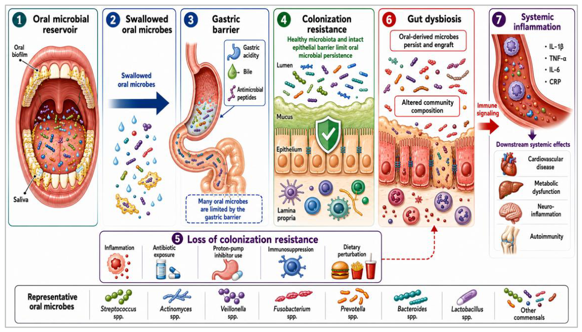
Figure 2.
Spatial organization of the oral microbiome. The oral microbiome is distributed across distinct ecological niches, including teeth/supragingival plaque, subgingival biofilm, tongue dorsum, saliva, and buccal mucosa. These habitats differ in oxygen availability, pH, nutrient availability, salivary flow, surface structure, and host immune factors, leading to site-specialized microbial communities. Representative taxa shown include Streptococcus, Actinomyces, Veillonella, Prevotella, Fusobacterium, and Porphyromonas. Dysbiosis in these niches, particularly within subgingival biofilms, can increase the burden of inflammatory oral taxa with potential downstream effects on gut and systemic health.
Figure 2.
Spatial organization of the oral microbiome. The oral microbiome is distributed across distinct ecological niches, including teeth/supragingival plaque, subgingival biofilm, tongue dorsum, saliva, and buccal mucosa. These habitats differ in oxygen availability, pH, nutrient availability, salivary flow, surface structure, and host immune factors, leading to site-specialized microbial communities. Representative taxa shown include Streptococcus, Actinomyces, Veillonella, Prevotella, Fusobacterium, and Porphyromonas. Dysbiosis in these niches, particularly within subgingival biofilms, can increase the burden of inflammatory oral taxa with potential downstream effects on gut and systemic health.
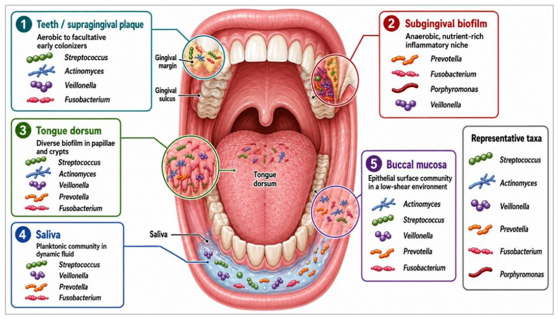
Figure 3.
Major mechanisms connecting oral dysbiosis with intestinal and systemic disease. Oral dysbiosis may influence the gut and distant organs through five interconnected mechanisms. First, microbial migration allows oral-derived microbes and microbial products to reach the gastrointestinal tract through swallowed saliva, shed biofilms, and microbial translocation. Under disrupted colonization resistance, some oral taxa may persist or engraft in the intestinal mucosa. Second, immune-mediated communication occurs when oral microbial products, including lipopolysaccharide and other microbe-associated molecular patterns, activate innate and adaptive immune pathways and promote inflammatory T helper responses, including Th1 and Th17 activation. Third, metabolite-mediated effects involve both the loss of protective microbial metabolites, such as short-chain fatty acids, and increased exposure to dysbiotic oral metabolites or endotoxins, which may alter epithelial and immune homeostasis. Fourth, barrier disruption affects epithelial tight junctions in oral and intestinal tissues, increasing permeability and facilitating the translocation of microbial or inflammatory signals. Finally, inflammatory feedback loops may connect oral, gut, and systemic inflammation, reinforcing dysbiosis across anatomical sites. Together, these mechanisms may contribute to altered gut microbial ecology, intestinal inflammation, immune dysregulation, and systemic disease processes. Abbreviations: LPS, lipopolysaccharide; MAMPs, microbe-associated molecular patterns; SCFAs, short-chain fatty acids; Th1, T helper 1; Th17, T helper 17; TNF-α, tumor necrosis factor-alpha; IL, interleukin; NAFLD, non-alcoholic fatty liver disease.
Figure 3.
Major mechanisms connecting oral dysbiosis with intestinal and systemic disease. Oral dysbiosis may influence the gut and distant organs through five interconnected mechanisms. First, microbial migration allows oral-derived microbes and microbial products to reach the gastrointestinal tract through swallowed saliva, shed biofilms, and microbial translocation. Under disrupted colonization resistance, some oral taxa may persist or engraft in the intestinal mucosa. Second, immune-mediated communication occurs when oral microbial products, including lipopolysaccharide and other microbe-associated molecular patterns, activate innate and adaptive immune pathways and promote inflammatory T helper responses, including Th1 and Th17 activation. Third, metabolite-mediated effects involve both the loss of protective microbial metabolites, such as short-chain fatty acids, and increased exposure to dysbiotic oral metabolites or endotoxins, which may alter epithelial and immune homeostasis. Fourth, barrier disruption affects epithelial tight junctions in oral and intestinal tissues, increasing permeability and facilitating the translocation of microbial or inflammatory signals. Finally, inflammatory feedback loops may connect oral, gut, and systemic inflammation, reinforcing dysbiosis across anatomical sites. Together, these mechanisms may contribute to altered gut microbial ecology, intestinal inflammation, immune dysregulation, and systemic disease processes. Abbreviations: LPS, lipopolysaccharide; MAMPs, microbe-associated molecular patterns; SCFAs, short-chain fatty acids; Th1, T helper 1; Th17, T helper 17; TNF-α, tumor necrosis factor-alpha; IL, interleukin; NAFLD, non-alcoholic fatty liver disease.
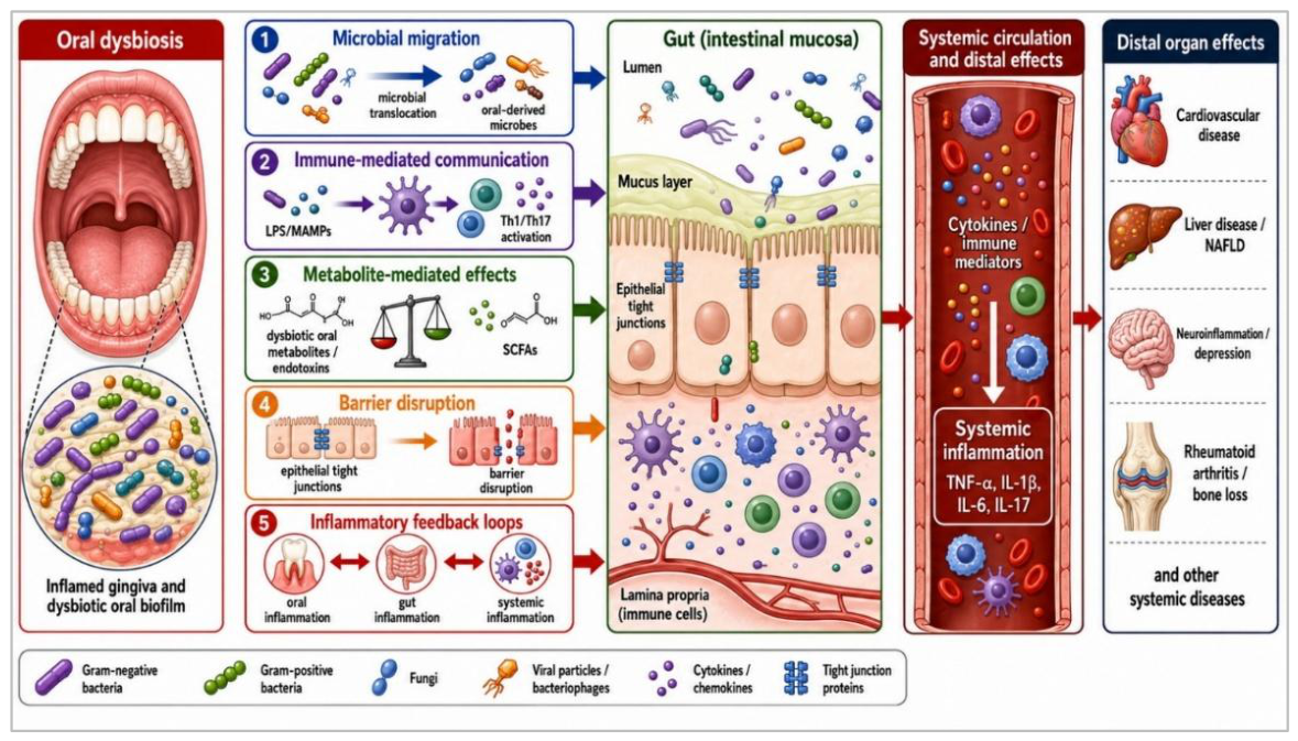
Figure 4.
Oral microbiome biomarker workflow for translational applications. This figure illustrates a stepwise workflow for translating oral microbiome data into potential diagnostic, monitoring, and intervention strategies. Non-invasive oral samples, including saliva, oral rinse, tongue coating, and oral swabs, may be collected alone or integrated with stool and blood specimens to provide broader oral–gut and systemic information. These samples can be analyzed using sequencing and multi-omics approaches, including metagenomics, metatranscriptomics, metabolomics, proteomics, and immune profiling. The resulting datasets may support biomarker discovery by identifying microbial signatures, diversity patterns, disease-associated taxa, metabolite biomarkers, and host-microbe interaction markers. These features can then be incorporated into risk-prediction models for disease-risk stratification, screening, longitudinal monitoring, prognostic assessment, and clinical decision support. The final step involves translation into personalized interventions, including colorectal cancer screening, inflammatory bowel disease monitoring, periodontal intervention, microbiome modulation using probiotics, prebiotics, or synbiotics, and individualized microbiome profiling. This workflow represents a translational framework rather than an established clinical pathway, and its implementation requires standardized sampling, analytical validation, longitudinal studies, and integration with established clinical biomarkers. Abbreviations: CRC, colorectal cancer; IBD, inflammatory bowel disease.
Figure 4.
Oral microbiome biomarker workflow for translational applications. This figure illustrates a stepwise workflow for translating oral microbiome data into potential diagnostic, monitoring, and intervention strategies. Non-invasive oral samples, including saliva, oral rinse, tongue coating, and oral swabs, may be collected alone or integrated with stool and blood specimens to provide broader oral–gut and systemic information. These samples can be analyzed using sequencing and multi-omics approaches, including metagenomics, metatranscriptomics, metabolomics, proteomics, and immune profiling. The resulting datasets may support biomarker discovery by identifying microbial signatures, diversity patterns, disease-associated taxa, metabolite biomarkers, and host-microbe interaction markers. These features can then be incorporated into risk-prediction models for disease-risk stratification, screening, longitudinal monitoring, prognostic assessment, and clinical decision support. The final step involves translation into personalized interventions, including colorectal cancer screening, inflammatory bowel disease monitoring, periodontal intervention, microbiome modulation using probiotics, prebiotics, or synbiotics, and individualized microbiome profiling. This workflow represents a translational framework rather than an established clinical pathway, and its implementation requires standardized sampling, analytical validation, longitudinal studies, and integration with established clinical biomarkers. Abbreviations: CRC, colorectal cancer; IBD, inflammatory bowel disease.
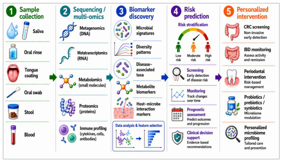

Table 1.
Core oral and gut microbial taxa relevant to the oral–gut axis.
| Microbial group/taxon | Primary niche | Key role in health or dysbiosis | Reported disease relevance | References |
|---|---|---|---|---|
| Streptococcus spp. | Oral cavity; minor gut presence | Early biofilm colonizer; some acid-tolerant species contribute to caries and dysbiosis. | Dental caries, IBD-associated oralization, gastric cancer-associated oral signatures. | [1,36,47] |
| Veillonella spp. | Oral cavity and gut | Lactate-utilizing commensal; may expand in inflammatory oral communities. | Rheumatoid arthritis, multiple sclerosis, IBD-associated dysbiosis. | [1,48,49] |
| Prevotella spp. | Oral, gut, and mucosal sites | Mucosal commensal with pro-inflammatory potential under dysbiosis. | Periodontitis, rheumatoid arthritis, IBD, metabolic syndrome, SLE. | [21,50,51] |
| Fusobacterium nucleatum | Oral cavity; ectopic gut/tumor colonizer | Bridging organisms in oral biofilms; can invade the mucosa and support inflammatory biofilms. | Colorectal cancer, IBD, adenomas, cancer therapy resistance. | [52,53,54,55] |
| Porphyromonas gingivalis | Oral subgingival biofilm | Keystone periodontal pathobiont; promotes immune disruption and polymicrobial dysbiosis. | Periodontitis, cardiovascular disease, rheumatoid arthritis, Alzheimer’s disease, IBD. | [21,56,57] |
| Actinomyces spp. | Oral tooth surfaces and plaque | Early colonizer; contributes to plaque formation and root-surface biofilms. | Root caries and altered oral communities in multiple sclerosis. | [1,49] |
| Bacteroides spp. | Gut colon | Major anaerobe involved in carbohydrate metabolism and immune regulation. | IBD, colorectal cancer, colitis, metabolic disease. | [58,59,60] |
| Faecalibacterium prausnitzii | Gut colon | Butyrate producer with anti-inflammatory and barrier-supporting functions. | Depleted in Crohn’s disease, IBD, CRC, and metabolic dysbiosis. | [58,59] |
| Akkermansia muciniphila | Gut mucus layer | Mucin degrader associated with barrier integrity and metabolic health. | Obesity, type 2 diabetes, IBD, NAFLD, metabolic syndrome. | [59,61] |
| Lactobacillus spp. | Oral cavity, gut, and vaginal mucosa | Lactic acid production, immune modulation, and probiotic potential. | IBD, Parkinson’s disease-associated oral dysbiosis, probiotic applications. | [59,62] |
| Escherichia/Shigella | Gut; minor oral presence | Low-abundance commensal or pathobiont; may bloom during inflammation. | IBD, CRC, enteric infection, gut dysbiosis severity. | [60,63] |
IBD, inflammatory bowel disease; CRC, colorectal cancer; NAFLD, non-alcoholic fatty liver disease; SLE, systemic lupus erythematosus; spp., species.
Table 2.
Summary of evidence linking oral dysbiosis with gastrointestinal and systemic diseases.
| Disease category | Key oral–gut findings | Key mechanisms | Evidence level | Clinical implications | References |
|---|---|---|---|---|---|
| Inflammatory bowel disease (IBD) | Oral pathobionts and oral-derived taxa are enriched in IBD-associated oral and gut communities. | Oral microbial migration, impaired colonization resistance, barrier disruption, Th1/Th17 activation. | Strong association with mechanistic support | Oral health monitoring may support IBD care. | [13,14,55,63] |
| Irritable bowel syndrome (IBS) | Oral microbial changes have been reported, but causality is unclear. | Stress, diet, gut-brain signaling, permeability, and motility-related microbial changes. | Limited to moderate | Possible biomarker role, but more evidence is needed. | [73,74] |
| Celiac disease | Oral microbial changes and oral manifestations have been reported. | Microbial-immune interactions and altered mucosal inflammation. | Emerging | Not ready for clinical use. | [74,75] |
| Colitis | Experimental studies suggest oral taxa may worsen intestinal inflammation. | Oral microbial translocation, immune activation, and barrier injury. | Mainly experimental | Human validation is needed. | [13,55,63] |
| Colorectal cancer (CRC) | Oral-associated taxa, especially Fusobacterium nucleatum, are enriched in CRC-associated microbiota. | Biofilm formation, inflammation, FadA/Fap2 signaling, immune modulation, and changes in the tumor microenvironment. | Strongest evidence. | Oral microbiome biomarkers may support CRC screening. | [52,76,77,78] |
| Cardiovascular disease | Periodontal dysbiosis is associated with inflammatory oral taxa. | Hematogenous spread, LPS, endothelial activation, oxidative stress, and inflammation. | Moderate; causality unresolved. | Periodontal care may reduce systemic inflammatory burden. | [79,80,81] |
| Metabolic disorders (diabetes, obesity, NAFLD, metabolic syndrome) | Periodontal pathogens and altered salivary microbiota are linked with diabetes, obesity, NAFLD, and metabolic syndrome. | Gut dysbiosis, intestinal permeability, metabolic endotoxemia, and insulin resistance. | Moderate mechanistic evidence. | Oral health may be a modifiable factor. | [61,69,82] |
| Neurodegenerative diseases (Alzheimer’s disease, Parkinson’s disease, multiple sclerosis) | Periodontal dysbiosis has been associated with Alzheimer’s disease, Parkinson’s disease, and MS. | Systemic inflammation, blood-brain barrier disruption, microbial metabolites, and gut-brain axis signaling. | Emerging; causality not established. | Possible risk prediction needs validation. | [49,83,84] |
| Autoimmune diseases (rheumatoid arthritis, SLE, Sjögren’s syndrome) | Oral dysbiosis involving Prevotella, Veillonella, and periodontal organisms is linked with RA, SLE, and Sjögren’s syndrome. | Molecular mimicry, microbial translocation, Th17 activation, cytokine amplification, and autoantigen generation. | Moderate; strongest for RA. | May support inflammatory monitoring in future. | [57,85,86] |
IBD, inflammatory bowel disease; IBS, irritable bowel syndrome; CRC, colorectal cancer; NAFLD, non-alcoholic fatty liver disease; RA, rheumatoid arthritis; SLE, systemic lupus erythematosus; MS, multiple sclerosis; LPS, lipopolysaccharide; Th1, T helper 1; Th17, T helper 17; spp., species; FadA, Fusobacterium adhesin A; Fap2, Fusobacterium autotransporter protein 2.
Table 3.
Translational applications, limitations, and future directions for oral microbiome-based biomarkers and interventions.
Table 3.
Translational applications, limitations, and future directions for oral microbiome-based biomarkers and interventions.
| Application | Candidate biomarker/intervention | Sample type | Potential use | Current limitations and future direction | References |
|---|---|---|---|---|---|
| Saliva-based screening |
Oral microbial signatures, microbial diversity patterns, disease-associated taxa (Fusobacterium, Porphyromonas, Prevotella, Veillonella) | Saliva, oral rinse, tongue coating, oral swabs | Non-invasive detection of disease-associated microbial changes and risk assessment | Results vary with diet, geography, oral hygiene, lifestyle, and sequencing methods; standardized sampling and validated prediction models are needed. | [145,151] |
| Periodontal intervention | Plaque control, periodontal therapy, reduction of gingival inflammation | Dental plaque, gingival samples, clinical periodontal assessment | Reduction of oral inflammatory burden and systemic microbial dissemination | Evidence for improvement of systemic disease outcomes remains limited; clinical trials are needed to test oral therapy as an adjunct intervention. | [75,90] |
| Probiotic/prebiotic/synbiotic therapy | Beneficial microbial strains, dietary substrates, microbiome-modulating approaches | Oral and gut microbiome samples | Restoration of microbial balance, improved barrier function, and modulation of inflammation | Effects are influenced by strain specificity; microbial persistence, and host response; future approaches should be personalized and ecology-based. | [75] |
| CRC/cancer risk biomarkers | Fusobacterium nucleatum and oral microbial community signatures | Saliva, oral swabs, tumor-associated samples | Early detection, cancer risk prediction, prognosis, and surveillance | Large-scale validation and standardized biomarker panels are lacking; integration with clinical, imaging, and host biomarkers is needed. | [78] |
| Multi-omics diagnostics | Metagenomics, metatranscriptomics, metabolomics, proteomics, immune profiling | Saliva, stool, blood, tissue samples | Identification of microbial function, metabolic pathways, and host-microbe interactions | High cost, complexity, and limited clinical interpretation remain challenges; machine-learning models may improve integration of microbial and host datasets. | [152,153] |
| Personalized oral–gut microbiome profiling | Individual microbial signatures combined with immune and clinical parameters | Longitudinal oral and gut samples | Disease prediction, treatment response monitoring, and precision prevention strategies | Longitudinal evidence is limited and causality remains incomplete; personalized microbiome-guided interventions require further validation | [63,150] |
CRC, colorectal cancer; multi-omics, integrated analysis of multiple biological data types, including metagenomics, metatranscriptomics, metabolomics, proteomics, and immune profiling.
Disclaimer/Publisher’s Note: The statements, opinions and data contained in all publications are solely those of the individual author(s) and contributor(s) and not of MDPI and/or the editor(s). MDPI and/or the editor(s) disclaim responsibility for any injury to people or property resulting from any ideas, methods, instructions or products referred to in the content. |
© 2026 by the authors. Licensee MDPI, Basel, Switzerland. This article is an open access article distributed under the terms and conditions of the Creative Commons Attribution (CC BY) license.
Copyright: This open access article is published under a Creative Commons CC BY 4.0 license, which permit the free download, distribution, and reuse, provided that the author and preprint are cited in any reuse.