

Submitted:
20 October 2026
Posted:
21 October 2026
You are already at the latest version
Abstract
Endometriosis is associated with increased cardiovascular risk, but how its subtype heterogeneity informs this association remains unclear. This narrative review examines the biological characteristics of superficial peritoneal endometriosis (SUP), ovarian endometrioma (OMA), and deep infiltrating endometriosis (DIE) and their potential cardiovascular relevance. English-language literature identified through PubMed, Scopus, Google Scholar, and reference screening was synthesized across susceptibility factors, molecular and vascular alterations, and treatment exposures. Subtype-related findings highlight differences in anatomical location and tissue microenvironment, including peritoneal immune interactions in superficial lesions, intracystic iron accumulation and oxidative injury in OMA, and inflammatory and fibromuscular remodeling in deep lesions. These features provide a basis for investigating how lesion heterogeneity may relate to systemic lipid dysregulation, inflammation, endothelial dysfunction, and coagulation abnormalities. Ovarian function, surgical history, and hormonal regimens further inform interpretation of these relationships. By integrating phenotype and treatment exposure while distinguishing local findings from circulating biomarkers, vascular measurements, and clinical events, this review identifies evidence gaps linking biological heterogeneity to cardiovascular outcomes. Emerging subtype-related evidence supports further investigation but does not establish differences in cardiovascular event risk among SUP, OMA, and DIE. Prospective studies are needed to determine whether incorporating endometriosis subtype improves cardiovascular risk assessment beyond conventional factors. (Figure 1)
Keywords:
endometriosis
; cardiovascular diseases
; dyslipidemias
; oxidative stress
; inflammation
1. Introduction
Endometriosis (EMs) is a chronic, estrogen-dependent inflammatory disorder affecting approximately 10% of women of reproductive age worldwide[1]. It is characterized by the presence of endometrial-like tissue outside the uterus and is commonly classified into superficial peritoneal endometriosis (SUP), ovarian endometrioma (OMA), and deep infiltrating endometriosis (DIE). These subtypes differ in lesion location, tissue involvement, and pathological features. Proposed mechanisms of disease development include retrograde menstruation and implantation[2,3], together with altered immune responses and local inflammatory processes.
Beyond its reproductive consequences, EMs has been associated with several adverse health outcomes, with cardiovascular disease (CVD) being a major concern due to its high morbidity and mortality[4,5,6,7]. These cardiovascular outcomes include hypertension, ischaemic heart disease, myocardial infarction (MI), and cerebrovascular accidents. However, the pathways underlying this association remain incompletely understood. In particular, it is unclear whether differences in the inflammatory, metabolic, and vascular characteristics of SUP, OMA, and DIE translate into differences in CVD risk. Distinguishing disease-associated mechanisms from shared susceptibility factors and treatment-related effects is therefore important for interpreting the available evidence.
Figure 1.
Graphical abstract. Proposed framework linking EMs to cardiovascular risk. Shared susceptibility factors, disease-associated metabolic and inflammatory alterations, and treatment exposures may interact. Abbreviations: EMs, endometriosis; SUP, superficial peritoneal endometriosis; OMA, ovarian endometrioma; DIE, deep infiltrating endometriosis.
Figure 1.
Graphical abstract. Proposed framework linking EMs to cardiovascular risk. Shared susceptibility factors, disease-associated metabolic and inflammatory alterations, and treatment exposures may interact. Abbreviations: EMs, endometriosis; SUP, superficial peritoneal endometriosis; OMA, ovarian endometrioma; DIE, deep infiltrating endometriosis.
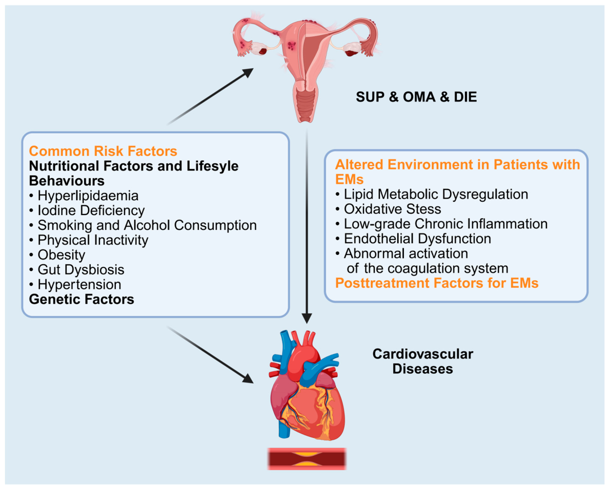
This narrative review examines the epidemiological association between EMs and CVD and critically discusses shared and potentially distinct molecular mechanisms linking EMs subtypes to CVD. It organizes the evidence around three interconnected domains: shared susceptibility factors, disease-associated metabolic and vascular alterations, and the effects of hormonal and surgical treatments. The aim is to identify biologically plausible links, clarify the limitations of current evidence, and discuss their implications for long-term cardiovascular health.
2. Results
2.1. EMs and CVD: Close Association
Large observational studies in different populations have associated EMs with increased cardiovascular risk [7,8,9,10,11,12] (Table 1). This evidence provides a population-level basis for examining the relationship between EMs and cardiovascular health, although it does not, by itself, establish causality.
The reported associations differ across cardiovascular outcomes and study populations. A UK cohort found increased risks of ischemic heart disease, arrhythmias, and cerebrovascular disease, without a significant increase in heart failure[11], whereas a Danish nationwide cohort reported an increased risk of heart failure[7]. Furthermore, when EMs coexists with other gynecological conditions such as uterine fibroids, a potential synergistic effect may further amplify the long-term cardiovascular mortality risk[12]. However, the coexistence of these conditions should not be interpreted as evidence of a synergistic effect without an interaction analysis. These findings highlight the need to consider specific outcomes and study contexts when interpreting the cardiovascular implications of EMs.
Site-specific genetic analyses provide a complementary perspective. Zheng and Zheng reported associations between genetic liability to ovarian, pelvic peritoneal, and rectovaginal septal/vaginal EMs and ischemic stroke in a Mendelian randomization (MR) study. These findings motivate investigation of anatomical heterogeneity, but the registry-defined sites do not map precisely onto mutually exclusive SUP, OMA, and DIE groups. Moreover, associations for separate genetic exposures do not constitute a direct comparison of clinical event rates between subtypes or demonstrate mediation by lesion pathology[13].
Taken together, these findings make cardiovascular health a relevant consideration in EMs, while leaving the basis of the association unresolved. Interpreting this relationship requires consideration of shared risk factors, disease-associated molecular changes, and treatment exposures, whose contributions may overlap. Within this broader framework, the clinical and biological heterogeneity of EMs raises a further question: whether SUP, OMA, and DIE involve different pathways with potential cardiovascular relevance. Direct comparisons of cardiovascular outcomes across these subtypes remain limited, so the cardiovascular implications of lesion-level differences require validation against systemic vascular changes and clinical outcomes. The following sections examine these possible connections, beginning with shared risk factors and susceptibility.
2.2. EMs and CVD: Potential Mechanisms
2.2.1. Common Risk Factors and Susceptibility
When investigating the association between comorbid conditions of EMs and CVD, it is initially crucial to recognize that a comprehensive understanding of the common risk factors is highly important.
Nutritional Factors and Lifestyle Behaviors
Hyperlipidemia. It is established hyperlipidemia is a cardiovascular risk factor and has a potentially bidirectional association with EMs. Mu et al.[10] reported a higher risk of EMs among women with hypercholesterolemia (relative risk [RR] = 1.22) and a higher risk of hyperlipidemia among women with EMs (RR = 1.25). These findings suggest an association between abnormal lipid homeostasis and EMs, without establishing a self-sustaining causal cycle.
Lipid availability may influence ectopic endometrial cell survival and invasion. Proposed mechanisms include increased peritoneal exposure to low-density lipoprotein (LDL), activation of pelvic macrophages by oxidized LDL (ox-LDL), and release of tumor necrosis factor-α (TNF-α) and interleukin-8 (IL-8). These processes may enhance adhesion molecule expression and vascular endothelial growth factor (VEGF) production, supporting adhesion, invasion, and angiogenesis. Increased LDL receptor mRNA expression has been reported in deep bowel endometriotic lesions[14], suggesting a potential alteration in cholesterol uptake, which may be associated with enhanced cholesterol uptake to meet the demands of cell proliferation. Meanwhile, cholesterol metabolic reprogramming may mutually reinforce local estrogen synthesis, thereby supporting the persistent growth of ectopic lesions[15].
Atorvastatin-related animal experiments (rat models) and in vitro culture studies of endometriotic stromal cells have provided a theoretical basis for exploring lipid-related pathways in endometriosis[16,17]. However, these findings cannot confirm the clinical efficacy of this agent. Evidence from randomized clinical trials is still required before lipid-lowering agents can be recommended for the treatment of EMs.
Iodine Deficiency. Iodine-related processes have been investigated in endometrial tissues, although their relevance to EMs remains uncertain. Trovato et al.[18] detected sodium/iodide symporter (NIS) expression in human endometrium, providing a biological basis for investigating local iodide handling; however, this study did not establish an EMs-specific abnormality. More directly, Feider et al. reported a lower-intensity signal tentatively assigned to iodide in ectopic lesions than in eutopic endometrium using desorption electrospray ionization mass spectrometry imaging[19]. This observation suggests differences in tissue iodide distribution, but does not establish systemic iodine deficiency or a causal contribution to lesion development.
Thus, although iodine deficiency is a known risk factor for CVD events[20,21,22], the available evidence is insufficient to classify EMs as an iodine-deficiency disorder or to establish iodine supplementation as an effective treatment. Whether iodine-related alterations differ among SUP, OMA, and DIE or contribute to EMs-associated cardiovascular risk remains unestablished.
Smoking and Alcohol Consumption. Current research shows alcohol may influence estrogen-dependent disease through endocrine and immune pathways. Proposed mechanisms include that, at the hormonal level, it enhances aromatase activity to accelerate testosterone-to-estrogen conversion and stimulates ovarian estradiol secretion, thereby amplifying estrogen exposure risk[23]. Immunologically, long-term intake disrupts immune homeostasis and induces pro-inflammatory cytokine dysregulation, linking to chronic inflammatory diseases[24,25]. A 2013 systematic review found drinkers had a significantly higher risk of EMs than non-drinkers[26]. However, this association may be influenced by reverse causation, as physical or emotional distress in patients could lead to increased alcohol use. As shown in a matched case-control study conducted by Heilier et al., daily alcohol consumption was significantly associated with an increased risk of deep endometriotic nodules (odds ratio [OR] = 5.82, 95% confidence interval [CI]: 1.20–28.3), whereas no statistically significant association was found between alcohol intake and peritoneal EMs (OR = 1.91, 95% CI: 0.30–12.0)[27]. These inconsistent findings may be attributed to differences in pathological mechanisms across EMs subtypes and limited statistical power.
The association between smoking and EMs is inconsistent. Some studies report an inverse association[28,29,30,31], possibly due to tobacco’s anti-estrogenic effects[32,33,34], whereas other analyses suggest confounding by factors such as infertility[35]. Reports also include null associations[36,37] and increased risk[38], with variation by genotype[39] and population characteristics, including infertility history[40]. These differences limit generalization across EMs subtypes.
Alcohol-related endocrine and immune effects provide possible links to EMs and CVD, but the subtype-specific findings require replication. Smoking remains relevant to cardiovascular assessment, whereas its relationship with EMs is unresolved.
Physical Inactivity. Epidemiological studies link moderate-to-vigorous physical activity associated with a lower risk of EMs[41,42,43,44], whereas prolonged sedentary time consistently correlates with increased risk.[41] Regular exercise modulates estrogen metabolism by reducing estradiol levels[45,46,47,48] and increasing the estrone-to-estradiol ratio[49], potentially inhibiting endometriotic lesion establishment. However, their relevance to lesion establishment may depend on exercise intensity[48] and age[47], and should not be assumed to demonstrate prevention of EMs.
Concurrently, physical inactivity promotes CVD risk[50] through mechanisms including obesity, insulin resistance, and chronic inflammation, which accelerate atherosclerosis and increase cardiovascular event incidence.
Physical inactivity is therefore a potential shared risk factor. Although physical activity is relevant to cardiovascular health, its ability to prevent EMs or EMs-associated cardiovascular events requires direct evaluation.
Obesity. Traditionally, body mass index (BMI) has been used to assess obesity. While many observational studies associate EMs with lower BMI[51,52,53,54,55], this finding is not universal[56,57,58]. For instance, some studies indicates that the average BMI of patients with DIE and OMA was significantly lower than that of controls (with no difference observed in women with SUP)[52], while others noted that individuals with EMs and obesity had more severe disease (e.g., higher Revised American Fertility Society scores) [59].
Critically, BMI has notable limitations, fluctuating across life stages and failing to identify individuals with “normal-weight obesity syndrome”[59,60,61], potentially masking the impact of metabolic obesity on disease. For example, a higher visceral adiposity index (VAI) demonstrated a significant positive association with EMs (OR=1.08[1.04-1.12])[62]. Given that obesity-related metabolic abnormalities (e.g., insulin resistance, dyslipidemia) are the shared pathophysiological basis of EMs and CVD, focusing solely on BMI may obscure true associations.
Genetic findings further complicate the interpretation of mechanisms underlying BMI-related associations. The CYP2C19*2 gene variant approximately doubles the risk of EMs, and this association is significantly stronger among obese women[63]. This suggests that the genetic correlation may vary depending on obesity status, while the existence of gene-obesity interactions still requires further verification. Studies focusing on early-life body phenotypes can provide additional evidence[53,64,65]; however, discrepancies in body size assessment criteria and potential recall bias should be taken into account in relevant research. Meanwhile, no common genetic pathogenic pathway linking obesity, EMs, and CVD has been clarified so far.
Current controversies reflect two opposing models: the mechanical model proposes that low BMI may promote retrograde menstruation, while the metabolic model emphasizes that obesity-related abnormal adipokine profiles exacerbate EMs progression through pathways such as inflammation promotion, angiogenesis, and insulin resistance—pathways overlapping with CVD pathophysiology. Neither model establishes obesity as protective or causal across all EMs phenotypes. Future research should move beyond BMI to focus on body fat distribution, metabolic phenotypes, and gene-environment interactions to systematically clarify the shared pathogenic network of obesity in EMs and CVD.
Notably, EMs-related chronic pain may restrict food intake, contributing to a low BMI phenotype[59]; animal experiments further indicate that EMs disrupts hepatic metabolic genes expression, reducing body weight and fat mass[66]. Thus, the exact causal relationship between the two remains to be clarified.
Gut Dysbiosis. Gut dysbiosis has been implicated in EMs[67,68,69] and CVD[70,71,72]. Effects on barrier function, inflammation, and metabolism provide a possible biological connection, although a shared causal pathway has not been established.
Tang et al.[73] used MR to examine associations between gut microbiota and site-specific EMs. For pelvic peritoneal EMs, genetically predicted Escherichia–Shigella abundance was associated with increased susceptibility (OR = 1.51; 95% CI, 1.10–2.07; p = 0.012), whereas Catenibacterium (OR = 0.76; 95% CI, 0.60–0.97; p = 0.025), the Eubacterium oxidoreducens group (OR = 0.75; 95% CI, 0.58–0.98; p = 0.038), and Paraprevotella (OR = 0.81; 95% CI, 0.68–0.95; p = 0.018) showed inverse associations. For ovarian EMs, Blautia was positively associated (OR = 2.958; 95% CI, 1.399–6.253; p = 0.0045), while Barnesiella (OR = 0.741; 95% CI, 0.573–0.958; p = 0.022) and Terrisporobacter (OR = 0.708; 95% CI, 0.527–0.952; p = 0.022) were inversely associated. For vaginal or rectovaginal septal EMs, Tyzzerella 3 showed a positive association; Erysipelotrichaceae UCG003, the Eubacterium oxidoreducens group, the Lachnospiraceae NK4A136 group, and Ruminococcaceae UCG003 showed inverse associations. For intestinal EMs, positive associations were reported for Intestinimonas, Lachnospiraceae NC2004 group, Lactococcus, Oscillospira, Phascolarctobacterium, and Ruminococcaceae UCG005, with an inverse association for Parabacteroides. These site-based genetic associations should not be equated with measured microbial profiles in SUP, OMA, and DIE.
Studies have reported patients with EMs exhibit gut microbial imbalance resembling that of CVD patients in general, characterized by alterations in the Firmicutes-to-Bacteroidetes ratio[74,75,76,77], alongside increased abundances of Proteobacteria[78], Enterobacteriaceae, and Escherichia coli[71,79]. These observations suggest overlapping features rather than a uniform microbial signature. This dysbiosis disrupts the intestinal barrier, permitting bacterial components like lipopolysaccharide (LPS) to enter circulation. Synergizing with elevated inflammatory markers (C-reactive protein [CRP], TNF-α), it exacerbates peritoneal and systemic inflammation and fostering a pro-EMs and pro-atherosclerotic microenvironment. Furthermore, aberrant metabolites like trimethylamine N-oxide (TMAO) directly accelerate endothelial inflammation and atherosclerosis by inhibiting reverse cholesterol transport[80] and promoting foam cell formation[81].
Microbial effects on estrogen metabolism provide another possible connection. Increased abundance of Gram-negative bacteria (e.g., Escherichia coli)[79] may involve abnormal β-glucuronidase (GUSB) secretion[67], which hydrolyzes estrogen conjugates to enhance enteric recapture of free estrogens[82,83,84], creating a high-estrogen environment for EMs progression. GUSB also promotes M2 macrophage polarization, driving endometrial stromal cell proliferation and migration[67,85]. Importantly, EMs-characteristic M2 polarization stimulates profibrotic factor secretion (transforming growth factor-β [TGF-β]), mirroring the molecular mechanisms of atherosclerotic smooth muscle cell activation and fibrous plaque formation, revealing cross-organ similarities in biological processes.
Thus, correcting gut dysbiosis through probiotics, dietary interventions, or targeted enzyme inhibitors may break this inflammation-metabolic vicious cycle, offering potential therapeutic strategies for both EMs and CVD[86]. However, their ability to reduce cardiovascular events or EMs remains unproven. (Figure 2)
Hypertension. Mu et al. reported a higher risk of EMs among women with hypertension than among those without hypertension (RR = 1.29; 95% CI, 1.18–1.41)[10].
Studies have associated hypertension with increased circulating inflammatory markers[87,88,89,90]. An inflammatory environment has been proposed to support endometrial cell adhesion and lesion progression through altered cytokine exposure [89]. The extent to which this mechanism explains the hypertension-EMs association is uncertain[91].
Genetic Factors
Genetic findings suggest possible overlap between EMs and CVD susceptibility. The 9p21[92,93] region has been implicated in both conditions [90,91]. Candidate-gene studies have also examined variants in pathways related to inflammation, immunity, angiogenesis, and hormonal signaling, including VEGF-related genes[94,95,96,97], IL6[98,99], IL10[100,101,102], IL16[103,104,105], CCL21[106,107,108], ESR1[109,110], and TP53[111,112]. These findings require gene- and variant-specific evaluation and do not by themselves demonstrate shared causal variants or subtype-specific cardiovascular susceptibility.
Non-coding RNAs and RNA modification pathways are additional candidates. CDKN2B-AS1, also known as ANRIL, may connect susceptibility loci with gene regulation[113]. Circulating microRNAs (miRNAs), including miR-199a, have been investigated in EMs[114], while separate studies implicate miR-199a in endothelial or cardiac biology and cardiovascular biomarkers[115,116,117]. N6-methyladenosine (m⁶A) regulation and the RNA-binding protein IGF2BP2 have also been studied in cardiac dysfunction[118,119,120]. In ovarian EMs, IGF2BP2 stabilizes m⁶A-modified MEIS2 and GATA6 transcripts and promotes disease progression[121]. These observations identify potentially convergent regulatory mechanisms; they do not establish that the same pathway mediates CVD in patients with EMs.
Genetic and epigenetic observations thus provide hypotheses for mechanistic studies. Demonstrating a shared causal mechanism will require appropriate genetic analyses and functional validation in relevant lesion and vascular cell types.
2.2.2. Altered Environment in Patients with EMs
Lipid Metabolic Dysregulation
Proatherogenic lipid alterations have been reported in systemic and peritoneal samples from women with EMs, including increased LDL[122,123,124] and reduced high-density lipoprotein (HDL) concentrations or function [123,125,126,127]. LDL oxidation and foam cell formation provide potential links to atherosclerosis. Reduced serum paraoxonase-1 (PON-1) activity has also been associated with EMs severity[128], suggesting impaired antioxidant functions relevant to HDL-mediated vascular protection.
Altered triglyceride (TG) metabolism may contribute to these associations. Elevated TG concentrations have been reported in EMs [129,130], and TG-derived products, including free fatty acids, can promote endothelial oxidative stress, inflammation, and prothrombotic responses[131,132]. Lipoprotein(a) has also been investigated in EMs[130,133] and has established relevance to plaque biology[134] and MI risk[135]. Population evidence also associates EMs with a 25% increased risk of hypercholesterolemia[10], supporting an association between EMs and dyslipidemia. However, overall associations may not capture differences among lesion phenotypes.
Metabolomic studies suggest phenotype-associated differences. In a pilot study, women with EMs had higher serum free fatty acid and ketone body concentrations than controls; the OMA group had higher concentrations of two ketone bodies, whereas the DIE group had higher free fatty acid and lipid concentrations[136]. Local estradiol metabolism may influence metabolic activity[137], but these studies do not directly demonstrate an estrogen-driven mechanism for the circulating metabolite differences or establish their cardiovascular consequences.
Complementing these metabolomic observations, the ENDO Cohort Study strengthens the rationale for phenotype-specific assessment of circulating lipids. Overall EMs status was not associated with adverse lipid profiles, whereas analyses by typology identified a pattern of higher TG, very-low-density lipoprotein cholesterol, and apolipoprotein B, particularly with OMA and coexisting ovarian and deep disease[138]. The combined phenotype comprised only 17 women, and lipid measurements were concurrent with diagnosis. Thus, these findings suggest that pooling phenotypes may obscure metabolic heterogeneity, while requiring replication and longitudinal evaluation before cardiovascular implications can be assigned.
Lipid-related composite indices provide additional clinical observations. Patients with OMA exhibit a significantly higher atherogenic index of plasma (AIP) than controls (0.25 ± 0.24 vs. −0.13 ± 0.19, p < 0.001) and have 2.31-fold higher odds of being in the high-risk AIP category (AIP ≥ 0.1; OR = 2.31, 95% CI 1.23–4.33, p = 0.008) [139]. At the broader EMs level, the cardiometabolic index (CMI)—calculated as the product of the waist-to-height ratio (WHtR) and the TG/high-density lipoprotein cholesterol (HDL-C) ratio—is significantly and positively associated with EMs (fully adjusted OR = 1.22 per unit increase, 95% CI 1.07–1.38). For CMI values above 0.67, each unit increase was associated with 20% higher odds of EMs (OR = 1.20, 95% CI 1.05–1.37) [129]. These findings concern lipid-related indices and EMs status; they do not directly establish cardiovascular event risk or differences between EMs subtypes.
Beyond these observational findings, genetic analyses address the possible direction of the lipid–EMs relationship. While the causal link between EMs and lipid profile changes requires further investigation, bidirectional MR analyses reveal a reciprocal relationship specifically for TG: genetically predicted higher TG concentrations increase EMs susceptibility (OR = 1.15–1.19 per SD increase), while genetic liability to EMs in turn modestly elevates TG levels (OR = 1.02, 95% CI 1.01–1.02). This suggests that TG dysregulation may be both a cause and a consequence of EMs[123,125], whereas elevated LDL cholesterol (LDL-C) appears to be a unidirectional risk factor and HDL-C a protective factor. Notably, the reverse effect is small and has not been replicated in all MR studies, warranting validation in larger, multi-ethnic cohorts.
At the molecular level, sphingolipid and phospholipid metabolism may provide further connections. Dysregulated sphingolipid metabolism has been reported in EMs[140], while separate cardiovascular studies have evaluated ceramides as predictors of adverse outcomes[141,142]. In an EMs mouse model, changes in polyunsaturated fatty acid-rich phosphatidylcholine (PC) suggest altered phospholipid metabolism involving phosphatidylethanolamine N-methyltransferase (PEMT) [143]. In ApoE-deficient mice, impaired PEMT-dependent PC synthesis was associated with a 30% reduction in atherosclerotic lesions and protection against lipotoxic cardiac dysfunction[144]. These experimental findings identify a pathway of interest but do not demonstrate cardiovascular benefit from PEMT inhibition in EMs. Together, the clinical and experimental findings support investigation of lipid-related vascular pathways, while leaving unresolved whether phenotype-associated metabolic differences translate into different cardiovascular outcomes.
Low-grade Chronic Inflammation
Chronic inflammation provides a biological link between EMs and CVD. Within the pelvic microenvironment, interactions between ectopic endometrial cells and immune cells contribute to inflammatory signaling and lesion persistence. Cytokines and chemokines, including IL-1β, IL-6, IL-8, TNF-α, and monocyte chemoattractant protein-1 (MCP-1) [145,146,147,148,149,150,151,152], participate in immune cell recruitment and stromal cell responses. Macrophage activation, together with impaired natural killer cell cytotoxicity[153,154,155,156], and altered lymphocyte responses, may further sustain this environment. These processes are relevant to peritoneal lesions, including SUP, while also providing a shared inflammatory background for other lesion phenotypes.
Against this common background, anatomical location and tissue composition shape the inflammatory features associated with each subtype. SUP is situated at the peritoneal surface, where lesion–immune cell interactions occur within the surrounding peritoneal environment. OMA provides an additional cyst-associated compartment in which inflammatory mediators and cyst contents may affect adjacent ovarian tissue. It is reported increases in IL-6, IL-8, IL-10, and matrix metalloproteinase-9 (MMP-9) [157,158,159], together with macrophage activation[160] in the cyst cavity. Rupture may extend exposure to inflammatory cyst contents within the peritoneal cavity. Whether this produces a sustained systemic inflammatory pathway relevant to CVD requires further investigation. In DIE, inflammation is closely associated with fibrosis and smooth muscle remodeling[161]. Santulli et al. reported higher serum IL-33 concentrations in women with DIE than in controls and women with SUP, as well as higher peritoneal fluid concentrations than in controls. Serum IL-33 also correlated with selected measures of painful symptoms and lesion extent, although its limited detectability warrants caution when interpreting it as a marker of disease burden[162]. These findings illustrate complementary aspects of inflammatory heterogeneity across lesion phenotypes.
Single-cell profiling provides direct molecular evidence of lesion heterogeneity. Fonseca et al. identified differences in epithelial and stromal transcriptional programs across endometriomas and extra-ovarian lesions. Endometrioma-associated cells showed increased complement signaling, whereas stromal cells in deep lesions, compared with superficial lesions, were enriched for complement signaling and glycosaminoglycan metabolism[163]. These findings extend the biological comparison beyond oxidative stress to immune regulation and tissue remodeling. They identify candidate pathways for paired lesion–vascular studies, although the study itself did not assess systemic vascular injury or cardiovascular events.
Hormonal signaling provides a mechanism through which local inflammation may be maintained. In endometriotic cells, prostaglandin E2 (PGE2) stimulates steroidogenic gene expression, including steroidogenic acute regulatory protein (StAR) and aromatase, elevating local estradiol and progesterone. Estrogen stimulates PGE2 that in turn upregulates steroidogenic enzymes[164]. Concurrently, inflammation lowers androgens, further promoting prostaglandin (PG) synthesis via cyclooxygenase (COX) activation[165]. This reciprocal relationship connects local estrogen production with inflammatory signaling. The identification of estrogen and progesterone receptors in the smooth muscle component of DIE[166], with variation across anatomical sites, further suggests that hormonal responsiveness extends beyond the glandular and stromal compartments, thus providing a distinctive tissue context in DIE.
Alongside these local processes, circulating inflammatory changes[167,168,169,170,171] have been reported in women with EMs. Increased serum TNF-α, IL-1β, and IL-6 concentrations, together with experimental evidence that EMs-associated miRNA alterations regulate macrophage cytokine production, support the possibility of inflammatory effects beyond the pelvic environment. Persistent inflammatory signaling could contribute to endothelial activation and atherogenesis, providing a plausible connection with CVD. The cardiovascular significance of this connection nevertheless depends on whether local inflammatory activity translates into sustained systemic exposure and vascular injury. Integrating lesion phenotype, circulating inflammatory profiles, and longitudinal vascular outcomes may clarify this relationship without assuming a fixed hierarchy of cardiovascular risk among SUP, OMA, and DIE.
The relationship between lesion heterogeneity and systemic inflammation may depend on features beyond the three major subtypes. In 566 participants from the WisE consortium, circulating inflammatory markers showed nominal associations with lesion color, vascularity, and location, but no significant associations with overall macrophenotype or revised American Society for Reproductive Medicine (rASRM) stage; the nominal findings did not survive correction for multiple testing. Limited representation of non-superficial disease constrained subtype comparisons. These results support more detailed lesion characterization alongside SUP, OMA, and DIE classification when investigating systemic inflammatory pathways[172].
Endothelial Dysfunction
The chronic inflammatory microenvironment in EMs induces endothelial dysfunction—a key driver of CVD through pathways including thrombosis, vasoconstriction, and vascular remodeling. Clinically, this is reflected by impaired flow-mediated dilation (FMD)[127,173], elevated brachial-ankle pulse wave velocity (baPWV)[126], and cutaneous advanced glycation end products (AGEs)[174], while structural markers such as carotid intima-media thickness typically remain unaltered[175]. OMA is further associated with localized hemodynamic changes in the ipsilateral uterine artery, including elevated uterine artery resistance and dilatation[176].
More specifically, a cross-sectional study of 20 women with stage III–IV ovarian EMs and 30 age-matched controls reported lower brachial FMD in the EMs group (10.80 ± 3.47% versus 15.26 ± 2.59%; p < 0.001), with no significant difference in central arterial stiffness[177]. These findings provide evidence of impaired endothelial function in a selected ovarian EMs population. However, most patients were receiving dienogest, and the absence of SUP and DIE comparator groups limits conclusions about the independent contribution of ovarian lesion biology or differences in vascular impairment across subtypes.
Serological studies further reveal systemic endothelial activation, characterized by elevated asymmetric dimethylarginine (ADMA), high-sensitivity C-reactive protein (hs-CRP), and IL-6, which reflect NO pathway inhibition and systemic inflammation[173]. Concurrent increases in vascular cell adhesion molecule-1 (VCAM-1), intercellular adhesion molecule-1 (ICAM-1), and E-selectin promote leukocyte adhesion, while heightened von Willebrand factor (vWF) and ristocetin cofactor levels indicate coagulation imbalance[178]. Collectively, these alterations constitute the biochemical basis of endothelial dysfunction, augmenting CVD susceptibility, but their predictive value for cardiovascular events in EMs remains uncertain.
Notably, surgical excision of endometriotic lesions can partially reverse this dysfunction, with studies showing restored FMD and normalized inflammatory markers two years post-operation[178]. However, whether this translates to reduced CVD events requires long-term validation.
Future studies could evaluate whether endothelial function measures, such as FMD and baPWV, add to cardiovascular assessment in younger patients with EMs. Short-term statin treatment has improved NO-mediated cutaneous microvascular function in an experimental clinical study[179], but this does not establish long-term cardiovascular benefit or justify routine treatment for EMs alone.
Abnormal activation of the coagulation system
Inflammation and coagulation are closely intertwined systems, and their bidirectional interaction is well-established in CVD pathogenesis, offering a key perspective for understanding the EMs-CVD link.
Evidence indicates a coexisting hypercoagulable-hyperfibrinolytic state in individuals with EMs. They exhibit significantly elevated plasma fibrinogen (FIB), D-dimer, and activated platelet percentage, alongside shortened prothrombin time (PT), thrombin time (TT), and activated partial thromboplastin time (APTT)[180,181,182]. Notably, patients with OMA demonstrate shorter APTT than other subtypes, suggesting phenotype-related variation in coagulation markers[181,183]. FIB, an independent cardiovascular event risk factor[184], also promotes migration, invasion, and angiogenesis of ectopic endometrial cells, exacerbating the disease[185,186,187].
Genetic evidence further supports this link. MR analyses suggest a negative causal relationship between a disintegrin and metalloproteinase with thrombospondin type 1 motif, member 13 (ADAMTS13) and ovarian, pelvic peritoneal, and uterine EMs, and a positive causality between vWF and ovarian/pelvic peritoneal EMs[188]. Given that the primary function of ADAMTS13 is to cleave vWF, its deficiency may lead to the accumulation of vWF multimers, thereby triggering platelet aggregation and microthrombosis[189,190]. Collectively, these multi-level findings indicate clear aberrant activation of the coagulation system in EMs patients, which may represent a critical pathological basis driving hypercoagulability and microthrombosis—key pathological bases of CVD. However, these laboratory findings do not establish clinical thrombosis or a uniform hypercoagulable–hyperfibrinolytic state across all patients. (Figure 3)
2.2.3. Posttreatment Factors for EMs
Treatment exposure may contribute to the association between EMs and CVD through changes in ovarian function, sex steroid exposure, and cardiometabolic regulation. The potential consequences depend on the type and timing of surgery, the hormonal regimen and its duration, and the patient’s baseline risk profile. Accordingly, treatment history provides an important context for interpreting cardiovascular outcomes alongside disease phenotype and severity.
The cardiovascular relevance of surgery depends partly on whether ovarian endocrine function is preserved. In the Nurses’ Health Study II[9], hysterectomy/oophorectomy was associated with a higher risk of coronary heart disease (CHD; RR, 1.51; 95% CI, 1.34–1.71). Greater frequency of these procedures and younger age at surgery statistically accounted for 42% of the association between EMs and CHD, highlighting the importance of both surgical exposure and its timing. Premenopausal bilateral oophorectomy causes abrupt loss of ovarian hormone production, whereas hysterectomy with ovarian conservation and ovarian cystectomy have different endocrine consequences. Cardiovascular and metabolic associations have been reported after ovarian-conserving hysterectomy[191]. In OMA, prospective studies have documented reductions in ovarian reserve after cystectomy, particularly in bilateral disease[192]. These ovarian reserve changes are distinct from surgical menopause, and their relationship with subsequent cardiovascular events remains uncertain.
Hormonal therapies may also influence cardiovascular health, with effects that vary according to their pharmacological properties. Danazol has been associated with adverse lipid changes[193], while the metabolic effects of progestins differ between agents; for example, a study of dienogest at 2 mg daily reported little influence on lipid metabolism[194]. The degree of ovarian suppression provides another relevant distinction. Gonadotropin-releasing hormone (GnRH) agonists can induce marked hypoestrogenism, making treatment duration and accompanying hormonal exposure important when considering longer-term endocrine and metabolic effects. These differences favor assessment of specific regimens rather than treating hormonal therapy as a single cardiovascular exposure.
Clinical outcome data provide complementary evidence beyond changes in metabolic markers. A Danish nationwide cohort associated several systemic hormonal contraceptives with ischemic stroke and, for some preparations, MI, although absolute risks remained low. The levonorgestrel-releasing intrauterine device was not associated with either outcome. As women with EMs were excluded, these results inform the broader cardiovascular context of hormonal treatment but do not directly quantify treatment-associated risk in EMs [195].
Lesion phenotype helps organize these treatment-related considerations. In OMA, ovarian involvement and the potential effects of cystectomy on ovarian reserve are particularly relevant. In SUP and DIE, symptom burden and recurrence may necessitate prolonged hormonal treatment, surgery, or both. Across all three phenotypes, postoperative hormonal therapy may add to cumulative treatment exposure. Cardiovascular assessment should therefore integrate the actual procedures and medications received, age at treatment, ovarian function, and conventional risk factors. This approach connects phenotype with treatment history while supporting individualized decisions that balance symptom control, fertility goals, and long-term health. Prospective studies incorporating these factors are needed to clarify how treatment contributes to cardiovascular outcomes in EMs (Figure 4).
3. Discussion
3.1. Integration of the Evidence
The evidence reviewed here supports an association between EMs and increased cardiovascular risk, but its underlying mechanisms remain incompletely understood. Cohort studies have reported higher risks of CHD and other cardiovascular outcomes after adjustment for measured confounders[7,8,9,10,11,12]. These findings establish the relevance of cardiovascular health in EMs, while leaving the contributions of shared susceptibility, disease-associated biological changes, and treatment exposure unresolved. Interpreting differences among EMs subtypes therefore requires consideration of both lesion biology and treatment patterns.
Altered lipid metabolism, oxidative stress, and inflammation provide plausible molecular connections to vascular dysfunction. Their potential significance lies in their interactions and effects on endothelial signaling and coagulation. In women with EMs, reduced FMD has been associated with increased circulating ADMA and inflammatory markers, implicating NO-related pathways[173]. This links systemic biochemical abnormalities with impaired vascular function, but does not establish that these changes originate in endometriotic lesions or mediate subsequent cardiovascular events.
Comparisons among the three major EMs subtypes—SUP, OMA, and DIE—highlight the importance of the local tissue environment. Peritoneal lesion–immune interactions in SUP, iron-associated intracystic oxidative stress in OMA, and inflammation accompanying fibrotic remodeling in DIE illustrate subtype-associated biological features, without implying that these processes are exclusive to individual subtypes. High free iron concentrations and oxidative damage have been reported in endometriotic cysts[196], whereas increased peritoneal protein oxidative stress markers have been observed particularly in DIE with intestinal involvement[197]. These findings support biological heterogeneity, but differences across tissues and fluid compartments cannot establish a hierarchy of systemic vascular injury or cardiovascular risk among subtypes.
Treatment selection provides a further source of heterogeneity. In OMA, ovarian surgery raises particular concerns about preservation of ovarian tissue and reserve; in DIE, organ involvement may influence the extent and complexity of surgery. For SUP, symptom burden and reproductive goals inform the choice between medical and surgical approaches. Hormonal treatment may be used across all three subtypes, including after surgery, so treatment exposures overlap rather than follow mutually exclusive subtype-specific pathways. In a prospective cohort, hysterectomy/oophorectomy and age at surgery statistically accounted for part of the association between EMs and CHD[9]. These findings should not be generalized to ovarian cystectomy or other lesion-directed procedures. Comparisons among subtypes should therefore account for the actual surgical procedures, ovarian conservation, age at treatment, and hormonal regimens and duration, alongside disease severity and treatment indication.
Together, these observations support a framework incorporating shared susceptibility, molecular disturbances, and treatment-related influences. Epidemiological associations, vascular abnormalities, and lesion-level findings provide complementary evidence, but the connections among them remain incompletely demonstrated. The central question is whether subtype-associated biological changes, treatment exposures, or their combined effects help explain any differences in cardiovascular outcomes among SUP, OMA, and DIE beyond conventional risk factors. Addressing this question requires joint assessment of EMs subtype, ovarian function, and longitudinal treatment history.
3.2. Clinical and Translational Implications
Cardiovascular assessment in women with EMs should remain grounded in conventional risk factors and treatment history. EMs subtype may inform research but does not currently justify subtype-specific cardiovascular risk categories or screening protocols.
For SUP, research should clarify whether peritoneal inflammation corresponds to systemic vascular abnormalities. For OMA, intracystic oxidative stress should be distinguished from systemic effects, with ovarian function and surgical history considered when interpreting vascular findings. For DIE, organ involvement and the extent of surgery are relevant considerations alongside inflammatory and fibrotic changes. These features do not establish greater cardiovascular risk.
Across subtypes, treatment decisions should integrate symptom control, fertility goals, ovarian function, and cardiovascular safety. Treatment exposures overlap and should be assessed by the actual procedures and hormonal regimens received. Neither biomarker differences nor short-term vascular responses to treatment establish subtype-specific preventive indications.
3.3. Strengths and Limitations
A strength of this review is its integration of epidemiological associations with molecular and vascular findings, organized around SUP, OMA, and DIE. This comparative approach helps distinguish shared biological processes from findings reported in particular EMs subtypes. By considering treatment exposure alongside disease characteristics, the review provides a framework for examining whether cardiovascular associations reflect shared susceptibility, disease-associated changes, treatment-related influences, or their combination.
Several limitations should be considered. First, as a narrative review, this synthesis is susceptible to selective inclusion and interpretation, and the restriction to English-language publications may exclude relevant evidence. It does not provide a pooled effect estimate or quantify between-study heterogeneity.
Second, differences in diagnostic methods, subtype definitions, participant characteristics, treatment histories, and cardiovascular endpoints constrain comparability. Coexisting subtypes further complicate classification, and anatomical site-based categories do not necessarily correspond to mutually exclusive SUP, OMA, and DIE groups. Molecular measurements derive from lesion tissue, cyst fluid, peritoneal fluid, and peripheral blood; differences across these compartments cannot establish differences in systemic disease burden. Direct comparisons of cardiovascular outcomes among subtypes remain limited.
Third, the evidence does not establish a continuous causal pathway from lesion biology to cardiovascular events. Cross-sectional studies cannot resolve temporal relationships, and experimental findings may not translate directly to human vascular disease. Subtype-associated treatment selection, incompletely characterized hormonal exposure, and differences in ovarian function may complicate attribution of cardiovascular findings to lesion biology. Reduced ovarian reserve after cystectomy should not be equated with the endocrine consequences of bilateral oophorectomy. Genetic and MR analyses also depend on instrument validity and other design assumptions.
Accordingly, this framework identifies plausible mechanisms and testable relationships; it does not establish that biological or treatment differences among subtypes cause differences in cardiovascular risk.
3.4. Future Directions
Future research should clarify whether and how SUP, OMA, and DIE differ in their cardiovascular implications through four complementary priorities.
First, establish comparable subtype definitions. Studies should consistently classify SUP, OMA, and DIE, document coexisting subtypes, and distinguish lesion location from disease extent. Detailed lesion characteristics should supplement subtype classification where relevant.
Second, compare cardiovascular outcomes across subtypes. Prospective cohorts should use standardized cardiovascular endpoints and formal tests of between-subtype differences, rather than infer differences from separate comparisons with unaffected controls. Repeated assessment of conventional risk factors, ovarian function, and treatment exposure is essential.
Third, investigate subtype-associated mechanisms. Research in SUP should examine whether peritoneal inflammatory activity corresponds to systemic vascular changes. In OMA, studies should distinguish intracystic oxidative and metabolic disturbances from circulating abnormalities. In DIE, investigations should assess whether inflammatory and fibrotic features, including intestinal involvement, relate to systemic inflammation or endothelial dysfunction. Paired local and blood sampling, combined with vascular assessments and functional validation, could test these connections without assuming that pathways are exclusive to individual subtypes.
Fourth, validate the clinical value of subtype classification and treatment history. Studies should determine whether these features improve cardiovascular prediction beyond conventional factors. Treatment analyses should distinguish surgical procedures, ovarian conservation, age at intervention, and hormonal regimens and duration. Adequately powered intervention studies should prespecify subtype-stratified analyses and assess clinically meaningful outcomes before subtype-specific surveillance or preventive strategies are proposed.
4. Materials and Methods
4.1. Literature Identification
English-language, peer-reviewed publications were identified through PubMed, Scopus, and Google Scholar using terms relating to endometriosis, endometrioma, and cardiovascular disease, with an emphasis on studies published within the past 20 years while retaining relevant earlier studies. Medical Subject Headings (MeSH) terms were used in PubMed and keywords in Scopus and Google Scholar. Reference lists were also examined.
4.2. Inclusion and Exclusion Criteria
Relevant cohort, case–control, cross-sectional, randomized controlled, and genetic epidemiological studies were considered. Animal and in vitro studies informed mechanistic discussions. Non-English publications were excluded. Potentially relevant articles underwent full-text assessment, with disagreements resolved through discussion.
4.3. Data Extraction
Extracted information included author, publication year, country, study design, sample type, principal findings, strengths, and limitations. Evidence was synthesized narratively across shared susceptibility factors, disease-associated alterations, and treatment exposures. Subtype comparisons distinguished lesion-level findings, circulating biomarkers, vascular measurements, and clinical outcomes, accounting for differences in study populations and sampling compartments.
5. Conclusions
Observational evidence associates EMs with increased cardiovascular risk, while clinical and experimental studies identify inflammatory, metabolic, oxidative, and vascular abnormalities that may help explain this association. Findings across SUP, OMA, and DIE support biological heterogeneity, while differences in treatment selection provide an additional context for interpreting cardiovascular findings. This review contributes an integrative framework that considers EMs subtype alongside ovarian function and treatment exposure, while distinguishing local molecular findings from circulating biomarkers, vascular measurements, and clinical events. Whether subtype-associated biological changes, treatment exposures, or their combined effects explain differences in cardiovascular outcomes remains unresolved. Prospective studies incorporating standardized subtype classification, paired molecular and vascular assessments, and detailed treatment histories are needed to determine whether these features improve cardiovascular risk assessment beyond conventional factors.
Author Contributions
Conceptualization, L.K., S.Z., W.Z. and H.X.; Investigation, Y.D., X.S. and R.W.; Visualization, Y.D., X.S., R.W. and L.L.; Writing – Original Draft Preparation, L.K., S.Z., L.L., W.Z. and H.X.; Writing – Review & Editing, L.K., S.Z., L.L., W.Z. and H.X. All authors have read and agreed to the published version of the manuscript.
Funding
This study was supported by the Shanghai Pujiang Program (21PJD006).
Institutional Review Board Statement
Not applicable.
Informed Consent Statement
Not applicable.
Data Availability Statement
No new data were created or analyzed in this study. Data sharing is not applicable to this article.
Acknowledgments
We thank the Shanghai Pujiang Program (21PJD006) for financial support and all team members for their contributions.
Conflicts of Interest
The authors declare no conflicts of interest. The sponsors had no role in the design, execution, interpretation, or writing of the study.
Abbreviations
The following abbreviations are used in this manuscript:
| 8-OHdG | 8-hydroxy-2′-deoxyguanosine |
| ADAMTS13 | a disintegrin and metalloproteinase with thrombospondin type 1 motif, member 13 |
| ADMA | asymmetric dimethylarginine |
| AGEs | advanced glycation end products |
| AIP | atherogenic index of plasma |
| AOPPs | advanced oxidation protein products |
| APTT | activated partial thromboplastin time |
| BMI | body mass index |
| CHD | coronary heart disease |
| CI | confidence interval |
| CMI | cardiometabolic index |
| COX | cyclooxygenase |
| CRP | C-reactive protein |
| CVD | cardiovascular disease |
| DIE | deep infiltrating endometriosis |
| EMs | endometriosis |
| FIB | fibrinogen |
| FMD | flow-mediated dilation |
| GnRH | gonadotropin-releasing hormone |
| GSH | glutathione |
| GUSB | β-glucuronidase |
| HDL | high-density lipoprotein |
| HDL-C | high-density lipoprotein cholesterol |
| hs-CRP | high-sensitivity C-reactive protein |
| ICAM-1 | intercellular adhesion molecule-1 |
| IL-10 | interleukin-10 |
| IL-1β | interleukin-1β |
| IL-33 | interleukin-33 |
| IL-6 | interleukin-6 |
| IL-8 | interleukin-8 |
| LDL | low-density lipoprotein |
| LDL-C | low-density lipoprotein cholesterol |
| LNG-IUD | levonorgestrel-releasing intrauterine device |
| Lp(a) | lipoprotein(a) |
| LPS | lipopolysaccharide |
| MCP-1 | monocyte chemoattractant protein-1 |
| MDA | malondialdehyde |
| MeSH | Medical Subject Headings |
| MMP-9 | matrix metalloproteinase-9 |
| MR | Mendelian randomization |
| NIS | sodium/iodide symporter |
| NO | nitric oxide |
| OMA | ovarian endometrioma |
| OR | odds ratio |
| ox-LDL | oxidized low-density lipoprotein |
| PC | phosphatidylcholine |
| PEMT | phosphatidylethanolamine N-methyltransferase |
| PG | prostaglandin |
| PGE2 | prostaglandin E2 |
| PON-1 | paraoxonase-1 |
| PT | prothrombin time |
| ROS | reactive oxygen species |
| RR | relative risk |
| SOD | superoxide dismutase |
| StAR | steroidogenic acute regulatory protein |
| SUP | superficial peritoneal endometriosis |
| TG | triglyceride |
| TGF-β | transforming growth factor-β |
| TMAO | trimethylamine N-oxide |
| TNF-α | tumor necrosis factor-α |
| TT | thrombin time |
| VAI | visceral adiposity index |
| VCAM-1 | vascular cell adhesion molecule-1 |
| VEGF | vascular endothelial growth factor |
| vWF | von Willebrand factor |
| WHtR | waist-to-height ratio |
References
- Shafrir, A.L.; Farland, L.V.; Shah, D.K.; Harris, H.R.; Kvaskoff, M.; Zondervan, K.; Missmer, S.A. Risk for and Consequences of Endometriosis: A Critical Epidemiologic Review. Best Pract. Res. Clin. Obstet. Gynaecol. 2018, 51, 1–15. [Google Scholar] [CrossRef]
- Burney, R.O.; Giudice, L.C. Pathogenesis and Pathophysiology of Endometriosis. Fertil. Steril. 2012, 98, 511–519. [Google Scholar] [CrossRef]
- Giudice, L.C. Clinical Practice. N. Engl. J. Med. 2010, 362, 2389–2398. [Google Scholar] [CrossRef]
- Shigesi, N.; Kvaskoff, M.; Kirtley, S.; Feng, Q.; Fang, H.; Knight, J.C.; Missmer, S.A.; Rahmioglu, N.; Zondervan, K.T.; Becker, C.M. The Association between Endometriosis and Autoimmune Diseases: A Systematic Review and Meta-Analysis. Hum. Reprod. Update 2019, 25, 486–503. [Google Scholar] [CrossRef]
- Vendittelli, F.; Barasinski, C.; Rivière, O.; Bourdel, N.; Fritel, X. Endometriosis and Risk of Adverse Pregnancy Outcomes: A Retrospective Multicenter Cohort Study. Fertil. Steril. 2025, 123, 137–147. [Google Scholar] [CrossRef]
- Barnard, M.E.; Farland, L.V.; Yan, B.; Wang, J.; Trabert, B.; Doherty, J.A.; Meeks, H.D.; Madsen, M.; Guinto, E.; Collin, L.J.; et al. Endometriosis Typology and Ovarian Cancer Risk. JAMA 2024, 332, 482–489. [Google Scholar] [CrossRef]
- Havers-Borgersen, E.; Hartwell, D.; Ekelund, C.; Butt, J.H.; Østergaard, L.; Holgersson, C.; Schou, M.; Køber, L.; Fosbøl, E.L. Endometriosis and Long-Term Cardiovascular Risk: A Nationwide Danish Study. Eur. Heart J. 2024, 45, 4734–4743. [Google Scholar] [CrossRef]
- Nagai, K.; Hayashi, K.; Yasui, T.; Katanoda, K.; Iso, H.; Kiyohara, Y.; Wakatsuki, A.; Kubota, T.; Mizunuma, H. Disease History and Risk of Comorbidity in Women’s Life Course: A Comprehensive Analysis of the Japan Nurses’ Health Study Baseline Survey. BMJ Open 2015, 5, e006360. [Google Scholar] [CrossRef]
- Mu, F.; Rich-Edwards, J.; Rimm, E.B.; Spiegelman, D.; Missmer, S.A. Endometriosis and Risk of Coronary Heart Disease. Circ. Cardiovasc. Qual. Outcomes 2016, 9, 257–264. [Google Scholar] [CrossRef]
- Mu, F.; Rich-Edwards, J.; Rimm, E.B.; Spiegelman, D.; Forman, J.P.; Missmer, S.A. Association between Endometriosis and Hypercholesterolemia or Hypertension. Hypertension 2017, 70, 59–65. [Google Scholar] [CrossRef]
- Okoth, K.; Wang, J.; Zemedikun, D.; Thomas, G.; Nirantharakumar, K.; Adderley, N. Risk of Cardiovascular Outcomes among Women with Endometriosis in the United Kingdom: A Retrospective Matched Cohort Study. Int. J. Obstet. Gynaecol. 2021, 128, 1598–1609. [Google Scholar] [CrossRef]
- Wang, Y.-X.; Farland, L.V.; Gaskins, A.J.; Wang, S.; Terry, K.L.; Rexrode, K.M.; Rich-Edwards, J.W.; Tamimi, R.; Chavarro, J.E.; Missmer, S.A. Endometriosis and Uterine Fibroids and Risk of Premature Mortality: Prospective Cohort Study. BMJ (clin. Res. Ed,) 2024, 387, e078797. [Google Scholar] [CrossRef]
- Zheng, M.; Zheng, S. Endometriosis Increases the Risk of Stroke: A Mendelian Randomization Study. Stroke 2023, 54, e30–e33. [Google Scholar] [CrossRef]
- Gibran, L.; Maranhão, R.C.; Tavares, E.R.; Carvalho, P.O.; Abrão, M.S.; Podgaec, S. mRNA Levels of Low-Density Lipoprotein Receptors Are Overexpressed in the Foci of Deep Bowel Endometriosis. Hum. Reprod. (oxf. Engl.) 2017, 32, 332–339. [Google Scholar] [CrossRef]
- Peng, H.-Y.; Lei, S.-T.; Hou, S.-H.; Weng, L.-C.; Yuan, Q.; Li, M.-Q.; Zhao, D. PrPC Promotes Endometriosis Progression by Reprogramming Cholesterol Metabolism and Estrogen Biosynthesis of Endometrial Stromal Cells through PPARα Pathway. Int. J. Biol. Sci. 2022, 18, 1755–1772. [Google Scholar] [CrossRef]
- Oktem, M.; Esinler, I.; Eroglu, D.; Haberal, N.; Bayraktar, N.; Zeyneloglu, H.B. High-Dose Atorvastatin Causes Regression of Endometriotic Implants: A Rat Model. Hum. Reprod. (oxf. Engl.) 2007, 22, 1474–1480. [Google Scholar] [CrossRef]
- Sharma, I.; Dhawan, V.; Mahajan, N.; Saha, S.C.; Dhaliwal, L.K. In Vitro Effects of Atorvastatin on Lipopolysaccharide-Induced Gene Expression in Endometriotic Stromal Cells. Fertil. Steril. 2010, 94, 1639–1646.e1. [Google Scholar] [CrossRef]
- Trovato, M.; Vitarelli, E.; Tripepi, M.; Abate, A.; Rizzo, P.; Benedetto, V.; Sciacchitano, S.; Grosso, M.; Barresi, G. Expression of NA/1 Symporter (NIS) in Endometrial Mucosa of Fertile, Sterile and Post-Menopausal Women. Histol. Histopathol. 2008, 23, 549–554. [Google Scholar] [CrossRef]
- Feider, C.L.; Woody, S.; Ledet, S.; Zhang, J.; Sebastian, K.; Breen, M.T.; Eberlin, L.S. Molecular Imaging of Endometriosis Tissues Using Desorption Electrospray Ionization Mass Spectrometry. Sci. Rep. 2019, 9, 15690. [Google Scholar] [CrossRef]
- Hak, A.E.; Pols, H.A.; Visser, T.J.; Drexhage, H.A.; Hofman, A.; Witteman, J.C. Subclinical Hypothyroidism Is an Independent Risk Factor for Atherosclerosis and Myocardial Infarction in Elderly Women: The Rotterdam Study. Ann. Intern. Med. 2000, 132, 270–278. [Google Scholar] [CrossRef]
- Walsh, J.P.; Bremner, A.P.; Bulsara, M.K.; O’Leary, P.; Leedman, P.J.; Feddema, P.; Michelangeli, V. Subclinical Thyroid Dysfunction as a Risk Factor for Cardiovascular Disease. Arch. Intern. Med. 2005, 165, 2467–2472. [Google Scholar] [CrossRef]
- Razvi, S.; Weaver, J.U.; Vanderpump, M.P.; Pearce, S.H.S. The Incidence of Ischemic Heart Disease and Mortality in People with Subclinical Hypothyroidism: Reanalysis of the Whickham Survey Cohort. J. Clin. Endocrinol. Metab. 2010, 95, 1734–1740. [Google Scholar] [CrossRef]
- Fernandez, S.V. Estrogen, Alcohol Consumption, and Breast Cancer. Alcohol. Clin. Exp. Res. 2011, 35, 389–391. [Google Scholar] [CrossRef]
- Cassano, N.; Vestita, M.; Apruzzi, D.; Vena, G.A. Alcohol, Psoriasis, Liver Disease, and Anti-Psoriasis Drugs. Int. J. Dermatol. 2011, 50, 1323–1331. [Google Scholar] [CrossRef]
- Di Giuseppe, D.; Alfredsson, L.; Bottai, M.; Askling, J.; Wolk, A. Long Term Alcohol Intake and Risk of Rheumatoid Arthritis in Women: A Population Based Cohort Study. BMJ (clin. Res. Ed,) 2012, 345, e4230. [Google Scholar] [CrossRef]
- Parazzini, F.; Cipriani, S.; Bravi, F.; Pelucchi, C.; Chiaffarino, F.; Ricci, E.; Viganò, P. A Metaanalysis on Alcohol Consumption and Risk of Endometriosis. Am. J. Obstet. Gynecol. 2013, 209, 106.e1–106.e10. [Google Scholar] [CrossRef]
- Heilier, J.-F.; Donnez, J.; Nackers, F.; Rousseau, R.; Verougstraete, V.; Rosenkranz, K.; Donnez, O.; Grandjean, F.; Lison, D.; Tonglet, R. Environmental and Host-Associated Risk Factors in Endometriosis and Deep Endometriotic Nodules: A Matched Case-Control Study. Environ. Res. 2007, 103, 121–129. [Google Scholar] [CrossRef]
- Parazzini, F.; Cipriani, S.; Bianchi, S.; Gotsch, F.; Zanconato, G.; Fedele, L. Risk Factors for Deep Endometriosis: A Comparison with Pelvic and Ovarian Endometriosis. Fertil. Steril. 2008, 90, 174–179. [Google Scholar] [CrossRef]
- Cramer, D.W.; Wilson, E.; Stillman, R.J.; Berger, M.J.; Belisle, S.; Schiff, I.; Albrecht, B.; Gibson, M.; Stadel, B.V.; Schoenbaum, S.C. The Relation of Endometriosis to Menstrual Characteristics, Smoking, and Exercise. JAMA 1986, 255, 1904–1908. [Google Scholar] [CrossRef]
- Aban, M.; Ertunc, D.; Tok, E.C.; Tamer, L.; Arslan, M.; Dilek, S. Modulating Interaction of Glutathione-S-Transferase Polymorphisms with Smoking in Endometriosis. J. Reprod. Med. 2007, 52, 715–721. [Google Scholar]
- Calhaz-Jorge, C.; Mol, B.W.; Nunes, J.; Costa, A.P. Clinical Predictive Factors for Endometriosis in a Portuguese Infertile Population. Hum. Reprod. (oxf. Engl.) 2004, 19, 2126–2131. [Google Scholar] [CrossRef]
- Baron, J.A.; La Vecchia, C.; Levi, F. The Antiestrogenic Effect of Cigarette Smoking in Women. Am. J. Obstet. Gynecol. 1990, 162, 502–514. [Google Scholar] [CrossRef]
- Sanders, S.R.; Cuneo, S.P.; Turzillo, A.M. Effects of Nicotine and Cotinine on Bovine Theca Interna and Granulosa Cells. Reprod. Toxicol. (elmsford N.Y.) 2002, 16, 795–800. [Google Scholar] [CrossRef]
- Vidal, J.D.; VandeVoort, C.A.; Marcus, C.B.; Lazarewicz, N.R.; Conley, A.J. In Vitro Exposure to Environmental Tobacco Smoke Induces CYP1B1 Expression in Human Luteinized Granulosa Cells. Reprod. Toxicol. (elmsford N.Y.) 2006, 22, 731–737. [Google Scholar] [CrossRef]
- Penzias, A.; Bendikson, K.; Butts, S.; Coutifaris, C.; Falcone, T.; Gitlin, S.; Gracia, C.; Hansen, K.; Jindal, S.; Kalra, S.; et al. Smoking and Infertility: A Committee Opinion. Fertil. Steril. 2018, 110, 611–618. [Google Scholar] [CrossRef]
- Chapron, C.; Souza, C.; de Ziegler, D.; Lafay-Pillet, M.-C.; Ngô, C.; Bijaoui, G.; Goffinet, F.; Borghese, B. Smoking Habits of 411 Women with Histologically Proven Endometriosis and 567 Unaffected Women. Fertil. Steril. 2010, 94, 2353–2355. [Google Scholar] [CrossRef]
- Bravi, F.; Parazzini, F.; Cipriani, S.; Chiaffarino, F.; Ricci, E.; Chiantera, V.; Viganò, P.; La Vecchia, C. Tobacco Smoking and Risk of Endometriosis: A Systematic Review and Meta-Analysis. BMJ Open 2014, 4, e006325. [Google Scholar] [CrossRef]
- Burghaus, S.; Klingsiek, P.; Fasching, P.A.; Engel, A.; Häberle, L.; Strissel, P.L.; Schmidt, M.; Jonas, K.; Strehl, J.D.; Hartmann, A.; et al. Risk Factors for Endometriosis in a German Case-Control Study. Geburtshilfe Frauenheilkd. 2011, 71, 1073–1079. [Google Scholar] [CrossRef]
- Tsai, C.-W.; Ho, C.-Y.; Shih, L.-C.; Ying, T.-H.; Hsieh, Y.-H.; Chen, Y.-C.; Chang, W.-S.; Huang, C.-Y.; Pan, S.-B.; Shui, H.-A.; et al. The Joint Effect of hOGG1 Genotype and Smoking Habit on Endometriosis in Taiwan. Chin. J. Physiol. 2013, 56, 263–268. [Google Scholar] [CrossRef]
- Missmer, S.A. Incidence of Laparoscopically Confirmed Endometriosis by Demographic, Anthropometric, and Lifestyle Factors. Am. J. Epidemiol. 2004, 160, 784–796. [Google Scholar] [CrossRef]
- Hemmert, R.; Schliep, K.C.; Willis, S.; Peterson, C.M.; Louis, G.B.; Allen-Brady, K.; Simonsen, S.E.; Stanford, J.B.; Byun, J.; Smith, K.R. Modifiable Life Style Factors and Risk for Incident Endometriosis. Paediatr. Perinat. Epidemiol. 2019, 33, 19–25. [Google Scholar] [CrossRef]
- Vitonis, A.F.; Hankinson, S.E.; Hornstein, M.D.; Missmer, S.A. Adult Physical Activity and Endometriosis Risk. Epidemiol. (camb. Mass,) 2010, 21, 16–23. [Google Scholar] [CrossRef]
- Dhillon, P.K.; Holt, V.L. Recreational Physical Activity and Endometrioma Risk. Am. J. Epidemiol. 2003, 158, 156–164. [Google Scholar] [CrossRef]
- Signorello, L.B.; Harlow, B.L.; Cramer, D.W.; Spiegelman, D.; Hill, J.A. Epidemiologic Determinants of Endometriosis: A Hospital-Based Case-Control Study. Ann. Epidemiol. 1997, 7, 267–741. [Google Scholar] [CrossRef]
- Dale, E.; Gerlach, D.H.; Wilhite, A.L. Menstrual Dysfunction in Distance Runners. Obstet. Gynecol. 1979, 54, 47–53. [Google Scholar] [CrossRef]
- Bonen, A.; Belcastro, A.N.; Ling, W.Y.; Simpson, A.A. Profiles of Selected Hormones during Menstrual Cycles of Teenage Athletes. J. Appl. Physiol. Respir. Environ. Exerc. Physiol. 1981, 50, 545–551. [Google Scholar] [CrossRef]
- Baker, E.R.; Mathur, R.S.; Kirk, R.F.; Williamson, H.O. Female Runners and Secondary Amenorrhea: Correlation with Age, Parity, Mileage, and Plasma Hormonal and Sex-Hormone-Binding Globulin Concentrations. Fertil. Steril. 1981, 36, 183–187. [Google Scholar] [CrossRef]
- Boyden, T.W.; Pamenter, R.W.; Stanforth, P.; Rotkis, T.; Wilmore, J.H. Sex Steroids and Endurance Running in Women. Fertil. Steril. 1983, 39, 629–632. [Google Scholar] [CrossRef]
- Schwartz, B.; Cumming, D.C.; Riordan, E.; Selye, M.; Yen, S.S.; Rebar, R.W. Exercise-Associated Amenorrhea: A Distinct Entity? Am. J. Obstet. Gynecol. 1981, 141, 662–670. [Google Scholar] [CrossRef]
- Stamatakis, E.; Gale, J.; Bauman, A.; Ekelund, U.; Hamer, M.; Ding, D. Sitting Time, Physical Activity, and Risk of Mortality in Adults. J. Am. Coll. Cardiol. 2019, 73, 2062–2072. [Google Scholar] [CrossRef]
- Ferrero, S.; Anserini, P.; Remorgida, V.; Ragni, N. Body Mass Index in Endometriosis. Eur. J. Obstet. Gynecol. Reprod. Biol. 2005, 121, 94–98. [Google Scholar] [CrossRef]
- Lafay Pillet, M.-C.; Schneider, A.; Borghese, B.; Santulli, P.; Souza, C.; Streuli, I.; de Ziegler, D.; Chapron, C. Deep Infiltrating Endometriosis Is Associated with Markedly Lower Body Mass Index: A 476 Case-Control Study. Hum. Reprod. (oxf. Engl.) 2012, 27, 265–272. [Google Scholar] [CrossRef]
- Shah, D.K.; Correia, K.F.; Vitonis, A.F.; Missmer, S.A. Body Size and Endometriosis: Results from 20 Years of Follow-up within the Nurses’ Health Study II Prospective Cohort. Hum. Reprod. 2013, 28, 1783–1792. [Google Scholar] [CrossRef]
- Shahbazi, S.; Shahrabi-Farahani, M. Evaluation of the Correlation between Body Mass Index and Endometriosis among Iranian Fertile Women. Gynecol. Endocrinol. Off. J. Int. Soc. Gynecol. Endocrinol. 2016, 32, 157–160. [Google Scholar] [CrossRef]
- Vaghar, M.I. Evaluation of Lifestyle and Endometriosis in Infertile Women Referring to the Selected Hospital of Tehran University Medical Sciences. J. Fam. Med. Prim. Care 2019, 8, 3574–3577. [Google Scholar] [CrossRef]
- Eisenberg, V.; Weil, C.; Chodick, G.; Shalev, V. Epidemiology of Endometriosis: A Large Population-based Database Study from a Healthcare Provider with 2 Million Members. BJOG Int. J. Obstet. Gynaecol. 2018, 125, 55–62. [Google Scholar] [CrossRef]
- Saha, R.; Kuja-Halkola, R.; Tornvall, P.; Marions, L. Reproductive and Lifestyle Factors Associated with Endometriosis in a Large Cross-Sectional Population Sample. J. Women’s Health (2002) 2017, 26, 152–158. [Google Scholar] [CrossRef]
- Tang, Y.; Zhao, M.; Lin, L.; Gao, Y.; Chen, G.Q.; Chen, S.; Chen, Q. Is Body Mass Index Associated with the Incidence of Endometriosis and the Severity of Dysmenorrhoea: A Case–Control Study in China? BMJ Open 2020, 10, e037095. [Google Scholar] [CrossRef]
- Holdsworth-Carson, S.J.; Rogers, P.A. The Complex Relationship between Body Mass Index and Endometriosis. J. Endometr. Pelvic Pain Disord. 2018, 10, 187–189. [Google Scholar] [CrossRef]
- De Lorenzo, A.; Martinoli, R.; Vaia, F.; Di Renzo, L. Normal Weight Obese (NWO) Women: An Evaluation of a Candidate New Syndrome. Nutr. Metab. Cardiovasc. Dis. NMCD 2006, 16, 513–523. [Google Scholar] [CrossRef]
- Franco, L.P.; Morais, C.C.; Cominetti, C. Normal-Weight Obesity Syndrome: Diagnosis, Prevalence, and Clinical Implications. Nutr. Rev. 2016, 74, 558–570. [Google Scholar] [CrossRef]
- Zhang, J.; Zhang, Q.; Chu, T.; Chen, X.; Zhou, H.; Xu, D.; Dong, C.; Wu, Y. Association between Visceral Adiposity Index and Endometriosis: A Population-Based Study. Front. Nutr. 2025, 12, 1602288. [Google Scholar] [CrossRef]
- Cardoso, J.V.; Abrão, M.S.; Berardo, P.T.; Ferrari, R.; Nasciutti, L.E.; Machado, D.E.; Perini, J.A. Role of Cytochrome P450 2C19 Polymorphisms and Body Mass Index in Endometriosis: A Case-Control Study. Eur. J. Obstet. Gynecol. Reprod. Biol. 2017, 219, 119–123. [Google Scholar] [CrossRef]
- Viganò, P.; Somigliana, E.; Panina, P.; Rabellotti, E.; Vercellini, P.; Candiani, M. Principles of Phenomics in Endometriosis. Hum. Reprod. Update 2012, 18, 248–259. [Google Scholar] [CrossRef]
- Aarestrup, J.; Jensen, B.W.; Ulrich, L.G.; Hartwell, D.; Trabert, B.; Baker, J.L. Birth Weight, Childhood Body Mass Index and Height and Risks of Endometriosis and Adenomyosis. Ann. Hum. Biol. 2020, 47, 173–180. [Google Scholar] [CrossRef]
- Goetz, T.G.; Mamillapalli, R.; Taylor, H.S. Low Body Mass Index in Endometriosis Is Promoted by Hepatic Metabolic Gene Dysregulation in Mice. Biol. Reprod. 2016, 95, 115. [Google Scholar] [CrossRef]
- Wei, Y.; Tan, H.; Yang, R.; Yang, F.; Liu, D.; Huang, B.; OuYang, L.; Lei, S.; Wang, Z.; Jiang, S.; et al. Gut Dysbiosis-Derived β-Glucuronidase Promotes the Development of Endometriosis. Fertil. Steril. 2023, 120, 682–694. [Google Scholar] [CrossRef]
- Ata, B.; Yildiz, S.; Turkgeldi, E.; Brocal, V.P.; Dinleyici, E.C.; Moya, A.; Urman, B. The Endobiota Study: Comparison of Vaginal, Cervical and Gut Microbiota between Women with Stage 3/4 Endometriosis and Healthy Controls. Sci. Rep. 2019, 9, 2204. [Google Scholar] [CrossRef]
- Chadchan, S.B.; Cheng, M.; Parnell, L.A.; Yin, Y.; Schriefer, A.; Mysorekar, I.U.; Kommagani, R. Antibiotic Therapy with Metronidazole Reduces Endometriosis Disease Progression in Mice: A Potential Role for Gut Microbiota. Hum. Reprod. (oxf. Engl.) 2019, 34, 1106–1116. [Google Scholar] [CrossRef]
- Sanchez Cruz, C.; Rojas Huerta, A.; Lima Barrientos, J.; Rodriguez, C.; Devani, A.; Boosahda, V.; Rasagna Mareddy, N.S.; Briceno Silva, G.; Del Castillo Miranda, J.C.; Reyes Gochi, K.A.; et al. Inflammatory Bowel Disease and Cardiovascular Disease: An Integrative Review with a Focus on the Gut Microbiome. Cureus 2024, 16, e65136. [Google Scholar] [CrossRef]
- Jie, Z.; Xia, H.; Zhong, S.-L.; Feng, Q.; Li, S.; Liang, S.; Zhong, H.; Liu, Z.; Gao, Y.; Zhao, H.; et al. The Gut Microbiome in Atherosclerotic Cardiovascular Disease. Nat. Commun. 2017, 8, 845. [Google Scholar] [CrossRef]
- Sinha, T.; Zain, Z.; Bokhari, S.F.H.; Waheed, S.; Reza, T.; Eze-Odurukwe, A.; Patel, M.; Almadhoun, M.K.I.K.; Hussain, A.; Reyaz, I. Navigating the Gut-Cardiac Axis: Understanding Cardiovascular Complications in Inflammatory Bowel Disease. Cureus 2024, 16, e55268. [Google Scholar] [CrossRef]
- Tang, Y.; Yang, J.; Hang, F.; Huang, H.; Jiang, L. Unraveling the Relationship between Gut Microbiota and Site-Specific Endometriosis: A Mendelian Randomization Analysis. Front. Microbiol. 2024, 15, 1363080. [Google Scholar] [CrossRef]
- Yuan, M.; Li, D.; Zhang, Z.; Sun, H.; An, M.; Wang, G. Endometriosis Induces Gut Microbiota Alterations in Mice. Hum. Reprod. (oxf. Engl.) 2018, 33, 607–616. [Google Scholar] [CrossRef]
- Walker, A.W.; Sanderson, J.D.; Churcher, C.; Parkes, G.C.; Hudspith, B.N.; Rayment, N.; Brostoff, J.; Parkhill, J.; Dougan, G.; Petrovska, L. High-Throughput Clone Library Analysis of the Mucosa-Associated Microbiota Reveals Dysbiosis and Differences between Inflamed and Non-Inflamed Regions of the Intestine in Inflammatory Bowel Disease. BMC Microbiol. 2011, 11, 7. [Google Scholar] [CrossRef]
- Cao, Y.; Jiang, C.; Jia, Y.; Xu, D.; Yu, Y. Letrozole and the Traditional Chinese Medicine, Shaofu Zhuyu Decoction, Reduce Endometriotic Disease Progression in Rats: A Potential Role for Gut Microbiota. Evid. Based Complement Altern. Med. 2020, 2020, 3687498. [Google Scholar] [CrossRef]
- Gao, K.; Wang, P.X.; Mei, X.; Yang, T.; Yu, K. Untapped Potential of Gut Microbiome for Hypertension Management. Gut Microbes 2024, 16, 2356278. [Google Scholar] [CrossRef]
- T44: The Intestinal Flora Characteristics of Endometriosis and the Intervention of Traditional Chinese Medicine. Am. J. Reprod. Immunol. 2018, 80, 37–37. [CrossRef]
- Leonardi, M.; Hicks, C.; El-Assaad, F.; El-Omar, E.; Condous, G. Endometriosis and the Microbiome: A Systematic Review. BJOG Int. J. Obstet. Gynaecol. 2020, 127, 239–249. [Google Scholar] [CrossRef]
- Brown, J.M.; Hazen, S.L. Metaorganismal Nutrient Metabolism as a Basis of Cardiovascular Disease. Curr. Opin. Lipidol. 2014, 25, 48–53. [Google Scholar] [CrossRef]
- Geng, J.; Yang, C.; Wang, B.; Zhang, X.; Hu, T.; Gu, Y.; Li, J. Trimethylamine N-Oxide Promotes Atherosclerosis via CD36-Dependent MAPK/JNK Pathway. BioMed Pharmacother. 2018, 97, 941–947. [Google Scholar] [CrossRef]
- Hu, S.; Ding, Q.; Zhang, W.; Kang, M.; Ma, J.; Zhao, L. Gut Microbial Beta-Glucuronidase: A Vital Regulator in Female Estrogen Metabolism. Gut Microbes 15, 2236749. [CrossRef]
- Kwa, M.; Plottel, C.S.; Blaser, M.J.; Adams, S. The Intestinal Microbiome and Estrogen Receptor-Positive Female Breast Cancer. J. Natl. Cancer Inst. 2016, 108, djw029. [Google Scholar] [CrossRef]
- Qi, X.; Yun, C.; Pang, Y.; Qiao, J. The Impact of the Gut Microbiota on the Reproductive and Metabolic Endocrine System. Gut Microbes 2021, 13, 1–21. [Google Scholar] [CrossRef]
- Cao, M.; Yan, H.; Han, X.; Weng, L.; Wei, Q.; Sun, X.; Lu, W.; Wei, Q.; Ye, J.; Cai, X.; et al. Ginseng-Derived Nanoparticles Alter Macrophage Polarization to Inhibit Melanoma Growth. J. ImmunoTher. Cancer 2019, 7, 326. [Google Scholar] [CrossRef]
- Xu, M.-Q.; Cao, H.-L.; Wang, W.-Q.; Wang, S.; Cao, X.-C.; Yan, F.; Wang, B.-M. Fecal Microbiota Transplantation Broadening Its Application beyond Intestinal Disorders. World J. Gastroenterol. 2015, 21, 102–111. [Google Scholar] [CrossRef]
- Pauletto, P.; Rattazzi, M. Inflammation and Hypertension: The Search for a Link. Nephrol. Dial. Transplant. Off. Publ. Eur. Dial. Transpl. Assoc.-Eur. Ren. Assoc. 2006, 21, 850–853. [Google Scholar] [CrossRef]
- Chae, C.U.; Lee, R.T.; Rifai, N.; Ridker, P.M. Blood Pressure and Inflammation in Apparently Healthy Men. Hypertens. 2001, 38, 399–403. [Google Scholar] [CrossRef]
- Schillaci, G.; Pirro, M.; Gemelli, F.; Pasqualini, L.; Vaudo, G.; Marchesi, S.; Siepi, D.; Bagaglia, F.; Mannarino, E. Increased C-Reactive Protein Concentrations in Never-Treated Hypertension: The Role of Systolic and Pulse Pressures. [CrossRef]
- Stumpf, C.; John, S.; Jukic, J.; Yilmaz, A.; Raaz, D.; Schmieder, R.E.; Daniel, W.G.; Garlichs, C.D. Enhanced Levels of Platelet P-Selectin and Circulating Cytokines in Young Patients with Mild Arterial Hypertension. J. Hypertens. 2005, 23, 995–1000. [Google Scholar] [CrossRef]
- Harada, T.; Iwabe, T.; Terakawa, N. Role of Cytokines in Endometriosis. Fertil. Steril. 2001, 76, 1–10. [Google Scholar] [CrossRef]
- Napoli, C.; Crudele, V.; Soricelli, A.; Al-Omran, M.; Vitale, N.; Infante, T.; Mancini, F.P. Primary Prevention of Atherosclerosis: A Clinical Challenge for the Reversal of Epigenetic Mechanisms? Circulation 2012, 125, 2363–2373. [Google Scholar] [CrossRef]
- Uno, S.; Zembutsu, H.; Hirasawa, A.; Takahashi, A.; Kubo, M.; Akahane, T.; Aoki, D.; Kamatani, N.; Hirata, K.; Nakamura, Y. A Genome-Wide Association Study Identifies Genetic Variants in the CDKN2BAS Locus Associated with Endometriosis in Japanese. Nat. Genet. 2010, 42, 707–710. [Google Scholar] [CrossRef]
- Bacci, M.; Capobianco, A.; Monno, A.; Cottone, L.; Di Puppo, F.; Camisa, B.; Mariani, M.; Brignole, C.; Ponzoni, M.; Ferrari, S.; et al. Macrophages Are Alternatively Activated in Patients with Endometriosis and Required for Growth and Vascularization of Lesions in a Mouse Model of Disease. Am. J. Pathol. 2009, 175, 547–556. [Google Scholar] [CrossRef]
- Liu, D.; Song, J.; Ji, X.; Liu, Z.; Cong, M.; Hu, B. Association of Genetic Polymorphisms on VEGFA and VEGFR2 with Risk of Coronary Heart Disease. Medicine 2016, 95, e3413. [Google Scholar] [CrossRef]
- Ma, H.; Marti-Gutierrez, N.; Park, S.-W.; Wu, J.; Hayama, T.; Darby, H.; Van Dyken, C.; Li, Y.; Koski, A.; Liang, D.; et al. Ma et al. Reply. Nature 2018, 560, E10–E23. [Google Scholar] [CrossRef]
- Shimizu, Y.; Arima, K.; Noguchi, Y.; Yamanashi, H.; Kawashiri, S.-Y.; Nobusue, K.; Nonaka, F.; Aoyagi, K.; Nagata, Y.; Maeda, T. Vascular Endothelial Growth Factor (VEGF) Polymorphism Rs3025039 and Atherosclerosis among Older with Hypertension. Sci. Rep. 2022, 12, 5564. [Google Scholar] [CrossRef]
- Lu, S.; Wang, Y.; Wang, Y.; Hu, J.; Di, W.; Liu, S.; Zeng, X.; Yu, G.; Wang, Y.; Wang, Z. The IL-6 Rs1800795 and Rs1800796 Polymorphisms Are Associated with Coronary Artery Disease Risk. J. Cell. Mol. Med. 2020, 24, 6191–6207. [Google Scholar] [CrossRef]
- Mao, T.; Zong, L.; Wang, Y.; Zhao, X.; Fu, Y.; Zeng, J.; Rao, X. [Association of the IL-6 Gene 634C/G Polymorphism with Susceptibility to Endometriosis]. Zhonghua Yi Xue Yi Chuan Xue Za Zhi = Zhonghua Yixue Yichuanxue Zazhi = Chin. J. Med. Genet. 2011, 28, 555–558. [Google Scholar] [CrossRef]
- Kumari, R.; Kumar, S.; Ahmad, M.K.; Singh, R.; Kant Kumar, S.; Pradhan, A.; Chandra, S.; Kumar, S. Promoter Variants of TNF-α Rs1800629 and IL-10 Rs1800871 Are Independently Associated with the Susceptibility of Coronary Artery Disease in North Indian. Cytokine 2018, 110, 131–136. [Google Scholar] [CrossRef]
- Juo, S.-H.H.; Wu, R.; Lin, C.-S.; Wu, M.-T.; Lee, J.-N.; Tsai, E.-M. A Functional Promoter Polymorphism in Interleukin-10 Gene Influences Susceptibility to Endometriosis. Fertil. Steril. 2009, 92, 1228–1233. [Google Scholar] [CrossRef]
- Zheng, J.; Chen, T.; Lin, H. IL-10, IL-18 Gene Polymorphisms Might Influence Predisposition to Coronary Artery Disease in East Asians: A Meta-Analysis. Immunol. Invest. 2021, 50, 37–46. [Google Scholar] [CrossRef]
- Matalliotakis, M.; Zervou, M.I.; Eliopoulos, E.; Matalliotaki, C.; Rahmioglu, N.; Kalogiannidis, I.; Zondervan, K.; Spandidos, D.A.; Matalliotakis, I.; Goulielmos, G.N. The Role of IL-16 Gene Polymorphisms in Endometriosis. Int. J. Mol. Med. 2018, 41, 1469–1476. [Google Scholar] [CrossRef]
- Hai-Feng, T.; Wei, W.; Yuan-Yuan, Y.; Jun, Z.; Su-Ping, G.; Hui-Ming, L. Association between Polymorphisms in IL-16 Genes and Coronary Heart Disease Risk. Pak. J. Med. Sci. 2013, 29, 1033–1037. [Google Scholar] [CrossRef]
- Chen, Y.; Huang, H.; Liu, S.; Pan, L.-A.; Zhou, B.; Zhang, L.; Zeng, Z. IL-16 Rs11556218 Gene Polymorphism Is Associated with Coronary Artery Disease in the Chinese Han Population. Clin. Biochem. 2011, 44, 1041–1044. [Google Scholar] [CrossRef]
- Li, G.; Zhao, J.; Li, B.; Ma, J.; Zhao, Q.; Wang, X.; Lv, Z.; Li, K.; Du, Z.; Ma, X.; et al. Associations between CCL21 Gene Polymorphisms and Susceptibility to Rheumatoid Arthritis: A Meta-Analysis. Rheumatol. Int. 2017, 37, 1673–1681. [Google Scholar] [CrossRef]
- Rodríguez-Rodríguez, L.; López-Mejías, R.; García-Bermúdez, M.; González-Juanatey, C.; González-Gay, M.A.; Martín, J. Genetic Markers of Cardiovascular Disease in Rheumatoid Arthritis. Mediat. Inflamm. 2012, 2012, 574817. [Google Scholar] [CrossRef]
- Sundqvist, J.; Falconer, H.; Seddighzadeh, M.; Vodolazkaia, A.; Fassbender, A.; Kyama, C.; Bokor, A.; Stephansson, O.; Padyukov, L.; Gemzell-Danielsson, K.; et al. Endometriosis and Autoimmune Disease: Association of Susceptibility to Moderate/Severe Endometriosis with CCL21 and HLA-DRB1. Fertil. Steril. 2011, 95, 437–440. [Google Scholar] [CrossRef]
- Eldafira, E.; Prasasty, V.D.; Abinawanto, A.; Syahfirdi, L.; Pujianto, D.A. Polymorphisms of Estrogen Receptor-α and Estrogen Receptor-β Genes and Its Expression in Endometriosis. Turk. J. Pharm. Sci. 2021, 18, 91–95. [Google Scholar] [CrossRef]
- Casazza, K.; Page, G.P.; Fernandez, J.R. The Association between the Rs2234693 and Rs9340799 Estrogen Receptor Alpha Gene Polymorphisms and Risk Factors for Cardiovascular Disease: A Review. Biol. Res. Nurs. 2010, 12, 84–97. [Google Scholar] [CrossRef]
- Omrani-Nava, V.; Hedayatizadeh-Omran, A.; Alizadeh-Navaei, R.; Mokhberi, V.; Jalalian, R.; Janbabaei, G.; Amjadi, O.; Rahmatpour, G.; Mozaffari, A. TP53 Single Nucleotide Polymorphism (Rs1042522) in Iranian Patients with Coronary Artery Disease. Biomed. Rep. 2018, 9, 259–265. [Google Scholar] [CrossRef]
- Li, J.; Chen, Y.; Mo, Z.; Li, L. TP53 Arg72Pro Polymorphism (Rs1042522) and Risk of Endometriosis among Asian and Caucasian Populations. Eur. J. Obstet. Gynecol. Reprod. Biol. 2015, 189, 73–78. [Google Scholar] [CrossRef]
- Cunnington, M.S.; Santibanez Koref, M.; Mayosi, B.M.; Burn, J.; Keavney, B. Chromosome 9p21 SNPs Associated with Multiple Disease Phenotypes Correlate with ANRIL Expression. PLoS Genet. 2010, 6, e1000899. [Google Scholar] [CrossRef]
- Wang, W.-T.; Zhao, Y.-N.; Han, B.-W.; Hong, S.-J.; Chen, Y.-Q. Circulating microRNAs Identified in a Genome-Wide Serum microRNA Expression Analysis as Noninvasive Biomarkers for Endometriosis. J. Clin. Endocrinol. Metab. 2013, 98, 281–289. [Google Scholar] [CrossRef]
- Haghikia, A.; Missol-Kolka, E.; Tsikas, D.; Venturini, L.; Brundiers, S.; Castoldi, M.; Muckenthaler, M.U.; Eder, M.; Stapel, B.; Thum, T.; et al. Signal Transducer and Activator of Transcription 3-Mediated Regulation of miR-199a-5p Links Cardiomyocyte and Endothelial Cell Function in the Heart: A Key Role for Ubiquitin-Conjugating Enzymes. Eur. Heart J. 2011, 32, 1287–1297. [Google Scholar] [CrossRef]
- Li, Z.; Song, Y.; Liu, L.; Hou, N.; An, X.; Zhan, D.; Li, Y.; Zhou, L.; Li, P.; Yu, L.; et al. miR-199a Impairs Autophagy and Induces Cardiac Hypertrophy through mTOR Activation. Cell Death Differ. 2017, 24, 1205–1213. [Google Scholar] [CrossRef]
- Xue, S.; Zhu, W.; Liu, D.; Su, Z.; Zhang, L.; Chang, Q.; Li, P. Circulating miR-26a-1, miR-146a and miR-199a-1 Are Potential Candidate Biomarkers for Acute Myocardial Infarction. Mol. Med. (camb. Mass,) 2019, 25, 18. [Google Scholar] [CrossRef]
- Krumbein, M.; Oberman, F.; Cinnamon, Y.; Golomb, M.; May, D.; Vainer, G.; Belzer, V.; Meir, K.; Fridman, I.; Haybaeck, J.; et al. RNA Binding Protein IGF2BP2 Expression Is Induced by Stress in the Heart and Mediates Dilated Cardiomyopathy. Commun. Biol. 2023, 6, 1229. [Google Scholar] [CrossRef]
- Dorn, L.E.; Lasman, L.; Chen, J.; Xu, X.; Hund, T.J.; Medvedovic, M.; Hanna, J.H.; van Berlo, J.H.; Accornero, F. The N6-Methyladenosine mRNA Methylase METTL3 Controls Cardiac Homeostasis and Hypertrophy. Circulation 2019, 139, 533–545. [Google Scholar] [CrossRef]
- Mathiyalagan, P.; Adamiak, M.; Mayourian, J.; Sassi, Y.; Liang, Y.; Agarwal, N.; Jha, D.; Zhang, S.; Kohlbrenner, E.; Chepurko, E.; et al. FTO-Dependent N6 -Methyladenosine Regulates Cardiac Function during Remodeling and Repair. Circulation 2019, 139, 518–532. [Google Scholar] [CrossRef]
- Zhao, S.; Zhang, B.; Yuan, H.; Yin, Y.; Qi, S.; Li, W.; Wu, X.; Yaling, F. IGF2BP2 Promotes the Progression of Ovarian Endometriosis by Regulating m6A-Modified MEIS2 and GATA6. Int. J. Biochem. Cell Biol. 2022, 152, 106296. [Google Scholar] [CrossRef]
- Cirillo, M.; Coccia, M.E.; Petraglia, F.; Fatini, C. Role of Endometriosis in Defining Cardiovascular Risk: A Gender Medicine Approach for Women’s Health. Hum. Fertil. (camb. Engl.) 2022, 25, 745–753. [Google Scholar] [CrossRef]
- Zhou, G.; Ren, J.; Huang, Q.; Nie, X.; Tong, X.; Cui, Y.W.; Hu, R.; Yao, Q. Gene Associations of Lipid Traits, Lipid-Lowering Drug-Target Genes and Endometriosis. Reprod. Biomed. Online 2024, 49, 103856. [Google Scholar] [CrossRef]
- Ma, C.; Huang, W.; Wang, H.; Yao, W.; Liang, M.; Yu, G.; Zhou, X. Oxidized LDL Promotes EMS-Induced Angiogenesis by Increasing VEGF-a Expression and Secretion by Endometrial Cells. Mol. Med. (camb. Mass,) 2022, 28, 151. [Google Scholar] [CrossRef]
- Peng, D.; Zhong, W.; Wang, Y.; Fu, Y.; Shang, W. The Relationship between Blood Lipids and Endometriosis: A Cross-Sectional Study from NHANES (1999-2006) and a Bidirectional Mendelian Randomization Study. J. Psychosom. Obstet. Gynaecol. 2024, 45, 2441196. [Google Scholar] [CrossRef]
- Tani, A.; Yamamoto, S.; Maegawa, M.; Kunimi, K.; Matsui, S.; Keyama, K.; Kato, T.; Uemura, H.; Kuwahara, A.; Matsuzaki, T.; et al. Arterial Stiffness Is Increased in Young Women with Endometriosis. J. Obstet. Gynaecol. 2015, 35, 711–715. [Google Scholar] [CrossRef]
- Santoro, L.; D’Onofrio, F.; Campo, S.; Ferraro, P.M.; Tondi, P.; Campo, V.; Flex, A.; Gasbarrini, A.; Santoliquido, A. Endothelial Dysfunction but Not Increased Carotid Intima-Media Thickness in Young European Women with Endometriosis. Hum. Reprod. 2012, 27, 1320–1326. [Google Scholar] [CrossRef]
- Verit, F.F.; Erel, O.; Celik, N. Serum Paraoxonase-1 Activity in Women with Endometriosis and Its Relationship with the Stage of the Disease. Hum. Reprod. 2007, 23, 100–104. [Google Scholar] [CrossRef]
- Wang, J.; Wang, B.; Liu, T.; Shang, J.; Gu, X.; Zhang, T.; Cong, H. Association between Cardiometabolic Index (CMI) and Endometriosis: A Cross-Sectional Study on NHANES. Lipids Health Dis. 2024, 23, 328. [Google Scholar] [CrossRef]
- Cirillo, M.; Coccia, M.E.; Petraglia, F.; Fatini, C. Role of Endometriosis in Defining Cardiovascular Risk: A Gender Medicine Approach for Women’s Health. Hum. Fertil. 2022, 25, 745–753. [Google Scholar] [CrossRef]
- Ginsberg, H.N.; Packard, C.J.; Chapman, M.J.; Borén, J.; Aguilar-Salinas, C.A.; Averna, M.; Ference, B.A.; Gaudet, D.; Hegele, R.A.; Kersten, S.; et al. Triglyceride-Rich Lipoproteins and Their Remnants: Metabolic Insights, Role in Atherosclerotic Cardiovascular Disease, and Emerging Therapeutic Strategies-a Consensus Statement from the European Atherosclerosis Society. Eur. Heart J. 2021, 42, 4791–4806. [Google Scholar] [CrossRef]
- Wadström, B.N.; Pedersen, K.M.; Wulff, A.B.; Nordestgaard, B.G. Elevated Remnant Cholesterol, Plasma Triglycerides, and Cardiovascular and Non-Cardiovascular Mortality. Eur. Heart J. 2023, 44, 1432–1445. [Google Scholar] [CrossRef]
- Crook, D.; Howell, R.; Sidhu, M.; Edmonds, D.K.; Stevenson, J.C. Elevated Serum Lipoprotein(a) Levels in Young Women with Endometriosis. Metab. Clin. Exp. 1997, 46, 735–739. [Google Scholar] [CrossRef]
- Leibundgut, G.; Arai, K.; Orsoni, A.; Yin, H.; Scipione, C.; Miller, E.R.; Koschinsky, M.L.; Chapman, M.J.; Witztum, J.L.; Tsimikas, S. Oxidized Phospholipids Are Present on Plasminogen, Affect Fibrinolysis, and Increase Following Acute Myocardial Infarction. J. Am. Coll. Cardiol. 2012, 59, 1426–1437. [Google Scholar] [CrossRef]
- Boffa, M.B. Beyond Fibrinolysis: The Confounding Role of Lp(a) in Thrombosis. Atherosclerosis 2022, 349, 72–81. [Google Scholar] [CrossRef]
- Maignien, C.; Santulli, P.; Kateb, F.; Caradeuc, C.; Marcellin, L.; Pocate-Cheriet, K.; Bourdon, M.; Chouzenoux, S.; Batteux, F.; Bertho, G.; et al. Endometriosis Phenotypes Are Associated with Specific Serum Metabolic Profiles Determined by Proton-Nuclear Magnetic Resonance. Reprod. Biomed. Online 2020, 41, 640–652. [Google Scholar] [CrossRef]
- Huhtinen, K.; Desai, R.; Ståhle, M.; Salminen, A.; Handelsman, D.J.; Perheentupa, A.; Poutanen, M. Endometrial and Endometriotic Concentrations of Estrone and Estradiol Are Determined by Local Metabolism Rather than Circulating Levels. J. Clin. Endocrinol. Metab. 2012, 97, 4228–4235. [Google Scholar] [CrossRef]
- Schliep, K.C.; Shaaban, M.; Adediran, E.; Pollack, A.Z.; Rexrode, K.M.; Hemmert, R.; Paulsen, M.; Treidl, J.; Baradaran, H.; Majersik, J.J.; et al. Endometriosis Phenotypes and Staging in Relation to Lipid Biomarkers: Findings from the ENDO Cohort Study. J. Gynecol. Obstet. Hum. Reprod. 2026, 55, 103087. [Google Scholar] [CrossRef]
- Yilmaz, G.; Acar, O. Evaluation of Atherogenic Indices in Patients with Endometrioma: A Case-Control Study. Taiwan. J. Obstet. Gynecol. 2024, 63, 896–899. [Google Scholar] [CrossRef]
- Lee, Y.H.; Tan, C.W.; Venkatratnam, A.; Tan, C.S.; Cui, L.; Loh, S.F.; Griffith, L.; Tannenbaum, S.R.; Chan, J.K.Y. Dysregulated Sphingolipid Metabolism in Endometriosis. J. Clin. Endocrinol. Metab. 2014, 99, E1913–E1921. [Google Scholar] [CrossRef]
- Hilvo, M.; Vasile, V.C.; Donato, L.J.; Hurme, R.; Laaksonen, R. Ceramides and Ceramide Scores: Clinical Applications for Cardiometabolic Risk Stratification. Front. Endocrinol. 2020, 11, 570628. [Google Scholar] [CrossRef]
- Poss, A.M.; Holland, W.L.; Summers, S.A. Risky Lipids: Refining the Ceramide Score That Measures Cardiovascular Health. Eur. Heart J. 2020, 41, 381–382. [Google Scholar] [CrossRef]
- Dutta, M.; Anitha, M.; Smith, P.B.; Chiaro, C.R.; Maan, M.; Chaudhury, K.; Patterson, A.D. Metabolomics Reveals Altered Lipid Metabolism in a Mouse Model of Endometriosis. J. Proteome Res. 2016, 15, 2626–2633. [Google Scholar] [CrossRef]
- Cole, L.K.; Dolinsky, V.W.; Dyck, J.R.B.; Vance, D.E. Impaired Phosphatidylcholine Biosynthesis Reduces Atherosclerosis and Prevents Lipotoxic Cardiac Dysfunction in ApoE-/- Mice. Circ. Res. 2011, 108, 686–694. [Google Scholar] [CrossRef]
- Van Langendonckt, A.; Casanas-Roux, F.; Donnez, J. Iron Overload in the Peritoneal Cavity of Women with Pelvic Endometriosis. Fertil. Steril. 2002, 78, 712–718. [Google Scholar] [CrossRef]
- Ueki, M.; Tsurunaga, T.; Ushiroyama, T.; Ueda, M. Macrophage Activation Factors and Cytokines in Peritoneal Fluid from Patients with Endometriosis. Asia. Ocean. J. Obstet. Gynaecol. 1994, 20, 427–432. [Google Scholar] [CrossRef]
- Dunselman, G.A.J.; Hendrix, M.G.R.; Bouckaert, P.X.J.M.; Evers, J.L.H. Functional Aspects of Peritoneal Macrophages in Endometriosis of Women. Reproduction 1988, 82, 707–710. [Google Scholar] [CrossRef]
- Halme, J.; Becker, S.; Wing, R. Accentuated Cyclic Activation of Peritoneal Macrophages in Patients with Endometriosis. Am. J. Obstet. Gynecol. 1984, 148, 85–90. [Google Scholar] [CrossRef]
- Olive, D.L.; Weinberg, J.B.; Haney, A.F. Peritoneal Macrophages and Infertility: The Association between Cell Number and Pelvic Pathology. Fertil. Steril. 1985, 44, 772–777. [Google Scholar] [CrossRef]
- Santulli, P.; Chouzenoux, S.; Fiorese, M.; Marcellin, L.; Lemarechal, H.; Millischer, A.-E.; Batteux, F.; Borderie, D.; Chapron, C. Protein Oxidative Stress Markers in Peritoneal Fluids of Women with Deep Infiltrating Endometriosis Are Increased. Hum. Reprod. (oxf. Engl.) 2015, 30, 49–60. [Google Scholar] [CrossRef]
- Osborn, B.H.; Haney, A.F.; Misukonis, M.A.; Weinberg, J.B. Inducible Nitric Oxide Synthase Expression by Peritoneal Macrophages in Endometriosis-Associated Infertility. Fertil. Steril. 2002, 77, 46–51. [Google Scholar] [CrossRef]
- Murphy, A.A.; Santanam, N.; Parthasarathy, S. Endometriosis: A Disease of Oxidative Stress? Semin. Reprod. Endocrinol. 1998, 16, 263–273. [Google Scholar] [CrossRef]
- Polak, G.; Barczyński, B.; Kwaśniewski, W.; Bednarek, W.; Wertel, I.; Derewianka-Polak, M.; Kotarski, J. Low-Density Lipoproteins Oxidation and Endometriosis. Mediat. Inflamm. 2013, 2013, 624540. [Google Scholar] [CrossRef]
- Mier-Cabrera, J.; Jiménez-Zamudio, L.; García-Latorre, E.; Cruz-Orozco, O.; Hernández-Guerrero, C. Quantitative and Qualitative Peritoneal Immune Profiles, T-Cell Apoptosis and Oxidative Stress-Associated Characteristics in Women with Minimal and Mild Endometriosis. BJOG Int. J. Obstet. Gynaecol. 2011, 118, 6–16. [Google Scholar] [CrossRef]
- Sharma, I.; Dhaliwal, L.K.; Saha, S.C.; Sangwan, S.; Dhawan, V. Role of 8-Iso-Prostaglandin F2alpha and 25-Hydroxycholesterol in the Pathophysiology of Endometriosis. Fertil. Steril. 2010, 94, 63–70. [Google Scholar] [CrossRef]
- Yamaguchi, K.; Mandai, M.; Toyokuni, S.; Hamanishi, J.; Higuchi, T.; Takakura, K.; Fujii, S. Contents of Endometriotic Cysts, Especially the High Concentration of Free Iron, Are a Possible Cause of Carcinogenesis in the Cysts through the Iron-Induced Persistent Oxidative Stress. Clin. Cancer Res. 2008, 14, 32–40. [Google Scholar] [CrossRef]
- Nasiri, N.; Moini, A.; Eftekhari-Yazdi, P.; Karimian, L.; Salman-Yazdi, R.; Arabipoor, A. Oxidative Stress Statues in Serum and Follicular Fluid of Women with Endometriosis. Cell J. 2017, 18, 582–587. [Google Scholar] [CrossRef]
- Prieto, L.; Quesada, J.F.; Cambero, O.; Pacheco, A.; Pellicer, A.; Codoceo, R.; Garcia-Velasco, J.A. Analysis of Follicular Fluid and Serum Markers of Oxidative Stress in Women with Infertility Related to Endometriosis. Fertil. Steril. 2012, 98, 126–130. [Google Scholar] [CrossRef]
- Jackson, L.W.; Schisterman, E.F.; Dey-Rao, R.; Browne, R.; Armstrong, D. Oxidative Stress and Endometriosis. Hum. Reprod. (oxf. Engl.) 2005, 20, 2014–2020. [Google Scholar] [CrossRef]
- Andrisani, A.; Donà, G.; Brunati, A.M.; Clari, G.; Armanini, D.; Ragazzi, E.; Ambrosini, G.; Bordin, L. Increased Oxidation-Related Glutathionylation and Carbonic Anhydrase Activity in Endometriosis. Reprod. Biomed. Online 2014, 28, 773–779. [Google Scholar] [CrossRef]
- Mier-Cabrera, J.; Genera-García, M.; De la Jara-Díaz, J.; Perichart-Perera, O.; Vadillo-Ortega, F.; Hernández-Guerrero, C. Effect of Vitamins C and E Supplementation on Peripheral Oxidative Stress Markers and Pregnancy Rate in Women with Endometriosis. Int. J. Gynaecol. Obstet. 2008, 100, 252–256. [Google Scholar] [CrossRef]
- Mori, H.; Sawairi, M.; Nakagawa, M.; Itoh, N.; Wada, K.; Tamaya, T. Peritoneal Fluid Interleukin-1 Beta and Tumor Necrosis Factor in Patients with Benign Gynecologic Disease. Am. J. Reprod. Immunol. (n. Y. N.Y.: 1989) 1991, 26, 62–67. [Google Scholar] [CrossRef]
- Arici, A.; Oral, E.; Attar, E.; Tazuke, S.I.; Olive, D.L. Monocyte Chemotactic Protein-1 Concentration in Peritoneal Fluid of Women with Endometriosis and Its Modulation of Expression in Mesothelial Cells. Fertil. Steril. 1997, 67, 1065–1072. [Google Scholar] [CrossRef]
- Tseng, J.F.; Ryan, I.P.; Milam, T.D.; Murai, J.T.; Schriock, E.D.; Landers, D.V.; Taylor, R.N. Interleukin-6 Secretion in Vitro Is up-Regulated in Ectopic and Eutopic Endometrial Stromal Cells from Women with Endometriosis. J. Clin. Endocrinol. Metab. 1996, 81, 1118–1122. [Google Scholar] [CrossRef]
- Akoum, A.; Jolicoeur, C.; Boucher, A. Estradiol Amplifies Interleukin-1-Induced Monocyte Chemotactic Protein-1 Expression by Ectopic Endometrial Cells of Women with Endometriosis. J. Clin. Endocrinol. Metab. 2000, 85, 896–904. [Google Scholar] [CrossRef]
- Forster, R.; Sarginson, A.; Velichkova, A.; Hogg, C.; Dorning, A.; Horne, A.W.; Saunders, P.T.K.; Greaves, E. Macrophage-Derived Insulin-like Growth Factor-1 Is a Key Neurotrophic and Nerve-Sensitizing Factor in Pain Associated with Endometriosis. FASEB J. Off. Publ. Fed. Am. Soc. Exp. Biol. 2019, 33, 11210–11222. [Google Scholar] [CrossRef]
- Cheong, Y.C.; Shelton, J.B.; Laird, S.M.; Richmond, M.; Kudesia, G.; Li, T.C.; Ledger, W.L. IL-1, IL-6 and TNF-α Concentrations in the Peritoneal FLuid of Women with Pelvic Adhesions. [CrossRef]
- Khorram, O.; Taylor, R.N.; Ryan, I.P.; Schall, T.J.; Landers, D.V. Peritoneal Fluid Concentrations of the Cytokine RANTES Correlate with the Severity of Endometriosis. Am. J. Obstet. Gynecol. 1993, 169, 1545–1549. [Google Scholar] [CrossRef]
- Arici, A.; Seli, E.; Zeyneloglu, H.B.; Senturk, L.M.; Oral, E.; Olive, D.L. Interleukin-8 Induces Proliferation of Endometrial Stromal Cells: A Potential Autocrine Growth Factor. J. Clin. Endocrinol. Metab. 1998, 83, 1201–1205. [Google Scholar] [CrossRef]
- Ho, H.N.; Chao, K.H.; Chen, H.F.; Wu, M.Y.; Yang, Y.S.; Lee, T.Y. Peritoneal Natural Killer Cytotoxicity and CD25+ CD3+ Lymphocyte Subpopulation Are Decreased in Women with Stage III-IV Endometriosis. Hum. Reprod. (oxf. Engl.) 1995, 10, 2671–2675. [Google Scholar] [CrossRef]
- Wilson, T.J.; Hertzog, P.J.; Angus, D.; Munnery, L.; Wood, E.C.; Kola, I. Decreased Natural Killer Cell Activity in Endometriosis Patients: Relationship to Disease Pathogenesis. Fertil. Steril. 1994, 62, 1086–1088. [Google Scholar] [CrossRef]
- Yu, J.-J.; Sun, H.-T.; Zhang, Z.-F.; Shi, R.-X.; Liu, L.-B.; Shang, W.-Q.; Wei, C.-Y.; Chang, K.-K.; Shao, J.; Wang, M.-Y.; et al. IL15 Promotes Growth and Invasion of Endometrial Stromal Cells and Inhibits Killing Activity of NK Cells in Endometriosis. Reprod. (camb. Engl.) 2016, 152, 151–160. [Google Scholar] [CrossRef]
- Du, Y.; Liu, X.; Guo, S.-W. Platelets Impair Natural Killer Cell Reactivity and Function in Endometriosis through Multiple Mechanisms. Hum. Reprod. (oxf. Engl.) 2017, 32, 794–810. [Google Scholar] [CrossRef]
- Chung, H.W.; Wen, Y.; Chun, S.H.; Nezhat, C.; Woo, B.H.; Lake Polan, M. Matrix Metalloproteinase-9 and Tissue Inhibitor of Metalloproteinase-3 mRNA Expression in Ectopic and Eutopic Endometrium in Women with Endometriosis: A Rationale for Endometriotic Invasiveness. Fertil. Steril. 2001, 75, 152–159. [Google Scholar] [CrossRef]
- Crispim, P.C.A.; Jammal, M.P.; Antão, P.K.A.; Micheli, D.C.; Tavares-Murta, B.M.; Murta, E.F.C.; Nomelini, R.S. IL6, IL8, and IL10 in the Distinction of Malignant Ovarian Neoplasms and Endometriomas. Am. J. Reprod. Immunol. 2020, 84, e13309. [Google Scholar] [CrossRef]
- Velasco, I.; Acién, P.; Campos, A.; Acién, M.I.; Ruiz-Maciá, E. Interleukin-6 and Other Soluble Factors in Peritoneal Fluid and Endometriomas and Their Relation to Pain and Aromatase Expression. J. Reprod. Immunol. 2010, 84, 199–205. [Google Scholar] [CrossRef]
- Ye, W.; Sun, Y.; Cai, J.; Yin, J.; Liu, J.; Liu, Y.; Zhang, S.; Xia, S.; Song, Y. Activation of cGAS/STING Drives Inflammation and Cellular Senescence of Macrophages in Ovarian Endometrioma Induced by Endometriotic Cyst Fluid. Adv. Biol. 2024, 8, e2300711. [Google Scholar] [CrossRef]
- Anaf, V. Smooth Muscles Are Frequent Components of Endometriotic Lesions. Hum. Reprod. 2000, 15, 767–771. [Google Scholar] [CrossRef]
- Santulli, P.; Borghese, B.; Chouzenoux, S.; Vaiman, D.; Borderie, D.; Streuli, I.; Goffinet, F.; De Ziegler, D.; Weill, B.; Batteux, F.; et al. Serum and Peritoneal Interleukin-33 Levels Are Elevated in Deeply Infiltrating Endometriosis. Hum. Reprod. 2012, 27, 2001–2009. [Google Scholar] [CrossRef]
- Fonseca, M.A.S.; Haro, M.; Wright, K.N.; Lin, X.; Abbasi, F.; Sun, J.; Hernandez, L.; Orr, N.L.; Hong, J.; Choi-Kuaea, Y.; et al. Single-Cell Transcriptomic Analysis of Endometriosis. Nat. Genet. 2023, 55, 255–267. [Google Scholar] [CrossRef]
- Attar, E.; Bulun, S.E. Aromatase and Other Steroidogenic Genes in Endometriosis: Translational Aspects. Hum. Reprod. Update 2006, 12, 49–56. [Google Scholar] [CrossRef]
- Evans, S.F.; Hull, M.L.; Hutchinson, M.R.; Rolan, P.E. Androgens, Endometriosis and Pain. Front. Reprod. Health 2021, 3, 792920. [Google Scholar] [CrossRef]
- Noël, J.-C.; Chapron, C.; Bucella, D.; Buxant, F.; Peny, M.-O.; Fayt, I.; Borghese, B.; Anaf, V. Estrogen and Progesterone Receptors in Smooth Muscle Component of Deep Infiltrating Endometriosis. Fertil. Steril. 2010, 93, 1774–1777. [Google Scholar] [CrossRef]
- Othman, E.E.-D.R.; Hornung, D.; Salem, H.T.; Khalifa, E.A.; El-Metwally, T.H.; Al-Hendy, A. Serum Cytokines as Biomarkers for Nonsurgical Prediction of Endometriosis. Eur. J. Obstet. Gynecol. Reprod. Biol. 2008, 137, 240–246. [Google Scholar] [CrossRef]
- Heidari, S.; Kolahdouz-Mohammadi, R.; Khodaverdi, S.; Tajik, N.; Delbandi, A.-A. Expression Levels of MCP-1, HGF, and IGF-1 in Endometriotic Patients Compared with Non-Endometriotic Controls. BMC Women’s Health 2021, 21, 422. [Google Scholar] [CrossRef]
- Kato, T.; Yasuda, K.; Matsushita, K.; Ishii, K.J.; Hirota, S.; Yoshimoto, T.; Shibahara, H. Interleukin-1/-33 Signaling Pathways as Therapeutic Targets for Endometriosis. Front. Immunol. 2019, 10. [Google Scholar] [CrossRef]
- Nematian, S.E.; Mamillapalli, R.; Kadakia, T.S.; Majidi Zolbin, M.; Moustafa, S.; Taylor, H.S. Systemic Inflammation Induced by microRNAs: Endometriosis-Derived Alterations in Circulating microRNA 125b-5p and Let-7b-5p Regulate Macrophage Cytokine Production. J. Clin. Endocrinol. Metab. 2018, 103, 64–74. [Google Scholar] [CrossRef]
- Rafi, U.; Ahmad, S.; Bokhari, S.S.; Iqbal, M.A.; Zia, A.; Khan, M.A.; Roohi, N. Association of Inflammatory Markers/Cytokines with Cardiovascular Risk Manifestation in Patients with Endometriosis. Mediat. Inflamm. 2021, 2021, 3425560. [Google Scholar] [CrossRef]
- Terry, K.L.; Shafrir, A.; Laliberte, A.; Vitonis, A.F.; Garbutt, K.; DePari, M.; Becker, C.; Sasamoto, N.; Zondervan, K.T.; Missmer, S.A. Circulating Inflammatory Biomarkers and Endometriosis Lesion Characteristics in the WisE Consortium. npj Women’s Health 2025, 3, 62. [Google Scholar] [CrossRef]
- Kinugasa, S.; Shinohara, K.; Wakatsuki, A. Increased Asymmetric Dimethylarginine and Enhanced Inflammation Are Associated with Impaired Vascular Reactivity in Women with Endometriosis. Atherosclerosis 2011, 219, 784–788. [Google Scholar] [CrossRef]
- Smyk, J.M.; Danielecka, Z.; Kotowska, M.; Zawadka, M.; Andruszkiewicz, P.; Grąt, M.; Główczyńska, R.; Grabowski, M.; Gąsecka, A.; Romejko-Wolniewicz, E. Cardiovascular Risks and Endothelial Dysfunction in Reproductive-Age Women with Endometriosis. Sci. Rep. 2024, 14, 24127. [Google Scholar] [CrossRef]
- Pretta, S.; Remorgida, V.; Abbamonte, L.H.; Anserini, P.; Ragni, N.; Del Sette, M.; Gandolfo, C.; Ferrero, S. Atherosclerosis in Women with Endometriosis. Eur. J. Obstet. Gynecol. Reprod. Biol. 2007, 132, 226–231. [Google Scholar] [CrossRef]
- Waratani, M.; Mori, T.; Ito, F.; Tanaka, Y.; Koshiba, A.; Takahata, A.; Kitawaki, J. Increased Ipsilateral Uterine Artery Vascular Resistance in Women with Ovarian Endometrioma. J. Obstet. Gynaecol. Res. 2017, 43, 736–743. [Google Scholar] [CrossRef]
- Majhi, S.; Chandran, D.S.; Patil, A.; Vatsa, R.; Bharti, J.; Jaryal, A.K. Preserved Large Elastic Arterial Stiffness and Impaired Conduit Artery Endothelial Function in Patients with Endometriosis Receiving Progestin Therapy: A Cross-Sectional Observational Study. Int. J. Gynecol. Obstet. 2026, ijgo.71204. [Google Scholar] [CrossRef]
- Santoro, L.; D’Onofrio, F.; Campo, S.; Ferraro, P.M.; Flex, A.; Angelini, F.; Forni, F.; Nicolardi, E.; Campo, V.; Mascilini, F.; et al. Regression of Endothelial Dysfunction in Patients with Endometriosis after Surgical Treatment: A 2-Year Follow-up Study. Hum. Reprod. 2014, 29, 1205–1210. [Google Scholar] [CrossRef]
- Dillon, G.A.; Stanhewicz, A.E.; Serviente, C.; Flores, V.A.; Stachenfeld, N.; Alexander, L.M. Seven Days of Statin Treatment Improves Nitric-Oxide Mediated Endothelial-Dependent Cutaneous Microvascular Function in Women with Endometriosis. Microvasc. Res. 2022, 144, 104421. [Google Scholar] [CrossRef]
- Lin, Q.; Ding, S.J.; Zhu, T.H.; Li, T.T.; Huang, X.F.; Zhang, X.M. [Role and Clinical Significance of Coagulation and Inflammatory Factors in Moderate and Severe Ovarian Endometriosis]. Zhonghua Fu Chan Ke Za Zhi 2018, 53, 167–171. [Google Scholar] [CrossRef]
- Viganò, P.; Ottolina, J.; Sarais, V.; Rebonato, G.; Somigliana, E.; Candiani, M. Coagulation Status in Women with Endometriosis. Reprod. Sci. (thousand Oaks Calif,) 2018, 25, 559–565. [Google Scholar] [CrossRef]
- Wu, Q.; Ding, D.; Liu, X.; Guo, S.-W. Evidence for a Hypercoagulable State in Women with Ovarian Endometriomas. Reprod. Sci. (thousand Oaks Calif,) 2015, 22, 1107–1114. [Google Scholar] [CrossRef]
- Ottolina, J.; Bartiromo, L.; Dolci, C.; Salmeri, N.; Schimberni, M.; Villanacci, R.; Viganò, P.; Candiani, M. Assessment of Coagulation Parameters in Women Affected by Endometriosis: Validation Study and Systematic Review of the Literature. Diagn. 2020, 10, 567. [Google Scholar] [CrossRef]
- Ang, L.; Behnamfar, O.; Palakodeti, S.; Lin, F.; Pourdjabbar, A.; Patel, M.P.; Reeves, R.R.; Mahmud, E. Elevated Baseline Serum Fibrinogen: Effect on 2-Year Major Adverse Cardiovascular Events Following Percutaneous Coronary Intervention. J. Am. Heart Assoc. 2017, 6, e006580. [Google Scholar] [CrossRef]
- Chen, Y.; Li, H.; Cheng, H.-Y.; Rui-Qiong, M.; Ye, X.; Cui, H.; Hong-Lan, Z.; Chang, X.-H. Fibrinogen Alpha Chain Is Up-Regulated and Affects the Pathogenesis of Endometriosis. Reprod. BioMed Online 2019, 39, 893–904. [Google Scholar] [CrossRef]
- Li, H.; Cai, E.; Cheng, H.; Ye, X.; Ma, R.; Zhu, H.; Chang, X. FGA Controls VEGFA Secretion to Promote Angiogenesis by Activating the VEGFR2-FAK Signalling Pathway. Front Endocrinol. 2022, 13, 791860. [Google Scholar] [CrossRef]
- Zhao, Y.; Liu, Y.-N.; Li, Y.; Tian, L.; Ye, X.; Cui, H.; Chang, X.-H. Identification of Biomarkers for Endometriosis Using Clinical Proteomics. Chin. Med. J. (Engl.) 2015, 128, 520–527. [Google Scholar] [CrossRef]
- Li, Y.; Liu, H.; Ye, S.; Zhang, B.; Li, X.; Yuan, J.; Du, Y.; Wang, J.; Yang, Y. The Effects of Coagulation Factors on the Risk of Endometriosis: A Mendelian Randomization Study. BMC Med. 2023, 21, 195. [Google Scholar] [CrossRef]
- Tsai, H.M.; Lian, E.C. Antibodies to von Willebrand Factor-Cleaving Protease in Acute Thrombotic Thrombocytopenic Purpura. N. Engl. J. Med. 1998, 339, 1585–1594. [Google Scholar] [CrossRef]
- Warwicker, P.; Goodship, J.A.; Goodship, T.H. Von Willebrand Factor-Cleaving Protease in Thrombotic Thrombocytopenic Purpura and the Hemolytic-Uremic Syndrome. N. Engl. J. Med. 1999, 340, 1368–1369. [Google Scholar] [CrossRef]
- Laughlin-Tommaso, S.K.; Khan, Z.; Weaver, A.L.; Smith, C.Y.; Rocca, W.A.; Stewart, E.A. Cardiovascular and Metabolic Morbidity after Hysterectomy with Ovarian Conservation: A Cohort Study. Menopause 2018, 25, 483–492. [Google Scholar] [CrossRef]
- Wang, Y.; Ruan, X.; Lu, D.; Sheng, J.; Mueck, A.O. Effect of Laparoscopic Endometrioma Cystectomy on Anti-Müllerian Hormone (AMH) Levels. Gynecol. Endocrinol. 2019, 35, 494–497. [Google Scholar] [CrossRef]
- Henzl, M.R.; Corson, S.L.; Moghissi, K.; Buttram, V.C.; Berqvist, C.; Jacobson, J. Administration of Nasal Nafarelin as Compared with Oral Danazol for Endometriosis. A Multicenter Double-Blind Comparative Clinical Trial. N Engl. J. Med. 1988, 318, 485–489. [Google Scholar] [CrossRef]
- Köhler, G.; Göretzlehner, G.; Brachmann, K. Lipid Metabolism during Treatment of Endometriosis with the Progestin Dienogest. Acta Obstet. Gynecol. Scand. 1989, 68, 633–635. [Google Scholar] [CrossRef]
- Yonis, H.; Løkkegaard, E.; Kragholm, K.; Granger, C.B.; Møller, A.L.; Mørch, L.S.; Torp-Pedersen, C.; Meaidi, A. Stroke and Myocardial Infarction with Contemporary Hormonal Contraception: Real-World, Nationwide, Prospective Cohort Study. BMJ 2025, 388, e082801. [Google Scholar] [CrossRef]
- Santulli, P.; Chouzenoux, S.; Fiorese, M.; Marcellin, L.; Lemarechal, H.; Millischer, A.-E.; Batteux, F.; Borderie, D.; Chapron, C. Protein Oxidative Stress Markers in Peritoneal Fluids of Women with Deep Infiltrating Endometriosis Are Increased. Hum. Reprod. 2015, 30, 49–60. [Google Scholar] [CrossRef]
Figure 2.
Gut dysbiosis as a potential link between EMs and CVD. Gut dysbiosis, through the promotion of systemic inflammation and metabolic disturbances, establishes a detrimental feedback loop, potentially serving as a critical link between the gut, the cardiovascular system, and gynecological disorders. Abbreviations: EMs, endometriosis; CVD, cardiovascular disease.
Figure 2.
Gut dysbiosis as a potential link between EMs and CVD. Gut dysbiosis, through the promotion of systemic inflammation and metabolic disturbances, establishes a detrimental feedback loop, potentially serving as a critical link between the gut, the cardiovascular system, and gynecological disorders. Abbreviations: EMs, endometriosis; CVD, cardiovascular disease.
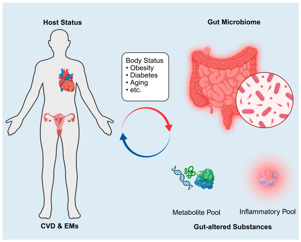
Figure 3.
Altered Pathological Environment in Patients with EMs. Patients with EMs exhibit an altered pathological microenvironment, primarily characterized by lipid metabolic dysregulation (purple), oxidative stress (blue), low-grade chronic inflammation (red), endothelial dysfunction (yellow), and abnormal activation of the coagulation system (green). These interconnected alterations collectively form the core pathological basis for the increased risk of CVD in people with EMs. Upward arrow: increased substance levels; Downward arrow: decreased substance levels. Abbreviations: LDL-C, low-density lipoprotein cholesterol; TG, triglycerides; Lp(a), lipoprotein(a); HDL-C, high-density lipoprotein cholesterol; PON-1, paraoxonase-1; SOD, superoxide dismutase; GSH, glutathione; AOPPs, advanced oxidation protein products; MDA, malondialdehyde; LOOHs, lipid hydroperoxides; ox-LDL, oxidized low-density lipoprotein; IL, interleukin; TNF-α, tumor necrosis factor-α; CCL, C-C motif chemokine ligand; VEGF, vascular endothelial growth factor; ER, estrogen receptor; PR, progesterone receptor; PDGF, platelet-derived growth factor; TGF-β, transforming growth factor-β; FMD, flow-mediated dilation; baPWV, brachial-ankle pulse wave velocity; AGEs, advanced glycation end products; UtA RI, uterine artery resistance index; CRP, C-reactive protein; vWF, von Willebrand factor; PT, prothrombin time; TT, thrombin time; APTT, activated partial thromboplastin time.
Figure 3.
Altered Pathological Environment in Patients with EMs. Patients with EMs exhibit an altered pathological microenvironment, primarily characterized by lipid metabolic dysregulation (purple), oxidative stress (blue), low-grade chronic inflammation (red), endothelial dysfunction (yellow), and abnormal activation of the coagulation system (green). These interconnected alterations collectively form the core pathological basis for the increased risk of CVD in people with EMs. Upward arrow: increased substance levels; Downward arrow: decreased substance levels. Abbreviations: LDL-C, low-density lipoprotein cholesterol; TG, triglycerides; Lp(a), lipoprotein(a); HDL-C, high-density lipoprotein cholesterol; PON-1, paraoxonase-1; SOD, superoxide dismutase; GSH, glutathione; AOPPs, advanced oxidation protein products; MDA, malondialdehyde; LOOHs, lipid hydroperoxides; ox-LDL, oxidized low-density lipoprotein; IL, interleukin; TNF-α, tumor necrosis factor-α; CCL, C-C motif chemokine ligand; VEGF, vascular endothelial growth factor; ER, estrogen receptor; PR, progesterone receptor; PDGF, platelet-derived growth factor; TGF-β, transforming growth factor-β; FMD, flow-mediated dilation; baPWV, brachial-ankle pulse wave velocity; AGEs, advanced glycation end products; UtA RI, uterine artery resistance index; CRP, C-reactive protein; vWF, von Willebrand factor; PT, prothrombin time; TT, thrombin time; APTT, activated partial thromboplastin time.
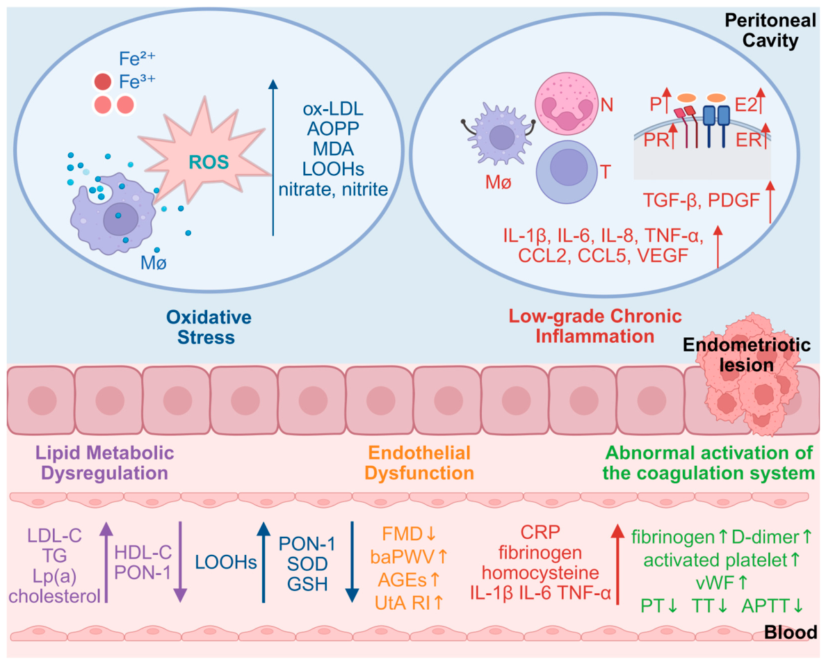
Figure 4.
Treatment-Related CVD Risk Assessment in Patients with Different Subtypes of EMs. Framework linking treatment exposure to cardiovascular risk in EMs. Surgical (blue) and hormonal (orange) exposures may modify CVD risk through altered ovarian function, sex-steroid exposure, and cardiometabolic–thromboembolic dysregulation (purple). The surgical axis predominates in OMA; SUP and DIE depend mainly on prolonged hormonal treatment, and postoperative hormonal therapy adds cumulative exposure across all three phenotypes (teal). Assessment should integrate the actual procedures and medications received, age at treatment, ovarian function, and conventional risk factors. Abbreviations: CHD, coronary heart disease; CVD, cardiovascular disease; DIE, deep infiltrating endometriosis; EMs, endometriosis; GnRH, gonadotropin-releasing hormone; LNG-IUD, levonorgestrel-releasing intrauterine device; MI, myocardial infarction; OMA, ovarian endometrioma; SUP, superficial peritoneal endometriosis.
Figure 4.
Treatment-Related CVD Risk Assessment in Patients with Different Subtypes of EMs. Framework linking treatment exposure to cardiovascular risk in EMs. Surgical (blue) and hormonal (orange) exposures may modify CVD risk through altered ovarian function, sex-steroid exposure, and cardiometabolic–thromboembolic dysregulation (purple). The surgical axis predominates in OMA; SUP and DIE depend mainly on prolonged hormonal treatment, and postoperative hormonal therapy adds cumulative exposure across all three phenotypes (teal). Assessment should integrate the actual procedures and medications received, age at treatment, ovarian function, and conventional risk factors. Abbreviations: CHD, coronary heart disease; CVD, cardiovascular disease; DIE, deep infiltrating endometriosis; EMs, endometriosis; GnRH, gonadotropin-releasing hormone; LNG-IUD, levonorgestrel-releasing intrauterine device; MI, myocardial infarction; OMA, ovarian endometrioma; SUP, superficial peritoneal endometriosis.
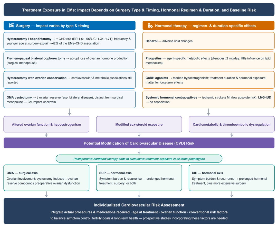

Table 1.
Summary of Observed Associations Between Endometriosis and Cardiovascular Risk Factors.
| Author(s) | Study Name | Location | Population | Design |
Data Collection |
Ascertainment Period (year) | Findings (OR/RR/aHR/HR, 95% CI) | |
| Nagai et al. (2015) | JNHS | Japan | 49927 female nurses (41.2±7.9 years) | Cross-sectional study | Self-completed questionnaire | 2001–2007 | 1.Transient ischaemic attack 2.Angina pectoris 3.Hypertension 4. Hypercholesterolaemia |
OR=1.91(1.26-2.90) OR=1.55(1.03-2.32) OR=1.26(1.07-1.47) OR=1.30(1.15-1.47) |
| Mu et al. (2016) | NHSII | USA | 116430 female nurses (25–42 years); 4244 EMs (laparoscopic-confirmed) vs 91554 controls | Prospective cohort study |
Self-completed questionnaire | 1989–2009 | 1.Myocardial Infarction 2.Angina pectoris 3.Coronary Bypass/Angioplasty/Stent 4.Combined Coronary Heart Disease |
RR=1.52(1.17-1.98) RR=1.91(1.59-2.29) RR=1.35(1.08-1.69) RR=1.62(1.39-1.89) |
| Mu et al. (2017) | NHSII | USA | 116430 female nurses (25–42 years); 4244 EMs (laparoscopic-confirmed) vs 91554 controls | Prospective cohort study |
Self-completed questionnaire | 1989–2009 | 1.Hypertension 2.Hypercholesterolemia |
RR=1.14(1.09-1.18) RR=1.25(1.21-1.30) |
| Okoth et al. (2021) | — | UK | 56090 EMs vs 223669 controls (16–50 years) | Population-based retrospective cohort study |
Electronic health records | 1995–2018 | 1.Composite CVD 2.IHD 3.Cerebrovascular Disease 4.Arrhythmia 5.Hypertension 6.Heart Failure |
aHR=1.24(1.13-1.37) aHR=1.40(1.22-1.61) aHR=1.19(1.04-1.36) aHR=1.26(1.11-1.43) aHR=1.12(1.07-1.17) aHR=0.76(0.54-1.07) |
| Wang et al. (2024b) | NHSII | USA | 110091 female nurses (25–42 years); 5185 EMs (laparoscopic-confirmed) vs 104906 controls | Prospective cohort study |
Self-completed questionnaire | 1989–2019 | 1.CVD mortality (EMs): 2.CVD mortality (EMs + UFs): 3.CVD mortality (EMs without UFs): 4.CVD mortality (UFs without EMs): |
HR=1.12(0.77-1.63) HR=1.61(0.93-2.76) HR=0.93(0.57-1.51) HR=1.07(0.78-1.48) |
| Havers-Borgersen et al. (2024) | — | Denmark | Main study: 60508 EMs vs 242032 controls (median 37.3 years, 1977–2021) Secondary study: 35190 EMs vs 140760 controls (median 36.3 years, 2000–2021) |
Population-based retrospective cohort study |
Danish Civil Registration System | 1977–2021 | 1.Composite outcome: AMI/ischaemic stroke: 2.Individual components: 1)AMI: 2)Ischaemic stroke: 3)Arrhythmias: 4)Heart failure: |
HR=1.15(1.11-1.20) HR=1.35(1.31-1.40) HR=1.18(1.12-1.23) HR=1.21(1.17-1.25) HR=1.11(1.05-1.18) |
| Note: Numbered outcomes in the Findings column correspond to the effect estimates (OR/RR/aHR/HR with 95% CI) listed in the same order in the adjacent column. a Same population, data collection method, and ascertainment period as Mu et al. (2016). Abbreviations: aHR, adjusted hazard ratio; AMI, acute myocardial infarction; CI, confidence interval; CVD, cardiovascular disease; EMs, endometriosis; HR, hazard ratio; IHD, ischaemic heart disease; JNHS, Japan Nurses’ Health Study; NHSII, Nurses’ Health Study II; OR, odds ratio; RR, relative risk; UFs, uterine fibroids. | ||||||||
Disclaimer/Publisher’s Note: The statements, opinions and data contained in all publications are solely those of the individual author(s) and contributor(s) and not of MDPI and/or the editor(s). MDPI and/or the editor(s) disclaim responsibility for any injury to people or property resulting from any ideas, methods, instructions or products referred to in the content. |
© 2026 by the authors. Licensee MDPI, Basel, Switzerland. This article is an open access article distributed under the terms and conditions of the Creative Commons Attribution (CC BY) license.
Copyright: This open access article is published under a Creative Commons CC BY 4.0 license, which permit the free download, distribution, and reuse, provided that the author and preprint are cited in any reuse.