

Submitted:
19 October 2026
Posted:
21 October 2026
You are already at the latest version
Abstract
The essay reviews current understanding of the role of metal-dependent forms of regulated cell death – ferroptosis and cuproptosis – in the pathogenesis of breast cancer and their therapeutic potential. The aim of the work was to analyze the molecular mechanisms of both processes, identify common regulatory nodes, and assess the prospects for their joint targeting to overcome chemoresistance. Methodologically, the work is based on a critical review of peer-reviewed scientific sources devoted to ferroptosis, cuproptosis, and their cross-talk in the context of oncology. It has been shown that ferroptosis, iron-dependent lipid peroxidation, and cuproptosis, copper-dependent proteotoxic death, have common regulatory nodes, in particular, glutathione metabolism, mitochondrial respiration, and the p53 signaling pathway. It has been established that induction of both processes allows overcoming resistance to cisplatin, doxorubicin, and taxanes, especially in aggressive subtypes such as triple-negative breast cancer. Current therapeutic strategies are reviewed, including the use of nanocarriers for targeted delivery of ferroptosis and cuproptosis inducers, as well as the results of clinical trials of disulfiram, elesclomol, and copper chelators. At the same time, limitations of clinical translation are emphasized: the lack of validated biomarkers, the risk of systemic toxicity, and tumor heterogeneity. It is concluded that co-targeting ferroptosis and cuproptosis is a promising strategy, but its widespread clinical application requires further prospective studies, biomarker identification, and development of targeted nanosystems.
Keywords:
ferroptosis
; cuproptosis
; breast cancer
; chemoresistance
; glutathione
; mitochondrial metabolism
; p53
; nanotechnology
; biomarkers
; triple-negative breast cancer
1. Introduction
Breast cancer remains the most common cancer among women worldwide, and despite significant progress in diagnosis and treatment, the problem of resistance to therapy and relapse remains extremely relevant. According to GLOBOCAN, in 2022, more than 2.3 million new cases of breast cancer were registered, and the mortality rate reaches 685 thousand cases per year [1]. Like many chronic non-communicable diseases, the initiation and progression of breast cancer are deeply rooted in systemic pathogenetic alterations, among which chronic inflammation plays a fundamental role [2]. Traditional treatment methods, including surgery, chemotherapy, radiotherapy and targeted therapy, often prove to be insufficiently effective due to the high heterogeneity of the tumor and the ability of cancer cells to evade apoptosis. This encourages researchers to search for new forms of regulated cell death that could be used to selectively destroy tumor cells (Figure 1).
Among such novel mechanisms, ferroptosis, an iron-dependent lipid peroxidation, and cuproptosis, a copper-dependent proteotoxic death resulting from the aggregation of lipoylated proteins in mitochondria, have attracted particular attention. Both processes belong to metal-dependent forms of regulated cell death (RCD) and demonstrate a close relationship at the level of metabolic pathways, in particular through glutathione (GSH), mitochondrial metabolism and p53 signaling cascades. In the context of breast cancer, where metabolic rearrangement is one of the key features, these pathways open up new therapeutic opportunities, especially for aggressive subtypes such as triple-negative breast cancer (TNBC) [3].
In this essay, we argue that co-targeting ferroptosis and cuproptosis is a promising strategy to overcome chemoresistance in breast cancer, but clinical translation of this approach remains limited due to the lack of validated biomarkers, the risk of systemic toxicity, and tumor heterogeneity. We review the molecular mechanisms of both processes, their cross-talk, therapeutic potential, and obstacles to clinical application.
2. Main Text
Molecular mechanisms of ferroptosis. Ferroptosis, first described in 2012, is an iron-dependent form of necrotic cell death characterized by the accumulation of lipid peroxides and disruption of plasma membrane integrity. Key regulators of ferroptosis are the /GSH/GPX4 system, as well as proteins that control iron and lipid metabolism [4,5].
Glutathione peroxidase 4 (GPX4) is a selenium-dependent enzyme that reduces lipid hydroperoxides to nontoxic alcohols using GSH as a cofactor. Decreased GPX4 activity due to GSH depletion, inhibition of the system (consisting of SLC7A11 and SLC3A2), or impaired GSH synthesis leads to the accumulation of lipid peroxides and the initiation of ferroptosis [6]. In addition, proteins regulating polyunsaturated fatty acid (PUFA) metabolism, in particular ACSL4 and LPCAT3, play an important role, which promote the incorporation of PUFA into membrane phospholipids, making them susceptible to peroxidation [7] (Figure 2).
Iron is critical for ferroptosis: excess Fe2+ catalyzes the Fenton reaction, generating hydroxyl radicals that attack PUFAs. Proteins that control iron homeostasis, such as transferrin receptor 1 (TFR1), ferritin (FTH1/FTL), and ferroportin (FPN), modulate cellular susceptibility to ferroptosis [8]. Importantly, there are GPX4-independent defense pathways, including FSP1/CoQ10, DHODH, and GCH1/BH4, that provide additional antioxidant defense [9].
In breast cancer, ferroptosis plays a dual role. On the one hand, tumor cells often have elevated levels of GPX4 and SLC7A11, which makes them resistant to ferroptosis. On the other hand, induction of ferroptosis, for example with inhibitors of GPX4 (RSL3) or the system (erastin, sulfasalazine), suppresses tumor growth and increases sensitivity to chemotherapy [10]. The use of ferroptosis to overcome resistance to cisplatin and doxorubicin is particularly promising, as has been shown in preclinical models of TNBC [11].
Molecular mechanisms of cuproptosis. Cuproptosis, discovered in 2022, is a new form of RCC resulting from excess intracellular copper. The mechanism of cuproptosis is distinct from apoptosis, necroptosis, pyroptosis, and ferroptosis and is dependent on mitochondrial respiration. Excess Cu+ binds to lipoylated proteins of the tricarboxylic acid cycle (TCA), particularly dihydrolipoamide S-acetyltransferase (DLAT), leading to their aggregation, loss of iron-sulfur clusters (Fe-S), and proteotoxic stress [12] (Figure 3).
A key regulator of cuproptosis is ferredoxin 1 (FDX1), which reduces Cu2+ to the more toxic Cu+ and regulates protein lipoylation through interaction with lipoyl synthase (LIAS). Inhibition of FDX1 or LIAS prevents cuproptosis [13]. Other genes that contribute to cuproptosis include LIPT1, DLD, DLAT, PDHA1, and PDHB, while MTF1, GLS, and CDKN2A have a protective function [14,15].
In breast cancer, cuproptosis is of particular importance because tumor cells, especially TNBC, exhibit an increased dependence on mitochondrial respiration and have higher copper content. This makes them sensitive to cuproptosis inducers such as elesclomol (ES) and disulfiram (DSF). Elesclomol transports Cu2+ into the mitochondria, where it induces DLAT aggregation and cell death [16]. Disulfiram, in combination with copper, forms a DSF/Cu complex that induces cuproptosis and apoptosis, and overcomes resistance to taxanes and anthracyclines [17].
Common regulatory nodes of ferroptosis and cuproptosis. Despite differences in molecular mechanisms, ferroptosis and cuproptosis share several common regulatory nodes, which creates the basis for their synergistic induction (Figure 4).
Glutathione (GSH) is a central mediator of both processes. In ferroptosis, GSH acts as an antioxidant, supporting GPX4 activity, whereas in cuproptosis, GSH chelates copper, preventing its binding to lipoylated proteins. Depletion of GSH, for example with the inhibitor buthionine sulfoximine (BSO), simultaneously increases the sensitivity of cells to both forms of death [18]. This makes GSH an attractive target for combination therapy.
Mitochondrial metabolism is another common node. Ferroptosis is accompanied by changes in mitochondrial morphology and metabolism, whereas cuproptosis is directly dependent on mitochondrial respiration and the TCA cycle. Electron transport chain inhibitors (e.g., rotenone and antimycin A) prevent cuproptosis but may affect ferroptosis [19]. In addition, mitochondrial iron and copper metabolism are closely intertwined, and disruption of one metal homeostasis affects the other.
The tumor suppressor p53 exhibits a multifaceted regulatory function in ferroptosis and cuproptosis. It can both induce and inhibit ferroptosis depending on the context: p53 suppresses SLC7A11, reducing GSH synthesis, but can also increase the expression of CDKN1A/p21, which promotes cell survival. In cuproptosis, p53 increases cell sensitivity by regulating the metabolism and expression of genes involved in the biogenesis of Fe-S clusters [20].
Autophagy also plays a role in regulating both processes. Autophagy can promote ferroptosis through ferritinophagy, releasing free iron, and also affect cuproptosis by degrading GPX4 and other proteins [21].
Cross-talk between ferroptosis and cuproptosis: synergy or antagonism? Recent studies have demonstrated that ferroptosis and cuproptosis can both synergize and antagonize each other. On the one hand, induction of ferroptosis can increase susceptibility to cuproptosis through GSH depletion and ROS accumulation. For example, erastin, an inhibitor of the system, reduces GSH levels, which enhances copper toxicity [22]. On the other hand, excess copper can induce GPX4 degradation through autophagy, which promotes ferroptosis [23]. Thus, there is a positive feedback loop that can be exploited therapeutically.
However, there are also antagonistic effects. Copper can inhibit ferroptosis by activating NRF2, which increases the expression of antioxidant genes. In addition, high concentrations of copper can inhibit caspases, which affects apoptosis but not ferroptosis [24]. This indicates the complexity of the interaction and the need for careful selection of combinations (Figure 5).
Therapeutic strategies: from nanotechnology to clinical trials. Targeting ferroptosis and cuproptosis together opens up new therapeutic possibilities. One of the most promising approaches is the use of nanocarriers to deliver inducers of both processes. For example, CuP/Er nanoparticles containing erastin and copper induce both ferroptosis and cuproptosis, causing immunogenic cell death and enhancing the efficacy of immunotherapy [25]. Another example is the MetaCell nanosystem, which uses neutrophils to deliver Fe-Cu MOFs that induce both types of death and activate antitumor immunity [26].
Clinical trials are also investigating modulators of ferroptosis and cuproptosis. For example, disulfiram in combination with copper is in phase II clinical trials in metastatic breast cancer (NCT03323346). Elesclomol, an inducer of cuproptosis, is being tested in combination with paclitaxel in melanoma and other solid tumors [27]. However, most studies remain in the preclinical stage (Figure 6).
Problems and limitations. Despite its promise, clinical translation of co-targeting ferroptosis and cuproptosis faces a number of obstacles. First, there is a lack of validated biomarkers for patient stratification. Although GPX4, SLC7A11, FDX1, and DLAT levels correlate with prognosis, their prognostic value needs to be confirmed in prospective studies [28].
Second, the risk of systemic toxicity. Induction of ferroptosis and cuproptosis can cause damage to healthy tissues, especially the liver, kidneys, and nervous system. For example, GPX4 inhibitors can cause neurodegeneration, and copper chelators can cause immune dysfunction [29].
Third, tumor heterogeneity. Different breast cancer subtypes have different sensitivities to ferroptosis and cuproptosis. TNBC is more sensitive to cuproptosis, while HER2-positive tumors may be more sensitive to ferroptosis [30]. This requires a personalized approach.
Finally, there is a lack of clinical data. Most studies have been conducted on cell lines and mouse models, and only a few clinical trials are in the early stages (Figure 7).
3. Conclusions
Ferroptosis and cuproptosis are two novel forms of metal-dependent regulated cell death that play important roles in the pathogenesis of breast cancer. Their shared regulatory landscape, involving GSH, mitochondrial metabolism, and p53, provides the basis for synergistic induction. Preclinical data suggest that combined targeting of both processes can overcome chemotherapy resistance, especially in aggressive subtypes such as TNBC.
However, the clinical translation of this approach remains limited due to the lack of validated biomarkers, the risk of systemic toxicity, and tumor heterogeneity. In our opinion, further research should focus on the development of targeted nanocarriers, the identification of biomarkers for patient stratification, and the conduct of prospective clinical trials. Only with an integrated approach can the joint targeting of ferroptosis and cuproptosis become a realistic therapeutic strategy to improve breast cancer outcomes.
Author Contributions
The author confirms sole responsibility for the conception, design, analysis, interpretation, drafting, and final approval of the manuscript.
Funding
This research received no external funding.
Data Availability Statement
No data were generated as a result of this study.
Acknowledgments
Not applicable.
Conflicts of Interest
The author declares that he has no competing interests.
Declaration on the Use of Artificial Intelligence: During the preparation of this work, the author used Google Gemini and Google Search tools solely to assist with literature search, data analysis, and language editing to improve readability. The tool was not used to generate scientific conclusions or original text. Following the use of these tools, the author reviewed, edited, and evaluated the content as needed and takes full responsibility for the final contents of the publication.
Abbreviations
The following abbreviations are used in this manuscript:
| ADC | Antibody-drug conjugate |
| ATP | Adenosine triphosphate |
| BH4 | Tetrahydrobiopterin |
| BSO | Buthionine sulfoximine |
| CRT | Calreticulin |
| DC | Dendritic cell |
| DSF | Disulfiram |
| ES | Elesclomol |
| Fe-S | Iron-sulfur |
| GPX4 | Glutathione peroxidase 4 |
| GSH | Glutathione |
| HER2 | Human epidermal growth factor receptor 2 |
| HMGB1 | High mobility group box 1 |
| ICD | Immunogenic cell death |
| IV | Intravenous |
| MOF | Metal-Organic Framework |
| MRP | Multidrug resistance-associated protein |
| MTs | Metallothioneins |
| NGS | Next-Generation Sequencing |
| P-gp | P-glycoprotein |
| PUFA | Polyunsaturated fatty acid |
| RCD | Regulated cell death |
| RCC | Regulated cell death |
| ROS | Reactive oxygen species |
| TCA | Tricarboxylic acid cycle |
| TFR1 | Transferrin receptor 1 |
| TNBC | Triple-negative breast cancer |
References
- Bray, F.; Laversanne, M.; Sung, H.; Ferlay, J.; Siegel, R.L.; Soerjomataram, I.; Jemal, A. Global Cancer Statistics 2022: GLOBOCAN Estimates of Incidence and Mortality Worldwide for 36 Cancers in 185 Countries. CA. Cancer J. Clin. 2024, 74, 229–263. [Google Scholar] [CrossRef]
- Ivchuk, V. Chronic Inflammation as an Underlying Pathogenetic Mechanism of Chronic Non-Communicable Diseases: Molecular, Cellular, and Therapeutic Dimensions. Preprints 2026, 2026092666. [Google Scholar] [CrossRef]
- Imam, M.; Ji, J.; Zhang, Z.; Yan, S. Targeting the Initiator to Activate Both Ferroptosis and Cuproptosis for Breast Cancer Treatment: Progress and Possibility for Clinical Application. Front. Pharmacol. 2025, 15, 1493188. [Google Scholar] [CrossRef]
- Dixon, S.J.; Lemberg, K.M.; Lamprecht, M.R.; Skouta, R.; Zaitsev, E.M.; Gleason, C.E.; Patel, D.N.; Bauer, A.J.; Cantley, A.M.; Yang, W.S.; et al. Ferroptosis: An Iron-Dependent Form of Nonapoptotic Cell Death. Cell 2012, 149, 1060–1072. [Google Scholar] [CrossRef]
- Ivchuk, V. Ferroptosis: Molecular Mechanisms of Regulation and Therapeutic Potential in the Pathogenesis of Multifactorial Diseases. Preprints 2026, 2026090771. [Google Scholar] [CrossRef]
- Koppula, P.; Zhuang, L.; Gan, B. Cystine Transporter SLC7A11/xCT in Cancer: Ferroptosis, Nutrient Dependency, and Cancer Therapy. Protein Cell 2021, 12, 599–620. [Google Scholar] [CrossRef]
- Doll, S.; Proneth, B.; Tyurina, Y.Y.; Panzilius, E.; Kobayashi, S.; Ingold, I.; Irmler, M.; Beckers, J.; Aichler, M.; Walch, A.; et al. ACSL4 Dictates Ferroptosis Sensitivity by Shaping Cellular Lipid Composition. Nat. Chem. Biol. 2017, 13, 91–98. [Google Scholar] [CrossRef]
- Fang, X.; Ardehali, H.; Min, J.; Wang, F. The Molecular and Metabolic Landscape of Iron and Ferroptosis in Cardiovascular Disease. Nat. Rev. Cardiol. 2023, 20, 7–23. [Google Scholar] [CrossRef]
- Bersuker, K.; Hendricks, J.M.; Li, Z.; Magtanong, L.; Ford, B.; Tang, P.H.; Roberts, M.A.; Tong, B.; Maimone, T.J.; Zoncu, R.; et al. The CoQ Oxidoreductase FSP1 Acts Parallel to GPX4 to Inhibit Ferroptosis. Nature 2019, 575, 688–692. [Google Scholar] [CrossRef]
- Chen, X.; Kang, R.; Kroemer, G.; Tang, D. Broadening Horizons: The Role of Ferroptosis in Cancer. Nat. Rev. Clin. Oncol. 2021, 18, 280–296. [Google Scholar] [CrossRef]
- Wang, L.; Wang, C.; Li, X.; Tao, Z.; Zhu, W.; Su, Y.; Choi, W.S. Melatonin and Erastin Emerge Synergistic Anti-Tumor Effects on Oral Squamous Cell Carcinoma by Inducing Apoptosis, Ferroptosis, and Inhibiting Autophagy through Promoting ROS. Cell. Mol. Biol. Lett. 2023, 28, 36. [Google Scholar] [CrossRef]
- Tsvetkov, P.; Coy, S.; Petrova, B.; Dreishpoon, M.; Verma, A.; Abdusamad, M.; Rossen, J.; Joesch-Cohen, L.; Humeidi, R.; Spangler, R.D.; et al. Copper Induces Cell Death by Targeting Lipoylated TCA Cycle Proteins. Science 2022, 375, 1254–1261. [Google Scholar] [CrossRef]
- Dreishpoon, M.B.; Bick, N.R.; Petrova, B.; Warui, D.M.; Cameron, A.; Booker, S.J.; Kanarek, N.; Golub, T.R.; Tsvetkov, P. FDX1 Regulates Cellular Protein Lipoylation through Direct Binding to LIAS. J. Biol. Chem. 2023, 299, 105046. [Google Scholar] [CrossRef]
- Tang, Y.; Lv, C.; Luo, Z.; Li, Z.; Yu, J. Construction of a Prognostic Model Based on Cuproptosis-Related Patterns for Predicting Survival, Immune Infiltration, and Immunotherapy Efficacy in Breast Cancer: Cuproptosis-Based Prognostic Modeling in Breast Cancer. Medicine 2024, 103, e40136. [Google Scholar] [CrossRef]
- Ivchuk, V. Cuproptosis: Biochemical Mechanisms, Therapeutic Potential, and Clinical Challenges. Preprints 2026, 2026101523. [Google Scholar] [CrossRef]
- Zheng, P.; Zhou, C.; Lu, L.; Liu, B.; Ding, Y. Elesclomol: A Copper Ionophore Targeting Mitochondrial Metabolism for Cancer Therapy. J. Exp. Clin. Cancer Res. 2022, 41, 271. [Google Scholar] [CrossRef]
- Guo, X.; Xu, B.; Pandey, S.; Goessl, E.; Brown, J.; Armesilla, A.L.; Darling, J.L.; Wang, W. Disulfiram/Copper Complex Inhibiting NFκB Activity and Potentiating Cytotoxic Effect of Gemcitabine on Colon and Breast Cancer Cell Lines. Cancer Lett. 2010, 290, 104–113. [Google Scholar] [CrossRef]
- Zhang, P.; Zhou, C.; Ren, X.; Jing, Q.; Gao, Y.; Yang, C.; Shen, Y.; Zhou, Y.; Hu, W.; Jin, F.; et al. Inhibiting the Compensatory Elevation of xCT Collaborates with Disulfiram/Copper-Induced GSH Consumption for Cascade Ferroptosis and Cuproptosis. Redox Biol. 2024, 69, 103007. [Google Scholar] [CrossRef]
- Tang, D.; Chen, X.; Kroemer, G. Cuproptosis: A Copper-Triggered Modality of Mitochondrial Cell Death. Cell Res. 2022, 32, 417–418. [Google Scholar] [CrossRef]
- Xiong, C.; Ling, H.; Hao, Q.; Zhou, X. Cuproptosis: P53-Regulated Metabolic Cell Death? Cell Death Differ. 2023, 30, 876–884. [Google Scholar] [CrossRef]
- Xue, Q.; Yan, D.; Chen, X.; Li, X.; Kang, R.; Klionsky, D.J.; Kroemer, G.; Chen, X.; Tang, D.; Liu, J. Copper-Dependent Autophagic Degradation of GPX4 Drives Ferroptosis. Autophagy 2023, 19, 1982–1996. [Google Scholar] [CrossRef]
- Wang, W.; Lu, K.; Jiang, X.; Wei, Q.; Zhu, L.; Wang, X.; Jin, H.; Feng, L. Ferroptosis Inducers Enhanced Cuproptosis Induced by Copper Ionophores in Primary Liver Cancer. J. Exp. Clin. Cancer Res. 2023, 42, 142. [Google Scholar] [CrossRef]
- Gao, W.; Huang, Z.; Duan, J.; Nice, E.C.; Lin, J.; Huang, C. Elesclomol Induces Copper-dependent Ferroptosis in Colorectal Cancer Cells via Degradation of ATP7A. Mol. Oncol. 2021, 15, 3527–3544. [Google Scholar] [CrossRef]
- Li, Y.; Du, Y.; Zhou, Y.; Chen, Q.; Luo, Z.; Ren, Y.; Chen, X.; Chen, G. Iron and Copper: Critical Executioners of Ferroptosis, Cuproptosis and Other Forms of Cell Death. Cell Commun. Signal. 2023, 21, 327. [Google Scholar] [CrossRef]
- Li, Y.; Liu, J.; Chen, Y.; Weichselbaum, R.R.; Lin, W. Nanoparticles Synergize Ferroptosis and Cuproptosis to Potentiate Cancer Immunotherapy. Adv. Sci. 2024, 11, 2310309. [Google Scholar] [CrossRef]
- Chen, K.; Zhou, A.; Zhou, X.; He, J.; Xu, Y.; Ning, X. Cellular Trojan Horse Initiates Bimetallic Fe-Cu MOF-Mediated Synergistic Cuproptosis and Ferroptosis against Malignancies. Sci. Adv. 2024, 10, eadk3201. [Google Scholar] [CrossRef]
- O’Day, S.J.; Eggermont, A.M.M.; Chiarion-Sileni, V.; Kefford, R.; Grob, J.J.; Mortier, L.; Robert, C.; Schachter, J.; Testori, A.; Mackiewicz, J.; et al. Final Results of Phase III SYMMETRY Study: Randomized, Double-Blind Trial of Elesclomol Plus Paclitaxel Versus Paclitaxel Alone As Treatment for Chemotherapy-Naive Patients With Advanced Melanoma. J. Clin. Oncol. 2013, 31, 1211–1218. [Google Scholar] [CrossRef]
- Hu, K.; Qiu, J.; Hu, Y.; Wang, Y.; Yu, C.; Wu, Y. Efficacy of FERscore in Predicting Sensitivity to Ferroptosis Inducers in Breast Cancer. npj Breast Cancer 2024, 10, 74. [Google Scholar] [CrossRef]
- Koppula, P.; Lei, G.; Zhang, Y.; Yan, Y.; Mao, C.; Kondiparthi, L.; Shi, J.; Liu, X.; Horbath, A.; Das, M.; et al. A Targetable CoQ-FSP1 Axis Drives Ferroptosis- and Radiation-Resistance in KEAP1 Inactive Lung Cancers. Nat. Commun. 2022, 13, 2206. [Google Scholar] [CrossRef]
- Yang, F.; Xiao, Y.; Ding, J.-H.; Jin, X.; Ma, D.; Li, D.-Q.; Shi, J.-X.; Huang, W.; Wang, Y.-P.; Jiang, Y.-Z.; et al. Ferroptosis Heterogeneity in Triple-Negative Breast Cancer Reveals an Innovative Immunotherapy Combination Strategy. Cell Metab. 2023, 35, 84–100.e8. [Google Scholar] [CrossRef]
Figure 1.
Comprehensive schematic overview of co-targeting ferroptosis and cuproptosis in breast cancer: from molecular mechanisms to therapeutic prospects. The diagram illustrates the integration of iron-dependent ferroptosis and copper-dependent cuproptosis pathways and their exploitation in oncology. Left panel (Ferroptosis): Highlights cystine transport via system (SLC7A11/SLC3A2), intracellular glutathione (GSH) synthesis, GPX4-mediated antioxidant defense, iron metabolism via Transferrin/TFR1 and ferritinophagy, and the Fenton reaction driving lipid peroxidation and membrane damage. Middle panel (Shared Nodes): Identifies critical intersection points between both pathways, specifically GSH metabolism (acting concurrently as an antioxidant and copper chelator), mitochondrial metabolism (TCA cycle and OXPHOS respiratory chain), and the p53 signaling pathway governing SLC7A11 repression and Fe-S cluster biogenesis. Right panel (Cuproptosis): Demonstrates copper import via CTR1, FDX1-mediated reduction of to toxic , pathological oligomerization and aggregation of lipoylated TCA enzymes (DLAT, PDHA1/PDHB), loss of essential Fe-S cluster proteins, and resulting proteotoxic stress. Bottom panel (Therapeutic Strategies & Barriers): Outlines advanced nanocarriers for co-delivery (e.g., CuP/Er nanoparticles, MetaCell, Cu-GA nanoparticles), ongoing clinical translation efforts involving copper-based compounds (Disulfiram + , Elesclomol, copper chelators), and major translational barriers including the lack of robust biomarkers, systemic toxicity, and intra-tumoral heterogeneity. Image created using Gemini tool (Google, 2026) based on the directions provided by the author to illustrate this concept. The final image was curated and verified for technical accuracy by the author.
Figure 1.
Comprehensive schematic overview of co-targeting ferroptosis and cuproptosis in breast cancer: from molecular mechanisms to therapeutic prospects. The diagram illustrates the integration of iron-dependent ferroptosis and copper-dependent cuproptosis pathways and their exploitation in oncology. Left panel (Ferroptosis): Highlights cystine transport via system (SLC7A11/SLC3A2), intracellular glutathione (GSH) synthesis, GPX4-mediated antioxidant defense, iron metabolism via Transferrin/TFR1 and ferritinophagy, and the Fenton reaction driving lipid peroxidation and membrane damage. Middle panel (Shared Nodes): Identifies critical intersection points between both pathways, specifically GSH metabolism (acting concurrently as an antioxidant and copper chelator), mitochondrial metabolism (TCA cycle and OXPHOS respiratory chain), and the p53 signaling pathway governing SLC7A11 repression and Fe-S cluster biogenesis. Right panel (Cuproptosis): Demonstrates copper import via CTR1, FDX1-mediated reduction of to toxic , pathological oligomerization and aggregation of lipoylated TCA enzymes (DLAT, PDHA1/PDHB), loss of essential Fe-S cluster proteins, and resulting proteotoxic stress. Bottom panel (Therapeutic Strategies & Barriers): Outlines advanced nanocarriers for co-delivery (e.g., CuP/Er nanoparticles, MetaCell, Cu-GA nanoparticles), ongoing clinical translation efforts involving copper-based compounds (Disulfiram + , Elesclomol, copper chelators), and major translational barriers including the lack of robust biomarkers, systemic toxicity, and intra-tumoral heterogeneity. Image created using Gemini tool (Google, 2026) based on the directions provided by the author to illustrate this concept. The final image was curated and verified for technical accuracy by the author.
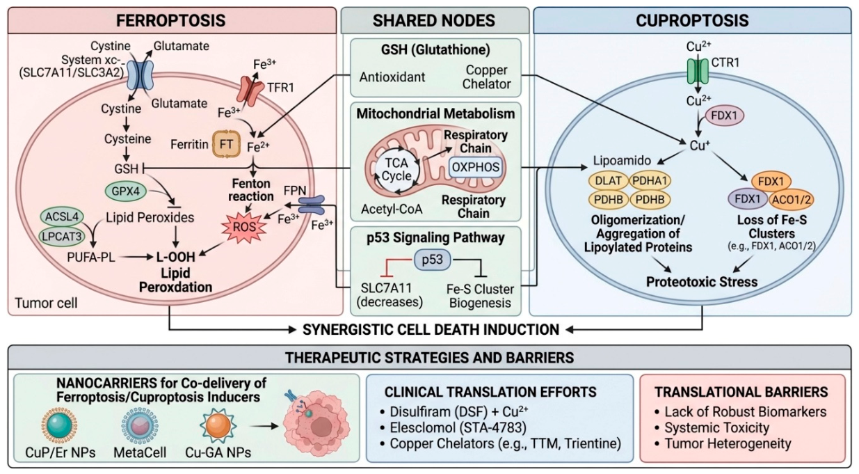
Figure 2.
Molecular mechanisms of ferroptosis execution, lipid peroxidation, and iron homeostasis in tumor cells. This flowchart details the biochemical cascade leading to iron-dependent necrotic cell death. Top-left block (Cysteine Transport & GSH Synthesis): Extracellular cystine is imported via system (SLC7A11/SLC3A2 heterodimer) in exchange for glutamate and subsequently reduced to cysteine, the rate-limiting precursor for intracellular glutathione (GSH) biosynthesis. Top-middle block (GPX4-Mediated Antioxidant Defense): Glutathione peroxidase 4 (GPX4) utilizes GSH as a cofactor to reduce toxic lipid hydroperoxides () into non-toxic lipid alcohols (); pharmacological inhibition by RSL3 blocks this defense. Top-right block (Iron Metabolism & Fenton Reaction): Extracellular binds Transferrin (Tf), enters via TFR1, is reduced by STEAP3 and transported via DMT1 into the labile iron pool (LIP), stored in ferritin (mobilized via NCOA4-mediated ferritinophagy), and exported by ferroportin (FPN). Excess intracellular reacts with hydrogen peroxide () via the Fenton reaction to generate reactive hydroxyl radicals (). Bottom-left block (Lipid Peroxidation Pathway): Polyunsaturated fatty acids (PUFAs, e.g., arachidonic acid AA, adrenic acid AdA) are esterified into membrane phospholipids (PUFA-PL) via ACSL4 and LPCAT3 enzymes. Bottom-middle block (Lipid Peroxidation Mechanism): Lipoxygenases (LOX) and Fenton-derived radicals abstract bis-allylic hydrogens from PUFA-PL, generating lipid radicals (), lipid peroxyl radicals (), and accumulating . Bottom-right block (Ferroptosis Execution): Concomitant GSH depletion/GPX4 inhibition and iron overload/Fenton-driven lipid peroxide accumulation cause catastrophic plasma membrane damage and cell death. Image created using Gemini tool (Google, 2026) based on the directions provided by the author to illustrate this concept. The final image was curated and verified for technical accuracy by the author.
Figure 2.
Molecular mechanisms of ferroptosis execution, lipid peroxidation, and iron homeostasis in tumor cells. This flowchart details the biochemical cascade leading to iron-dependent necrotic cell death. Top-left block (Cysteine Transport & GSH Synthesis): Extracellular cystine is imported via system (SLC7A11/SLC3A2 heterodimer) in exchange for glutamate and subsequently reduced to cysteine, the rate-limiting precursor for intracellular glutathione (GSH) biosynthesis. Top-middle block (GPX4-Mediated Antioxidant Defense): Glutathione peroxidase 4 (GPX4) utilizes GSH as a cofactor to reduce toxic lipid hydroperoxides () into non-toxic lipid alcohols (); pharmacological inhibition by RSL3 blocks this defense. Top-right block (Iron Metabolism & Fenton Reaction): Extracellular binds Transferrin (Tf), enters via TFR1, is reduced by STEAP3 and transported via DMT1 into the labile iron pool (LIP), stored in ferritin (mobilized via NCOA4-mediated ferritinophagy), and exported by ferroportin (FPN). Excess intracellular reacts with hydrogen peroxide () via the Fenton reaction to generate reactive hydroxyl radicals (). Bottom-left block (Lipid Peroxidation Pathway): Polyunsaturated fatty acids (PUFAs, e.g., arachidonic acid AA, adrenic acid AdA) are esterified into membrane phospholipids (PUFA-PL) via ACSL4 and LPCAT3 enzymes. Bottom-middle block (Lipid Peroxidation Mechanism): Lipoxygenases (LOX) and Fenton-derived radicals abstract bis-allylic hydrogens from PUFA-PL, generating lipid radicals (), lipid peroxyl radicals (), and accumulating . Bottom-right block (Ferroptosis Execution): Concomitant GSH depletion/GPX4 inhibition and iron overload/Fenton-driven lipid peroxide accumulation cause catastrophic plasma membrane damage and cell death. Image created using Gemini tool (Google, 2026) based on the directions provided by the author to illustrate this concept. The final image was curated and verified for technical accuracy by the author.
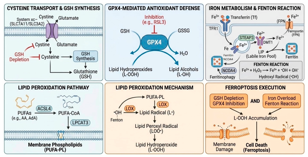
Figure 3.
Molecular mechanisms of cuproptosis and copper-induced proteotoxic stress in cancer cells. The schematic outlines the pathway of copper-dependent regulated cell death. Top-left block (Copper Import and Reduction): Extracellular is transported into the cytosol via high-affinity copper transporter 1 (CTR1). Top-right block (Cytosolic & Mitochondrial Chelation): Intracellular glutathione (GSH) buffers excess copper ions by forming complexes to mitigate acute toxicity. Bottom-left block (Canonical Protein Lipoylation & Pathological Aggregation): Under normal physiological conditions, lipoic acid (LPA) is synthesized and attached via LIAS and LIPT1 to the E2 subunit (DLAT) of the tricarboxylic acid (TCA) cycle pyruvate dehydrogenase (PDH) complex (including PDHA1 and PDHB subunits), ensuring mitochondrial respiration. In cuproptosis, excess intracellular binds directly to lipoylated moieties of DLAT, triggering massive pathological oligomerization and aggregation of lipoylated proteins, alongside the depletion of essential iron-sulfur (Fe-S) cluster proteins (e.g., FDX1, ACO1/2). Bottom-right block (Defense & Regulatory Mechanisms): Transcription factor MTF1 translocates to the nucleus to induce metallothioneins (MTs) and copper detoxification genes, while GLS and CDKN2A modulate cellular sensitivity to cuproptosis. Image created using Gemini tool (Google, 2026) based on the directions provided by the author to illustrate this concept. The final image was curated and verified for technical accuracy by the author.
Figure 3.
Molecular mechanisms of cuproptosis and copper-induced proteotoxic stress in cancer cells. The schematic outlines the pathway of copper-dependent regulated cell death. Top-left block (Copper Import and Reduction): Extracellular is transported into the cytosol via high-affinity copper transporter 1 (CTR1). Top-right block (Cytosolic & Mitochondrial Chelation): Intracellular glutathione (GSH) buffers excess copper ions by forming complexes to mitigate acute toxicity. Bottom-left block (Canonical Protein Lipoylation & Pathological Aggregation): Under normal physiological conditions, lipoic acid (LPA) is synthesized and attached via LIAS and LIPT1 to the E2 subunit (DLAT) of the tricarboxylic acid (TCA) cycle pyruvate dehydrogenase (PDH) complex (including PDHA1 and PDHB subunits), ensuring mitochondrial respiration. In cuproptosis, excess intracellular binds directly to lipoylated moieties of DLAT, triggering massive pathological oligomerization and aggregation of lipoylated proteins, alongside the depletion of essential iron-sulfur (Fe-S) cluster proteins (e.g., FDX1, ACO1/2). Bottom-right block (Defense & Regulatory Mechanisms): Transcription factor MTF1 translocates to the nucleus to induce metallothioneins (MTs) and copper detoxification genes, while GLS and CDKN2A modulate cellular sensitivity to cuproptosis. Image created using Gemini tool (Google, 2026) based on the directions provided by the author to illustrate this concept. The final image was curated and verified for technical accuracy by the author.
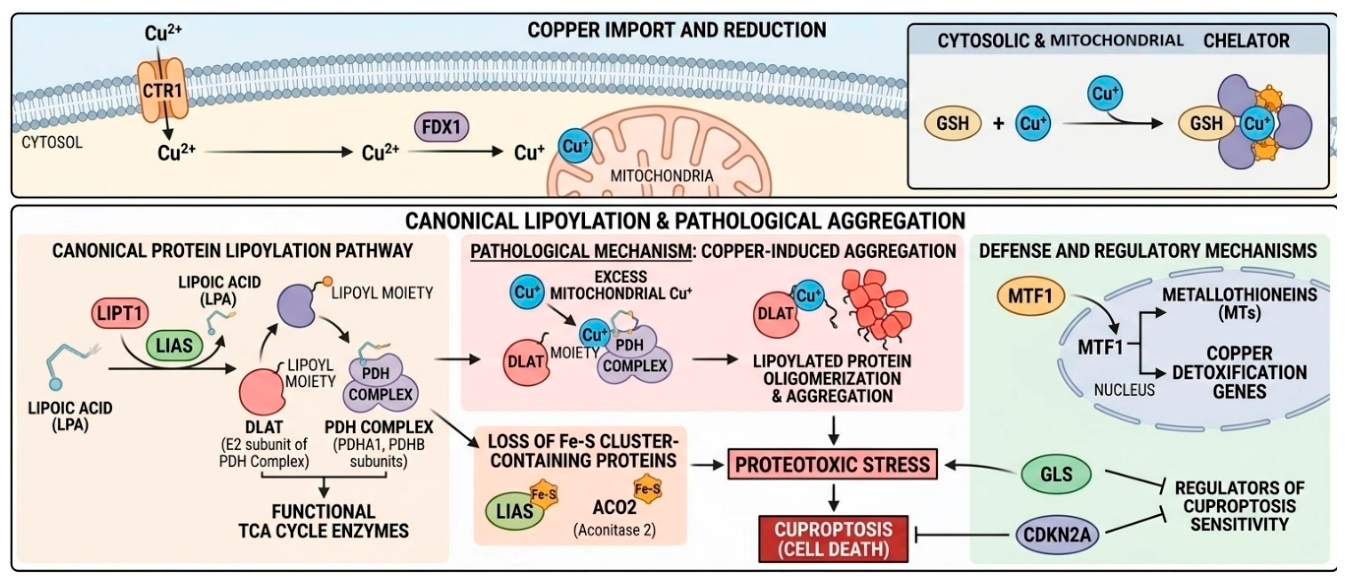
Figure 4.
Common regulatory nodes linking ferroptosis and cuproptosis metabolic networks. The diagram highlights intersecting biochemical hubs that enable synergistic cell death induction. Central Hub (Glutathione & Convergent Pathways): GSH serves as a dual-functional master regulator acting as an antioxidant protecting against ferroptotic lipid peroxidation and as a copper-chelating buffer mitigating cuproptotic stress; GSH depletion sensitizes cells to both modalities. Top Sector (Mitochondrial Metabolism): The TCA cycle and electron transport chain (OXPHOS respiratory chain) generate metabolic flux, reactive oxygen species (ROS), and mitochondrial stress that modulate both iron- and copper-dependent pathways. Bottom Sector (Regulatory Modules): Autophagy pathways (such as ferritinophagy releasing iron) and the p53 signaling network act as overarching regulators by transcriptionally suppressing SLC7A11 and regulating iron-sulfur cluster biogenesis in response to metabolic stress. Image created using Gemini tool (Google, 2026) based on the directions provided by the author to illustrate this concept. The final image was curated and verified for technical accuracy by the author.
Figure 4.
Common regulatory nodes linking ferroptosis and cuproptosis metabolic networks. The diagram highlights intersecting biochemical hubs that enable synergistic cell death induction. Central Hub (Glutathione & Convergent Pathways): GSH serves as a dual-functional master regulator acting as an antioxidant protecting against ferroptotic lipid peroxidation and as a copper-chelating buffer mitigating cuproptotic stress; GSH depletion sensitizes cells to both modalities. Top Sector (Mitochondrial Metabolism): The TCA cycle and electron transport chain (OXPHOS respiratory chain) generate metabolic flux, reactive oxygen species (ROS), and mitochondrial stress that modulate both iron- and copper-dependent pathways. Bottom Sector (Regulatory Modules): Autophagy pathways (such as ferritinophagy releasing iron) and the p53 signaling network act as overarching regulators by transcriptionally suppressing SLC7A11 and regulating iron-sulfur cluster biogenesis in response to metabolic stress. Image created using Gemini tool (Google, 2026) based on the directions provided by the author to illustrate this concept. The final image was curated and verified for technical accuracy by the author.
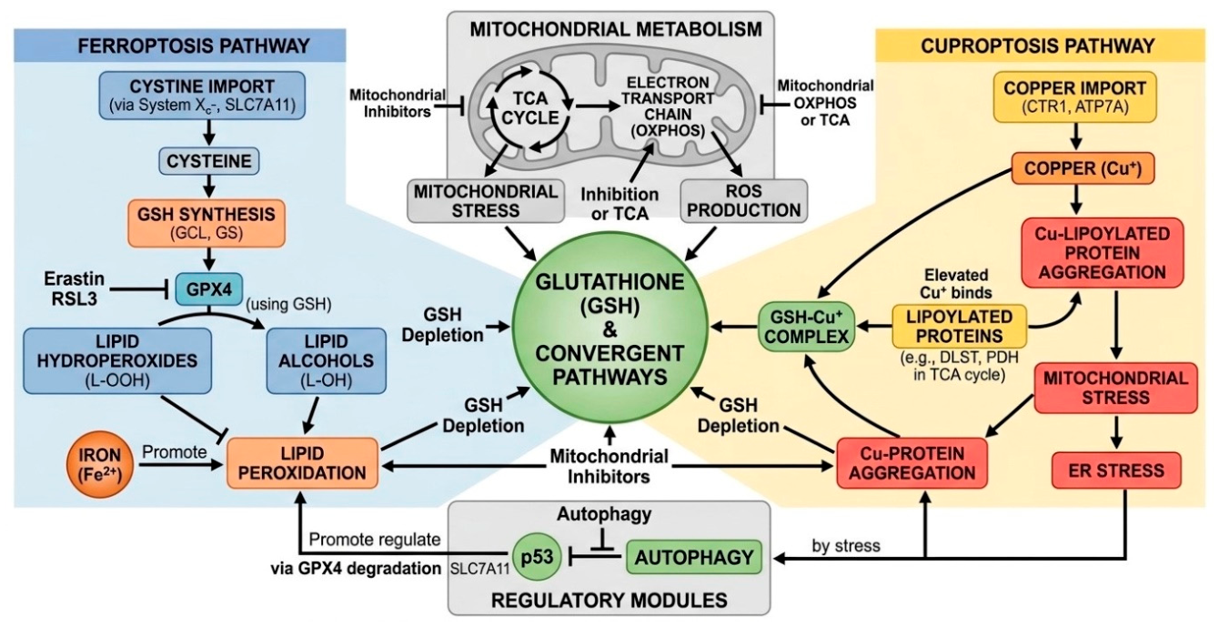
Figure 5.
Crosstalk, synergy, and antagonism between ferroptosis and cuproptosis pathways. This integrative scheme maps the bidirectional regulatory interactions between the two forms of cell death. Left Domain (Ferroptosis): Encompasses iron-dependent lipid peroxidation driven by system inhibition, cysteine depletion, and GPX4 inactivation. Right Domain (Cuproptosis): Encompasses copper-dependent proteotoxic stress driven by excessive intracellular (e.g., via elesclomol/Cu), abnormal DLAT lipoylation, and TCA cycle dysfunction. Central Interconnections (Synergy & Antagonism): Thick green promotional arrows indicate that ferroptosis induction (elevated ROS and GSH depletion) potentiates cuproptosis sensitivity, while GPX4 degradation promotes ferroptosis via increased lipid peroxidation (LPO). Conversely, inhibitory blunt-ended lines show antagonistic loops, including NRF2 activation (which upregulates GSH and GPX4 to protect against both pathways) and caspase inhibition. Image created using Gemini tool (Google, 2026) based on the directions provided by the author to illustrate this concept. The final image was curated and verified for technical accuracy by the author.
Figure 5.
Crosstalk, synergy, and antagonism between ferroptosis and cuproptosis pathways. This integrative scheme maps the bidirectional regulatory interactions between the two forms of cell death. Left Domain (Ferroptosis): Encompasses iron-dependent lipid peroxidation driven by system inhibition, cysteine depletion, and GPX4 inactivation. Right Domain (Cuproptosis): Encompasses copper-dependent proteotoxic stress driven by excessive intracellular (e.g., via elesclomol/Cu), abnormal DLAT lipoylation, and TCA cycle dysfunction. Central Interconnections (Synergy & Antagonism): Thick green promotional arrows indicate that ferroptosis induction (elevated ROS and GSH depletion) potentiates cuproptosis sensitivity, while GPX4 degradation promotes ferroptosis via increased lipid peroxidation (LPO). Conversely, inhibitory blunt-ended lines show antagonistic loops, including NRF2 activation (which upregulates GSH and GPX4 to protect against both pathways) and caspase inhibition. Image created using Gemini tool (Google, 2026) based on the directions provided by the author to illustrate this concept. The final image was curated and verified for technical accuracy by the author.
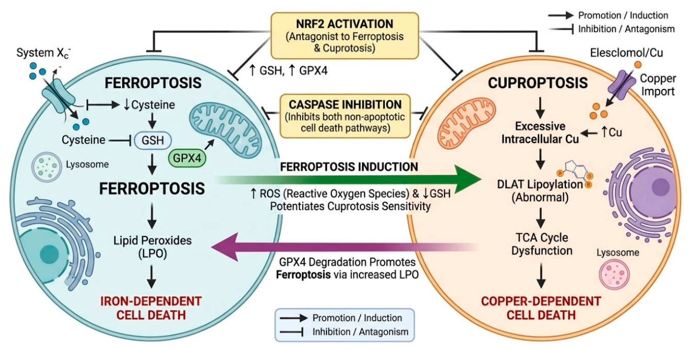
Figure 6.
Therapeutic strategies combining ferroptosis and cuproptosis induction and mechanisms of resistance overcoming. The illustration categorizes modern anti-tumor strategies. Block 1 (Nanocarriers for Co-delivery of Inducers): Highlights advanced delivery platforms such as CuP/Er nanoparticles (Copper(II) Porphyrin/Erbium-doped upconversion nanoparticles), MetaCell (Metal-Organic Framework-encapsulated cell membrane nanoplatform), and Cu-GA nanoparticles (Copper-Gallic Acid coordination polymer nanoparticles). Block 2 (Clinical Trials of Copper-Based Compounds): Details pharmacological interventions including Disulfiram (DSF) + , Elesclomol (STA-4783) + , and copper chelators (Tetrathiomolybdate – TTM, Trientine) targeting primary tumors, metastases, cancer stem cells, and tumor vasculature via intravenous, oral, or intratumoral routes. Block 3 (Mechanisms to Overcome Resistance & Activate Immune Response): Demonstrates how co-induction inhibits P-glycoprotein (P-gp) and multidrug resistance-associated proteins (MRP), depletes GSH, triggers mitochondrial dysfunction and ROS production, and induces immunogenic cell death (ICD) characterized by calreticulin (CRT) exposure, ATP release, HMGB1 secretion, and subsequent dendritic cell (DC) maturation and T-cell activation. Image created using Gemini tool (Google, 2026) based on the directions provided by the author to illustrate this concept. The final image was curated and verified for technical accuracy by the author.
Figure 6.
Therapeutic strategies combining ferroptosis and cuproptosis induction and mechanisms of resistance overcoming. The illustration categorizes modern anti-tumor strategies. Block 1 (Nanocarriers for Co-delivery of Inducers): Highlights advanced delivery platforms such as CuP/Er nanoparticles (Copper(II) Porphyrin/Erbium-doped upconversion nanoparticles), MetaCell (Metal-Organic Framework-encapsulated cell membrane nanoplatform), and Cu-GA nanoparticles (Copper-Gallic Acid coordination polymer nanoparticles). Block 2 (Clinical Trials of Copper-Based Compounds): Details pharmacological interventions including Disulfiram (DSF) + , Elesclomol (STA-4783) + , and copper chelators (Tetrathiomolybdate – TTM, Trientine) targeting primary tumors, metastases, cancer stem cells, and tumor vasculature via intravenous, oral, or intratumoral routes. Block 3 (Mechanisms to Overcome Resistance & Activate Immune Response): Demonstrates how co-induction inhibits P-glycoprotein (P-gp) and multidrug resistance-associated proteins (MRP), depletes GSH, triggers mitochondrial dysfunction and ROS production, and induces immunogenic cell death (ICD) characterized by calreticulin (CRT) exposure, ATP release, HMGB1 secretion, and subsequent dendritic cell (DC) maturation and T-cell activation. Image created using Gemini tool (Google, 2026) based on the directions provided by the author to illustrate this concept. The final image was curated and verified for technical accuracy by the author.
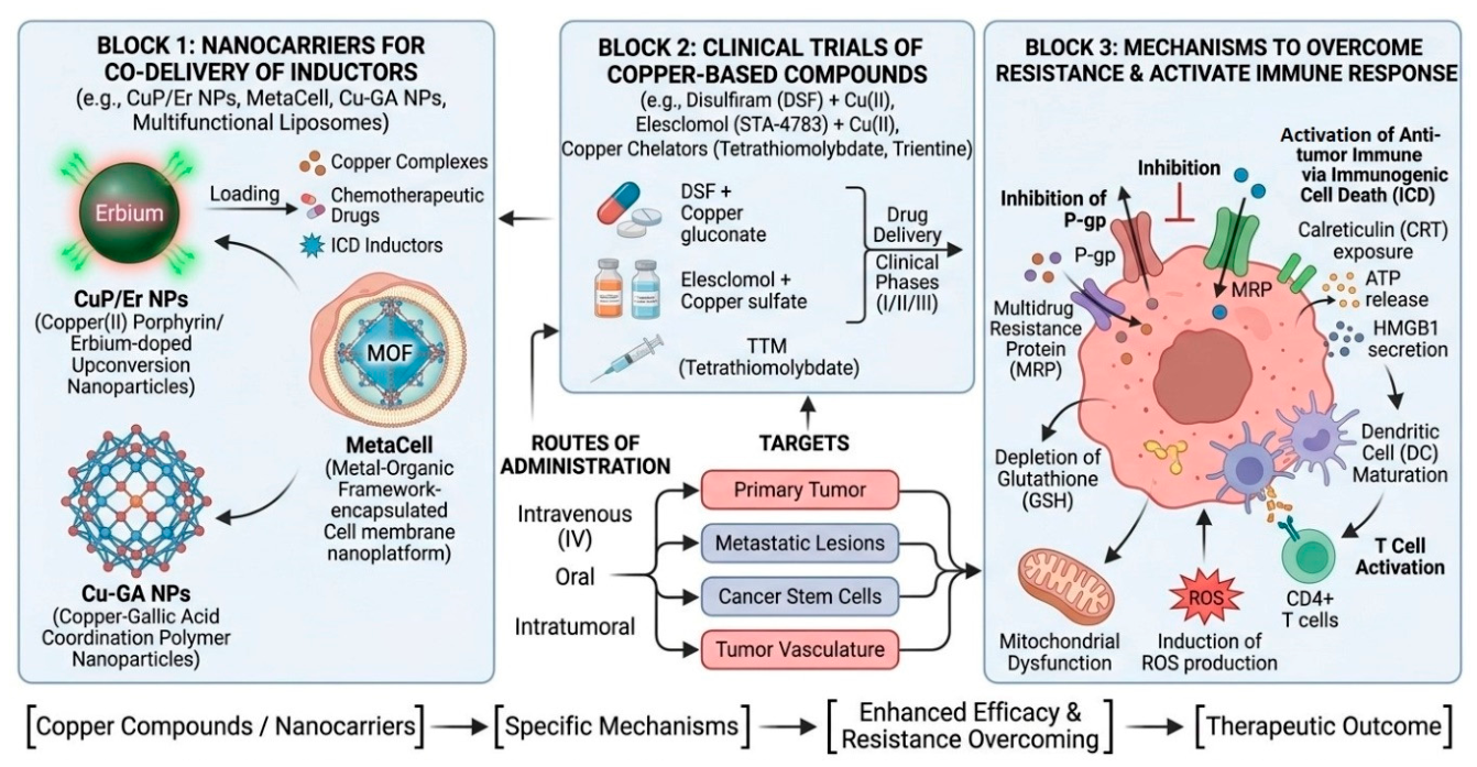
Figure 7.
Challenges, limitations, and overcoming strategies for clinical translation of ferroptosis and cuproptosis co-targeting. This structured flowchart contrasts biological barriers with precision oncology solutions. Barrier 1 & Overcoming Strategy 1: Addresses the lack of validated prognostic biomarkers and insufficient molecular markers for patient selection by proposing high-throughput sequencing (NGS), proteomics, and bioinformatics to discover specific gene and protein signatures. Barrier 2 & Overcoming Strategy 2: Addresses the risk of systemic toxicity in healthy tissues (liver, kidneys, nervous system) through targeted drug delivery systems and nanocarriers (such as antibody-drug conjugates [ADCs] and lipid nanoparticles) that maximize tumor accumulation. Barrier 3 & Overcoming Strategy 3: Addresses intra-tumoral heterogeneity across breast cancer subtypes (TNBC, HER2+, Luminal A/B) by implementing personalized medicine and molecular profiling to design tailored combination therapies and immune checkpoint inhibitors. Image created using Gemini tool (Google, 2026) based on the directions provided by the author to illustrate this concept. The final image was curated and verified for technical accuracy by the author.
Figure 7.
Challenges, limitations, and overcoming strategies for clinical translation of ferroptosis and cuproptosis co-targeting. This structured flowchart contrasts biological barriers with precision oncology solutions. Barrier 1 & Overcoming Strategy 1: Addresses the lack of validated prognostic biomarkers and insufficient molecular markers for patient selection by proposing high-throughput sequencing (NGS), proteomics, and bioinformatics to discover specific gene and protein signatures. Barrier 2 & Overcoming Strategy 2: Addresses the risk of systemic toxicity in healthy tissues (liver, kidneys, nervous system) through targeted drug delivery systems and nanocarriers (such as antibody-drug conjugates [ADCs] and lipid nanoparticles) that maximize tumor accumulation. Barrier 3 & Overcoming Strategy 3: Addresses intra-tumoral heterogeneity across breast cancer subtypes (TNBC, HER2+, Luminal A/B) by implementing personalized medicine and molecular profiling to design tailored combination therapies and immune checkpoint inhibitors. Image created using Gemini tool (Google, 2026) based on the directions provided by the author to illustrate this concept. The final image was curated and verified for technical accuracy by the author.
Disclaimer/Publisher’s Note: The statements, opinions and data contained in all publications are solely those of the individual author(s) and contributor(s) and not of MDPI and/or the editor(s). MDPI and/or the editor(s) disclaim responsibility for any injury to people or property resulting from any ideas, methods, instructions or products referred to in the content. |
© 2026 by the author. Licensee MDPI, Basel, Switzerland. This article is an open access article distributed under the terms and conditions of the Creative Commons Attribution (CC BY) license.
Copyright: This open access article is published under a Creative Commons CC BY 4.0 license, which permit the free download, distribution, and reuse, provided that the author and preprint are cited in any reuse.