
Submitted:
19 October 2026
Posted:
21 October 2026
You are already at the latest version
Abstract
Background/Objectives: We examined whether lower linear endovenous energy density (LEED) in the anterior saphenous vein (ASV) was proportional to its smaller diameter during simultaneous great saphenous vein (GSV) and ASV ablation. Methods: This exploratory retrospective study used 136 procedural records from a 433-patient source cohort. The primary comparison included 56 ipsilateral GSV–ASV pairs from 46 procedures. Nominal endovenous fluence equivalent (EFE) was calculated from LEED and pre-tumescence diameter measured with patients recumbent. Bilateral log ratios were averaged within procedures. Patient-linked clinical outcomes were not analyzed. Results: Procedure-weighted ASV/GSV ratios were 0.58 for diameter (95% confidence interval [CI], 0.55–0.62), 0.75 for LEED (95% CI, 0.70–0.80), and 1.29 for nominal EFE (95% CI, 1.19–1.40). Lower ASV LEED with higher nominal EFE occurred in 39 of 56 limbs. The between-wavelength EFE-ratio contrast was 0.97 (95% CI, 0.82–1.15). Excluding ambiguous records yielded an EFE ratio of 1.27 (95% CI, 1.17–1.39). Conclusions: The reduction in ASV LEED was smaller than the reduction in recorded diameter. Reporting measurement conditions alongside diameter and LEED improves dosimetric interpretation. These findings do not establish optimal dosing or clinical benefit.
Keywords:
endovenous laser ablation
; anterior saphenous vein
; great saphenous vein
; energy density
; vein diameter
1. Introduction
Endovenous laser ablation (EVLA) is widely used to treat superficial truncal reflux. European and North American guidelines support endovenous treatment in appropriately selected patients, based on symptoms, duplex findings, and venous anatomy [1,2]. When more than one refluxing trunk is treated during a procedure, energy delivery must be described separately for veins that differ in caliber and treated length. Comparisons based only on joules per centimeter can obscure these anatomical differences.
Retrospective comparisons of 1470- and 1940-nm flush EVLA have reported differences in linear endovenous energy density (LEED) and early local outcomes [3]. LEED describes energy delivered per unit length, but it does not account for vein diameter. A lower LEED in a smaller vein can therefore coexist with a higher value after geometric normalization. The calculation itself is established; the empirical question is how operator-selected LEED and recorded diameter relate within simultaneously treated veins.
The anterior saphenous vein (ASV) is recognized as a truncal vein, and multisocietal recommendations favor this term over anterior accessory saphenous vein [4]. Its treatment requires attention to its reflux source, course, and relationship to the saphenous compartment [5]. Comparing the ASV and great saphenous vein (GSV) within the same limb provides a way to describe relative energy allocation under a shared treatment setting, although local anatomy and procedural requirements can still differ.
Proebstle and colleagues described the endovenous fluence equivalent (EFE), which relates delivered energy to an idealized cylindrical venous surface area [6]. Mathematical modeling has also incorporated diameter when estimating energy requirements [7]. These approaches do not directly measure the energy absorbed by the vein wall. Tumescence changes venous geometry and protects surrounding tissue; fiber characteristics, blood, wall contact, and pullback also influence energy transfer [8]. Experimental comparisons of 1470- and 1940-nm systems have not established a universal conversion between nominal energy and tissue injury [9,10].
We examined whether the proportional reduction in ASV LEED matched the proportional reduction in recorded pre-tumescence diameter during paired GSV–ASV treatment. Secondary analyses described wavelength-specific ratios, cumulative energy across treated vein entries, and the association between diameter and LEED. The purpose was to assess the information added by diameter-aware reporting. The study was not designed to determine a therapeutic window or an optimal energy prescription.
2. Materials and Methods
2.1. Study Design and Setting
The source cohort comprised 433 patients: 127 treated with a 1470-nm system and 306 with a 1940-nm system. This exploratory retrospective study reports dosimetric analyses of 136 procedural records: 77 at 1470 nm and 59 at 1940 nm. These records were selected by random sampling separately within each wavelength group. This sampling concerned record selection rather than randomized assignment to a laser wavelength. These analyzed records dated from July 20, 2022, to February 28, 2024. The source-cohort patient totals describe the clinical population; the analysis-specific procedure, limb, and vein-entry counts define the observations used for each estimate. The reporting framework was informed by the STROBE statement [11]. Ethics approval was obtained from the participating hospitals under TABED Ethics Committee approval no. 1-251264, dated May 7, 2025.
The study addressed procedural dosimetry within the source cohort. Patient-linked closure, recurrence, pain, and quality-of-life outcomes were not analyzed. The cohort-wide report of no complications was retained as an aggregate clinical observation. Complication definitions, ascertainment methods, and follow-up duration were not available for the present analysis, and this observation was not linked to individual energy settings. It therefore could not support an assessment of dose-related safety or efficacy.
2.2. Procedural Records and Diameter Measurements
Records included age, sex, previous intervention, number of vascular accesses, and separate right- and left-sided entries for the GSV, small saphenous vein (SSV), and ASV. A treated vein entry, termed a segment in the source records, contained a treatment indicator, diameter in millimeters, recorded treated length in centimeters, and LEED in J/cm. Calculations used the entire recorded treated length for each entry; no arbitrary fixed-length subdivision was introduced. An additional accessory-vein field did not specify reliable laterality. We used ASV throughout for the corresponding source fields. A companion summary file contained 50 records already represented in the 1940-nm dataset and was not counted as an additional patient series.
Procedures used ultrasound-guided radial-fiber flush EVLA with perivenous tumescent anesthesia and individualized energy settings. For the primary GSV–ASV comparison, ultrasound diameters were measured at the saphenofemoral junction level, with patients in a recumbent position, before tumescent infiltration, between morning and noon. These measurements describe pre-tumescence caliber. Post-tumescence diameter was not available for the dosimetric calculation. Power, pullback speed, tumescent volume, and exact fiber-tip position were not consistently represented in the analyzed records. We therefore interpreted EFE as a nominal geometric index based on the recorded pre-tumescence diameter.
2.3. Eligibility and Paired Observations
A treated vein entry was eligible if it was marked as treated and had positive diameter, length, and LEED values. Decimal commas and decimal points were converted to numeric values. Zeros in untreated entries were treated as absence of treatment. One entry marked as treated had zero measurements and was excluded. One unmarked ASV entry had complete positive measurements; it was excluded from the principal analysis and included in a sensitivity analysis. An unmarked GSV entry with a diameter value alone was not counted as treated.
The paired analysis required an eligible GSV and ASV on the same side during the same procedure. If both legs contained eligible pairs, the two log ratios were averaged before analysis so that each procedure contributed equally. A repeated name appeared on two procedure dates in the 1470-nm dataset. Both encounters were retained and conservatively assigned to one provisional patient cluster; only one contributed an eligible GSV–ASV pair. Patient linkage was therefore provisional.
2.4. Dosimetry and Endpoints
Energy for each treated vein entry was calculated as LEED multiplied by its entire recorded treated length. For the paired GSV–ASV comparison, nominal EFE was calculated by dividing this energy by the lateral surface area of an idealized cylinder, using the recorded pre-tumescence diameter and treated length. With diameter in millimeters, nominal EFE in J/cm² equals 10 times LEED divided by the product of pi and diameter. Treated length cancels because both total energy and cylindrical surface area are proportional to length under this model. Thus, using separate vein entries preserves the different diameters of individual trunks; pooling the lengths of the GSV and ASV would obscure the within-limb comparison. For these veins, a single SFJ-level diameter does not represent variation along the entire treated length.
The primary endpoint was the procedure-weighted geometric mean ASV/GSV nominal EFE ratio. LEED and diameter ratios were calculated using the same weighting. We also counted limbs in which ASV LEED was lower but nominal EFE was higher than the corresponding GSV value. Total procedure energy was the sum of eligible vein-entry energies; length-weighted LEED was total energy divided by total treated length. Four procedures with unresolved discrepancies in treatment indicators, access counts, or entry completeness were excluded from cumulative-energy summaries. Their complete ipsilateral pairs remained eligible for the principal analysis, with exclusion assessed in a sensitivity analysis.
2.5. Statistical Analysis
Continuous variables are presented as mean ± standard deviation or median and interquartile range. Geometric means and 95% confidence intervals (CIs) were calculated on the log scale using the t distribution. Wavelength groups were compared using Welch’s test of procedure-level log EFE ratios. An exploratory regression modeled log LEED using log diameter, log length, vein type, and wavelength. Standard errors were clustered by provisional patient identifier, with a finite-sample correction and t inference [12]. The model included GSV, ASV, and SSV entries; the six entries without a defined truncal type were excluded. Estimates describe recorded energy allocation and are not clinical relative risks.
Sensitivity analyses excluded ambiguous procedures, omitted the possible repeat encounter, included the unmarked entry with complete measurements, and analyzed individual limbs with clustered inference. A separate scenario increased ASV diameter and decreased GSV diameter by equal percentages while holding LEED constant. This scenario assessed dependence on relative diameter; it did not estimate measurement error or simulate the physical effect of tumescence. The primary endpoint was selected after review of the available variables. All analyses were exploratory, without a prospective sample-size calculation or multiplicity adjustment. Tests were two-sided and performed in Python using NumPy, pandas, and SciPy.
2.6. Manuscript Preparation
Generative artificial intelligence assisted with revision of the scientific prose and presentation, reference formatting, and reconstruction of figures from reported summary statistics. During this revision, the statistical estimates were retained from the existing analysis; no patient-level reanalysis was performed. Figure 4B presents a deterministic transformation of the reported pooled estimate and confidence limits under the stated diameter assumptions.
3. Results
3.1. Source Cohort and Analytical Samples
The source cohort comprised 433 patients, including 127 in the 1470-nm group and 306 in the 1940-nm group. Dosimetric results below derive from the 136 analyzed procedural records, which contained 288 entries marked as treated. Of these, 287 had positive diameter, length, and LEED values and were eligible: 199 GSV, 57 ASV, 25 SSV, and six other accessory-vein entries. Table 1 and Figure 1 distinguish the overall clinical population from each analytical sample. The SSV entries contributed to secondary analyses where eligible, including the vein-type regression; they did not contribute to the primary comparison, which specifically required ipsilateral GSV and ASV treatment.
3.2. Paired GSV and ASV Dosimetry
There were 56 eligible ipsilateral GSV–ASV pairs from 46 procedures. At 1470 nm, 25 procedures contributed 28 pairs, including three bilateral procedures. At 1940 nm, 21 procedures contributed 28 pairs, including seven bilateral procedures. After averaging bilateral log ratios within procedures, the ASV/GSV diameter ratio was 0.58 (95% CI, 0.55–0.62), whereas the LEED ratio was 0.75 (95% CI, 0.70–0.80). The proportional reduction in recorded LEED was therefore smaller than the reduction in recorded pre-tumescence diameter.
The pooled ASV/GSV nominal EFE ratio was 1.29 (95% CI, 1.19–1.40; p < 0.001). Ratios were 1.31 (95% CI, 1.16–1.48) at 1470 nm and 1.27 (95% CI, 1.13–1.43) at 1940 nm (Table 2; Figure 2). The ratio of these contrasts, 1940 nm/1470 nm, was 0.97 (95% CI, 0.82–1.15; p = 0.716). Lower ASV LEED with higher nominal EFE occurred in 19 of 28 paired limbs at 1470 nm and 20 of 28 at 1940 nm, or 39 of 56 overall (69.6%).
3.3. Cumulative Energy and Exploratory Regression
Cumulative energy was assessed in 132 procedures: 74 at 1470 nm and 58 at 1940 nm. Median energy increased from 3.29 kJ for one treated vein entry to 6.84 kJ for at least three entries at 1470 nm, and from 1.88 to 4.53 kJ at 1940 nm (Table 3; Figure 3).
In the exploratory model of 281 entries from 135 provisional patient clusters, the coefficient for log diameter was 0.172 (95% CI, 0.096–0.248); a doubling of diameter was associated with a 1.13-fold LEED (95% CI, 1.07–1.19; Table 4). This association describes operator-selected energy settings.
3.4. Sensitivity Analyses and Clinical Context
Excluding ambiguous procedures retained 43 paired procedures and an EFE ratio of 1.27 (95% CI, 1.17–1.39). Other data-handling analyses yielded ratios of 1.29–1.30. Assumed opposing diameter changes of 10% reduced the pooled ratio to 1.06; changes of 15% reduced it to 0.95 (Figure 4). No complications were reported in the 433-patient source cohort. Complication definitions, ascertainment methods, and follow-up duration were not available for the present analysis; this aggregate report therefore could not support a complication-rate estimate or linkage of safety to dosimetry. Patient-linked closure and other clinical outcomes were not analyzed.
4. Discussion
4.1. Principal Findings
Among 46 procedures with paired GSV–ASV measurements, ASV LEED was approximately 25% lower and recorded pre-tumescence diameter approximately 42% smaller than in the ipsilateral GSV. Nominal EFE was consequently 29% higher. The direction of this geometric comparison was similar in both wavelength groups and persisted after exclusion of ambiguous procedures. These findings apply to the analyzed records and characterize energy reporting rather than treatment efficacy.
The study’s contribution is the paired description of how recorded energy and diameter varied between simultaneously treated trunks. The identity is straightforward: the ASV/GSV nominal EFE ratio equals the LEED ratio divided by the diameter ratio. Higher nominal EFE follows when the proportional reduction in diameter exceeds the reduction in LEED. This is not independent evidence of greater thermal injury. The observed frequency of this pattern shows why LEED alone can give an incomplete account of relative energy allocation in multitruncal procedures.
4.2. Clinical Interpretation and Measurement Conditions
A clinically useful dosing strategy must achieve durable occlusion with acceptable adverse effects. Different nominal EFE values may remain compatible with effective, well-tolerated treatment within a therapeutic range. Tumescence can alter lumen size, blood content, and heat transfer while protecting surrounding tissue [7,8]. Our calculations cannot locate that therapeutic range, classify a treatment as excessive or insufficient, or justify reducing ASV energy to equalize EFE. The aggregate report of no complications does not resolve this question without defined surveillance, closure assessment, and patient-linked dosimetry.
Pre-tumescence diameter is central to interpreting the result. Measurements were obtained with patients recumbent, at the saphenofemoral junction level, between morning and noon. Specifying these conditions improves reproducibility, but it does not make the recorded value a measure of the lumen during energy delivery. Tumescence can compress the vein and induce spasm, and the magnitude of narrowing need not be identical in the ASV and GSV [8]. Earlier modeling explicitly considered post-tumescence diameter [7]. The present EFE values therefore normalize energy to recorded baseline geometry rather than to the actual treatment-time surface area.
Total procedure energy increased with the number of treated vein entries and their aggregate length. This is expected from the calculation and should not be interpreted as an independent biological discovery or evidence of increased harm. Reporting total energy, treated length, LEED, and diameter separately allows readers to distinguish procedural extent from energy per unit length and from a nominal geometric normalization.
4.3. Context Within the Clinical Literature
Clinical studies of 1940-nm EVLA provide context for the energy values used in practice. Park and the group led by Viarengo reported clinical outcomes with this wavelength [13,14]. Setia and colleagues prospectively studied 139 patients with 177 treated veins, with comparison against published 1470-nm cohorts [15]. Keo and colleagues reported feasibility and early efficacy [16], while a systematic review and a multicenter experience highlighted variation in techniques, devices, and outcome assessment [17,18]. These reports cannot establish that an identical nominal EFE produces an identical tissue response across different veins or treatment systems.
The 1470-nm radial-fiber literature likewise evaluates clinically used treatment systems [19,20]. Some comparisons differ in both wavelength and fiber configuration [21], while others compare EVLA with radiofrequency ablation [22]. Such designs cannot isolate every determinant of energy transfer. In the present study, the between-wavelength EFE contrast was imprecise: its 95% CI ranged from 0.82 to 1.15. The nonsignificant test does not demonstrate equivalence, and no equivalence margin was specified.
Junctional technique and recurrence are separate clinical questions. Rits and colleagues reported less stump reflux after flush ablation [23]. In a randomized trial, Alozai and colleagues found shorter stumps after flush EVLA but no reduction in ASV reflux at 24 months; reflux occurred in 30% of the flush group and 21% of the standard group (p = 0.34) [24]. Because the present records did not provide stump length or exact fiber-tip distance from the junction, our dosimetric findings cannot explain differences in recurrence.
Reports of major ascending tributary ablation and a systematic review of flush EVLA address the extent of treatment and its clinical consequences [25,26]. The veins in our study had already been selected for ablation. The study provides no evidence for prophylactic ablation of a competent ASV; treatment selection remains based on symptoms, duplex-confirmed reflux, and individual anatomy [1,2,5].
4.4. Limitations and Future Research
The retrospective, exploratory design limits interpretation. The 433-patient source cohort provides the clinical context, whereas dosimetric estimates derive from 136 procedural records and the primary paired comparison from 46 procedures. These analytical samples have not been shown to represent the entire source cohort. Although records were sampled randomly within each wavelength group, a comparison with patients outside the analytical sample was not available. Eligibility for the paired analysis further restricted the sample to procedures with complete ipsilateral GSV–ASV observations; selection bias therefore remains possible. Within-limb pairing reduces variation in the overall treatment setting but cannot control for local anatomy, treatment indication, or operator choice. Provisional patient linkage, post hoc endpoint selection, and multiple exploratory comparisons further limit inference.
A single junction-level diameter cannot represent tapering, segmental dilation, or the post-tumescence shape of the treated vein. Morning-to-noon scheduling defines a measurement window but does not eliminate physiological variation. The opposing-diameter scenario brought the nominal EFE ratio close to unity at 10% and reversed its direction at 15%, demonstrating dependence on the relative diameter assumptions. These percentages were selected for illustration and are not measured errors or observed changes caused by tumescence. The geometric model also omits wall thickness and longitudinal variation in energy delivery. Power, pullback, and fiber–wall interactions were incompletely characterized.
Clinical validation would require standardized measurement sites and patient position, documentation of pre- and post-tumescence caliber, and prospective linkage of dosimetry to duplex-confirmed closure, symptoms, and adverse events. Thrombotic outcomes should use defined ascertainment and follow-up [27], and symptom change could be assessed with the revised Venous Clinical Severity Score [28]. Until such data are analyzed, the practical implication is more complete reporting of treatment conditions. The present results do not establish a lower ASV energy target or a preferred wavelength.
5. Conclusions
In the analyzed paired GSV–ASV procedures, the reduction in ASV LEED was smaller than the reduction in recorded pre-tumescence diameter, producing a higher nominal EFE. Reporting diameter together with its measurement site, patient position, and timing relative to tumescence clarifies this geometric comparison. The findings do not determine optimal dosing or establish differences in closure or complications.
Supplementary Materials
The following supporting information can be downloaded at the website of this paper posted on Preprints.org. STROBE reporting checklist.
Author Contributions
Conceptualization, E.K. and A.B.B.; methodology, E.K. and A.B.B.; writing—original draft preparation, E.K. and A.B.B.; writing—review and editing, E.K. and A.B.B.
Funding
This research received no external funding.
Institutional Review Board Statement
The study was approved by the participating hospitals under TABED Ethics Committee approval no. 1-251264, dated 7 May 2025.
Informed Consent Statement
Informed consent was obtained for participation in the research.
Data Availability Statement
The data supporting the findings of this study are available from the corresponding author upon request by the journal editors.
Conflicts of Interest
The authors declare no conflicts of interest.
Abbreviations
ASV, anterior saphenous vein; CI, confidence interval; EFE, endovenous fluence equivalent; EVLA, endovenous laser ablation; GSV, great saphenous vein; LEED, linear endovenous energy density; SFJ, saphenofemoral junction; SSV, small saphenous vein; STROBE, Strengthening the Reporting of Observational Studies in Epidemiology.
References
- De Maeseneer, M.G.; Kakkos, S.K.; Aherne, T.; Baekgaard, N.; Black, S.; Blomgren, L.; Giannoukas, A.; Gohel, M.; de Graaf, R.; Hamel-Desnos, C.; et al. European Society for Vascular Surgery (ESVS) 2022 Clinical Practice Guidelines on the Management of Chronic Venous Disease of the Lower Limbs. Eur. J. Vasc. Endovasc. Surg. 2022, 63, 184–267. [Google Scholar] [CrossRef] [PubMed]
- Gloviczki, P.; Lawrence, P.F.; Wasan, S.M.; Meissner, M.H.; Almeida, J.; Brown, K.R.; Bush, R.L.; Di Iorio, M.; Fish, J.; Fukaya, E.; et al. The 2023 Society for Vascular Surgery, American Venous Forum, and American Vein and Lymphatic Society clinical practice guidelines for the management of varicose veins of the lower extremities. Part II. J. Vasc. Surg. Venous Lymphat. Disord. 2024, 12, 101670. [Google Scholar] [CrossRef] [PubMed]
- Budak, A.B.; Yıldırım, A.; Gül, E.B. Impact of laser wavelength on outcomes of flush endovenous laser ablation: comparative evaluation of 1470 nm and 1940 nm systems and associated complications. Ann. Vasc. Surg. 2026, 130, 30–37. [Google Scholar] [CrossRef] [PubMed]
- Meissner, M.; Boyle, E.M.; Labropoulos, N.; Caggiati, A.; Drgastin, R.; Doganci, S.; Gasparis, A. The anterior saphenous vein. Part 1. A position statement endorsed by the American Vein and Lymphatic Society, the American Venous Forum, and the International Union of Phlebology. Phlebology 2024, 39, 310–312. [Google Scholar] [CrossRef] [PubMed]
- Boyle, E.M.; Drgastin, R.; Labropoulos, N.; Caggiati, A.; Gasparis, A.; Doganci, S.; Meissner, M. The anterior saphenous vein. Part 4. Clinical and technical considerations in treatment. Endorsed by the American Vein and Lymphatic Society, the American Venous Forum, and the International Union of Phlebology. Phlebology 2024, 39, 333–341. [Google Scholar] [CrossRef] [PubMed]
- Proebstle, T.M.; Moehler, T.; Herdemann, S. Reduced recanalization rates of the great saphenous vein after endovenous laser treatment with increased energy dosing: definition of a threshold for the endovenous fluence equivalent. J. Vasc. Surg. 2006, 44, 834–839. [Google Scholar] [CrossRef] [PubMed]
- Mordon, S.R.; Wassmer, B.; Zemmouri, J. Mathematical modeling of endovenous laser treatment (ELT). Biomed. Eng. Online 2006, 5, 26. [Google Scholar] [CrossRef] [PubMed]
- Vuylsteke, M.E.; Mordon, S.R. Endovenous laser ablation: a review of mechanisms of action. Ann. Vasc. Surg. 2012, 26, 424–433. [Google Scholar] [CrossRef] [PubMed]
- de Araujo, W.J.B.; Timi, J.R.R.; Kotze, L.R.; Vieira da Costa, C.R. Comparison of the effects of endovenous laser ablation at 1470 nm versus 1940 nm and different energy densities. Phlebology 2019, 34, 162–170. [Google Scholar] [CrossRef] [PubMed]
- Whiteley, M.S.; Cross, A.C.; Whiteley, V.C. No significant difference between 1940 and 1470 nm in endovenous laser ablation using an in vitro porcine liver model. Lasers Med. Sci. 2022, 37, 1899–1906. [Google Scholar] [CrossRef] [PubMed]
- von Elm, E.; Altman, D.G.; Egger, M.; Pocock, S.J.; Gøtzsche, P.C.; Vandenbroucke, J.P.; for the STROBE Initiative. The Strengthening the Reporting of Observational Studies in Epidemiology (STROBE) statement: guidelines for reporting observational studies. PLoS Med. 2007, 4, e296. [Google Scholar] [CrossRef] [PubMed]
- Cameron, A.C.; Miller, D.L. A practitioner’s guide to cluster-robust inference. J. Hum. Resour. 2015, 50, 317–372. [Google Scholar] [CrossRef]
- Park, I. Initial outcomes of endovenous laser ablation with 1940 nm diode laser in the treatment of incompetent saphenous veins. Vascular 2019, 27, 27–32. [Google Scholar] [CrossRef] [PubMed]
- Viarengo, L.M.A.; Viarengo, G.; Martins, A.M.; Mancini, M.W.; Lopes, L.A. Medium and long-term outcomes of endovenous treatment of varicose veins with a 1940nm diode laser: critical analysis and technical considerations. J. Vasc. Bras. 2017, 16, 23–30. [Google Scholar] [CrossRef] [PubMed]
- Setia, A.; Schmedt, C.G.; Beisswenger, A.; Dikic, S.; Demhasaj, S.; Setia, O.; Schmitz-Rixen, T.; Sroka, R. Safety and efficacy of endovenous laser ablation (EVLA) using 1940 nm and radial emitting fiber: 3-year results of a prospective, non-randomized study and comparison with 1470 nm. Lasers Surg. Med. 2022, 54, 511–522. [Google Scholar] [CrossRef] [PubMed]
- Keo, H.H.; Somma, C.; Regli, C.; Staub, D.; Diehm, N.; Lindenberg, J.; Gaehwiler, R.; Uthoff, H. Safety, feasibility, and early efficacy of the water-specific 1940-nm laser wavelength for ablation of saphenous incompetence. J. Vasc. Surg. Cases Innov. Tech. 2023, 9, 101125. [Google Scholar] [CrossRef] [PubMed]
- Setia, A.; Schmedt, C.G.; Sroka, R. Endovenous laser ablation using laser systems emitting at wavelengths >1900 nm: a systematic review. Lasers Med. Sci. 2022, 37, 3473–3483. [Google Scholar] [CrossRef] [PubMed]
- Palombi, L.; Morelli, M.; Bruzzese, D.; Martinelli, F.; Quarto, G.; Bianchi, P.G. Endovenous laser ablation (EVLA) for vein insufficiency: two-year results of a multicenter experience with 1940-nm laser diode and a novel optical fiber. Lasers Med. Sci. 2024, 39, 61. [Google Scholar] [CrossRef] [PubMed]
- Pannier, F.; Rabe, E.; Rits, J.; Kadiss, A.; Maurins, U. Endovenous laser ablation of great saphenous veins using a 1470 nm diode laser and the radial fibre: follow-up after six months. Phlebology 2011, 26, 35–39. [Google Scholar] [CrossRef] [PubMed]
- von Hodenberg, E.; Zerweck, C.; Knittel, M.; Zeller, T.; Schwarz, T. Endovenous laser ablation of varicose veins with the 1470 nm diode laser using a radial fiber: 1-year follow-up. Phlebology 2015, 30, 86–90. [Google Scholar] [CrossRef] [PubMed]
- Doganci, S.; Demirkilic, U. Comparison of 980 nm laser and bare-tip fibre with 1470 nm laser and radial fibre in the treatment of great saphenous vein varicosities: a prospective randomised clinical trial. Eur. J. Vasc. Endovasc. Surg. 2010, 40, 254–259. [Google Scholar] [CrossRef] [PubMed]
- Lawson, J.A.; Gauw, S.A.; van Vlijmen, C.J.; Pronk, P.; Gaastra, M.T.W.; Tangelder, M.J.; Mooij, M.C. Prospective comparative cohort study evaluating incompetent great saphenous vein closure using radiofrequency-powered segmental ablation or 1470-nm endovenous laser ablation with radial-tip fibers (Varico 2 study). J. Vasc. Surg. Venous Lymphat. Disord. 2018, 6, 31–40. [Google Scholar] [CrossRef] [PubMed]
- Rits, J.; Maurins, U.; Rabe, E.; Kadiss, A.; Prave, S.; Vigants, R.; Brunenieks, I.; Pannier, F. Lower prevalence of stump reflux after endovenous laser flush ablation of the great saphenous vein. Vasa 2022, 51, 222–228. [Google Scholar] [CrossRef] [PubMed]
- Alozai, T.; Oud, S.; Eggen, C.A.M.; Pullens, R.; Schreve, M.A.; Ünlü, Ç.; Mooij, M.C.; van Vlijmen, C.J. Editor’s Choice - A randomised, single blind, controlled trial comparing flush endovenous laser ablation with standard endovenous laser ablation of the great saphenous vein. Eur. J. Vasc. Endovasc. Surg. 2025, 69, 768–774. [Google Scholar] [CrossRef] [PubMed]
- Müller, L.; Alm, J. Feasibility and potential significance of prophylactic ablation of the major ascending tributaries in endovenous laser ablation (EVLA) of the great saphenous vein: a case series. PLoS ONE 2021, 16, e0245275. [Google Scholar] [CrossRef] [PubMed]
- Bontinis, V.; Bontinis, A.; Giannopoulos, A.; Pitoulias, A.G.; Chorti, A.; Ktenidis, K. Flush endovenous laser ablation (fEVLA) in the treatment of lower limb venous insufficiency: a systematic review and meta-analysis. Ann. Vasc. Surg. 2025, 112, 352–362. [Google Scholar] [CrossRef] [PubMed]
- Healy, D.A.; Twyford, M.; Moloney, T.; Kavanagh, E.G. Systematic review on the incidence and management of endovenous heat-induced thrombosis following endovenous thermal ablation of the great saphenous vein. J. Vasc. Surg. Venous Lymphat. Disord. 2021, 9, 1312–1320.e10. [Google Scholar] [CrossRef] [PubMed]
- Vasquez, M.A.; Rabe, E.; McLafferty, R.B.; Shortell, C.K.; Marston, W.A.; Gillespie, D.; Meissner, M.H.; Rutherford, R.B. Revision of the venous clinical severity score: venous outcomes consensus statement: special communication of the American Venous Forum Ad Hoc Outcomes Working Group. J. Vasc. Surg. 2010, 52, 1387–1396. [Google Scholar] [CrossRef] [PubMed]
Figure 1.
Source cohort and analysis-specific samples. The source cohort comprised 433 patients. The reported calculations used 136 procedural records, yielding 287 eligible treated vein entries. The paired analysis comprised 46 procedures and 56 limbs. Cumulative-energy and regression analyses applied separate completeness criteria. Patients, procedures, and vein entries are distinct units; the diagram does not imply unique-patient coverage of the source cohort.
Figure 1.
Source cohort and analysis-specific samples. The source cohort comprised 433 patients. The reported calculations used 136 procedural records, yielding 287 eligible treated vein entries. The paired analysis comprised 46 procedures and 56 limbs. Cumulative-energy and regression analyses applied separate completeness criteria. Patients, procedures, and vein entries are distinct units; the diagram does not imply unique-patient coverage of the source cohort.
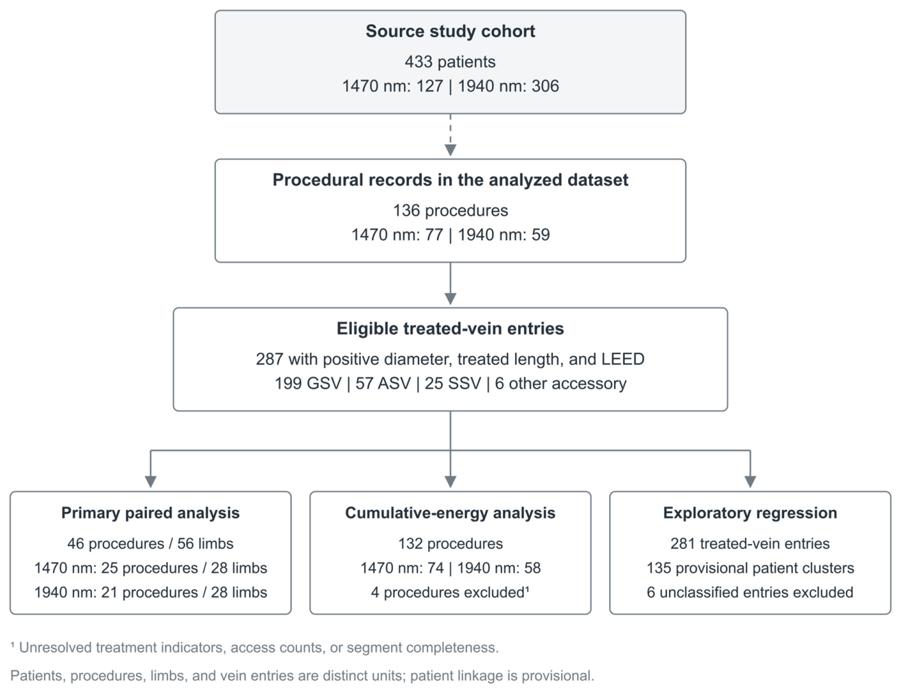
Figure 2.
Procedure-weighted ASV/GSV dosimetric ratios. Points and horizontal bars show geometric means and 95% CIs from 25 procedures at 1470 nm, 21 at 1940 nm, and 46 pooled procedures. The paired sample comprised 56 limbs, with bilateral log ratios averaged within each procedure. The vertical reference denotes a ratio of 1. Nominal EFE is based on recorded pre-tumescence diameter.
Figure 2.
Procedure-weighted ASV/GSV dosimetric ratios. Points and horizontal bars show geometric means and 95% CIs from 25 procedures at 1470 nm, 21 at 1940 nm, and 46 pooled procedures. The paired sample comprised 56 limbs, with bilateral log ratios averaged within each procedure. The vertical reference denotes a ratio of 1. Nominal EFE is based on recorded pre-tumescence diameter.
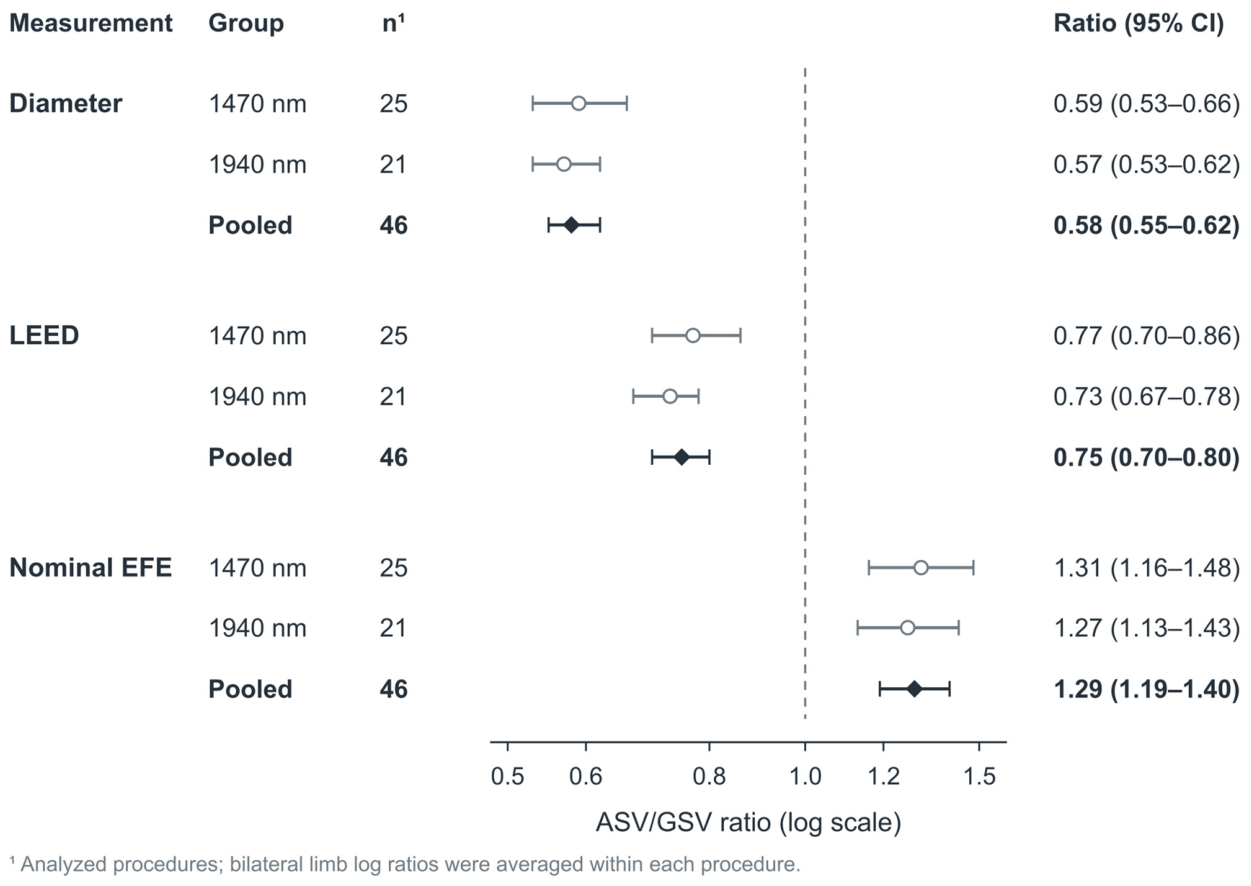
Figure 3.
Total laser energy by number of treated vein entries. Points show medians and bars show interquartile ranges from 132 procedures with adequate completeness: 74 at 1470 nm and 58 at 1940 nm. Counts are shown for each group. Energy uses the full recorded treated length of each eligible entry. Bars represent distributional quartiles, not confidence intervals.
Figure 3.
Total laser energy by number of treated vein entries. Points show medians and bars show interquartile ranges from 132 procedures with adequate completeness: 74 at 1470 nm and 58 at 1940 nm. Counts are shown for each group. Energy uses the full recorded treated length of each eligible entry. Bars represent distributional quartiles, not confidence intervals.
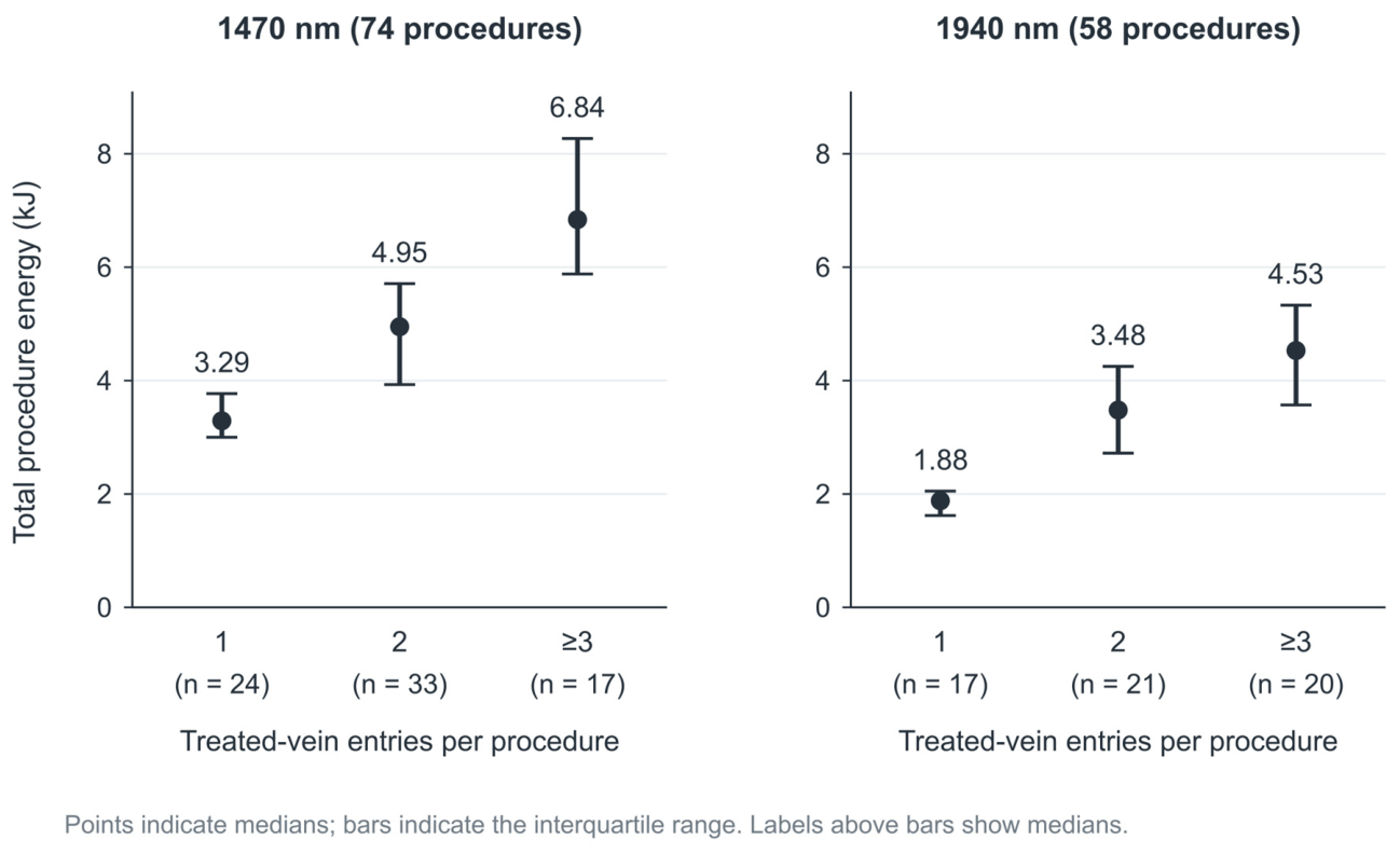
Figure 4.
Sensitivity of the paired ASV/GSV nominal EFE ratio. A: Geometric mean ratios and 95% CIs for the primary analysis (46 procedures) and after exclusion of ambiguous procedures (43 procedures). B: An illustrative scenario increasing ASV diameter and decreasing GSV diameter by the same percentage while holding LEED constant. The curve and shaded band transform the reported pooled estimate and CI; they do not estimate measurement error or post-tumescence collapse. The scenario uses rounded reported summary estimates.
Figure 4.
Sensitivity of the paired ASV/GSV nominal EFE ratio. A: Geometric mean ratios and 95% CIs for the primary analysis (46 procedures) and after exclusion of ambiguous procedures (43 procedures). B: An illustrative scenario increasing ASV diameter and decreasing GSV diameter by the same percentage while holding LEED constant. The curve and shaded band transform the reported pooled estimate and CI; they do not estimate measurement error or post-tumescence collapse. The scenario uses rounded reported summary estimates.
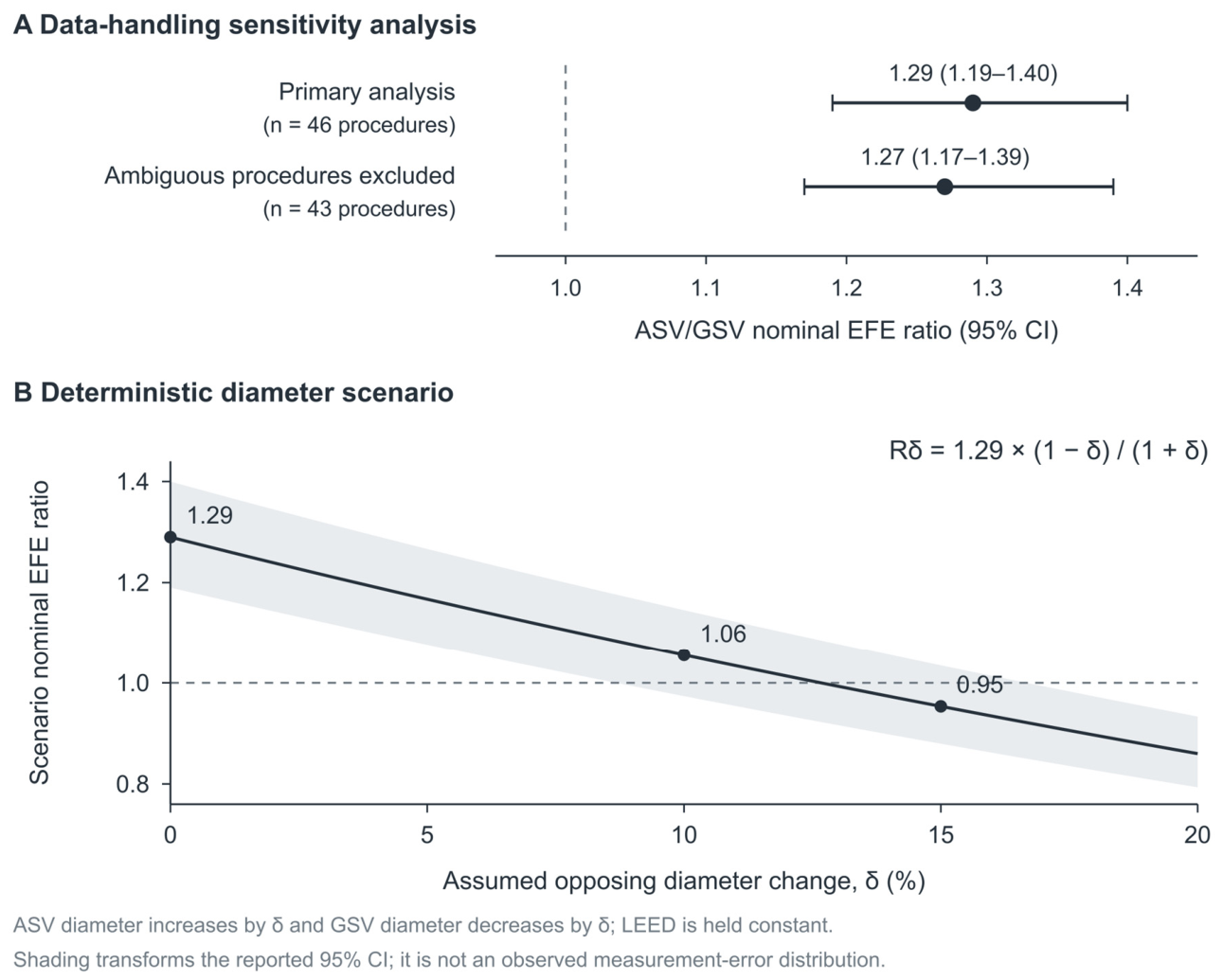

Table 1.
Source cohort and analyzed procedural records.
| Characteristic | 1470 nm | 1940 nm |
|---|---|---|
| Source cohort, patients, n | 127 | 306 |
| Procedural records analyzed, n | 77 | 59 |
| Age in analyzed records, years | 46.3 ± 13.2 | 47.9 ± 12.7 |
| Female sex in analyzed records, n/N (%) | 55/77 (71.4) | 43/59 (72.9) |
| Previous intervention in analyzed records, n/N (%) | 19/77 (24.7) | 14/59 (23.7) |
| Eligible treated vein entries, n | 157 | 130 |
| Procedures with paired GSV-ASV data, n | 25 | 21 |
The source cohort comprised 433 patients. Detailed analyses presented here used 136 procedural records. Age is mean ± standard deviation; other entries are counts or n/N (%). Patients, procedures, limbs, and treated vein entries are distinct units; record counts should not be interpreted as unique-patient coverage. GSV, great saphenous vein; ASV, anterior saphenous vein.
Table 2.
Paired anterior-to-great saphenous vein dosimetric ratios.
| Measurement | 1470 nm 25 procedures |
1940 nm 21 procedures |
Pooled 46 procedures |
|---|---|---|---|
| Analyzed procedures, n | 25 | 21 | 46 |
| Paired limbs, n | 28 | 28 | 56 |
| Diameter ratio | 0.59 (0.53-0.66) | 0.57 (0.53-0.62) | 0.58 (0.55-0.62) |
| LEED ratio | 0.77 (0.70-0.86) | 0.73 (0.67-0.78) | 0.75 (0.70-0.80) |
| Nominal EFE ratio | 1.31 (1.16-1.48) | 1.27 (1.13-1.43) | 1.29 (1.19-1.40) |
Values are procedure-weighted geometric means (95% CI), except counts. The analytical sample comprised 46 procedures and 56 paired limbs. Bilateral limb log ratios were averaged within each procedure. Nominal EFE uses pre-tumescence diameter and is not measured wall fluence. LEED, linear endovenous energy density; EFE, endovenous fluence equivalent.
Table 3.
Cumulative laser energy by number of treated vein entries.
| Wavelength / treated entries | n | Total length (cm) |
Total energy (kJ) |
Weighted LEED (J/cm) |
|---|---|---|---|---|
| 1470 nm / 1 entry | 24 | 52.0 (41.8-55.2) | 3.29 (3.00-3.77) | 67.8 (63.0-72.6) |
| 1470 nm / 2 entries | 33 | 82.0 (69.0-91.0) | 4.95 (3.93-5.71) | 59.0 (56.7-63.2) |
| 1470 nm / 3+ entries | 17 | 111.0 (94.0-128.0) | 6.84 (5.88-8.27) | 58.7 (56.6-62.6) |
| 1940 nm / 1 entry | 17 | 44.0 (42.0-49.0) | 1.88 (1.62-2.05) | 40.9 (37.7-48.6) |
| 1940 nm / 2 entries | 21 | 83.0 (60.5-92.0) | 3.48 (2.72-4.25) | 43.4 (40.0-46.1) |
| 1940 nm / 3+ entries | 20 | 110.5 (94.0-128.5) | 4.53 (3.57-5.33) | 41.9 (38.6-43.5) |
The analytical sample comprised 132 procedures: 74 at 1470 nm and 58 at 1940 nm. Values are medians (interquartile ranges); n denotes procedures. Each vein entry uses its entire recorded treated length. Total energy is the sum of LEED multiplied by treated length across eligible entries. Weighted LEED is total energy divided by total treated length.
Table 4.
Exploratory factors associated with recorded linear energy density.
| Variable | Adjusted LEED ratio |
95% CI | p value |
|---|---|---|---|
| Doubling of diameter | 1.13 | 1.07-1.19 | <0.001 |
| Doubling of treated length | 1.11 | 1.04-1.19 | 0.004 |
| ASV vs GSV | 0.93 | 0.83-1.04 | 0.193 |
| SSV vs GSV | 0.92 | 0.82-1.02 | 0.120 |
| 1940 nm vs 1470 nm | 0.70 | 0.67-0.74 | <0.001 |
The model included 281 GSV, ASV, and SSV entries from 135 provisional patient clusters; six unclassified accessory-vein entries were excluded. CIs used cluster-robust covariance with finite-sample correction and t inference. R² = 0.692. Ratios describe recorded LEED, not clinical risk or causal effects. GSV, great saphenous vein; ASV, anterior saphenous vein; SSV, small saphenous vein; CI, confidence interval.
Disclaimer/Publisher’s Note: The statements, opinions and data contained in all publications are solely those of the individual author(s) and contributor(s) and not of MDPI and/or the editor(s). MDPI and/or the editor(s) disclaim responsibility for any injury to people or property resulting from any ideas, methods, instructions or products referred to in the content. |
© 2026 by the authors. Licensee MDPI, Basel, Switzerland. This article is an open access article distributed under the terms and conditions of the Creative Commons Attribution (CC BY) license.
Copyright: This open access article is published under a Creative Commons CC BY 4.0 license, which permit the free download, distribution, and reuse, provided that the author and preprint are cited in any reuse.