
Submitted:
19 October 2026
Posted:
21 October 2026
You are already at the latest version
Abstract
We introduce a patented pylon-type implant that is intended to support osseointegration of prostheses with involvement of a neural interface. This conceptual/patent publication details its development and importance. The main objective was to improve the safety of the existing skin and bone integrated pylon (SBIP), developed earlier by the authors, which was achieved with a hermetic seal that prevents infection migration through the interface into the surrounding skin. The neural interface consists of a plurality of wires that conduct signals between the internal nervous system and external robotic/prosthetic mechanisms and controls. Each wire is electrically insulated through the channel of a single-, double- or multi-bore insulator ceramic rod. The wires are extended outside on both ends of the rod, forming leads for further connection with corresponding wires that conduct signals between the body and its external environment. The rod assembly is inserted inside a channel of a solid tubular load bearing titanium frame. The outside surface of the rod is hermetically brazed to the inside of the tubular frame at both ends. The frame is surrounded by a porous silver-coated titanium cladding of the pylon, ensuring safe integration with skin deeply ingrown into the implant.
Keywords:
transcutaneous osseointegration
; lower limb prostheses
; powered prostheses
; bidirectional neural control
; infection-protective hermetic seal
1. Introduction
1.1. Infection as the Critical Barrier to Progress in Osseointegration
Due to the significant increase in the functionality of direct skeletal attachment / osseointegration (OI) versus socket-type attachments, in 2015 the FDA approved the OPRA platform, the first system for direct skeletal attachment of limb prostheses [1]. The approval was made despite the unfortunate reality that, in the words of the FDA, “the most common adverse event [was] infection.” A systematic review of 22 articles comprising 13 unique studies in OI showed a wide range of infection rates, from 1 to 77 percent, and concluded that the current technology is associated with an increased risk of soft tissue infection [2]. In the OPRA technology the skin-implant interface is established as a stoma that slides along the abutment with a layer of pus between the skin and abutment. No attachment of skin to the abutment (the external portion of the implant) is percued, since the abutment is smooth. That circumferential opening in the skin to the outside environment requires daily care and frequent antibiotic treatment to partially mitigate infection. Despite the unsolved risk of infection at the implantation site, amputees accept OI attachment of prostheses, because of its great functionality.
Even more serious is the issue of infection in osseointegrated powered prostheses with bidirectional neurocontrol of their motor(s). There is always a real possibility of infection penetrating through the neural interface from the outside environment into the body if the interface is not adequately sealed. Such seal is extremely difficult to build, since the implanted abutment has longitudinal channels for electrical wires/cables transmitting signals from nerves/muscles to the control system of the powered prosthesis. For example, the implant in [3] describes an abutment with a sealed neural interface. However, the seal is achieved by screw joints, which leaves a physical gap between opposite threads, is therefore not hermetic, and cannot protect against infection.
1.2. Prior Research
Animal studies by other research groups have attempted to reduce the infection rate in OI, some experimenting with an implant constructed to be superficially porous. The most effective published study in sheep showed a reduction in the skin infection rate down to 16.7% over a 24-month period [4]. However, 25% of the sheep were removed due to early complications. Despite initial skin ingrowth into the pylon, a down-growth of skin epithelium along the implant broke the skin seal and introduced the potential risk of future infection and implant failure.
Within our prior research project undertaken by Poly-Orth International, Sharon, MA, we built a biotechnological OI platform that takes advantage of the natural anisotropy of skin regeneration [5]. The platform centers around the patented, deeply porous transcutaneous implant called the Skin and Bone Integrated Pylon (SBIP) [6,7,8,9,10,11]. Our animal studies [12] demonstrated its strong potential to develop into a marketable product and process. Recent improvements in the functionality and safety of the interface between the SBIP and tissues of the residuum were accomplished by introducing 3D-printing instead of sintering and optimizing the parameters of the implant’s antimicrobial silver coating [9,13,14]. Several weeks after implantation are required until the ingrowth is deep enough to make this barrier. During this vulnerable period, silver coating provides the protection needed. Once the ingrowth is deep and sufficiently protective, the coating is not needed. We ensured that the coat dissolved within 8 weeks after implantation to avoid negative influence of the indefinite presence of silver in the body, after which deep ingrowth of skin into the porous cladding of the implant is finalized.
1.3. Objectives
In order to develop an osseointegrated implant for attaching a powered prosthesis to a hermetically sealed neural interface, several design features needed to be introduced.
- (1)
- Permanent implantation during OI, necessitates the hermetical seal.
- (2)
- (3)
- A new transcutaneous pilon has to be ready for implantation “off-the-shelf”.
2. Materials and Methods
Designing a ready-for-implantation, “off-the-shelf” device, permits to use materials for insulating and filling gaps without limitations of temperature or the timing of curing. These limitations exist when insulation is performed during the implantation procedure, as the patient’s hosting tissues cannot be heated above 39 °C - 43 °C.
Specific control system for a patient may require a bundle of multiple wires whose thickness inside the implant is the same as for their segments passing through the implant into the residuum or to the external power control.
In the new device, wires can be significantly thinner compared to traditional methodology, since the portion of wires inside the implant can be placed there without being pulled and possibly damaged. Additional thinning of the wire is achieved can be achieved by using without individual insulation cover. For that, insulation should be provided by placement of wires in the channels of the ceramic cylindrical insert.
3. Discussion of Features and Benefits
We choose the specification that meets our objectives and developed the infection-protective hermetically sealed neural interface of the Skin and Bone Integrated Pylon for osseointegrated lower limb powered prosthesis. The device has been patented under the name “load-bearing skin and bone integrated pylon with hermetically sealed neural interface” [18].
The “load-bearing” in the title of the patent characterizes a difference between this device and implants with a neural interface that are used for internal placement. Those are not designed to withstand loads.
The neural interface may consist of a plurality of wires conducting signals between the nervous system and external prosthetic mechanisms and controls (Figure 3, Figure 4 and Figure 5). The wires may be made with medically approved alloys such as stainless steel, MP35, or pure and alloyed platinum.
Each of such wires is electrically insulated, but not with individual plasic cover, but the implant’s ceramic frame rod. Each wire is threading through the channel of a single, double or multi bore insulator rod, such as a ceramic cylinder (see Figure 2), that may be made with alumina (Al2O3). All wires are extended outside on both ends of the rod, forming leads to further connect with wires conducting signals to and from outside and inside the body (see Figure 2, Figure 3 and Figure 4). The wires are hermetically braze-sealed to inside of each bore at both ends of the rod.
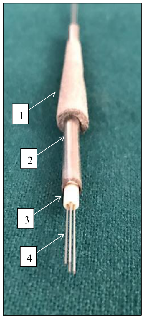
The assembly of wires and ceramic channels inside the solid tubular load-bearing frame is made with pure or alloyed titanium. The solid tubular frame is surrounded by a porous titanium portion of the SBIP pylon (Figure 3, Figure 4 and Figure 5) ensuring integration with the residuum’s bone and skin.
Assembly of the components described above is presented in Figure 6.
4.
An option of having a ready for implantation “off-the-shelf” device seems attractive from both the lead time and the cost stand points. The conical shape of the pylon probably eliminates in most cases the necessity for customization of the design, as such a pylon may fit the variety of bone shapes and sizes. The technology behind the manufacturing of hermetically sealed pylon interface appears practical and logical:
- The pylon consisting of solid titanium tubular frame that is surrounded by porous titanium is prepared first;
- The ceramic such as alumina insert with threaded wires is placed inside the channel of tubular frame;
- A short brazing cycle is applied to the assembly to hermetically seal titanium-to-ceramic, and ceramic-to-each wire on both ends of the pylon.
- The recent advances in direct metal laser sintering (DMLS or 3D printing) allow a more precise manufacturing of the pylon where the titanium part is built around the ceramic insert followed by a brazing cycle.
The ceramic-to-metal brazing is a well-known operation that yields a hermitically sealed joint. Both the alumina-to titanium and alumina-to-platinum joint may be achieved by using pure gold as a filler metal [19] in the shape of a pre-form or paste, or a combination of both. The brazing needs to be done in vacuum mainly to protect titanium from oxidation. An additional advantage of using vacuum is that the final sealed pylon contains vacuum inside eliminating gas pressure build-up during heat treatment.
This presentation is limited by the early stage of translation from the bench to the animal. In vivo, the quality of the seal has not yet been verified, and mechanical resistance sufficient to withstand the vertical and bending loads in the range observed in normal gait, running and jumping, must be assessed in preclinical laboratory and animal studies.
The assembly outlined in Figure 6 must be fully completed, for which the authors are finalizing specifications for all components and for test samples to be fabricated. In anticipation of the future implementation of the new device, we present the current publication to bring our work to light for colleagues working in the field who would be interested in further collaborative or independent investigations.
5. Conclusions
Here, the authors describe the patented design of an implant for transcutaneous ossointegration using powered limb prostheses with bidirectional neural control. Therapeutic advantages of the implant have not been yet experimentally validated and therefore remain hypothetical. However, when comparing to current technologies, the ability to fabricate a ready-to-use assembly prior to implantation seems promising in mitigating the high risk of post-implantation infections. Future studies should include in vitro and in vivo testing, and clinical investigations to establish safety, performance, and clinical utility.
6. Patents
Pitkin M, Raykhtsaum G, inventors; LOAD-BEARING SKIN AND BONE INTEGRATED PYLON WITH HERMETICALLY SEALED NEURAL INTERFACE. US Patent 12,611,307. 2026.
Author Contributions
Participation of both authors, M.P and G.R. was equal in conceptualization of the design, patenting, original draft preparation review and in editing the manuscript.
Funding
NIAMS/NIH GRANT 1SB1AR086711.
Conflicts of Interest
Author Mark Pitkin was employed by the company Poly-Orth International. Author Grigory Raykhtsaum was employed by the company Noble Metallurgical Consultancy. Neither company provided financial support for this study in the form of funding, consulting fees, equity, or any other economic benefits. Author Mark Pitkin and Author Grigory Raykhtsaum hold U.S. Patent No. 12,611,307 (2026) related to the content of this article.
Abbreviations
| OI | Osseointegration |
| SBIP | Skin and Bone Integrated Pylon |
| SBIP-PNI | Skin and Bone Integrated Pylon with Peripheral Neural Interface |
| OPRA | Osseointegrated Prostheses for the Rehabilitation of Amputees |
| FDA | Food and Drug Administration |
References
- FDA. FDA authorizes use of prosthesis for rehabilitation of above-the-knee amputations. 2015. Available online: https://www.fda.gov/NewsEvents/Newsroom/PressAnnouncements/UCM455103.
- Kunutsor, S.; Gillatt, D.; Blom, A. Systematic review of the safety and efficacy of osseointegration prosthesis after limb amputation. Br. J. Surg. 2018. [Google Scholar] [CrossRef] [PubMed]
- Ortiz-Catalan, M.; Mastinu, E.; Sassu, P.; Aszmann, O.; Branemark, R. Self-Contained Neuromusculoskeletal Arm Prostheses. N Engl. J. Med. 2020, 382(18), 1732–8. [Google Scholar] [CrossRef] [PubMed]
- Jeyapalina, S.; Beck, J.P.; Agarwal, J.; Bachus, K.N. A 24-month evaluation of a percutaneous osseointegrated limb-skin interface in an ovine amputation model. J. Mater. Sci. Mater. Med. 2017, 28(11), 179. [Google Scholar] [CrossRef] [PubMed]
- Pitkin, M. Addressing anisotropy of tissues regeneration in the implants design. Biomim. Biomater. Tissue Eng. 2015, 18, 3. [Google Scholar]
- Pitkin, M. On the way to total integration of prosthetic pylon with residuum. J. Rehabil. Res. Dev. 2009, 46(3), 345–60. [Google Scholar] [CrossRef] [PubMed]
- Pitkin, M.; Raykhtsaum, G.; Galibin, O.V.; Protasov, M.V.; Chihovskaya, J.V.; Belyaeva, I.G. Skin and bone integrated prosthetic pylon: a pilot animal study. J. Rehabil. Res. Dev. 2006, 43(4), 573–80. [Google Scholar] [CrossRef] [PubMed]
- Pitkin, M.; Pilling, J.; Raykhtsaum, G. Mechanical properties of totally permeable titanium composite pylon for direct skeletal attachment. J. Biomed. Mater. Res. Part B Appl. Biomater. 2012, 100B(4), 993–9. [Google Scholar] [CrossRef]
- Shevtsov, M.; Pitkin, E.; Combs, S.; Meulen, G.; Preucil, C.; Pitkin, M. Comparison in-vitro study on the interface between skin and bone cell cultures and microporous titanium samples of Skin and Bone Integrated Pylons (SBIP) manufactured with 3-D printing technology versus sintered samples. Nanomaterals 2024, 14(18), 1484. [Google Scholar] [CrossRef] [PubMed] [PubMed Central]
- Pitkin M, inventor In-bone implantable shaft for prosthetic joints or for direct skeletal attachment of external limb prostheses and method of its installation. 2015. Available online: https://patents.google.com/patent/US8992615B2/en.
- Pitkin, M.; Raykhtsaum, G. inventors; Skin Integrated Device. 2012. Available online: http://www.google.com/patents/US8257435.
- Pitkin, M.; Cassidy, C.; Shevtsov, M.A.; et al. Recent Progress in Animal Studies of the Skin- and Bone-integrated Pylon With Deep Porosity for Bone-Anchored Limb Prosthetics With and Without Neural Interface. Mil. Med. 2021, 186(S_1), 688–95. [Google Scholar] [CrossRef] [PubMed]
- Shevtsov, M.; Pitkin, E.; Combs, S.E.; et al. Biocompatibility Analysis of the Silver-Coated Microporous Titanium Implants Manufactured with 3D-Printing Technology. Nanomaterials 2024, 14(23), 1876. [Google Scholar] [CrossRef] [PubMed] [PubMed Central]
- Shevtsov, M.; Bozhokina, E.; Yudintceva, N.; et al. Enhancing the Antibacterial and Biointegrative Properties of Microporous Titanium Surfaces Using Various Metal Coatings: A Comparative Study. Prosthesis 2025, 7(6), 133. Available online: https://www.mdpi.com/2673-1592/7/6/133. [CrossRef] [PubMed]
- Pitkin, M.; Cassidy, C.; Muppavarapu, R.; Edell, D. Recording of Electric Signal Passing Through a Pylon in Direct Skeletal Attachment of Leg Prostheses with Neuromuscular Control. IEEE Trans. Bio-Med. Eng. 2012, 59(5), 1349–53. Available online: https://ieeexplore.ieee.org/stamp/stamp.jsp?tp=&arnumber=6151814. [CrossRef] [PubMed]
- Pitkin, M.; Park, H.; Frossard, L.; Klishko, A.N.; Prilutsky, B.I. Transforming the anthropomorphic passive free-flow foot prosthesis into a powered foot prosthesis with intuitive control and sensation (Bionic FFF). Mil. Med. 2024, 189 (Supplement_3), 439–47. [Google Scholar] [CrossRef] [PubMed] [PubMed Central]
- Park, H.; Klishko, A.N.; Oh, K.; et al. Electrical stimulation of distal tibial nerve during stance phase of walking may reverse effects of unilateral paw pad anesthesia in the cat. Mot. Control 2022, 27(1), 71–95. [Google Scholar] [CrossRef] [PubMed]
- Pitkin, M.; Raykhtsaum, G. inventors; LOAD-BEARING SKIN AND BONE INTEGRATED PYLON WITH HERMETICALLY SEALED NEURAL INTERFACE. US patent 12,611,307, 2026. [Google Scholar]
- Bian, H.; Song, X.; Hu, S.; et al. Microstructure evolution and mechanical properties of titanium/alumina brazed joints for medical implants. Metals 2019, 9(6), 644. [Google Scholar] [CrossRef]
Figure 1.
(A) - Illustrations of the implanted SBIP-PNI pylon with loose wires leads and nerve cuff electrodes in passive prosthesis, and in the powered transtibial prosthesis (B) (adopted from [16]).
Figure 1.
(A) - Illustrations of the implanted SBIP-PNI pylon with loose wires leads and nerve cuff electrodes in passive prosthesis, and in the powered transtibial prosthesis (B) (adopted from [16]).
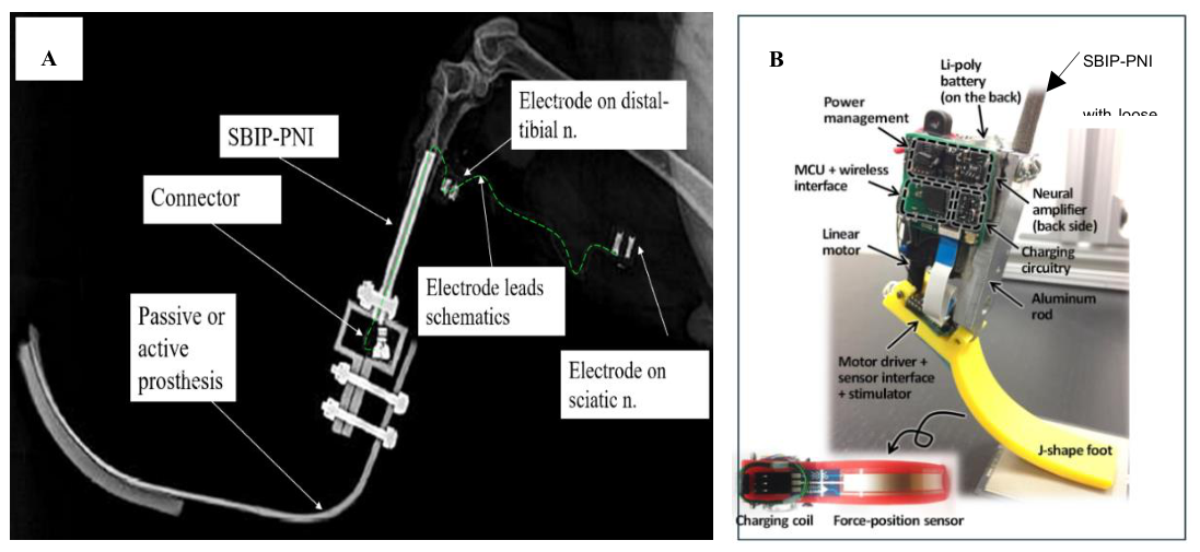
Figure 2.
Longitudinal cross section of the: 1 – circumference of the solid titanium tube; 2 – ceramic cylindrical insert; 3 -upper socket for the wires in the channels of the insert 2; 4 – wires in the insert 2 (adopted from [18]).
Figure 2.
Longitudinal cross section of the: 1 – circumference of the solid titanium tube; 2 – ceramic cylindrical insert; 3 -upper socket for the wires in the channels of the insert 2; 4 – wires in the insert 2 (adopted from [18]).
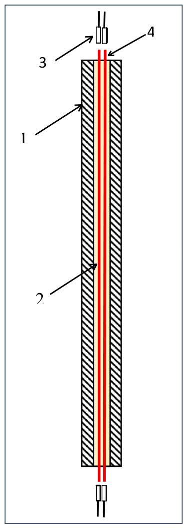
Figure 3.
Assembly of the SBIP-PNI implant: 1 –solid titanium tube; 2 – ceramic cylindrical insert; 3 -upper socket for the wires in the channels of the insert 2; 4 – wires in the insert 2. 5 – porous titanium cladding. Portion of the SBIP-PNI above green dashed line is implanted to the bone marrow canal of the residuum, and the portion below line is devoted to the interface with residuum skin (adopted from [18]).
Figure 3.
Assembly of the SBIP-PNI implant: 1 –solid titanium tube; 2 – ceramic cylindrical insert; 3 -upper socket for the wires in the channels of the insert 2; 4 – wires in the insert 2. 5 – porous titanium cladding. Portion of the SBIP-PNI above green dashed line is implanted to the bone marrow canal of the residuum, and the portion below line is devoted to the interface with residuum skin (adopted from [18]).
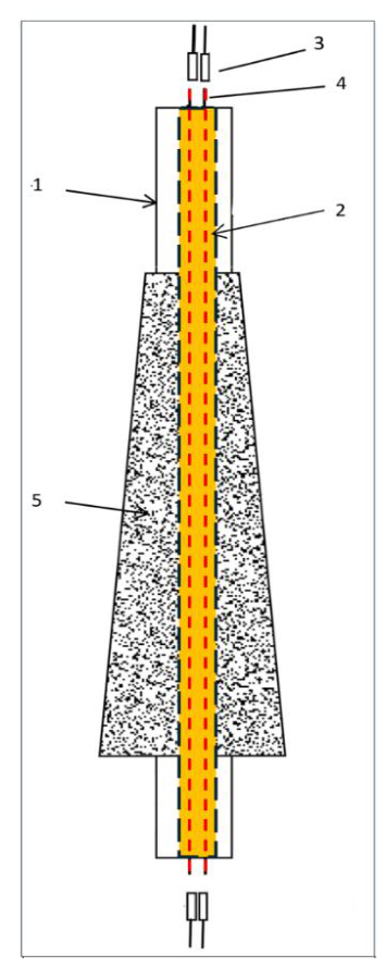
Figure 4.
1- Cross section (see Figure 2 and Figure 3) of the solid titanium tube; 2 - ceramic cylindrical insert with wires 3 after cured insulation 4, protecting from outside infectious agents (adopted from [18]).
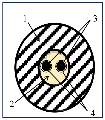
Figure 5.
Assembly of material components of the SBIP-PNI implant: 1 – porous cladding covering solid titanium tube 2; 3 - ceramic cylindrical insert with channels for wires 4.
Figure 5.
Assembly of material components of the SBIP-PNI implant: 1 – porous cladding covering solid titanium tube 2; 3 - ceramic cylindrical insert with channels for wires 4.
Disclaimer/Publisher’s Note: The statements, opinions and data contained in all publications are solely those of the individual author(s) and contributor(s) and not of MDPI and/or the editor(s). MDPI and/or the editor(s) disclaim responsibility for any injury to people or property resulting from any ideas, methods, instructions or products referred to in the content. |
© 2026 by the authors. Licensee MDPI, Basel, Switzerland. This article is an open access article distributed under the terms and conditions of the Creative Commons Attribution (CC BY) license (http://creativecommons.org/licenses/by/4.0/).
Copyright: This open access article is published under a Creative Commons CC BY 4.0 license, which permit the free download, distribution, and reuse, provided that the author and preprint are cited in any reuse.